Embed Size (px)
Citation preview
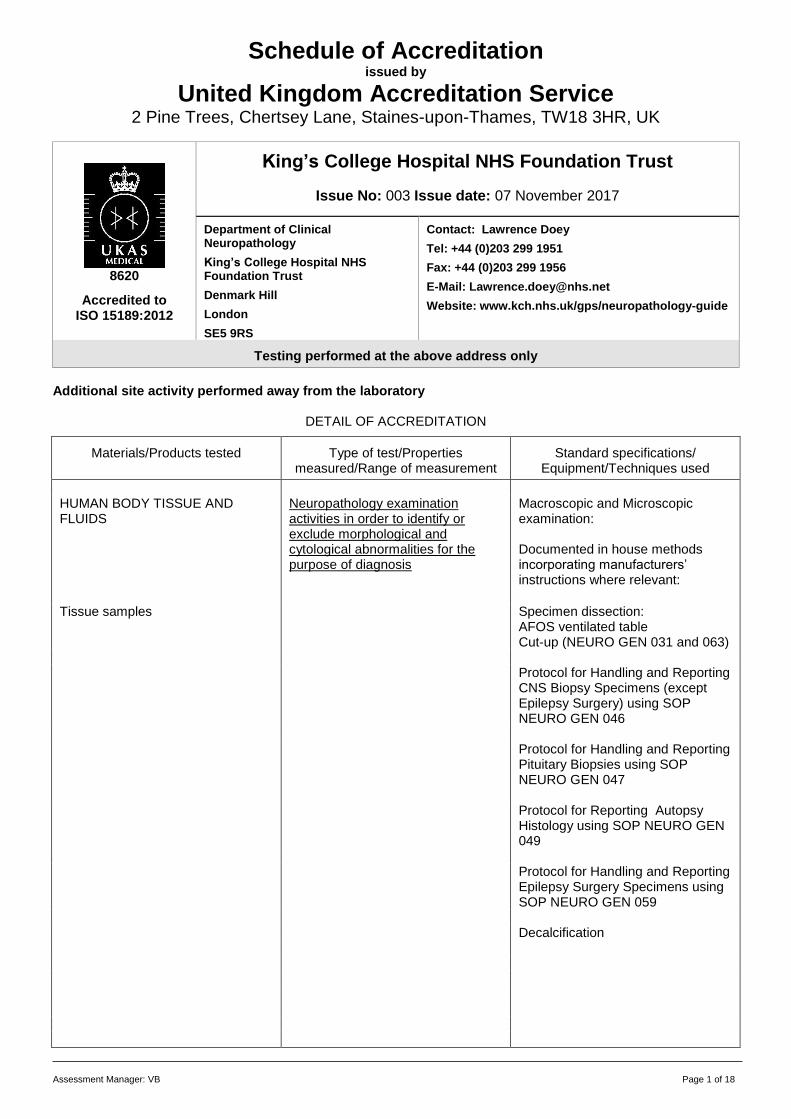
Assessment Manager: VB Page 1 of 18
Schedule of Accreditation issued by
United Kingdom Accreditation Service
2 Pine Trees, Chertsey Lane, Staines-upon-Thames, TW18 3HR, UK
8620
Accredited to ISO 15189:2012
King’s College Hospital NHS Foundation Trust
Issue No: 003 Issue date: 07 November 2017
Department of Clinical Neuropathology
King’s College Hospital NHS Foundation Trust
Denmark Hill
London
SE5 9RS
Contact: Lawrence Doey
Tel: +44 (0)203 299 1951
Fax: +44 (0)203 299 1956
E-Mail: [email protected]
Website: www.kch.nhs.uk/gps/neuropathology-guide
Testing performed at the above address only
Additional site activity performed away from the laboratory
DETAIL OF ACCREDITATION
Materials/Products tested
Type of test/Properties
measured/Range of measurement
Standard specifications/
Equipment/Techniques used
HUMAN BODY TISSUE AND FLUIDS
Neuropathology examination activities in order to identify or exclude morphological and cytological abnormalities for the purpose of diagnosis
Macroscopic and Microscopic examination: Documented in house methods incorporating manufacturers’ instructions where relevant:
Tissue samples Specimen dissection:
AFOS ventilated table Cut-up (NEURO GEN 031 and 063)
Protocol for Handling and Reporting
CNS Biopsy Specimens (except Epilepsy Surgery) using SOP NEURO GEN 046
Protocol for Handling and Reporting
Pituitary Biopsies using SOP NEURO GEN 047
Protocol for Reporting Autopsy
Histology using SOP NEURO GEN 049
Protocol for Handling and Reporting
Epilepsy Surgery Specimens using SOP NEURO GEN 059
Decalcification
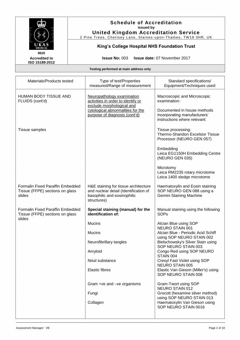
8620
Accredited to ISO 15189:2012
Schedule of Accreditation issued by
United Kingdom Accreditation Service 2 P ine Trees , Cher t sey Lane, S ta i nes -upon-Thames , TW 18 3HR, UK
King’s College Hospital NHS Foundation Trust
Issue No: 003 Issue date: 07 November 2017
Testing performed at main address only
Assessment Manager: VB Page 2 of 18
Materials/Products tested
Type of test/Properties
measured/Range of measurement
Standard specifications/
Equipment/Techniques used
HUMAN BODY TISSUE AND FLUIDS (cont’d)
Neuropathology examination activities in order to identify or exclude morphological and cytological abnormalities for the purpose of diagnosis (cont’d)
Macroscopic and Microscopic examination: Documented in house methods incorporating manufacturers’ instructions where relevant:
Tissue samples Tissue processing: Thermo-Shandon Excelsior Tissue Processor (NEURO GEN 057)
Embedding Leica EG1150H Embedding Centre (NEURO GEN 035)
Microtomy
Leica RM2235 rotary microtome Leica 1400 sledge microtome
Formalin Fixed Paraffin Embedded Tissue (FFPE) sections on glass slides
H&E staining for tissue architecture and nuclear detail (Identification of basophilic and eosinophilic structures)
Haematoxylin and Eosin staining SOP NEURO GEN 088 using a Gemini Staining Machine
Formalin Fixed Paraffin Embedded Tissue (FFPE) sections on glass slides
Special staining (manual) for the identification of:
Manual staining using the following SOPs
Mucins
Alcian Blue using SOP NEURO STAIN 001
Mucins
Alcian Blue - Periodic Acid Schiff using SOP NEURO STAIN 002
Neurofibrillary tangles Bielschowsky's Silver Stain using SOP NEURO STAIN 003
Amyloid Congo Red using SOP NEURO STAIN 004
Nissl substance Cresyl Fast Violet using SOP NEURO STAIN 005
Elastic fibres Elastic Van Gieson (Miller's) using SOP NEURO STAIN 008
Gram +ve and –ve organisms Gram-Twort using SOP
NEURO STAIN 012 Fungi Grocott (hexamine silver method)
using SOP NEURO STAIN 013 Collagen Haematoxylin Van Gieson using
SOP NEURO STAIN 0016
8620
Accredited to ISO 15189:2012
Schedule of Accreditation issued by
United Kingdom Accreditation Service 2 P ine Trees , Cher t sey Lane, S ta i nes -upon-Thames , TW 18 3HR, UK
King’s College Hospital NHS Foundation Trust
Issue No: 003 Issue date: 07 November 2017
Testing performed at main address only
Assessment Manager: VB Page 3 of 18
Materials/Products tested
Type of test/Properties
measured/Range of measurement
Standard specifications/
Equipment/Techniques used
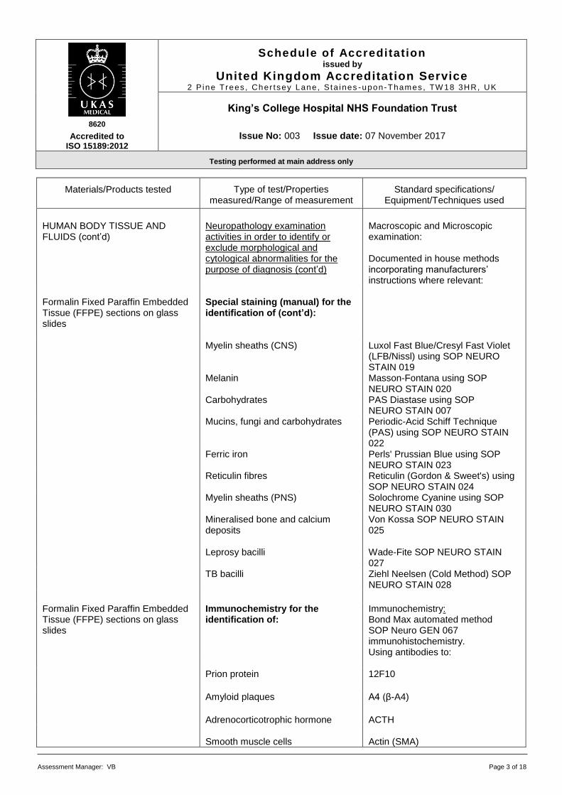
HUMAN BODY TISSUE AND FLUIDS (cont’d)
Neuropathology examination activities in order to identify or exclude morphological and cytological abnormalities for the purpose of diagnosis (cont’d)
Macroscopic and Microscopic examination: Documented in house methods incorporating manufacturers’ instructions where relevant:
Formalin Fixed Paraffin Embedded Tissue (FFPE) sections on glass slides
Special staining (manual) for the identification of (cont’d):
Myelin sheaths (CNS) Luxol Fast Blue/Cresyl Fast Violet
(LFB/Nissl) using SOP NEURO STAIN 019
Melanin Masson-Fontana using SOP NEURO STAIN 020
Carbohydrates PAS Diastase using SOP NEURO STAIN 007
Mucins, fungi and carbohydrates Periodic-Acid Schiff Technique (PAS) using SOP NEURO STAIN 022
Ferric iron Perls' Prussian Blue using SOP NEURO STAIN 023
Reticulin fibres Reticulin (Gordon & Sweet's) using SOP NEURO STAIN 024
Myelin sheaths (PNS) Solochrome Cyanine using SOP NEURO STAIN 030
Mineralised bone and calcium deposits
Von Kossa SOP NEURO STAIN 025
Leprosy bacilli Wade-Fite SOP NEURO STAIN
027 TB bacilli Ziehl Neelsen (Cold Method) SOP
NEURO STAIN 028
Formalin Fixed Paraffin Embedded Tissue (FFPE) sections on glass slides
Immunochemistry for the identification of:
Immunochemistry: Bond Max automated method SOP Neuro GEN 067 immunohistochemistry. Using antibodies to:
Prion protein 12F10
Amyloid plaques A4 (β-A4)
Adrenocorticotrophic hormone ACTH
Smooth muscle cells Actin (SMA)
8620
Accredited to ISO 15189:2012
Schedule of Accreditation issued by
United Kingdom Accreditation Service 2 P ine Trees , Cher t sey Lane, S ta i nes -upon-Thames , TW 18 3HR, UK
King’s College Hospital NHS Foundation Trust
Issue No: 003 Issue date: 07 November 2017
Testing performed at main address only
Assessment Manager: VB Page 4 of 18
Materials/Products tested
Type of test/Properties
measured/Range of measurement
Standard specifications/
Equipment/Techniques used
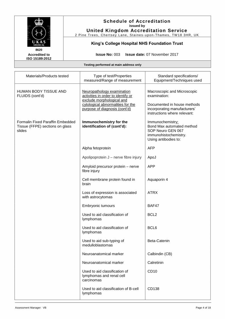
HUMAN BODY TISSUE AND FLUIDS (cont’d)
Neuropathology examination activities in order to identify or exclude morphological and cytological abnormalities for the purpose of diagnosis (cont’d)
Macroscopic and Microscopic examination: Documented in house methods incorporating manufacturers’ instructions where relevant:
Formalin Fixed Paraffin Embedded Tissue (FFPE) sections on glass slides
Immunochemistry for the identification of (cont’d):
Immunochemistry: Bond Max automated method SOP Neuro GEN 067 immunohistochemistry. Using antibodies to:
Alpha fetoprotein AFP
Apolipoprotein J – nerve fibre injury ApoJ
Amyloid precursor protein – nerve fibre injury
APP
Cell membrane protein found in
brain Aquaporin 4
Loss of expression is associated
with astrocytomas ATRX
Embryonic tumours BAF47
Used to aid classification of
lymphomas BCL2
Used to aid classification of
lymphomas BCL6
Used to aid sub-typing of
medulloblastomas Beta-Catenin
Neuroanatomical marker Calbindin (CB) Neuroanatomical marker Calretinin Used to aid classification of
lymphomas and renal cell carcinomas
CD10
Used to aid classification of B-cell
lymphomas CD138
8620
Accredited to ISO 15189:2012
Schedule of Accreditation issued by
United Kingdom Accreditation Service 2 P ine Trees , Cher t sey Lane, S ta i nes -upon-Thames , TW 18 3HR, UK
King’s College Hospital NHS Foundation Trust
Issue No: 003 Issue date: 07 November 2017
Testing performed at main address only
Assessment Manager: VB Page 5 of 18
Materials/Products tested
Type of test/Properties
measured/Range of measurement
Standard specifications/
Equipment/Techniques used
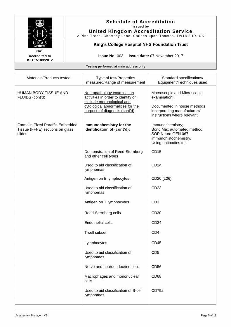
HUMAN BODY TISSUE AND FLUIDS (cont’d)
Neuropathology examination activities in order to identify or exclude morphological and cytological abnormalities for the purpose of diagnosis (cont’d)
Macroscopic and Microscopic examination: Documented in house methods incorporating manufacturers’ instructions where relevant:
Formalin Fixed Paraffin Embedded Tissue (FFPE) sections on glass slides
Immunochemistry for the identification of (cont’d):
Immunochemistry: Bond Max automated method SOP Neuro GEN 067 immunohistochemistry. Using antibodies to:
Demonstration of Reed-Sternberg
and other cell types CD15
Used to aid classification of
lymphomas CD1a
Antigen on B lymphocytes CD20 (L26)
Used to aid classification of
lymphomas CD23
Antigen on T lymphocytes CD3
Reed-Sternberg cells CD30 Endothelial cells CD34
T-cell subset CD4
Lymphocytes CD45 Used to aid classification of
lymphomas CD5
Nerve and neuroendocrine cells CD56 Macrophages and mononuclear
cells CD68
Used to aid classification of B-cell
lymphomas CD79a

8620
Accredited to ISO 15189:2012
Schedule of Accreditation issued by
United Kingdom Accreditation Service 2 P ine Trees , Cher t sey Lane, S ta i nes -upon-Thames , TW 18 3HR, UK
King’s College Hospital NHS Foundation Trust
Issue No: 003 Issue date: 07 November 2017
Testing performed at main address only
Assessment Manager: VB Page 6 of 18
Materials/Products tested
Type of test/Properties
measured/Range of measurement
Standard specifications/
Equipment/Techniques used
HUMAN BODY TISSUE AND FLUIDS (cont’d)
Neuropathology examination activities in order to identify or exclude morphological and cytological abnormalities for the purpose of diagnosis (cont’d)
Macroscopic and Microscopic examination: Documented in house methods incorporating manufacturers’ instructions where relevant:
Formalin Fixed Paraffin Embedded Tissue (FFPE) sections on glass slides
Immunochemistry for the identification of (cont’d):
Immunochemistry: Bond Max automated method SOP Neuro GEN 067 immunohistochemistry. Using antibodies to:
T-cell subset CD8 Ewing’s Sarcoma marker CD99 Antigen in intestinal epithelium CDX2
Protein in gastro-intestinal cells CEA Granules in neuroendocrine cells Chromogranin A
Pan-cytokeratin marker CK(Pan) Cytokeratin in specific epithelium CK07
Cytokeratin in gastro-intestinal cells CK20 Cytokeratin in epithelium CK5/6 Cytomegalovirus CMV Used to aid classification of
lymphomas Cyclin D1
Smooth and striated muscle cells Desmin
Epstein-Barr virus EBV
Protein in epithelial cells EMA Follicle Stimulating Hormone FSH Used to aid identification of rare
Dementias FUS
8620
Accredited to ISO 15189:2012
Schedule of Accreditation issued by
United Kingdom Accreditation Service 2 P ine Trees , Cher t sey Lane, S ta i nes -upon-Thames , TW 18 3HR, UK
King’s College Hospital NHS Foundation Trust
Issue No: 003 Issue date: 07 November 2017
Testing performed at main address only
Assessment Manager: VB Page 7 of 18
Materials/Products tested
Type of test/Properties
measured/Range of measurement
Standard specifications/
Equipment/Techniques used
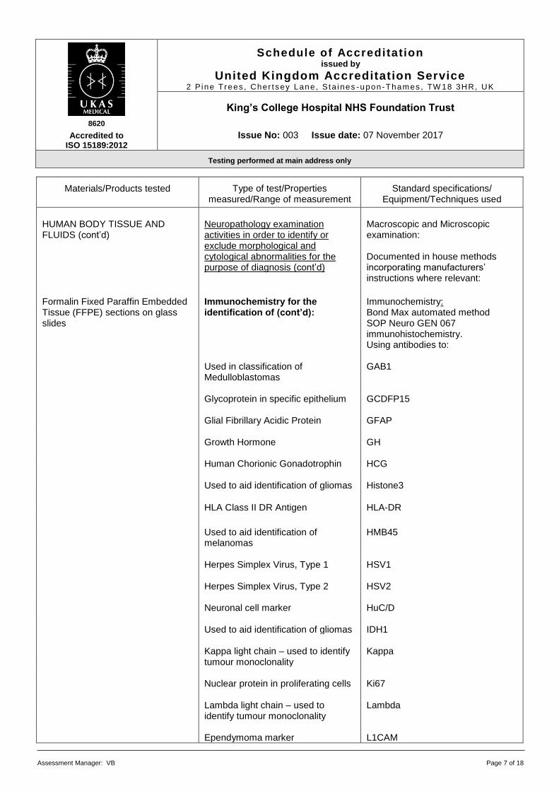
HUMAN BODY TISSUE AND FLUIDS (cont’d)
Neuropathology examination activities in order to identify or exclude morphological and cytological abnormalities for the purpose of diagnosis (cont’d)
Macroscopic and Microscopic examination: Documented in house methods incorporating manufacturers’ instructions where relevant:
Formalin Fixed Paraffin Embedded Tissue (FFPE) sections on glass slides
Immunochemistry for the identification of (cont’d):
Immunochemistry: Bond Max automated method SOP Neuro GEN 067 immunohistochemistry. Using antibodies to:
Used in classification of
Medulloblastomas GAB1
Glycoprotein in specific epithelium GCDFP15 Glial Fibrillary Acidic Protein GFAP Growth Hormone GH Human Chorionic Gonadotrophin HCG Used to aid identification of gliomas Histone3
HLA Class II DR Antigen HLA-DR
Used to aid identification of melanomas
HMB45
Herpes Simplex Virus, Type 1 HSV1 Herpes Simplex Virus, Type 2 HSV2 Neuronal cell marker HuC/D Used to aid identification of gliomas IDH1 Kappa light chain – used to identify
tumour monoclonality Kappa
Nuclear protein in proliferating cells Ki67 Lambda light chain – used to
identify tumour monoclonality Lambda
Ependymoma marker L1CAM

8620
Accredited to ISO 15189:2012
Schedule of Accreditation issued by
United Kingdom Accreditation Service 2 P ine Trees , Cher t sey Lane, S ta i nes -upon-Thames , TW 18 3HR, UK
King’s College Hospital NHS Foundation Trust
Issue No: 003 Issue date: 07 November 2017
Testing performed at main address only
Assessment Manager: VB Page 8 of 18
Materials/Products tested
Type of test/Properties
measured/Range of measurement
Standard specifications/
Equipment/Techniques used
HUMAN BODY TISSUE AND FLUIDS (cont’d)
Neuropathology examination activities in order to identify or exclude morphological and cytological abnormalities for the purpose of diagnosis (cont’d)
Macroscopic and Microscopic examination: Documented in house methods incorporating manufacturers’ instructions where relevant:
Formalin Fixed Paraffin Embedded Tissue (FFPE) sections on glass slides
Immunochemistry for the identification of (cont’d):
Immunochemistry: Bond Max automated method SOP Neuro GEN 067 immunohistochemistry. Using antibodies to:
Leutinzing Hormone LH Microtubule Associated Protein –
neuronal cell marker MAP2
Measles virus Measles Used as an aid in tumour
identification MGMT
Cytokeratin expressed in a range of
epithelial cells MNF116
Mismatch repair protein MSH2 Used to aid classification of
lymphomas MUM1
Skeletal muscle Myogenin
Sub-typing muscle fibres Myosin (Fast)
Sub-typing muscle fibres Myosin (Slow) Neuronal cell marker Nestin
Neuronal cell marker NeuN Nerve fibres NF(Pan)
Nerve fibres NF200KD
8620
Accredited to ISO 15189:2012
Schedule of Accreditation issued by
United Kingdom Accreditation Service 2 P ine Trees , Cher t sey Lane, S ta i nes -upon-Thames , TW 18 3HR, UK
King’s College Hospital NHS Foundation Trust
Issue No: 003 Issue date: 07 November 2017
Testing performed at main address only
Assessment Manager: VB Page 9 of 18
Materials/Products tested
Type of test/Properties
measured/Range of measurement
Standard specifications/
Equipment/Techniques used
HUMAN BODY TISSUE AND FLUIDS (cont’d)
Neuropathology examination activities in order to identify or exclude morphological and cytological abnormalities for the purpose of diagnosis (cont’d)
Macroscopic and Microscopic examination: Documented in house methods incorporating manufacturers’ instructions where relevant:
Formalin Fixed Paraffin Embedded Tissue (FFPE) sections on glass slides
Immunochemistry for the identification of (cont’d):
Immunochemistry: Bond Max automated method SOP Neuro GEN 067 immunohistochemistry. Using antibodies to:
Neuronal cell marker NSE Human Immunodeficiency Virus P24 Cell proliferation marker P53 Used to aid classification of
Dementias P62
Used as a marker in Pituitary
tumours Pan-α
Neuronal cell marker Parvalbumin Demonstration of small nerve fibres
in skin biopsies PGP9.5
Progesterone receptor expression
in tumours PGR
Enzyme produced by trophoblasts PLAP Demonstration of Pneumocystis
organism Pneumocystis
Used to aid classification of
Dementias Polyglutamine
Prolactin hormone Prolactin
Used to aid classification of metastatic carcinomas
PSA
Expressed in a range of cell types
including glial cells S100
8620
Accredited to ISO 15189:2012
Schedule of Accreditation issued by
United Kingdom Accreditation Service 2 P ine Trees , Cher t sey Lane, S ta i nes -upon-Thames , TW 18 3HR, UK
King’s College Hospital NHS Foundation Trust
Issue No: 003 Issue date: 07 November 2017
Testing performed at main address only
Assessment Manager: VB Page 10 of 18
Materials/Products tested
Type of test/Properties
measured/Range of measurement
Standard specifications/
Equipment/Techniques used
HUMAN BODY TISSUE AND FLUIDS (cont’d)
Neuropathology examination activities in order to identify or exclude morphological and cytological abnormalities for the purpose of diagnosis (cont’d)
Macroscopic and Microscopic examination: Documented in house methods incorporating manufacturers’ instructions where relevant:
Formalin Fixed Paraffin Embedded Tissue (FFPE) sections on glass slides
Immunochemistry for the identification of (cont’d):
Immunochemistry: Bond Max automated method SOP Neuro GEN 067 immunohistochemistry. Using antibodies to:
Diagnostic marker for
haemangiopericytomas Stat6
SV40 virus SV40 Demonstration of synaptophysin
producing cells Synaptophysin
Used to aid classification of Dementias
Synuclein
Used to aid classification of
Dementias Tau (AT8)
Used to aid classification of
Dementias Tau3
Used to aid classification of
Dementias Tau4
Used to aid classification of
Dementias TDP43-P
Demonstration of Toxoplasma
organism Toxoplasma
Thyroid stimulating hormone TSH
Nuclear protein in lung and thyroid TTF1
Used to aid classification of
Dementias Ubiquitin
8620
Accredited to ISO 15189:2012
Schedule of Accreditation issued by
United Kingdom Accreditation Service 2 P ine Trees , Cher t sey Lane, S ta i nes -upon-Thames , TW 18 3HR, UK
King’s College Hospital NHS Foundation Trust
Issue No: 003 Issue date: 07 November 2017
Testing performed at main address only
Assessment Manager: VB Page 11 of 18
Materials/Products tested
Type of test/Properties
measured/Range of measurement
Standard specifications/
Equipment/Techniques used
HUMAN BODY TISSUE AND FLUIDS (cont’d)
Neuropathology examination activities in order to identify or exclude morphological and cytological abnormalities for the purpose of diagnosis (cont’d)
Macroscopic and Microscopic examination: Documented in house methods incorporating manufacturers’ instructions where relevant:
Formalin Fixed Paraffin Embedded Tissue (FFPE) sections on glass slides
Immunochemistry for the identification of (cont’d):
Immunochemistry: Bond Max automated method SOP Neuro GEN 067 immunohistochemistry. Using antibodies to:
Used in classification of
Medulloblastomas YAP1
Filament in mesenchymal cells Vimentin Stained slides prepared as above Morphological assessment and
interpretation/diagnosis Microscopic examination: Assorted range of diagnostic bright-field microscopes Protocol for Handling and Reporting CNS Biopsy Specimens (except Epilepsy Surgery) using SOP NEURO GEN 046 Protocol for Handling and Reporting Pituitary Biopsies using SOP NEURO GEN 047 Protocol for Reporting Autopsy Histology using SOP NEURO GEN 049 Protocol for Handling and Reporting Epilepsy Surgery Specimens using SOP NEURO GEN 059
8620
Accredited to ISO 15189:2012
Schedule of Accreditation issued by
United Kingdom Accreditation Service 2 P ine Trees , Cher t sey Lane, S ta i nes -upon-Thames , TW 18 3HR, UK
King’s College Hospital NHS Foundation Trust
Issue No: 003 Issue date: 07 November 2017
Testing performed at main address only
Assessment Manager: VB Page 12 of 18
Materials/Products tested
Type of test/Properties
measured/Range of measurement
Standard specifications/
Equipment/Techniques used
HUMAN BODY TISSUE AND FLUIDS (cont’d)
Neuropathology examination activities in order to identify or exclude morphological and cytological abnormalities for the purpose of diagnosis (cont’d)
Macroscopic and Microscopic examination: Documented in house methods incorporating manufacturers’ instructions where relevant:
Brain biopsy
H&E staining for tissue architecture and nuclear detail (Identification of basophilic and eosinophilic structures)
Preparation of Smears and Frozen Sections for Intra-operative Diagnosis Howarth Class I Safety Cabinet Bright BM Cryostat Staining smears and frozen sections for intra-operative diagnosis (NEURO STAIN 025) Haematoxylin and Eosin
Stained brain biopsy slides prepared as above
Morphological assessment and interpretation/diagnosis
Protocol for intra-operative smears and frozen sections (NEURO GEN 048)
Muscle biopsy Preparation of Frozen Sections
Howarth Class I Safety Cabinet Bright BM Cryostat Preparation of muscle biopsies using the following Laboratory Protocol for Handling Muscle Biopsies, SOP NEURO MUSCLE 008
8620
Accredited to ISO 15189:2012
Schedule of Accreditation issued by
United Kingdom Accreditation Service 2 P ine Trees , Cher t sey Lane, S ta i nes -upon-Thames , TW 18 3HR, UK
King’s College Hospital NHS Foundation Trust
Issue No: 003 Issue date: 07 November 2017
Testing performed at main address only
Assessment Manager: VB Page 13 of 18
Materials/Products tested
Type of test/Properties
measured/Range of measurement
Standard specifications/
Equipment/Techniques used
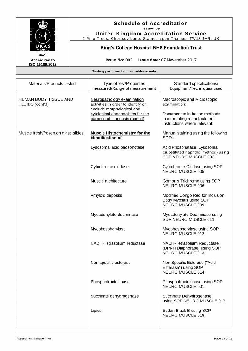
HUMAN BODY TISSUE AND FLUIDS (cont’d)
Neuropathology examination activities in order to identify or exclude morphological and cytological abnormalities for the purpose of diagnosis (cont’d)
Macroscopic and Microscopic examination: Documented in house methods incorporating manufacturers’ instructions where relevant:
Muscle fresh/frozen on glass slides Muscle Histochemistry for the
identification of: Manual staining using the following SOPs
Lysosomal acid phosphotase Acid Phosphatase, Lysosomal
(substituted naphthol method) using SOP NEURO MUSCLE 003
Cytochrome oxidase Cytochrome Oxidase using SOP NEURO MUSCLE 005
Muscle architecture Gomori's Trichrome using SOP NEURO MUSCLE 006
Amyloid deposits Modified Congo Red for Inclusion Body Myositis using SOP NEURO MUSCLE 009
Myoadenylate deaminase Myoadenylate Deaminase using SOP NEURO MUSCLE 011
Myophosphorylase Myophosphorylase using SOP NEURO MUSCLE 012
NADH-Tetrazolium reductase NADH-Tetrazolium Reductase (DPNH Diaphorase) using SOP NEURO MUSCLE 013
Non-specific esterase Non Specific Esterase ("Acid
Esterase") using SOP NEURO MUSCLE 014
Phosphofructokinase
Phosphofructokinase using SOP NEURO MUSCLE 001
Succinate dehydrogenase Succinate Dehydrogenase
using SOP NEURO MUSCLE 017 Lipids Sudan Black B using SOP
NEURO MUSCLE 018
8620
Accredited to ISO 15189:2012
Schedule of Accreditation issued by
United Kingdom Accreditation Service 2 P ine Trees , Cher t sey Lane, S ta i nes -upon-Thames , TW 18 3HR, UK
King’s College Hospital NHS Foundation Trust
Issue No: 003 Issue date: 07 November 2017
Testing performed at main address only
Assessment Manager: VB Page 14 of 18
Materials/Products tested
Type of test/Properties
measured/Range of measurement
Standard specifications/
Equipment/Techniques used
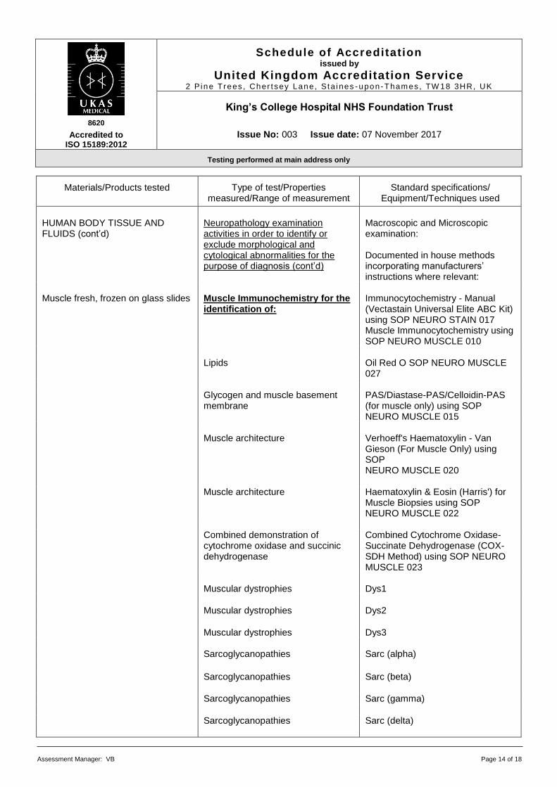
HUMAN BODY TISSUE AND FLUIDS (cont’d)
Neuropathology examination activities in order to identify or exclude morphological and cytological abnormalities for the purpose of diagnosis (cont’d)
Macroscopic and Microscopic examination: Documented in house methods incorporating manufacturers’ instructions where relevant:
Muscle fresh, frozen on glass slides Muscle Immunochemistry for the
identification of:
Immunocytochemistry - Manual (Vectastain Universal Elite ABC Kit) using SOP NEURO STAIN 017 Muscle Immunocytochemistry using SOP NEURO MUSCLE 010
Lipids Oil Red O SOP NEURO MUSCLE
027 Glycogen and muscle basement
membrane PAS/Diastase-PAS/Celloidin-PAS (for muscle only) using SOP NEURO MUSCLE 015
Muscle architecture Verhoeff's Haematoxylin - Van
Gieson (For Muscle Only) using SOP NEURO MUSCLE 020
Muscle architecture Haematoxylin & Eosin (Harris') for
Muscle Biopsies using SOP NEURO MUSCLE 022
Combined demonstration of
cytochrome oxidase and succinic dehydrogenase
Combined Cytochrome Oxidase-Succinate Dehydrogenase (COX-SDH Method) using SOP NEURO MUSCLE 023
Muscular dystrophies Dys1
Muscular dystrophies Dys2
Muscular dystrophies Dys3 Sarcoglycanopathies Sarc (alpha)
Sarcoglycanopathies Sarc (beta)
Sarcoglycanopathies Sarc (gamma)
Sarcoglycanopathies Sarc (delta)
8620
Accredited to ISO 15189:2012
Schedule of Accreditation issued by
United Kingdom Accreditation Service 2 P ine Trees , Cher t sey Lane, S ta i nes -upon-Thames , TW 18 3HR, UK
King’s College Hospital NHS Foundation Trust
Issue No: 003 Issue date: 07 November 2017
Testing performed at main address only
Assessment Manager: VB Page 15 of 18
Materials/Products tested
Type of test/Properties
measured/Range of measurement
Standard specifications/
Equipment/Techniques used
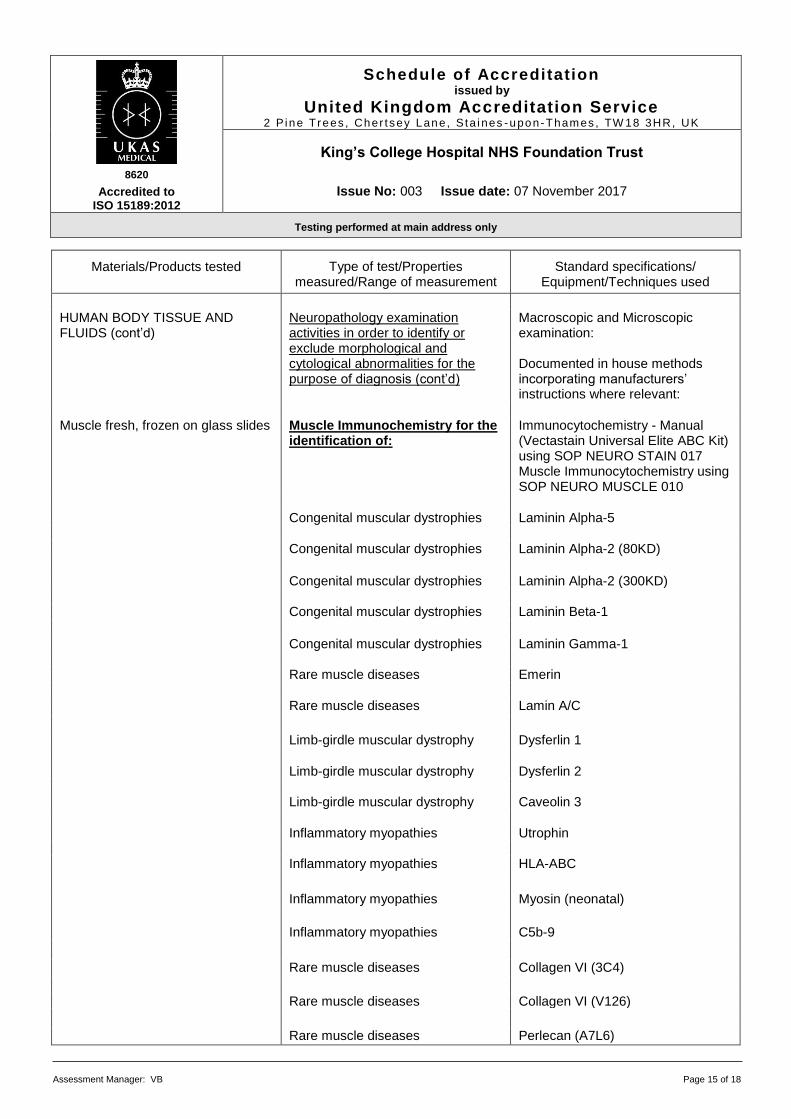
HUMAN BODY TISSUE AND FLUIDS (cont’d)
Neuropathology examination activities in order to identify or exclude morphological and cytological abnormalities for the purpose of diagnosis (cont’d)
Macroscopic and Microscopic examination: Documented in house methods incorporating manufacturers’ instructions where relevant:
Muscle fresh, frozen on glass slides Muscle Immunochemistry for the identification of:
Immunocytochemistry - Manual (Vectastain Universal Elite ABC Kit) using SOP NEURO STAIN 017 Muscle Immunocytochemistry using SOP NEURO MUSCLE 010
Congenital muscular dystrophies Laminin Alpha-5
Congenital muscular dystrophies Laminin Alpha-2 (80KD)
Congenital muscular dystrophies Laminin Alpha-2 (300KD) Congenital muscular dystrophies Laminin Beta-1
Congenital muscular dystrophies Laminin Gamma-1 Rare muscle diseases Emerin
Rare muscle diseases Lamin A/C
Limb-girdle muscular dystrophy Dysferlin 1
Limb-girdle muscular dystrophy Dysferlin 2 Limb-girdle muscular dystrophy Caveolin 3
Inflammatory myopathies Utrophin Inflammatory myopathies HLA-ABC
Inflammatory myopathies Myosin (neonatal) Inflammatory myopathies C5b-9
Rare muscle diseases Collagen VI (3C4)
Rare muscle diseases Collagen VI (V126)
Rare muscle diseases Perlecan (A7L6)
8620
Accredited to ISO 15189:2012
Schedule of Accreditation issued by
United Kingdom Accreditation Service 2 P ine Trees , Cher t sey Lane, S ta i nes -upon-Thames , TW 18 3HR, UK
King’s College Hospital NHS Foundation Trust
Issue No: 003 Issue date: 07 November 2017
Testing performed at main address only
Assessment Manager: VB Page 16 of 18
Materials/Products tested
Type of test/Properties
measured/Range of measurement
Standard specifications/
Equipment/Techniques used
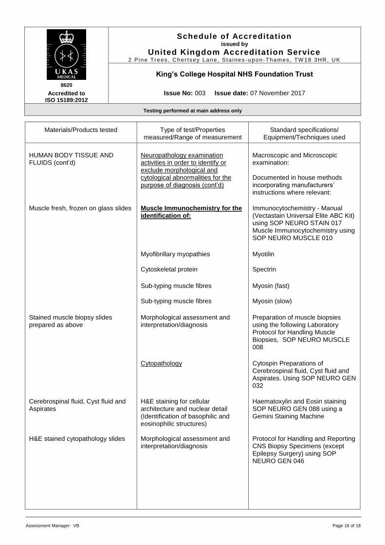
HUMAN BODY TISSUE AND FLUIDS (cont’d)
Neuropathology examination activities in order to identify or exclude morphological and cytological abnormalities for the purpose of diagnosis (cont’d)
Macroscopic and Microscopic examination: Documented in house methods incorporating manufacturers’ instructions where relevant:
Muscle fresh, frozen on glass slides Muscle Immunochemistry for the identification of:
Immunocytochemistry - Manual (Vectastain Universal Elite ABC Kit) using SOP NEURO STAIN 017 Muscle Immunocytochemistry using SOP NEURO MUSCLE 010
Myofibrillary myopathies Myotilin
Cytoskeletal protein Spectrin
Sub-typing muscle fibres Myosin (fast)
Sub-typing muscle fibres Myosin (slow)
Stained muscle biopsy slides prepared as above
Morphological assessment and interpretation/diagnosis
Preparation of muscle biopsies using the following Laboratory Protocol for Handling Muscle Biopsies, SOP NEURO MUSCLE 008
Cytopathology Cytospin Preparations of
Cerebrospinal fluid, Cyst fluid and Aspirates. Using SOP NEURO GEN 032
Cerebrospinal fluid, Cyst fluid and Aspirates
H&E staining for cellular architecture and nuclear detail (Identification of basophilic and eosinophilic structures)
Haematoxylin and Eosin staining SOP NEURO GEN 088 using a Gemini Staining Machine
H&E stained cytopathology slides Morphological assessment and
interpretation/diagnosis Protocol for Handling and Reporting CNS Biopsy Specimens (except Epilepsy Surgery) using SOP NEURO GEN 046
8620
Accredited to ISO 15189:2012
Schedule of Accreditation issued by
United Kingdom Accreditation Service 2 P ine Trees , Cher t sey Lane, S ta i nes -upon-Thames , TW 18 3HR, UK
King’s College Hospital NHS Foundation Trust
Issue No: 003 Issue date: 07 November 2017
Testing performed at main address only
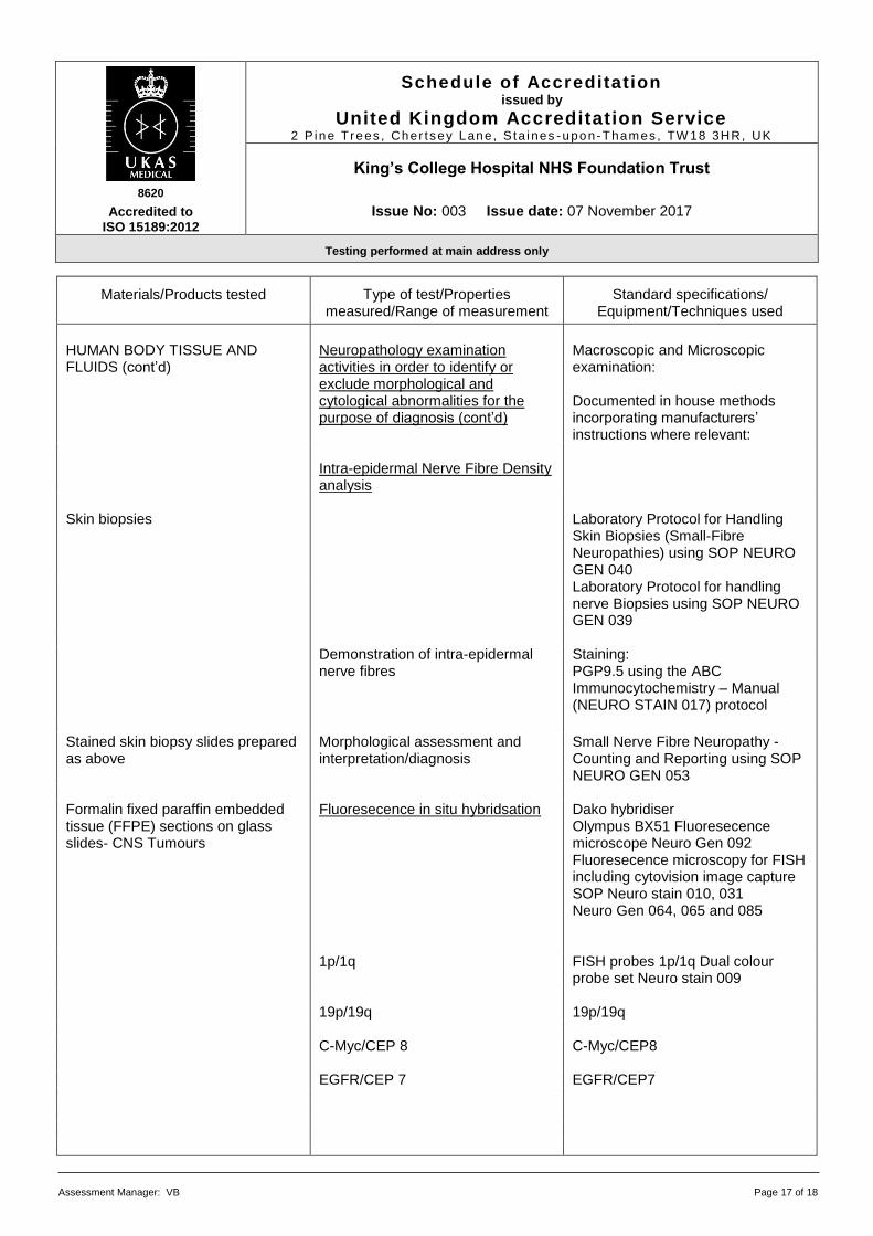
Assessment Manager: VB Page 17 of 18
Materials/Products tested
Type of test/Properties
measured/Range of measurement
Standard specifications/
Equipment/Techniques used
HUMAN BODY TISSUE AND FLUIDS (cont’d)
Neuropathology examination activities in order to identify or exclude morphological and cytological abnormalities for the purpose of diagnosis (cont’d)
Macroscopic and Microscopic examination: Documented in house methods incorporating manufacturers’ instructions where relevant:
Intra-epidermal Nerve Fibre Density
analysis
Skin biopsies Laboratory Protocol for Handling
Skin Biopsies (Small-Fibre Neuropathies) using SOP NEURO GEN 040
Laboratory Protocol for handling nerve Biopsies using SOP NEURO GEN 039
Demonstration of intra-epidermal
nerve fibres
Staining: PGP9.5 using the ABC Immunocytochemistry – Manual (NEURO STAIN 017) protocol
Stained skin biopsy slides prepared as above
Morphological assessment and interpretation/diagnosis
Small Nerve Fibre Neuropathy - Counting and Reporting using SOP NEURO GEN 053
Formalin fixed paraffin embedded tissue (FFPE) sections on glass slides- CNS Tumours
Fluoresecence in situ hybridsation Dako hybridiser Olympus BX51 Fluoresecence microscope Neuro Gen 092 Fluoresecence microscopy for FISH including cytovision image capture SOP Neuro stain 010, 031 Neuro Gen 064, 065 and 085
1p/1q FISH probes 1p/1q Dual colour
probe set Neuro stain 009 19p/19q 19p/19q C-Myc/CEP 8 C-Myc/CEP8 EGFR/CEP 7 EGFR/CEP7
8620
Accredited to ISO 15189:2012
Schedule of Accreditation issued by
United Kingdom Accreditation Service 2 P ine Trees , Cher t sey Lane, S ta i nes -upon-Thames , TW 18 3HR, UK
King’s College Hospital NHS Foundation Trust
Issue No: 003 Issue date: 07 November 2017
Testing performed at main address only
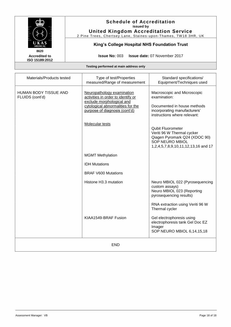
Assessment Manager: VB Page 18 of 18
Materials/Products tested
Type of test/Properties
measured/Range of measurement
Standard specifications/
Equipment/Techniques used
HUMAN BODY TISSUE AND FLUIDS (cont’d)
Neuropathology examination activities in order to identify or exclude morphological and cytological abnormalities for the purpose of diagnosis (cont’d)
Macroscopic and Microscopic examination: Documented in house methods incorporating manufacturers’ instructions where relevant:
Molecular tests Qubit Fluorometer
Veriti 96 W Thermal cycker Qiagen Pyromark Q24 (XDOC 90) SOP NEURO MBIOL 1,2,4,5,7,8,9,10,11,12,13,16 and 17
MGMT Methylation IDH Mutations
BRAF V600 Mutations Histone H3.3 mutation Neuro MBIOL 022 (Pyrosequencing
custom assays) Neuro MBIOL 023 (Reporting pyrosequencing results)
RNA extraction using Veriti 96 W
Thermal cycler KIAA1549-BRAF Fusion Gel electrophoresis using
electrophoresis tank Gel Doc EZ Imager SOP NEURO MBIOL 6,14,15,18
END