Embed Size (px)
Citation preview
Published by ISGE ISSN: 2736-5530
ISSUE 1 Volume 2 (March 2021)

0
Index Issue 1 Volume 2 TheTrocar March 2021
After a year of COVID19, the medical societies are fighting for funding. Guenter Noé
page 1
Original Articles 1. Improved reproductive outcomes in women with adenomyosis undergoing in vitrofertilisation following long term GnRH agonist downregulation: A case series
Catherine Schepisi
page 2-14
2. Laparoscopic ischial spine colpopexy: a new approach and first single centerexperience
Sanjay Shanbhag
page 15-22
Review: 3. Identical ovarian and deep pelvic endometriosis with colorectal involvement inmonozygotic twins: a case report and review of the literature
Adel Shervin
page 23-32
Case reports: 4. Hysteroscopic removal of Retained Intra-uterine Fetal Bone which causes secondaryinfertility: A case report
Ameneh Haghgoo
page 33-38
5. Laparoscopic management of cesarean scar pregnancy: a case report and literaturereviewAhmed Mimouni
page 39-46
6. Antenatal ultrasound diagnosis of ‘Iniencephaly Apertus
Glossy Sabharwal, Meenu Agarwal (general topic)
page 47-51
page 52-54
page 55
page 56-57
7. Learning for our complication
Adel Sedrati
Video Articles:
8. Hemorrhagic ascites and pleural effusion: an uncommon presentation of endometriosis
Hind Ennasser 9. Laparoscopic management of a case of accessory cavitated uterine mass (ACUM)
Sanket Pisat
Erratum: Issue 1 Vol2 Erratum (December 2020) Article: Page (25-30) Intra-ovarian direct trocar penetration and drainage for access prior to laparoscopic surgery for giant ovarian cyst
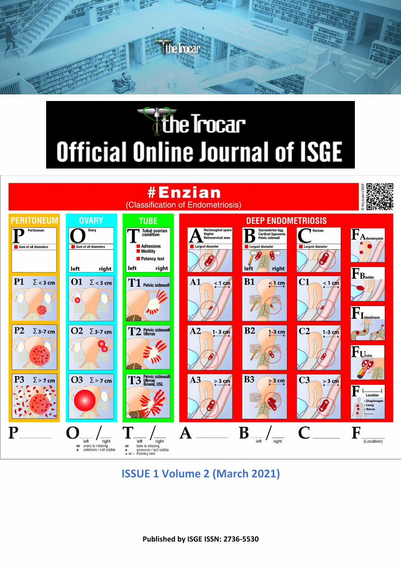
The Titlepage shows the new extended ENZIAN Score for Endometriosis classification
page 58
1
After a year of COVID19, the medical societies are fighting for funding.
On the one hand, the COVID19 crisis brought a so-called digitalization boost, but on the other
hand it hit the traditional funding of the societies hard. At the beginning everyone wanted to
show that they are still active and there were countless free teaching opportunities. The
conference technology made it possible to register 500, 1000 or 5000 colleagues for
conferences. The industry was also initially enthusiastic about the large number of participants
at one event. With the latter, however, meanwhile a hangover mood did take over. The lack
of personal contact at online events has apparently had a negative impact on the Return of
Investment (ROI) for the industry. The large companies in particular complain about it, while
smaller ones are happy about the greater exposure. With the new technology, live surgeries
can also be financed by small companies and exciting workshops can be designed. For
Societies like ISGE, however, all of this becomes a major challenge. The membership fees are
adapted to the financial possibilities of our members and their countries of origin. The
accreditation, another pillar of the financing, is severely restricted by COVID19, so that new
course formats have to be found. For organizers like AAGL or ESGE, congresses are usually an
additional source of income, for ISGE these only played a small role in the financing.
Meanwhile, the competition on the web is so great that it is hardly possible to cover the costs.
Numerous providers conduct courses online and issue certificates, which is ultimately very
problematic. For accreditation and certification, colleagues should contact the professional
associations that offer structured and Evidence-based education. ISGE is continuously working
on an optimized training of our members, here we want to use the use of IT technologies in
order to be able to offer excellent and affordable surgical education globally. Support us and
become a member or a sponsor.
Sincerely
Guenter Noé
Incoming President ISGE 2021
2 Corresponding author: Dr Catherine Schepisi [email protected] DOI: 10.36205/trocar1.2021002 Received 28/01/2021- Accepted 05/02/2021
TheTrocar Issue 1 2021 / Page 2 ISSN: 2736-5530
Improved reproductive outcomes in women with adenomyosis
undergoing in vitro fertilisation following long term GnRH agonist
downregulation: A case series
Author: Catherine Schepisi1, Oshri Bar El2, Shagun Narula1, Roni Ratner3, Jim Tsaltas1,3
Affiliation: 1 Department of Obstetrics and Gynaecology, Monash University, Australia 2 Division of Obstetrics and Gynaecology, Assuta Medical Centre, Ashdod, Israel 3 Gynaecological Endosurgery Unit, Monash Health, Melbourne Vic, Australia
Abstract
Background: Adenomyosis is a common condition that is often associated with poor reproductive
outcome. Repeated implantation failure in in vitro fertilization (IVF) cycles can result from impaired
implantation caused by adenomyosis. The evidence regarding the role of long-term suppression (LTS)
with gonadotropin-releasing hormone agonists (GNRHa) prior to embryo transfer (ET) in cases of
adenomyosis is limited and conflicting.
Aim: The aim of this study was to assess the efficacy of long-term GNRHa therapy on livebirth rates in
women with adenomyosis and infertility.
Design: The following case series includes 15 women with infertility and known adenomyosis undergoing
in vitro fertilisation (IVF) that underwent at least three months downregulation with GNRHa prior to ET.
Outcomes were compared to previous cycles performed without LTS.
Results: LTS with GNRHa was given in 16 cases (94.1%) prior to ET. In one case (5.9%) 6 months
suppression was given. The majority of these patients had previous unsuccessful IVF cycles prior to LTS
protocol with GnHRa. 17 embryo transfers (16 frozen, 1 fresh from donor) following LTS protocol
resulted in 10 liveborn deliveries at term (58.8%) vs. a live birth rate of only 7.7% per ET without LTS (26
embryo transfers resulting in 2 live births; P<0.0001). Only three women who underwent long term
GnRHa downregulation had no successful embryo transfers.
Conclusion: Our successful outcomes support the use of LTS with GnRHa to improve reproductive
outcomes.
Keywords: Adenomyosis, infertility, in vitro fertilisation, gonadotropin releasing hormone agonist
-14
2
Introduction
Adenomyosis is a benign estrogen dependent
disease whereby ectopic endometrial glands or
stroma are found within the myometrium,
surrounded by myometrial smooth muscle cell
hyperplasia and hypertrophy [1-3]. It can be
diffuse or focal depending on extent of uterine
spread [4, 5]. Women with adenomyosis typically
present with dysmenorrhea, heavy menstrual
bleeding or infertility, however a large
proportion can be asymptomatic [3]. Therefore,
true prevalence is unknown, with large variations
between 5-70% cited within the literature [3, 6,
7]. Previously thought to be a disease associated
with older multiparous women, delayed fertility
and advancements in imaging techniques have
led to an increased diagnosis in younger women
of reproductive age being investigated for
infertility [8]. Adenomyosis has been associated
with poor reproductive prognoses [9, 10].
Multiple factors have been implicated including
impaired implantation and utero-tubal
disruption [11, 12]. Of particular interest is the
impact of poor endometrial receptivity on
implantation, which has been suggested to be
more significant than embryo quality [13].
Increasing evidence is emerging to support the
role of long-term GnRHa downregulation in
improved reproductive outcomes, especially
when used in conjunction with IVF or
intracytoplasmic sperm injection [7, 14-16]. The
following case series details successful
pregnancy outcomes following GnRHa down
regulation prior to embryo transfer (ET), further
supporting the existing evidence and theorises its
main mechanism of action is by improving
endometrial receptivity.
Materials and methods
This is a retrospective case series that includes 15
patients diagnosed with adenomyosis and
treated with long acting GnRHa prior to their IVF
treatment. These patients were chosen from a
population of patients attending a private IVF
clinic. All women included required an
ultrasound diagnosis of adenomyosis. Patients
were treated with at least 3 months of Goserelin
Acetate Implant (Zoladex®) as part of the IVF
protocol prior to ET. The study was reviewed by
the Epworth Healthcare ethical review board and
registered as a quality assurance study. Data was
retrospectively collated from patient files and
included patient demographics, previous
obstetric history, prior fertility treatment and
outcomes as well as significant past medical
history. Data were analysed to assess the effect
of long-term GnRHa downregulation on
reproductive outcomes. Other factors impacting
fertility were also recorded for each patient (See
Table 1).
Data analysis was performed using SPSS
Statistics, version 11. Descriptive characteristics
of data are presented as median and
interquartile range. Test of statistical significance
for categorical variables was done using
Pearson’s chi-square test and T-Test for non-
categorical variables. Statistical significance was
set at a p value <0.05.
Results
received long acting GnRHa down regulation
were reviewed. The average age of the patients
was 37.5 years (range 28-48, SD 6.5). Most of the
patients had concurrent diagnosis of
endometriosis (13 patients, 86.7%) that was also
treated prior to the commencement of the
current long-acting suppression (LTS) protocol,
none of the patients had surgery immediately
prior to the commencement of the LTS protocol.
All of the endometriosis surgeries that were
performed occurred prior to cycles with regular
ET without LTS. Diffuse adenomyosis was
3
reported in 14 of the women (93.3%) while one
patient had focal adenomyosis. All patients were
diagnosed using ultrasound scans that were
performed by a certified obstetrics and
gynecology ultrasound specialists. Further data
regarding patient characteristics can be found in
Table 1.
Three months of long-term suppression with
Zoladex was given in 16 cases (94.1%) prior to ET
(Table 2). In one case (5.9%) 6 months
suppression was given due to significant
abnormal uterine bleeding. Frozen embryos
were transferred in 16 (94.1%) patients after long
term suppression (LTS) and a fresh embryo was
transferred in one case (5.9%). Embryo transfers
without LTS protocol included 15(57.7%) frozen
embryos and 11 (42.3%) fresh embryos.
The majority of these patients had previous
unsuccessful IVF cycles prior to LTS protocol with
GnRHa down regulation. All patients had a single
embryo transfer in all cycles. 17 embryo transfers
(16 frozen, 1 fresh from donor) following LTS
protocol resulted in 10 liveborn deliveries at
term (58.8%) vs. a live birth rate of only 7.7% per
ET without long term down regulation (26
embryo transfers resulting in 2 live births), this
was statistically significant (P<0.0001). Only
three women who underwent long term GnRHa
downregulation had no successful embryo
transfers. Total pregnancy rates were
significantly better as well in the long-term
suppression group (58.8% vs 19.2%, P=0.008
excluding chemical pregnancies, or 64.7% vs
26.9%, P=0.014 including chemical pregnancies).
Although most patients in this study had
concurrent endometriosis, none of the LTS ET
cycles were performed immediately following
surgery for endometriosis. Furthermore, even
though 9 patients underwent excision of
endometriosis prior to other cycles, none of the
patients had a live birth following the surgery
(0%) while 6 of them (66.7%) had a live birth
following LTS protocol. This superiority of LTS in
cases of adenomyosis compared with
endometriosis surgery was statistically
significant (p=0.003)
Discussion
The pathogenesis of adenomyosis is not
completely understood.[2] Adenomyosis is
associated with a higher prevalence of recurrent
pregnancy loss, failed assisted-reproductive
treatment (ART) and poorer IVF reproductive
outcomes [16-19]. Adenomyosis is suspected to
impact fertility through a range of molecular
mechanisms resulting in recurrent implantation
failure (Figure 1). Adenomyosis related junctional
zone disturbance causes dysperistalsis, impairing
sperm transport and blastocyst implantation [12,
20, 21]. Hypoestrogenism, found in women with
adenomyosis, perpetuates this dysperistalsis
[22].
Impaired endometrial receptivity is also
associated with implantation failure [23].
Adenomyosis has been associated with reduced
endometrial receptivity markers, such as integrin
ß3 and leukaemia-inhibiting factor, HOXA10 and
HOXA11, which play critical roles in implantation
as well as endometrial growth, differentiation
and decidualisation[24, 25]. Pinopodes are
morphological markers of endometrial
receptivity seen on the endometrial surface at
the time of implantation [25]. Decreased
numbers and poorly formed pinopodes have
been seen in human and mice studies with
adenomyosis [25-27].
There however remains conflicting reviews in
current literature regarding the impact of
adenomyosis on infertility, with several authors
concluding that cases with concurrent
4
endometriosis confound and limit available
evidence [16, 22, 28]. A 2014 meta-analysis
found a 28% reduction in likelihood of clinical
pregnancy in women with adenomyosis
undergoing IVF or ICSI and suggests screening for
adenomyosis prior to ART [15]. A retrospective
cohort study of 213 women showed a
significantly decreased rate of viable clinical
pregnancies in women with adenomyosis
undergoing IVF with GnRH antagonist for ovarian
stimulation [29]. Furthermore, Dueholm’s review
found an overall reduction in pregnancy rate with
adenomyosis (RR 0.63) and an increased risk of
miscarriage [20]. In contrast, a case-control
retrospective study of 49 women with
adenomyosis having oocyte donation showed no
significant differences in implantation rates [30].
Benaglia’s [31] prospective cohort study also
showed no significant difference in clinical or
ongoing pregnancy rates in women with
adenomyosis undergoing IVF. Similar findings
have been found in other studies [32, 33].
Overall, the heterogeneity of studies
investigating adenomyosis and infertility make it
difficult to compare results. There are no high
quality studies, with those published being
limited by their retrospective nature, differing
diagnostic criteria, small sample sizes, differing
ages and concurrent endometriosis [7, 20, 34,
35]. There is also a lack of studies looking at the
effect of adenomyosis on natural conception
[20].
There are no current guidelines specific to the
management of adenomyosis, especially for
those seeking fertility assistance [36]. Medical
treatment is limited in patients that wish to
conceive and hysterectomy is not a valid option
for these patients.[6] Growing desire for fertility
preservation has seen an increase in
cytoreductive surgeries performed and
development of safer surgical techniques [37].
Whilst surgical excision of adenomyosis improves
symptoms and fertility outcomes they are not
without risk, including uterine adhesions and
pregnancy complications such as uterine rupture
[37, 38].
It is thought that GnRHa improves fertility in
women with adenomyosis through reduction in
hyper estrogenic states both indirectly through
to hypothalamic-pituitary axis and directly at the
level of the tissues by normalising the
endometrial over-expression of aromatase
cytochrome P450 that occurs in adenomyosis
[39]. GnRHa has also been shown to have an
antiproliferative and apoptotic effect on
endometrial cells in vitro and Khan[40] identified
suppressed pathologic lesions in women with
adenomyosis. GnRHa therapy may improve
endometrial receptivity by reducing the extent of
basal endometrium dislocation that occurs in
adenomyosis [41]. A retrospective cohort study
by Bao et al[26] also found an increase in
endometrial receptivity markers following long-
acting GnRHa protocol and significant increase in
clinical pregnancy rates in women with
decreased ovarian reserve.
Over the last decade, four retrospective cohort
studies have evaluated the use of GnRHa
downregulation on fertility and IVF outcomes in
adenomyosis[41-44]. Niu [41] compared clinical
pregnancy rates in the first IVF cycle post
treatment with GnRHa and HRT versus HRT
alone. They found that one month of GnRHa
downregulation doubled implantation and
ongoing clinical pregnancy rates. Small case
series further support the benefit of GnRHa
downregulation prior to IVF [45, 46]. However
following 2-3 months of monthly goserelin, Park
[44] found no statistical difference in clinical
pregnancy rates. A small RCT found no significant

5
difference in outcomes[47]. Data regarding the
effect of long term down regulation with GnRHa
is limited [48].
We believe that adenomyosis can severely impair
fertility and that our results further support this.
Our cases were chosen for intervention following
repeated failed IVF cycles, thought to be a result
of their severe adenomyosis. Almost all of these
patients had recurrent implantation failures prior
to treatment with long term GnRHa
downregulation. Our results show significant
highly successful fertility outcomes post three
months of GnRHa therapy prior to embryo
transfer supporting its use in management of
infertility. This differs from other evidence where
successful cases with GnRHa downregulation
were seen only following cytoreductive surgery
[49-51]. Use of GnRHa therapy alone would likely
be associated with less risks than in combination
with surgical management, however whilst the
women in our case series tolerated GnRHa
therapy well (all successfully completing a
minimum of three-months treatment), the side
effects from a hypooestrogenic state may not be
tolerated by all. Tolerance may be regime
dependant and currently there remain large
discrepancies in GnRHa downregulation regimes
[41, 42, 44]. Our study has several limitations
being retrospective in nature with a small sample
size and high concurrent endometriosis rate.
There was also selection bias as most patients
had recurrent implantation failures in multiple
cycles, this is also accentuated as the patients are
their own controls. However, this significantly
high success rate in a population that had very
poor outcomes prior to this treatment is very
promising. Acknowledging that outcomes will
always be better after introduction of an
intervention, we believe it supports further
research to explore the impact of adenomyosis
and GnRHa on implantation.
1.5 CONCLUSION
Our cases show promising results and support
the use of long term GnRHa downregulation prior
to frozen embryo transfer. The increased rate of
pregnancy seen post GnRHa downregulation in
this cohort may be attributed to increased
endometrial receptivity promoted by GnRHa.
Furthermore, whilst we believe that GnRHa
downregulation is a promising therapy for
women with adenomyosis and infertility, further
evidence is still required to assess its impact on
pregnancy rates.
6
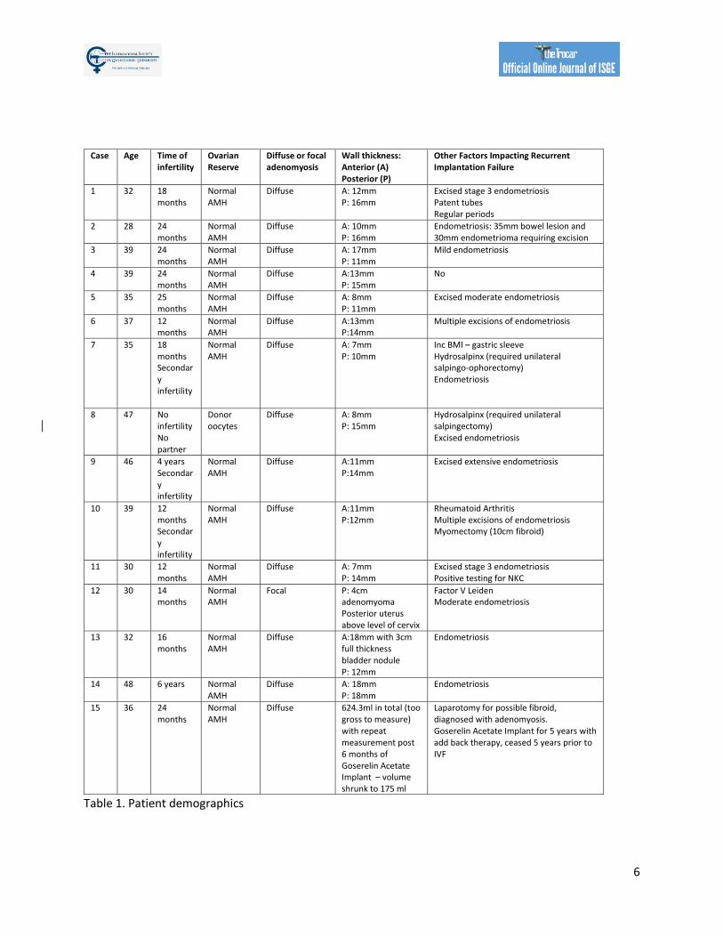
Table 1. Patient demographics
Case Age Time of infertility
Ovarian Reserve
Diffuse or focal adenomyosis
Wall thickness: Anterior (A) Posterior (P)
Other Factors Impacting Recurrent Implantation Failure
1 32 18 months
Normal AMH
Diffuse A: 12mm P: 16mm
Excised stage 3 endometriosis Patent tubes Regular periods
2 28 24 months
Normal AMH
Diffuse A: 10mm P: 16mm
Endometriosis: 35mm bowel lesion and 30mm endometrioma requiring excision
3 39 24 months
Normal AMH
Diffuse A: 17mm P: 11mm
Mild endometriosis
4 39 24 months
Normal AMH
Diffuse A:13mm P: 15mm
No
5 35 25 months
Normal AMH
Diffuse A: 8mm P: 11mm
Excised moderate endometriosis
6 37 12 months
Normal AMH
Diffuse A:13mm P:14mm
Multiple excisions of endometriosis
7 35 18 months Secondary infertility
Normal AMH
Diffuse A: 7mm P: 10mm
Inc BMI – gastric sleeve Hydrosalpinx (required unilateral salpingo-ophorectomy) Endometriosis
8 47 No infertility No partner
Donor oocytes
Diffuse A: 8mm P: 15mm
Hydrosalpinx (required unilateral salpingectomy) Excised endometriosis
9 46 4 years Secondary infertility
Normal AMH
Diffuse A:11mm P:14mm
Excised extensive endometriosis
10 39 12 months Secondary infertility
Normal AMH
Diffuse A:11mm P:12mm
Rheumatoid Arthritis Multiple excisions of endometriosis Myomectomy (10cm fibroid)
11 30 12 months
Normal AMH
Diffuse A: 7mm P: 14mm
Excised stage 3 endometriosis Positive testing for NKC
12 30 14 months
Normal AMH
Focal P: 4cm adenomyoma Posterior uterus above level of cervix
Factor V Leiden Moderate endometriosis
13 32 16 months
Normal AMH
Diffuse A:18mm with 3cm full thickness bladder nodule P: 12mm
Endometriosis
14 48 6 years Normal AMH
Diffuse A: 18mm P: 18mm
Endometriosis
15 36 24 months
Normal AMH
Diffuse 624.3ml in total (too gross to measure) with repeat measurement post 6 months of Goserelin Acetate Implant – volume shrunk to 175 ml
Laparotomy for possible fibroid, diagnosed with adenomyosis. Goserelin Acetate Implant for 5 years with add back therapy, ceased 5 years prior to IVF
7
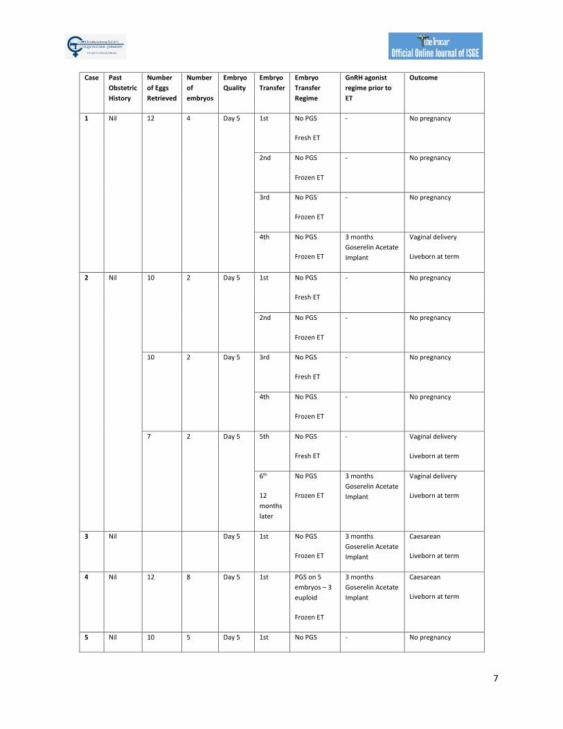
Case Past
Obstetric
History
Number
of Eggs
Retrieved
Number
of
embryos
Embryo
Quality
Embryo
Transfer
Embryo
Transfer
Regime
GnRH agonist
regime prior to
ET
Outcome
1 Nil 12 4 Day 5 1st No PGS
Fresh ET
- No pregnancy
2nd No PGS
Frozen ET
- No pregnancy
3rd No PGS
Frozen ET
- No pregnancy
4th No PGS
Frozen ET
3 months
Goserelin Acetate
Implant
Vaginal delivery
Liveborn at term
2
Nil 10 2 Day 5 1st No PGS
Fresh ET
- No pregnancy
2nd No PGS
Frozen ET
- No pregnancy
10 2 Day 5 3rd No PGS
Fresh ET
- No pregnancy
4th No PGS
Frozen ET
- No pregnancy
7 2 Day 5 5th No PGS
Fresh ET
- Vaginal delivery
Liveborn at term
6th
12
months
later
No PGS
Frozen ET
3 months
Goserelin Acetate
Implant
Vaginal delivery
Liveborn at term
3 Nil Day 5 1st No PGS
Frozen ET
3 months
Goserelin Acetate
Implant
Caesarean
Liveborn at term
4 Nil 12 8 Day 5 1st PGS on 5
embryos – 3
euploid
Frozen ET
3 months
Goserelin Acetate
Implant
Caesarean
Liveborn at term
5 Nil 10 5 Day 5 1st No PGS - No pregnancy

8
Frozen ET
2nd No PGS
Frozen ET
- No pregnancy
10 6 Day 5 3rd No PGS
Frozen ET
3 months
Goserelin Acetate
Implant
Vaginal delivery
Liveborn at term
6 Nil 11 6 Day 5 1st No PGS
Fresh ET
- No pregnancy
2nd No PGS
Frozen ET
- No pregnancy
21 9 Day 5 3rd PGS on 3
embryos – 2
euploid
Frozen ET
3 months
Goserelin Acetate
Implant
Vaginal delivery
Liveborn at term
7 1 NVD at
term
14 13 Day 5 1st No PGS
Fresh ET
- Biochemical pregnancy
2nd PGS on 9
embryos – 5
euploid
Frozen ET
3 months
Goserelin Acetate
Implant
No pregnancy
3rd PGS as above
Frozen ET
3 months
Goserelin Acetate
Implant
No pregnancy
8 Nil
- - - 1st No PGS
Fresh ET
(from donor)
3 months
Goserelin Acetate
Implant
Caesarean
Liveborn at term
9 1 NVD at
term
- - - 1st No PGS
Frozen ET
3 months
Goserelin Acetate
Implant
Biochemical pregnancy
10 1 NVD at
term
8 6 Day 5 1st No PGS
Fresh ET
- No pregnancy
2nd No PGS - 8 weeks miscarriage

9
Fresh ET
6 3 Day 5
Poor
quality
not
used
- - - -
7 4 Day 5 3rd PGS on 1
embryo –
euploid
Frozen ET
- 8 weeks miscarriage
19 9 Day 5 4th PGS on 4
embryos - 1
euploid
Frozen ET
3 months
Goserelin Acetate
Implant
No pregnancy
11 Nil 12 7 Day 5 1st No PGS
Frozen ET
- 8 weeks miscarriage
2nd No PGS
Frozen ET
- No pregnancy
*Nb went to another
unit following this and
had 10 unsuccessful
ETs
- 2 left - 3rd with
this unit
PGS on 2
embryos – 2
euploid
Frozen ET
3 months
Goserelin Acetate
Implant
No pregnancy
4th with
this unit
PGS as above
Frozen ET
3 months
Goserelin Acetate
Implant
Vaginal delivery
Liveborn at term
12
Nil 8 6
Day 5 1st No PGS
Fresh ET
-
No pregnancy
2nd No PGS
Frozen ET
*FVL
heterozygous
diagnosed –
commenced
enoxaparin
- No pregnancy
10
from time of
ET
3rd No PGS
Frozen ET
- No pregnancy
4th No PGS
Frozen ET
- Vaginal delivery
Liveborn at term
8 4 Day 5
Only 2
good
embryos
1st No PGS
Fresh ET
- Biochemical pregnancy
2nd No PGS
Frozen ET
- No pregnancy
12 5 Day 5
Only 4
good
embryos
3rd No PGS
Fresh ET
- No pregnancy
4th No PGS
Frozen ET
- No pregnancy
5th No PGS
Frozen ET
3 months
Goserelin Acetate
Implant
No pregnancy
13 Nil 6 1st PGS
Frozen ET
3 months
Goserelin Acetate
Implant
Caesarean
Liveborn at term
14 Nil 1 1st Frozen ET 3 months
Goserelin Acetate
Implant
Not pregnant
15 Nil 4 1st PGS
Frozen ET
6 months
Goserelin Acetate
Implant
Caesarean
Liveborn at term
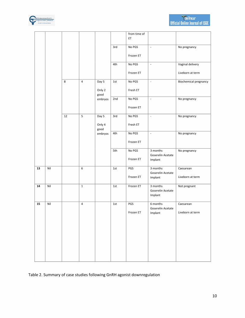
Table 2. Summary of case studies following GnRH agonist downregulation
11
The above table outlines the case series of seventeen pregnancies undergoing GnRH agonist down-
regulation for adenomyosis prior to IVF.
Definitions: ET- endometrial thickness, Zoladex – goserelin acetate, PGS – Pre-implantation Genetic
Screening
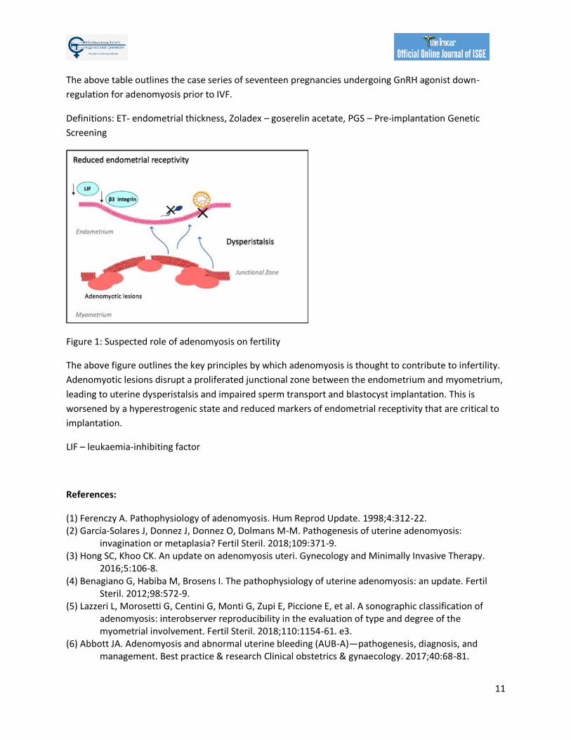
Figure 1: Suspected role of adenomyosis on fertility
The above figure outlines the key principles by which adenomyosis is thought to contribute to infertility.
Adenomyotic lesions disrupt a proliferated junctional zone between the endometrium and myometrium,
leading to uterine dysperistalsis and impaired sperm transport and blastocyst implantation. This is
worsened by a hyperestrogenic state and reduced markers of endometrial receptivity that are critical to
implantation.
LIF – leukaemia-inhibiting factor
References:
(1) Ferenczy A. Pathophysiology of adenomyosis. Hum Reprod Update. 1998;4:312-22. (2) García-Solares J, Donnez J, Donnez O, Dolmans M-M. Pathogenesis of uterine adenomyosis:
invagination or metaplasia? Fertil Steril. 2018;109:371-9. (3) Hong SC, Khoo CK. An update on adenomyosis uteri. Gynecology and Minimally Invasive Therapy.
2016;5:106-8. (4) Benagiano G, Habiba M, Brosens I. The pathophysiology of uterine adenomyosis: an update. Fertil
Steril. 2012;98:572-9. (5) Lazzeri L, Morosetti G, Centini G, Monti G, Zupi E, Piccione E, et al. A sonographic classification of
adenomyosis: interobserver reproducibility in the evaluation of type and degree of the myometrial involvement. Fertil Steril. 2018;110:1154-61. e3.
(6) Abbott JA. Adenomyosis and abnormal uterine bleeding (AUB-A)—pathogenesis, diagnosis, and management. Best practice & research Clinical obstetrics & gynaecology. 2017;40:68-81.

12
(7) Soave I, Wenger J-M, Pluchino N, Marci R. Treatment options and reproductive outcome for adenomyosis-associated infertility. Curr Med Res Opin. 2018;34:839-49.
(8) Naftalin J, Hoo W, Pateman K, Mavrelos D, Holland T, Jurkovic D. How common is adenomyosis? A prospective study of prevalence using transvaginal ultrasound in a gynaecology clinic. Hum Reprod. 2012;27:3432-9.
(9) Mochimaru A, Aoki S, Oba MS, Kurasawa K, Takahashi T, Hirahara F. Adverse pregnancy outcomes associated with adenomyosis with uterine enlargement. J Obstet Gynaecol Res. 2015;41:529-33.
(10) Hashimoto A, Iriyama T, Sayama S, Nakayama T, Komatsu A, Miyauchi A, et al. Adenomyosis and adverse perinatal outcomes: increased risk of second trimester miscarriage, preeclampsia, and placental malposition. The Journal of Maternal-Fetal & Neonatal Medicine. 2018;31:364-9.
(11) Kuijsters NPM, Methorst WG, Kortenhorst MSQ, Rabotti C, Mischi M, Schoot BC. Uterine peristalsis and fertility: current knowledge and future perspectives: a review and meta-analysis. Reprod Biomed Online. 2017;35:50-71.
(12) Kissler S, Zangos S, Vogl T, Hamscho N, Gruenwald F, Kohl J, et al. Impaired utero-tubal sperm transport in adenomyosis and endometriosis—a cause for infertility. International Congress Series: Elsevier; 2004. p. 229-31.
(13) Messaoudi S, Kasmi IE, Bourdiec A, Crespo K, Bissonnette L, Le Saint C, et al. 15 years of transcriptomic analysis on endometrial receptivity: what have we learnt? Fertility Research and Practice. 2019;5:9.
(14) Dueholm M, Aagaard J. Adenomyosis and IVF/ICSI treatment: clinical considerations and recommendations. Taylor & Francis; 2018.
(15) Vercellini P, Consonni D, Dridi D, Bracco B, Frattaruolo MP, Somigliana E. Uterine adenomyosis and in vitro fertilization outcome: a systematic review and meta-analysis. Hum Reprod. 2014;29:964-77.
(16) Vercellini P, Consonni D, Barbara G, Buggio L, Frattaruolo MP, Somigliana E. Adenomyosis and reproductive performance after surgery for rectovaginal and colorectal endometriosis: a systematic review and meta-analysis. Reprod Biomed Online. 2014;28:704-13.
(17) Puente J, Fabris A, Patel J, Patel A, Cerrillo M, Requena A, et al. Adenomyosis in infertile women: prevalence and the role of 3D ultrasound as a marker of severity of the disease. Reprod Biol Endocrinol. 2016;14:1-9.
(18) Salim R, Riris S, Saab W, Abramov B, Khadum I, Serhal P. Adenomyosis reduces pregnancy rates in infertile women undergoing IVF. Reprod Biomed Online. 2012;25:273-7.
(19) Shimizu Y, Fukuda J, Kawamura K, Tanaka T. Retrospectively analysis of the fertility of adenomyosis and the outcome of adenomyosis complicated pregnancy in in vitro fertilization patients. Fertil Steril. 2002;77:S50.
(20) Dueholm M. Uterine adenomyosis and infertility, review of reproductive outcome after in vitro fertilization and surgery. Acta Obstet Gynecol Scand. 2017;96:715-26.
(21) Larsen SB, Lundorf E, Forman A, Dueholm M. Adenomyosis and junctional zone changes in patients with endometriosis. European Journal of Obstetrics & Gynecology and Reproductive Biology. 2011;157:206-11.
(22) Yan L, Ding L, Tang R, Chen Z-J. Effect of adenomyosis on in vitro fertilization/intracytoplasmic sperm injection outcomes in infertile women: a retrospective cohort study. Gynecol Obstet Invest. 2014;77:14-8.
(23) Neykova K, Tosto V, Giardina I, Tsibizova V, Vakrilov G. Endometrial receptivity and pregnancy outcome. The Journal of Maternal-Fetal & Neonatal Medicine. 2020:1-15.
(24) Xiao Y, Sun X, Yang X, Zhang J, Xue Q, Cai B, et al. Leukemia inhibitory factor is dysregulated in the endometrium and uterine flushing fluid of patients with adenomyosis during implantation window. Fertil Steril. 2010;94:85-9.
13
(25) Guo S, Li Z, Yan L, Sun Y, Feng Y. GnRH agonist improves pregnancy outcome in mice with induced adenomyosis by restoring endometrial receptivity. Drug Des Devel Ther. 2018;12:1621.
(26) Bao H, Qu Q, Guo Z, Li W, Zhao H. Long-Acting GnRH Agonist Improves IVF Outcomes of Young Patients with Diminished Ovarian Reserve by Increasing Endometrial Receptivity. 2020.
(27) Zhou W, Li D, Ziwei Z, Pan Y, Jiang Y, Zhu C. GnRH-a induced changes in endometrial pinopodes. 2020.
(28) Maheshwari A, Gurunath S, Fatima F, Bhattacharya S. Adenomyosis and subfertility: a systematic review of prevalence, diagnosis, treatment and fertility outcomes. Hum Reprod Update. 2012;18:374-92.
(29) Thalluri V, Tremellen K. Ultrasound diagnosed adenomyosis has a negative impact on successful implantation following GnRH antagonist IVF treatment. Hum Reprod. 2012;27:3487-92.
(30) Vergara V, Garcia G, Meseguer M, Garrido N, Simon C, Pellicer A. The presence of uterine adenomyosis does not affect implantation in an oocyte donation program. Fertil Steril. 2008;90:S376.
(31) Benaglia L, Cardellicchio L, Paffoni A, Leonardi M, Faulisi S, Somigliana E. Adenomyosis does not influence pregnancy rate in women undergoing IVF. Fertil Steril. 2013;100:S292.
(32) O'Connor D, Bedaiwy M, Dunne C, Taylor B, Havelock J, Lawrence C, et al. Outcomes for in vitro fertilization in uterine adenomyosis: a retrospective cohort study. Fertil Steril. 2016;106:e333-e4.
(33) Benaglia L, Cardellicchio L, Leonardi M, Faulisi S, Vercellini P, Paffoni A, et al. Asymptomatic adenomyosis and embryo implantation in IVF cycles. Reprod Biomed Online. 2014;29:606-11.
(34) Kishi Y, Yabuta M. The benefit of adenomyomectomy on fertility outcomes in women with rectovaginal endometriosis with coexisting adenomyosis. Gynecology and minimally invasive therapy. 2017;6:20-4.
(35) Mavrelos D, Holland TK, O'Donovan O, Khalil M, Ploumpidis G, Jurkovic D, et al. The impact of adenomyosis on the outcome of IVF–embryo transfer. Reprod Biomed Online. 2017;35:549-54.
(36) Donnez J, Donnez O, Dolmans M-M. Introduction: Uterine adenomyosis, another enigmatic disease of our time. Fertil Steril. 2018;109:369-70.
(37) Osada H. Uterine adenomyosis and adenomyoma: the surgical approach. Fertil Steril. 2018;109:406-17.
(38) Harada T, Khine YM, Kaponis A, Nikellis T, Decavalas G, Taniguchi F. The impact of adenomyosis on women's fertility. Obstet Gynecol Surv. 2016;71:557.
(39) Ishihara H, Kitawaki J, Kado N, Koshiba H, Fushiki S, Honjo H. Gonadotropin-releasing hormone agonist and danazol normalize aromatase cytochrome P450 expression in eutopic endometrium from women with endometriosis, adenomyosis, or leiomyomas. Fertil Steril. 2003;79:735-42.
(40) Khan KN, Kitajima M, Hiraki K, Fujishita A, Nakashima M, Ishimaru T, et al. Cell proliferation effect of GnRH agonist on pathological lesions of women with endometriosis, adenomyosis and uterine myoma. Hum Reprod. 2010;25:2878-90.
(41) Niu Z, Chen Q, Sun Y, Feng Y. Long-term pituitary downregulation before frozen embryo transfer could improve pregnancy outcomes in women with adenomyosis. Gynecol Endocrinol. 2013;29:1026-30.
(42) Al Jama FE. Management of adenomyosis in subfertile women and pregnancy outcome. Oman Med J. 2011;26:178.
(43) Wang PH, Fuh JL, Chao HT, Liu WM, Cheng MH, Chao KC. Is the surgical approach beneficial to subfertile women with symptomatic extensive adenomyosis? J Obstet Gynaecol Res. 2009;35:495-502.
(44) Park CW, Choi MH, Yang KM, Song IO. Pregnancy rate in women with adenomyosis undergoing fresh or frozen embryo transfer cycles following gonadotropin-releasing hormone agonist treatment. Clinical and experimental reproductive medicine. 2016;43:169.
14
(45) Hirata JD, Moghissi KS, Ginsburg KA. Pregnancy after medical therapy of adenomyosis with a gonadotropin-releasing hormone agonist. Fertil Steril. 1993;59:444-5.
(46) Huang F, Kung F, Chang S, Hsu T. Effects of short-course buserelin therapy on adenomyosis. A report of two cases. The Journal of reproductive medicine. 1999;44:741-4.
(47) Davar R, Dashti S, Omidi M. Endometrial preparation using gonadotropin-releasing hormone agonist prior to frozen-thawed embryo transfer in women with repeated implantation failure: An RCT. International Journal of Reproductive BioMedicine (IJRM). 2020:319–26-–26.
(48) Sallam HN, Garcia‐Velasco JA, Dias S, Arici A, Abou‐Setta AM. Long‐term pituitary down‐regulation before in vitro fertilization (IVF) for women with endometriosis. Cochrane Database Syst Rev. 2006.
(49) Shiau C-S, Hsieh C-C, Chang M-Y, Hsieh C-L, Hsieh T-T. Conservative treatment of uterine adenomyosis in the infertile women with combined preoperative gonadotropin-releasing hormone agonist therapy and cytoreductive surgery. Fertil Steril. 2007;88:S221.
(50) Wang P-H, Yang T-S, Lee W-L, Chao H-T, Chang S-P, Yuan C-C. Treatment of infertile women with adenomyosis with a conservative microsurgical technique and a gonadotropin-releasing hormone agonist. Fertil Steril. 2000;73:1061-2.
(51) Wang C-J, Yuen L-T, Chang S-D, Lee C-L, Soong Y-K. Use of laparoscopic cytoreductive surgery to treat infertile women with localized adenomyosis. Fertil Steril. 2006;86:462. e5-. e8.
15 Corresponding author: Sanjay Shanbhag e-mail: [email protected] DOI: 10.36205/trocar1.2021005 Received 20.12.2020 - Accepted 10.01.2021
TheTrocar Issue 1 2021 / Page 15-22 ISSN: 2736-5530
Laparoscopic ischial spine colpopexy: a new approach and first single
center experience
Author: Sanjay Shanbhag1, Swati Shanbhag1 Guenter Noé2
Affiliation: 1Tara Nursing Home India
2 University of Witten Herdecke
Abstract:
The laparoscopic treatment of prolapse is dominated by Sacral colpopexy, the latter is known as current
Gold Standard”. Various new approaches have been described in the last years with different fixation areas
and different combinations. The ischial ligament is used successfully via vaginal route since a long time.
We have developed a laparoscopic approach to use the structure for apical repair without mesh. This
paper describes the technique and the first single center data for this new surgical technique.
Keywords: vault prolapse; laparoscopy, ischial spine,
16
Introduction:
As mesh Issues caused by vaginal approaches
occurred in the last two decades, more
laparoscopic procedures were developed and
published[1-3].Most of them were mesh based
using long mesh arms (Dubuisson) or the
pectineal ligament (Noé). There is a long tradition
for abdominal fixation (laparotomy and vaginal
fixation either [4, 5]. The ischial ligament is
successfully used uni- or bi-lateral since decades
but has not been approached abdominally yet.
Due to mesh discussion, we have adopted the
thread based vaginal technique to an abdominal
approach. According to Delancy there is
evidence that the apex of the vagina (level 1 by
delaneys classification) is at the level of S2
vertebra or at the level of ischial spine. Also, the
mid portion of the vagina (level 2 Delaney)
derives a lot of support from the white line over
the obturator fascia. Both the ischial spine and
the white line are anatomically bellow the
obturator nerve. With the advent of high-
definition cameras, these areas can be precisely
dissected and thereby we have a concept of
repairing the prolapse to correct the anatomical
position. Since the braided polyester suture
stimulates a reasonable amount of fibrosis, we
use it to replace the lack of collagenization at the
vaginal end.
Surgical approach and technique
Trocar position
1) A 10 mm optic trocar is placed in the midline
one inch above the umbilicus.
2) One 5 mm trocar is placed above and lateral
to the left anterior superior iliac spine for the
surgeon's left hand.
3) A second 5 mm trocar is added 2 inches to
the left and two cm above the optical 10 mm
trocar for the surgeon's right hand.
4) A third 5mm trocar is placed just above the
MacBurneys point on the right side for the
assistant.
5) A Fourth 5mm trocar is placed two inches
below and to the right of the umbilicus. This is
exclusively used for the safe and comfortable
passage of the 40 mm 1/2 circle taper cut heavy
needle, attached to the number 2 braided
polyester suture.
STEP 1: Dissection to approach the ischial bone
A 10 mm 50-degree scope is passed through a
supraumbilical canula. The retroperitoneum is
entered by cutting the round ligament on the
right side and incising the peritoneum cranially
parallel to the right infundibulopelvic ligament
( right ovarian vessels) and caudally to meet the
uterovesical fold.
The para rectal space (Latzko's space) is
developed between the uterine and internal iliac
artery (lateral border) and ureter (medial
border).
Further dissection is carried out at a level caudal
to the obturator nerve just lateral to the internal
iliac artery and the umbilical artery.
The dissection is to be carried out with great
caution taking very small steps and sometimes
even excising small bits of the fat (in view of the
vast variations in the anatomy as mentioned
above), until the ischial bone is reached.
Further careful dissection of this part of the
ischial bone anteriorly and parallel to the
obturator nerve is commenced.
17
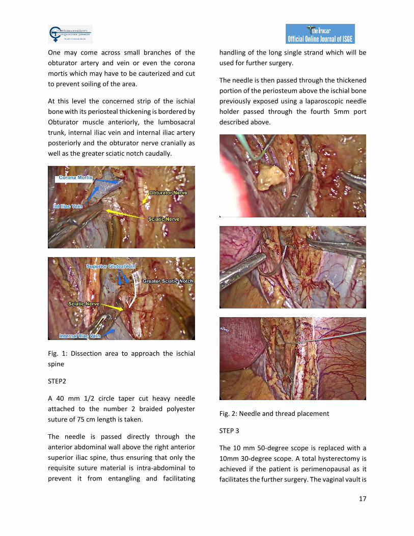
One may come across small branches of the
obturator artery and vein or even the corona
mortis which may have to be cauterized and cut
to prevent soiling of the area.
At this level the concerned strip of the ischial
bone with its periosteal thickening is bordered by
Obturator muscle anteriorly, the lumbosacral
trunk, internal iliac vein and internal iliac artery
posteriorly and the obturator nerve cranially as
well as the greater sciatic notch caudally.
Fig. 1: Dissection area to approach the ischial
spine
STEP2
A 40 mm 1/2 circle taper cut heavy needle
attached to the number 2 braided polyester
suture of 75 cm length is taken.
The needle is passed directly through the
anterior abdominal wall above the right anterior
superior iliac spine, thus ensuring that only the
requisite suture material is intra-abdominal to
prevent it from entangling and facilitating
handling of the long single strand which will be
used for further surgery.
The needle is then passed through the thickened
portion of the periosteum above the ischial bone
previously exposed using a laparoscopic needle
holder passed through the fourth 5mm port
described above.
Fig. 2: Needle and thread placement
STEP 3
The 10 mm 50-degree scope is replaced with a
10mm 30-degree scope. A total hysterectomy is
achieved if the patient is perimenopausal as it
facilitates the further surgery. The vaginal vault is
18
sutured in two layers to reduce the extrusion of
the poly-filament polyester suture.
STEP4
A 1.5-inch broad malleable copper retractor is
introduced into the vagina from bellow. This
facilitates the further dissection and suturing.
The vesico vaginal space is dissected out up to
the trigone of the bladder. The peritoneum on
the posterior vaginal wall is cut above the fat and
the rectovaginal space is dissected out up to the
lowermost part of the prolapse (not up to the
levator ani) as guided by the assistant's finger
behind the malleable retractor.
STEP5
The aforementioned needle on polyester which
is already passed through the thickened
periosteum of the ischial bone is now passed by
multiple bites through the fascia on the anterior
vaginal wall (plication of fascia) taking care not to
enter the vagina avoiding full thickness bites.
The suture material is deliberately kept on the
right side of the operative field to avoid
entangling. Bites are taken in a parallel manner
to facilitate laparoscopic suturing. The polyester
suture is pulled through to leave at least about
30 to 35 cm to do a similar plication of the
posterior vaginal wall fascia.
The suture on the anterior vaginal wall is now
pulled to tighten the plicated area thus
correcting the cystocoele. The needle is now held
and passed in a manner similar to the anterior
vaginal wall, posteriorly starting from fascia over
the lowermost part of the prolapsed posterior
vaginal wall (as guided by the assistant’s finger
placed posteriorly behind the malleable
retractor).
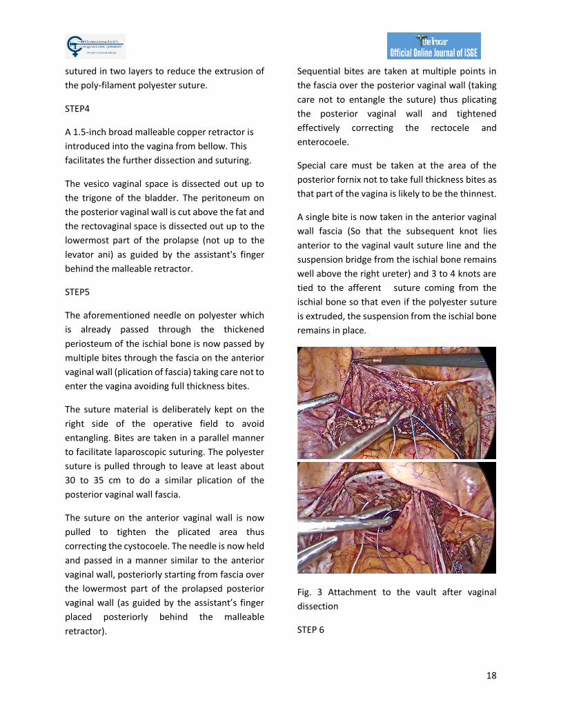
Sequential bites are taken at multiple points in
the fascia over the posterior vaginal wall (taking
care not to entangle the suture) thus plicating
the posterior vaginal wall and tightened
effectively correcting the rectocele and
enterocoele.
Special care must be taken at the area of the
posterior fornix not to take full thickness bites as
that part of the vagina is likely to be the thinnest.
A single bite is now taken in the anterior vaginal
wall fascia (So that the subsequent knot lies
anterior to the vaginal vault suture line and the
suspension bridge from the ischial bone remains
well above the right ureter) and 3 to 4 knots are
tied to the afferent suture coming from the
ischial bone so that even if the polyester suture
is extruded, the suspension from the ischial bone
remains in place.
Fig. 3 Attachment to the vault after vaginal
dissection
STEP 6
19
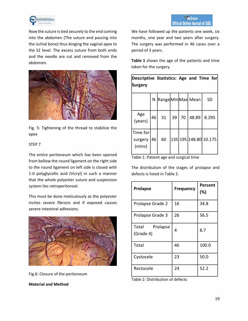
Now the suture is tied securely to the end coming
into the abdomen (The suture end passing into
the ischial bone) thus binging the vaginal apex to
the S2 level. The excess suture from both ends
and the needle are cut and removed from the
abdomen.
Fig. 5: Tightening of the thread to stabilize the
apex
STEP 7
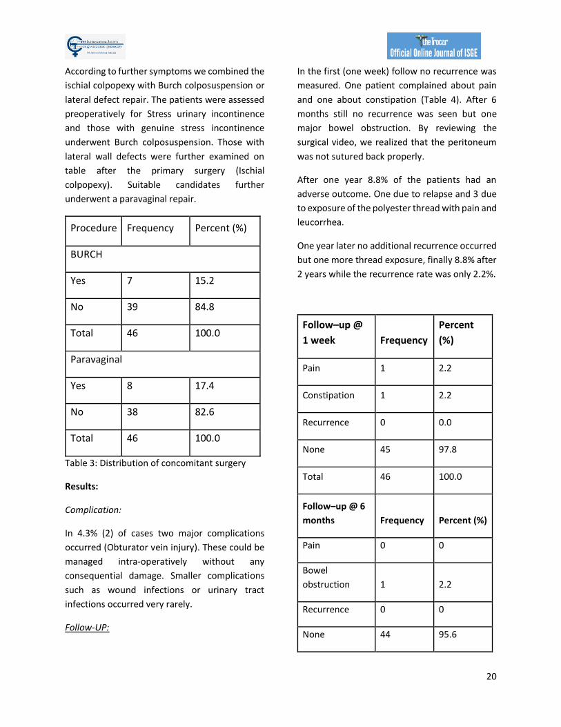
The entire peritoneum which has been opened
from bellow the round ligament on the right side
to the round ligament on left side is closed with
1-0 polyglycollic acid (Vicryl) in such a manner
that the whole polyester suture and suspension
system lies retroperitoneal.
This must be done meticulously as the polyester
incites severe fibrosis and if exposed causes
severe intestinal adhesions.
Fig.6: Closure of the peritoneum
Material and Method
We have followed up the patients one week, six
months, one year and two years after surgery.
The surgery was performed in 46 cases over a
period of 3 years.
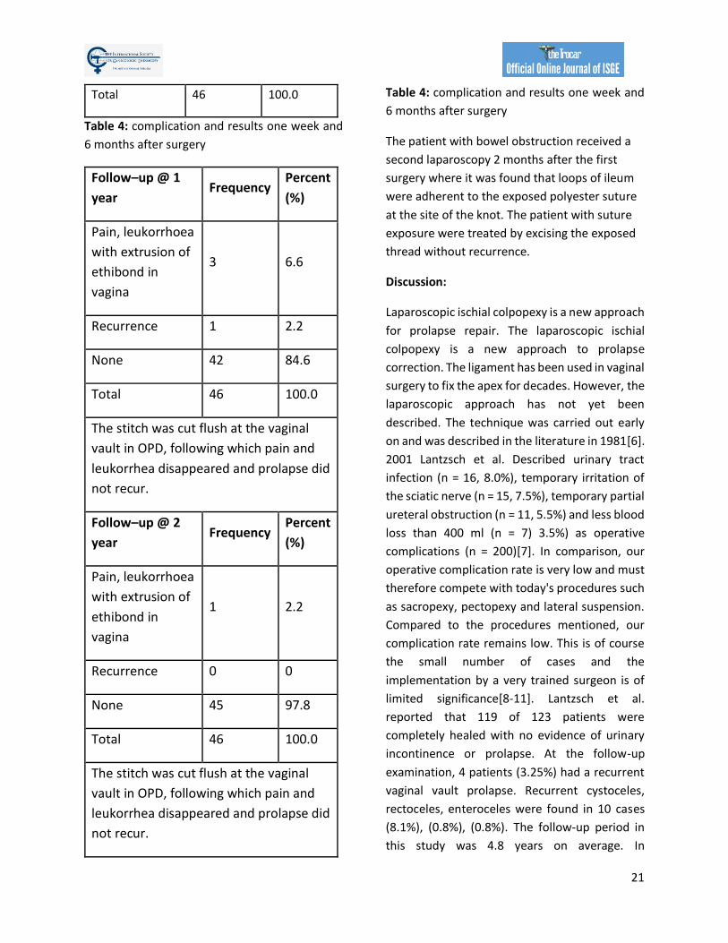
Table 1 shows the age of the patients and time
taken for the surgery.
Descriptive Statistics: Age and Time for
Surgery
N Range Min Max Mean SD
Age
(years) 46 31 39 70 48.89 8.295
Time for
surgery
(mins)
46 60 135 195 148.80 10.175
Table 1: Patient age and surgical time
The distribution of the stages of prolapse and
defects is listed in Table 2.
Prolapse Frequency Percent
(%)
Prolapse Grade 2 16 34.8
Prolapse Grade 3 26 56.5
Total Prolapse
(Grade 4) 4 8.7
Total 46 100.0
Cystocele 23 50.0
Rectocele 24 52.2
Table 2: Distribution of defects
20
According to further symptoms we combined the
ischial colpopexy with Burch colposuspension or
lateral defect repair. The patients were assessed
preoperatively for Stress urinary incontinence
and those with genuine stress incontinence
underwent Burch colposuspension. Those with
lateral wall defects were further examined on
table after the primary surgery (Ischial
colpopexy). Suitable candidates further
underwent a paravaginal repair.
Procedure Frequency Percent (%)
BURCH
Yes 7 15.2
No 39 84.8
Total 46 100.0
Paravaginal
Yes 8 17.4
No 38 82.6
Total 46 100.0
Table 3: Distribution of concomitant surgery
Results:
Complication:
In 4.3% (2) of cases two major complications
occurred (Obturator vein injury). These could be
managed intra-operatively without any
consequential damage. Smaller complications
such as wound infections or urinary tract
infections occurred very rarely.
Follow-UP:
In the first (one week) follow no recurrence was
measured. One patient complained about pain
and one about constipation (Table 4). After 6
months still no recurrence was seen but one
major bowel obstruction. By reviewing the
surgical video, we realized that the peritoneum
was not sutured back properly.
After one year 8.8% of the patients had an
adverse outcome. One due to relapse and 3 due
to exposure of the polyester thread with pain and
leucorrhea.
One year later no additional recurrence occurred
but one more thread exposure, finally 8.8% after
2 years while the recurrence rate was only 2.2%.
Follow–up @
1 week Frequency
Percent
(%)
Pain 1 2.2
Constipation 1 2.2
Recurrence 0 0.0
None 45 97.8
Total 46 100.0
Follow–up @ 6
months Frequency Percent (%)
Pain 0 0
Bowel
obstruction 1 2.2
Recurrence 0 0
None 44 95.6
21
Total 46 100.0
Table 4: complication and results one week and
6 months after surgery
Follow–up @ 1
year Frequency
Percent
(%)
Pain, leukorrhoea
with extrusion of
ethibond in
vagina
3 6.6
Recurrence 1 2.2
None 42 84.6
Total 46 100.0
The stitch was cut flush at the vaginal
vault in OPD, following which pain and
leukorrhea disappeared and prolapse did
not recur.
Follow–up @ 2
year Frequency
Percent
(%)
Pain, leukorrhoea
with extrusion of
ethibond in
vagina
1 2.2
Recurrence 0 0
None 45 97.8
Total 46 100.0
The stitch was cut flush at the vaginal
vault in OPD, following which pain and
leukorrhea disappeared and prolapse did
not recur.
Table 4: complication and results one week and
6 months after surgery
The patient with bowel obstruction received a
second laparoscopy 2 months after the first
surgery where it was found that loops of ileum
were adherent to the exposed polyester suture
at the site of the knot. The patient with suture
exposure were treated by excising the exposed
thread without recurrence.
Discussion:
Laparoscopic ischial colpopexy is a new approach
for prolapse repair. The laparoscopic ischial
colpopexy is a new approach to prolapse
correction. The ligament has been used in vaginal
surgery to fix the apex for decades. However, the
laparoscopic approach has not yet been
described. The technique was carried out early
on and was described in the literature in 1981[6].
2001 Lantzsch et al. Described urinary tract
infection (n = 16, 8.0%), temporary irritation of
the sciatic nerve (n = 15, 7.5%), temporary partial
ureteral obstruction (n = 11, 5.5%) and less blood
loss than 400 ml (n = 7) 3.5%) as operative
complications (n = 200)[7]. In comparison, our
operative complication rate is very low and must
therefore compete with today's procedures such
as sacropexy, pectopexy and lateral suspension.
Compared to the procedures mentioned, our
complication rate remains low. This is of course
the small number of cases and the
implementation by a very trained surgeon is of
limited significance[8-11]. Lantzsch et al.
reported that 119 of 123 patients were
completely healed with no evidence of urinary
incontinence or prolapse. At the follow-up
examination, 4 patients (3.25%) had a recurrent
vaginal vault prolapse. Recurrent cystoceles,
rectoceles, enteroceles were found in 10 cases
(8.1%), (0.8%), (0.8%). The follow-up period in
this study was 4.8 years on average. In
22
comparison to the data from Lantzsch and the
data from comparable laparoscopic studies, our
success rate in the examined collective is very
satisfactory. However, since we wanted to work
without mesh, the high exposure rate of 8.8%
must be viewed critically.
Limitations of the technique:
The thread material is supposed to produce good
fibrosis, but appears to favor exposure due to its
strong local irritation. Therefore, we have to look
for alternatives regarding the material. The
suturing technique appears to be effective in
correcting even a combined prolapse. However,
the proximity to the ureter, pelvic floor nerves
and lymphatic tissue requires a high level of
expertise from the surgeon, which will make it
more difficult to spread.
Conclusion:
The laparoscopic ischial colpopexy can expand
the portfolio of laparoscopic techniques for
correcting pelvic floor defects. The early
experiences with the technology show the
feasibility but also the limitations. The material
and safety of the technology still has to be
examined on a larger scale before further
statements can be made. Polyester sutures
seem to be of risk to be used close to the
vagina.
References:
1. Banerjee, C. and K.G. Noe, Laparoscopic pectopexy: a new technique of prolapse surgery for obese patients. Arch Gynecol Obstet, 2011. 284(3): p. 631-5.
2. FDA. 2011. 3. Dubuisson, J., et al., Laparoscopic repair of vaginal vault prolapse by lateral suspension with
mesh. Arch Gynecol Obstet, 2013. 287(2): p. 307-12. 4. Lane, F.E., Repair of posthysterectomy vaginal-vault prolapse. Obstetrics and Gynecology, 1962.
20(72). 5. Richardson, A.C. and G.A. Williams, Treatment of prolapse of the vagina following hysterectomy.
Am J Obstet Gynecol, 1969. 105: p. 90-93. 6. Richter, K. and W. Albrich, Long-term results following fixation of the vagina on the sacrospinal
ligament by the vaginal route (vaginaefixatio sacrospinalis vaginalis). Am J Obstet Gynecol, 1981. 141(7): p. 811-6.
7. Lantzsch, T., et al., Sacrospinous ligament fixation for vaginal vault prolapse. Arch Gynecol Obstet, 2001. 265(1): p. 21-5.
8. Noe, K.G., C. Spuntrup, and M. Anapolski, Laparoscopic pectopexy: a randomised comparative clinical trial of standard laparoscopic sacral colpo-cervicopexy to the new laparoscopic pectopexy. Short-term postoperative results. Arch Gynecol Obstet, 2013. 287(2): p. 275-80.
9. Matthews, C.A., Minimally Invasive Sacrocolpopexy: How to Avoid Short- and Long-Term Complications. Curr Urol Rep, 2016. 17(11): p. 81.
10. Moore, R., et al., Laparoscopic sacrocolpopexy: operative times and efficiency in a high-volume female pelvic medicine and laparoscopic surgery practice. Int Urogynecol J, 2017. 28(6): p. 887-892.
11. Noé, G.K., et al., Prospective international multicenter pectopexy trial: interim results and findings post surgery. European Journal of Obstetrics and Gynecology and Reproductive Biology, 2019. 244: p. 81-86.
Corresponding author: Joseph Miller [email protected]; Ashford SA 5035, Australia DOI: 10.36205/trocar1.2021001
TheTrocar Issue 1 2021 / Page 23 -32 ISSN: 2736-5530
Identical ovarian and deep pelvic endometriosis with colorectal
involvement in monozygotic twins: a case report and review of the
literature Author: Adel Shervin, M.D.a; Joseph Miller, B.Psych (Hons).b; Fariba Behnia-Willison, M.D.c
Affiliation: a Department of Obstetrics, Gynecology Farmanieh Hospital, Tehran, Iran.
Tehran Advanced Laparoscopic Surgery and Endometriosis Referral and Treatment
Center, Tehran, Iran. b FBW Gynaecology Plus. Ashford, Australia c FBW Gynaecology Plus; Flinders Medical Centre; Flinders University Adelaide, Australia.
Abstract:
Endometriosis is a common benign gynecologic disease characterised by the presence of ectopic
endometrial tissue outside the uterus. We present a brief review on the genetic factors underlying
endometriosis, followed by a case report on concordant anatomical distribution of deep
infiltrating endometriosis (DIE) in a pair of monozygotic (MZ) twins. To our knowledge, this is
one of the first reported cases of DIE in MZ twins. The remarkable concordance and resemblance
of deep disease involving the same anatomical sites, ovaries, pelvic floor and rectosigmoid colon
in our MZ twins reiterates the role and impact of genetic factors in the pathogenesis of
endometriosis.
Keywords: Deep infiltrating endometriosis, sigmoid resection, monozygotic twins, genetics.
24
Introduction
Endometriosis is a polygenic multifactorial
disease. Incidence of deep infiltrating
endometriosis (DIE) involving the GI tract is
estimated at 8-12% (1, 2) and commonly involves
the rectosigmoid colon. Endometriosis is one of
the most common benign gynecologic diseases.
It causes pelvic pain and subfertility, and is
characterised by the presence of ectopic
endometrial tissue outside the uterus (3).
Endometriosis is under-diagnosed and
associated with a mean latency of 6.7 years from
onset of symptoms to definitive diagnosis (4).
Most estimates of prevalence are made on the
basis of surgical cases or small samples, and are
highly selective, ranging between 5% and 10% in
women of reproductive age, and up to 50%
among infertile women (5-7). Endometriosis has
an important socio-economic impact, because it
greatly lowers quality of life for a significant
portion of the population, and is responsible for
substantial health expenditure, diagnosis,
treatment, and loss of economic performance.
The cost of endometriosis to the US health care
system was $69.4 billion in 2009 (8).
Despite 150 years of hypothesis-driven research,
the cause of endometriosis remains uncertain.
Therapeutic options are therefore limited, often
lacking unanimous consensus. However, there is
mounting evidence that endometriosis is a
complex multifactorial disease, with both genetic
and environmental components contributing to
disease susceptibility (7).
In 1980, Simpson et al. published the first formal
genetic study of endometriosis (9). Studying 123
probands with histologically proven
endometriosis, they found that 5.9% of female
siblings over the age of 18 years had
endometriosis; the mothers were affected in
8.1% of cases. However, only 1% of the patients’
husband’s first-degree relatives (controls) had
the disease. Women with an affected sibling or
parent were more likely to have a severe form of
endometriosis (10). Severe endometriosis was
present in 61% of probands who had an affected
first-degree relative, whereas it was only present
in 23% of the affected probands with no affected
first-degree relatives.
One recent meta-analysis combining results from
a genome-wide association study and replication
studies showed that of the nine loci found to be
associated with endometriosis in at least one of
the studies, six remained statistically significant
genome-wide, and two showed borderline
statistically significant genome-wide association
with moderate/severe disease (11).
In an Australian twin-based study, a twofold
increase in endometriosis risk in monozygotic
(MZ) compared with dizygotic (DZ) twin pairs was
reported (7), which suggests that the genetic
component contributing to phenotypic
variability in endometriosis is about 52%.
These data imply that endometriosis is a complex
genetic trait, and indicate that a number of genes
interact with each other to form disease
susceptibility, with the phenotype emerging in
the presence of environmental risk factors which
in themselves account for 53% of disease liability.
Environmental chemicals, as well as food, have
been discussed as possible contributing factors
(12-14). However, there is no existing evidence
as to the nature of this environmental
contribution. Only one study to date has used
quantitative analysis to examine the contribution
of genetic and environmental factors to
endometriosis, using a small twin sample (7). A
larger twin sample is expected to provide further
clarification on the role of genetic and
environmental factors (12-14).
25
In this report, we present a case of deep
infiltrating endometriosis (DIE) in a pair of
monozygotic (MZ) twins.
Materials and Methodes
A pair of monozygotic twins was referred to our
office within the same year, mainly due to severe
chronic pelvic pain and infertility, with the
following pertinent clinical information.
Twin A
A 31-year-old nulliparous woman was admitted
to Farmanieh Hospital (Tehran, Iran) complaining
of heavy menstrual blood loss, progressive
chronic severe pelvic pain and dyspareunia for
the past six years, painful defecation with
passage of narrow, occasionally blood-tinged
stool, and history of failed hormonal medical
treatment on and off during the past six years.
The patient had been infertile for the past two
years. She had a history of hypothyroidism,
thalassemia minor, cervical spinal cord tumour
surgery, rhinoplasty and eye surgery.
Transvaginal ultrasound detected a 25 x 22 mm
cyst in the right ovary and two heterogeneous
hypoechoic foci (32 x 21 mm and 29 x 18 mm
respectively) in the left ovary; these observations
were compatible with endometrioma.
Colonoscopy results were negative.
Haemoglobin level was 11.4 g/dL, serum CA 125
level was 60.67 U/mL, serum CA 19-9 level was
6.4 U/mL, and AMH level was 5.7 ng/mL.
Intravenous pyelogram (IVP) results were
negative. Magnetic Resonance Imaging (MRI)
revealed two T1 foci in both ovaries (25 x 15 mm
and 20 x 10 mm high respectively), which was
suggestive of a hemorrhagic cyst or, more
probably, a dermoid cyst. No other pathology
was noted, including for the intestinal tract.
Twin B
A 31-year-old nulliparous woman was admitted
to Farmanieh Hospital (Tehran, Iran) complaining
of menorrhagia, progressive severe pelvic pain
with rectal radiation for six years, painful
defecation with occasional blood-tinged stool,
severe dyspareunia for the past two years and
failed hormonal medical treatment for the past
six years. She had a history of hypothyroidism,
thalassemia minor, mitral valve prolapse
rhinoplasty and eye surgery.
Two transvaginal pelvic ultrasounds revealed a
small anterior wall myoma; there were no other
findings. Haemoglobin level was 10.8 g/dL,
serum CA 125 level was 21 U/mL, serum CA 19-9
level was 9.4 U/mL, and AMG level was 7.4
ng/mL.
IVP results were within normal limits. MRI of the
pelvis detected three small myomas (10-15 mm)
at the anterior uterine wall and a 20 mm follicle
in the left ovary. There were no other findings,
including for the bowel. Colonoscopy results
were within normal limits.
A double-contrast barium enema detected
segmental luminal narrowing with upward
displacement of the sigmoid area, with mucosal
thinning. These findings suggested extrinsic
pressure, mostly due to endometriosis, which
seemed to have involved the posterior wall of the
sigmoid colon.
Results
Case reports
26
Twin A
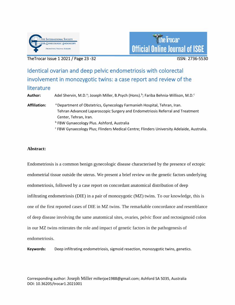
A laparoscopic segmental sigmoid resection was
performed, with staple re-anastomosis,
resection of pelvic floor DIE, and resection of a
bilateral endometrioma. The procedure took 152
minutes. The patient was discharged after four
days.
Figure 1. Laparoscopic images for Twin A. (A)
Anterior uterine myoma; (B) attached left adnexa
to stenotic RS colon; (C) left adnexal colonic
adhesion; (D) left ovarian endometrioma; (E)
right ovarian endometrioma; (F) segmental
resection of RS colon; (G) resected bowel with
deep endometriosis; (H) DIE pelvic floor; and (I)
DIE at peritoneal site of invagination.
Twin B
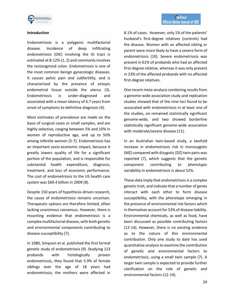
Twin B’s initial procedure was performed in the
same month as Twin A’s procedure. Laparoscopic
adhesiolysis was performed, with resection of
pelvic floor DIE, resection of ovarian
endometrioma, and shaving of rectal deep
endometriosis. Bowel resection was deferred
because of inadequate bowel preparation, and
laparoscopic sigmoid resection with staple
anastomosis was performed 8 weeks after the
initial procedure.
Figure 2. Laparoscopic images for Twin B. (A)
Myoma at anterior uterine wall; (B) left ovarian
endometrioma severely attached to sigmoid
colon; (C) ovarian attachment to sigmoid colon;
(D) left ovarian endometriosis; (E) right ovarian
endometriosis; (F) shaving DIE off rectal surface;
(G) defect at rectal surface; (H) DIE pelvic floor
with rectal involvement; (I) DIE peritoneal site on
invagination, right side; (J) segmental resection
of RS colon; (K) resected bowel with undivided DIE
nodule; and (L) resected bowel with divided DIE
nodule.
Discussion
Anatomical involvement of the relevant pelvic
organ in this pair of identical twins was nearly
identical. The twins had grown up in the same
environment all their lives, both worked as
secretaries at the same institution, and both
were experiencing not only the symptoms of DIE,
but also signs and symptoms of intestinal
involvement.
The ultrasound and MRI for both patients was
reported negative, which clearly indicates the
importance of experienced radiologists for the
detection of deep endometriosis (15), and
reiterates the importance of bimanual exam by
clinicians, particularly at the time of
menstruation, to achieve an accurate diagnosis,.
27
In fact, available data clearly indicates that the
accuracy of good physical examination (PE) is not
much different from that of transvaginal
ultrasound (TVS), rectal endoscopic sonography
(RES) and MRI, all within the 80% range (16).
Twin A was managed by laparoscopic segmental
sigmoid resection and staple re-anastomosis,
along with global resection of DIE from the pelvic
floor and endometriomas. We were intending to
do the same for Twin B, but in OR the lack of
adequate bowel preparation was noted; thus
bowel resection was postponed and was
performed 8 weeks later under adequate bowel
preparation. However, after resection of
endometriomas and pelvic floor deep lesions,
larger DIE at the posterior cervix with extension
to the rectum was managed by laparoscopic
deep shaving, and the defect was closed in two
layers. No intraoperative or postoperative
complications were encountered in either twin.
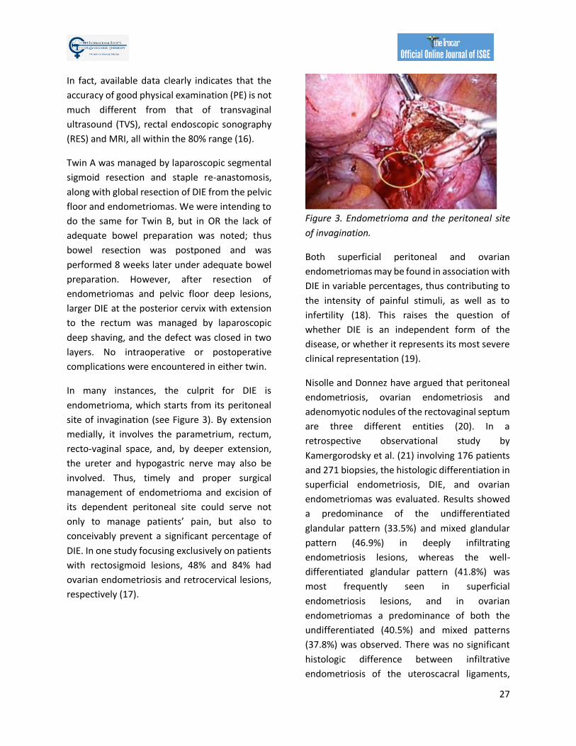
In many instances, the culprit for DIE is
endometrioma, which starts from its peritoneal
site of invagination (see Figure 3). By extension
medially, it involves the parametrium, rectum,
recto-vaginal space, and, by deeper extension,
the ureter and hypogastric nerve may also be
involved. Thus, timely and proper surgical
management of endometrioma and excision of
its dependent peritoneal site could serve not
only to manage patients’ pain, but also to
conceivably prevent a significant percentage of
DIE. In one study focusing exclusively on patients
with rectosigmoid lesions, 48% and 84% had
ovarian endometriosis and retrocervical lesions,
respectively (17).
Figure 3. Endometrioma and the peritoneal site
of invagination.
Both superficial peritoneal and ovarian
endometriomas may be found in association with
DIE in variable percentages, thus contributing to
the intensity of painful stimuli, as well as to
infertility (18). This raises the question of
whether DIE is an independent form of the
disease, or whether it represents its most severe
clinical representation (19).
Nisolle and Donnez have argued that peritoneal
endometriosis, ovarian endometriosis and
adenomyotic nodules of the rectovaginal septum
are three different entities (20). In a
retrospective observational study by
Kamergorodsky et al. (21) involving 176 patients
and 271 biopsies, the histologic differentiation in
superficial endometriosis, DIE, and ovarian
endometriomas was evaluated. Results showed
a predominance of the undifferentiated
glandular pattern (33.5%) and mixed glandular
pattern (46.9%) in deeply infiltrating
endometriosis lesions, whereas the well-
differentiated glandular pattern (41.8%) was
most frequently seen in superficial
endometriosis lesions, and in ovarian
endometriomas a predominance of both the
undifferentiated (40.5%) and mixed patterns
(37.8%) was observed. There was no significant
histologic difference between infiltrative
endometriosis of the uteroscacral ligaments,

28
recto-vaginal endometriosis, or bowel
endometriosis. There was a predominance of the
well-differentiated pattern in more superficial
lesions and the undifferentiated pattern in
deeper lesions, suggesting a similar mechanism
to explain the invasive potential of
endometriosis.
Recently, Sapkota et al. (22) analysed genetic risk
scores derived from two large European
genome-wide association (GWA) datasets from a
previous multi-ethnic GWA meta-analysis of
endometriosis. They found that genetic factors
contributing to minimal disease might differ from
those contributing to more severe
endometriosis, and that more severe
endometriosis cases exhibit greater genetic load
than minimal or mild disease. The genetic burden
generally increased from less severe (minimal) to
more severe disease, consistent with disease
progression.
Considering the fact that endometriotic lesions
undergo repeated cyclic bleeding (injury) and
repair like other organs leading to fibrosis (23), it
has been suggested that endometriotic lesions
are fundamentally wounds with repeated tissue
injury resulting in smooth muscle metaplasia
(SMM) and ultimately fibrosis via EMT (epithelial
mesenchymal transition), MET (mesenchymal
epithelial transition) mechanism securing
disease progression (24). This points to the
complex microenvironment and cross-reaction
of endometrioc lesions with other cells, i.e.
platelets and macrophages, their gradual but
progressive evolution to SMM and fibrosis and,
on occasion, to malignancy and the new
traits/phenotypes they may acquire while losing
old ones. This dynamic feature of endometriotic
lesions may explain the reason for some
conflicting results in this area of research,
including the fact that development of
biomarkers for the diagnosis or prognosis of
endometriosis has posed a challenge, at least
until now.
Endometriosis appears to be a progressive
disease and its progression is mediated by
EMT,MET mechanism and stemness ability of
mesenchymal cells capable of creating local
invasion and progression, intravasation and
extravasation providing new sites or distant
metastasis, as evidenced by longitudinal
laparoscopies in female baboons (25). In regard
to human evidence there are at least eight
studies reported (with a total of 162 patients) on
repeat laparoscopy in women assigned to
placebo treatment. The results indicate nearly
equal distribution among women whose disease
stage deteriorated (31%), was unchanged (32%),
or improved (38%). In fact, all but one study
noted that 23% of placebo patients had complete
regression of the disease over intervals of 4 to 39
months, meaning that in over two thirds of
women the disease will either persist or progress
(26-33).
The clinical presentation of the disease followed
a similar trend and evolution in both of our cases.
Each twin had undergone a long history of slowly
progressive pelvic pain from the point of
intensity, developing bowel symptoms and
dyschesia as time passed, with dyspareunia and
change in stool diameter to the point of bowel
stenosis and impending obstruction. All of this is
consistent with the progressive nature of the
disease reported in the existing literature.
Deep infiltrating endometriosis, contrary to
being an estrogen-dependent disease, usually
does not respond well to hormonal suppressive
therapy. Adequate surgical excision of the lesions
provides the best long-term results and
symptomatic relief (31, 34) but surgical
treatment of colorectal endometriosis has been
29
quite controversial, and there is no consensus on
whether the treatment should be conservative
(shaving or discoid resection) or radical
(segmental bowel resection and anastomosis)
(35). Lately, there has been a great deal of effort
to advocate shaving as the treatment of choice,
mainly because of higher early and late
complications in segmental colorectal resection.
(36)
The efficacy of segmental colorectal resection is
very debatable; data in this regard is
controversial and, to a great deal, dependent
upon the surgeon’s experience (37, 38).
Findings from histopathologic analysis of
specimens from segmental bowel resection
indicates that in 50% of cases there is a satellite
lesion independent of the primary deep nodule.
The deepest layer of the bowel wall containing
endometriotic foci at the primary lesion is in the
submucosal layer in 70% of cases and the internal
circular muscle layer in 30%. Furthermore,
persistent lesions are present in 50% of patients
treated with discoid or shaving resection (39).
The latest systematic review of different surgical
approaches to bowel and rectovaginal
endometriosis showed that the complication
rate is variable for conservative and radical
treatment. Recurrence rate for shaving was
reported at 22.2%, for discoid resection 5.17%,
and in segmental resection 2.19%; these rates
were significantly different (37) but
comparatively complete resection of bladder DIE
had no recurrence reported by different authors.
Positive bowel resection margins as well as age
<31 years and body mass index ≥23 kg/m2
appear to be important independent predictors
of disease recurrence (40).
Our patients were managed according to the
extent of their pathology, intensity of pelvic pain,
and future fertility, thus minimising the chance of
recurrence. Most importantly, patients’ wishes
and decisions regarding the type of clinical
management to be pursued were incorporated,
following proper counselling and explanation.
It is crucial to keep in mind that there is currently
no scientifically proven best treatment technique
for bowel endometriosis, and comparison of
different currently practised techniques is not
possible. According to our current knowledge
regarding the nature of disease pathology and its
progression, recurrence should be as much a
source of concern as complications for any given
surgical technique. Resection of DIE should be
global, not local, and limited to the bowel.
Operations should be performed by an
experienced surgeon and a well-organised team,
in a multidisciplinary fashion.
In summary, we present a case of advanced DIE
in monozygotic twins; one of the first such cases
to our knowledge. Our findings reiterate the
importance of genetic factors in the aetiology of
endometriosis. These cases also reinforce the
importance of the following considerations for
surgeons considering conservative treatment for
colorectal endometriosis:
• The extent of lesions cannot be assessed
macroscopically with certainty.
• Assessment of the depth of infiltration of
endometriosis foci in the bowel requires
an expert radiologist.
• In the majority of rectal endometriosis
nodules, the fibrosis which is the
landmark for shaving does not surround,
but follows behind the glandular
epithelium and stroma.
• More than 2 cm of bowel tissue must be
removed from the main lesion to obtain
clear margins in two thirds of patients.
30
• Low rectal lesions (<5–8 cm from the
anal verge) are associated with a higher
risk of complications.
• A great majority of patients have been
through previous surgeries and have a
good chance of recovering and becoming
pain-free.
Acknowledgements: None.
References
1. Seracchioli R, Poggioli G, Pierangeli F, et
al. Surgical outcome and long-term follow up
after laparoscopic rectosigmoid resection in
women with deep infiltrating endometriosis.
BJOG. 2007;114(7):889-95.
2. Wills HJ, Reid GD, Cooper MJ, Morgan M.
Fertility and pain outcomes following
laparoscopic segmental bowel resection for
colorectal endometriosis: a review. Aust N Z J
Obstet Gynaecol. 2008;48(3):292-5.
3. Kennedy S, Bergqvist A, Chapron C, et al.
ESHRE guideline for the diagnosis and treatment
of endometriosis. Hum Reprod.
2005;20(10):2698-704.
4. Nnoaham KE, Hummelshoj L, Webster P,
et al. Impact of endometriosis on quality of life
and work productivity: a multicenter study
across ten countries. Fertil Steril.
2011;96(2):366-73.e8.
5. Eskenazi B, Warner ML. Epidemiology of
endometriosis. Obstet Gynecol Clin North Am.
1997;24(2):235-58.
6. Meuleman C, Vandenabeele B, Fieuws S,
Spiessens C, Timmerman D, D'Hooghe T. High
prevalence of endometriosis in infertile women
with normal ovulation and normospermic
partners. Fertil Steril. 2009;92(1):68-74.
7. Treloar SA, O'Connor DT, O'Connor VM,
Martin NG. Genetic influences on endometriosis
in an Australian twin sample. Fertil Steril.
1999;71(4):701-10.
8. Simoens S, Dunselman G, Dirksen C, et al.
The burden of endometriosis: costs and quality
of life of women with endometriosis and treated
in referral centres. Hum Reprod.
2012;27(5):1292-9.
9. Simpson JL, Elias S, Malinak LR, Buttram
VC, Jr. Heritable aspects of endometriosis. I.
Genetic studies. Am J Obstet Gynecol.
1980;137(3):327-31.
10. Stefansson H, Geirsson RT,
Steinthorsdottir V, et al. Genetic factors
contribute to the risk of developing
endometriosis. Hum Reprod. 2002;17(3):555-9.
11. Rahmioglu N, Nyholt DR, Morris AP,
Missmer SA, Montgomery GW, Zondervan KT.
Genetic variants underlying risk of
endometriosis: insights from meta-analysis of
eight genome-wide association and replication
datasets. Hum Reprod Update. 2014;20(5):702-
16.
12. Louis GM, Weiner JM, Whitcomb BW, et
al. Environmental PCB exposure and risk of
endometriosis. Hum Reprod. 2005;20(1):279-85.
13. Parazzini F, Chiaffarino F, Surace M, et al.
Selected food intake and risk of endometriosis.
Hum Reprod. 2004;19(8):1755-9.
14. Porpora MG, Medda E, Abballe A, et al.
Endometriosis and organochlorinated
environmental pollutants: a case-control study
31
on Italian women of reproductive age. Environ
Health Perspect. 2009;117(7):1070-5.
15. Fraser MA, Agarwal S, Chen I, Singh SS.
Routine vs. expert-guided transvaginal
ultrasound in the diagnosis of endometriosis: a
retrospective review. Abdom Imaging.
2015;40(3):587-94.
16. Bazot M, Lafont C, Rouzier R, Roseau G,
Thomassin-Naggara I, Darai E. Diagnostic
accuracy of physical examination, transvaginal
sonography, rectal endoscopic sonography, and
magnetic resonance imaging to diagnose deep
infiltrating endometriosis. Endometriosis.
2009;92(6).
17. Goncalves MO, Podgaec S, Dias JA, Jr.,
Gonzalez M, Abrao MS. Transvaginal
ultrasonography with bowel preparation is able
to predict the number of lesions and
rectosigmoid layers affected in cases of deep
endometriosis, defining surgical strategy. Hum
Reprod. 2010;25(3):665-71.
18. Chapron C, Santulli P, de Ziegler D, et al.
Ovarian endometrioma: severe pelvic pain is
associated with deeply infiltrating
endometriosis. Hum Reprod. 2012;27(3):702-11.
19. Acién P, Velasco I. Endometriosis: A
disease that remains enigmatic. ISRN Obstet
Gynecol. 2013;2013:242149.
20. Nisolle M, Donnez J. Peritoneal
endometriosis, ovarian endometriosis, and
adenomyotic nodules of the rectovaginal septum
are three different entities. Fertil Steril.
1997;68(4):585-96.
21. Kamergorodsky G, Ribeiro PA, Galvao
MA, et al. Histologic classification of specimens
from women affected by superficial
endometriosis, deeply infiltrating endometriosis,
and ovarian endometriomas. Fertil Steril.
2009;92(6):2074-7.
22. Sapkota Y, Attia J, Gordon SD, et al.
Genetic burden associated with varying degrees
of disease severity in endometriosis. Mol Hum
Reprod. 2015;21(7):594-602.
23. Ding D, Liu X, Duan J, Guo SW. Platelets
are an unindicted culprit in the development of
endometriosis: clinical and experimental
evidence. Hum Reprod. 2015;30(4):812-32.
24. Mehal WZ, Iredale J, Friedman SL.
Scraping fibrosis: expressway to the core of
fibrosis. Nat Med. 2011;17(5):552-3.
25. D'Hooghe TM, Bambra CS, Raeymaekers
BM, Koninckx PR. Serial laparoscopies over 30
months show that endometriosis in captive
baboons (Papio anubis, Papio cynocephalus) is a
progressive disease. Fertil Steril. 1996;65(3):645-
9.
26. Telimaa S, Ronnberg L, Kauppila A.
Placebo-controlled comparison of danazol and
high-dose medroxyprogesterone acetate in the
treatment of endometriosis after conservative
surgery. Gynecol Endocrinol. 1987;1(4):363-71.
27. Thomas EJ, Cooke ID. Impact of
gestrinone on the course of asymptomatic
endometriosis. Br Med J (Clin Res Ed).
1987;294(6567):272-4.
28. Cooke ID, Thomas EJ. The medical
treatment of mild endometriosis. Acta Obstet
Gynecol Scand Suppl. 1989;150:27-30.
29. Mahmood TA, Templeton A. The impact
of treatment on the natural history of
endometriosis. Hum Reprod. 1990;5(8):965-70.
30. Overton CE, Lindsay PC, Johal B, et al. A
randomized, double-blind, placebo-controlled
32
study of luteal phase dydrogesterone
(Duphaston) in women with minimal to mild
endometriosis. Fertil Steril. 1994;62(4):701-7.
31. Sutton CJ, Pooley AS, Ewen SP, Haines P.
Follow-up report on a randomized controlled
trial of laser laparoscopy in the treatment of
pelvic pain associated with minimal to moderate
endometriosis. Fertil Steril. 1997;68(6):1070-4.
32. Harrison RF, Barry-Kinsella C. Efficacy of
medroxyprogesterone treatment in infertile
women with endometriosis: a prospective,
randomized, placebo-controlled study. Fertil
Steril. 2000;74(1):24-30.33. Abbott J, Hawe J,
Hunter D, Holmes M, Finn P, Garry R.
Laparoscopic excision of endometriosis: a
randomized, placebo-controlled trial. Fertil
Steril. 2004;82(4):878-84.
34. Koninckx PR, Ussia A, Adamyan L,
Wattiez A, Donnez J. Deep endometriosis:
definition, diagnosis, and treatment. Fertil Steril.
2012;98(3):564-71.
35. Abrão MS, Petraglia F, Falcone T,
Keckstein J, Osuga Y, Chapron C. Deep
endometriosis infiltrating the recto-sigmoid:
critical factors to consider before management.
Hum Reprod Update. 2015.
36. Roman H, Bridoux V, Tuech JJ, et al.
Bowel dysfunction before and after surgery for
endometriosis. Am J Obstet Gynecol.
2013;209(6):524-30.
37. Moustafa MM, Elnasharty MAA.
Systematic review of the outcome associated
with the different surgical treatment of bowel
and rectovaginal endometriosis. Gynecol Surg.
2014;11(1):37-52.
38. Keckstein J, Wiesinger H. Deep
endometriosis, including intestinal involvement-
-the interdisciplinary approach. Minim Invasive
Ther Allied Technol. 2005;14(3):160-6.
39. Jinushi M, Arakawa A, Matsumoto T, et
al. Histopathologic . J Minim Invasive
Gynecol.18(1):48-53.
40. Nirgianakis K, McKinnon B, Imboden S,
Knabben L, Gloor B, Mueller MD. Laparoscopic
management of bowel endometriosis: resection
margins as a predictor of recurrence. Acta Obstet
Gynecol Scand. 2014;93(12):1262-7
33 Corresponding author: Ameneh Haghgoo, email: [email protected] DOI: 10.36205/trocar1.2021003 Received 16/12/2020 - Accepted 05/02/2021
CASEREPORT TheTrocar Issue 1 2021 / Page 33-38 ISSN: 2736-5530
Hysteroscopic removal of Retained Intra-uterine Fetal Bone which
causes secondary infertility: A case report
Author: Ameneh Haghgoo1, MD, Bruno J. van Herendael, MD2, Adel Shervin, MD3, Mehran
Ghahremani3, MD, Kaveh Hojjat Jalali3, MD, Saeed Nasiri4, MD, Farnaz Akhbari, BSc5
Affiliation: 1 Department of Obstetrics & Gynecology. Nikan hospital Tehran, Iran
2 Consultant Gynaecologic Surgery ZNA Stuivenberg Antwerp Belgium
3 Department of Obstetrics & Gynecology. Nikan Education and Research Center, Iran
4 Department of imaging, shahid Beheshti university of medical science, Tehran, Iran
5 BSc of midwifery, researcher. Nikan Education and Research Center, Nikan hospital
Tehran, Iran
Abstract:
Intrauterine retention of fetal bones is a rare complication of unsafe abortion that can cause secondary
infertility. We are presenting the case of a young woman who underwent an illegal abortion 4 years ago.
Transvaginal ultrasonography suggested the presence of bone-like structures in the uterine cavity.
Retained bony structure from a previous abortion should be considered as a cause of secondary infertility.
Removal of the bony products and polypectomy was done by hysteroscopy.
Keywords: abortion, infertility, fetal bones, hysteroscopy, osseous metaplasia
34
Introduction
Bone within the uterine cavity is an unusual
finding in women with secondary infertility,
which is usually associated with a past history of
termination of pregnancy. The etiology of bone
like calcification is unknown, but the most
common ones include retained fetal bone and
osseous metaplasia of endometrial tissue.
Calcification appear as hyperechoic area on
ultrasound but the gynecologist should also be
aware of other differential diagnosis of
endometrial ossification which includes mixed
Mullerian mesenchymal tumor (MMMT), IUCD,
Osseous metaplasia or endometrial tuberculosis.
Hysteroscopy is gold standard as diagnostic and
therapeutic method.
Retained fetal bone as an entity is not adequately
reported in the literature. Prolonged retention of
intrauterine bone is recognized as a cause of
secondary infertility. One study reported that
retained fetal bones may act as an intrauterine
synechia or intrauterine device, increasing
endometrial prostaglandin F2-alpha and
preventing implantation, resulting in infertility
(1). The reported incidence is 0.15% at diagnostic
hysteroscopy (2). These patients may present
with pelvic pain, dysmenorrhea, abnormal
uterine bleeding and infertility (3). Apart from
retained intrauterine, endometrial calcified
lesions and the presence of ectopic bones can
also occur by metaplasia in association with
chronic inflammation and tissue destruction,
mostly presented after repeated spontaneous or
therapeutic abortion (4-6)
A Procedure of termination of a pregnancy can at
times result in unwanted immediate
complications like hemorrhage, uterine
perforation, cervical injury and at times in late
complications like infections, bleeding,
menstrual abnormalities and uterine synechia.
As 50% of patients undergoing medical
termination are young adults, there is a
possibility of secondary infertility due to an
abortion that has become complicated (7).
Occasionally if the abortion is done in the second
trimester or if the fetus was removed by
destructive means, some parts of the fetus may
inadvertently be left in the uterine cavity, these
could cause pelvic pain, abnormal uterine
bleeding, infection, dyspareunia or even
vaginally expulsion of fetal bones (8). Few
reports have been published describing
secondary infertility after abortion. It is expected
that retained fetal bones would induce a uterine
reaction in the form of vaginal discharge and pain
in the majority of such patients while also leading
to secondary infertility.
We are presenting the case of a young female
who had secondary infertility without any other
associated complaints. She had a previous
second-trimester abortion four years ago. shad
failed to conceive again after previous curettage.
Retained fetal bone was confirmed by
histopathological analysis of bone fragment
collected by hysteroscopy.
Case presentation
A 34 years old lady G4 P3 Ab1, presented in our
office with secondary infertility. She had
cesarean section delivery 3 times, according to
the patient she had a history of an illegal
termination of pregnancy at about 4th months of
gestation by dilatation and curettage four years
before. Following the abortion, she experienced
abnormal uterine bleeding for several months
and thereafter she could not conceive again.
Even she had experienced vaginal expulsion of
fetal bones several days after the curettage.
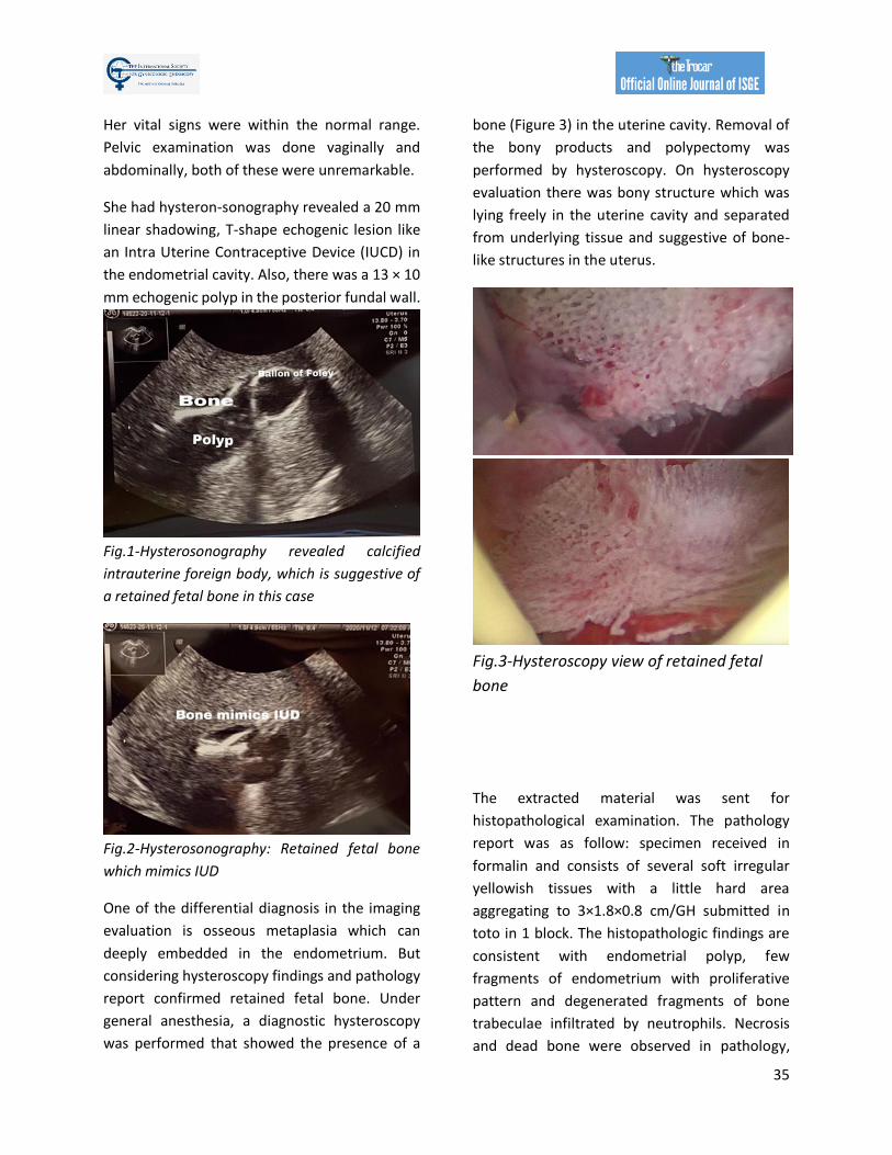
35
Her vital signs were within the normal range.
Pelvic examination was done vaginally and
abdominally, both of these were unremarkable.
She had hysteron-sonography revealed a 20 mm
linear shadowing, T-shape echogenic lesion like
an Intra Uterine Contraceptive Device (IUCD) in
the endometrial cavity. Also, there was a 13 × 10
mm echogenic polyp in the posterior fundal wall.
Fig.1-Hysterosonography revealed calcified
intrauterine foreign body, which is suggestive of
a retained fetal bone in this case
Fig.2-Hysterosonography: Retained fetal bone
which mimics IUD
One of the differential diagnosis in the imaging
evaluation is osseous metaplasia which can
deeply embedded in the endometrium. But
considering hysteroscopy findings and pathology
report confirmed retained fetal bone. Under
general anesthesia, a diagnostic hysteroscopy
was performed that showed the presence of a
bone (Figure 3) in the uterine cavity. Removal of
the bony products and polypectomy was
performed by hysteroscopy. On hysteroscopy
evaluation there was bony structure which was
lying freely in the uterine cavity and separated
from underlying tissue and suggestive of bone-
like structures in the uterus.
Fig.3-Hysteroscopy view of retained fetal
bone
The extracted material was sent for
histopathological examination. The pathology
report was as follow: specimen received in
formalin and consists of several soft irregular
yellowish tissues with a little hard area
aggregating to 3×1.8×0.8 cm/GH submitted in
toto in 1 block. The histopathologic findings are
consistent with endometrial polyp, few
fragments of endometrium with proliferative
pattern and degenerated fragments of bone
trabeculae infiltrated by neutrophils. Necrosis
and dead bone were observed in pathology,
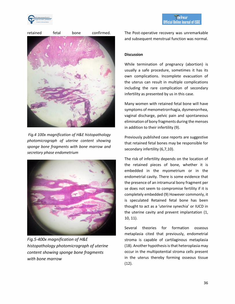
36
retained fetal bone confirmed.
Fig.4 100x magnification of H&E histopathology
photomicrograph of uterine content showing
sponge bone fragments with bone marrow and
secretory phase endometrium
Fig.5-400x magnification of H&E
histopathology photomicrograph of uterine
content showing sponge bone fragments
with bone marrow
The Post-operative recovery was unremarkable
and subsequent menstrual function was normal.
Discussion
While termination of pregnancy (abortion) is
usually a safe procedure, sometimes it has its
own complications. Incomplete evacuation of
the uterus can result in multiple complications
including the rare complication of secondary
infertility as presented by us in this case.
Many women with retained fetal bone will have
symptoms of menometrorrhagia, dysmenorrhea,
vaginal discharge, pelvic pain and spontaneous
elimination of bony fragments during the menses
in addition to their infertility (9).
Previously published case reports are suggestive
that retained fetal bones may be responsible for
secondary infertility (6,7,10).
The risk of infertility depends on the location of
the retained pieces of bone, whether it is
embedded in the myometrium or in the
endometrial cavity. There is some evidence that
the presence of an intramural bony fragment per
se does not seem to compromise fertility if it is
completely embedded (9) However commonly, it
is speculated Retained fetal bone has been
thought to act as a ‘uterine synechia’ or IUCD in
the uterine cavity and prevent implantation (1,
10, 11).
Several theories for formation osseous
metaplasia cited that previously, endometrial
stroma is capable of cartilaginous metaplasia
(18). Another hypothesis is that heteroplasia may
occur in the multipotential stroma cells present
in the uterus thereby forming osseous tissue
(12).
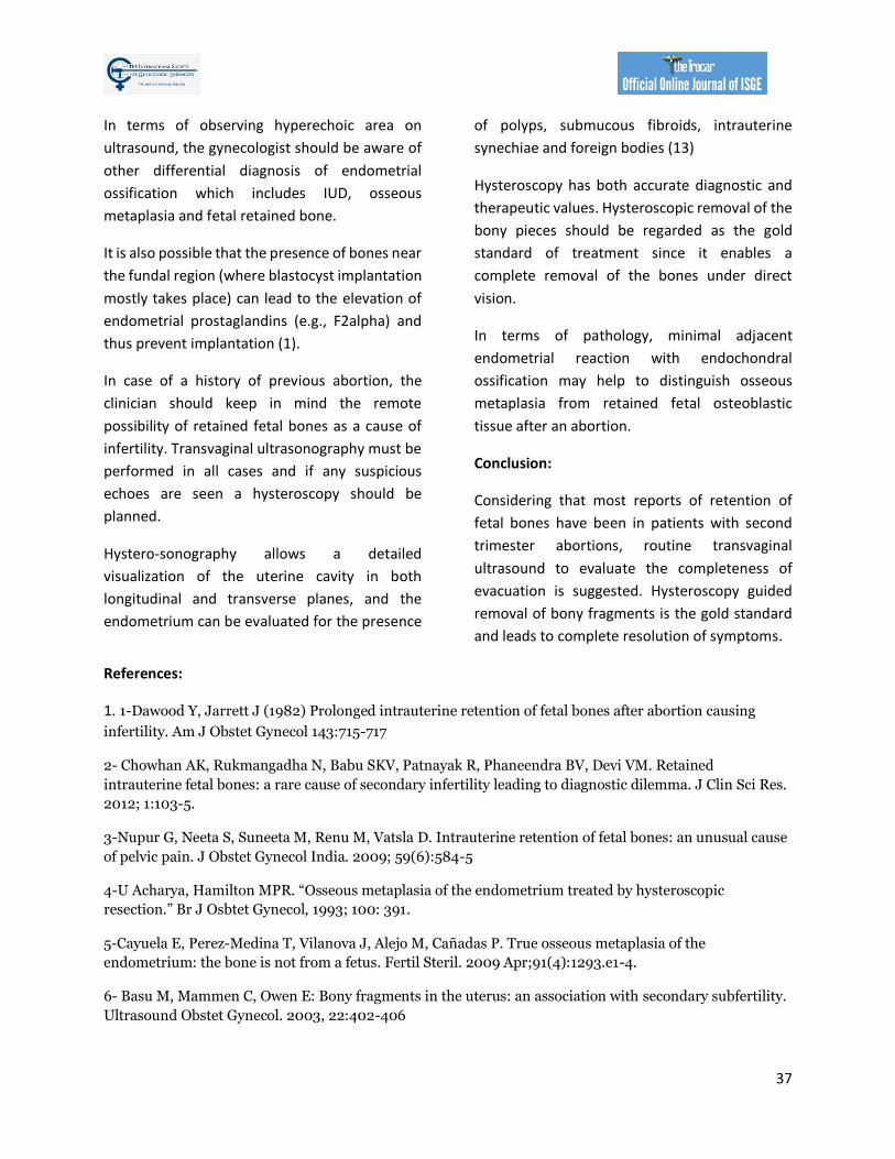
37
In terms of observing hyperechoic area on
ultrasound, the gynecologist should be aware of
other differential diagnosis of endometrial
ossification which includes IUD, osseous
metaplasia and fetal retained bone.
It is also possible that the presence of bones near
the fundal region (where blastocyst implantation
mostly takes place) can lead to the elevation of
endometrial prostaglandins (e.g., F2alpha) and
thus prevent implantation (1).
In case of a history of previous abortion, the
clinician should keep in mind the remote
possibility of retained fetal bones as a cause of
infertility. Transvaginal ultrasonography must be
performed in all cases and if any suspicious
echoes are seen a hysteroscopy should be
planned.
Hystero-sonography allows a detailed
visualization of the uterine cavity in both
longitudinal and transverse planes, and the
endometrium can be evaluated for the presence
of polyps, submucous fibroids, intrauterine
synechiae and foreign bodies (13)
Hysteroscopy has both accurate diagnostic and
therapeutic values. Hysteroscopic removal of the
bony pieces should be regarded as the gold
standard of treatment since it enables a
complete removal of the bones under direct
vision.
In terms of pathology, minimal adjacent
endometrial reaction with endochondral
ossification may help to distinguish osseous
metaplasia from retained fetal osteoblastic
tissue after an abortion.
Conclusion:
Considering that most reports of retention of
fetal bones have been in patients with second
trimester abortions, routine transvaginal
ultrasound to evaluate the completeness of
evacuation is suggested. Hysteroscopy guided
removal of bony fragments is the gold standard
and leads to complete resolution of symptoms.
References:
1. 1-Dawood Y, Jarrett J (1982) Prolonged intrauterine retention of fetal bones after abortion causing
infertility. Am J Obstet Gynecol 143:715-717
2- Chowhan AK, Rukmangadha N, Babu SKV, Patnayak R, Phaneendra BV, Devi VM. Retained
intrauterine fetal bones: a rare cause of secondary infertility leading to diagnostic dilemma. J Clin Sci Res.
2012; 1:103-5.
3-Nupur G, Neeta S, Suneeta M, Renu M, Vatsla D. Intrauterine retention of fetal bones: an unusual cause
of pelvic pain. J Obstet Gynecol India. 2009; 59(6):584-5
4-U Acharya, Hamilton MPR. “Osseous metaplasia of the endometrium treated by hysteroscopic
resection.” Br J Osbtet Gynecol, 1993; 100: 391.
5-Cayuela E, Perez-Medina T, Vilanova J, Alejo M, Cañadas P. True osseous metaplasia of the
endometrium: the bone is not from a fetus. Fertil Steril. 2009 Apr;91(4):1293.e1-4.
6- Basu M, Mammen C, Owen E: Bony fragments in the uterus: an association with secondary subfertility.
Ultrasound Obstet Gynecol. 2003, 22:402-406

38
7-Kramer HM, Rhemrev JP: Secondary infertility caused by the retention of fetal bones after an abortion:
a case report. J Med Case Rep. 2008, 2:2080
8-Graham O, Cheng LC, Parsons JH: The ultrasound diagnosis of retained fetal bones in West African
patients complaining of infertility. BJOG. 2000, 107:122-124
9-Chervenak FA, Amin HKo, Neuwirth RS (1982) Symptomatic intrauterine retention of fetal bones.
Obstet Gynecol 59 [Suppl]:58S–61 8
10- Tulandi T, Sammour A: Retained fetal bones in the uterine cavity. J Am Assoc Gynecol Laparosc. 2001,
8:179-180.
11- Lewis V, Khan-Dawood F, King M, Beckmann C, Dawood MY: Retention of intrauterine fetal bone
increases menstrual prostaglandins. Obstet Gynecol. 1990, 75:561-563
12- Marcus SF, Bhattacharya J, William G, et al. “Endometrial ossification: a cause of secondary infertility.
Report of two cases”. Am J Obstet Gynecol 1994; 170: 138.
13-Allahbadia GN, Tibrewala S, Mangeshikar P, PaiDhungat PB, Desai SK (1996) Prolonged intrauterine
retention of fetal bones after abortion—vaginosonographic diagnosis and hysteroscopic removal.
Singapore J Obstet Gynaecol 27:83–86

Corresponding author: Hind Ennasser E-mail address: mailto:[email protected] DOI: 10.36205/trocar1.2021004 Received 05.01.2021 accepted 24.02.2021
TheTrocar Issue 1 2020 / Page 39 -46 ISSN: 2736-5530
Laparoscopic management of cesarean scar pregnancy: a case report
and literature review Author: Ahmed Mimouni1, Hind Ennasser1, Imane Skiker2, Hafsa Taheri1, Hanane Saadi1
Affiliation: 1Department of Obstetrics and Gynecology, Mohammed VI University Hospital Center,
Oujda Morocco 2Department of Radiology, Mohammed VI University Hospital Center, Oujda Morocco
Abstract
Background: Cesarean section scar pregnancy is one of the rarest forms of ectopic pregnancy where the
gestational sac is fully or partially implanted within the scar caused by a previous caesarean section. Its
incidence is on the rise due to the increasing rate of cesarean sections (CS) and also to the increasing
awareness and the better ultrasound diagnosis
Case presentation: A 28-year-old woman G3P2 with a history of 2 cesarean deliveries who was diagnosed
by ultrasound scan at 8 weeks gestation with cesarean scar ectopic pregnancy that was confirmed by pelvic
magnetic resonance imaging (MRI). At 4 weeks the patient had a pelvic ultrasound suggested an
intrauterine pregnancy with a gestational sac visualized in the lower uterine segment suggesting a cervical
stage of miscarriage. Surgical management was opted for a combination of laparoscopy and hysteroscopy.
A ligation of bilateral ascending uterine artery ligation was performed.
Conclusion: Cesarean section scar pregnancy (CSP) is rare but life threating complication. It should be
diagnosed and treated early because of the increased risk of massive hemorrhage or uterine rupture that
engages the vital prognosis and the functional prognosis.
Keywords: Cesarean scar pregnancy, ectopic pregnancy, laparoscopy, hysteroscopy
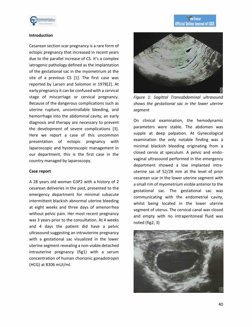
40
Introduction
Cesarean section scar pregnancy is a rare form of
ectopic pregnancy that increased in recent years
due to the parallel increase of CS. It’s a complex
iatrogenic pathology defined as the implantation
of the gestational sac in the myometrium at the
site of a previous CS [1]. The first case was
reported by Larsen and Solomon in 1978[2]. At
early pregnancy it can be confused with a cervical
stage of miscarriage or cervical pregnancy.
Because of the dangerous complications such as
uterine rupture, uncontrollable bleeding, and
hemorrhage into the abdominal cavity; an early
diagnosis and therapy are necessary to prevent
the development of severe complications [3].
Here we report a case of this uncommon
presentation of ectopic pregnancy with
laparoscopic and hysteroscopic management in
our department, this is the first case in the
country managed by laparoscopy.
Case report
A 28 years old woman G3P2 with a history of 2
cesarean deliveries in the past, presented to the
emergency department for minimal subacute
intermittent blackish abnormal uterine bleeding
at eight weeks and three days of amenorrhea
without pelvic pain. Her most recent pregnancy
was 3 years prior to the consultation. At 4 weeks
and 4 days the patient did have a pelvic
ultrasound suggesting an intrauterine pregnancy
with a gestational sac visualized in the lower
uterine segment revealing a non-viable detached
intrauterine pregnancy (fig1) with a serum
concentration of human chorionic gonadotropin
(HCG) at 8306 mUI/ml.
Figure 1: Sagittal Transabdominal ultrasound
shows the gestational sac in the lower uterine
segment
On clinical examination, the hemodynamic
parameters were stable. The abdomen was
supple at deep palpation. At Gynecological
examination the only notable finding was a
minimal blackish bleeding originating from a
closed cervix at speculum. A pelvic and endo-
vaginal ultrasound performed in the emergency
department showed a low implanted intra-
uterine sac of 52/28 mm at the level of prior
cesarean scar in the lower uterine segment with
a small rim of myometrium visible anterior to the
gestational sac. The gestational sac was
communicating with the endometrial cavity,
whilst being located in the lower uterine
segment of uterus. The cervical canal was closed
and empty with no intraperitoneal fluid was
noted (fig2, 3)
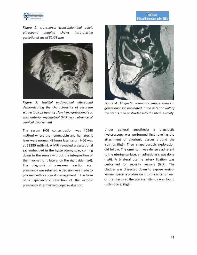
41
Figure 2: transversal transabdominal pelvic
ultrasound imaging shows intra-uterine
gestational sac of 52/28 mm
Figure 3: Sagittal endovaginal ultrasound
demonstrating the characteristics of cesarean
scar ectopic pregnancy : low lying gestational sac
with anterior myometrial thickness , absence of
cervical involvement
The serum HCG concentration was 40540
mUI/ml where the hemoglobin and hematocrit
level were normal, 48 hours later serum HCG was
at 53280 mUI/ml. A MRI revealed a gestational
sac embedded in the hysterotomy scar, coming
down to the serosa without the interposition of
the myometrium, lateral on the right side (fig4).
The diagnosis of caesarean section scar
pregnancy was retained. A decision was made to
proceed with a surgical management in the form
of a laparoscopic resection of the ectopic
pregnancy after hysteroscopic evaluation.
Figure 4: Magnetic resonance image shows a
gestational sac implanted in the anterior wall of
the uterus, and protruded into the uterine cavity.
Under general anesthesia a diagnostic
hysteroscopy was performed first reveling the
attachment of chorionic tissues around the
isthmus (fig5). Then a laparoscopic exploration
did follow. The omentum was densely adherent
to the uterine surface, an adhesiolysis was done
(fig6). A bilateral uterine artery ligation was
performed for security reasons (fig7). The
bladder was dissected down to expose vesico-
vaginal space, a protrusion into the anterior wall
of the uterus at the uterine isthmus was found
(isthmocele) (fig8).
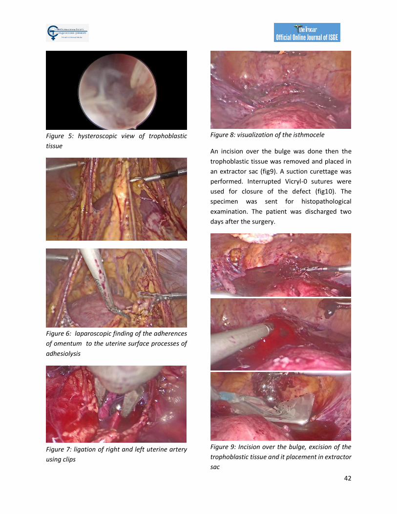
42
Figure 5: hysteroscopic view of trophoblastic
tissue
Figure 6: laparoscopic finding of the adherences
of omentum to the uterine surface processes of
adhesiolysis
Figure 7: ligation of right and left uterine artery
using clips
Figure 8: visualization of the isthmocele
An incision over the bulge was done then the
trophoblastic tissue was removed and placed in
an extractor sac (fig9). A suction curettage was
performed. Interrupted Vicryl-0 sutures were
used for closure of the defect (fig10). The
specimen was sent for histopathological
examination. The patient was discharged two
days after the surgery.
Figure 9: Incision over the bulge, excision of the
trophoblastic tissue and it placement in extractor
sac
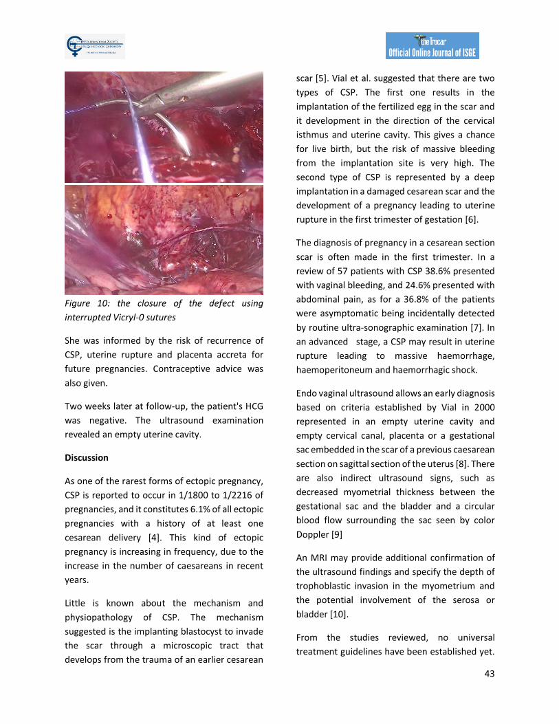
43
Figure 10: the closure of the defect using
interrupted Vicryl-0 sutures
She was informed by the risk of recurrence of
CSP, uterine rupture and placenta accreta for
future pregnancies. Contraceptive advice was
also given.
Two weeks later at follow-up, the patient's HCG
was negative. The ultrasound examination
revealed an empty uterine cavity.
Discussion
As one of the rarest forms of ectopic pregnancy,
CSP is reported to occur in 1/1800 to 1/2216 of
pregnancies, and it constitutes 6.1% of all ectopic
pregnancies with a history of at least one
cesarean delivery [4]. This kind of ectopic
pregnancy is increasing in frequency, due to the
increase in the number of caesareans in recent
years.
Little is known about the mechanism and
physiopathology of CSP. The mechanism
suggested is the implanting blastocyst to invade
the scar through a microscopic tract that
develops from the trauma of an earlier cesarean
scar [5]. Vial et al. suggested that there are two
types of CSP. The first one results in the
implantation of the fertilized egg in the scar and
it development in the direction of the cervical
isthmus and uterine cavity. This gives a chance
for live birth, but the risk of massive bleeding
from the implantation site is very high. The
second type of CSP is represented by a deep
implantation in a damaged cesarean scar and the
development of a pregnancy leading to uterine
rupture in the first trimester of gestation [6].
The diagnosis of pregnancy in a cesarean section
scar is often made in the first trimester. In a
review of 57 patients with CSP 38.6% presented
with vaginal bleeding, and 24.6% presented with
abdominal pain, as for a 36.8% of the patients
were asymptomatic being incidentally detected
by routine ultra-sonographic examination [7]. In
an advanced stage, a CSP may result in uterine
rupture leading to massive haemorrhage,
haemoperitoneum and haemorrhagic shock.
Endo vaginal ultrasound allows an early diagnosis
based on criteria established by Vial in 2000
represented in an empty uterine cavity and
empty cervical canal, placenta or a gestational
sac embedded in the scar of a previous caesarean
section on sagittal section of the uterus [8]. There
are also indirect ultrasound signs, such as
decreased myometrial thickness between the
gestational sac and the bladder and a circular
blood flow surrounding the sac seen by color
Doppler [9]
An MRI may provide additional confirmation of
the ultrasound findings and specify the depth of
trophoblastic invasion in the myometrium and
the potential involvement of the serosa or
bladder [10].
From the studies reviewed, no universal
treatment guidelines have been established yet.
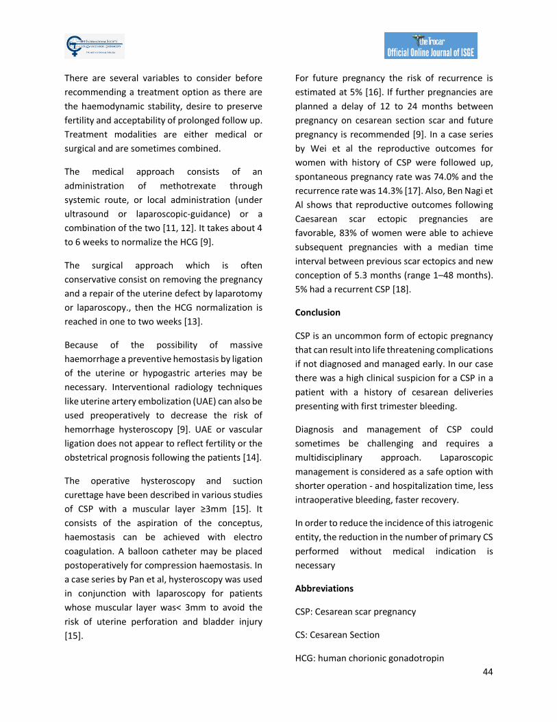
44
There are several variables to consider before
recommending a treatment option as there are
the haemodynamic stability, desire to preserve
fertility and acceptability of prolonged follow up.
Treatment modalities are either medical or
surgical and are sometimes combined.
The medical approach consists of an
administration of methotrexate through
systemic route, or local administration (under
ultrasound or laparoscopic-guidance) or a
combination of the two [11, 12]. It takes about 4
to 6 weeks to normalize the HCG [9].
The surgical approach which is often
conservative consist on removing the pregnancy
and a repair of the uterine defect by laparotomy
or laparoscopy., then the HCG normalization is
reached in one to two weeks [13].
Because of the possibility of massive
haemorrhage a preventive hemostasis by ligation
of the uterine or hypogastric arteries may be
necessary. Interventional radiology techniques
like uterine artery embolization (UAE) can also be
used preoperatively to decrease the risk of
hemorrhage hysteroscopy [9]. UAE or vascular
ligation does not appear to reflect fertility or the
obstetrical prognosis following the patients [14].
The operative hysteroscopy and suction
curettage have been described in various studies
of CSP with a muscular layer ≥3mm [15]. It
consists of the aspiration of the conceptus,
haemostasis can be achieved with electro
coagulation. A balloon catheter may be placed
postoperatively for compression haemostasis. In
a case series by Pan et al, hysteroscopy was used
in conjunction with laparoscopy for patients
whose muscular layer was< 3mm to avoid the
risk of uterine perforation and bladder injury
[15].
For future pregnancy the risk of recurrence is
estimated at 5% [16]. If further pregnancies are
planned a delay of 12 to 24 months between
pregnancy on cesarean section scar and future
pregnancy is recommended [9]. In a case series
by Wei et al the reproductive outcomes for
women with history of CSP were followed up,
spontaneous pregnancy rate was 74.0% and the
recurrence rate was 14.3% [17]. Also, Ben Nagi et
Al shows that reproductive outcomes following
Caesarean scar ectopic pregnancies are
favorable, 83% of women were able to achieve
subsequent pregnancies with a median time
interval between previous scar ectopics and new
conception of 5.3 months (range 1–48 months).
5% had a recurrent CSP [18].
Conclusion
CSP is an uncommon form of ectopic pregnancy
that can result into life threatening complications
if not diagnosed and managed early. In our case
there was a high clinical suspicion for a CSP in a
patient with a history of cesarean deliveries
presenting with first trimester bleeding.
Diagnosis and management of CSP could
sometimes be challenging and requires a
multidisciplinary approach. Laparoscopic
management is considered as a safe option with
shorter operation - and hospitalization time, less
intraoperative bleeding, faster recovery.
In order to reduce the incidence of this iatrogenic
entity, the reduction in the number of primary CS
performed without medical indication is
necessary
Abbreviations
CSP: Cesarean scar pregnancy
CS: Cesarean Section
HCG: human chorionic gonadotropin
45
MRI: Magnetic Resonance Imaging
UAE : Uterine artery embolization
Acknowledgements: None
Authors’ contributions
Ahmed Mimouni performed the surgery as a
main surgeon. The manuscript was prepared by
Hind Ennasser. Imane Skiker analyzed and
interpreted the MRI. Hanane Saadi and Hafsa
Taheri were the second and third surgeon.
All authors read and approved the final
manuscript
Funding: None of the author received any
funding for this study.
Ethics approval and consent to participate
The privacy of the patient was considered, and
the manuscript does not include any identifying
information.
Consent for publication: Informed consent for
publication of the patient’s clinical data and the
accompanying/ images was obtained from the
patient.
Competing interests: The authors declare that
they have no competing interests.
References:
1. Maymon R, Halperin R, Mendlovic S, Schneider D, Herman A. Ectopic pregnancies in a Caesarean scar: review of the medical approach to an iatrogenic complication. Hum Reprod Update 2004 Nov-Dec;10(6):515-523.
2. Ash A, Smith A, Maxwell D. Caesarean scar pregnancy. BJOG 2007 Mar;114(3):253-263 3. Herman A, Weinraub Z, Avrech O, Mamon R, Ron-El R, Bukovsky Y. Follow up and outcome of
isthmic pregnancy located in a previous caesarean section scar. Br J Obstet Gynaecol 1995; 102:839.
4. Rotas MA, Haberman S, Levgur M. Cesarean Scar Ectopic Pregnancies: Etiology, Diagnosis and Management. Obstetrics & Gynecology. 2006;107(6):1373–81
5. Qian ZD, Guo QY, Huang LL. Identifying risk factors for recurrent caesarean scar pregnancy: a case-control study. Fertil Steril. 2014 ;102(1):129–134
6. Vial Y, Petignat P, Hohlfeld P. Pregnancy in a cesarean scar. Ultrasound Obstet Gynecol. 2000;16:592–593
7. Rotas MA, Haberman S, Levgur M. Cesarean scar ectopic pregnancies: etiology, diagnosis, and management. Obstet Gynecol. 2006;107(6):1373‒1381
8. Vial Y, Petignat P, Hohlfeld P. Pregnancy in a cesarean scar. Ultrasound in Obstetrics and Gynecology: The Official Journal of the International Society of Ultrasound in Obstetrics and Gynecology. 2000 Nov;16(6):592-3
9. Seow KM, Hwang JL, Tsai YL, Huang LW, Lin YH, Hsieh BC. Subsequent pregnancy outcome after conservative treatment of a previous cesarean scar pregnancy. Acta obstetricia et gynecologica Scandinavica. 2004 Dec;83(12):1167-72
10. Nawroth F, Foth D, Wilhelm L, Schmidt T, Warm M, Römer T. Conservative treatment of ectopic pregnancy in a cesarean section scar with methotrexate: a case report. EJOGRB. 2001 Nov 1;99(1):135-7.
11. Ravhon A, Ben‐Chetrit A, Rabinowitz R, Neuman M, Seller U. Successful methotrexate treatment of a viable pregnancy within a thin uterine scar. BJOG: An International Journal of Obstetrics & Gynaecology. 1997 May 1;104(5):628-9.

46
12. Lam PM, Lo KW, Lau TK. Unsuccessful medical treatment of cesarean scar ectopic pregnancy with systemic methotrexate: a report of two cases. Acta obstetricia et gynecologica Scandinavica. 2004 Jan 1;83(1):108-11.
13. Maheut L, Seconda S, Bauville E, Leveque J. Cesarean scar pregnancy: a case report of conservative management. Journal de gynecologie, obstetrique et biologie de la reproduction. 2010 May;39(3):254-8.
14. L. Sentilhes , G. Kayem, B. Resch. Fertility and pregnancy after pelvic arterial embolisation vessel ligation and uterine compression for postpartum hemorrhage. La Lettre du Gynecologue- 2010 juin n°353 :22-26
15. Pan Y, Liu M. The value of hysteroscopic management of cesarean scar pregnancy: a report of 44 cases. Taiwanese J Obstet Gynecol. 2017;56(2):139-142.
16. Ben Nagi J, Helmy S, Ofili-Yebovi D, Yazbek J, Sawyer E, Jurkovic D. Reproductive outcomes of women with a previous history of Caesarean scar ectopic pregnancies. Human Reprod. 2007 Apr 20;22(7):2012-5
17. Wei LK, Yu LM, Mu RM, Xue FX. [Reproductive outcomes following women with previous cesarean scar pregnancy]. Zhonghua Yi Xue Za Zhi. 2018 Jul 17;98(27):2194-2197
18. Ben Nagi J, Helmy S, Ofili-Yebovi D, Yazbek J, Sawyer E, Jurkovic D. Reproductive outcomes of women with a previous history of Caesarean scar ectopic pregnancies. Hum Reprod. 2007 Jul;22(7):2012-5
Corresponding author: Dr Meenu Agarwal e-mail: [email protected] 47 DOI: 10.36205/trocar1.2021006 Received 28/01/2021- Accepted 05/02/2021
TheTrocar Issue 1 2021 / Page 47-51 ISSN: 2736-5530
Antenatal ultrasound diagnosis of ‘Iniencephaly Apertus’ Author: Glossy Sabharwal1, Meenu Agarwal2
Affiliation: 1Apollo Spectra Hospital Jeewan Mala Hospital New Delhi 2Dept of OBGY, Ruby Hall Clinic, Pune
Abstract
Iniencephaly is a rare neural tube defect characterized by extreme retroflexion of the head with absence
of the neck due to spinal deformities. On antenatal ultrasonography, features that clinch the diagnosis of
iniencephaly include deficit of occipital bone with an enlarged foramen magnum, fusion of malformed
cervical and thoracic vertebrae, and upward turned face with chin being continuous with the chest
because of absence of neck. Differential diagnoses include anencephaly with spinal retroflexion, Klippel-
Fiel syndrome, nuchal tumours (e.g. teratoma, goitre, and lymphangioma), and Jarcho-Levin syndrome.
We present a case of antenatal diagnosis iniencephaly apertus with discussion of the ultrasound features
and postnatal clinical and radiological correlation.
Key words: anencephaly, Iniencephaly apertus, star gazing foetus
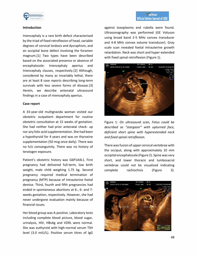
48
Introduction
Iniencephaly is a rare birth defect characterised
by the triad of fixed retroflexion of head, variable
degrees of cervical lordosis and dysraphism, and
an occipital bone defect involving the foramen
magnum.[1] Two types have been described
based on the associated presence or absence of
encephalocele: Iniencephaly apertus and
Iniencephaly clauses, respectively.[2] Although,
considered by many as invariably lethal, there
are at least 8 case reports describing long-term
survivals with less severe forms of disease.[3]
Herein, we describe antenatal ultrasound
findings in a case of iniencephaly apertus.
Case report
A 33-year-old multigravida woman visited our
obstetric outpatient department for routine
obstetric consultation at 15 weeks of gestation.
She had neither had prior antenatal check- up
nor any folic acid supplementation. She had been
a hypothyroid for 4 years and was on thyroxine
supplementation (50 mcg once daily). There was
no h/o consanguinity. There was no history of
teratogen exposure.
Patient’s obstetric history was G6P1A4L1. First
pregnancy had delivered full-term, low birth
weight, male child weighing 1.75 kg. Second
pregnancy required medical termination of
pregnancy (MTP) because of intrauterine foetal
demise. Third, fourth and fifth pregnancies had
ended in spontaneous abortions at 6-, 6- and 7-
weeks gestation, respectively. However, she had
never undergone evaluation mainly because of
financial issues.
Her blood group was A-positive. Laboratory tests
including complete blood picture, blood sugar,
urinalysis, HIV, HBsAg and VDRL were normal.
She was euthyroid with high-normal serum TSH
level (3.0 mIU/L). Positive serum titres of IgG
against toxoplasma and rubella were found.
Ultrasonography was performed (GE Voluson
using broad band 2-5 MHz convex transducer
and 4-8 MHz convex volume transducer). Gray
scale scan revealed foetal intrauterine growth
retardation. Neck was short and hyper-extended
with fixed spinal retroflexion (Figure 1).
Figure 1: On ultrasound scan, Fetus could be
described as “stargazer” with upturned face,
deficient short spine with hyperextended neck
and fixed spinal retroflexion.
There was fusion of upper cervical vertebrae with
the occiput, along with approximately 10 mm
occipital encephalocele (Figure 2). Spine was very
short, and lower thoracic and lumbosacral
vertebrae could not be visualized indicating
complete rachischisis (Figure 3).
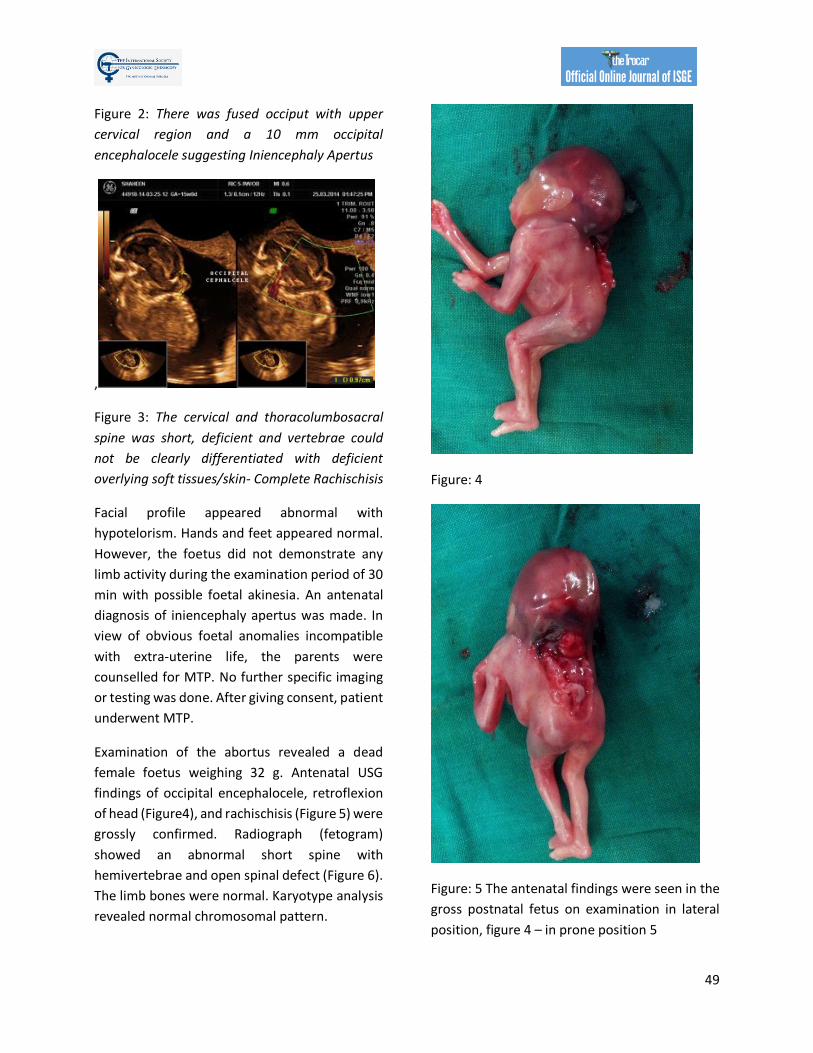
49
Figure 2: There was fused occiput with upper
cervical region and a 10 mm occipital
encephalocele suggesting Iniencephaly Apertus
,
Figure 3: The cervical and thoracolumbosacral
spine was short, deficient and vertebrae could
not be clearly differentiated with deficient
overlying soft tissues/skin- Complete Rachischisis
Facial profile appeared abnormal with
hypotelorism. Hands and feet appeared normal.
However, the foetus did not demonstrate any
limb activity during the examination period of 30
min with possible foetal akinesia. An antenatal
diagnosis of iniencephaly apertus was made. In
view of obvious foetal anomalies incompatible
with extra-uterine life, the parents were
counselled for MTP. No further specific imaging
or testing was done. After giving consent, patient
underwent MTP.
Examination of the abortus revealed a dead
female foetus weighing 32 g. Antenatal USG
findings of occipital encephalocele, retroflexion
of head (Figure4), and rachischisis (Figure 5) were
grossly confirmed. Radiograph (fetogram)
showed an abnormal short spine with
hemivertebrae and open spinal defect (Figure 6).
The limb bones were normal. Karyotype analysis
revealed normal chromosomal pattern.
Figure: 4
Figure: 5 The antenatal findings were seen in the
gross postnatal fetus on examination in lateral
position, figure 4 – in prone position 5
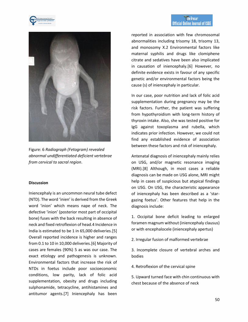
50
Figure: 6 Radiograph (Fetogram) revealed
abnormal undifferentiated deficient vertebrae
from cervical to sacral region.
Discussion
Iniencephaly is an uncommon neural tube defect
(NTD). The word ‘inien’ is derived from the Greek
word ‘inion’ which means nape of neck. The
defective ‘inion’ (posterior most part of occipital
bone) fuses with the back resulting in absence of
neck and fixed retroflexion of head.4 Incidence in
India is estimated to be 1 in 65,000 deliveries.[5]
Overall reported incidence is higher and ranges
from 0.1 to 10 in 10,000 deliveries.[6] Majority of
cases are females (90%) 5 as was our case. The
exact etiology and pathogenesis is unknown.
Environmental factors that increase the risk of
NTDs in foetus include poor socioeconomic
conditions, low parity, lack of folic acid
supplementation, obesity and drugs including
sulphonamide, tetracycline, antihistamines and
antitumor agents.[7] Iniencephaly has been
reported in association with few chromosomal
abnormalities including trisomy 18, trisomy 13,
and monosomy X.2 Environmental factors like
maternal syphilis and drugs like clomiphene
citrate and sedatives have been also implicated
in causation of iniencephaly.[6] However, no
definite evidence exists in favour of any specific
genetic and/or environmental factors being the
cause (s) of iniencephaly in particular.
In our case, poor nutrition and lack of folic acid
supplementation during pregnancy may be the
risk factors. Further, the patient was suffering
from hypothyroidism with long-term history of
thyroxin intake. Also, she was tested positive for
IgG against toxoplasma and rubella, which
indicates prior infection. However, we could not
find any established evidence of association
between these factors and risk of iniencephaly.
Antenatal diagnosis of iniencephaly mainly relies
on USG, and/or magnetic resonance imaging
(MRI).[8] Although, in most cases a reliable
diagnosis can be made on USG alone, MRI might
help in cases of suspicious but atypical findings
on USG. On USG, the characteristic appearance
of iniencephaly has been described as a ‘star-
gazing foetus’. Other features that help in the
diagnosis include:
1. Occipital bone deficit leading to enlarged
foramen magnum without (iniencephaly clausus)
or with encephalocele (iniencephaly apertus)
2. Irregular fusion of malformed vertebrae
3. Incomplete closure of vertebral arches and
bodies
4. Retroflexion of the cervical spine
5. Upward turned face with chin continuous with
chest because of the absence of neck
51
Our case had all these features, and thus, was
diagnosed as iniencephaly apertus. Iniencephaly
apertus should be differentiated from
anencephaly with retroflexion of spine.
Anencephaly shows a total or partial absence of
neurocranium and retroflexed head is not
covered with skin. However, in iniencephaly the
retroflexed head is completely covered with skin.
Cervical vertebrae are abnormal in iniencephaly,
but almost normal in anencephaly. In the present
case the retroflexed head was completely
covered with skin and there was no
encephalocele.
Iniencephaly carries bad prognosis, with the
classic malformation-complex being
incompatible with extra-uterine life. However,
few instances of long-term survival have been
reported in cases of lesser severity of
iniencephaly & associated malformations.7
References: 1. Romero R, Pilu G, Jeanty P, Ghidini A, Hobbins JC. Iniencephaly. In:Romero R, Pilu G, Jeanty P, Ghidini A, Hobbins JC (eds). Prenatal Diagnosis of Congenital Anomalies. Norwalk, CT: Appleton & Lange; 1988:65-7. 2. Chen CP. Prenatal diagnosis of iniencephaly. Taiwan J Obstet Gynecol 2007; 46:199-207. 3. Ayter MH, Dogulu F, Cemil B, Ergun E, Kurt G, Baykaner K. Iniencephaly and long-term survival: A rare case report. Childs Nerv Syst 2007; 23:719-21. 4. Shams Amiri R, Faghih Jouibari M, Nejat F, El Khashab M. Iniencephaly: clinical, radiological and surgical findings. Pediatr Neurosurg 2010; 46: 290-3. 5. Jayant K, Mehta A, Sanghvi LD. A study of congenital malformations in Mumbai. J Obstet Gynaecol India 1961; 11:280-94. 6. Nyberg DA, McGahan JP, Pretorius DH, Pilu G. Diagnostic imaging of fetal anomalies. Philadelphia: Lippincott Williams & Wilkins 2003:315-6. 7. Semi T, Mehmet U, Oya P, Husamettin U, Cem C, Firat E. Iniencephaly: Prenatal diagnosis with postmortem findings. J Obstet Gybocol 2007; 33:566-9. 8. Pungavkar SA, Sainani NI, Karnik AS, Mohanty PH, Lawande MA, Patkar DP, et al. Antenatal diagnosis of
iniencephaly: Sonographic MR correlation: A case Report. Korean J Radiol 2004; 8:351-5.
Corresponding author: Adel Sedrati DOI: 10.36205/trocar1.2021007
casereportTheTrocar Issue 1 2021 / Page 52-54 ISSN: 2736-5530
Learning for our complication Author: Adel Sedrati
Affiliation: Independent Consultant. Obstetrics and Gynecology. Constantine, Algeria
Keywords: bleeding, via false, hysteroscopy
Introduction
At our reference centre of hysteroscopy in
Constantine Algeria we perform more than a
thousand hysteroscopies per a year most of
these are office procedures.
The complication rate for all the procedures is
about 05 per thousand failures and 15 per
thousand incomplete diagnosis and operative
procedures, 04 cases of vagal reactions had to be
managed, 59 per thousand have had a painful
hysteroscopy up to a score of 07/10 (Analogue
Visual score 1 – 10 where the patients rate there
pain) no premedication nor analgesics were
used, the local vocal was the sole method
applied (1).
All the procedures were performed by
vaginoscopy (2) using 05 mm and 04mm
Bettocchi hysteroscopes a Gubbini 14.5 French
resectoscope and 05 French bipolar needle, also
hardly ever a resectoscope of 18,5 was used
when the cervix allowed an easy passage.
Case report
A very rare complication is described as a case
report.
A woman aged 41, married with a story of one
caesarean section four years ago and one year
later she had an incomplete abortion followed by
a curettage, a false route was made accidently
during the curettage as described in her
operatory protocol.
From that time the patient had probably
developed synechia because her symptoms of
hypo and oligomenorrhea. An
hysterosalpingography was performed one year
ago and failed because of the possible passage in
the old false route. Her ultrasound showed a
normal shaped uterus where the endometrial
lining is seen. She was referred to the centre in
order to treat the possible synechia (3),
At first, on day nine of her cycle, an office
hysteroscopy was performed and the scar of the
cervical false passage was clearly visible.
However, a successful passage into the uterine
cavity through the cervical canal averred to be
possible. The endometrium was clearly seen and

Corresponding author: Adel Sedrati DOI: 10.36205/trocar1.2021007
the uterine cavity did look tunnel shaped without
seeing the two ostia possibly covered by the
evident fibrotic tissue. The procedure was
stopped when the patient started to feel some
pain when a trial to cut this fibrotic tissue was
started.
Four months later (because of Covid-19
pandemic) she was scheduled for hysteroscopy
under spinal anaesthesia.
At the theatre a Bettocchi five mm hysteroscope
was used and after getting into the cervical canal
the scope did pass directly into and through the
false passage to finish outside the uterus into the
peritoneal cavity as witnessed by the intra pelvic
fatty tissue clearly visible (4).
The procedure was stopped immediately and the
patient was followed for 24 hours and she was
discharged home in a very good clinical
condition.
Seven days later the patient came back with an
abnormal vaginal bleeding, vaginal and pelvic
ultrasound did show a big hematoma in the
broad ligament measuring some 15cm over 10
cm. the patient was hospitalized for a close
follow up, her Haemoglobin (Hb) was at 11mg/dl
at intake. She was placed under tow different
antibiotics (cefalexin and metronidazole) since
her first hysteroscopy.
One day later early in the morning the team was
called in for an important vaginal bleeding and a
low blood pressure, her Hb was 07mg/dl and the
clotting tests were normal. The abdominal and
vaginal ultrasound revealed no hematoma at all,
so a decision to go to the OR and open the
patient (the centre does not allow to perform
laparoscopy when the blood pressure is down)
was made. through the ancient Pfannenstiel the
patient was opened and after adhesiolysis the
peritoneum of the left broad ligament has just a
bluish colour with the same findings behind the
bladder. A bilateral uterine artery ligation was
performed and an unusual cervical cerclage with
Vicryl 1 in order to close the possible false route,
the uterus had a normal aspect and a clear
opening of the perforation couldn’t be found, a
drain was brought in. A transfusion by three units
of blood was made.
The follow up was normal for three days, and the
patient was discharged home in a satisfactory
clinical condition. A vaginal ultrasound was done
three and five days after the second surgery and
the situation did remain normal.
Aknowledgments
The author does thank Bruno van Herendael for
his critical advise.
References
1- Amer-Cuenca JJ, Marín-Buck A, Vitale SG, La Rosa VL, Caruso S, Cianci A, Lisón JF. Non-pharmacological
pain control in outpatient hysteroscopies. Minimally Invasive Therapy & Allied Technologies. 2020 Jan
2;29(1):10-9.
2- Bettocchi S, Selvaggi L. A vaginoscopic approach to reduce the pain of office hysteroscopy. The Journal
of the American Association of Gynecologic Laparoscopists. 1997 Feb 1;4(2):255-8.
3- Khan Z, Goldberg JM. Hysteroscopic management of Asherman's syndrome. Journal of minimally
invasive gynecology. 2018 Feb 1;25(2):218-28.

Corresponding author: Adel Sedrati DOI: 10.36205/trocar1.2021007
4- Agostini A, Cravello L, Bretelle F, Shojai R, Roger V, Blanc B. Risk of uterine perforation during
hysteroscopic surgery. The Journal of the American Association of Gynecologic Laparoscopists. 2002 Aug
1;9(3):264-7.
1 Corresponding author: Hind Ennasser email: [email protected] DOI: 10.36205/trocarvid1.2021001
Videoarticle TheTrocar Issue 1 2020 / Page 55 ISSN: 2736-5530
Hemorrhagic ascites and pleural effusion: an uncommon presentation of
endometriosis Author: Hind Ennasser1; Ahmed Mimouni1,2; Hanane Saadi1,2; Hafsa Taheri1,2
Affiliation: 1 Department of Obstetrics and Gynecology, Mohammed VI University Hospital Center
Oujda،Morocco
2 Department Mother-child Faculty of Medicine and Pharmacy of Oujda, Morocco
Case report: We present a case of a 29-year-old woman with a history of primary infertility and
laparoscopic excision for recurrent ovarian endometrioma. She presented to the emergency department
for asthenia, abdominal swelling and pain. On examination she was stable with signs of massive ascites
and right-sided pleural effusion.
This was confirmed on ultrasound scan and computed tomography. Paracentesis was done reveling a
bloody ascetic fluid. She declined a pleurodesis for the pleural effusions. The patient was treated with a
GnRH analogues.
Conclusion: This case highlights the importance of evoking endometriosis in reproductive-age women
presenting with a massive ascites, with or without pleural effusions in differential diagnosis. Treating these
patients can be difficult as they are usually of childbearing age and nulliparous and so wish to preserve
their fertility based on conservative surgery and suppressing ovarian function with mainly GnRH agonist
Keywords: Endometriosis, hemorrhagic ascites, pleural effusion, hemothorax
Video Article TheTrocar Issue 1 2021 / Page 56-57
Laparoscopic management of a case of accessory cavitated uterine mass
(ACUM) Author: Sanket Pisat1, Suman Bijlani2, Mohit Shah3, Pranay Desai4
Affiliation: 1Department of Minimally invasive gynecological surgery, Akanksha Hospital,
Maharashtra, Mumbai, India 2Suraj Hospital, Mumbai, India 3Abhipraay Imaging Centre, Mumbai, India 4Nirmala Women’s Hospital, Mumbai, India (Dr Pranay Desai)
Keywords: laparoscopy, juvenile cystic adenomyoma, accessory cavitated uterine mass
Abstract
Study Objective: To demonstrate a technique of laparoscopic management of accessory cavitated uterine
mass, along with a clinical, imaging and histopathology co relation.
Design: A step by step description of the surgery using an instructional video Setting:
Patient: Twenty-seven-year-old woman married for 1 year with complaints of severe dysmenorrhoea.
Ultrasound showed a hype echoic mass lesion in the uterus, likely to be a broad ligament fibroid,
adenomyoma or a functional non communicating uterine horn.
Intervention: Diagnostic hysteroscopy revealed a normal uterine cavity. Diagnostic laparoscopy revealed
a mass approximately 5 cm in size in the right lateral wall, below the right round ligament. There was no
distortion of pelvic anatomy by endometriosis. The mass was infiltrated with diluted vasopressin solution
and excised completely. Chocolate fluid in the mass was drained during removal. The myometrial defect
was sutured in two layers, and covered with an adhesion barrier membrane. Histopathology revealed
features specific to endometriosis. Since all aspects of the ACIEN criteria diagnostic of ACUM were
satisfied, a retrospective diagnosis of ACUM was made.
Measurements and main results: Intra operative blood loss was 20 ml, and intra operative time was 90
minutes. The patient had complete relief of dysmenorrhoea from the very next menstrual cycle.
Corresponding author: Dr Sanket Pisat, Akanksha Mumbai 400053, Maharashtra, India 56E mail: [email protected] DOI: 10.36205/trocarvid1.2021002

C 57
Conclusion: ACUM (previously called Juvenile Cystic Adenomyoma, JCA) is a rare congenital uterine
malformation that usually presents in young women less than 30 years of age. Common presenting
symptoms are dysmenorrhoea and chronic pelvic pain. Treatment is laparoscopic excision. Diagnosis is
confirmed by the presence of a normal uterus, absence of pelvic endometriosis, and the identification of
endometriotic tissue on histopathology.
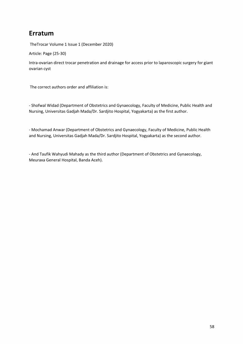
58
Erratum
TheTrocar Volume 1 Issue 1 (December 2020)
Article: Page (25-30)
Intra-ovarian direct trocar penetration and drainage for access prior to laparoscopic surgery for giant
ovarian cyst
The correct authors order and affiliation is:
- Shofwal Widad (Department of Obstetrics and Gynaecology, Faculty of Medicine, Public Health and
Nursing, Universitas Gadjah Mada/Dr. Sardjito Hospital, Yogyakarta) as the first author.
- Mochamad Anwar (Department of Obstetrics and Gynaecology, Faculty of Medicine, Public Health
and Nursing, Universitas Gadjah Mada/Dr. Sardjito Hospital, Yogyakarta) as the second author.
- And Taufik Wahyudi Mahady as the third author (Department of Obstetrics and Gynaecology,
Meuraxa General Hospital, Banda Aceh).