Embed Size (px)
Citation preview
Baseline Penile Ultrasound and Color Doppler Parameters – A Comparison Between Psychogenic
and Vasculogenic Erectile Dysfunction PatientsNarinder Kaur1*, Janu Arora2, Ravinder Kaur3, Nitin Gupta4, Vidur Bhalla5, Navdeep Kaur6 and
Bhavneet Singh2
1Professor, Department of Radiodiagnosis, Government Medical College and Hospital, Sector 32, Chandigarh, India, [email protected]
2Junior Resident, Government Medical College and Hospital, Sector 32, Chandigarh, India 3Professor and Head, Government Medical College and Hospital, Sector 32, Chandigarh, India
4Professor, Department of Psychiatry, Government Medical College and Hospital, Sector 32, Chandigarh, India 5Professor, Department of Urology, Government Medical College and Hospital, Sector 32, Chandigarh, India
6Senior Resident, Department of Radiodiagnosis, Government Medical College and Hospital, Sector 32, Chandigarh, India
AbstractBackground and Objectives: There are many causes of erectile dysfunction (ED) like congenital anomalies, neurological, endocrinal, pharmacological, psychological, pathological or hemodynamic. Penile doppler ultrasound provides real-time evaluation of cavernosal vascular flow dynamics. Purpose of our study was to evaluate and compare the baseline penile ultrasound and color doppler vascular parameters in patients of ED to see if there was any significant difference between patients of psychogenic and vasculogenic ED (diagnosed on post Papaverine injection color doppler) without the use of intracavernosal Papaverine injection. To best of our knowledge such detailed comparison of baseline penile ultrasound and doppler parameters have not been published in literature. Methods: We have prospectively studied 32 patients of ED who underwent ultrasound, and color doppler pre-and post-papaverine intracavernosal injection. Baseline pre papaverine diameters of right and left cavernosal arteries were measured on grey scale ultrasound. Peak systolic velocity (PSV), end diastolic velocity (EDV) and resistive index (RI) of right and left cavernosal arteries were measured on color doppler before and after injecting intracavernosal papaverine injection. Patients were divided into normal study group (psychogenic ED), arterial insufficiency and venous leakage groups (vasculogenic ED) on the basis of post papaverine color doppler findings. Results: Eighteen patients showed normal study, 11 showed arterial insufficiency and 3 patients had venous leakage on post Papaverine injection color doppler. When base line prepapaverine vascular diameters and color doppler parameters were compared statistically, no significant difference was detected between and within these study groups. We have also compared Ed duration and IIEF scores among various study groups and found statistically significant difference between and within the groups. Conclusion: Use of a vasoactive agent like Papaverine with color Doppler is must to diagnose the vasculogenic ED.
*Author for correspondence
1. Background and ObjectivesPenile erection is a coordinated function of the nervous,
arterial, venous and sinusoidal systems. Penile erectile tissue is composed of cavernous smooth musculature and smooth muscles of arterial and arteriolar walls and
Keywords: Baseline, Color Doppler, Erectile Dysfunction, Penile Ultrasound, Psychogenic, Vasculogenic
International Journal of Medical and Dental Sciences, Vol 10(2), DOI: 10.18311/ijmds/2021/27292, July 2021ISSN (Print) : 2454-8952
ISSN (Online) : 2320-1118Original Article
Baseline Penile Ultrasound and Color Doppler Parameters – A Comparison Between Psychogenic...
International Journal of Medical and Dental SciencesVol 10 (2) | July 2021 | http://www.informaticsjournals.com/index.php/ijmds/index1982
it plays a key role in erection1. A defect in any of these systems can give rise to erectile dysfunction (ED). Erectile Dysfunction is defined as the consistent inability to generate or maintain an erection of sufficient rigidity to perform sexual intercourse2.
In the age group of 40-70 years 10% of men have complete, 17.1% have mild and 25.2% have moderate erectile dysfunction as estimated by Massachusetts Male Aging Study (MMAS). MMAS has estimated the prevalence of ED as 52% in this age group3.
There are many causes of ED like congenital anomalies, neurological, endocrinal, pharmacological, psychological, pathological or hemodynamic. Penile doppler ultrasound (US) provides real-time evaluation of cavernosal vascular flow dynamics. Color and spectral penile doppler US is a highly accurate tool for assessing patients with erectile dysfunction4,5, it is a high performing noninvasive or minimally invasive imaging investigation that allows depiction of normal anatomy and macroscopic pathological abnormalities in real time and can depict functional changes in blood flow which can determine vascular cause of erectile dysfunction6. Hemodynamic dysfunction is responsible for most of the organic impotence and have many causes, here we are referring to the vascular causes only including arterial insufficiency (Peak systolic velocity less than 25cm/s), Venous leakage (end diastolic velocity > 5cm/s), or combined arterial insufficiency and venous leakage1,2 during post papaverine induced erection color doppler.
In literature, organic causes of ED are seen in 50-90% cases of which vascular cause accounts for 50-70% cases. Arteriogenic impotence accounts for about 30% cases, only venogenic impotence is found in about 15% cases. And often ED is caused by combined arteriogenic and venogenic causes1.
Vascular cause of ED can be further classified using penile color doppler parameters as normal vascular response, arterial insufficiency, vascular leakage or combined arterial insufficiency and venous leakage. This method requires intracorporeal injection of a vasoactive substance such as papaverine hydrochloride or prostaglandin E1 alone or in combination with phentolamine (Lue and Tanagho 19877,8). Papaverine hydrochloride when injected in penile tissue causes direct smooth muscle relaxation and consequent filling of the corpora cavernosum with blood resulting in erection9. It may lead to persistent painful erection (priapism) in
some patients which can be treated successfully and conservatively8.
Purpose of our study was to evaluate and compare the baseline pre papaverine injection grey scale ultrasound and color doppler vascular parameters (diameter, peak systolic velocity, end diastolic velocity and resistive index of right and left cavernosal arteries) among the normal and abnormal post papaverine injection color doppler groups in patients presenting for evaluation of erectile dysfunction (ED) by color doppler combined with intracavernosal Papaverine injection.
2. Materials and MethodsPatients were referred from the department of Psychiatry and department of Urology to department of Radiology for color doppler evaluation of erectile dysfunction. 32 patients were enrolled who had history of ED and did not have any exclusion criteria of our study from December 2017 to December 2019.Inclusion criteria: Patients having ED as per IIEF (International Index of Erectile Function) score < 21, and age > 20 to < 65 years were included in our study. Exclusion criteria: Patients with coronary artery disease, hypospadias, epispadias, history of-drug allergy, drug abuse, sickle cell anemia, bleeding disorders, taking anticoagulants, penile trauma, spinal trauma and spinal surgery were excluded from our study.
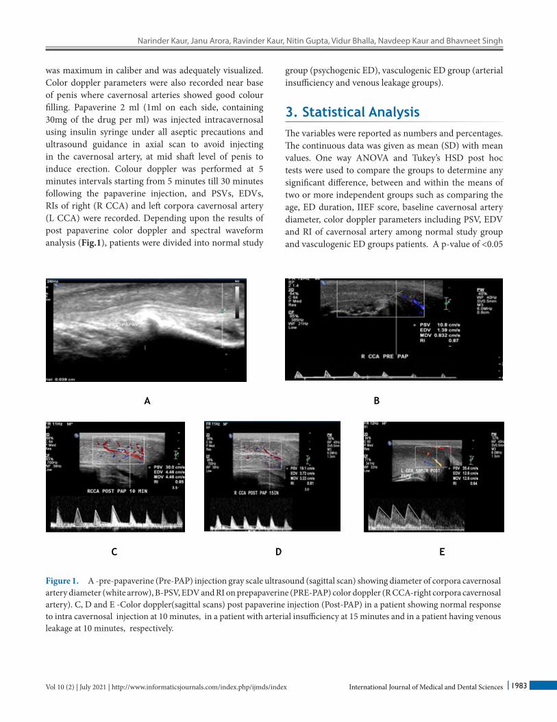
After obtaining detailed history and informed consent of the patient in an atmosphere of privacy, real time grey scale penile ultrasound and color doppler US was performed with 17-5 MHz broadband linear array high-frequency transducer on PHILIPS IU22 high resolution ultrasound machine in adequate privacy and comfortable environment. Grey scale ultrasonography was performed in axial and sagittal (Figure 1) planes to rule out any morphological abnormalities of penis and to measure the baseline prepapaverine diameters of cavernosal arteries in sagittal plane. Color doppler was performed to record the prepapaverine and post Papaverine injection peak systolic velocities (PSVs), end diastolic velocities (EDVs) and resistive indexes (RIs). Grey scale ultrasound and color Doppler US were performed in supine position of the patient with penis in anatomical position lying on the anterior abdominal wall. Diameter of cavernosal artery was measured near the base of penis where it
Narinder Kaur, Janu Arora, Ravinder Kaur, Nitin Gupta, Vidur Bhalla, Navdeep Kaur and Bhavneet Singh
International Journal of Medical and Dental Sciences 1983Vol 10 (2) | July 2021 | http://www.informaticsjournals.com/index.php/ijmds/index
was maximum in caliber and was adequately visualized. Color doppler parameters were also recorded near base of penis where cavernosal arteries showed good colour filling. Papaverine 2 ml (1ml on each side, containing 30mg of the drug per ml) was injected intracavernosal using insulin syringe under all aseptic precautions and ultrasound guidance in axial scan to avoid injecting in the cavernosal artery, at mid shaft level of penis to induce erection. Colour doppler was performed at 5 minutes intervals starting from 5 minutes till 30 minutes following the papaverine injection, and PSVs, EDVs, RIs of right (R CCA) and left corpora cavernosal artery (L CCA) were recorded. Depending upon the results of post papaverine color doppler and spectral waveform analysis (Fig.1), patients were divided into normal study
group (psychogenic ED), vasculogenic ED group (arterial insufficiency and venous leakage groups).
3. Statistical AnalysisThe variables were reported as numbers and percentages. The continuous data was given as mean (SD) with mean values. One way ANOVA and Tukey’s HSD post hoc tests were used to compare the groups to determine any significant difference, between and within the means of two or more independent groups such as comparing the age, ED duration, IIEF score, baseline cavernosal artery diameter, color doppler parameters including PSV, EDV and RI of cavernosal artery among normal study group and vasculogenic ED groups patients. A p-value of <0.05
Figure 1. A -pre-papaverine (Pre-PAP) injection gray scale ultrasound (sagittal scan) showing diameter of corpora cavernosal artery diameter (white arrow), B-PSV, EDV and RI on prepapaverine (PRE-PAP) color doppler (R CCA-right corpora cavernosal artery). C, D and E -Color doppler(sagittal scans) post papaverine injection (Post-PAP) in a patient showing normal response to intra cavernosal injection at 10 minutes, in a patient with arterial insufficiency at 15 minutes and in a patient having venous leakage at 10 minutes, respectively.
A B
C D E
Baseline Penile Ultrasound and Color Doppler Parameters – A Comparison Between Psychogenic...
International Journal of Medical and Dental SciencesVol 10 (2) | July 2021 | http://www.informaticsjournals.com/index.php/ijmds/index1984
was considered statistically significant. The statistical software SPSS 22.0 was used for data analysis.
4. ResultsObservations regarding age, complaints with duration, marital status, comorbidities, medication/drug history, findings of greyscale ultrasonography and color doppler study were made. Statistics analysis was applied wherever applicable.
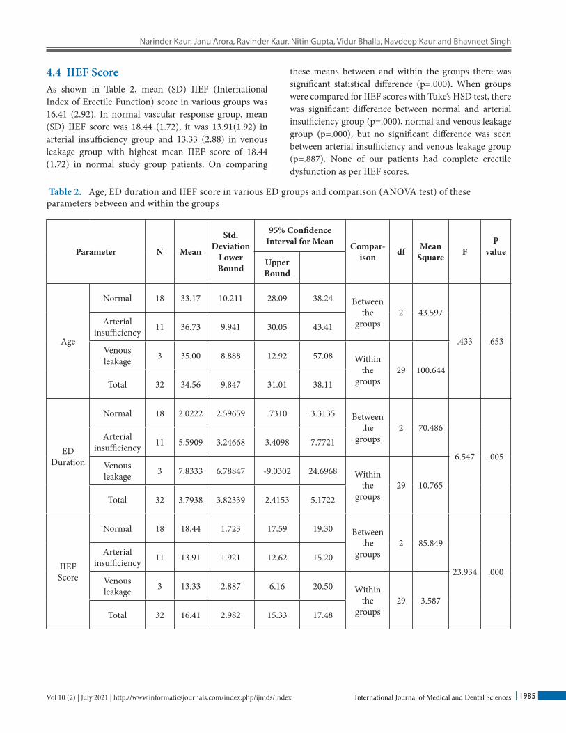
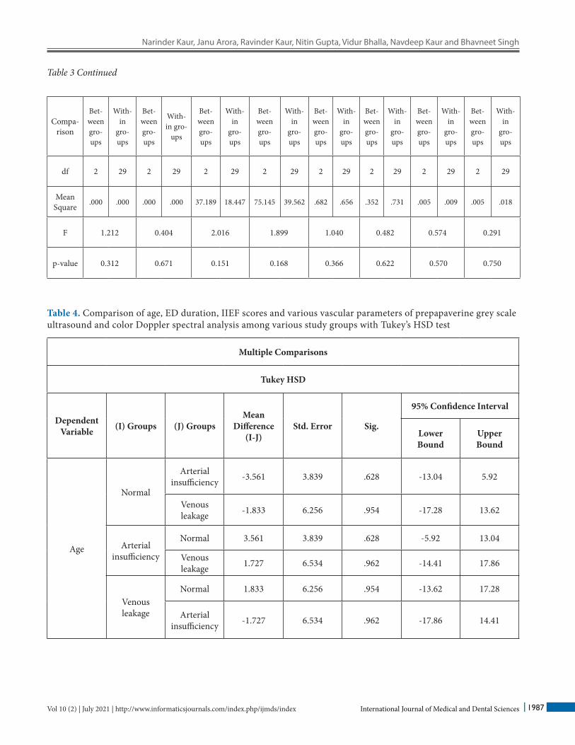
4.1 AgeOverall mean age of the patients in our study was 34.56 (9.84) years with range 31.01 – 38.11 years. It was 33.7 (10.21) years (Range: 28.09 – 38.24 years) in normal study group, 36.73(9.94) years (Range: 30.05 – 43.41 years) in arterial insufficiency group and 35.00 (8.89) years (Range: 12.92 – 57.08) years in venous leakage group. When compared age wise, there was no statistically significant difference between and within these groups (p=0.653) as shown in Table 2 and Table 4.
Maximum number of patients was in age group of 21-30 years i.e., 14 (43.75%). More cases (n=30) were of age between 21-50 years making 93.75% of total patients, and only two cases (6.25% of total) were aged between 51-65 years (Table 1).
4.2 Marital StatusIn this study 20(62.50%) patients were married and 12 (37.50%) patients were unmarried. Ten married patients belonged to normal color doppler study group, 7 patients
were in arterial insufficiency group and all three venous leakage group patients were married.
Out of 10 normal Doppler study group married patients, six had two children, two had one child each and two patients did not have any child.
In arterial insufficiency group six patients had 2 children and one patient did not have any child.
In Venous leakage group one patient had no child, one had 4 children and third patient had 2 children.
Among the unmarried patients 08 belonged to normal Doppler study group and 04 patients were in arterial insufficiency group.
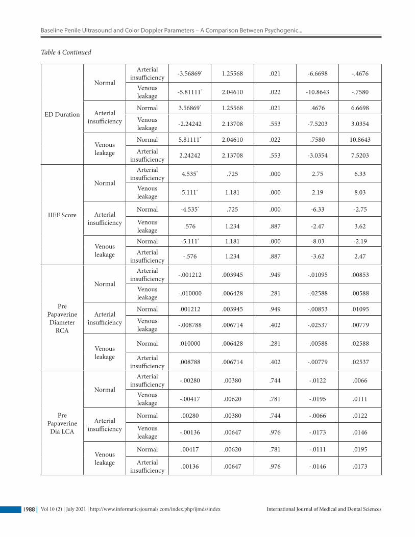
4.3 Duration of EDOverall combined ED duration Mean (SD) was 8 (1.72) years with a range 2.42-5.17 years in total study group of 32 patients. Mean (SD) duration of ED was 2.02 (2.59) years in normal study group, 5.60 (3.25) years in arterial insufficiency group, and 7.83 (6.78) years in venous leakage group. ED duration was shortest in normal study group and longest in venous leakage group patients. When various groups were compared for ED duration, there was statistically significant difference between and within the groups (p=.005) as shown in (Table 2). Statistical analysis with Tuke’s HSD test (Table 4) showed that there was significant difference of ED duration when normal group was compared with arterial insufficiency and venous leakage groups (p value .021 and .022 respectively). No statistically significant difference was seen between arterial insufficiency and venous leakage groups (p=.553).
Age Group in years Number of patients Percentage (%)
21-30 14 43.75
31-40 09 28.12
41-50 07 21.88
51-60 01 3.12
61-65 01 3.12
Total 32 100
Table 1. Various age group of the patients of ED in general
Narinder Kaur, Janu Arora, Ravinder Kaur, Nitin Gupta, Vidur Bhalla, Navdeep Kaur and Bhavneet Singh
International Journal of Medical and Dental Sciences 1985Vol 10 (2) | July 2021 | http://www.informaticsjournals.com/index.php/ijmds/index
4.4 IIEF ScoreAs shown in Table 2, mean (SD) IIEF (International Index of Erectile Function) score in various groups was 16.41 (2.92). In normal vascular response group, mean (SD) IIEF score was 18.44 (1.72), it was 13.91(1.92) in arterial insufficiency group and 13.33 (2.88) in venous leakage group with highest mean IIEF score of 18.44 (1.72) in normal study group patients. On comparing
these means between and within the groups there was significant statistical difference (p=.000). When groups were compared for IIEF scores with Tuke’s HSD test, there was significant difference between normal and arterial insufficiency group (p=.000), normal and venous leakage group (p=.000), but no significant difference was seen between arterial insufficiency and venous leakage group (p=.887). None of our patients had complete erectile dysfunction as per IIEF scores.
Parameter N Mean
Std. Deviation
Lower Bound
95% Confidence Interval for Mean Compar-
ison df Mean Square F
P value
Upper Bound
Age
Normal 18 33.17 10.211 28.09 38.24 Between the
groups2 43.597
.433 .653
Arterial insufficiency 11 36.73 9.941 30.05 43.41
Venous leakage 3 35.00 8.888 12.92 57.08 Within
the groups
29 100.644Total 32 34.56 9.847 31.01 38.11
ED Duration
Normal 18 2.0222 2.59659 .7310 3.3135 Between the
groups2 70.486
6.547 .005
Arterial insufficiency 11 5.5909 3.24668 3.4098 7.7721
Venous leakage 3 7.8333 6.78847 -9.0302 24.6968 Within
the groups
29 10.765Total 32 3.7938 3.82339 2.4153 5.1722
IIEF Score
Normal 18 18.44 1.723 17.59 19.30 Between the
groups2 85.849
23.934 .000
Arterial insufficiency 11 13.91 1.921 12.62 15.20
Venous leakage 3 13.33 2.887 6.16 20.50 Within
the groups
29 3.587Total 32 16.41 2.982 15.33 17.48
Table 2. Age, ED duration and IIEF score in various ED groups and comparison (ANOVA test) of these parameters between and within the groups

Baseline Penile Ultrasound and Color Doppler Parameters – A Comparison Between Psychogenic...
International Journal of Medical and Dental SciencesVol 10 (2) | July 2021 | http://www.informaticsjournals.com/index.php/ijmds/index1986
4.5 Co-MorbidityIn normal study group, 3 patients had depression, one had fracture ulna, one had fracture humerus and another patient had inguinal hernia making it to 6 (33.33%) out of 18 patients having co-morbidity.
In arterial insufficiency group no co-morbidity was seen. In venous leakage group one patient had insomnia and other patient had fracture humerus making it to 2 out of 3 patients i.e. 66.66% having co morbidity.
4.6 MedicationThere was history of taking medicine -tablet Tadalafil for last 5 years in 1(5.56%) of 18 normal study group patients. In arterial insufficiency group 3 (27.27%) of 11 patients gave history of taking medicine-one patient was taking tablet Tadalafil for past 5 years, one patient was on tablet al. prax and third patient was taking Ayurvedic medicine for his current complaint of ED.
4.7 Grey scale Ultrasonography FindingsOne of 32 (3.12%) patients, aged 47 year, showed a calcified hypoechoeic plaque in right corpora cavernosa in mid shaft region suggestive of peyronie’s disease and showed normal vascular response on post Papaverine color doppler. He was not a known case of peyronie’s disease prior to his penile ultrasound and doppler study.
There was no past history of taking Papaverine or any other medication in this patient. Rest of our patients (96.97%) showed normal penile anatomy and no focal penile lesion on ultrasonography.
4.8 Baseline Vascular -Ultrasound and Color Doppler Parameters
Bilateral cavernosal arteries were better visualized near base of penis and on post Papaverine status, however in 5 (15.63%) patients visualization of these arteries took longer than usual time in prepapaverine status on grayscale as well as on color doppler US.
Baseline prepapaverine diameters of bilateral cavernosal arteries were measured on gray scale ultrasonography in sagittal scan. Pre Papaverine color doppler was performed and PSVs, EDVs and RIs of right and left cavernosal arteries (RCA and LCA) were recorded. Means of these parameters are shown in (Table 3) .
Post Papaverine injection color doppler PSVs, EDVs, and RIs were recorded for right and left cavernosal arteries. Eighteen patients had normal study; 14 patients had abnormal post-Papaverine color doppler study. Out of abnormal study 14 patients -11 patients had arterial insufficiency and 03 had venous leakage.
Groups (N)
Prepap-averine RCA
diameter Mean (SD)
Prepap-averine LCA
diameter Mean (SD)
Prepap-averine RCA
PSV Mean (SD)
Prepap-averine LCA PSV Mean
(SD)
Prepap-averine
RCA EDV Mean (SD)
Prepap-averine
LCA EDV Mean (SD)
Prepap-averine RCA
RI Mean (SD)
Prepap-averine LCA RI Mean (SD
Normal (18) 0.047 (0.002) 0.048 (0.002) 7.42(1.048) 6.79(0.750) 1.20(0.23) 1.11(0.23) 0.86(0.02) 0.84(0.03)
Arterial insuffi-ciency (11)
0.048 (0.004) 0.051(0.003) 6.2(0.905) 7.63(2.38) 0.59(0.08) 0.79(1.71) 0.90(0.02) 0.86(0.04)
Venous leakage
(3)0.057 (0.006) 0.052(0.006) 11.78(4.261) 14.42(7.65) 1.04(0.65) 0.99(0.49) 0.89(0.09) 0.90(0.07)
Table 3. Baseline Prepapaverine penile vascular parameters of right and left cavernosal arteries on gray scale ultrasonography and color doppler in various groups and comparison of these parameters among various study groups by ANNOVA test
Narinder Kaur, Janu Arora, Ravinder Kaur, Nitin Gupta, Vidur Bhalla, Navdeep Kaur and Bhavneet Singh
International Journal of Medical and Dental Sciences 1987Vol 10 (2) | July 2021 | http://www.informaticsjournals.com/index.php/ijmds/index
Compa-rison
Bet-ween gro-ups
With-in
gro-ups
Bet-ween gro-ups
With-in gro-
ups
Bet-ween gro-ups
With-in
gro-ups
Bet-ween gro-ups
With-in
gro-ups
Bet-ween gro-ups
With-in
gro-ups
Bet-ween gro-ups
With-in
gro-ups
Bet-ween gro-ups
With-in
gro-ups
Bet-ween gro-ups
With-in
gro-ups
df 2 29 2 29 2 29 2 29 2 29 2 29 2 29 2 29
Mean Square .000 .000 .000 .000 37.189 18.447 75.145 39.562 .682 .656 .352 .731 .005 .009 .005 .018
F 1.212 0.404 2.016 1.899 1.040 0.482 0.574 0.291
p-value 0.312 0.671 0.151 0.168 0.366 0.622 0.570 0.750
Table 3 Continued
Table 4. Comparison of age, ED duration, IIEF scores and various vascular parameters of prepapaverine grey scale ultrasound and color Doppler spectral analysis among various study groups with Tukey’s HSD test
Multiple Comparisons
Tukey HSD
Dependent Variable (I) Groups (J) Groups
Mean Difference
(I-J)Std. Error Sig.
95% Confidence Interval
Lower Bound
Upper Bound
Age
Normal
Arterial insufficiency -3.561 3.839 .628 -13.04 5.92
Venous leakage -1.833 6.256 .954 -17.28 13.62
Arterial insufficiency
Normal 3.561 3.839 .628 -5.92 13.04
Venous leakage 1.727 6.534 .962 -14.41 17.86
Venous leakage
Normal 1.833 6.256 .954 -13.62 17.28
Arterial insufficiency -1.727 6.534 .962 -17.86 14.41
Baseline Penile Ultrasound and Color Doppler Parameters – A Comparison Between Psychogenic...
International Journal of Medical and Dental SciencesVol 10 (2) | July 2021 | http://www.informaticsjournals.com/index.php/ijmds/index1988
ED Duration
Normal
Arterial insufficiency -3.56869* 1.25568 .021 -6.6698 -.4676
Venous leakage -5.81111* 2.04610 .022 -10.8643 -.7580
Arterial insufficiency
Normal 3.56869* 1.25568 .021 .4676 6.6698
Venous leakage -2.24242 2.13708 .553 -7.5203 3.0354
Venous leakage
Normal 5.81111* 2.04610 .022 .7580 10.8643
Arterial insufficiency 2.24242 2.13708 .553 -3.0354 7.5203
IIEF Score
Normal
Arterial insufficiency 4.535* .725 .000 2.75 6.33
Venous leakage 5.111* 1.181 .000 2.19 8.03
Arterial insufficiency
Normal -4.535* .725 .000 -6.33 -2.75
Venous leakage .576 1.234 .887 -2.47 3.62
Venous leakage
Normal -5.111* 1.181 .000 -8.03 -2.19Arterial
insufficiency -.576 1.234 .887 -3.62 2.47
Pre Papaverine Diameter
RCA
Normal
Arterial insufficiency -.001212 .003945 .949 -.01095 .00853
Venous leakage -.010000 .006428 .281 -.02588 .00588
Arterial insufficiency
Normal .001212 .003945 .949 -.00853 .01095
Venous leakage -.008788 .006714 .402 -.02537 .00779
Venous leakage
Normal .010000 .006428 .281 -.00588 .02588
Arterial insufficiency .008788 .006714 .402 -.00779 .02537
Pre Papaverine Dia LCA
Normal
Arterial insufficiency -.00280 .00380 .744 -.0122 .0066
Venous leakage -.00417 .00620 .781 -.0195 .0111
Arterial insufficiency
Normal .00280 .00380 .744 -.0066 .0122
Venous leakage -.00136 .00647 .976 -.0173 .0146
Venous leakage
Normal .00417 .00620 .781 -.0111 .0195
Arterial insufficiency .00136 .00647 .976 -.0146 .0173
Table 4 Continued
Narinder Kaur, Janu Arora, Ravinder Kaur, Nitin Gupta, Vidur Bhalla, Navdeep Kaur and Bhavneet Singh
International Journal of Medical and Dental Sciences 1989Vol 10 (2) | July 2021 | http://www.informaticsjournals.com/index.php/ijmds/index
Pre Papaverine PSV RCA
Normal
Arterial insufficiency 1.2597 1.6437 .726 -2.800 5.319
Venous leakage -4.3567 2.6784 .251 -10.971 2.258
Arterial insufficiency
Normal -1.2597 1.6437 .726 -5.319 2.800
Venous leakage -5.6164 2.7975 .128 -12.525 1.292
Venous leakage
Normal 4.3567 2.6784 .251 -2.258 10.971
Arterial insufficiency 5.6164 2.7975 .128 -1.292 12.525
Pre Papaverine PSV LCA
Normal
Arterial insufficiency -.84480 2.40716 .935 -6.7896 5.1000
Venous leakage -7.63389 3.92239 .144 -17.3208 2.0530
Arterial insufficiency
Normal .84480 2.40716 .935 -5.1000 6.7896
Venous leakage -6.78909 4.09680 .239 -16.9068 3.3286
Venous leakage
Normal 7.63389 3.92239 .144 -2.0530 17.3208
Arterial insufficiency 6.78909 4.09680 .239 -3.3286 16.9068
Pre Papaverine EDV RCA
Normal
Arterial insufficiency .43221 .31001 .357 -.3334 1.1978
Venous leakage -.01728 .50516 .999 -1.2648 1.2303
Arterial insufficiency
Normal -.43221 .31001 .357 -1.1978 .3334
Venous leakage -.44948 .52762 .674 -1.7525 .8536
Venous leakage
Normal .01728 .50516 .999 -1.2303 1.2648
Arterial insufficiency .44948 .52762 .674 -.8536 1.7525
Table 4 Continued
Baseline Penile Ultrasound and Color Doppler Parameters – A Comparison Between Psychogenic...
International Journal of Medical and Dental SciencesVol 10 (2) | July 2021 | http://www.informaticsjournals.com/index.php/ijmds/index1990
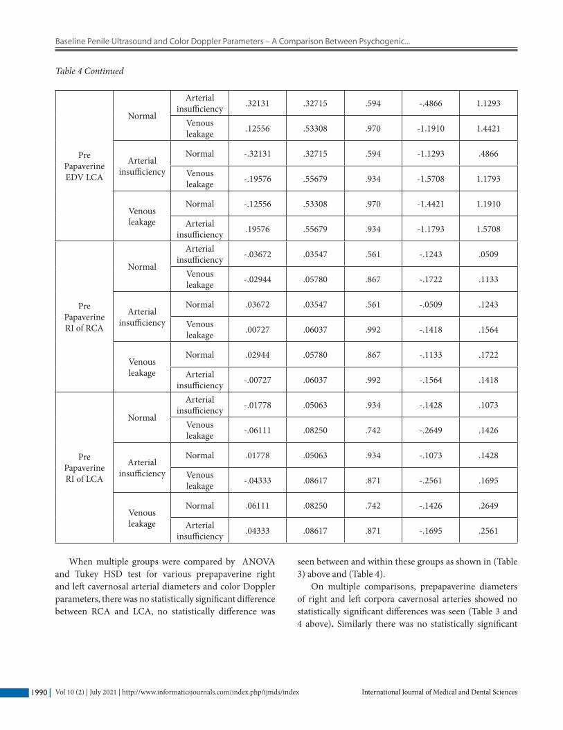
When multiple groups were compared by ANOVA and Tukey HSD test for various prepapaverine right and left cavernosal arterial diameters and color Doppler parameters, there was no statistically significant difference between RCA and LCA, no statistically difference was
seen between and within these groups as shown in (Table 3) above and (Table 4).
On multiple comparisons, prepapaverine diameters of right and left corpora cavernosal arteries showed no statistically significant differences was seen (Table 3 and 4 above). Similarly there was no statistically significant
Pre Papaverine EDV LCA
Normal
Arterial insufficiency .32131 .32715 .594 -.4866 1.1293
Venous leakage .12556 .53308 .970 -1.1910 1.4421
Arterial insufficiency
Normal -.32131 .32715 .594 -1.1293 .4866
Venous leakage -.19576 .55679 .934 -1.5708 1.1793
Venous leakage
Normal -.12556 .53308 .970 -1.4421 1.1910
Arterial insufficiency .19576 .55679 .934 -1.1793 1.5708
Pre Papaverine RI of RCA
Normal
Arterial insufficiency -.03672 .03547 .561 -.1243 .0509
Venous leakage -.02944 .05780 .867 -.1722 .1133
Arterial insufficiency
Normal .03672 .03547 .561 -.0509 .1243
Venous leakage .00727 .06037 .992 -.1418 .1564
Venous leakage
Normal .02944 .05780 .867 -.1133 .1722
Arterial insufficiency -.00727 .06037 .992 -.1564 .1418
Pre Papaverine RI of LCA
Normal
Arterial insufficiency -.01778 .05063 .934 -.1428 .1073
Venous leakage -.06111 .08250 .742 -.2649 .1426
Arterial insufficiency
Normal .01778 .05063 .934 -.1073 .1428
Venous leakage -.04333 .08617 .871 -.2561 .1695
Venous leakage
Normal .06111 .08250 .742 -.1426 .2649
Arterial insufficiency .04333 .08617 .871 -.1695 .2561
Table 4 Continued
Narinder Kaur, Janu Arora, Ravinder Kaur, Nitin Gupta, Vidur Bhalla, Navdeep Kaur and Bhavneet Singh
International Journal of Medical and Dental Sciences 1991Vol 10 (2) | July 2021 | http://www.informaticsjournals.com/index.php/ijmds/index
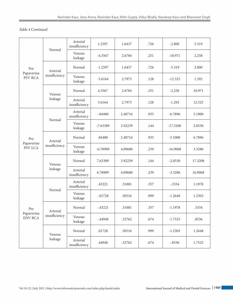
difference when we compared the means of prepapaverine color doppler parameters i.e. PSV, EDV and RI of various groups as shown in (Table 3 and 4).
5. ComplicationsTwo (6.25%) of 32 patients, belonging to normal study group developed priapism and were managed conservatively. No other complications were seen our study.
6. DiscussionErectile dysfunction (ED) is the most common sexual disorder in males with the prevalence rates of 2.59% -6% in the age between 20-29 years and prevalence of complete impotence increased from 5% to 15% in the age of 40-70 years. The prevalence of 39% at the age of 40 years, and 67% prevalence at the age of 70 years shows the association of ED with age10,11 and 12.
In present study mean age (SD) of the patients presenting with ED was 34.86 (9.86) years and it varied between 21-63 years. Maximum cases were in the age group of 21-30 years. Overall maximum patients i.e., 30 (93. 75%) were between 21-50 years of age, hence our results are varying from that found in literature, it might be because of relatively small sample of our study. Capogrosso P et al., in 2013 studied 439 patients and found that new onset ED as primary disorder was found in 26% males younger than 40 years and at IIEF, severe ED rates were found in 48.8% younger men and 40% older men respectively (p>0.05)13.
More of our patients were married n=20 (62.5%). This finding is in agreement to a study carried out by Suresh et al.14 in which they have reported erectile dysfunction as 70% in married and 30% in unmarried patients of total 33 patients.
Mean (SD) duration of ED was 2.02 (2.59) years in normal response group, 5.60 (3.25) years in arterial insufficiency group, and 7.83 (6.78) years in venous leakage group. When various groups were compared for ED duration, there was statistically significant difference between and within the groups (p=.005). Statistical analysis also showed that there was significant difference of ED duration when normal group was compared with arterial insufficiency and venous leakage groups (p value .021 and 0.022 respectively). No statistically significant
difference was seen between arterial insufficiency and venous leakage groups (p= .553).
The patient with organic impotence describes his problems with erection that progress over months to years, organic impotence is constant and non selective, meaning it is not better or worse with any specific partner or type of stimulation. On the other hand, psychogenic impotence is abrupt in onset, intermittent and not progressive (Ende J. 1990)15. Hence our findings of ED duration lowest in the normal study group (psychogenic ED), higher in arterial insufficiency, and highest in venous group, and statistically significant difference among the groups may be explained by the presentation of patients as described in this book to some extent.
Mean (SD) IIEF score in various groups was 16.41 (2.92). It was 18.44 (1.72) in normal study group, 13.91 (1.92) in arterial insufficiency group and, 13.33 (2.88) in venous leakage group. On comparing these means between and within the groups there was significant statistical difference (p=.000) with highest mean IIEF score in normal study group. Statistically significant difference was found between normal and arterial insufficiency group (p=.000), normal and venous leakage group (p=.000) but no significant difference was seen between arterial insufficiency and venous leakage groups (p=.887). In a study conducted by Atinbas KN et al. in 201816, IIEF score was found to be 17.03+3.64, 15+27 and 13.85+3.24 in various groups similar to the groups of present study, hence our findings are in agreement with the finding of Atinbas KN et al.
The erectile dysfunction has mainly two etiologies – Psychogenic and organic. Color doppler ultrasonography is established method of evaluating the ED patients to rule out the vasculogenic organic cause. It is performed during an induced erection by papaverine/prostaglandin E1 injected in the corpora cavernosa tissue of the penis. In the present study we aimed to evaluate and compare the baseline prepapaverine grey scale and color doppler vascular parameters of penis including morphological abnormalities if any, cavernosal arterial diameters, PSVs, EDVs and RIs in patients presenting with ED. Eighteen (56.25%) of 32 patients responded normally on post papaverine color doppler, hence labeled as having psychogenic ED. Rest 14 patients (43.75%) showed abnormal color doppler study indicating vasculogenic ED, out of these 11 (34.38%) patients had arterial insufficiency and 3 (9.38%) patients had venous leakage.
Baseline Penile Ultrasound and Color Doppler Parameters – A Comparison Between Psychogenic...
International Journal of Medical and Dental SciencesVol 10 (2) | July 2021 | http://www.informaticsjournals.com/index.php/ijmds/index1992
Organic causes of ED are seen in 50-90% cases, and vascular cause accounts for 50 -70% cases. In present study 43.75% patients had vasculogenic ED which is close to the prevalence of 50% – 70% of organic causes of ED as found in literature1, 8, 9 and10. Our study is also in agreement with a study conducted in 2017 by Usman Khanzada et al.17 in 97 patients where they found that 50.5% patients had normal color doppler hence psychogenic ED and 48.5% patients had vasculogenic organic cause for their erectile dysfunction.
In literature arterial insufficiency is seen in 30% cases and venous insufficiency is seen in 15% cases among the vasculogenic ED. In our study 34.38% patients had arterial insufficiency and 9.38% patients had venous leakage as cause of vasculogenic ED. Our study is in agreement with the results found in literature. Our study has found more number of arterial insufficiency cases compared to venous leakage patients and this is similar to a study conducted by Bari V et al.,18 which has reported a relatively large number of cases of arterial erectile dysfunction compared to venous insufficiency ED.
A study done by Gall et al.19 has reported accuracy of 95% in localization of penile arteries in the duplex ultrasound and high agreement (90%) in detection of vascular lesions with angiography. Moreover, duplex ultrasound may avoid the risks associated with angiography especially in arteriosclerotic patients and it also required general anesthesia and exposure to ionizing radiations. In our study we could locate the bilateral cavernosal arteries successfully on ultrasound in all 32 patients (100%) in flaccid state of penis, however it took more than usual time in locating these arteries in 5 (15.63%) patients.
Sidhu P.S. et al.,20 have recommended that the penile ultrasound must be conducted in an atmosphere of privacy, and evaluation begins with scanning of flaccid penis shaft in transverse plane to measure the diameter of cavernosal arteries and the diameters ranged from 0.2 to 1 mm in their study.
We measured the diameters of cavernosal arteries in flaccid state in sagittal plane near base of penis and mean diameters of right and left cavernosal arteries were .047 (.00) cm and .048 (.00) cm, .048 (.00) cm and .051 (.00) cm and .057 (.01) cm and .052 (.01) cm in normal, arterial insufficiency and venous leakage groups respectively which is close to that found by Sidhu P.S. et al.20.
Pre papaverine baseline PSVs, EDVs and RI are as shown in Table 3. We have not come across any studies showing such detailed prepapaverine baseline vascular parameters comparison among various groups. In our study when we compared these prepapaverine vascular parameters including the diameters of corpora cavernosal arteries among various groups of study, no statistically significant difference was found indicating that vasculogenic erectile dysfunction cannot be predicted by prepapaverine color doppler. Therefore, use of a vasoactive agent like papaverine combined with color doppler is essential to diagnose and classify the vasculogenic erectile dysfunction.
Priapism is a persistent erection lasting for more than 4 hours and a common and serious complication of color doppler with papaverine induced erection (Montague et al. 200321. Its occurrence is more in patients having relatively better baseline erection dysfunction (Lomas and Jarow, 199222). Secil M et al. [2001] stated that occurrence of priapism was between 1.8-18% in various studies and recorded its incidence as 11.9% during evaluation of erectile dysfunction by color doppler and papaverine8, 23. In present study 2 (6.24%) patients of normal study group developed priapism and were managed conservatively. Hence use of intracavernosal papaverine is safe for color doppler ED evaluation.
Peyronie’s disease is a fibrotic disease of penis causing deformity of the penis that is associated with pain, impaired ability to perform sexual intercourse, mean age at presentation is 52-57 years and estimated prevalence is 0.39-13 .1% and even higher in some group of population e.g., up to 16% in post radical prostatectomy men24. One (3.12%) of our 32 patients, aged 47 years, showed a calcified plaque within right corpora cavernosal parenchyma at mid shaft region on ultrasound and normal color doppler on post papaverine study. Incidence of 3.12% here is well within the incidence range shown in above study2.
7. LimitationsOur study had following limitations:
Small number of patients of our study might have influenced the statistical analysis.
Penile Doppler is an operator dependant modality and might have influenced the measurements and hence analysis.
Narinder Kaur, Janu Arora, Ravinder Kaur, Nitin Gupta, Vidur Bhalla, Navdeep Kaur and Bhavneet Singh
International Journal of Medical and Dental Sciences 1993Vol 10 (2) | July 2021 | http://www.informaticsjournals.com/index.php/ijmds/index
It was not a blinded study.
8. Conclusion We conclude that routine ultrasound can show abnormal anatomical changes/pathologies of penis. Routine colour doppler ultrasonography can give assessment of normal penile vascularity, however, it cannot diagnose vasculogenic ED as no statistically significant difference was found in prepapaverine penile color doppler vascular parameters among the normal and abnormal vascular response groups which were diagnosed on post papaverine colour doppler only.
Hence, use of vasoactive agents like papaverine is must with color doppler evaluation of ED to diagnose and classify its vasculogenic cause. This will help in early diagnosis and in starting early appropriate treatment for the patients of erectile dysfunction. Priapism occurred in two of our patients on post papaverine injection which could be managed conservatively hence we conclude that papaverine use is safe for evaluation of ED patients.
We have also compared age, Ed duration and IIEF in various study groups and found statistically significant difference in ED duration and IIEF score between and within the groups. ED duration was shortest in the normal doppler study group and IIEF score was highest in this group.
9. Ethical ConsiderationsThe study was conducted on the ethical guidelines for biomedical research on human subjects as given by Central Ethics Committee on Human Research (CECHR) followed by the institutional ethic committee, and followed the tenets of “Declaration of Helinsiki.
10. FundingNone
11. Conflict of InterestNone
12. References1. Dean RC, Lue TF. Physiology of penile erection and
pathophysiology of erectile dysfunction. Urol Clin North Am 2005; 32: 379–80. https://doi.org/10.1016/j.ucl.2005.08.007
2. Juszczak K, Wyczolkowski M, Filipek M, Thor PJ. Pathophysiology of erectile dysfunction in men. Folia Med Cracov 2008; 49(3-4): 67–77
3. Gupta BP, Murad MH, Clifton MM, Prokop L, Nehra A, Kopecky SL. The effect of lifestyle modification and cardiovascular risk factor reduction on erectile dysfunction: a systematic review and meta –analysis. Arch Intern Med 14 Nov 2011; 171(20): 1797–1803. https://doi.org/10. 1001/archinternmed.2011.440. Epub 2011 S Sep 12. PMID: 21911624
4. Bookstein JJ, Valji K, Parsons L, Kessler W. Pharmacoarteriography in the evaluation of impotence. J Urol. Feb 1987; 137(2): 333–7. https://doi.org/10.1016/s0022-5347(17)44017-1. PMID: 3100825.
5. Fitzgerald SW, Erickson SJ, Foley WD, Liipchik EO, Lawson TL. Color Doppler sonography in the evaluation of erectile dysfunction. Radiographics. Jan 1992; 12(1): 3–17: discussion 18-9. https://doi.org/10-1148/radiographics.12.1.1734478. PMID: 1734478.
6. Jung DC, Park SY, Lee JY. Penile Doppler Ultrasonography revisited. Ultrasonography. 2018 Jan;37(1): 16-24. https://doi.org/10.14366/usg.17022. Epub 10 Jun 2017. PMID: 28736428; PMCID: PMC5769945.
7. Lue FT, Tanagho EA. Physiology of erection and pharmacological management of impotence. J.Urol; 1987, 137: 829–836. Google Scholar; https://doi.org/10.1016/S0022-5347(17)44267-4.
8. M. Kilic, E.C. Serefoglu, A.T. Ozdemir, M.D. Balbay. The actual incidence of Papaverine-induced priapism in patients with erectile dysfunction following penile colour Doppler Ultrasonography. Andrologia. 11 January 2010. 41(2): http://doi.org/10.1111/j.1439-0272.2009.00940.x
9. Phani Chakaravarty Mutnuru, Harshavardhana Kuruba Ramanjaneyulu, Rammurti Susurala, Jyotsna Yarlagadda Rahul Devraj, Prabakaran Palanisamy. Pharmaco Penile Duplex Ultrasonography in Evaluation of Erectile Dysfunction: J. Clin. Diagnostic Res., 1 Jan 2017, 11(1): TC07 -TC10. https://doi.org/10.7860/JCDR/2017/25092.9270
10. Johannes CB, Araujo AB, Feldman HA, Derby CA, Kleinmann KP, McKinlay JB. Incidence of erectile dysfunction in men 40–90-year-old. Longitudinal Results from Massachusetts Male Aging Study. J Urol Feb 2000; 163(2): 460–3. PMID: 10647654. https://doi.org/10.1016/S0022-5347(05)67900-1
Baseline Penile Ultrasound and Color Doppler Parameters – A Comparison Between Psychogenic...
International Journal of Medical and Dental SciencesVol 10 (2) | July 2021 | http://www.informaticsjournals.com/index.php/ijmds/index1994
11. Feldman HA, Goldstein I, Hatzichristou DG, Krane RJ, McKinlay JB. Impotence and its medical and psychosocial correlates: results of Massachusetts Male Aging Study. J Urol Jan 1994; 151(1): 54–61. https://doi.org/10. 1016/s0022-5347(17)34871-1.
12. Golijanin D, Singer E, Davis R, Bhatt S, Seftel A, and Dogra V. Doppler evaluation of erectile dysfunction – Part 1. Int. J. Impot. Res.; Jan-Feb 2007; 19(1): 37–42. https://doi.org/10.1038/sj.ijir.3901477. Epub 20 Apr 2006.
13. Capogrosso P, Colicchia M, Ventimiglia E, Damiano R, Montorsi F, Salonia A et.al. One Patient Out of Four with Newly Diagnosed Erectile Dysfunction Is a Young Man-Worrisome Picture from the Everyday Clinical Practice. J Sex Med.: 1 July 2013; 10(7): 1833–1841. https://doi.org/10.1111/jsm.12179
14. Suresh A, Abishek Balachandran, N Indira, H V Ramprakash. Role of Penile Color Doppler in the Evaluation of Erectile Dysfunction. Int. J. Sci. Study”: October 2015; 3(7).
15. Ende J. Organic Impotence. In: Walker HK, Hall WD, Hurst JW, editors. Clinical Methods: The History, Physical, and Laboratory Examinations. 3rd ed. Bosten: Butterworths; 1990. Chapter 187.
16. Antinbas KN, Hamidi N. Penile Doppler ultrasonography and elastography evaluation in patients with erectile dysfunction. Pol J Radiol. 2018; 83: e491–499. https://doi.org/10.511/pjr.2018.80301
17. Khanzada U, Khan SA, Hussain M, Adel H, Masood K, Adil SO, et al. Evaluation of the causes of erectile dysfunction in patients undergoing penile doppler Ultrasonography in Pakistan. World J Mens Health 2017; 35: 22–27. https://doi.org/10.5534/wjmh.2017.35.1.22
18. Bari V, Ahmed MN, Rafique MZ, Ashraf K, Memon WA, Usman MU. Evaluation of erectile dysfunction with color Doppler sonography. J Pak Med Assoc June 2006; 56(6): 258–61.
19. Gall H, Bahren W, Scherb W, Steif C, Thon W. Diagnostic accuracy of Doppler ultrasound technique of penile arteries in correlation to selective Arteriography. Cardiovascular intervent Radiol. 1988; 11: 225–31. https://doi.org/10.1007/BF02577007
20. Sidhu P.S., Wilkins C.J. (2013) Disorders of Erectile Function. In: Hamm B, Ros P.R. (eds.) Abdominal Imaging. Springer: Berlin, Heidelberg. 2013; p. 1911–24. https://doi.org/10.1007/978-3-642-13327-5_210
21. Montague DK, Jarow J, Broderick GA, Dmochowski RR, Heaton JP, Lue TF, et.al. American Urological Association guidelines on the management of priapism. J Urol. Oct 2003; 170(4pt1): 1318–24. https://doi.org/10.1097/01.ju.0000087608.07371.ca. PMID: 14501756
22. Lomas GM, Jarow JP. Risk factors for Papaverine –induced priapism. J Urol. May 1992; 147(5): 1280–1281. https://doi.org/10.1016/S0022-5347(17)37542-0
23. Secil M, Arslan D, Gotky Y A. Esen A A, Dicle O, Pirnar T. The prediction of Papaverine induced priapism by color Doppler sonography. The J. Urol. 2001: 165(2): 416– 418; DOI: 10.1097/00005392-200102000-00015
24. Bilgutay NA, Pastuszak WA. Peyronie’s disease: What’s around the bend? Indian J Urol.: 2016; 32(1): 6-14. https://doi.org/10.4103/0970-1591.173107
How to cite this article: Kaur N, Arora J, Kaur R, Gupta N, Bhalla V, Kaur N and Singh B. Baseline Penile Ultrasound and Color Doppler Parameters – A Comparison Between Psychogenic and Vasculogenic Erectile Dysfunction Patients. Int. J. Med. Dent. Sci. 2021; 10(2): 1982-1994.


![Penile Anomalies in Adolescencedownloads.hindawi.com/journals/tswj/2011/704129.pdf · normal penile size[1]. The literature suggests that a stretched penile length of 12 cm is a sensible](https://img.pdfslide.us/doc/110x75/5ecdac72e6a6dc1a70663eeb/penile-anomalies-in-normal-penile-size1-the-literature-suggests-that-a-stretched.jpg)