Embed Size (px)
Citation preview

2455-0191 / JACS Directory©2016. All Rights Reserved
Cite this Article as: R. Kirupagaran, A. Saritha, S. Bhuvaneswari, Green synthesis of selenium nanoparticles from leaf and stem extract of Leucas lavandulifolia Sm. and their application, J. Nanosci. Tech. 2(5) (2016) 224–226.
Journal of Nanoscience and Technology 2(5) (2016) 224–226
Contents List available at JACS Directory
Journal of Nanoscience and Technology
journal homepage: www.jacsdirectory.com/jnst
Special Issue on “National Conference on Advanced Materials and Their Applications” Issue Editor: Dr. C. Vedhi
Green Synthesis of Selenium Nanoparticles from Leaf and Stem Extract of Leucas lavandulifolia Sm. and Their Application
R. Kirupagaran*, A. Saritha, S. Bhuvaneswari
PG and Research, Department of Chemistry, Govt. Arts College, Dharmapuri – 636 705, Tamilnadu, India.
A R T I C L E D E T A I L S
A B S T R A C T
Article history: Received 22 November 2016 Accepted 12 December 2016 Available online 31 December 2016
The present study an effort has been made to developed nanoparticles used to biological and industrial application. The green synthesis of selenium nanoparticles using aqueous extract of Leucas lavandulifolia leaf has been demonstrated. The high biological activity of selenium and its nanoparticles have an extensive range of applications. The healthy Leucas lavandulifolialeaf was collected from Nallampalli, in Dharmapuri district of Tamil Nadu in India. The phytochemically isolated components and the water soluble heterocyclic components such as alkaloid and flavones were principally responsible for the reduction of selenium ions and the stabilization of the nanoparticles. The synthesized selenium nanoparticles are characterized by UV-Visible spectroscopy, FT-IR, SEM, EDX and antibacterial activity. The phytochemical screening test displayed the presence of dynamic phytoconstituent of Leucas lavandulifolia leaf and stem aqueous extracts. The synthesized selenium nanoparticles exhibits spherical shape with average diameter range is 56 nm - 75 nm. Green synthesized selenium nanoparticles could be a potential antibacterial agent.
Keywords: Nanoparticles Selenium Antibacterial
1. Introduction
The need for biosynthesis of nanoparticles rose as the physical and chemical processes were costly. Often, chemical synthesis method leads to presence of some of the toxic chemical absorbed on the surface that may have adverse effect in the medical applications [1]. This is not an issue when it comes to biosynthesized nanoparticles via green synthesis route [2].
Selenium and its nanoparticles have an extensive range of applications. Selenium nanoparticles have high biological activity [3], including anti-hydroxyl radical property [4] and a protective effect against the oxidation of DNA [5]. It has been also reported that SeNPs have better bioavailability and less toxicity than other organic and inorganic selenocompounds [6]. Reported remarkable photocatalytic activity of selenium nanoparticles/nanorods for degradation of methylene blue under UV light irradiation, whereas Yang et al [7] reported that selenium nanoparticles can catalyze the decolorization of congo red efficiently in the presence of UV light. SeNPs has been shown to be an anticancer agent especially for prostate, colon, and lung cancers.
Biological synthesized nanoparticles have upsurge applications in various sectors [8]. Biomolecules present in plant extracts can be used to reduce metal ions to nanoparticles in a single-step green synthesis process. Silver (Ag) and gold (Au) nanoparticles have been the particular focus of plant-based syntheses. Extracts of a diverse range of plant species have been successfully used in making nanoparticles [9]. Green synthesis of selenium nanoparticles (SeNPs) was achieved by a simple biological procedure using the reducing power of fenugreek seed extract. The cytotoxicity of SeNPs was assayed against human breast-cancer cells (MCF-7). It was found that SeNPs are able to inhibit the cell growth by dose-dependent manner. In addition, combination of SeNPs and doxorubicin shows better anticancer effect than individual treatments [10]. An environmentally friendly route has been used for synthesizing selenium nanoparticles using an orange peel extract as both reducing and stabilizing agent. The orange peel extract was found to be more efficient in reducing sodium selenite to selenium nanoparticle of spherical shape. The
anti-algal activity of the selenium nanoparticles were tested and found to be effective in inhibiting algal blooms [11].
2. Experimental Methods
Selenious acid, ascorbic acid and ethanol were purchased from Merck and used without further purification. Distilled and deionized water was used in all experimental work.
2.1 Collection and Preparation of Plant Extract
Healthy Leucas lavandulifolia leaf was collected from Nallampalli, Dharmapuri district, Tamil Nadu, India. The plant materials were identified and confirmed by Botanical Survey of India (BSI), Coimbatore, Tamil Nadu, India. The voucher specimen number (BSI / SRC/5/23/2014-15/Tech.1460).
Leucas lavandulifolia leaf was washed several times with distilled water to remove dust particles and then shade dried. Leucas lavandulifolia leaf extract was prepared by placing 10 g of dried fine cut in 500 mL glass beaker along with 400 mL of sterile distilled water. The mixture was then boiled for 5 minutes until the color of aqueous solution changed from watery to yellow. Then the mixture was cooled to room temperature and filtered with Whatman No.1 filter paper before centrifuging at 1200 rpm for 2 minutes to remove biomaterials. The extract was stored at room temperature in order to be used for further experiments. 2.2 Synthesis of Selenium Nanoparticles
About 2 mL of plant extract was mixed with 10 mL of 50 mM selenious acid solution, along with 200 µL of 40 mM ascorbic acid which was used as an initiator of reduction reaction. Standard positive control was maintained using selenious acid and 200 µL of 40 mM ascorbic acid for the synthesis of selenium nanoparticles. While plant extract + 200 µL of 40 mM ascorbic acid was used as negative control. The ruby red Se NPs (Fig. 1) were suspended then centrifuged. The powder form of the selenium nanoparticles was used for further analysis.
*Corresponding Author Email Address: [email protected] (R. Kirupagaran)
ISSN: 2455-0191

225
R. Kirupagaran et al / Journal of Nanoscience and Technology 2(5) (2016) 224–226
Cite this Article as: R. Kirupagaran, A. Saritha, S. Bhuvaneswari, Green synthesis of selenium nanoparticles from leaf and stem extract of Leucas lavandulifolia Sm. and their application, J. Nanosci. Tech. 2(5) (2016) 224–226.
Fig. 1 Schematic illustration of synthesized selenium nanoparticles
The characterization of synthesized selenium nanoparticles are
characterized by using following parameter techniques such as UV-Visible spectroscopy, FT-IR, SEM, EDX and examined the antibacterial activity. 2.3 Antibacterial Activity
Four gram-positive bacterial strains Bacillus subtillis (B.subtillis), Enterococcus faedalis (E. Faedalis), Staphylococcus aureus (S. aureus) and three gram-negative bacterial strains Salmonella typhi (S. typhi, Klebsiella pneumonia (K. pneumonia), Shigella boydii (S. boydii) were used. All the bacterial strains were obtained from clinical laboratories, Salem District, Tamil Nadu. The test organisms were prepared by in inoculating a loopfall of culture in a 5 mL of Mueller Hinton broth and incubated (37 °C) for 14 hours.
The bacterial activities of the various extracts were evaluated by means of the agar well diffusion assay. The assay was carried out according to the method. Approximately 25 mL of Mueller Hinton Agar (MHA) (HiMedia) were poured into sterile petridish and allowed to solidify. About 100 µL bacterial inoculums were poured than swabbed on the MHA mediia by using sterile cotton swab. In each of these plates four wells (5 nm diameter) were punched in to the agar by using sterile cork borer. Than 50 µL of each extract (50 mg/mL) was separately added into wells and allowed to diffuse at room temperature. Equal volume of DMSO was served as negative control and standard antibiotic (Ciproflaoxacin) used as positive control. The plates were incubated of 24 hours at 37 °C and the diameter (in nm) of clear zone of growth inhibition was recorded [12]. 2.4 Statistical Analysis
Statistical analyses were conducted using SPPS software (16.0 version). Analysis of Variance (ANOVA) in a completely randomized design and Tukey’s multiple range tests were used to compare any significant differences between samples. Values were expressed as means ± standard deviations. All determinations were done at least in triplicate and all were averaged. The confident limits used in this study were based on 95% (p<0.05).
3. Results and Discussion
3.1 UV-Visible Spectrophotometer Analysis
Reduction of selenium ions into selenium nanoparticles during exposure to plant extracts was observed as a result of the colour change. The colour change is due to the surface plasmon resonance phenomenon (SPR). The metal nanoparticles have free electrons, which give the SPR absorption band, due to the combined vibration of electrons of metal nanoparticles in resonance with light wave.
Fig. 2 UV-Visible spectrum of Leucas lavandulifolia leaf extract mediated selenium nanoparticle
The UV-vis spectrophotometer in a range of wavelength from 200 to
500 nm was observed for Leucas lavandulifolia leaf extract mediated selenium nanoparticle (Fig. 2). Previous studies have shown that the spherical Se-NPs contribute to the absorption bands at around 250-400 nm in the UV-visible spectra Fesharaki et al. [13] reported λ max at 280 nm, Lin et al. [14] at 355 nm, Shen et al. [15] at 380 nm. The broad peak of selenium nanoparticles were appeared at 293 nm. This broad peak is
corresponding to the selenium nanoparticles. The small peak observed in the UV region may be due to the small organic molecules present in reaction mixture. The UV data may support to further characterization of Leucas lavandulifolia leaf extract mediated selenium nanoparticle. 3.2 FT-IR Analysis
The major absorption band appeared at 3420, 3250, 3025, 2660, 2361, 1654, 1362, 1224, 1078, 724 cm-1 The strong band at 3420 cm-1 is due to O-H stretching H-bonded alcohols and phenols. The band at 3250 cm-1is due to stretching of O-H groups in water, alcohol and phenols and N-H stretching in amines [16]. The band at 3025 cm-1 corresponds to O-H stretch carboxylic acids. The band at 2660 cm-1 is due to N-H stretching of amino acid. The band at 2361 cm-1 is due to C-H stretching of aryl acid. The strong band at 1654 cm-1 is attributed to the C=C stretch in aromatic ring, N-H bending in amine and C=O stretch in polyphenols. The C-N stretch of amide-I in protein gives the band at 1362 cm-1. The band at 1224 cm-1 is due to the C-O stretching of ether, C-O stretching in amino acid causes a band at 1078 cm-1. Finally the weak band at 724 cm-1is the result of C-H out of plane bending [17, 18]
The chemical constituents present in Leucas lavandulifolia leaf aqueous extract such as polyphenol components and the water soluble heterocyclic components such as alkaloid, flavones, flavonoids, alkaloids and fatty acids are responsible for the reduction of selenium ions(Se4+) to selenium nanoparticles(Se0) due to their capping and reducing capacity. Therefore, the FT-IR results imply that the Se-NPs were successfully synthesized and capped with bio-compounds present in the Leucas lavandulifolia leaf aqueous extract by using a green method.
Fig. 3 FT-IR spectrum of Leucas lavandulifolia leaf aqueous extract mediated selenium nanoparticle
3.3 Field Emission Scanning Electron Microscopic (FESEM) Analysis
Scanning electron microscope is employed to analyze the shape of the synthesized selenium nanoparticles. Fig. 4 shows the FE-SEM image of selenium nanoparticle. Majority of the particle were spherical in shape with diameter range 56-75 nm. These particles were well distributed with good aggregation. From these studies, the synthesized selenium nanoparticles may efficient applications in pharmacology
Fig. 4 SEM image of Leucas lavandulifolia leaf aqueous extract mediated Se-NPs
3.4 Antibacterial Studies
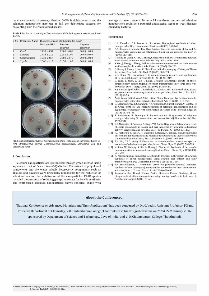
The antibacterial effect of Leucas lavandulifolia leaf aqueous extract mediated used Se nanoparticles were examined by disc diffusion method against to gram positive bacteria like Staphylococcus aureus, Staphylococcus epidermidis and gram negative bacteria like Escherichia coli, Salmonella typhi and sample was taken in three different concentration to display its antibacterial potential.
The SeNPs zones of inhibition against to selected pathogenic strains are shown in Fig. 5. The zone of inhibitions is measured in diameter (mm) for all organisms are shown in Table 1. Leucas lavandulifolia leaf aqueous extract mediated Se nanoparticles is exhibited efficient zone of inhibition against to all organisms. The higher zone of inhibition (15.33 ± 0.57) is observed specifically to E. coil and S. epidremidies than other two organisms. The high antibacterial activity of selenium nanoparticles is due to their extremely large surface area, which provides better contact with microorganisms [18]. Moreover, selenium nanoparticles act as effective antibacterial agent. The antibacterial study exposed the bacterial
F:\SOWMIYA\PH.D\1A.0 1A SOLID 17/06/2015
3851
.47
3795
.15
3729
.24
3668
.01
3420
.91
3250
.47
3025
.75
2660
.59
2361
.97
1654
.41
1549
.43
1512
.81
1430
.62
1352
.08
1224
.69
1078
.44
953.
76
724.
61
598.
30
486.
40
420.
43
500100015002000250030003500
Wavenumber cm-1
2040
6080
100
Tran
smitt
ance
[%]
Page 1/1
226
R. Kirupagaran et al / Journal of Nanoscience and Technology 2(5) (2016) 224–226
Cite this Article as: R. Kirupagaran, A. Saritha, S. Bhuvaneswari, Green synthesis of selenium nanoparticles from leaf and stem extract of Leucas lavandulifolia Sm. and their application, J. Nanosci. Tech. 2(5) (2016) 224–226.
resistance potential of green synthesized SeNPs is highly potential and the selenium nanoparticle may use to kill the deleterious bacteria for preventing from their mediated diseases. Table 1 Antibacterial activity of Leucas lavandulifolia leaf aqueous extract mediated Se-NPs
S. No Organisms Name Diameter of zone of inhibition (in mm)*
MA-L/Se-NPS Positive
control#
Negative
control@
1 E.coil 15.33 ± 0.57 22.00 ± 0.81 00.00 ± 0.00
2 S. aureus 13.33 ± 1.15 22.50 ± 1.00 00.00 ± 0.00
3 S.epidremidies 15.33 ± 0.57 35.00 ± 2.16 00.00 ± 0.00
4 S.typhi 12.66 ± 1.15 31.50 ± 1.00 00.00 ± 0.00
Fig. 5 Antibacterial activity of Leucas lavandulifolia leaf aqueous extract mediated Se-NPs Streptococcus aureus, Staphylococcus epidremidies, Escherichia coli and Salmonella typhi
4. Conclusion
Selenium nanoparticles are synthesized through green method using aqueous extract of Leucas lavandulifolia leaf. The extract of polyphenol components and the water soluble heterocyclic components such as alkaloid and flavones were principally responsible for the reduction of selenium ions and the stabilization of the nanoparticles. FT-IR spectra revealed the presence of reducing groups in extract for Se NPs synthesis. The synthesized selenium nanoparticles shows spherical shape with
average diameter range is 56 nm – 75 nm. Green synthesized selenium nanoparticles could be a potential antibacterial agent to treat diseases caused by bacteria.
References
[1] U.K. Parashar, P.S. Saxena, A. Srivastava, Bioinspired synthesis of silver nanoparticles, Dig. J. Nanomater. Biostruc. 4 (2009) 159-166.
[2] N.A. Begum, S. Mondal, R.A. Basu Laskar, Biogenic synthesis of Au and Ag nanoparticles using aqueous solutions of black tea leaf extracts, Biointer. 71 (2009) 113-118.
[3] J. Zhang, H. Wang, X. Yan, L. Zhang, Comparison of short-term toxicity between Nano-Se and selenite in mice, Life. Sci. 76 (2005) 1099-1109.
[4] X. Gao, J. Zhang, L. Zhang, Hollow sphere selenium nanoparticles: their in-vitro anti hydroxyl radical effect, Adv. Mater. 14 (2002) 290-293.
[5] B. Huang, J. Zhang, J. Hou, C. Chen, Free radical scavenging efficiency of Nano-Se in vitro, Free Radic. Bio. Med. 35 (2003) 805-813.
[6] Y.D. Chiou, Y.J. Hsu, Advances in nanotechnology research and application: 2012 Ed., Appl. Cataly. Environ. B 105 (2011) 211-219.
[7] L. Yang, Y. Shen, A. Xie, J. Liang, Oriented attachment growth of three-dimensionally packed trigonal selenium microspheres into large area wire networks, Euro. J. Inorg. Chem. 28 (2007) 4438-4444.
[8] K.S. Kavitha, Syed Baker, D. Rakshith, H.U. Kavitha. H.C. Yashwantha Rao, Plants as green source towards synthesis of nanoparticles, Inter. Res. J. Bio. Sci. 2 (2013) 66-76.
[9] Amit Kumar Mittal, Yusuf Chisti, Uttam Chand Banerjee, Synthesis of metallic nanoparticles using plant extracts, Biotechnol. Adv. 31 (2013) 346-356.
[10] C.H. Ramamurthy, K.S. Sampath, P. Arunkumar, M. Suresh Kumar, V. Sujatha, et al, Green synthesis and characterization of selenium nanoparticles and its augmented cytotoxicity with doxorubicin on cancer cells, Biosyst. Engg. 36 (2013) 1131-1139.
[11] S. Sasidharan, R. Sowmiya, R. Balakrishnaraja, Biosynthesis of selenium nanoparticles using Citrus reticulata peel extract, World J. Pharm. Res. 4 (2014) 1322-1330.
[12] K.S. Natarajan, S. Samman, S. Singh, Y.K. Gupta, Nagendran Balasundram, et al, Phenolic compounds in plants and agri-industrial by-products: antioxidant activity, occurrence, and potential uses, Food Chem. 99 (2005) 191-203.
[13] P.J. Fesharaki, P. Nazari, M. Shakibaie, S. Rezaie, M. Banoee, et al, Biosynthesis of selenium nanoparticles using Klebsiella pneumoniae and their recovery by a simple sterilization process, Braz. J. Microbio. 41 (2010) 461-466.
[14] Z.H. Lin, C.R.C. Wang, Evidence on the size-dependent absorption spectral evolution of selenium nanoparticles, Mater. Chem. Phys. 92 (2005) 591-594.
[15] Y. Shen, W. Xiufang, A. Xie, L. Huang, J. Zhu, et al, Synthesis of dextran/Se nanocomposites for nanomedicine application, Mater. Chem. Phys. 109 (2008) 534-540.
[16] K. Mallikarjuna, G. Narasimha, G.R. Dillip, B. Praveen, B. Shreedhar, et al, Green synthesis of silver nanoparticles using ocimum leaf extract and their characterization, Dig. J. Nanomat. Biostruc. 6 (2011) 181-186.
[17] S.R. Senthilkumar, T. Sivakumar, Green tea (Camellia sinensis) mediated synthesis of zinc oxide (zno) nanoparticles and studies on their antimicrobial activities, Inter. J. Pharm. Pharm. Sci. 6 (2014) 461-465.
[18] Smaranika Das, Umesh Kumar Parida, Birendra Kumar Bindhani, Green biosynthesis of silver nanoparticles using Moringa oleifera L. leaf, Inter. J. Nanotechnol. Appl. 2 (2013) 51-62.
About the Conference…
“National Conference on Advanced Materials and Their Applications” has been convened by Dr. C. Vedhi, Assistant Professor, PG and
Research Department of Chemistry, V.O.Chidambaram College, Thoothukudi at his designated venue on 21st & 22nd January 2016,
sponsored by Department of Science and Technology, Govt. of India, and V. O. Chidambaram College, Thoothukudi.