Embed Size (px)
Citation preview

World Journal ofClinical Cases
ISSN 2307-8960 (online)
World J Clin Cases 2021 April 6; 9(10): 2160-2418
Published by Baishideng Publishing Group Inc

WJCC https://www.wjgnet.com I April 6, 2021 Volume 9 Issue 10
World Journal of
Clinical CasesW J C CContents Thrice Monthly Volume 9 Number 10 April 6, 2021
MINIREVIEWS
Tertiary peritonitis: A disease that should not be ignored2160
Marques HS, Araújo GRL, da Silva FAF, de Brito BB, Versiani PVD, Caires JS, Milet TC, de Melo FF
SARS-CoV-2, surgeons and surgical masks2170
Khalil MI, Banik GR, Mansoor S, Alqahtani AS, Rashid H
ORIGINAL ARTICLE
Case Control Study
Iguratimod promotes transformation of mononuclear macrophages in elderly patients with rheumatoid arthritis by nuclear factor-κB pathway
2181
Liu S, Song LP, Li RB, Feng LH, Zhu H
Retrospective Study
Factors associated with overall survival in early gastric cancer patients who underwent additional surgery after endoscopic submucosal dissection
2192
Zheng Z, Bu FD, Chen H, Yin J, Xu R, Cai J, Zhang J, Yao HW, Zhang ZT
Epidemiological and clinical characteristics of 65 hospitalized patients with COVID-19 in Liaoning, China2205
Zhang W, Ban Y, Wu YH, Liu JY, Li XH, Wu H, Li H, Chen R, Yu XX, Zheng R
Comprehensive clinicopathologic characteristics of intraabdominal neurogenic tumors: Single institution experience
2218
Simsek C, Uner M, Ozkara F, Akman O, Akyol A, Kav T, Sokmensuer C, Gedikoglu G
Distribution and drug resistance of pathogens in burn patients in China from 2006 to 20192228
Chen H, Yang L, Cheng L, Hu XH, Shen YM
Observational Study
Impact of simethicone on bowel cleansing during colonoscopy in Chinese patients2238
Zhang H, Liu J, Ma SL, Huang ML, Fan Y, Song M, Yang J, Zhang XX, Song QL, Gong J, Huang PX, Zhang H
Prospective Study
Effect of suspension training on neuromuscular function, postural control, and knee kinematics in anterior cruciate ligament reconstruction patients
2247
Huang DD, Chen LH, Yu Z, Chen QJ, Lai JN, Li HH, Liu G
CASE REPORT
Turner syndrome with positive SRY gene and non-classical congenital adrenal hyperplasia: A case report2259
He MN, Zhao SC, Li JM, Tong LL, Fan XZ, Xue YM, Lin XH, Cao Y
WJCC https://www.wjgnet.com II April 6, 2021 Volume 9 Issue 10
World Journal of Clinical CasesContents
Thrice Monthly Volume 9 Number 10 April 6, 2021
Mechanical thrombectomy for acute occlusion of the posterior inferior cerebellar artery: A case report2268
Zhang HB, Wang P, Wang Y, Wang JH, Li Z, Li R
Bilateral retrocorneal hyaline scrolls secondary to asymptomatic congenital syphilis: A case report2274
Jin YQ, Hu YP, Dai Q, Wu SQ
Recurrent undifferentiated embryonal sarcoma of the liver in adult patient treated by pembrolizumab: A case report
2281
Yu XH, Huang J, Ge NJ, Yang YF, Zhao JY
Adult onset type 2 familial hemophagocytic lymphohistiocytosis with PRF1 c.65delC/c.163C>T compound heterozygous mutations: A case report
2289
Liu XY, Nie YB, Chen XJ, Gao XH, Zhai LJ, Min FL
Salvage of vascular graft infections via vacuum sealing drainage and rectus femoris muscle flap transposition: A case report
2296
Zhang P, Tao FL, Li QH, Zhou DS, Liu FX
Innovative chest wall reconstruction with a locking plate and cement spacer after radical resection of chondrosarcoma in the sternum: A case report
2302
Lin CW, Ho TY, Yeh CW, Chen HT, Chiang IP, Fong YC
Changes in sleep parameters following biomimetic oral appliance therapy: A case report2312
Singh GD, Kherani S
Bone remodeling in sigmoid sinus diverticulum after stenting for transverse sinus stenosis in pulsatile tinnitus: A case report
2320
Qiu XY, Zhao PF, Ding HY, Li XS, Lv H, Yang ZH, Gong SS, Jin L, Wang ZC
Prolonged use of bedaquiline in two patients with pulmonary extensively drug-resistant tuberculosis: Two case reports
2326
Gao JT, Xie L, Ma LP, Shu W, Zhang LJ, Ning YJ, Xie SH, Liu YH, Gao MQ
Low-grade mucinous appendiceal neoplasm mimicking an ovarian lesion: A case report and review of literature
2334
Borges AL, Reis-de-Carvalho C, Chorão M, Pereira H, Djokovic D
Granulomatosis with polyangiitis presenting as high fever with diffuse alveolar hemorrhage and otitis media: A case report
2344
Li XJ, Yang L, Yan XF, Zhan CT, Liu JH
Primary intramedullary melanoma of lumbar spinal cord: A case report2352
Sun LD, Chu X, Xu L, Fan XZ, Qian Y, Zuo DM
Proliferative glomerulonephritis with monoclonal immunoglobulin G deposits in a young woman: A case report
2357
Xu ZG, Li WL, Wang X, Zhang SY, Zhang YW, Wei X, Li CD, Zeng P, Luan SD

WJCC https://www.wjgnet.com III April 6, 2021 Volume 9 Issue 10
World Journal of Clinical CasesContents
Thrice Monthly Volume 9 Number 10 April 6, 2021
Nocardia cyriacigeorgica infection in a patient with pulmonary sequestration: A case report2367
Lin J, Wu XM, Peng MF
Long-term control of melanoma brain metastases with co-occurring intracranial infection and involuntary drug reduction during COVID-19 pandemic: A case report
2373
Wang Y, Lian B, Cui CL
Solitary bone plasmacytoma of the upper cervical spine: A case report 2380
Li RJ, Li XF, Jiang WM
Two-stage transcrestal sinus floor elevation-insight into replantation: Six case reports2386
Lin ZZ, Xu DQ, Ye ZY, Wang GG, Ding X
Programmed cell death protein-1 inhibitor combined with chimeric antigen receptor T cells in the treatment of relapsed refractory non-Hodgkin lymphoma: A case report
2394
Niu ZY, Sun L, Wen SP, Song ZR, Xing L, Wang Y, Li JQ, Zhang XJ, Wang FX
Pancreatic cancer secondary to intraductal papillary mucinous neoplasm with collision between gastric cancer and B-cell lymphoma: A case report
2400
Ma YH, Yamaguchi T, Yasumura T, Kuno T, Kobayashi S, Yoshida T, Ishida T, Ishida Y, Takaoka S, Fan JL, Enomoto N
Acquired haemophilia in patients with malignant disease: A case report2409
Krašek V, Kotnik A, Zavrtanik H, Klen J, Zver S
WJCC https://www.wjgnet.com IX April 6, 2021 Volume 9 Issue 10
World Journal of Clinical CasesContents
Thrice Monthly Volume 9 Number 10 April 6, 2021
ABOUT COVER
Editorial Board Member of World Journal of Clinical Cases, Deb Sanjay Nag, Senior Consultant, Department of Anaesthesiology, Tata Main Hospital, C-Road (West), Bistupur, Jamshedpur 831 001, India. [email protected]
AIMS AND SCOPE
The primary aim of World Journal of Clinical Cases (WJCC, World J Clin Cases) is to provide scholars and readers from various fields of clinical medicine with a platform to publish high-quality clinical research articles and communicate their research findings online. WJCC mainly publishes articles reporting research results and findings obtained in the field of clinical medicine and covering a wide range of topics, including case control studies, retrospective cohort studies, retrospective studies, clinical trials studies, observational studies, prospective studies, randomized controlled trials, randomized clinical trials, systematic reviews, meta-analysis, and case reports.
INDEXING/ABSTRACTING
The WJCC is now indexed in Science Citation Index Expanded (also known as SciSearch®), Journal Citation Reports/Science Edition, Scopus, PubMed, and PubMed Central. The 2020 Edition of Journal Citation Reports® cites the 2019 impact factor (IF) for WJCC as 1.013; IF without journal self cites: 0.991; Ranking: 120 among 165 journals in medicine, general and internal; and Quartile category: Q3. The WJCC's CiteScore for 2019 is 0.3 and Scopus CiteScore rank 2019: General Medicine is 394/529.
RESPONSIBLE EDITORS FOR THIS ISSUE
Production Editor: Yan-Xia Xing; Production Department Director: Yun-Xiaojian Wu; Editorial Office Director: Jin-Lei Wang.
NAME OF JOURNAL INSTRUCTIONS TO AUTHORS
World Journal of Clinical Cases https://www.wjgnet.com/bpg/gerinfo/204
ISSN GUIDELINES FOR ETHICS DOCUMENTS
ISSN 2307-8960 (online) https://www.wjgnet.com/bpg/GerInfo/287
LAUNCH DATE GUIDELINES FOR NON-NATIVE SPEAKERS OF ENGLISH
April 16, 2013 https://www.wjgnet.com/bpg/gerinfo/240
FREQUENCY PUBLICATION ETHICS
Thrice Monthly https://www.wjgnet.com/bpg/GerInfo/288
EDITORS-IN-CHIEF PUBLICATION MISCONDUCT
Dennis A Bloomfield, Sandro Vento, Bao-Gan Peng https://www.wjgnet.com/bpg/gerinfo/208
EDITORIAL BOARD MEMBERS ARTICLE PROCESSING CHARGE
https://www.wjgnet.com/2307-8960/editorialboard.htm https://www.wjgnet.com/bpg/gerinfo/242
PUBLICATION DATE STEPS FOR SUBMITTING MANUSCRIPTS
April 6, 2021 https://www.wjgnet.com/bpg/GerInfo/239
COPYRIGHT ONLINE SUBMISSION
© 2021 Baishideng Publishing Group Inc https://www.f6publishing.com
© 2021 Baishideng Publishing Group Inc. All rights reserved. 7041 Koll Center Parkway, Suite 160, Pleasanton, CA 94566, USA
E-mail: [email protected] https://www.wjgnet.com
WJCC https://www.wjgnet.com 2409 April 6, 2021 Volume 9 Issue 10
World Journal of
Clinical CasesW J C CSubmit a Manuscript: https://www.f6publishing.com World J Clin Cases 2021 April 6; 9(10): 2409-2418
DOI: 10.12998/wjcc.v9.i10.2409 ISSN 2307-8960 (online)
CASE REPORT
Acquired haemophilia in patients with malignant disease: A case report
Veronika Krašek, Aleša Kotnik, Hana Zavrtanik, Jasna Klen, Samo Zver
ORCID number: Veronika Krašek 0000-0002-6126-6625; Aleša Kotnik 0000-0001-7857-8310; Hana Zavrtanik 0000-0003-0469-3978; Jasna Klen 0000-0002-8807-7242; Samo Zver 0000-0003-2932-7923.
Author contributions: Klen J has conceived and designed the manuscript; Krašek V, Kotnik A and Zavrtanik H contributed to the acquisition, analysis and interpretation of the data; all authors participated in drafting the manuscript; Klen J and Zver S critically revised the manuscript; All authors read and approved the final version of the manuscript.
Informed consent statement: Written informed consent was obtained from the patient before the surgery.
Conflict-of-interest statement: The authors declare that they have no conflicts of interest with regard to this case report.
CARE Checklist (2016) statement: The authors have read the CARE Checklist (2016), and the manuscript was prepared and revised according to the CARE Checklist (2016).
Open-Access: This article is an open-access article that was selected by an in-house editor and fully peer-reviewed by external
Veronika Krašek, Aleša Kotnik, Hana Zavrtanik, Jasna Klen, Samo Zver, Medical Faculty, University of Ljubljana, Ljubljana 1000, Slovenia
Hana Zavrtanik, Jasna Klen, Department of Abdominal Surgery, University Medical Centre Ljubljana, Ljubljana 1000, Slovenia
Samo Zver, Department of Hematology, University Medical Centre Ljubljana, Ljubljana 1000, Slovenia
Corresponding author: Hana Zavrtanik, MD, Doctor, Department of Abdominal Surgery, University Medical Centre Ljubljana, Zaloška cesta 7, Ljubljana 1000, Slovenia. [email protected]
AbstractBACKGROUND Acquired haemophilia is a rare coagulation disorder characterized by autoantibodies against coagulation factor VIII leading to severe and potentially life-threatening haemorrhages. The underlying disorder causing the development of an autoimmune phenomenon is not always known, but 10%-15% could be linked to malignancies. Patients with cancer who require surgical resection represent a treatment challenge not solely due to increased risk of bleeding but also due to adverse events of immunosuppressive therapy.
CASE SUMMARY We present the case of a 67-year-old man with non-metastatic adenocarcinoma of the distal bile duct who developed concomitant acquired haemophilia a month after having been diagnosed with malignant disease. Haemostasis was established with recombinant activated factor VII, and immunosuppressive therapy was started immediately. An extensive surgical procedure was performed in order to remove the cancer and, therefore, eliminate the inhibitory autoantibodies. Due to a complicated postoperative course, relatively short period of treatment and likelihood of micrometastases, no improvement in the patient’s status was observed. Diagnosis and treatment of acquired haemophilia as well as other coagulation disorders in patients with cancer are discussed.
CONCLUSION Prompt diagnosis of acquired haemophilia is required in order to start appropriate treatment and reduce mortality. Among patients with cancer, other causes of abnormal bleeding related to malignancy should be considered.
Krašek V et al. Acquired haemophilia in malignancy
WJCC https://www.wjgnet.com 2410 April 6, 2021 Volume 9 Issue 10
reviewers. It is distributed in accordance with the Creative Commons Attribution NonCommercial (CC BY-NC 4.0) license, which permits others to distribute, remix, adapt, build upon this work non-commercially, and license their derivative works on different terms, provided the original work is properly cited and the use is non-commercial. See: http://creativecommons.org/Licenses/by-nc/4.0/
Manuscript source: Unsolicited manuscript
Specialty type: Medicine, research and experimental
Country/Territory of origin: Slovenia
Peer-review report’s scientific quality classificationGrade A (Excellent): 0 Grade B (Very good): 0 Grade C (Good): C, C Grade D (Fair): 0 Grade E (Poor): 0
Received: December 21, 2020 Peer-review started: December 21, 2020 First decision: December 30, 2020 Revised: January 4, 2021 Accepted: January 26, 2021 Article in press: January 26, 2021 Published online: April 6, 2021
P-Reviewer: Fu TL, Hamada Y S-Editor: Gao CC L-Editor: Filipodia P-Editor: Xing YX
Key Words: Malignancy; Haemostasis; Bleeding diathesis; Eradication therapy; Case report
©The Author(s) 2021. Published by Baishideng Publishing Group Inc. All rights reserved.
Core Tip: Acquired haemophilia is a rare coagulation disorder characterized by autoantibodies against coagulation factor VIII. Immediate consultation with a reference haemophilia centre should be made for obtaining early diagnosis and treatment. In patients with cancer, a wide range of haemostatic disorders should be taken into consideration and distinguished from acquired haemophilia. Although invasive procedures should be avoided in patients with acquired haemophilia, surgical resection of the underlying malignancy is sometimes required. This subset of patients represents a treatment challenge not solely due to increased risk of bleeding but also due to adverse events of immunosuppressive therapy.
Citation: Krašek V, Kotnik A, Zavrtanik H, Klen J, Zver S. Acquired haemophilia in patients with malignant disease: A case report. World J Clin Cases 2021; 9(10): 2409-2418URL: https://www.wjgnet.com/2307-8960/full/v9/i10/2409.htmDOI: https://dx.doi.org/10.12998/wjcc.v9.i10.2409
INTRODUCTIONAcquired haemophilia (AH) is a coagulation disorder caused by neutralizing autoantibodies against coagulation factor (F) VIII leading to severe and potentially life-threatening haemorrhages. It occurs at a rate of approximately 1.5/million/year[1] and is predominantly seen in elderly patients with reported median age at diagnosis being 68-78 years[2-6]. In contrast to congenital haemophilia, it affects both sexes equally and has a distinct pattern of bleeding that is unusual and unlike anything that has been seen before. The mortality rate is high, reaching above 20%, making early diagnosis and treatment of vital importance[3,4]. The underlying cause is unknown in about 50% of cases, while the remaining cases may be associated with malignancies, autoimmune diseases, pregnancy, certain medications or infections[3]. Solid cancers most commonly linked to AH are prostate, lung and colon neoplasms, but it can also be seen in lymphoproliferative malignancies[2]. Herein, AH should be distinguished from other causes of abnormal bleeding related to cancer in order to start appropriate treatment and reduce mortality[7]. Without effective treatment of the underlying malignancy, complete elimination of inhibitory autoantibodies is unlikely. Perioperative management of patients with AH and cancer who require surgical resection is challenging.
We present the case of a 67-year-old man with AH due to non-metastatic adenocarcinoma of the distal bile duct and discuss a diagnostic and therapeutic approach to a cancer patient with bleeding diathesis.
CASE PRESENTATIONChief complaintsA 67-year-old Caucasian male patient presented to the Emergency Department with fever, pain, swelling and bruising of his left thigh. He denied trauma to the leg.
History of present illnessA month before presentation, he was admitted for evaluation of painless jaundice. At the time of admission, his laboratory results showed acute kidney failure (serum creatinine 446 µmol/L, urea 10.7 mmol/L), normocytic anaemia (haemoglobin 90 g/L, mean corpuscular volume 86 fL), elevated liver enzymes [aspartate aminotransferase (AST) 2.6 µkat/L, alanine aminotransferase (ALT) 3.6 µkat/L, gamma-glutamyl transferase (GGT) 12.1 µkat/L, alkaline phosphatase (AP) 12.1 µkat/L] and elevated bilirubin levels (186/145 µmol/L), indicating cholestasis. Malignant stenosis of the

Krašek V et al. Acquired haemophilia in malignancy
WJCC https://www.wjgnet.com 2411 April 6, 2021 Volume 9 Issue 10
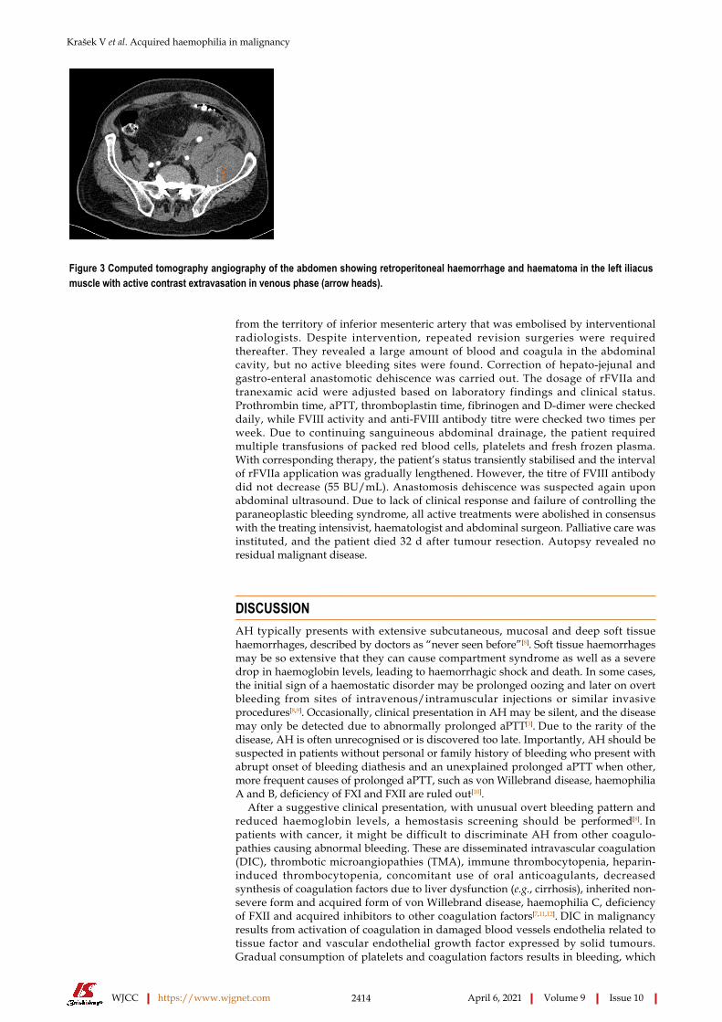
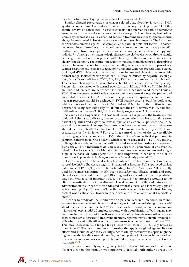
distal common bile duct was discovered upon abdominal computed tomography (CT) scan with no evidence of distant metastases (Figure 1). He underwent endoscopic retrograde cholangiopancreatography with stent placement in order to overcome the stenosis (Figure 2). Kidney function tests (serum creatinine 369 µmol/L), liver enzymes (AST 1.9 µkat/L, ALT 3.67 µkat/L, GGT 3.87 µkat/L, AP 9.86 µkat/L) and bilirubin levels (149/124 µmol/L) improved, however, levels of haemoglobin remained low (86 g/L). His case was discussed at a multidisciplinary cancer team meeting and surgical treatment was proposed.
History of past illnessHis background medical history was remarkable for arterial hypertension, hypercholesterolemia and stage 3 chronic kidney disease.
Physical examinationUpon admission the patient was alert, oriented and cooperative. He was febrile with body temperature of 38.1 °C, blood pressure of 135/72 mmHg, heart rate of 71/min and oxygen saturation of 100%. Physical examination revealed swelling and large suffusion on his left thigh.
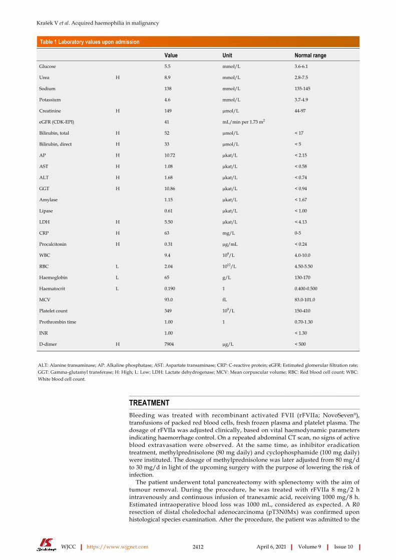
Laboratory examinationsLaboratory results showed severe normocytic anaemia (haemoglobin 65 g/L, mean corpuscular volume 93.0 fL), elevated liver enzymes (AST 1.08 µkat/L, ALT 1.68 µkat/L, GGT 10.86 µkat/L, AP 10.72 µkat/L) and bilirubin levels (52/33 µmol/L) and moderately raised inflammatory parameters (C-reactive protein 63 mg/L, white blood cell 9.4 × 10⁹/L, procalcitonin 0.31 ng/mL). Coagulation tests showed normal prothrombin time/international normalized ratio (1.03). Laboratory values upon admission are summarized in Table 1.
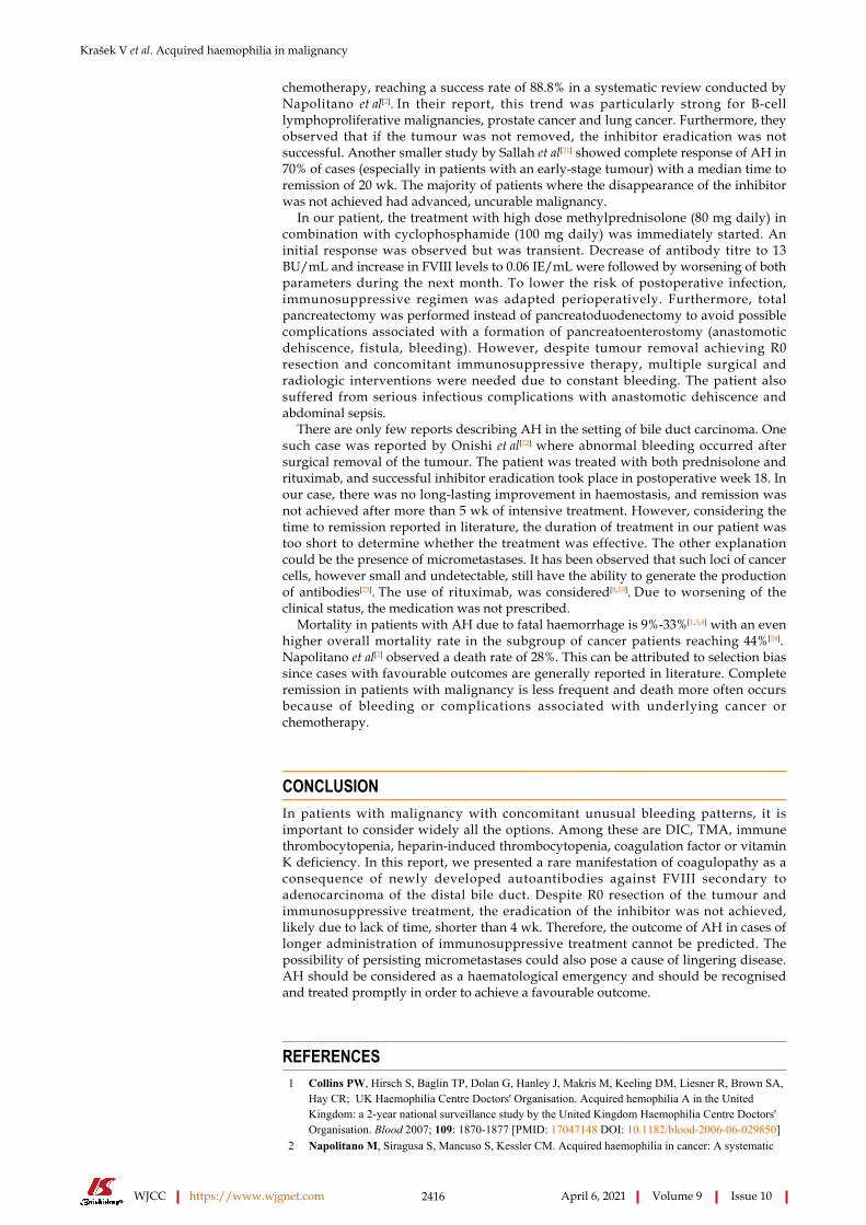
Imaging examinationsDeep vein thrombosis was excluded with a venous point-of-care ultrasound. Ultrasound of the soft tissues of the left thigh revealed a large hematoma (13 cm × 5 cm × 3 cm) in one of the muscles of the anterior/extensor compartment. Later on, when no significant improvement in haemoglobin level (73 g/L) was observed despite treatment with multiple erythrocyte transfusions, a CT angiography of the abdomen and lower limbs was performed. Here, hematoperitoneum with a large haematoma in the left iliacus muscle (5 cm × 9 cm × 29 cm) and left femoral rectus muscle (7 cm × 5 cm × 30 cm; previously described) was discovered, both with multiple active bleeding sites (Figure 3).
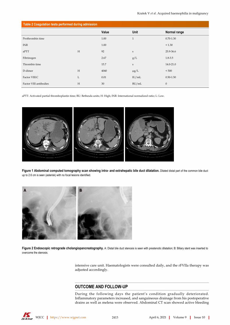
Further work-upAfter presentation, the patient received three units of packed red blood cells and was admitted for further treatment and evaluation. Antibiotic treatment due to cholangitis, confirmed upon abdominal ultrasound, was initiated and another endoscopic retrograde cholangiopancreatography was performed to replace the poorly functioning choledochal stent. A haematologist was consulted and ordered additional coagulation tests that revealed elevated activated partial thromboplastin time (aPTT; 92 s), normal prothrombin time (1.00) and international normalized ratio (1.00), solely reduced FVIII activity (1%) and appearance of anti-FVIII antibodies, with titre of 30 Bethesda units (BU)/mL (Table 2). Von Willebrand factor (vWF) antigen, vWF activity and vWF ristocetin cofactor activity were normal.
FINAL DIAGNOSISDue to reduced FVIII activity (1%) and the appearance of anti-FVIII antibodies, with titre of 30 BU/mL, AH was diagnosed, likely secondary to bile duct cancer. Other coagulation disorders were excluded as specific bleeding-related clinical picture with prolonged aPTT, in combination with low FVIII activity and the appearance of anti-FVIII antibodies, is a specific finding in AH.
Krašek V et al. Acquired haemophilia in malignancy
WJCC https://www.wjgnet.com 2412 April 6, 2021 Volume 9 Issue 10
Table 1 Laboratory values upon admission
Value Unit Normal range
Glucose 5.5 mmol/L 3.6-6.1
Urea H 8.9 mmol/L 2.8-7.5
Sodium 138 mmol/L 135-145
Potassium 4.6 mmol/L 3.7-4.9
Creatinine H 149 µmol/L 44-97
eGFR (CDK-EPI) 41 mL/min per 1.73 m2
Bilirubin, total H 52 µmol/L < 17
Bilirubin, direct H 33 µmol/L < 5
AP H 10.72 µkat/L < 2.15
AST H 1.08 µkat/L < 0.58
ALT H 1.68 µkat/L < 0.74
GGT H 10.86 µkat/L < 0.94
Amylase 1.15 µkat/L < 1.67
Lipase 0.61 µkat/L < 1.00
LDH H 5.50 µkat/L < 4.13
CRP H 63 mg/L 0-5
Procalcitonin H 0.31 µg/mL < 0.24
WBC 9.4 109/L 4.0-10.0
RBC L 2.04 1012/L 4.50-5.50
Haemoglobin L 65 g/L 130-170
Haematocrit L 0.190 1 0.400-0.500
MCV 93.0 fL 83.0-101.0
Platelet count 349 109/L 150-410
Prothrombin time 1.00 1 0.70-1.30
INR 1.00 < 1.30
D-dimer H 7904 µg/L < 500
ALT: Alanine transaminase; AP: Alkaline phosphatase; AST: Aspartate transaminase; CRP: C-reactive protein; eGFR: Estimated glomerular filtration rate; GGT: Gamma-glutamyl transferase; H: High; L: Low; LDH: Lactate dehydrogenase; MCV: Mean corpuscular volume; RBC: Red blood cell count; WBC: White blood cell count.
TREATMENTBleeding was treated with recombinant activated FVII (rFVIIa; NovoSeven®), transfusions of packed red blood cells, fresh frozen plasma and platelet plasma. The dosage of rFVIIa was adjusted clinically, based on vital haemodynamic parameters indicating haemorrhage control. On a repeated abdominal CT scan, no signs of active blood extravasation were observed. At the same time, as inhibitor eradication treatment, methylprednisolone (80 mg daily) and cyclophosphamide (100 mg daily) were instituted. The dosage of methylprednisolone was later adjusted from 80 mg/d to 30 mg/d in light of the upcoming surgery with the purpose of lowering the risk of infection.
The patient underwent total pancreatectomy with splenectomy with the aim of tumour removal. During the procedure, he was treated with rFVIIa 8 mg/2 h intravenously and continuous infusion of tranexamic acid, receiving 1000 mg/8 h. Estimated intraoperative blood loss was 1000 mL, considered as expected. A R0 resection of distal choledochal adenocarcinoma (pT3N0Mx) was confirmed upon histological species examination. After the procedure, the patient was admitted to the
Krašek V et al. Acquired haemophilia in malignancy
WJCC https://www.wjgnet.com 2413 April 6, 2021 Volume 9 Issue 10
Table 2 Coagulation tests performed during admission
Value Unit Normal range
Prothrombin time 1.00 1 0.70-1.30
INR 1.00 < 1.30
aPTT H 92 s 25.9-36.6
Fibrinogen 2.67 g/L 1.8-3.5
Thrombin time 15.7 s 14.0-21.0
D-dimer H 4040 µg/L < 500
Factor VIII:C L 0.01 IU/mL 0.50-1.50
Factor VIII antibodies H 30 BU/mL 0
aPTT: Activated partial thromboplastin time; BU: Bethesda units; H: High; INR: International normalized ratio; L: Low.
Figure 1 Abdominal computed tomography scan showing intra- and extrahepatic bile duct dilatation. Dilated distal part of the common bile duct up to 2.6 cm is seen (asterisk) with no focal lesions identified.
Figure 2 Endoscopic retrograde cholangiopancreatography. A: Distal bile duct stenosis is seen with prestenotic dilatation; B: Biliary stent was inserted to overcome the stenosis.
intensive care unit. Haematologists were consulted daily, and the rFVIIa therapy was adjusted accordingly.
OUTCOME AND FOLLOW-UPDuring the following days the patient’s condition gradually deteriorated. Inflammatory parameters increased, and sanguineous drainage from his postoperative drains as well as melena were observed. Abdominal CT scan showed active bleeding
Krašek V et al. Acquired haemophilia in malignancy
WJCC https://www.wjgnet.com 2414 April 6, 2021 Volume 9 Issue 10
Figure 3 Computed tomography angiography of the abdomen showing retroperitoneal haemorrhage and haematoma in the left iliacus muscle with active contrast extravasation in venous phase (arrow heads).
from the territory of inferior mesenteric artery that was embolised by interventional radiologists. Despite intervention, repeated revision surgeries were required thereafter. They revealed a large amount of blood and coagula in the abdominal cavity, but no active bleeding sites were found. Correction of hepato-jejunal and gastro-enteral anastomotic dehiscence was carried out. The dosage of rFVIIa and tranexamic acid were adjusted based on laboratory findings and clinical status. Prothrombin time, aPTT, thromboplastin time, fibrinogen and D-dimer were checked daily, while FVIII activity and anti-FVIII antibody titre were checked two times per week. Due to continuing sanguineous abdominal drainage, the patient required multiple transfusions of packed red blood cells, platelets and fresh frozen plasma. With corresponding therapy, the patient’s status transiently stabilised and the interval of rFVIIa application was gradually lengthened. However, the titre of FVIII antibody did not decrease (55 BU/mL). Anastomosis dehiscence was suspected again upon abdominal ultrasound. Due to lack of clinical response and failure of controlling the paraneoplastic bleeding syndrome, all active treatments were abolished in consensus with the treating intensivist, haematologist and abdominal surgeon. Palliative care was instituted, and the patient died 32 d after tumour resection. Autopsy revealed no residual malignant disease.
DISCUSSIONAH typically presents with extensive subcutaneous, mucosal and deep soft tissue haemorrhages, described by doctors as “never seen before”[8]. Soft tissue haemorrhages may be so extensive that they can cause compartment syndrome as well as a severe drop in haemoglobin levels, leading to haemorrhagic shock and death. In some cases, the initial sign of a haemostatic disorder may be prolonged oozing and later on overt bleeding from sites of intravenous/intramuscular injections or similar invasive procedures[8,9]. Occasionally, clinical presentation in AH may be silent, and the disease may only be detected due to abnormally prolonged aPTT[3]. Due to the rarity of the disease, AH is often unrecognised or is discovered too late. Importantly, AH should be suspected in patients without personal or family history of bleeding who present with abrupt onset of bleeding diathesis and an unexplained prolonged aPTT when other, more frequent causes of prolonged aPTT, such as von Willebrand disease, haemophilia A and B, deficiency of FXI and FXII are ruled out[10].
After a suggestive clinical presentation, with unusual overt bleeding pattern and reduced haemoglobin levels, a hemostasis screening should be performed[8]. In patients with cancer, it might be difficult to discriminate AH from other coagulo-pathies causing abnormal bleeding. These are disseminated intravascular coagulation (DIC), thrombotic microangiopathies (TMA), immune thrombocytopenia, heparin-induced thrombocytopenia, concomitant use of oral anticoagulants, decreased synthesis of coagulation factors due to liver dysfunction (e.g., cirrhosis), inherited non-severe form and acquired form of von Willebrand disease, haemophilia C, deficiency of FXII and acquired inhibitors to other coagulation factors[7,11,12]. DIC in malignancy results from activation of coagulation in damaged blood vessels endothelia related to tissue factor and vascular endothelial growth factor expressed by solid tumours. Gradual consumption of platelets and coagulation factors results in bleeding, which
Krašek V et al. Acquired haemophilia in malignancy
WJCC https://www.wjgnet.com 2415 April 6, 2021 Volume 9 Issue 10
may be the first clinical symptom indicating the presence of DIC[11,12].Similar clinical presentation of cancer-related coagulopathy is seen in TMA
syndrome in the form of secondary thrombotic thrombocytopenic purpura. The latter should always be considered in case of concomitant microangiopathic haemolytic anaemia and thrombocytopenia. As an entity among TMA syndromes, haemolytic uremic syndrome is rare in advanced cancer[12]. Immune thrombocytopenia should always be considered in isolated and cancer-related thrombocytopenia. The formation of antibodies directed against the complex of heparin and platelet factor 4 is named heparin-induced thrombocytopenia and may occur more often in cancer patients[7]. Furthermore, thrombocytopenia may also be a consequence of chemotherapy and radiation[13]. Among other haematologic diseases, myelodysplastic syndrome should be recognized, as it also can present with bleeding diathesis and is common in the elderly population[14]. The clinical presentation ranging from bleedings to thrombosis can also be seen in acute traumatic coagulopathy, where a sterile injury provokes a cellular response and changes coagulation[15]. Patients with AH present with isolated prolonged aPTT, while prothrombin time, thrombin time and platelet count are within normal range. Isolated prolongation of aPTT may be caused by heparin use, single coagulation factor deficiency (FVIII, FIX, FXI, FXII) or the presence of an inhibitor[8,9]. True factor deficiency as in haemophilia A should be ruled out with a mixing study. Patient plasma is mixed with normal pool plasma in 1:1 ratio[16]. Since FVIII inhibitors are time- and temperature-dependent, the mixture is then incubated for two hours at 37 °C. If after incubation aPTT fails to correct within the normal range, the presence of an inhibitor is suspected. At this point, the presence of lupus anticoagulant and heparin presence should be excluded[8,9]. FVIII activity assay should be performed which shows reduced activity of FVIII below 50%. The inhibitor titre is then determined using Bethesda assay[10,17]. In our case the FVIII activity was 1% and anti-FVIII antibodies titre was 30 BU/mL, both clearly suggestive of AH diagnosis.
As soon as the diagnosis of AH was established in our patient, the treatment was initiated. Being a rare disease, current recommendations are based on data from patient registries and expert consensus opinions[3]. Generally, patients should be treated at a reference haemophilia centre or at least consultation with such centre should be established[8]. The treatment of AH consists of bleeding control and eradication of the inhibitor[10]. For bleeding control, either of the two available bypassing agents is recommended, rFVIIa (Novo Seven®) and activated prothrombin complex concentrate (aPCC; FEIBA®), which contains activated FII, FVII, FIX and FX. Both agents are safe and effective with reported rates of haemostasis achievement being above 90%[3]. Insufficient data exist to support the preference of one over the other[4,18]. The lack of adequate laboratory test for measuring over- and underdosing is a major setback for both agents[10]. It is also important to consider the risk of thrombogenic potential in both agents, especially in elderly patients[4,8,18].
rFVIIa is reported to be relatively safe combined with tranexamic acid in case of severe bleeding[18]. The dosage regimen is standard as in other rFVIIa approved clinical indications, 90-120 μg/kg/2-3 h until the bleeding stops[8]. In Slovenia, rFVIIa is mostly used for haemostasis control in AH due to the safety and efficacy profile and good clinical experience with the drug[19]. Bleeding and its severity cannot be predicted based on FVIII level or inhibitor titre, so the treatment is directed according to the clinical manifestation of the disease[4]. The dosages of rFVIIa and intervals of administration in our patient were adjusted towards clinical and laboratory signs of active bleeding (90 µg/kg every 2-3 h with the extension of the interval when bleeding control was established). Tranexamic acid was added as an additional haemostatic agent[10].
In order to eradicate the inhibitors and prevent recurrent bleeding, immuno-suppressive therapy should be initiated at diagnosis and the underlying cause of AH should be identified and treated[2,5,8]. Corticosteroids are used, often in combination with cyclophosphamide[5]. Complete response with a combination of both agents may be more frequent than with corticosteroids alone[5], although some other authors showed no such difference[1,4]. In current literature, reported remission rates were 61%-72% when treated with either of the two regimens in a median time of 5-6 wk[4,5,10,20]. This may, however, take longer for patients with lower FVIII activity levels at presentation[20]. The use of immunosuppressive therapy is weighted against its side effects and should be applied carefully since mortality secondary to sepsis might be higher than the bleeding-related mortality in those patients[4]. Rituximab can be added to corticosteroids and/or cyclophosphamide if no response is seen after 2-3 wk of treatment[8,10,20].
In patients with underlying malignancy, higher rates in inhibitor eradication were observed when the tumour was effectively treated with either surgery or
Krašek V et al. Acquired haemophilia in malignancy
WJCC https://www.wjgnet.com 2416 April 6, 2021 Volume 9 Issue 10
chemotherapy, reaching a success rate of 88.8% in a systematic review conducted by Napolitano et al[2]. In their report, this trend was particularly strong for B-cell lymphoproliferative malignancies, prostate cancer and lung cancer. Furthermore, they observed that if the tumour was not removed, the inhibitor eradication was not successful. Another smaller study by Sallah et al[21] showed complete response of AH in 70% of cases (especially in patients with an early-stage tumour) with a median time to remission of 20 wk. The majority of patients where the disappearance of the inhibitor was not achieved had advanced, uncurable malignancy.
In our patient, the treatment with high dose methylprednisolone (80 mg daily) in combination with cyclophosphamide (100 mg daily) was immediately started. An initial response was observed but was transient. Decrease of antibody titre to 13 BU/mL and increase in FVIII levels to 0.06 IE/mL were followed by worsening of both parameters during the next month. To lower the risk of postoperative infection, immunosuppressive regimen was adapted perioperatively. Furthermore, total pancreatectomy was performed instead of pancreatoduodenectomy to avoid possible complications associated with a formation of pancreatoenterostomy (anastomotic dehiscence, fistula, bleeding). However, despite tumour removal achieving R0 resection and concomitant immunosuppressive therapy, multiple surgical and radiologic interventions were needed due to constant bleeding. The patient also suffered from serious infectious complications with anastomotic dehiscence and abdominal sepsis.
There are only few reports describing AH in the setting of bile duct carcinoma. One such case was reported by Onishi et al[22] where abnormal bleeding occurred after surgical removal of the tumour. The patient was treated with both prednisolone and rituximab, and successful inhibitor eradication took place in postoperative week 18. In our case, there was no long-lasting improvement in haemostasis, and remission was not achieved after more than 5 wk of intensive treatment. However, considering the time to remission reported in literature, the duration of treatment in our patient was too short to determine whether the treatment was effective. The other explanation could be the presence of micrometastases. It has been observed that such loci of cancer cells, however small and undetectable, still have the ability to generate the production of antibodies[23]. The use of rituximab, was considered[8,10]. Due to worsening of the clinical status, the medication was not prescribed.
Mortality in patients with AH due to fatal haemorrhage is 9%-33%[1,3,4] with an even higher overall mortality rate in the subgroup of cancer patients reaching 44%[24]. Napolitano et al[2] observed a death rate of 28%. This can be attributed to selection bias since cases with favourable outcomes are generally reported in literature. Complete remission in patients with malignancy is less frequent and death more often occurs because of bleeding or complications associated with underlying cancer or chemotherapy.
CONCLUSIONIn patients with malignancy with concomitant unusual bleeding patterns, it is important to consider widely all the options. Among these are DIC, TMA, immune thrombocytopenia, heparin-induced thrombocytopenia, coagulation factor or vitamin K deficiency. In this report, we presented a rare manifestation of coagulopathy as a consequence of newly developed autoantibodies against FVIII secondary to adenocarcinoma of the distal bile duct. Despite R0 resection of the tumour and immunosuppressive treatment, the eradication of the inhibitor was not achieved, likely due to lack of time, shorter than 4 wk. Therefore, the outcome of AH in cases of longer administration of immunosuppressive treatment cannot be predicted. The possibility of persisting micrometastases could also pose a cause of lingering disease. AH should be considered as a haematological emergency and should be recognised and treated promptly in order to achieve a favourable outcome.
REFERENCESCollins PW, Hirsch S, Baglin TP, Dolan G, Hanley J, Makris M, Keeling DM, Liesner R, Brown SA, Hay CR; UK Haemophilia Centre Doctors' Organisation. Acquired hemophilia A in the United Kingdom: a 2-year national surveillance study by the United Kingdom Haemophilia Centre Doctors' Organisation. Blood 2007; 109: 1870-1877 [PMID: 17047148 DOI: 10.1182/blood-2006-06-029850]
1
Napolitano M, Siragusa S, Mancuso S, Kessler CM. Acquired haemophilia in cancer: A systematic 2
Krašek V et al. Acquired haemophilia in malignancy
WJCC https://www.wjgnet.com 2417 April 6, 2021 Volume 9 Issue 10
and critical literature review. Haemophilia 2018; 24: 43-56 [PMID: 28960809 DOI: 10.1111/hae.13355]Knoebl P, Marco P, Baudo F, Collins P, Huth-Kühne A, Nemes L, Pellegrini F, Tengborn L, Lévesque H; EACH2 Registry Contributors. Demographic and clinical data in acquired hemophilia A: results from the European Acquired Haemophilia Registry (EACH2). J Thromb Haemost 2012; 10: 622-631 [PMID: 22321904 DOI: 10.1111/j.1538-7836.2012.04654.x]
3
Borg JY, Guillet B, Le Cam-Duchez V, Goudemand J, Lévesque H; SACHA Study Group. Outcome of acquired haemophilia in France: the prospective SACHA (Surveillance des Auto antiCorps au cours de l'Hémophilie Acquise) registry. Haemophilia 2013; 19: 564-570 [PMID: 23574453 DOI: 10.1111/hae.12138]
4
Collins P, Baudo F, Knoebl P, Lévesque H, Nemes L, Pellegrini F, Marco P, Tengborn L, Huth-Kühne A; EACH2 registry collaborators. Immunosuppression for acquired hemophilia A: results from the European Acquired Haemophilia Registry (EACH2). Blood 2012; 120: 47-55 [PMID: 22517903 DOI: 10.1182/blood-2012-02-409185]
5
Reeves BN, Key NS. Acquired hemophilia in malignancy. Thromb Res 2012; 129 Suppl 1: S66-S68 [PMID: 22682137 DOI: 10.1016/S0049-3848(12)70019-1]
6
DeLoughery TG. Management of acquired bleeding problems in cancer patients. Emerg Med Clin North Am 2009; 27: 423-444 [PMID: 19646646 DOI: 10.1016/j.emc.2009.04.004]
7
Collins P, Baudo F, Huth-Kühne A, Ingerslev J, Kessler CM, Castellano ME, Shima M, St-Louis J, Lévesque H. Consensus recommendations for the diagnosis and treatment of acquired hemophilia A. BMC Res Notes 2010; 3: 161 [PMID: 20529258 DOI: 10.1186/1756-0500-3-161]
8
Franchini M, Castaman G, Coppola A, Santoro C, Zanon E, Di Minno G, Morfini M, Santagostino E, Rocino A; AICE Working Group. Acquired inhibitors of clotting factors: AICE recommendations for diagnosis and management. Blood Transfus 2015; 13: 498-513 [PMID: 26192778 DOI: 10.2450/2015.0141-15]
9
Kruse-Jarres R, Kempton CL, Baudo F, Collins PW, Knoebl P, Leissinger CA, Tiede A, Kessler CM. Acquired hemophilia A: Updated review of evidence and treatment guidance. Am J Hematol 2017; 92: 695-705 [PMID: 28470674 DOI: 10.1002/ajh.24777]
10
Kvolik S, Jukic M, Matijevic M, Marjanovic K, Glavas-Obrovac L. An overview of coagulation disorders in cancer patients. Surg Oncol 2010; 19: e33-e46 [PMID: 19394816 DOI: 10.1016/j.suronc.2009.03.008]
11
Falanga A, Marchetti M, Vignoli A. Coagulation and cancer: biological and clinical aspects. J Thromb Haemost 2013; 11: 223-233 [PMID: 23279708 DOI: 10.1111/jth.12075]
12
Ayesh Haj Yousef MH, Alawneh K, Zahran D, Aldaoud NH, Khader Y. Immune thrombocytopenia among patients with cancer and its response to treatment. Eur J Haematol 2018 [PMID: 29729102 DOI: 10.1111/ejh.13091]
13
Byrne M, Liu X, Carter CM, Zumberg MS. Myelodysplastic syndrome and associated coagulopathy: a case report and review. Blood Coagul Fibrinolysis 2014; 25: 172-175 [PMID: 23867346 DOI: 10.1097/MBC.0b013e32836419a7]
14
Moore HB, Winfield RD, Aibiki M, Neal MD. Is Coagulopathy an Appropriate Therapeutic Target During Critical Illness Such as Trauma or Sepsis? Shock 2017; 48: 159-167 [PMID: 28234791 DOI: 10.1097/SHK.0000000000000854]
15
Miller CH. Mixing Studies. In: Shaz BH, Hillyer CD, Reyes Gil M. Transfusion Medicine and Hemostasis: Clinical and Laboratory Aspects. 3rd ed. Elsevier; 2019: 783-784
16
Duncan E, Collecutt M, Street A. Nijmegen-Bethesda assay to measure factor VIII inhibitors. Methods Mol Biol 2013; 992: 321-333 [PMID: 23546724 DOI: 10.1007/978-1-62703-339-8_24]
17
Baudo F, Collins P, Huth-Kühne A, Lévesque H, Marco P, Nemes L, Pellegrini F, Tengborn L, Knoebl P; EACH2 registry contributors. Management of bleeding in acquired hemophilia A: results from the European Acquired Haemophilia (EACH2) Registry. Blood 2012; 120: 39-46 [PMID: 22618709 DOI: 10.1182/blood-2012-02-408930]
18
Zver S, Škerget M. Acquired hemophilia: overview and case report. Anaesthesiol Res Med 2011; 5: 181-186
19
Tiede A, Klamroth R, Scharf RE, Trappe RU, Holstein K, Huth-Kühne A, Gottstein S, Geisen U, Schenk J, Scholz U, Schilling K, Neumeister P, Miesbach W, Manner D, Greil R, von Auer C, Krause M, Leimkühler K, Kalus U, Blumtritt JM, Werwitzke S, Budde E, Koch A, Knöbl P. Prognostic factors for remission of and survival in acquired hemophilia A (AHA): results from the GTH-AH 01/2010 study. Blood 2015; 125: 1091-1097 [PMID: 25525118 DOI: 10.1182/blood-2014-07-587089]
20
Sallah S, Wan JY. Inhibitors against factor VIII in patients with cancer. Analysis of 41 patients. Cancer 2001; 91: 1067-1074 [PMID: 11267950 DOI: 10.1002/1097-0142(20010315)91:6<1067::aid-cncr1101>3.0.co;2-4]
21
Onishi I, Kayahara M, Munemoto M, Sakai S, Makino I, Hayashi H, Nakagawara H, Tajima H, Takamura H, Kitagawa H, Tani T, Ohta T. Management of postoperative hemorrhage associated with factor VIII inhibitor: report of a case. Surg Today 2013; 43: 1058-1061 [PMID: 22886607 DOI: 10.1007/s00595-012-0286-6]
22
Kreuter M, Retzlaff S, Enser-Weis U, Berdel WE, Mesters RM. Acquired haemophilia in a patient with gram-negative urosepsis and bladder cancer. Haemophilia 2005; 11: 181-185 [PMID: 15810923 DOI: 10.1111/j.1365-2516.2005.01066.x]
23
Bitting RL, Bent S, Li Y, Kohlwes J. The prognosis and treatment of acquired hemophilia: a 24

Krašek V et al. Acquired haemophilia in malignancy
WJCC https://www.wjgnet.com 2418 April 6, 2021 Volume 9 Issue 10
systematic review and meta-analysis. Blood Coagul Fibrinolysis 2009; 20: 517-523 [PMID: 19644360 DOI: 10.1097/MBC.0b013e32832ca388]

Published by Baishideng Publishing Group Inc
7041 Koll Center Parkway, Suite 160, Pleasanton, CA 94566, USA
Telephone: +1-925-3991568
E-mail: [email protected]
Help Desk: https://www.f6publishing.com/helpdesk
https://www.wjgnet.com
© 2021 Baishideng Publishing Group Inc. All rights reserved.