Embed Size (px)
Citation preview

Published by Baishideng Publishing Group Inc
World Journal of Clinical Infectious DiseasesWorld J Clin Infect Dis 2015 May 25; 5(2): 14-50
ISSN 2220-3176 (online)
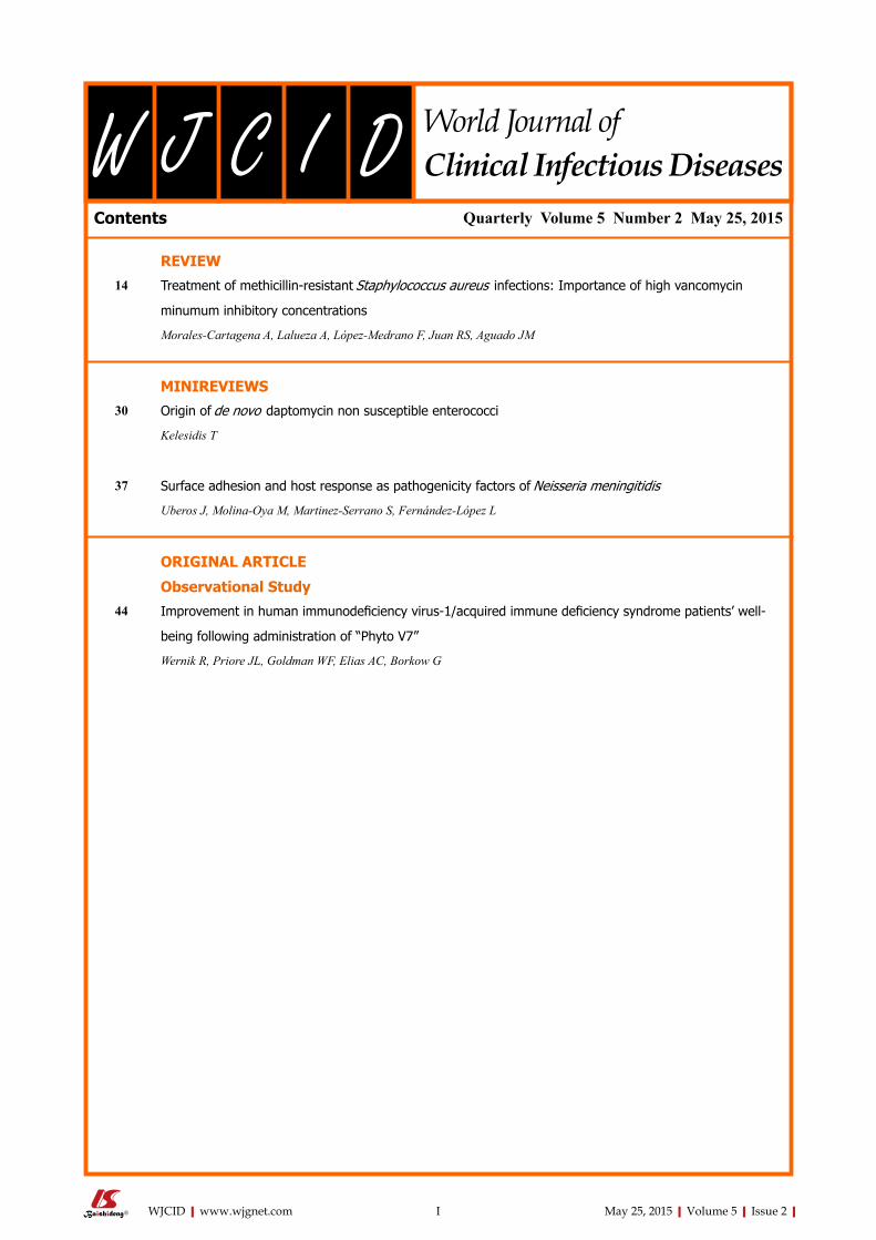
Contents
IWJCID|www.wjgnet.com May 25, 2015|Volume 5|Issue 2|
World Journal ofClinical Infectious DiseasesW J C I D
Quarterly Volume 5 Number 2 May 25, 2015
REVIEW14 Treatmentofmethicillin-resistantStaphylococcusaureus infections:Importanceofhighvancomycin
minumuminhibitoryconcentrations
Morales-Cartagena A, Lalueza A, López-Medrano F, Juan RS, Aguado JM
MINIREVIEWS30 Originofdenovo daptomycinnonsusceptibleenterococci
Kelesidis T
37 SurfaceadhesionandhostresponseaspathogenicityfactorsofNeisseriameningitidis
Uberos J, Molina-Oya M, Martinez-Serrano S, Fernández-López L
ORIGINAL ARTICLE
Observational Study44 Improvementinhumanimmunodeficiencyvirus-1/acquiredimmunedeficiencysyndromepatients’well-
beingfollowingadministrationof“PhytoV7”
Wernik R, Priore JL, Goldman WF, Elias AC, Borkow G
ContentsWorld Journal of Clinical Infectious Diseases
Volume 5 Number 5 May 25, 2015
EDITORS FOR THIS ISSUE
Responsible Assistant Editor: Xiang Li Responsible Science Editor: Fang-Fang Ji Responsible Electronic Editor: Huan-Liang Wu Proofing Editorial Office Director: Xiu-Xia SongProofing Editor-in-Chief: Lian-Sheng Ma
Rd, Atlanta, GA 30333, United States
EDITORIALOFFICEJin-Lei Wang, DirectorXiu-Xia Song, Vice DirectorWorld Journal of Clinical Infectious DiseasesRoom 903, Building D, Ocean International Center,No. 62 Dongsihuan Zhonglu, Chaoyang District, Beijing 100025, ChinaTelephone: +86-10-85381891Fax: +86-10-85381893E-mail: [email protected] Desk: http://www.wjgnet.com/esps/helpdesk.aspxhttp://www.wjgnet.com
PUBLISHERBaishideng Publishing Group Inc8226 Regency Drive, Pleasanton, CA 94588, USATelephone: +1-925-223-8242Fax: +1-925-223-8243E-mail: [email protected] Desk: http://www.wjgnet.com/esps/helpdesk.aspxhttp://www.wjgnet.com
PUBLICATIONDATEMay 25, 2015
COPYRIGHT© 2015 Baishideng Publishing Group Inc. Articles pub-lished by this Open-Access journal are distributed under the terms of the Creative Commons Attribution Non-commercial License, which permits use, distribution, and reproduction in any medium, provided the original work is properly cited, the use is non commercial and is otherwise in compliance with the license.
SPECIALSTATEMENTAll articles published in journals owned by the Baishideng Publishing Group (BPG) represent the views and opinions of their authors, and not the views, opinions or policies of the BPG, except where otherwise explicitly indicated.
INSTRUCTIONSTOAUTHORSFull instructions are available online at http://www.wjgnet.com/2220-3176/g_info_20100722180909.htm.
ONLINESUBMISSIONhttp://www.wjgnet.com/esps/
IIWJCID|www.wjgnet.com
ABOUT COVER
AIM AND SCOPE
FLYLEAF
May 25, 2015|Volume 5|Issue 2|
NAMEOFJOURNALWorld Journal of Clinical Infectious Diseases
ISSNISSN 2220-3176 (online)
LAUNCHDATEDecember 30, 2011
FREQUENCYQuarterly
EDITORS-IN-CHIEFShyam Sundar, MD, FRCP (London), FAMS, FNASc, FASc, FNA, Professor, Department of Medicine, Institute of Medical Sciences, Banaras Hindu University, Varanasi 221005, India
Lihua Xiao, DVM, PhD, Senior Scientist, Divi-sion of Foodborne, Waterborne, and Environmental Diseases, National Center for Emerging and Zoonotic Infectious Diseases, Centers for Disease Control and Prevention, Bldg 23, Rm 9-168, MS D66, 1600 Clifton
EditorialBoardMemberofWorldJournalofClinicalInfectiousDiseases ,GadiBorkow,PhD,ChiefMedicalScientist,CupronScientific,Hameyasdim44,Gibton
76910,Israel
World Journal of Clinical Infectious Diseases (World J Clin Infect Dis, WJCID, online ISSN 2220-3176, DOI: 10.5495) is a peer-reviewed open access (OA) academic journal that aims to guide clinical practice and improve diagnostic and therapeutic skills of clinicians.
WJCID will focus on a broad spectrum of topics on infectious diseases that will cover epidemiology, immune-pathogenesis, genetic factors, host susceptibility to infection, vector control, novel approaches of treatment, molecular diagnostic and vaccines. It will provide a common stage to share the visions, new approaches, most advanced techniques, and to discuss research problems that will help everyone working in the field of various infections to exchange their views and to improve public health. WJCID will also focus on broad range of infections like opportunistic infections, zoonotic infections, tropical and neglect-ed tropical diseases, emerging infections, etc. and following topics related to these issues: (1) Causative agents discussing various pathogens; (2) Vectors and Mode of transmission; (3) Host-pathogen interaction and immune-pathogenesis of the disease; (4) Epidemiology of the infection and vector control strategies; (5) Genetic factors covering both host and pathogen; (6) Molecular diagnostic techniques vaccines; and (7) Recent advances in cell tissue culture, lab techniques, etc. Various other related fields like medical microbiology, pharmacology of herbs, bioinformatics, etc. will be included.
We encourage authors to submit their manuscripts to WJCID. We will give priority to manuscripts that are supported by major national and international foundations and those that are of great basic and clinical significance.
World Journal of Clinical Infectious Diseases is now indexed in Digital Object Identifier.
I-III EditorialBoard
INDEXING/ABSTRACTING
Surface adhesion and host response as pathogenicity factors of Neisseria meningitidis
Jose Uberos, M Molina-Oya, S Martinez-Serrano, L Fernández-López
Jose Uberos, M Molina-Oya, S Martinez-Serrano, L Fernández-López, Department of Paediatrics, School of Medicine, University of Granada, 18012 Granada, SpainAuthor contributions: All authors contributed to this manuscript.Open-Access: This article is an open-access article which was selected by an in-house editor and fully peer-reviewed by external reviewers. It is distributed in accordance with the Creative Commons Attribution Non Commercial (CC BY-NC 4.0) license, which permits others to distribute, remix, adapt, build upon this work non-commercially, and license their derivative works on different terms, provided the original work is properly cited and the use is non-commercial. See: http://creativecommons.org/licenses/by-nc/4.0/Correspondence to: Dr. Jose Uberos, Department of Paediatrics, School of Medicine, University of Granada, Avda. de Madrid s/n, 18012 Granada, Spain. [email protected] Telephone: +34-95-8243066Received: July 3, 2014 Peer-review started: July 3, 2014First decision: July 21, 2014Revised: January 26, 2015 Accepted: March 5, 2015Article in press: March 9, 2015Published online: May 25, 2015
AbstractNeisseria meningitidis (N. meningitidis ) is an exclusively human pathogen that has been identified in 10%-35% of the adult population and in 5.9% of the child popu-lation. Despite the high prevalence of carriers of N. meningitidis , it only occasionally causes meningococcal disease in the context of endemic disease, in certain geographic areas or in isolated epidemic outbreaks. After the N. meningitidis genome is described, progress has been made toward understanding the pathogenic mechanisms of the bacteria, although some aspects concerning its interaction with the environment and the host remain unclear. Some studies have reported that oxidative stress in the environment can modify the surface characteristics of N. meningitidis , increasing its adhesive properties and favouring an asymptomatic
carrier state. The antigenic structure of N. meningitidis can be modified by its importing genetic material from other bacteria in its ecological niche. Some structures of lipopolysaccharides help it to evade the immune response, and these are observed more frequently in N. meningitidis isolated from blood than in healthy nasopharyngeal carriers. There is evidence that pili and capsule are downregulated upon contact with target cells. This paper reviews current knowledge on host-environment-bacteria mechanisms and interactions, with the aim of contributing to our understanding of the pathogenic mechanisms of N. meningitidis .
Key words: Bacterial adhesion; Neisseria meningitidis ; Virulence
© The Author(s) 2015. Published by Baishideng Publishing Group Inc. All rights reserved.
Core tip: After the Neisseria meningitidis (N. meningitidis) genome is described, progress has been made toward understanding the pathogenic mechanisms of the bacteria, although some aspects concerning its interaction with the environment and the host remain unclear. This paper reviews current knowledge on host-environment-bacteria mechanisms and interactions, with the aim of contributing to our understanding of the pathogenic mechanisms of N. meningitidis .
Uberos J, Molina-Oya M, Martinez-Serrano S, Fernández-López L. Surface adhesion and host response as pathogenicity factors of Neisseria meningitidis. World J Clin Infect Dis 2015; 5(2): 37-43 Available from: URL: http://www.wjgnet.com/2220-3176/full/v5/i2/37.htm DOI: http://dx.doi.org/10.5495/wjcid.v5.i2.37
BACTERIAL ADHESION AND PATHOGENICITYThe ability of bacteria to attach and grow on almost
MINIREVIEWS
Submit a Manuscript: http://www.wjgnet.com/esps/Help Desk: http://www.wjgnet.com/esps/helpdesk.aspxDOI: 10.5495/wjcid.v5.i2.37
World J Clin Infect Dis 2015 May 25; 5(2): 37-43ISSN 2220-3176 (online)
© 2015 Baishideng Publishing Group Inc. All rights reserved.
World Journal ofClinical Infectious DiseasesW J C I D
37 May 25, 2015|Volume 5|Issue 2|WJCID|www.wjgnet.com
any surface has been known for decades. The importance of adhesion in the colonisation of specific substrates, and its role in the pathogenesis of bacterial infections and in the maintenance of the carrier state has been studied widely in recent years[1]. Neisseria meningitidis (N. meningitidis) is only found in humans, suggesting that its ability to cause disease is likely an casual side effect of its life cycle. Globally, the carrier rate of N. meningitidis ranges from 10%35% among healthy adults[2]; the mean carrier rate in children is 5.9%, peaking at 10.3% in children aged under 3 years[3]. This situation has been associated with the genetic characteristics of the circulating strains of N. meningitidis, the immune pressure exerted by vaccination programmes and the hygienic and social conditions within a community. Compared with the rates of colonisation, meningococcal disease is less common, its development being affected by interacting factors such as the virulence of the bacterium, host defence mechanisms, the age of the host and the history of previous viral infections[2]. The best-defined virulence factor of N. meningitidis is its polysaccharide capsule that indicates its serogroup. Although 13 serogroups of N. meningitidis have been described (A, B, C, D, 29E, H, I, K, L, Y, W135, X and Z), invasive meningococcal disease is most frequently caused by serogroups A, B, C, Y and W135, to which 10% mortality has been attributed.
The adhesion of bacteria to epithelial surfaces is an initial step in the colonisation of microbial habitats, and it ensures the survival of N. meningitidis in its ecological niche[4]. Adherence can be defined as a phenomenon resulting from the interaction between two surfaces, with the participation of physical, chemical and biological factors, with contact between the bacterium and the cell being necessary for adherence to take place. The adhesion occurs in several steps: (1) N. meningitidis attaches to the surface of target cells to form small colonies. This step is essentially a pilusmediated process; and (2) after N. meningitidis has been attached, it comes into close contact with surface of the target cells (intimate adhesion). The adhesive interaction is present both in commensal bacteria and in pathogens and so for N. meningitidis to adhere, it has developed proprietary adhesive mechanisms that allow it to compete with flora of the same ecological niche[5]. Meningococcal pili are of type IV and are composed of pilin subunits that are encoded by pilE gene. Other homologous proteins, PilC1 and PilC2, are also involved in pilus assembly and adhesion. PilC1containing may involve interaction with CD46, a human transmembrane glycoprotein involved in complement regulation. The expression of pilC1 is induced following the contact of N. meningitidis with viable target cells. Both pili and capsule are downregulated upon contact with target cells. This downregulation seems to be associated with intimate adhesion of N. meningitidis to target cells[5,6].
The “adhesion process” can be defined in terms of
the adhesion affinity of bacteria to epithelial surfaces, as has been described in the MichaelisMenten equations. The maximum point of adhesion (and affinity) can be determined graphically by the LineaweverBurk equations, using simple experimental models[7]. This first phase would be a reversible process in which Van der Waals and electrostatic forces are responsible for a wide range of interactions, including chemical bonding, dipolar interaction and hydrophobicity. Surface molecules as Nacetylneuraminic acid may alter the initial adhesion strength, reducing electrostatic repulsive forces or increasing attractive ones[8]. The cell surfaces of both prokaryotic and eukaryotic cells are negatively charged. Electrostatic repulsive forces between the cell and the bacteria can be overcome by long and shortrange attractive forces, and so the specific binding of fimbriae with cell surface receptors must overcome the repulsive forces between the two surfaces. According to Smyth et al[9], the reduction of the surface bacteria potential by the intervention of hydrophobic adhesins probably facilitates adhesion, with the hydrophobic forces being a first step in the interaction of the organism with mucosal surfaces. Surface hydrophobicity is a nonspecific adhesion factor which is important to the adhesion and growth of microorganisms on epithelial surfaces. The hydrophobichydrophilic environment of the bacterial surface is modulated by hydrophobic or hydrophilic agents (increasing or reducing hydrophobicity, respectively), which may coexist on the surface of the outer membrane[5]. Generally, the strains that formed biofilms show highlevel cell surface hydrophobicity. Many studies have examined the contribution of surface hydrophobicity to bacterial adhesion, with particular attention to Salmonella[10,11], Escherichia coli (E. coli), N. gonorrhoeae[12,13] and N. meningitidis[14,15].
The type IV pili of N. meningitidis are crucial determinants of the adhesion of these pathogens to epithelial and endothelial cells[16]. Under natural conditions, pili are the only means by which encapsulated N. meningitidis can adhere to human mucosal surfaces[17,18].
SURFACE MODULATION AND INTERACTION WITH THE HOSTWhen N. meningitidis adheres to epithelial cells, it becomes resistant to the bactericidal effect of the antimicrobial peptide LL37, which is the first line of defence of innate immunity. The decreased binding of LL37 to the adhered bacteria can result from its degradation by proteases released at the site of infection. Furthermore, N. meningitidis induces the formation in the nasopharynx of cholesterolrich membrane microdomains, which are essential to the antimicrobial resistance induced by bacterial adhesion[2].
To avoid immune detection, the surface components of N. meningitidis may be modified. The structural and antigenic modification of surface molecules can
Uberos J et al . Pathogenic mechanisms of N. meningitidis
38 May 25, 2015|Volume 5|Issue 2|WJCID|www.wjgnet.com
involve changes in gene alleles. Studies have reported the import of genetic material from other bacteria or via intragenomic recombination[2]. N. meningitidis may be encapsulated or unencapsulated. N. meningitidis isolated from blood or cerebrospinal fluid is invariably encapsulated. The existence of the capsule enables it to withstand the effects of antibodies and complements and to resist serum opsonic activity. Some lipopolysaccharide structures help N. meningitidis to evade the immune response, and are more frequently observed in N. meningitidis isolated from blood than in healthy nasopharyngeal carriers. Both the capsule and some lipopolysaccharide immunotypes (L1, L8 and L10) of N. meningitidis may influence bacterial adhesion and invasive capacity. It has been found the inhibitory role of capsule in biofilm formation[14]. The capsule genes are located in a single chromosomal locus (cps) divided into three regions. The capsular polysaccharides B, C, W135 and Y contain sialic acid, which contributes to make the lipopolysaccharides of the capsule less visible to the immune system, since sialic acid is a common component of the host cell surfaces. Moreover, the serogroup B capsule contains a homopolymer that is structurally identical to the neural adhesion molecule, which is responsible for the poor immune response generated by serogroup B in humans. However, the genetic similarities of the loci of serogroups B, C, W and Y (not A) favour the horizontal exchange of fragments of the capsule between different serogroups[2].
N. meningitidis expresses and secretes various surface molecules that bind to epithelial molecules, and some of these proteins include lactoferrin and the proteins bound to the transferrin that enable the meningococcus to acquire iron from the environment. Iron is a crucial element for bacterial growth in the surface colonisation stage and during the production of disease[19,20], although some adherent properties, such as hydrophobicity and adherence to inert surfaces like nitrocellulose remain unchanged after incubation in culture media supplemented with Fe[21]. Other authors have described nine porin complexes formed by different combinations of the meningococcal porin protein (Por) A, PorB and RmpM proteins[22]. N. meningitidis expresses two types of outer membrane proteins (Opa and Opc) which give an opaque appearance to colonies in agar. Opa and Opc are of a similar size (2731 kDa). Most Opa molecules recognise one or more members of the family of carcinoembryonic antigenrelated cell adhesion molecules (CEACAM). The CEACAM1 receptor is found in epithelial and endothelial cells, while other family members such as CEACAM3 and CEACAM6 are expressed in neutrophils. CEACAM receptor density in the epithelial cells is modulated by the secretion of inflammatory cytokines, such that a high expression of CEACAM receptors takes place in response to inflammation, which could influence the development of meningococcal disease. Furthermore, some Opa proteins can interact with the heparan sulphate proteoglycan that is present in most epithelial
cells[2,23]. Over the past 50 years, our understanding of the
importance of serogroup B (MenB) disease per se, the social impact of fear caused by the devastating effects of the disease. The difficulty of inducing an effective immune response against the MenB capsular polysaccharide has lead to the search in vaccines for this serogroup based on outer membrane proteins (OMP). Public health interventions in Cuba, Norway and New Zealand have demonstrated that these proteinbased vaccines can prevent MenB disease.
By combining a pangenome analysis with an extensive experimental validation to identify new potential vaccine candidates, genes coding for antigens likely to be exposed on the surface of the MenB were selected after a multistep comparative analysis of entire Neisseria genomes. Again, in the quest for vaccine candidates are successfully identified a significant number of new genes. Recent studies with meningococcal membrane proteins have centered on conserved antigens in order to obtain a universal vaccine that confers protection against a broad range of strains. There are several recent reports about the use of conserved minor OMP from N. meningitidis as immunogens[24].
The classical bioinformatic approaches, in combination with proteomic data, conventional protein purification and immunological evaluation are powerful tools for the identification of novel meningococcal antigens and open reading frames and potential vaccine components[25].
We now know that OMP based vaccines are most effective when are used against epidemics due to a homologous or clonal strain carrying the same PorA as that present in the vaccine. When used against endemic disease or outbreaks due to a number of different strains (heterologous epidemiologic situations), the level of effectiveness will generally be too low to rely on the effects of a conventional OMP vaccine alone for protection.
The general strategy in the Pajon et al[25] study, was to maximize the chance of identifying bacterial surface components by selecting not only proteins predicted by protein localization algorithms in outer membrane components of gramnegative bacteria, but also those predicte as periplasmic or inner membrane proteins. However, we must stress that while the most attention in the development of meningococcal vaccines has been devoted to major OMPs. The impact of conserved protein components in the induction of a significant immune response, and their potential as adjuvants, it must not be overlooked. The success in expressing all cloned genes came from the use of a highly optimized expression/purification platform designed precisely for this scenario, but also from the stringent selection procedure of potential vaccine candidates. Finally, five proteins are capable of inducing a functional antibody response vs N. meningitidis strain CU385: NMB0606 a potential YajC orthologue, NMB0928 the
39 May 25, 2015|Volume 5|Issue 2|WJCID|www.wjgnet.com
Uberos J et al . Pathogenic mechanisms of N. meningitidis
are located. In general, a pathogen becomes less virulent on passing from a natural environment to an artificial culture medium; in these circumstances, it is said to be attenuated, and the same effect can be observed in unfavourable environmental conditions. The virulence of a microorganism can be reduced, either by the use of certain culture media, or by exploiting its successive passage through animals. Numerous studies support this; thus, Horská et al[29] have reported that three different bacterial strains of Pseudomonas are capable of changing the surface charge and their hydrophobicity. By contrast, an attenuated microorganism can acquire greater virulence by its prior passage through certain animal species; specifically, pneumococcal virulence is enhanced by its passage through mice[30]. N. meningitidis requires iron, and in the absence of iron alters its gene expression to increase iron acquisition[19,31]. When N. meningitidis, has grown in an ironrestricted environment, it synthesises new outer membrane proteins, which are necessary for its survival. Some of these proteins Tbp A and Tbp B are examples of meningococcal surface antigens regulated by iron, which are not expressed after culture in common laboratory media[32]. TbpA was found to possess a similar architecture to the siderophore and is highly immunogenic, allowing for prediction of potentially important ligandbinding epitopes[33].
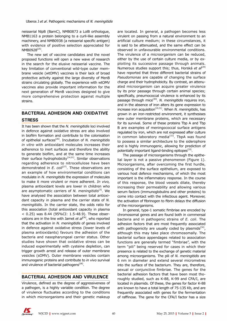
The passage of microorganisms through the epithelial layer is not a passive phenomenon (Figure 1). Microorganisms, after overcoming the first hurdle, consisting of the surface epithelium, are exposed to various host defence mechanisms, of which the most important is the inflammatory response. In the course of this response, the blood vessels dilate, thereby increasing their permeability and allowing various serum factors (immunoglobulins and other proteins) to come into contact with the infectious agent. Moreover, the activation of fibrinogen to fibrin delays the diffusion of the microorganisms.
In general, type-1 somatic fimbriae are encoded by chromosomal genes and are found both in commensal bacteria and in pathogenic strains of E. coli. The adhesion factors that are most frequently associated with pathogenicity are usually coded by plasmids[34], although this may take place chromosomally. The bacterial surface appendages related to association functions are generally termed “fimbriae”, with the term “pili” being reserved for cases in which their presence is related to the exchange of genetic material among microorganisms. The pili of N. meningitidis are 6 nm in diameter and extend several micrometres into the surface of the bacterium. They are, therefore, sexual or conjunctive fimbriae. The genes for the bacterial adhesion factors that have been most thoroughly studied, such as K88, K99 and CFA/I, are located in plasmids. Of these, the genes for factor K88 are known to have a total length of 75135 Kb, and are frequently associated with genes for the fermentation of raffinose. The gene for the CFA/I factor has a size
neisserial NlpB (BamC), NMB0873 a LolB orthologue, NMB1163 a protein belonging to a curlilike assembly machinery, and NMB0938 (a neisserial specific antigen) with evidence of positive selection appreciated for NMB0928[25].
The new set of vaccine candidates and the novel proposed functions will open a new wave of research in the search for the elusive neisserial vaccine. The key limitation of conventional wildtype outer membrane vesicle (wtOMV) vaccines is their lack of broad protective activity against the large diversity of MenB strains circulating globally. The experience with wtOMV vaccines also provide important information for the next generation of MenB vaccines designed to give more comprehensive protection against multiple strains.
BACTERIAL ADHESION AND OXIDATIVE STRESSIt has been shown that the N. meningitidis loci involved in defence against oxidative stress are also involved in biofilm formation and contribute to the colonisation of epithelial surfaces[26]. Incubation of N. meningitidis in vitro with antioxidant molecules increases their adherence to inert surfaces and therefore the ability to generate biofilm, and at the same time increases their surface hydrophobicity[14,21]. Similar observations regarding adherence to nitrocellulose have been demonstrated in E. coli[27]. These observations are an example of how environmental conditions can modulate in N. meningitidis the expression of molecules to make it more virulent or more adherent. In vivo, plasma antioxidant levels are lower in children who are asymptomatic carriers of N. meningitidis[3]. We have analysed the association between total antioxidant capacity in plasma and the carrier state of N. meningitidis. In the carrier state, the odds ratio for this association (total antioxidant capacity in plasma < 0.25) was 8.44 (95%CI: 1.548.9). These observations are in the line with Jamet et al[26], who reported that the activation in N. meningitidis of genes involved in defence against oxidative stress (lower levels of plasma antioxidants) favours the adhesion of the bacteria and nasopharyngeal carrier status. Other studies have shown that oxidative stress can be induced experimentally with cysteine depletion, can trigger growth arrest and release of outer membrane vesicles (sOMV). Outer membrane vesicles contain immunogenic proteins and contribute to in vivo survival and virulence of bacterial pathogens[28].
BACTERIAL ADHESION AND VIRULENCEVirulence, defined as the degree of aggressiveness of a pathogen, is a highly variable condition. The degree of virulence fluctuates according to the conditions in which microorganisms and their genetic makeup
40 May 25, 2015|Volume 5|Issue 2|WJCID|www.wjgnet.com
Uberos J et al . Pathogenic mechanisms of N. meningitidis
of approximately 90 Kb, and it is bound to a gene for a stable enterotoxin[35]. For adhesion factor K88, three plasmids have been shown to be responsible for the three known antigenic variants: K88ab, K88ac and K88ad. Mooi et al[36], designed experiments to determine which genes of the plasmid chain were responsible for the formation of the K88 factor in each of the variants. For this purpose, each of the three K88 plasmids was digested with restriction enzymes, and the fragments obtained from each one were then cloned by inclusion in the PBR322 vector. The bacterial clones carrying each of the K88 antigens were then identified. This procedure revealed that the expression of the K88 factor depends of the orientation of the DNA chain responsible and on the variant in question. In the case of K88ab, its insertion into the vector PBR322 in a direction or another modifies the quantity of antigen expressed. The lipopolysaccharide of N. meningitidis is known as the major determinant of its virulence, and the use of monoclonal antibodies, together with structural studies, have highlighted the heterogeneity and complexity of meningococcal lipopolysaccharides, which can be divided into 12 immunotypes[37].
Studies by McGee et al[38], have underlined the importance of gonococcal fimbriae in cell colonisation and destruction in cultures of cells from the human fallopian tube. These assays show that both fimbriate and nonfimbriate gonococci bind epithelial cells, although in the former case cell destruction is produced more quickly, this process being mediated by one or more toxic factors, such as surface lipopolysaccharides. Type IV pili, which are protein structures associated
with the surface, have also been associated with the adhesion of N. meningitidis to endothelial cells and the development of fulminant meningococcal disease[39,40]. The pili of E. coli, which have been studied in detail, consist of protein subunits that are thought to play an important role in the interaction with specific surface carbohydrates in eukaryotic cells, and some of them are K antigens. D(+)Mannose inhibits the in vitro adhesion of bacteria with type-1 fimbriae on the surface of eukaryotic cells containing mannose residue[41]. This is an indiscriminate mechanism of adhesion, as oligosaccharide chains containing mannose are very commonly present in cell surface oligoproteins, including phagocytic cells. Preincubation of bacteria with inhibitor sugars does not affect the adhesiveness, while the pretreatment of cells with carbohydrates effectively prevents adhesion. This indicates that the cell surface structures recognise the radicals of fucose and glucose in the bacterial lipopolysaccharides.
Some authors[42,43], have analysed phenotypic changes in bacteria associated with epigenetic changes. Aspects such as virulence, response to oxidative stress and the formation of biofilm have been observed among epigenetic modifications. Unfortunately, these processes and their relationship with pathogenic changes in N. meningitidis are as yet incompletely understood.
Despite the high prevalence of carriers of N. meningitidis, it only occasionally causes meningococcal disease in the context of endemic disease, in certain geographic areas or in isolated epidemic outbreaks. Some studies have reported that oxidative stress in the environment can modify the surface characteristics
41 May 25, 2015|Volume 5|Issue 2|WJCID|www.wjgnet.com
Capsule Opc
PilusOpa
Pilus
receptor
Epithelium
MspA APP
NhhA
NadA
HSPGCEACAM Integrin
Internalisation via hostreceptor interactions
Interaction with matrix proteinsVn/Fn
1
2
Figure 1 Schematic representation of the interaction mechanisms of Neisseria meningitidis with cellular receptors. The first adherence phase would be a reversible process in which Van der Waals and electrostatic forces are responsible for a wide range of interactions, including chemical bonding. Finally we added a summary at the endding, dipolar interaction and hydrophobicity. Pili extending beyond the capsule are considered to mediate the primary interaction with epithelial cells. Opa proteins may bind to carcinoembryonic antigen-related cell-adhesion molecule (CEACAMs) and heparan sulphate proteoglycan (HSPGs), and Opc proteins can interact with HSPGs and, via vitronectin and fibronectin, to their integrin receptors. Engagement of CEACAMs, integrins and HSPGs can result in meningococcal internalization by epithelial cells. MSP: Meningococcal serine protease A; App: Adhesion and penetration protein; NadA: Neisserial adhesin; NhhA: Neisseria hia/hsf homologue A.
Uberos J et al . Pathogenic mechanisms of N. meningitidis
of N. meningitidis. Also the antigenic structure can be modified by its importing genetic material from other bacteria in its ecological niche, and some structures of lipopolysaccharides, pili and capsule change the immune response. This paper reviews current knowledge on hostenvironmentbacteria mechanisms and interactions, with the aim of contributing to our understanding of the pathogenic mechanisms of N. meningitidis.
REFERENCES1 Hernandez DM, Matos PP, Hernandez JC, Muñoz JL, Villasana
Lde C. Persistence of an infected urachus presenting as acute abdominal pain. Case report. Arch Esp Urol 2009; 62: 589-592 [PMID: 19815963]
2 Geörg M, Maudsdotter L, Tavares R, Jonsson AB. Meningococcal resistance to antimicrobial peptides is mediated by bacterial adhesion and host cell RhoA and Cdc42 signalling. Cell Microbiol 2013; 15: 1938-1954 [PMID: 23834289]
3 Uberos J, Molina-Carballo A, Galdo-Muñoz G, Muñoz-Hoyos A. Total antioxidant capacity of plasma in asymptomatic carrier state of Neisseria meningitidis. Epidemiol Infect 2007; 135: 857-860 [PMID: 17109775 DOI: 10.1017/S0950268806007539]
4 Van Wamel WJ, Vandenbroucke-Grauls CM, Verhoef J, Fluit AC. The effect of culture conditions on the in-vitro adherence of methicillin-resistant Staphylococcus aureus. J Med Microbiol 1998; 47: 705-709 [PMID: 9877191 DOI: 10.1099/00222615-47-8-705]
5 Bartley SN, Tzeng YL, Heel K, Lee CW, Mowlaboccus S, Seemann T, Lu W, Lin YH, Ryan CS, Peacock C, Stephens DS, Davies JK, Kahler CM. Attachment and invasion of Neisseria meningitidis to host cells is related to surface hydrophobicity, bacterial cell size and capsule. PLoS One 2013; 8: e55798 [PMID: 23405216 DOI: 10.1371/journal.pone.0055798]
6 Marrie TJ, Lam J, Costerton JW. Bacterial adhesion to uroepithelial cells: a morphologic study. J Infect Dis 1980; 142: 239-246 [PMID: 6774033 DOI: 10.1093/infdis/142.2.239]
7 Cohen C, Phillips GN. Spikes and fimbriae: alpha-helical proteins form surface projections on microorganisms. Proc Natl Acad Sci USA 1981; 78: 5303-5304 [PMID: 6117855 DOI: 10.1073/pnas.78.9.5303]
8 Liu F, Lee HJ, Strynadka NC, Tanner ME. Inhibition of Neisseria meningitidis sialic acid synthase by a tetrahedral intermediate analogue. Biochemistry 2009; 48: 9194-9201 [PMID: 19719325 DOI: 10.1021/bi9012758]
9 Smyth CJ, Siegel J, Salton MR, Owen P. Immunochemical analysis of inner and outer membranes of Escherichia coli by crossed immunoelectrophoresis. J Bacteriol 1978; 133: 306-319 [PMID: 338583]
10 Edebo L, Hed J, Kihlström E, Magnusson KE, Stendahl O. The adhesion of enterobacteria and the effect of antibodies of different immunoglobulin classes. Scand J Infect Dis Suppl 1980; Suppl 24: 93-99 [PMID: 7010568]
11 Grundström T, Jaurin B, Edlund T, Normark S. Physical mapping and expression of hybrid plasmids carrying chromosomal beta-lactamase genes of Escherichia coli K-12. J Bacteriol 1980; 143: 1127-1134 [PMID: 6251026]
12 Lambden PR, Heckels JE, James LT, Watt PJ. Variations in surface protein composition associated with virulence properties in opacity types of Neisseria gonorrhoeae. J Gen Microbiol 1979; 114: 305-312 [PMID: 120407 DOI: 10.1099/00221287-114-2-305]
13 Ellen RP, Gibbons RJ. M protein-associated adherence of Streptococcus pyogenes to epithelial surfaces: prerequisite for virulence. Infect Immun 1972; 5: 826-830 [PMID: 4564883]
14 Yi K, Rasmussen AW, Gudlavalleti SK, Stephens DS, Stojiljkovic I. Biofilm formation by Neisseria meningitidis. Infect Immun 2004; 72: 6132-6138 [PMID: 15385518 DOI: 10.1128/IAI.72.10.6132-6138.2004]
15 Beachey EH. Bacterial adherence: adhesin-receptor interactions mediating the attachment of bacteria to mucosal surface. J Infect Dis 1981; 143: 325-345 [PMID: 7014727 DOI: 10.1093/infdis/143.3.325]
16 Nassif X, Beretti JL, Lowy J, Stenberg P, O’Gaora P, Pfeifer J, Normark S, So M. Roles of pilin and PilC in adhesion of Neisseria meningitidis to human epithelial and endothelial cells. Proc Natl Acad Sci USA 1994; 91: 3769-3773 [PMID: 7909606 DOI: 10.1073/pnas.91.9.3769]
17 Virji M, Makepeace K, Peak IR, Ferguson DJ, Jennings MP, Moxon ER. Opc- and pilus-dependent interactions of meningococci with human endothelial cells: molecular mechanisms and modulation by surface polysaccharides. Mol Microbiol 1995; 18: 741-754 [PMID: 8817495 DOI: 10.1111/j.1365-2958.1995.mmi_18040741.x]
18 Koomey M, Gotschlich EC, Robbins K, Bergström S, Swanson J. Effects of recA mutations on pilus antigenic variation and phase transitions in Neisseria gonorrhoeae. Genetics 1987; 117: 391-398 [PMID: 2891588]
19 Jordan PW, Saunders NJ. Host iron binding proteins acting as niche indicators for Neisseria meningitidis. PLoS One 2009; 4: e5198 [PMID: 19352437 DOI: 10.1371/journal.pone.0005198]
20 Criado MT, del Río MC, Ferreirós CM, Pintor M, Sáinz V, Carballo J. Iron and outer membrane proteins in the susceptibility of Neisseria meningitidis to human serum. FEMS Microbiol Lett 1990; 58: 145-150 [PMID: 2121585 DOI: 10.1111/j.1574-6968.1990.tb13968.x]
21 Uberos J, Molina A, Liébana J, Augustin MC, Muñoz A. The influence of different concentrations of melatonin on the cell surface hydrophobic characteristics of Neisseria meningitidis. Lett Appl Microbiol 2000; 31: 294-298 [PMID: 11068910 DOI: 10.1046/j.1472-765x.2000.00813.x]
22 Marzoa J, Sánchez S, Ferreirós CM, Criado MT. Identification of Neisseria meningitidis outer membrane vesicle complexes using 2-D high resolution clear native/SDS-PAGE. J Proteome Res 2010; 9: 611-619 [PMID: 19888731 DOI: 10.1021/pr9006409]
23 Johswich KO, McCaw SE, Islam E, Sintsova A, Gu A, Shively JE, Gray-Owen SD. In vivo adaptation and persistence of Neisseria meningitidis within the nasopharyngeal mucosa. PLoS Pathog 2013; 9: e1003509 [PMID: 23935487 DOI: 10.1371/journal.ppat.1003509]
24 Holst J, Oster P, Arnold R, Tatley MV, Næss LM, Aaberge IS, Galloway Y, McNicholas A, O’Hallahan J, Rosenqvist E, Black S. Vaccines against meningococcal serogroup B disease containing outer membrane vesicles (OMV): lessons from past programs and implications for the future. Hum Vaccin Immunother 2013; 9: 1241-1253 [PMID: 23857274 DOI: 10.4161/hv.24129]
25 Pajon R, Yero D, Niebla O, Climent Y, Sardiñas G, García D, Perera Y, Llanes A, Delgado M, Cobas K, Caballero E, Taylor S, Brookes C, Gorringe A. Identification of new meningococcal serogroup B surface antigens through a systematic analysis of neisserial genomes. Vaccine 2009; 28: 532-541 [PMID: 19837092 DOI: 10.1016/j.vaccine.2009.09.128]
26 Jamet A, Euphrasie D, Martin P, Nassif X. Identification of genes involved in Neisseria meningitidis colonization. Infect Immun 2013; 81: 3375-3381 [PMID: 23817612 DOI: 10.1128/IAI.00421-13]
27 Uberos J, Augustin C, Liébana J, Molina A, Muñoz-Hoyos A. Comparative study of the influence of melatonin and vitamin E on the surface characteristics of Escherichia coli. Lett Appl Microbiol 2001; 32: 303-306 [PMID: 11328494 DOI: 10.1046/j.1472-765X.2001.00908.x]
28 van de Waterbeemd B, Zomer G, van den Ijssel J, van Keulen L, Eppink MH, van der Ley P, van der Pol LA. Cysteine depletion causes oxidative stress and triggers outer membrane vesicle release by Neisseria meningitidis; implications for vaccine development. PLoS One 2013; 8: e54314 [PMID: 23372704 DOI: 10.1371/journal.pone.0054314]
29 Horská E, Pokorný J, Labajová M. Effect of cultivation medium on some physicochemical parameters of outer bacterial membrane. Microbios 1995; 81: 203-211 [PMID: 7770007]
30 Gibbons RJ, Qureshi JV. Virulence-related physiological changes and antigenic variation in populations of Streptococcus mutans
42 May 25, 2015|Volume 5|Issue 2|WJCID|www.wjgnet.com
Uberos J et al . Pathogenic mechanisms of N. meningitidis
colonizing gnotobiotic rats. Infect Immun 1980; 29: 1082-1091 [PMID: 7429627]
31 Livorsi DJ, Stenehjem E, Stephens DS. Virulence factors of gram-negative bacteria in sepsis with a focus on Neisseria meningitidis. Contrib Microbiol 2011; 17: 31-47 [PMID: 21659746 DOI: 10.1159/000324008]
32 Pintor M, Ferrón L, Gómez JA, Powell NB, Ala’Aldeen DA, Borriello SP, Criado MT, Ferreirós CM. Blocking of iron uptake from transferrin by antibodies against the transferrin binding proteins in Neisseria meningitidis. Microb Pathog 1996; 20: 127-139 [PMID: 8965674 DOI: 10.1006/mpat.1996.0012]
33 Oakhill JS, Sutton BJ, Gorringe AR, Evans RW. Homology modelling of transferrin-binding protein A from Neisseria meningitidis. Protein Eng Des Sel 2005; 18: 221-228 [PMID: 15820975 DOI: 10.1093/protein/gzi024]
34 McNeish AS, Turner P, Fleming J, Evans N. Mucosal adherence of human enteropathogenic Escherichia coli. Lancet 1975: 2: 946-948 [DOI: 10.1016/S0140-6736(75)90360-8]
35 Smith HW, Parsell Z. Transmissible substrate-utilizing ability in enterobacteria. J Gen Microbiol 1975; 87: 129-140 [PMID: 1094091 DOI: 10.1099/00221287-87-1-129]
36 Mooi FR, de Graaf FK, van Embden JD. Cloning, mapping and expression of the genetic determinant that encodes for the K88ab antigen. Nucleic Acids Res 1979; 6: 849-865 [PMID: 375197 DOI: 10.1093/nar/6.3.849]
37 Verheul AF, Snippe H, Poolman JT. Meningococcal lipopoly-
saccharides: virulence factor and potential vaccine component. Microbiol Rev 1993; 57: 34-49 [PMID: 8464406]
38 McGee ZA, Gross J, Dourmashkin RR, Taylor-Robinson D. Nonpilar surface appendages of colony type 1 and colony type 4 gonococci. Infect Immun 1976; 14: 266-270 [PMID: 820643]
39 Melican K, Duménil G. A humanized model of microvascular infection. Future Microbiol 2013; 8: 567-569 [PMID: 23642111 DOI: 10.2217/fmb.13.35]
40 Ryll RR, Rudel T, Scheuerpflug I, Barten R, Meyer TF. PilC of Neisseria meningitidis is involved in class II pilus formation and restores pilus assembly, natural transformation competence and adherence to epithelial cells in PilC-deficient gonococci. Mol Microbiol 1997; 23: 879-892 [PMID: 9076726 DOI: 10.1046/j.1365-2958.1997.2631630.x]
41 Firon N, Ofek I, Sharon N. Interaction of mannose-containing oligosaccharides with the fimbrial lectin of Escherichia coli. Biochem Biophys Res Commun 1982; 105: 1426-1432 [PMID: 6125146 DOI: 10.1016/0006-291X(82)90947-0]
42 Chen P, Jeannotte R, Weimer BC. Exploring bacterial epigenomics in the next-generation sequencing era: a new approach for an emerging frontier. Trends Microbiol 2014; 22: 292-300 [PMID: 24725482 DOI: 10.1016/j.tim.2014.03.005]
43 Davis BM, Chao MC, Waldor MK. Entering the era of bacterial epigenomics with single molecule real time DNA sequencing. Curr Opin Microbiol 2013; 16: 192-198 [PMID: 23434113 DOI: 10.1016/j.mib.2013.01.011]
P- Reviewer: Callegan MC, Weng CF S- Editor: Yu J L- Editor: A E- Editor: Wu HL
43 May 25, 2015|Volume 5|Issue 2|WJCID|www.wjgnet.com
Uberos J et al . Pathogenic mechanisms of N. meningitidis

© 2015 Baishideng Publishing Group Inc. All rights reserved.
Published by Baishideng Publishing Group Inc8226 Regency Drive, Pleasanton, CA 94588, USA
Telephone: +1-925-223-8242Fax: +1-925-223-8243
E-mail: [email protected] Desk: http://www.wjgnet.com/esps/helpdesk.aspx
http://www.wjgnet.com