Embed Size (px)
Citation preview
生物圏科学広島大学大学院生物圏科学研究科紀要 第56巻 2017
広島大学大学院生物圏科学研究科 東広島市2017年12月発行
ISSN 1348-1371
原著論文Kazuya Nagasawa and Shinji TaNaka
1 A rare infection of Ceratothoa verrucosa (Isopoda: Cymothoidae) on red seabream, Pagrus major, cultured in central Japan
Kazuya Nagasawa and Hiroki Nakao
7 Chub mackerel, Scomber japonicus (Perciformes: Scombridae), a new host record for Nerocila phaiopleura (Isopoda: Cymothoidae)
Kazuya Nagasawa 13 Two species of copepods, Lernanthropus atrox and Hatschekia pagrosomi, parasitic on crimson seabream, Evynnis tumifrons, in Hiroshima Bay,western Japan
新田理人 23 東広島市におけるミカドケナガノミ Chaetopsylla mikado の初記録
米谷まり・飯田 健・藤 太稀・平野勝士・近藤裕介・大塚 攻・中口和光・山口修平・加藤幹雄・広瀬雅人・藤田敏彦
27 大島新曽根で採集されたトヨシオマリヒトデPodosphaeraster toyoshiomaruae の行動観察
総 説Kazuya Nagasawa and Hirotaka kaTaHira
33 A revised and updated checklist of the parasites of eels (Anguilla spp.) (Anguilliformes:Anguillidae) in Japan(1915-2017)
Kazuya Nagasawa 71 A synopsis of the parasites of medaka (Oryzias latipes) of Japan(1929-2017)
長澤和也・上野大輔 87 日本産魚類に寄生するサメジラミ科カイアシ類の目録(1898-2017年)
長澤和也 105 日本に定着したサンフィッシュ科魚類3種(ブルーギル,オオクチバス,コクチバス)の寄生虫目録
(1962-2017年)
資 料123 博士論文要旨176 修士論文題目179 研究科長裁量経費による助成研究報告185 広島大学大学院生物圏科学研究科教員業績目録
(2016︲2017年)
目 次

A rare infection of Ceratothoa verrucosa (Isopoda: Cymothoidae) on red seabream, Pagrus major, cultured in central Japan
Kazuya Nagasawa1)* and Shinji Tanaka
2)
1) Graduate School of Biosphere Science, Hiroshima University, 1-4-4 Kagamiyama, Higashi-Hiroshima, Hiroshima 739-8528, Japan
2) Mie Prefecture Fisheries Research Institute, Hamajima, Shima, Mie 517-0404, Japan
Abstract An immature female of the cymothoid isopod, Ceratothoa verrucosa (Schioedte and Meinert, 1883), was found to be attached ventrally to the roof of the buccal cavity of a red seabream, Pagrus major (Temminck and Schlegel, 1843), cultured in Kamisakiura Cove, Mie Prefecture, central Japan, in July 2008. Since April 1985, data on the diseases of marine fishes cultured in this prefecture have been taken at two prefectural organizations, but only two records of C. verrucosa infection, including the present case, were found in those long-term data from an examination of more than 14,591 farmed red seabream from April 1985 to July 2017. This indicates that C. verrucosa is an extremely rare parasite of farmed red seabream in Mie Prefecture.Key words: aquaculture, Ceratothoa verrucosa, Cymothoidae, fish parasite, Isopoda, Pagrus major
INTRODUCTION
Isopods of the family Cymothoidae are found on marine fishes cultured in various countries (e.g., Horton and Okamura, 2001). In Japan, three species of cymothoid isopods have been reported to date: Mothocya parvostis Bruce, 1986 from Japanese amberjack, Seriola quinqueradiata Temminck and Schlegel, 1845 and mejina, Girella punctata Gray, 1835 (Hatai and Yasumoto, 1980, 1981, 1982 [reported as Irona melanosticta]; Bruce, 1986); Ceratothoa verrucosa (Schioedte and Meinert, 1883) from red seabream, Pagrus major (Temminck and Schlegel, 1843) (Hatai, 1989, 2006 [as Rhexanella verrucosa]); and Nerocila phaiopleura Bleeker, 1857 from Pacific bluefin tuna, Thunnus orientalis (Temminck and Schlegel, 1844) (Nagasawa and Shirakashi, 2017). Of these species, little is known about C. verrucosa because the available information on this species in aquaculture is only Hatai’s (1989) one-page account in a reference book about fish diseases of Japan. A similar account (Hatai, 2006) was used in a revised version of the book. In other words, no scientific paper has been published on the infection of C. verrucosa on red seabream cultured in Japan. Red seabream is one of the major fishes cultured in coastal marine waters of Mie Prefecture, central Japan. For their efficient treatment and control, fish diseases are routinouly diagnosed at the Mie Prefecture Fisheries Research Institute, Hamajima, and its Owase Branch, Owase. During a recent fish examination, we found an infection of C. verrucosa on farmed red seabream, which is reported herein. We also report that this parasite is very rare in red seabream farming based on long-term data on the diseases of marine fishes cultured in this prefecture.
生物圏科学Biosphere Sci. 56:1-5 (2017)
Accepted on September 19, 2017 *E-mail: [email protected]
2 Kazuya NAGASAWA and Shinji TANAKA
A CASE REPORT
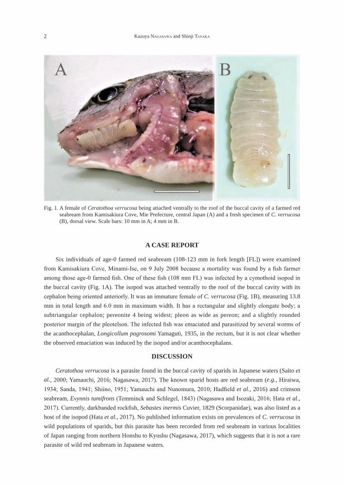
Six individuals of age-0 farmed red seabream (108-123 mm in fork length [FL]) were examined from Kamisakiura Cove, Minami-Ise, on 9 July 2008 because a mortality was found by a fi sh farmer among those age-0 farmed fi sh. One of these fi sh (108 mm FL) was infected by a cymothoid isopod in the buccal cavity (Fig. 1A). The isopod was attached ventrally to the roof of the buccal cavity with its cephalon being oriented anteriorly. It was an immature female of C. verrucosa (Fig. 1B), measuring 13.8 mm in total length and 6.0 mm in maximum width. It has a rectangular and slightly elongate body; a subtriangular cephalon; pereonite 4 being widest; pleon as wide as pereon; and a slightly rounded posterior margin of the pleotelson. The infected fi sh was emaciated and parasitized by several worms of the acanthocephalan, Longicollum pagrosomi Yamaguti, 1935, in the rectum, but it is not clear whether the observed emaciation was induced by the isopod and/or acanthocephalans.
DISCUSSION
Ceratothoa verrucosa is a parasite found in the buccal cavity of sparids in Japanese waters (Saito et al., 2000; Yamauchi, 2016; Nagasawa, 2017). The known sparid hosts are red seabream (e.g., Hiraiwa, 1934; Sanda, 1941; Shiino, 1951; Yamauchi and Nunomura, 2010; Hadfi eld et al., 2016) and crimson seabream, Evynnis tumifrons (Temminck and Schlegel, 1843) (Nagasawa and Isozaki, 2016; Hata et al., 2017). Currently, darkbanded rockfi sh, Sebastes inermis Cuvier, 1829 (Scorpanidae), was also listed as a host of the isopod (Hata et al., 2017). No published information exists on prevalences of C. verrucosa in wild populations of sparids, but this parasite has been recorded from red seabream in various localities of Japan ranging from northern Honshu to Kyushu (Nagasawa, 2017), which suggests that it is not a rare parasite of wild red seabream in Japanese waters.
Fig. 1. A female of Ceratothoa verrucosa being attached ventrally to the roof of the buccal cavity of a farmed red seabream from Kamisakiura Cove, Mie Prefecture, central Japan (A) and a fresh specimen of C. verrucosa (B), dorsal view. Scale bars: 10 mm in A; 4 mm in B.
A cymothoid infection in red seabream culture 3
Since 1985, data on the diseases of farmed marine fishes including red seabream have been accumulated at the Mie Prefecture Fisheries Research Institute, Hamajima, and its Owase Branch, Owase (Tanaka, 2001), and more than 14,591 individuals of red seabream were examined a total of 3,822 times for 32 years between April 1985 and July 2017. Nevertheless, only two cases of infection of C. verrucosa, including the case reported herein, were found in those data, which indicates that this parasite is extremely rare in red seabream farming of Mie Prefecture. The other case was recored as occurring in two individuals of age-0 fish (78 and 189 g in body weight) cultured in Hikimotoura Cove, Miyama (currently Kihoku), in September 1985: these fish were emaciated and harbored cymothoid isopods identifiable as C. verrucosa in the gill operculum region (not in the buccal cavity). No further information, such as the morphology of the parasite, was present. A similar rare occurrence of C. verrucosa on farmed red seabream may occur in such other prefectures as Oita and Ehime, whose aquaculture production of red seabream is high, because there is no report of the isopod from farmed fish in these prefectures (Fukuda, 1999; Matsuoka, 2000). Unlike C. verrucosa, another species of crustacean parasite, Caligus sclerotinosus Roubal, Armitage and Rohde, 1983 (Copepoda: Caligidae) frequently and heavily parasitizes red seabream cultured in Mie Prefecture (Tanaka et al., 2013). As both parasites have direct life cycles without any intermediate hosts (Sanada, 1941; Maran et al., 2012), they are considered to easily find and profierate on their hosts when once they succeed in invading the culture cages. This is, however, not the case with C. verrucosa, and at present, the reason why the species cannot establish its populations within the cages is unknown.
REFERENCES
Bruce, N. L., 1986. Revision of the isopod crustacean genus Mothocya Costa in Hope, 1851 (Cymothoidae: Flabellifera), parasitic on marine fishes. Journal of Natural History, 20: 1089-1192.
Fukuda, Y., 1999. Diseases of marine fishes and shellfishes cultured in Oita Prefecture diagnosed from 1980 to 1997. Bulletin of Oita Institute of Marine and Fisheries Science, 2: 41-73. (In Japanese with English title).
Hadfield, K. A., Bruce, N. L., Smit, N. J., 2016. Redescription of poorly known species of Ceratothoa Dana, 1852 (Crustacea, Isopoda, Cymothoidae), based on original type material. ZooKeys, 592: 39-91.
Hata, H., Sogabe, A., Tada, S., Nishimoto, R., Nakano, R., Kohya, N., Takeshima, H., Kawanishi, R., 2017. Molecular phylogeny of obligate fish parasites of the family Cymothoidae (Isopoda, Crustacea): evolution of the attachment mode to host fish and the habitat shift from saline water to freshwater. Marine Biology, 164: 105. DOI 10.1007/s00227-017-3138-5.
Hatai, K., 1989. [Rhexanellosis]. In “Atlas of Fish Diseases” (ed. by K. Hatai, K. Ogawa, H. Hirose). Midori Shobo, Tokyo, p. 41. (In Japanese).
Hatai, K., 2006. Rhexanellosis. In “New Atlas of Fish Diseases” (ed. by K. Hatai, K. Ogawa). Midori Shobo, Tokyo, p. 189. (In Japanese with English title).
Hatai, K., Yasumoto, S., 1980. A parasitic isopod, Irona melanosticta isolated from the gill chamber of fingerlings of cultured yellowtail, Seriola quinqueradiata. Bulletin of the Nagasaki Prefectural Institute of Fisheries, 6: 87-96. (In Japanese with English title).
Hatai, K., Yasumoto, S., 1981. Some notes on the ironasis of cultured young yellowtail, Seriola quinqueradiata. Bulletin of the Nagasaki Prefectural Institute of Fisheries, 7: 77-81. (In Japanese with English title).
4 Kazuya NAGASAWA and Shinji TANAKA
Hatai, K., Yasumoto, S., 1982. Effects of Irona melanosticta on the growth of young rudder fish, Girella punctata. Bulletin of the Nagasaki Prefectural Institute of Fisheries, 8: 75-79. (In Japanese with English title).
Hiraiwa, Y. K., 1934. [Rexana (sic) verrucosa and Irona melanosticta]. Shokubutsu oyobi Dobutsu, 2: 380-384. (In Japanese).
Horton, T., Okamura, B., 2001. Cymothoid isopod parasites in aquaculture: a review and case study of a Turkish sea bass (Dicentrarchus labrax) and sea bream (Sparus auratus) farm. Diseases of Aquatic Organisms, 46: 181-188.
Maran, B. V., Oh, S. Y., Soh, H. Y., Choi, H. J., Myoung, J. G., 2012. Caligus sclerotinosus (Copepoda: Caligidae), a serious pest of cultured red seabream Pagrus major (Sparidae) in Korea. Veterinary Parasitology, 188: 355-361.
Matsuoka, M., 2000. Studies on disease occurrence in cultured marine fin-fish in Ehime Prefecture and Pasteurella piscicida infection. Bulletin of the Ehime Prefectural Fisheries Experimental Station, 8: 1-177. (In Japanese with English abstract).
Nagasawa, K., 2017. Ceratothoa verrucosa (Isopoda: Cymothoidae) parasitic on red seabream Pagrus major in Kagoshima Bay, Kyushu, Japan. Nature of Kagoshima, 43: 311-315. (In Japanese with English abstract).
Nagasawa, K., Isozaki, S., 2016. Crimson seabream Evynnis tumifrons (Temminck & Schlegel, 1843) (Perciformes, Pagridae), a new host for Ceratothoa verrucosa (Schioedte & Meinert, 1883) (Isopoda, Cymothoidae). Crustaceana, 89: 1229-1232.
Nagasawa, K., Shirakashi, S., 2017. Nerocila phaiopleura (Isopoda: Cymothoidae), a cymothoid isopod parasitic on Pacific bluefin tuna, Thunnus orientalis, cultured in Japan. Crustacean Research, 46: 95-101.
Saito, N., Itani, G., Nunomura, N., 2000. A preliminary check list of isopod crustaceans in Japan. Bulletin of the Toyama Science Museum, 23: 11-207. (In Japanese with English abstract).
Sanada, M., 1941. On sexuality in Cymothoidae, Isopoda I. Rhexana verrucosa Schioedte & Meinert parasitic in the buccal cavity of the porgy, Pagrosomus major (Temminck & Schlegel). Journal of Science of the Hiroshima University, Series B, Division 1, Zoology, 9: 209-217.
Shiino, S. M., 1951.On the cymothoid Isopoda parasitic on Japanese fishes. Bulletin of the Japanese Society of Scientific Fisheries, 16: 81-89. (In Japanesr with English abstract).
Tanaka, S., 2001. Changes in diseases occurring in cultured marine fin-fish in Mie Prefecture from April, 1985 to March, 2000. Bulletin of the Fisheries Research Institute of Mie, 9: 15-33. (In Japanese with English abstract).
Tanaka, S., Yamamoto, S., Ogawa, K., 2013. The occurrence of Caligus sclerotinosus (Caligidae) infection in cultured red sea bream Pagrus major and involvement of phototaxis in fish-to-fish transfer of the adults. Fish Pathology, 48: 75-80.
Yamauchi, T., 2016. Cymothoid isopods (Isopoda: Cymothoidae) from fishes in Japanese waters. Cancer, 25: 113-119. (In Japanese with English title).
Yamauchi, T., Nunomura, N., 2010. Cymothoid isopods (Crustacea: Isopoda) collected by Dr. Y. Kano in Toyama Bay of the Sea of Japan. Bulletin of the Toyama Science Museum, 33: 71-76.
A cymothoid infection in red seabream culture 5
養殖マダイにおけるタイノエの稀な寄生
長澤 和也1)・田中 真二2)
1)広島大学大学院生物圏科学研究科,〒739-8528 広島県東広島市鏡山1-4-42)三重県水産研究所,〒517-0404 三重県志摩市浜島町浜島3564-3
要 旨 三重県南伊勢町神前浦で養殖されていたマダイ当歳魚の口腔に等脚類ウオノエ科のタイノエCeratothoa verrucosa(Schioedte and Meinert, 1883)の寄生を認めた。タイノエは雌で腹面を宿主の口蓋に向けて寄生していた。三重県では養殖海水魚の魚病診断記録が1985年4月から蓄積されている。2017年7月までの32年間に調べられた14,591尾以上の養殖マダイにタイノエの寄生が認められたのは本件を含めて僅か2件であった。これは,タイノエが養殖マダイの極めて稀な寄生虫であることを示している。キーワード:ウオノエ類,魚類寄生虫,水産養殖,タイノエ,等脚類,マダイ

Chub mackerel, Scomber japonicus (Perciformes: Scombridae), a new host record for Nerocila phaiopleura (Isopoda: Cymothoidae)
Kazuya Nagasawa1)* and Hiroki Nakao
2)
1) Graduate School of Biosphere Science, Hiroshima University, 1-4-4 Kagamiyama, Higashi-Hiroshima, Hiroshima 739-8528, Japan
2) Fisheries Research Division, Oita Prefectural Agriculture, Forestry and Fisheries Research Center, Kamiura, Saeki, Oita 879-2602, Japan
Abstract An ovigerous female of Nerocila phaiopleura Bleeker, 1857 was collected from the caudal peduncle of a chub mackerel, Scomber japonicus Houttuyn, 1782 (Perciformes: Scombridae), at the Hōyo Strait located between the western Seto Inland Sea and the Bungo Channell in western Japan. This represents a new host record for N. phaioplueura and its fourth record from the Seto Inland Sea and adjacent region.Key words: Cymothoidae, fish parasite, Isopoda, Nerocila phaiopleura, new host record, Scomber
japonicus
INTRODUCTION
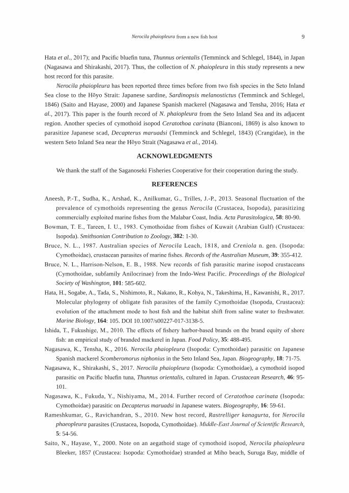
The Hōyo Strait is located between the western Seto Inland Sea and the Bungo Channell in western Japan. This strait is famous as a fishing ground of two perciform fishes of high quality, viz., chub mackerel, Scomber japonicus Houttuyn, 1782 (Scombridae), and Japanese jack mackerel, Trachurus japonicus (Temminck and Schlegel, 1844) (Carangidae), both of which are currently called “Seki-saba” and “Seki-aji”, respectively, as registered brands (e.g., Ishida and Fukushige, 2010). The brand names are well known nationwide, and the price of the fishes is very high (up to 5,000 yen per kg). Under these situations, the fishermen working in the strait pay much attention to the parasites of the fishes they catch because those fishes are almost exclusively eaten raw as “sashimi.” Recently, a chub mackerel infected by a large parasite on the body surface (Fig. 1A) was caught by a fisherman in the Hōyo Strait and was sent to us for identification. The parasite was identified as the cymothoid isopod Nerocila phaiopleura Bleeker, 1857, which is reported herein as a new host record.
MATERIALS AND METHODS
The fish was commercially caught using hook and line in the Hōyo Strait off Saganoseki, Oita Prefecture, on 30 January 2017. It was found to harbor a large skin parasite before auction and immediately transported to the Oita Prefectural Agriculture, Forestry and Fisheries Research Center, Saeki, where it was examined for the parasite after being photographed and measured for total length (TL). The parasite was carefully removed from the fish, fixed and preserved in 70% ethanol. This parasite specimen was sent to Hiroshima University, Higashi-Hiroshima, for identification. It is deposited in the Crustacea (Cr) collection of the National Museum of Nature and Science, Tsukuba, Ibaraki Prefecture (NSMT-Cr 25583).
生物圏科学Biosphere Sci. 56:7-11 (2017)
Accepted on September 19, 2017 *E-mail: [email protected]
8 Kazuya NAGASAWA and Hiroki NAKAO
RESULTS AND DISCUSSION
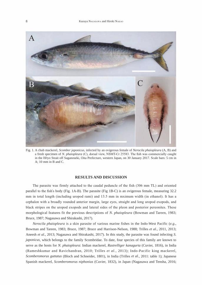
The parasite was firmly attached to the caudal peduncle of the fish (306 mm TL) and oriented parallel to the fi sh’s body (Fig. 1A-B). The parasite (Fig 1B-C) is an ovigerous female, measuring 32.2 mm in total length (including uropod rami) and 13.5 mm in mximum width (in ethanol). It has a cephalon with a broadly rounded anterior margin, large eyes, straight and long uropod exopods, and black stripes on the uropod exopods and lateral sides of the pleon and posterior pereonites. These morphological features fit the previous descriptions of N. phaiopleura (Bowman and Tareen, 1983; Bruce, 1987; Nagasawa and Shirakashi, 2017). Nerocila phaiopleura is a skin parasite of various marine fishes in the Indo-West Pacific (e.g., Bowman and Tareen, 1983; Bruce, 1987; Bruce and Harrison-Nelson, 1988; Trilles et al., 2011, 2013; Aneesh et al., 2013; Nagasawa and Shirakashi, 2017). In this study, the parasite was found infecting S. japonicus, which belongs to the family Scombridae. To date, four species of this family are known to serve as the hosts for N. phaiopleura: Indian mackerel, Rastrelliger kanagurta (Cuvier, 1816), in India (Rameshkumar and Ravichandran, 2010; Trilles et al., 2013); Indo-Pacific king mackerel, Scomberomorus guttatus (Bloch and Schneider, 1801), in India (Trilles et al., 2011: table 1); Japanese Spanish mackerel, Scomberomorus niphonius (Cuvier, 1832), in Japan (Nagasawa and Tensha, 2016;
Fig. 1. A chub mackerel, Scomber japonicus, infected by an ovigerous female of Nerocila phaiopleura (A, B) and a fresh specimen of N. phaiopleura (C), dorsal view, NSMT-Cr 25583. The fi sh was commercially caught in the Hōyo Strait off Saganoseki, Oita Prefecture, western Japan, on 30 January 2017. Scale bars: 5 cm in A; 10 mm in B and C.
Nerocila phaiopleura from a new fish host 9
Hata et al., 2017); and Pacific bluefin tuna, Thunnus orientalis (Temminck and Schlegel, 1844), in Japan (Nagasawa and Shirakashi, 2017). Thus, the collection of N. phaiopleura in this study represents a new host record for this parasite. Nerocila phaiopleura has been reported three times before from two fish species in the Seto Inland Sea close to the Hōyo Strait: Japanese sardine, Sardinopsis melanostictus (Temminck and Schlegel, 1846) (Saito and Hayase, 2000) and Japanese Spanish mackerel (Nagasawa and Tensha, 2016; Hata et al., 2017). This paper is the fourth record of N. phaiopleura from the Seto Inland Sea and its adjacent region. Another species of cymothoid isopod Ceratothoa carinata (Bianconi, 1869) is also known to parasitize Japanese scad, Decapterus maruadsi (Temminck and Schlegel, 1843) (Crangidae), in the western Seto Inland Sea near the Hōyo Strait (Nagasawa et al., 2014).
ACKNOWLEDGMENTS
We thank the staff of the Saganoseki Fisheries Cooperative for their cooperation during the study.
REFERENCES
Aneesh, P.-T., Sudha, K., Arshad, K., Anilkumar, G., Trilles, J.-P., 2013. Seasonal fluctuation of the prevalence of cymothoids representing the genus Nerocila (Crustacea, Isopoda), parasitizing commercially exploited marine fishes from the Malabar Coast, India. Acta Parasitologica, 58: 80-90.
Bowman, T. E., Tareen, I. U., 1983. Cymothoidae from fishes of Kuwait (Arabian Gulf) (Crustacea: Isopoda). Smithsonian Contribution to Zoology, 382: 1-30.
Bruce, N. L., 1987. Australian species of Nerocila Leach, 1818, and Creniola n. gen. (Isopoda: Cymothoidae), crustacean parasites of marine fishes. Records of the Australian Museum, 39: 355-412.
Bruce, N. L., Harrison-Nelson, E. B., 1988. New records of fish parasitic marine isopod crustaceans (Cymothoidae, subfamily Anilocrinae) from the Indo-West Pacific. Proceedings of the Biological Society of Washington, 101: 585-602.
Hata, H., Sogabe, A., Tada, S., Nishimoto, R., Nakano, R., Kohya, N., Takeshima, H., Kawanishi, R., 2017. Molecular phylogeny of obligate fish parasites of the family Cymothoidae (Isopoda, Crustacea): evolution of the attachment mode to host fish and the habitat shift from saline water to freshwater. Marine Biology, 164: 105. DOI 10.1007/s00227-017-3138-5.
Ishida, T., Fukushige, M., 2010. The effects of fishery harbor-based brands on the brand equity of shore fish: an empirical study of branded mackerel in Japan. Food Policy, 35: 488-495.
Nagasawa, K., Tensha, K., 2016. Nerocila phaiopleura (Isopoda: Cymothoidae) parasitic on Japanese Spanish mackerel Scomberomorus niphonius in the Seto Inland Sea, Japan. Biogeography, 18: 71-75.
Nagasawa, K., Shirakashi, S., 2017. Nerocila phaiopleura (Isopoda: Cymothoidae), a cymothoid isopod parasitic on Pacific bluefin tuna, Thunnus orientalis, cultured in Japan. Crustacean Research, 46: 95-101.
Nagasawa, K., Fukuda, Y., Nishiyama, M., 2014. Further record of Ceratothoa carinata (Isopoda: Cymothoidae) parasitic on Decapterus maruadsi in Japanese waters. Biogeography, 16: 59-61.
Rameshkumar, G., Ravichandran, S., 2010. New host record, Rastrelliger kanagurta, for Nerocila phaeopleura parasites (Crustacea, Isopoda, Cymothoidae). Middle-East Journal of Scientific Research, 5: 54-56.
Saito, N., Hayase, Y., 2000. Note on an aegathoid stage of cymothoid isopod, Nerocila phaiopleura Bleeker, 1857 (Crustacea: Isopoda: Cymothoidae) stranded at Miho beach, Suruga Bay, middle of
10 Kazuya NAGASAWA and Hiroki NAKAO
Japan. I. O. P. Diving News, 11(10): 2-6. (in Japanese with English abstract).Trilles, J.-P., Ravichandran, S., Rameshkumar, G., 2011. A checklist of the Cymothoidae (Crustacea,
Isopoda) recorded from Indian fishes. Acta Parasitologica, 56: 446-459.Trilles, J.-P., Rameshkumar, G., Ravichandran, S., 2013. Nerocila species (Crustacea, Isopoda,
Cymothoidae) from Indian marine fishes. Parasitology Research, 112: 1273-1286.
Nerocila phaiopleura from a new fish host 11
マサバはイワシノコバンの新宿主
長澤 和也1)・中尾 拓貴2)
1)広島大学大学院生物圏科学研究科,〒739-8528 広島県東広島市鏡山1-4-42)大分県農林水産研究指導センター水産研究部,〒879-2602 大分県佐伯市上浦大字津井浦194-6
要 旨 大分県佐賀関沖の豊予海峡で漁獲されたマサバの尾柄部に等脚類ウオノエ科のイワシノコバン Nerocila phaiopleura Bleeker, 1857の寄生を認めた。マサバはイワシノコバンの新宿主である。本報告は,瀬戸内海と周辺水域からのイワシノコバンの第4記録となる。キーワード:イワシノコバン,ウオノエ類,魚類寄生虫,新宿主,等脚類,マサバ

Two species of copepods, Lernanthropus atrox and Hatschekia pagrosomi, parasitic on crimson seabream, Evynnis tumifrons, in Hiroshima Bay, western Japan
Kazuya Nagasawa*
Graduate School of Biosphere Science, Hiroshima University, 1-4-4 Kagamiyama, Higashi-Hiroshima, Hiroshima 739-8528, Japan
Abstract Two species of copepods, Lernanthropus atrox Heller, 1865, and Hatschekia pagrosomi Yamaguti, 1939, were collected from the gills of crimson seabream, Evynnis tumifrons (Temminck and Schlegel, 1843), in Hiroshima Bay, the Seto Inland Sea, western Japan. This collection represents a new host record for L. atrox and the first record of H. pagrosomi from E. tumifrons in Japan. The hosts and geographical distribution of these copepods are also reviewed.Key words: Copepoda, Evynnis tumifrons, fish parasite, Hatschekia pagrosomi, Hiroshima Bay,
Lernanthropus atrox
INTRODUCTION
Sparids are widely distributed and commercially caught in coastal temperate and subtropical waters of Japan, where they consist of 13 species in three subfamilies and four genera (Nakabo, 2013). Of these species, red seabream, Pagrus major (Temminck and Schlegel, 1843), is the most important species in fisheries and abundantly caught in various waters of Japan. The parasite fauna of this species has been well studied in Japan: for example, as many as 24 species of metazoan parasitic helminths (3 monogeneans, 13 digneans, 4 cestodes, 3 nametodes, and 1 acanthocephalan) were reported only by Dr. Satyu Yamaguti (Kamegai and Ichihara, 1972), and four species of parasitic copepods are known to infect the fish species in the Seto Inland Sea, western Japan (Nagasawa, 2011). In contrast, much remains poorly known about the parasites of other Japanese sparids. Crimson seabream, Evynnis tumifrons (Temminck and Schlegel, 1843), is one of such sparids, and as its crustacean parasites, only the cymothoid isopod, Ceratothoa verrucosa (Schioedte and Meinert, 1883), and some unidentified parasitic copepods have been reported in Japan (Madinabeitia and Nagasawa, 2013; Nagasawa and Isozaki, 2016). The latter unidentified copepods were recorded from Hiroshima Bay, part of the western Seto Inland Sea, and belong to five families (Bomolochidae, Philichthyidae including “Colobomatus sp. 1”, Lernaeopodidae, Lernanthropidae, and Caligidae) (Madinabeitia and Nagasawa, 2013: tables 1-2), but their identification has not been made to species level. Recently, I examined individuals of E. tumifrons caught in Hiroshima Bay and collected two species of parasitic copepods, Lernanthropus atrox Heller, 1865, and Hatschekia pagrosomi Yamaguti, 1939.
MATERIALS AND METHODS
Eleven fresh individuals of E. tumifrons commercially caught in Hiroshima Bay on 8 April 2015
生物圏科学Biosphere Sci. 56:13-21 (2017)
Accepted on September 19, 2017 *E-mail: [email protected]
14 Kazuya NAGASAWA
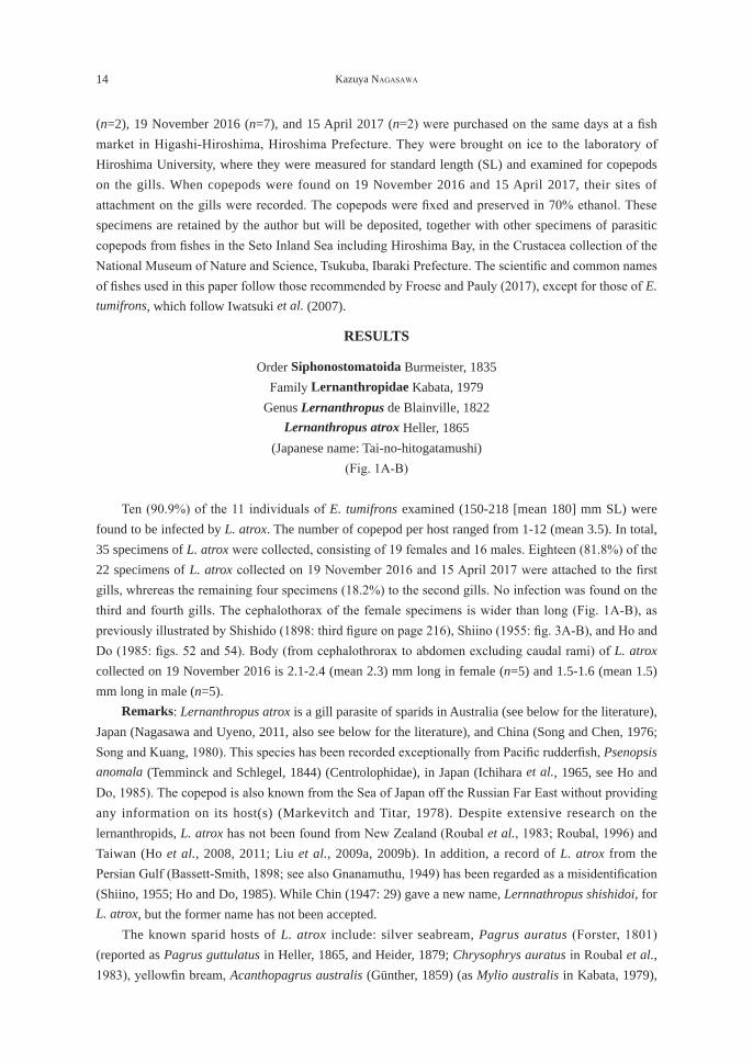
(n=2), 19 November 2016 (n=7), and 15 April 2017 (n=2) were purchased on the same days at a fish market in Higashi-Hiroshima, Hiroshima Prefecture. They were brought on ice to the laboratory of Hiroshima University, where they were measured for standard length (SL) and examined for copepods on the gills. When copepods were found on 19 November 2016 and 15 April 2017, their sites of attachment on the gills were recorded. The copepods were fixed and preserved in 70% ethanol. These specimens are retained by the author but will be deposited, together with other specimens of parasitic copepods from fishes in the Seto Inland Sea including Hiroshima Bay, in the Crustacea collection of the National Museum of Nature and Science, Tsukuba, Ibaraki Prefecture. The scientific and common names of fishes used in this paper follow those recommended by Froese and Pauly (2017), except for those of E. tumifrons, which follow Iwatsuki et al. (2007).
RESULTS
Order Siphonostomatoida Burmeister, 1835Family Lernanthropidae Kabata, 1979
Genus Lernanthropus de Blainville, 1822Lernanthropus atrox Heller, 1865
(Japanese name: Tai-no-hitogatamushi)(Fig. 1A-B)
Ten (90.9%) of the 11 individuals of E. tumifrons examined (150-218 [mean 180] mm SL) were found to be infected by L. atrox. The number of copepod per host ranged from 1-12 (mean 3.5). In total, 35 specimens of L. atrox were collected, consisting of 19 females and 16 males. Eighteen (81.8%) of the 22 specimens of L. atrox collected on 19 November 2016 and 15 April 2017 were attached to the first gills, whrereas the remaining four specimens (18.2%) to the second gills. No infection was found on the third and fourth gills. The cephalothorax of the female specimens is wider than long (Fig. 1A-B), as previously illustrated by Shishido (1898: third figure on page 216), Shiino (1955: fig. 3A-B), and Ho and Do (1985: figs. 52 and 54). Body (from cephalothrorax to abdomen excluding caudal rami) of L. atrox collected on 19 November 2016 is 2.1-2.4 (mean 2.3) mm long in female (n=5) and 1.5-1.6 (mean 1.5) mm long in male (n=5). Remarks: Lernanthropus atrox is a gill parasite of sparids in Australia (see below for the literature), Japan (Nagasawa and Uyeno, 2011, also see below for the literature), and China (Song and Chen, 1976; Song and Kuang, 1980). This species has been recorded exceptionally from Pacific rudderfish, Psenopsis anomala (Temminck and Schlegel, 1844) (Centrolophidae), in Japan (Ichihara et al., 1965, see Ho and Do, 1985). The copepod is also known from the Sea of Japan off the Russian Far East without providing any information on its host(s) (Markevitch and Titar, 1978). Despite extensive research on the lernanthropids, L. atrox has not been found from New Zealand (Roubal et al., 1983; Roubal, 1996) and Taiwan (Ho et al., 2008, 2011; Liu et al., 2009a, 2009b). In addition, a record of L. atrox from the Persian Gulf (Bassett-Smith, 1898; see also Gnanamuthu, 1949) has been regarded as a misidentification (Shiino, 1955; Ho and Do, 1985). While Chin (1947: 29) gave a new name, Lernnathropus shishidoi, for L. atrox, but the former name has not been accepted. The known sparid hosts of L. atrox include: silver seabream, Pagrus auratus (Forster, 1801) (reported as Pagrus guttulatus in Heller, 1865, and Heider, 1879; Chrysophrys auratus in Roubal et al., 1983), yellowfin bream, Acanthopagrus australis (Günther, 1859) (as Mylio australis in Kabata, 1979),

Two species of copepods parasitic on crimson seabream 15
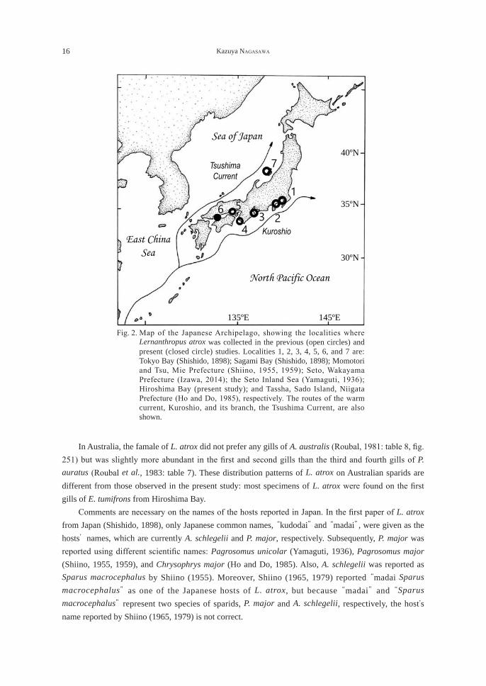
black bream, Acanthopagrus butcheri (Munro, 1949), and yellowfin seabream, Acanthopagrus latus (Houttuyn, 1782), from Australia (Heller, 1865; Heider, 1879; Kabata, 1979; Roubal, 1981, 1986, 1989, 1990a, 1990b, 1995, 1996; Roubal et al., 1983; Byrnes, 1988; Byrnes and Rohde, 1992); blackhead seabream, Acanthopagrus schlegelii (Bleeker, 1854), and red seabream, P. major, from Japan (Shishido, 1898; Yamaguti, 1936; Shiino, 1955, 1959; Ho and Do, 1985); and blackhead seabream, A. schlegelii (as Sparus macrocephalus), from China (Song and Chen, 1976; Song and Kuang, 1980). In the present study, L. atrox was collected for the first time from E. tumifrons, which represents a new host record for the copepod. This fish species is a third sparid host of L. atrox in Japan. The unidentified species of Lernanthropidae reported from E. tumifrons in Hiroshima Bay (Madinabeitia and Nagasawa, 2013: table 1) may be identifiable as L. atrox because the present material of copepod was collected from the same host species of the same locality. The localities of L. atrox recorded from Japan are: Tokyo Bay and Sagami Bay (Shishido, 1898, see Nagasawa and Uyeno, 2011); the Seto Inland Sea including Hiroshima Bay (Yamaguti, 1936; this paper); Momotori and Tsu, Mie Prefecture (Shiino, 1955, 1959); Tassha, Sado Island, Niigata Prefecture (Ho and Do, 1985); and Seto, Wakayama Prefecture (Izawa, 2014) (Fig. 2). These localities are in the temperate region of Japan and more or less affected by the warm current, Kuroshio, and its branch, the Tsushima Current (Fig. 2). Toward a further understanding of the geographical distribution of L. atrox in Japanese waters, it is desirable to examine sparids in southern Japan ranging from Shikoku through Kyushu to the Ryukyu Islands. It is interesting to note that L. atrox occurs in coastal waters of Hainan Island off southern China (Song and Chen, 1976; Song and Kuang, 1980) but has not been discovered from Taiwan (Ho et al., 2008, 2011; Liu et al., 2009a, 2009b).
A B C
Fig. 1. An ovigerous female of Lernanthropus atrox (A, dorsal view; B, ventral view) and an ovigerous female of Hatschekia pagrosomi (C, dorsal view) removed from the gills of Evynnis tumifrons in Hiroshima Bay, western Japan, on 19 November 2016. Scale bars: 1 mm in A and B; 0.5 mm in C.
16 Kazuya NAGASAWA
In Australia, the famale of L. atrox did not prefer any gills of A. australis (Roubal, 1981: table 8, fig. 251) but was slightly more abundant in the first and second gills than the third and fourth gills of P. auratus (Roubal et al., 1983: table 7). These distribution patterns of L. atrox on Australian sparids are different from those observed in the present study: most specimens of L. atrox were found on the first gills of E. tumifrons from Hiroshima Bay. Comments are necessary on the names of the hosts reported in Japan. In the first paper of L. atrox from Japan (Shishido, 1898), only Japanese common names, “kudodai” and “madai”, were given as the hosts’ names, which are currently A. schlegelii and P. major, respectively. Subsequently, P. major was reported using different scientific names: Pagrosomus unicolar (Yamaguti, 1936), Pagrosomus major (Shiino, 1955, 1959), and Chrysophrys major (Ho and Do, 1985). Also, A. schlegelii was reported as Sparus macrocephalus by Shiino (1955). Moreover, Shiino (1965, 1979) reported “madai Sparus macrocephalus” as one of the Japanese hosts of L. atrox, but because “madai” and “Sparus macrocephalus” represent two species of sparids, P. major and A. schlegelii, respectively, the host's name reported by Shiino (1965, 1979) is not correct.
Sea of Japan
North Pacific Ocean
Kuroshio
Tsushima Current
145ºE 135ºE
35ºN
40ºN
30ºN
East China Sea
235
16
4
7
Fig. 2. Map of the Japanese Archipelago, showing the localities where Lernanthropus atrox was collected in the previous (open circles) and present (closed circle) studies. Localities 1, 2, 3, 4, 5, 6, and 7 are: Tokyo Bay (Shishido, 1898); Sagami Bay (Shishido, 1898); Momotori and Tsu, Mie Prefecture (Shiino, 1955, 1959); Seto, Wakayama Prefecture (Izawa, 2014); the Seto Inland Sea (Yamaguti, 1936); Hiroshima Bay (present study); and Tassha, Sado Island, Niigata Prefecture (Ho and Do, 1985), respectively. The routes of the warm current, Kuroshio, and its branch, the Tsushima Current, are also shown.
Two species of copepods parasitic on crimson seabream 17
Order Siphonostomatoida Burmeister, 1835Family Hatschekiidae Kabata, 1979
Genus Hatschekia Poche, 1902Hatschekia pagrosomi Yamaguti, 1939
(Japanese name: Madai-no-eranomi)(Fig. 1C)
Two (11.1%) of the 11 individuals of E. tumifrons examined were found individually to harbor three and one ovigerous females of H. pagrosomi on the gills (three and one on the first and fourth gills, respectively). These females measure 1.5-2.1 (mean 1.9) mm (n=4) in body length (from cephalothorax to abdomen excluding caudal rami). Remarks: Hatschekia pagrosomi is a gill parasite of sparids in Japan (Yamaguti, 1939; Nagasawa and Uyeno, 2012), Korea (Kim, 1998), Australia (Roubal et al., 1983; Kabata, 1991; Roubal, 1996), and New Zealand (Roubal et al., 1983). The species has also been reported from two non-sparid fishes in Japan: Chinese emperor, Lethrinus haematopterus (Temminck and Schlegel, 1844) (Lethrinidae) (Yamaguti, 1939), and Japanese jack mackerel, Trachurus japonicus (Temminck and Schlegel, 1844) (Carangidae) (as Trachurus trachuri) (Yamaguti and Yamasu, 1960; see Jones, 1985, for synonymy). The known sparid hosts of H. pagrosomi are: red seabream, P. major, from Japan (Yamaguti, 1936); crimson seabream, E. tumifrons (as E. tanaka), from Korea (Kim, 1998); and silver seabream, P. auratus (as Chrysophrys auratus in Roubal et al., 1983; Kabata, 1991), from Australia and New Zealand (Roubal et al., 1983; Kabata, 1991; Roubal, 1996). The collection of H. pagrosomi in this study represents its second record from E. tumifrons and its first record from this fish species in Japan. The Seto Inland Sea is the only known locality of H. pagrosomi in Japan (Yamaguti, 1939; Yamaguti and Yamasu, 1960; this paper). While Kabata (1991) states that H. pagrosomi was collected by Ichihara et al. (1964) from T. japonicus in Sagami Bay, central Japan, his citation is wrong because the latter authors did not collect the copepod from the bay.
REFERENCES
Bassett-Smith, P. W., 1898. Further new parasitic copepods found on fish in the Indo-tropical region. Annals and Magazine of Natural History, 7: 77-98.
Byrnes, T., 1988. Lernanthropids and lernaeopodids (Copepoda) parasitic on Australian bream (Acanthopagrus spp.). Publications of the Seto Marine Biological Laboratory, 33: 97-120.
Byrnes, T., Rohde, K., 1992. Geographical distribution and host specificity of ectoparasites of Australian bream, Acanthopagrus spp. (Sparidae). Folia Parasitologica, 39: 249-264.
Chin, J.-H., 1947. On two new species of Lernanthropus (Copepoda parasitica) from Chinese marine fishes. Sinensia, 18: 21-33.
Froese, R., Pauly, D., eds. 2017. FishBase. World Wide Web electronic publication. www.fishbase.org, version (06/2017). (accessed on 3 August 2017).
Gnanamuthu, C. P., 1949. Lernanthropus sciaenae sp. nov., a copepod parasitic on the gills of the fish Sciaena glauca from Madras. Records of the Indian Museum, 45: 291-298.
Ho, J.-S., Do, T. T., 1985. Copepods of the family Lernanthropidae parasitic on Japanese marine fishes, with a phylogenetic analysis of the lernanthropid genera. Report of the Sado Marine Biological Station, Niigata University, 15: 31-76.
18 Kazuya NAGASAWA
Heller, C., 1865. Crustacean. In “Reise der Öesterreichischen Fregatte “Novara” um die Erde in der Jahren 1857, 1858, 1859, (Zoologischer Theil)”, 2(3): 1-280.
Heider. C., 1879. Die Gattung Lernanthropus. Arbeiten aus dem Zoologischen Institut der Universität Wien, 2(3): 269-368.
Ho, J.-S., Liu, W.-C., Lin, C.-L., 2008. Six species of lernanthropid copepods (Siphonostomatoida) parasitic on marine fishes of Taiwan. Journal of the Fisheries Society of Taiwan, 35: 251-280.
Ho, J.-S., Liu, W.-C., Lin, C.-L., 2011. Six species of Lernanthropidae (Crustacea: Copepoda) parasitic on marine fishes of Taiwan, with a key to 18 species of the family known from Taiwan. Zoological Studies, 50: 611-635.
Ichihara, A., Kato, K., Kamegai, Sh., Kamegai, S., Nonobe, H., Machida, M., 1964. On the parasites of fishes and shell-fishes in Sagami Bay (No. 2). Parasites of Trachurus trachurus (Tem. et Schl.) (I). Monthly Reports of the Meguro Parasitological Museum, 65: 2-5. (In Japanese with English title).
Ichihara, A., Kato, K., Kamegai, Sh., Kamegai, S., Nonobe, H., Machida, M., 1965. On the parasites of fishes and shell-fishes in Sagami Bay (No. 3). Part 2. Parasites of Psenopsis anomala (Temm. et Sch.), Part 3. Parasites of Gephyroberyx japaonicus (Döderiein). Monthly Reports of the Meguro Parasitological Museum, 78-80: 2-14. (In Japanese with English title).
Iwatsuki, Y., Akazaki, M., Taniguchi, N., 2007. Review of the species of the genus Dentex (Perciformes: Sparidae) in the western Pacific defined as the D. hypselosomus complex with the description of a new species, Dentex abei and a redescription of Evynnis tumifrons. Bulletin of the National Museum of Nature and Science, Series A, Supplement, 1: 29-49.
Izawa, K., 2014. Some new and known species of the Lernanthropidae (Copepoda, Siphonostomatoida) parasitic on the branchial lamellae of Japanese actinopterygian fishes, with revision of two known species of the family and discussion on the insemination mode in the Siphonostomatoida. Crustaceana, 87: 1521-1558.
Jones, J. B., 1985. A revision of Hatschekia Poche, 1902 (Copepoda: Hatschekiidae), parasitic on marine fishes. New Zealand Journal of Zoology, 12: 213-271.
Kabata, Z., 1979. Parasitic Copepoda of Australian fishes, XII. Family Lernanthropidae. Crustaceana, 37: 198-213.
Kabata, Z., 1991. Parasitic Copepoda of Australian fishes, XIII: family Hatschekiidae. Journal of Natural History, 25: 91-121.
Kamegai, S., Ichihara, A., 1972. A check list of the helminths from Japan and adjacent areas. Part I. Fish parasites reported by S. Yamaguti from Japanese waters and adjacent areas. Research Bulletin of the Meguro Parasitological Museum, 6: 1-43.
Kim, I.-H., 1998. Illustrated Encyclopedia of Fauna and Flora of Korea. Vol. 38. Cirripedia, Symbiotic Copepoda, Pycnogonida. Ministry of Education, Seoul. 1038 pp. (In Korean).
Liu, W.-C., Ho, J.-S., Lin, C.-L., 2009a. Three species of Lernanthropus de Blainville, 1822 (Copepoda, Lernanthropidae) parasitic on marine fishes of Taiwan. Journal of the Fisheries Society of Taiwan, 36: 29-48.
Liu, W.-C., Ho, J.-S., Lin, C.-L., 2009b. Another three species of Lernanthropus de Blainville, 1822 (Copepoda, Lernanthropidae) parasitic on marine fishes of Taiwan, with a key to the species of the genus Lernanthropus found in Taiwan. Journal of the Fisheries Society of Taiwan, 36: 119-134.
Madinabeitia, Y., Nagasawa, K., 2013. Double-netting: an alternative approach for the recovery of parasitic copepods from finfishes. Journal of Natural History, 47: 529-541.
Two species of copepods parasitic on crimson seabream 19
Markevitch, A. P., Titar, V. M., 1978. Copepod parasites of marine fishes from the Soviet Far East. In “Proceedings of the 4th International Congress of Parasitology, Section H.” Warsaw, Poland. pp. 38-39.
Nagasawa, K., 2011. A checklist of the parasitic copepods (Crustacea) of fishes and invertebrates of the Seto Inland Sea, Japan (1935-2011), with a new locality record for Caligus macarovi (Caligidae). Bulletin of the Hiroshima University Museum, 3: 113-128.
Nagasawa, K., Isozaki, S., 2016. Crimson seabream Evynnis tumifrons (Temminck & Schlegel, 1843) (Perciformes, Pagridae), a new host for Ceratothoa verrucosa (Schioedte & Meinert, 1883) (Isopoda, Cymothoidae). Crustaceana, 89: 1229-1232.
Nagasawa, K., Uyeno, D., 2011. A checklist of copepods of the family Lernanthropidae (Siphonostomatoida) from fishes in Japanese waters (1898-2011). Bulletin of the Biogeographical Society of Japan, 66: 17-25. (In Japanese with English abstract).
Nagasawa, K., Uyeno, D., 2012. A checklist of copepods of the families Dichelesthiidae, Hatschekiidae and Pseudohatschekiidae (Siphonostomatoida) from fishes in Japanese waters (1916-2012). Biosphere Science, 51: 37-59. (In Japanese with English abstract).
Nakabo, T., ed., 2013. Fishes of Japan with Pictorial Keys to the Species. Third Edition. Tokai University Press, Hadano. l + 2428 pp. (In Japanese with English title).
Roubal, F. R., 1981. The taxonomy and site specificity of the metazoan ectoparasites on the black bream, Acanthopagrus australis (Günther), in northern New South Wales. Australian Journal of Zoology, Supplement Series, 84: 1-100.
Roubal, F. R., 1986. Studies on monogeneans and copepods parasitizing the gills of a sparid (Acanthopagrus australis (Günther)) in northern New South Wales. Canadian Journal of Zoology, 64: 841-849.
Roubal, F. R., 1989. Comparative pathology of some monogenean and copepod ectoparasites on the gills of Acanthopagrus australis (family Sparidae). Journal of Fish Biology, 34: 503-514.
Roubal, F. R., 1990a. The parasites of the sparid Acanthopagrus australis in Australia. Bulletin of the European Association of Fish Pathologists, 10: 110-111.
Roubal, F. R., 1990b. Seasonal changes in ectoparasite infection of juvenile yellowfin bream, Acanthopagrus australis (Günther) (Pisces: Sparidae), from a small estuary in northern New South Wales. Australian Journal of Marine and Freshwater Research, 41: 411-427.
Roubal, F. R., 1995. Changes in monogenean and copepod infestation on captive Acanthopagrus australis (Sparidae). Journal of Fish Biology, 46: 423-431.
Roubal, F. R., 1996. A comparison of the ectoparasite assemblage on snapper, Pagrus austratus, from different regions in Australia and from New Zealand. International Journal for Parasitology, 26: 661-665.
Roubal, F. R., Armitage, J., Rohde, K., 1983. Taxonomy of metazoan ectoparasites of snapper, Chrysophrys auratus (family Sparidae), from southern Australia and New Zealand. Australian Journal of Zoology, Supplement Series, 94: 1-68.
Shiino, S. M., 1955. Copepods parasitic on Japanese fishes. 8. The Anthosomidae. Report of Faculty of Fisheries, Prefectural University of Mie, 2: 50-69.
Shiino, S. M., 1959. Sammlung der parasitischen Copepoden in der Präfekturuniversität von Mie. Report of Faculty of Fisheries, Prefectural University of Mie, 3: 334-374.
20 Kazuya NAGASAWA
Shiino, S. M., 1965. Lernanthropus atrox Heller. In “New Illustrated Encylopedia of the Fauna of Japan [II]” (ed. by Y. Okada, S. Uchida, T. Uchida). Hokuryu-kan, Tokyo. p. 498. (In Japanese).
Shiino, S. M., 1979. Lernanthropus atrox Heller. In “Illustrated Encylopedia of the Fauna of Japan, Newly Complied” (ed. by M. Imajima, M. Takeda). Hokuryu-kan, Tokyo. p. 401. (In Japanese).
Shishido, I., 1898. [Parasitic copepods, Lernanthropus]. Dobutsugaku Zasshi (Zoological Magazine), 10: 82-87, 120-125, 148-151, 215-218, 254-256, 337-340, 1 pl. (In Japanese).
Song, D. S., Chen, G., 1976. Some parasitic copepods of marine fishes of China. Acta Zoologica Sinica, 22: 406-424. (In Chinese with English abstract).
Song, D. S., Kuang, P. R., 1980. Illustrations of Chinese Animals - Crustacea. Volume 4. Science Publisher, Beijing. 90 pp. (In Chinese).
Yamaguti, S., 1936. Parasitic copepods from fishes of Japan. Part 3. Caligoida, II. Published by the author, Kyoto. 21 pp., 9 pls.
Yamaguti, S., 1939. Parasitic copepods from fishes of Japan. Part 5. Caligoida, III. Volumen Jubilare Pro Professor Sadao Yoshida, 2: 443-487, 20 pls.
Yamaguti, S., Yamasu, T., 1960. New parasitic copepods from Japanese fishes. Publications of the Seto Marine Biological Laboratory, 8: 141-152.
Two species of copepods parasitic on crimson seabream 21
広島湾産チダイに寄生していたカイアシ類2種,タイノヒトガタムシとマダイノエラノミ
長澤 和也
広島大学大学院生物圏科学研究科,〒739-8528 広島県東広島市鏡山1-4-4
要 旨 広島湾で漁獲されたチダイの鰓に寄生するカイアシ類を調べたところ,ヒトガタムシ科のタイノヒトガタムシ Lernanthropus atrox Heller, 1865とエラノミ科のマダイノエラノミHatschekia pagrosomi Yamaguti, 1939の寄生を認めた。チダイはタイノヒトガタムシの新宿主であり,わが国のチダイからマダイノエラノミが見出されたのは初めてある。寄生率や寄生数を示すとともに,両寄生虫の宿主や地理的分布に関する考察を行った。キーワード:カイアシ類,魚類寄生虫,タイノヒトガタムシ,チダイ,広島湾,マダイノエラノミ

東広島市におけるミカドケナガノミ Chaetopsylla mikadoの初記録
新田理人*
広島大学大学院生物圏科学研究科,〒739-8528 広島県東広島市鏡山1-4-4
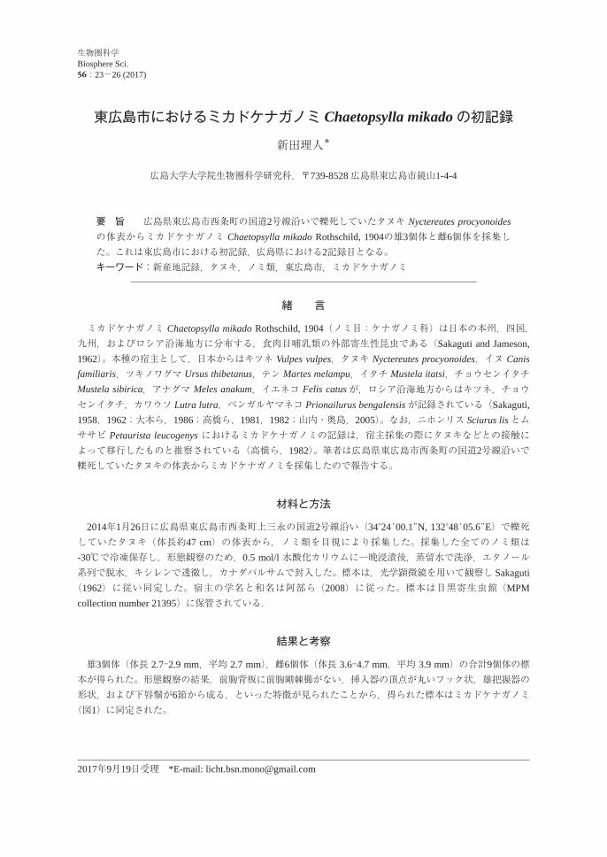
要 旨 広島県東広島市西条町の国道2号線沿いで轢死していたタヌキ Nyctereutes procyonoidesの体表からミカドケナガノミ Chaetopsylla mikado Rothschild, 1904の雄3個体と雌6個体を採集した。これは東広島市における初記録,広島県における2記録目となる。キーワード:新産地記録,タヌキ,ノミ類,東広島市,ミカドケナガノミ
緒 言
ミカドケナガノミ Chaetopsylla mikado Rothschild, 1904(ノミ目:ケナガノミ科)は日本の本州,四国,九州,およびロシア沿海地方に分布する,食肉目哺乳類の外部寄生性昆虫である(Sakaguti and Jameson, 1962)。本種の宿主として,日本からはキツネ Vulpes vulpes,タヌキ Nyctereutes procyonoides,イヌ Canis familiaris,ツキノワグマ Ursus thibetanus,テン Martes melampu,イタチ Mustela itatsi,チョウセンイタチ Mustela sibirica,アナグマ Meles anakum,イエネコ Felis catusが,ロシア沿海地方からはキツネ,チョウセンイタチ,カワウソ Lutra lutra,ベンガルヤマネコ Prionailurus bengalensisが記録されている(Sakaguti, 1958,1962;大本ら,1986;高橋ら,1981,1982;山内・奥島,2005)。なお,ニホンリス Sciurus lis とムササビ Petaurista leucogenys におけるミカドケナガノミの記録は,宿主採集の際にタヌキなどとの接触によって移行したものと推察されている(高橋ら,1982)。筆者は広島県東広島市西条町の国道2号線沿いで轢死していたタヌキの体表からミカドケナガノミを採集したので報告する。
材料と方法
2014年1月26日に広島県東広島市西条町上三永の国道2号線沿い(34°24'00.1"N, 132°48'05.6"E)で轢死していたタヌキ(体長約47 cm)の体表から,ノミ類を目視により採集した。採集した全てのノミ類は-30℃で冷凍保存し,形態観察のため,0.5 mol/l 水酸化カリウムに一晩浸漬後,蒸留水で洗浄,エタノール系列で脱水,キシレンで透徹し,カナダバルサムで封入した。標本は,光学顕微鏡を用いて観察し Sakaguti(1962)に従い同定した。宿主の学名と和名は阿部ら(2008)に従った。標本は目黒寄生虫館(MPM collection number 21395)に保管されている.
結果と考察
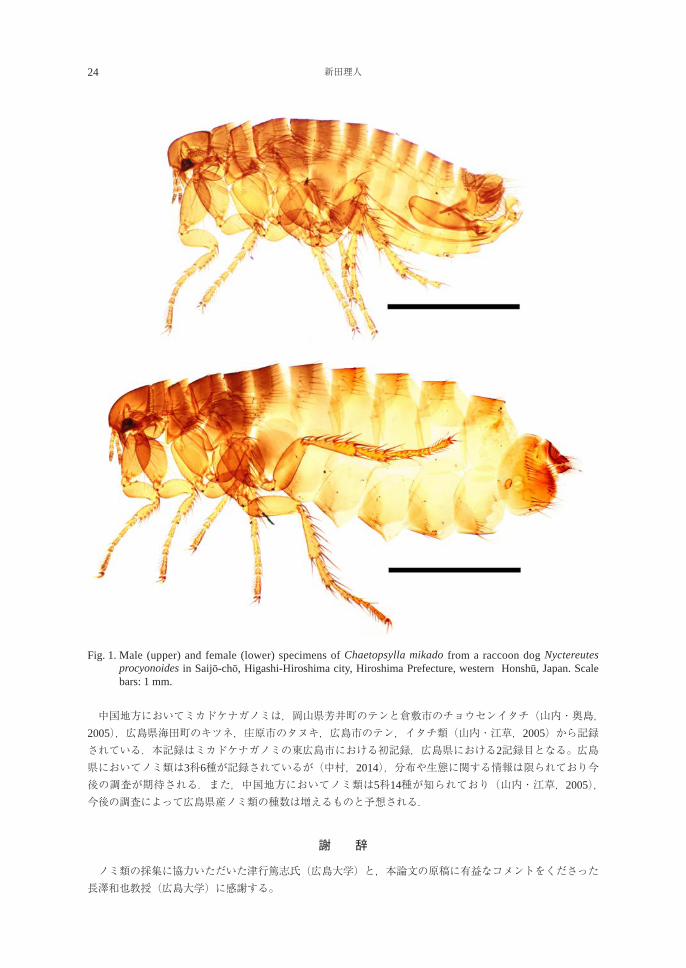
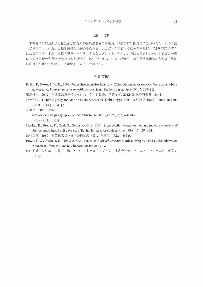
雄3個体(体長 2.7-2.9 mm,平均 2.7 mm),雌6個体(体長 3.6-4.7 mm,平均 3.9 mm)の合計9個体の標本が得られた。形態観察の結果,前胸背板に前胸剛棘櫛がない,挿入器の頂点が丸いフック状,雄把握器の形状,および下唇鬚が6節から成る,といった特徴が見られたことから,得られた標本はミカドケナガノミ (図1)に同定された。
2017年9月19日受理 *E-mail: [email protected]
生物圏科学Biosphere Sci. 56:23-26 (2017)
24 新田理人
Fig. 1. Male (upper) and female (lower) specimens of Chaetopsylla mikado from a raccoon dog Nyctereutes procyonoides in Saijō-chō, Higashi-Hiroshima city, Hiroshima Prefecture, western Honshū, Japan. Scale bars: 1 mm.
中国地方においてミカドケナガノミは,岡山県芳井町のテンと倉敷市のチョウセンイタチ(山内・奥島,2005),広島県海田町のキツネ,庄原市のタヌキ,広島市のテン,イタチ類(山内・江草,2005)から記録されている.本記録はミカドケナガノミの東広島市における初記録,広島県における2記録目となる。広島県においてノミ類は3科6種が記録されているが(中村,2014),分布や生態に関する情報は限られており今後の調査が期待される.また,中国地方においてノミ類は5科14種が知られており(山内・江草,2005),今後の調査によって広島県産ノミ類の種数は増えるものと予想される.
謝 辞
ノミ類の採集に協力いただいた津行篤志氏(広島大学)と,本論文の原稿に有益なコメントをくださった長澤和也教授(広島大学)に感謝する。
東広島市のミカドケナガノミ 25
引用文献
阿部 永・石井信夫・伊藤徹魯・金子之史・前田喜四雄・三浦慎悟・米田政明.2008.日本の哺乳類 改訂2版.東海大学出版会,秦野:xvi + 206 pp.
中村慎吾.2014.広島県昆虫誌 改訂増補版.比婆科学教育振興会,庄原:2840 pp.大本雅由・入佐鋭昭・矢富謙治・大西富雄.1986.兵庫県下における,イヌ,ネコの外部寄生虫について.食品衛生研究.37(4): 73-80.
Sakaguti, K., 1958. On the genus Chaetopsylla of Japan, with description of a new species (Siphonaptera), studies on the Japanease Siphonaptera V. Medical Journal of Osaka University 8: 771-783.
Sakaguti, K., 1962. A monograph of the Siphonaptera of Japan. The Nippon Printing and Publishing Co. Ltd., Osaka: 255 pp.
Sakaguti, K., Jameson, E. W. Jr. 1962. The Siphonaptera of Japan. Pacific Insects Monograph, 3: 1-169.高橋 守・斎藤 貴・町田和彦・大沢賢治・井上茂樹.1981.埼玉県における中・大型哺乳類寄生ノミ相(I).昆虫と自然.16(8): 31-33.
高橋 守・山本貞司・斎藤 貴・町田和彦.1982.埼玉および群馬県下における哺乳類および鳥類に寄生するノミ類 I 中・大型哺乳類に寄生するノミ類.大原綜合病院年報.25: 7-24.
山内健生・江草真治.2005.広島県の中型哺乳類および鳥類に寄生するノミ類.昆蟲 ニューシリーズ.8(2): 37-42.
山内健生・奥島雄一.2005.倉敷市立自然史博物館に所蔵されている岡山県産の哺乳類外部寄生昆虫標本(シラミ目,ノミ目,ハエ目).倉敷市立自然史博物館研究報告.20: 33-35.
26 新田理人
A new record of Chaetopsylla mikado from Higashi-Hiroshima city, Hiroshima Prefecture
Masato Nitta
Graduate School of Biosphere Science, Hiroshima University 1-4-4 Kagamiyama, Higashi-Hiroshima, Hiroshima 739-8528, Japan
Abstract: Three male and six female specimens of Chaetopsylla mikado Rothschild, 1904 (Siphonaptera: Vermipsyllidae) were collected from a raccoon dog Nyctereutes procyonoides in Saijō-chō, Higashi-Hiroshima city, Hiroshima Prefecture, western Honshū, Japan, on 26 January 2014. This collection represents the first record of C. mikado from Higashi-Hiroshima city and the second report from Hiroshima Prefecture.Key words: Chaetopsylla mikado, Higashi-Hiroshima city, new locality record, Nyctereutes procyonoides,
Siphonaptera
大島新曽根で採集されたトヨシオマリヒトデ Podosphaeraster toyoshiomaruae の行動観察
米谷まり1)・飯田 健1)・藤 太稀1)・平野勝士1)・近藤裕介1)・大塚 攻1)*中口和光2)・山口修平2)・加藤幹雄2)・広瀬雅人3)・藤田敏彦4)
1)広島大学大学院生物圏科学研究科附属瀬戸内圏フィールド科学教育研究センター竹原ステーション〒725-0024 広島県竹原市港町5-8-1
2)広島大学生物生産学部附属練習船豊潮丸 〒737-0029 広島県呉市宝町7-43)北里大学海洋生命科学部 〒252-0373 神奈川県相模原市南区北里1-15-1
4)国立科学博物館動物研究部 〒305-0005 茨城県つくば市天久保4-1-1
要 旨 トヨシオマリヒトデ Podosphaeraster toyoshiomaruae Fujita and Rowe, 2002は体がほぼ球形で,腕が発達しないという特殊な形態を持つ小型ヒトデ類である。鹿児島県奄美大島北西部に位置する大島新曽根水深100-200 m程度の堆に生息している。この堆は ROVで観察するとカイメン類,八放サンゴ類などで覆われている。2017年5月21日に本種の生きた個体が大島新曽根で採集され,管足を伸ばした状態や歩行が観察されたのでその行動を記載した。管足を体内にしまった状態とは異なり,体がやや口・肛門軸方向に扁平になり,約1.65 cm/minの速度で歩行した。通常の腕の発達したヒトデ類の歩行速度と比較すると相対的に著しく遅い。管足には少なくとも2種類が認められ,歩行用と感覚用と考えられる。キーワード:ROV,大島新曽根,管足,トヨシオマリヒトデ,歩行
緒 言
鹿児島県奄美大島の北西約39 kmに大島新曽根(28°53′N,129°32′E)という水深約100-200 m程度の堆がある(Fujita and Rowe, 2002; JAMSTEC, 2009)。この堆にはカイメン類などが底質一面を覆っていることがROVでの調査で判明している(JAMSTEC, 2009)。この堆から1999年5月29日に,腕が顕著でないため体全体がほぼ球形(直径21.5 mmまで)を呈した小型ヒトデ類が発見され,Fujita and Rowe(2002)によって新種 Podosphaeraster toyoshiomaruae Fujita and Rowe, 2002(和名 トヨシオマリヒトデ)として記載され,さらに本属をタイプとする新科が設立された。 2012年5月29日に同海域で生きているトヨシオマリヒトデが4個体採集されたので行動を観察したところ,形が口・肛門軸方向にやや扁平になり,管足を伸ばしていることが確認された(広瀬,2012)。さらに,2017年5月21日,同海域で再度,本種1個体が生きたまま採集された。その際に運動時の体の形態を記述し,管足による歩行速度などを測定したのでここに報告する。
材料と方法
トヨシオマリヒトデの採集は,2017年5月21日,大島新曽根(28°53′N,129°33′E,水深159-166 m)においてドレッジ(口幅50 cm,メッシュサイズ5 mm)で行った。ドレッジは広島大学生物生産学部附属練習船豊潮丸によって2ノットで航行させながらワイヤーを徐々に繰り出し,着底をワイヤーの振動具合で確認後
2017年9月19日受理 *Corresponding author: [email protected]
生物圏科学Biosphere Sci. 56:27-32 (2017)
28米谷まり・飯田 健・藤 太稀・平野勝士・近藤裕介・大塚 攻
中口和光・山口修平・加藤幹雄・広瀬雅人・藤田敏彦
(ワイヤー長350 m),3分間曳網した。この時の海表面水温は23.6℃(表層連続観測装置,日本海洋株式会社)であった。トヨシオマリヒトデを採集した後,採集地点の表層の海水を満たした円柱形の小型プラスチック容器(丸型 V式容器:内径78 mm,高さ45.7 mm)に入れて歩行行動を観察した。この時の室温は船内空調設備により約27℃に保たれていた。また,照度は470-580 lxであった(デジタル照度計 LX-1108,株式会社マザーツール)。行動はデジタルビデオカメラ(Handycam HDR-CX550,ソニー株式会社)で記録した。ヒトデの歩行速度はビデオで撮影し,水平方向に移動した距離(ビデオ画面にスケールを写し込んだ)と時間から計算した。10秒間の直線移動距離から歩行速度を算出し,これを16回分測定して平均,標準偏差を求めた。計測はヒトデがある程度歩行し,ペースが安定してから開始した。 採集地点近傍の海底の映像は,2009年9月29日,海洋研究開発機構(JAMSTEC)所有の ROV Hyper-Dolphinによる調査(Dive #1059,研究代表者東京大学松永茂樹教授)によって撮影され,松永教授より本論文に掲載の許可をいただいた。
結果と考察
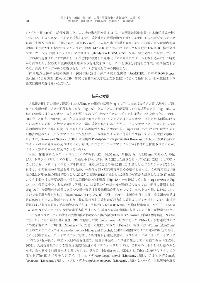
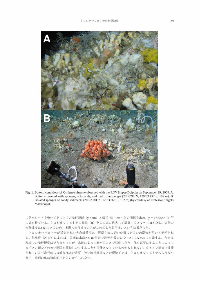
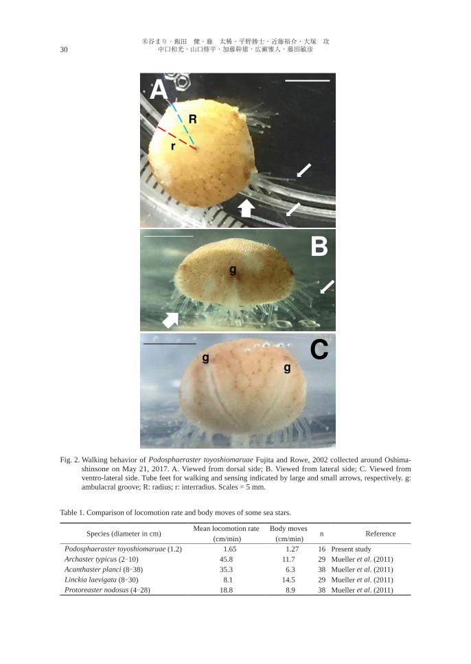
大島新曽根付近の調査で観察された水深182 mの海底の状態をFig. 1に示す。海底はカイメン類,八放サンゴ類,ヒドロ虫類のポリプで一面覆われており(Fig. 1A),ところどころ砂が蓄積している場所もある(Fig. 1B)。これらの映像にはトヨシオマリヒトデが写っておらず,そのマイクロハビタットは特定できなかった。1999年,2000年,2001年,2012年,2015年にほぼ同一地点で行ったドレッジではトヨシオマリヒトデが本画像に映っているカイメン類,八放サンゴ類などと一緒に採集されていることから,トヨシオマリヒトデはこれらの固着動物の体上かそれらに接して生息している可能性が高いと思われる。Fujita and Rowe(2002)はカイメンの体表の窪みからトヨシオマリヒトデを見いだし,本種がカイメンに付着して生活している可能性を示唆した。また,Rowe and Nichols(1980)は同属の Podosphaeraster pulvinatus Rowe and Nichols, 1980を枝状のカイメンの体の隙間から見つけている。なお,これまでトヨシオマリヒトデが9個体以上採集されているが,カイメン類の体内からは見つかっていない。 今回,採集されたトヨシオマリヒトデの輻長(R)は6.50 mm,間輻長(r)は5.85 mmであった(Fig. 2A)。トヨシオマリヒトデの Rと rの差は小さい。以下,Rを2倍した長さをヒトデの直径(2R)として扱うことにする。トヨシオマリヒトデを採集後,速やかに現場の海水215 mLを満たしたプラスチック容器に入れると,その直後から管足を伸ばし始め,体全体も口・肛門軸方向にやや扁平となった。この時の高さ /直径の比は0.75-0.88の範囲で変化した。2012年に広瀬(2012)が撮影した2個体の写真から計算した比(0.47, 0.55)よりも本個体は扁平度が高い。管足は口側の5つの歩帯溝(Fig. 2A)から伸ばしている(large arrows in Fig. 2A, B)。管足は少なくとも2種類に区別され,口周辺のものは先端が吸盤状になっており歩行に使用するが(Fig. 2C),歩帯溝の先端部にあるやや細い管足は吸盤状構造が明らかでなく,体の上方や側方に伸ばしているので感覚用と考えられる(small arrows in Fig. 2A, B)(西村,1995)。本種が歩行する間,感覚用の管足は常に体のやや上方に伸ばされており,特に進行方向の管足は反対方向の管足より長く伸長していた。歩行用管足および進行方向側の感覚用管足の長さは,それぞれ2.68 ± 0.96 mm(平均±標準偏差,N = 16),1.56 ± 0.48 mm(N = 5)であった。歩行は水平方向だけでなく,垂直な容器の側面にも登っていく様子が観察された。 トヨシオマリヒトデの10秒毎の移動距離を平均すると歩行速度は1.65 ± 3.23 cm/min(平均±標準偏差,N = 16)であった。この平均値を体の直径(2R)で除算した比(body move)は1.27であった(Table 1)。歩行速度およびこの比を他のヒトデ類4種(Mueller et al. 2011)と比較してみた(Table 1)。輻長(R)が1 cm(直径2 cm)以下のカスリモミジガイ Archaster typicus Müller and Troschel, 1840の小型個体ではこの比が約6-12であり,それと比較するとトヨシオマリヒトデは著しく相対的歩行速度が遅い。カスリモミジガイはトヨシオマリヒトデに比べ腕が長く,中型~大型の浅海性種で,底質が砂底やサンゴ礁に生息している種である(佐波ら,2002)。大島新曽根のような複雑な底質に生息するトヨシオマリヒトデは,これらのヒトデとは形態のみならず,全く異なる行動を示すと考えられる。さらに,Mueller et al.(2011)は Table 1に挙げたフィリピン産ヒトデ類4種 カスリモミジガイ,オニヒトデ Acanthaster planci(Linnaeus, 1758),アオヒトデ Linckia laevigata(Linnaeus, 1758),コブヒトデ Protoreaster nodosus(Linnaeus, 1758)について,生息海域の海底
トヨシオマリヒトデの行動観察 29
B
A
Fig. 1
Fig. 1. Bottom conditions of Oshima-shinsone observed with the ROV Hyper-Dolphin on September 29, 2009. A. Bottoms covered with sponges, octocorals, and hydrozoan polyps (28°52′98″N, 129°33′136″E, 182 m); B. Isolated sponges on sandy sediments (28°52′181″N, 129°33′63″E, 182 m) (by courtesy of Professor Shigeki Matsunaga).
に防水シートを敷いてその上での歩行距離(y : cm)と輻長(R : cm)との関係を求め,y = 17.822× R-1.261
の式を得ている。トヨシオマリヒトデの輻長(R)をこの式に代入して計算すると y = 1.68となる。実際の歩行速度は1.65であるため,実際の歩行速度の方がこの式より若干遅いという結果だった。 トヨシオマリヒトデが採集された大島新曽根は,黒潮主流に近い位置にあるため潮流が早いと予想される。気象庁(2017)によれば,黒潮は水深200 m付近で流速が最大になり2.0-2.5 m/sにも達する。今回は現場での歩行観察はできなかったが,水流によって転がることで移動したり,体を扁平にすることによってカイメン類などの狭い間隙を移動したりすることが可能となっているのかもしれない。カイメン類等で被覆されている三次元的に複雑な海底の底質,速い流速潮流などの環境下では,トヨシオマリヒトデのような小型で,球形の体は適応的であるのかもしれない。
30米谷まり・飯田 健・藤 太稀・平野勝士・近藤裕介・大塚 攻
中口和光・山口修平・加藤幹雄・広瀬雅人・藤田敏彦
Fig. 2. Walking behavior of Podosphaeraster toyoshiomaruae Fujita and Rowe, 2002 collected around Oshima-shinsone on May 21, 2017. A. Viewed from dorsal side; B. Viewed from lateral side; C. Viewed from ventro-lateral side. Tube feet for walking and sensing indicated by large and small arrows, respectively. g: ambulacral groove; R: radius; r: interradius. Scales = 5 mm.
B
C
A
g
gg
R
r
Fig. 2Table 1. Comparison of locomotion rate and body moves of some sea stars.
Species (diameter in cm)Mean locomotion rate
(cm/min)Body moves
(cm/min)n Reference
Podosphaeraster toyoshiomaruae (1.2) 1.65 1.27 16 Present studyArchaster typicus (2-10) 45.8 11.7 29 Mueller et al. (2011)Acanthaster planci (8-38) 35.3 6.3 38 Mueller et al. (2011)Linckia laevigata (8-30) 8.1 14.5 29 Mueller et al. (2011)Protoreaster nodosus (4-28) 18.8 8.9 38 Mueller et al. (2011)
トヨシオマリヒトデの行動観察 31
謝 辞
本調査では広島大学生物生産学部附属練習船豊潮丸の乗組員,乗船者には採集でご協力いただいたので記して感謝申し上げる。大島新曽根の海底の映像を提供いただいた東京大学松永茂樹教授,JAMSTECの方々には深謝する。また,原稿を査読いただき,貴重なコメントをいただいた方にも深謝したい。本研究の一部は日本学術振興会科学研究費(基盤研究 C,No.16K07825,代表 大塚攻),国立科学博物館総合研究「黒潮に注目した地史・生物史・人類史」によって行われた。
引用文献
Fujita, T., Rowe, F. W. E., 2002. Podosphaerasteridae fam. nov. (Echinodermata: Asteroidea: Valvatida), with a new species, Podosphaeraster toyoshiomaruae, from Southern Japan. Spec. Div. 7: 317-332.
広瀬雅人.2012.南西諸島海域で得られたコケムシ動物.豊潮丸 No 2012-05 航海報告書:38-41.JAMSTEC (Japan Agency for Marine-Earth Science & Technology). 2009. NATSUSHIMA: Cruise Report:
NT09-17, Leg. 1, 34 pp.気象庁.2017.黒潮. http://www.data.jma.go.jp/kaiyou/shindan/sougou/html_vol2/2_2_2_vol2.html (2017年6月1日閲覧)Mueller, B., Bos, A. R., Graf, G., Gumanao, G. S., 2011. Size-specific locomotion rate and movement pattern of
four common Indo-Pacific sea stars (Echinodermata; Asteoidea). Aquat. Biol. 12: 157-164.西村三郎.1995.原色検索日本海岸動物図鑑(Ⅱ).保育社,大阪:663 pp.Rowe, F. W., Nichols, D., 1980. A new species of Podosphaeraster Clark & Wright, 1962 (Echinodermata:
Asteroidea) from the Pacific. Micronesica 16: 289-295.佐波征機・入村精一・楚山 勇.2002.ヒトデガイドブック.株式会社ティビーエス・ブリタニカ,東京;
135 pp.
32米谷まり・飯田 健・藤 太稀・平野勝士・近藤裕介・大塚 攻
中口和光・山口修平・加藤幹雄・広瀬雅人・藤田敏彦
An observation of the walking behavior of Podosphaeraster toyoshiomaruae collected from the bank Oshima-shinsone, Kagoshima Prefecture, Japan
Mari Yonetani1), Ken Iida
1), Taiki Fuji1), Katsushi Hirano
1), Yusuke Kondo1), Susumu ohtsuKa
1), Kazumitsu naKaguchi
2), Shuhei Yamaguchi2), Mikio Kato
2), Masato hirose3) and Toshihiko Fujita
4)
1)Takehara Station, Setouchi Field Science Center, School of Biosphere Science, Hiroshima University, 5-8-1 Minato-machi, Takehara, Hiroshima 725-0024, Japan
2)Training and Research Vessel Toyoshio-maru, Faculty of Applied Biological Science, Hiroshima University, 7-4 Takara-machi, Kure, Hiroshima 737-0029, Japan
3)School of Marine Biosciences, Kitasato University, 1-15-1 Kitasato, Minami-ku, Sagamihara, Kanagawa 252-0373, Japan4)Department of Zoology, National Museum of Nature and Science, 4-1-1 Amakubo, Tsukuba, Ibaraki 305-0005, Japan
Abstract The podosphaerastrid asteroid Podosphaeraster toyoshiomaruae Fujita and Rowe, 2002 is small-sized, nearly spherical in shape. The species exclusively inhabits on the bank Oshima-shinsone at depths of 100 to 200 m, northwest of Amami-Oshima Island, Kagoshima Prefecture, Japan, where the bottom is entirely covered with sponges, octocorals, and hyrozoan polyps with patches of sandy bottom. The bottom conditions were clearly observed with a ROV on September 29, 2009. A living specimen was collected from this locality on May 21, 2017. During locomotion by tube feet, the body was depressed dorso-ventrally in contrast with the spherical body shape with tube feet unexpanded. The locomotion rate of the specimen was about 1.65 cm/min. Two types of tube feet were identified. Presumably one is for locomotion with a sucker terminally, while the other for sensing.Key words: locomotion, Oshima-shinsone, Podosphaeraster toyoshiomaruae, ROV, tube foot
REVIEW
A revised and updated checklist of the parasites of eels (Anguilla spp.)(Anguilliformes: Anguillidae) in Japan (1915-2017)
Kazuya Nagasawa1)* and Hirotaka Katahira
2)
1) Graduate School of Biosphere Science, Hiroshima University,1-4-4 Kagamiyama, Higashi-Hiroshima, Hiroshima 739-8528, Japan
2) Faculty of Bioresources, Mie University, 1577 Kurima machiya-cho, Tsu, Mie 514-8507, Japan
Abstract Information on the protistan and metazoan parasites of four species of eels (the Japanese eel Anguilla japonica, the giant mottled eel Anguilla marmorata, the European eel Anguilla anguilla, and the short-finned eel Anguilla australis) in Japan is summarized in the Parasite-Host and Host-Parasite lists, based on the literature published for 103 years between 1915 and 2017. This is a revised and updated version of the checklist published in 2007. Anguilla japonica and A. marmorata are native to Japan, whereas A. anguilla and A. australis are introduced species from Europe and Australia, respectively. The parasites, including 54 nominal species and those not identified to species level, are listed by higher taxa as follows: Sarcomastigophora (no. of nominal species: 0), Ciliophora (6), Microspora (1), Myxozoa (6), Trematoda (12), Monogenea (8), Cestoda (3), Nematoda (7), Acanthocephala (6), Hirudinida (3), Bivalvia (1), and Copepoda (1). For each parasite species listed, the following information is given: its currently recognized scientific name, any original combination, synonym(s), or other previous identification used for the parasite from Japanese eels; habitat (freshwater, brackish, or marine); site(s) of infection within or on the host; known geographical distribution in Japanese waters; and the published source of each locality record. Of the 54 nominal species of parasites listed, 50 are from A. japonica, six from A. marmorata, nine from A. anguilla, and one from A. australis. Five species, viz., Gyrodactylus anguillae, Gyrodactylus nipponensis, Pseudodactylogyrus mundayi (Monogenea), Bothriocephalus claviceps (Cestoda), and Raphidascaris acus (Nematoda), have been regarded as introduced parasites from other countries, and the remaining 49 nominal species are indigenous parasites of Japan. Nine nominal species of marine and/or brackish-water origin, viz., Lecithochrium musculus, Proctotrematoides pisodontophidis, Tubulovesicula anguillae (Trematoda), Gyrodactylus nipponensis, Pseudodactylogyrus kamegaii (Monogenea), Nybelinia angullicola (Cestoda), Cucullanus filiformis, Heliconema anguillae (Nematoda), and Limnotrachelobdella okae (Hirudinida), have been reported from A. japonica. Individiduals of A. japonica known as “sea eels” and “estuarine eels” inhabiting coastal marine and riverine brackish waters are considered to serve as hosts for those marine and/or brackish-water parasites.Key words: Anguilla anguilla, Anguilla australis, Anguilla japonica, Anguilla marmorata, bibliography,
checklist, eels, parasites
生物圏科学Biosphere Sci. 56:33-69 (2017)
Accepted on September 19, 2017 *E-mail: [email protected]
34 Kazuya NAGASAWA and Hirotaka KATAHIRA
INTRODUCTION
In 2007, A checklist of the parasites of eels (Anguilla spp.) (Anguilliformes: Anguillidae) in Japan (1915-2007) was published based on the literature published for 93 years between 1915 and 2007 (Nagasawa et al., 2007). This checklist contained the information on both protistan and metazoan parasites reported from three species of freshwater eels (the Japanese eel Anguilla japonica Temminck and Schlegel; the giant mottled eel Anguilla marmorata Quoy and Gaimard; and the European eel Anguilla anguilla (Linnaeus)) in Japan, and 44 nominal species of parasites were listed by higher taxa as follows: Ciliophora (6), Microspora (1), Myxozoa (6), Trematoda (7), Monogenea (7), Cestoda (3), Nematoda (7), Acanthocephala (4), Hirudinida (2), and Copepoda (1). It also contained the information on unidentified species of Sarcomastigophora, Ciliophora, Microspora, Myxozoa, Trematoda, Monogenea, Cestoda, and Nematoda. The checklist is revised and updated herein based on three sources of the literature: 1) the papers cited in the 2007 version; 2) those overlooked in the 2007 version (Nagao, 1956; Isobe, 1956, 1962; Irie, 1958; Egusa, 1958; Furukawa and Kobayashi, 1966; Ito, 1968; Horiuchi et al., 1988; Nagasawa, 1991; Rahhou et al., 2005; Shimazu and Araki, 2006; Shimazu, 2007); and 3) those published between the years 2008 and 2017 (Shimazu, 2008; Wielgross et al., 2008; Fang et al., 2008; Tanaka et al., 2009; Shimazu et al., 2011; Katahira et al., 2011, 2012, 2016: Laetsch et al., 2012; Nagasawa et al., 2013; Shimazu, 2014a, 2014b, 2015, 2016a, 2016b; Katahira and Nagasawa, 2014, 2015; Nagasawa and Utsumi, 2015; Ogawa et al., 2015; Kan et al., 2016; Nagasawa and Kan, 2017). In this revised checklist, we deal with the parasites reported from A. japonica, A. marmorata, A. anguilla, and the short-finned eel Anguilla australis Richardson. Anguilla japonica and A. marmorata are native to Japan, whereas A. anguilla and A. australis are introduced species from Europe and Australia, respectively. A total of 54 nominal species of parasites and those not identified to species level are listed herein, and the following 11 nominal species are newly included: 1. Coitocaecum plagiorchis Ozaki, 1926 (Trematoda) from Anguilla japonica (Shimazu et al., 2011); 2. Genarchopsis anguillae Yamaguti, 1938 (Trematoda) from Anguilla japonica (Shimazu, 2015); 3. Genarchopsis chubuensis Shimazu, 2015 (Trematoda) from Anguilla japonica (Shimazu, 2015); 4. Genarchopsis gigi Yamaguti, 1938 (Trematoda) from Anguilla japonica (Shimazu, 2015); 5. Isoparorchis eurytremus (Kobayashi, 1915) (Trematoda) from Anguilla japonica (Nagasawa et al.,
2013); 6. Palaeorchis diplorchis (Yamaguti, 1936) (Trematoda) from Anguilla japonica (Shimazu et al.,
2011); 7. Pseudodactylogyrus mundayi Ogawa, Iwashita, Hayward and Kurashima, 2015 (Monogenea) from
Anguilla australis (Ogawa et al., 2015); 8. Acanthocephalus longiacanthus Katahira and Nagasawa, 2014 (Acanthocephala) from Anguilla
marmorata (Katahira and Nagasawa, 2014); 9. Southwellina hispida (Van Cleave, 1925) (Acanthocephala) from Anguilla marmorata (Katahira
and Nagasawa, 2014; Nagasawa and Kan, 2017); 10. Limnotrachelobdella okae (Moore, 1924) (Hirudinida) from Anguilla japonica (Nagasawa and
Utsumi, 2015); and 11. Hyriopsis schlegeli (Martens, 1861) (Mollusca) from Anguilla japonica (Furukawa and Kobayashi,
1966).
A revised and updated checklist of the parasites of eels in Japan 35
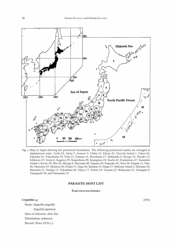
A new scientific name is adopted herein for each of the following species because their scientific name has currently been changed: Pseudophyllodistomum macrobrachicola (Yamaguti, 1934) (Trematoda), Anguillicola crassus Kuwahara, Niimi and Itagaki, 1974 (Nematoda), and Heliconema anguillae Yamaguti, 1935 (Nematoda). These species were reported as Phyllodistomum anguilae, Anguillicoloides crassus, and Heliconema longissimum, respectively, in the 2007 version. Moreover, Genarchopis goppo Ozaki, 1925 (Trematoda) listed in the 2007 version has been re-identified and separated by Shimazu (2015) into three species, itself, Genarchopsis gigi Yamaguti, 1939, and Genarchopsis chubuensis Shimazu, 2015, the latter two species of which are listed herein. Like in Nagasawa et al. (2007), the information on the parasites reported from Japanese Anguilla spp. is assembled as Parasite-Host and Host-Parasite lists. In the PARASITE-HOST LIST, the parasites are arranged by higher taxa in the following order: Sarcomastigophora, Ciliophora, Microspora, Myxozoa, Trematoda, Monogenea, Cestoda, Nematoda, Acanthocephala, Hirudinida, Bivalvia, and Copepoda. Within each higher taxa, genera and species are listed alphabetically. For each species of parasite, the following information is provided: 1) The current scientific name, including author(s) and date(s), followed by any original combination, recognized synonym(s), or other identifications(s) that have been used in establishing records from Anguilla spp. in Japan. 2) The habitat in which the parasite was acquired and normally completes its life cycle is given as FW for fresh waters, B for brackish waters, and M for marine waters. 3) The Site(s) of infection of the parasite in or on its host. If the site was not given in the original record, the likely site was determined from other records and is enclosed in square brackets. 4) The Distribution of the parasite is indicated by prefecture (boundaries shown in Fig. 1), in geographical order from northeast to southwest in Japan. 5) The Record(s). The authors responsible for the records are listed in chronological order. If a parasite has been reported more than once, the references are numbered, but not when there has been only one record of the parasite. Each reference is followed by the locality or localities given in two parts, first the prefecture and then the detailed collection locality or localities from which the parasite was reported. If no locality record was given, the geographical locality is shown by a dash (–). When all records are from the same prefecture, only the detailed collection locality or localities are listed. 6) Under Remarks, explanatory comments are given on systematics, nomenclature, useful references, and notes on specific items such as tentative parasite identifications in the original reports. In the HOST-PARASITE LIST, Anguilla japonica is first listed, followed by A. marmorata, A. anguilla, A. australis, and Anguilla sp. The scientific and English common names of the four nominal species of Anguilla follow Froese and Pauly (2017). After these names, a Japanese name is also provided for each eel species excluding A. australis. Based on the Parasite-Host List, all the parasites reported from each of Anguilla spp. are listed in alphabetical order in each higher taxa, and after the name of each parasite, its geographical distribution in Japan is given in parentheses. Under Remarks, the parasite fauna of each eel species is summarized. The REFERENCES section includes works directly cited in the Parasite-Host List. If only a Japanese title was given by the original author(s), our translation of the title into English is provided in square brackets.
36 Kazuya NAGASAWA and Hirotaka KATAHIRA
PARASITE-HOST LIST
SarcomaStigophora
Cryptobia sp. (FW) Hosts: Anguilla anguilla Anguilla japonica Sites of infection: skin, fins Distribution: unknown Record: Niwa 1979 (–)
Fig. 1. Map of Japan showing the prefectural boundaries. The following prefectural names are arranged in alphabetical order: Aichi-24; Akita-7; Aomori-5; Chiba-15; Ehime-41; Etorofu Island-1; Fukui-26; Fukuoka-43; Fukushima-10; Gifu-23; Gunma-14; Hiroshima-37; Hokkaido-4; Hyogo-33; Ibaraki-12; Ishikawa-25; Iwate-6; Kagawa-39; Kagoshima-49; Kanagawa-18; Kochi-42; Kumamoto-47; Kunashiri Island-3; Kyoto-29; Mie-28; Miyagi-8; Miyazaki-48; Nagano-20; Nagasaki-45; Nara-30; Niigata-11; Oita-46; Okayama-35; Okinawa-50; Osaka-31; Saga-44; Saitama-16; Shiga-27; Shikotan Island-2; Shimane-36; Shizuoka-21; Tochigi-13; Tokushima-40; Tokyo-17; Tottori-34; Toyama-22; Wakayama-32; Yamagata-9; Yamaguchi-38; and Yamanashi-19.
A revised and updated checklist of the parasites of eels in Japan 37
Ichthyobodo sp. (FW) Includes: Costia sp. (erroneously as “Chostia”) of Niwa, 1979 Hosts: Anguilla anguilla Anguilla japonica Sites of infection: skin, fins Distribution: unknown Record: Niwa 1979 (–)
Trypanosoma sp. (FW) Host: Anguilla japonica Site of infection: blood Distribution: Shizuoka Records: 1. Hoshina and Sano 1957 (Yoshida); 2. Egusa 1967 (Yoshida)
ciliophora
Ambiphrya sp. (FW) Host: Anguilla japonica Sites of infection: gills, skin Distribution: unknown Record: Egusa 1978 (–)
Apiosoma sp. (FW) Includes: Glossatella sp. of Nishio et al., 1970; Egusa, 1970; Hatai and Egusa, 1973; Niwa, 1979 Hosts: Anguilla anguilla (3, 5) Anguilla japonica (1, 2, 4) Site of infection: gills Distribution: Shizuoka Records: 1. Nishio et al. 1970 (Yoshida); 2. Egusa 1970 (Yoshida); 3. Hatai and Egusa 1973 (Yaizu,
Yoshida); 4. Egusa 1978 (–); 5. Niwa 1979 (–)
Capriniata piscium (Buetschli, 1889) Jankowski, 1973 (FW) Previous identification: Trichophrya piscium of Egusa, 1978 Includes: Trichophrya sp. of Egusa and Ahmed, 1970; Nishio et al., 1970; Egusa, 1970, 1971; Niwa,
1979 Hosts: Anguilla anguilla (1, 2, 4, 5, 6) Anguilla japonica (1, 3, 4, 6) Site of infection: gills Distribution: Shizuoka Records: 1. Egusa and Ahmed 1970 (Yaizu); 2. Nishio et al. 1970 (Yoshida); 3. Egusa 1970 (Yoshida);
4. Egusa 1971 (–); 5. Egusa 1978 (–); 6. Niwa 1979 (–) Remarks: Matsui (1972: 577-578, figs. 27.44, 27.45) reported, in addition to Capriniata piscium (as
Trichophrya sp.), two species of ciliates, “Sayphidia or Sayphydia sp.” and “Sudonia sp.” were
38 Kazuya NAGASAWA and Hirotaka KATAHIRA
found on the gills of A. japonica. His identification of the latter two species, however, is definitely not correct.
Carchesium polypinum Linnaeus, 1758 (FW) Host: Anguilla japonica Site of infection: skin Distribution: Tokushima Record: Naruto Station, Fish. Exp. St. Tokushima Pref. 1966 (–)
Chilodonella sp. (FW) Hosts: Anguilla anguilla (2) Anguilla japonica (1, 2) Sites of infection: gills, skin Distribution: unknown Records: 1. Egusa 1978 (–); 2. Niwa 1979 (–)
Ichthyophthirius multifiliis Fouquet, 1876 (FW) Hosts: Anguilla anguilla (1, 3, 4, 5, 6, 7, 8, 9) Anguilla japonica (1, 2, 6, 7, 8, 9) Sites of infection: skin, fins, gills, buccal cavity Distribution: Shizuoka Records 1. Egusa et al. 1970 (Yaizu); 2. Nishio et al. 1970 (Yoshida); 3. Egusa 1971 (–); 4. Oka 1973a
(near Lake Hamana); 5. Oka 1973b (–); 6. Egusa 1978 (–); 7. Egusa 1979 (–); 8. Niwa 1979 (–); 9. Egusa 1983 (–)
Trichodina acuta Lom, 1961 (FW) Host: Anguilla japonica Site of infection: gills Distribution: Mie Record: Imai et al. 1991 (Tsu)
Trichodina jadranica Haider, 1964 (FW) Host: Anguilla japonica Site of infection: gills Distribution: Mie Record: Imai et al. 1991 (Tsu) Remarks: This trichodinid was reported from the gills of A. japonica cultured in freshwater ponds in
central Japan (Imai et al., 1991). However, it was later found on marine fishes (the bastard halibut Paralichthys olivaceus and the stone flounder Kareius bicoloratus) in China (Xu et al., 2001), suggesting that T. jadranica is a euryhaline species.
Trichodina japonica Imai, Miyazaki and Nomura, 1991 (FW) Host: Anguilla japonica
A revised and updated checklist of the parasites of eels in Japan 39
Site of infection: gills Distribution: Mie Record: Imai et al. 1991 (Tsu) Remarks: This trichodinid was described from the gills of A. japonica cultured in freshwater ponds in
central Japan (Imai et al., 1991). However, it also occurs on marine fishes (the Japanese seabass Lateolabrax japonicus and the red seabream Pagrus major [as Chrysophyrys major]) and a brackish-water fish (the barramundi Lates calcarifer) in China and India, respectively (Xu et al., 1999, 2001; Mitra and Bandyopadhyay, 2005), indicating that T. japonica is a euryhaline species, like T. jadranica (see above).
Trichodina sp. (FW) Hosts: Anguilla anguilla (3, 6, 7, 8) Anguilla japonica (1, 2, 3, 4, 5, 6, 8) Sites of infection: gills Distribution: Shizuoka Records: 1. Egusa 1967 (Yoshida); 2. Egusa 1968 (Yoshida); 3. Nishio et al. 1970 (Yoshida); 4. Egusa
1970 (Yoshida); 5. Egusa et al. 1971 (Yoshida); 6. Egusa 1971 (–); 7. Hatai and Egusa 1973 (Yaizu, Yoshida); 8. Niwa 1979 (–)
microSpora
Heterosporis anguillarum (Hoshina, 1951) Lom, Dyková, Körting and Klinger, 1989 (FW) Original combination: Plistophora anguillarum Hoshima, 1951 Previous identification: Plistophora anguillarum of Hoshina, 1972; Awakura, 1974; Hashimoto and
Takinami, 1976; Hashimoto et al., 1976; Niwa, 1979Pleistophora anguillarum of Kano and Fukui, 1982; Kano et al., 1982; Buchmann et al., 1992
Includes: Plistophora sp. of Niwa, 1979 Hosts: Anguilla anguilla (6) Anguilla japonica (1, 2, 3, 4, 5, 6, 7, 8, 9) Site of infection: musculature Distribution: Hokkaido, Kanagawa, Shizuoka, Aichi, Kagoshima Records: 1. Hoshina 1951a (Kangawa: near Odawara; Shizuoka: Yoshida); 2. Hoshima 1972
(Kanagawa:–; Shizuoka:–; Aichi:–); 3. Awakura 1974 (Hokkaido: Shikabe); 4. Hashimoto and Takinami 1976 (Shizuoka: Hamanko Branch of Shzuoka Pref. Fish. Exp. St.); 5. Hashimoto et al. 1976 (Shizuoka: Hamanko Branch of Shzuoka Pref. Fish. Exp. St.); 6. Niwa 1979 (Shizuoka:–; Aichi:–; Kagoshima:–); 7. Kano and Fukui 1982 (–); 8. Kano et al. 1982 (–); 9. Buchmann et al. 1992 (Shizuoka:–)
Remarks: The present species was transferred from the genus Pleistophora to Hetrosporis by Lom et al. (1989). Although Awakura (1974) found this parasite in Hokkaido, the infected fish had been transported from Shizuoka, central Honshu (see Fig. 1). The species is known to infect A. japonica in Taiwan (T’sui and Wang, 1988; T’sui et al., 1988; Tsai et al., 2002) and Korea (Suh and Chun, 1988; Joh et al., 2007) as well. Hoshima (1972) reported the presence of this parasite in young A. japonica imported from Taiwan to Japan.
40 Kazuya NAGASAWA and Hirotaka KATAHIRA
Unidentified Microspora (FW) Host: Anguilla japonica Site of infection: gills Distribution: Shizuoka Record: Egusa 1967 (Yoshida)
myxozoa
Myxidium giardi Cépède, 1906 (FW) Synonyms: Myxidium anguillae Ishii, 1915; Myxidium enchelypterygii Hoshina, 1952 Previous identification: Myxidium anguillae of Ishii, 1915 Myxidium enchelypterygii of Hoshina, 1952 Includes: Myxidium sp. of Ishii, 1916b; Iwata, 1972 Hosts: Anguilla anguilla (5) Anguilla japonica (1, 2, 3, 4, 6) Sites of infection: skin, fins, gills Distribution: Tokyo, Shizuoka, Miyazaki Records 1. Ishii 1915a (Shizuoka: Numazu); 2. Ishii 1916b (Tokyo:–); 3. Hoshina 1952 (Shizuoka:
Yoshida Fish-Cultural Laboratory); 4. Iwata 1972 (Miyazaki: Hosoda River); 5. Hine 1980 (–); 6. Oka and Egusa 1983 (Shizuoka: Hamamatsu)
Remarks: Although Hoshina (1952) reported that the spores of Myxidium enchelypterygii were clearly differentiated from those of M. anguillae by thier size and shape, Hine (1980) regarded both taxa as identical, which was supported by Oka and Egusa (1983). Hine (1980: table 1) listed a record of M. giardi from the gall bladder and musculature of the American eel Anguilla rostrata from Japan, but this record is not included herein because no references were found to support it.
Myxidium lentiforme Fujita, 1929 (FW) Synonym: Myxidium fusiforme Fujita, 1927 Host: Anguilla japonica Site of infection: kidney Distribution: Shiga Record: Fujita 1927 (Lake Biwa) Remarks: This parasite had been originally described by Fujita (1927) as M. fusiforme, but it was later
renamed as Myxidium lentiforme by Fujita (1929: 249-250) because the former had been preoccupied.
Myxidium matsuii Fujita, 1929 (FW) Host: Anguilla japonica Site of infection: skin Distribution: Kanagawa, Shizuoka, Aichi Records: 1. Fujita 1929 (Shizuoka: near Lake Hamana; Aichi: Toyohashi); 2. Hoshina 1952
(Kanagawa: Odawara); 3. Egusa 1978 (–); 4. Hine 1980 (–)
A revised and updated checklist of the parasites of eels in Japan 41
Myxidium uchiyamae Fujita, 1927 (FW) Host: Anguilla japonica Site of infection: kidney Distribution: Shiga Record: Fujita 1927 (Lake Biwa)
Myxidium sp. (FW) Hosts: Anguilla anguilla (3, 4, 6) Anguilla japonica (1, 2, 5, 6) Sites of infection: gills, kidney, liver Distribution: Shizuoka Records: 1. Egusa 1967 (Yoshida); 2. Egusa 1970 (Yoshida); 3. Oka 1973a (near Lake Hamana); 4.
Oka 1973b (–); 5. Ushiyama and Misaki 1977 (suburb of Hamamatsu); 6. Niwa 1979 (–) Remarks: There is no information on the morphology and identification of this parasite. Niwa (1979)
reported that its spores are more commonly found in the kidney of A. anguilla than A. japonica.
Myxobolus dermatobius (Ishii, 1915) Landsberg and Lom, 1991 (FW) Original combination: Lentospora dermatobia Ishii, 1915 Previous identification: Myxosoma (Lentospora) dermatobia of Hoshina, 1952 Host: Anguilla japonica Site of infection: skin Distribution: Tochigi, Shizuoka Records: 1. Ishii 1915b (Shizuoka: Numazu); 2. Hoshina 1952 (Tochigi: Lake Chuzenji) Remarks: The present species originally described as Lentospora dermatobia by Ishii (1915b) was
transferred to the genus Myxobolus by Landsberg and Lom (1991).
Myxobolus fujitai (Fujita, 1929) Eiras, Molnár and Lu, 2005 (FW) Synonym: Lentospora anguillae Fujita, 1929 Previous identification: Lentospora anguillae of Fujita, 1929 Host: Anguilla japonica Site of infection: skin Distribution: Ibaraki Record: Fujita 1929 (Lake Hinuma) Remarks: The present species originally described as Lentospora anguillae by Fujita (1929) was
renamed as Myxobolus anguilli by Landsberg and Lom (1991). However, because of the preoccupation of the latter name, Eiras et al. (2005) proposed a new name, Mybobolus fujitai, for M. anguilli.
Unidentified Myxozoa (FW) Host: Anguilla japonica Site of infection: gills Distribution: Shizuoka, Gifu Records: 1. Nishio et al. 1970 (Shizuoka: Yoshida); 2. Nishio et al. 1971 (Shizuoka: Yoshida); 3.
42 Kazuya NAGASAWA and Hirotaka KATAHIRA
Anonymous 2002 (Gifu: a tributary of the Kiso River)
trematoda
Azygia gotoi (Ariake, 1922) Shimazu, 1979 (FW) Synonym: Azygia anguillae Ozaki, 1924 Previous identification: Azygia anguillae of Ozaki, 1924; Yamaguti, 1934a; Iwashita et al., 2003;
Shimazu, 2007 Includes: Azygia gotoi-like trematodes of Shimazu, 1979 Host: Anguilla japonica Sites of infection: stomach, esophagus Distribution: Aomori, Ibaraki, Chiba, Tokyo, Nagano, Shiga Records: 1. Ozaki 1924 (Tokyo:–); 2. Yamaguti 1934a (Ibaraki: Lake Kasumiga-ura [as “Kasumiga-
ura”]); 3. Shimazu 1979 (Aomori: Lake Hira-numa; Nagano: Lake Kizaki, Lake Suwa; Shiga: Lake Biwa); 4. Iwashita et al. 2003 (Chiba: mouth of the Tone River); 5. Shimazu 2007 (Nagano: Lake Kizaki, Lake Suwa); 6. Shimazu et al. 2011 (Shiga: Lake Biwa, Uso River); 7. Shimazu 2014b (Aomori: Hiranuma; Ibaraki: Lake Kasumigaura; Chiba: Tone River; Tokyo: vicinity of Tokyo; Nagano: Lake Nakatsuna and Lake Kizaki; Shiga: Lake Biwa basin)
Remarks: The taxonomy and life history of this trematode was reported in details by Shimazu (1979). Although A. anguillae was proposed by Shimazu (2007) as the scientific name of the species, A. gotoi has been currently adopted (see Shimazu et al., 2011). Information on the species is available from Shimazu (1999a, 2003).
Bucephalus sp. (M) Host: Anguilla japonica Site of infection: digestive tract Distribution: Chiba Record: Iwashita et al. 2003 (mouth of the Minato River) Remarks: This species has been suggested to be a marine parasite (Iwashita et al., 2003).
Centrocestus formosanus (Nishigori, 1924) Price, 1932 (metacercaria) (FW) Host: Anguilla japonica Habitat: gills Distribution: Kagoshima Records: 1. Yanohara and Kagei 1983 (Tanegashima Island); 2. Kagei and Yanohara 1995
(Tanegashima Island)
Coitocaecum plagiorchis Ozaki, 1926 (FW) Host: Anguilla japonica Habitat: intestine Distribution: Shiga Records: 1. Shimazu et al. 2011 (Uso River); 2. Shimazu 2016b (Uso River)
A revised and updated checklist of the parasites of eels in Japan 43
Genarchopsis anguillae Yamaguti, 1938 (FW) Host: Anguilla japonica Site of infection: intestine Distribution: Ibaraki Records: 1. Yamaguti 1938 (Tsuchiura [as Tutiura]); 2. Shimazu 1995 (Tsuchiura); 3. Shimazu 2015
(Tuchiura)
Genarchopsis chubuensis Shimazu, 2015 (FW) Previous identification: Genarchopsis goppo of Shimazu, 1995 Host: Anguilla japonica Site of infection: stomach Distribution: Nagano Records: 1. Shimazu 1995 (Lake Suwa); 2. Shimazu 2015 (Lake Suwa)
Genarchopsis gigi Yamaguti, 1939 (FW) Previous identification: Genarchopsis goppo of Shimazu, 1995; Shimasu et al., 2011 Host: Anguilla japonica Site of infection: intestine Distribution: Shiga Records: 1. Shimazu 1995 (Omatsu); 2. Shimazu et al. 2011 (Omatsu); 3. Shimazu 2015 (Omatsu)
Hemiuridae gen. sp. (FW?) Host: Anguilla japonica Site of infection: stomach Distribution: Tokyo Record: Ozaki 1924 (–) Remarks: When Azygia gotoi (as A. anguillae) was described, Ozaki (1924: 426) reported that another
trematode beloging to the family Hemiuridae occurred in the stomach of A. japonica. No description of this trematode is yet available.
Isoparorchis eurytremus (Kobayashi, 1915) Shimazu, Cribb, Miller, Urabe, Ha, Binnh and Shed’ko, 2014 (FW) Synonym: Isoparorchis hypselobagri (Billet, 1898) Previous identification: Isoparorchis hypselobagri of Nagasawa et al., 2013 Host: Anguilla japonica Sites of infection: stomach wall tissue, mesentery, outer surface of airbaldder wall Distribution: Shimane, Ehime Record: Nagasawa et al. 2013 (Shimane: Lake Shinji, Lake Nakaumi; Ehime: Sozu River) Remarks: Information on this species (as I. hypselobagri) is available in Nagasawa et al. (2013).
Lasiotocus sp. (M) Host: Anguilla japonica Site of infection: intestine (digestive tract) Distribution: Aomori, Chiba
44 Kazuya NAGASAWA and Hirotaka KATAHIRA
Records: 1. Iwashita et al. 2003 (Chiba: mouth of the Tone River); 2. Shimazu 2005 (Aomori: Lake Ogawara)
Remarks: This species has been suggested to be a marine parasite (Iwashita et al., 2003).
Lecithochrium musculus (Looss, 1907) Nasir and Diaz, 1971 (M) Synonym: Sterrhurus musculus Looss, 1907 Previous identification: Sterrhurus musculus of Yamaguti, 1934a Host: Anguilla japonica Site of infection: stomach Distribution: Mie, unspecified prefecture facing the Seto Inland Sea Record: Yamaguti 1934a (Mie: Ise Bay; unspecified prefecture: Seto Inland Sea [as Inland Sea]) Remarks: The identification of this trematode by Yamaguti (1934a) needs confirmation (Gibson and
Bray, 1986: 83-90).
Metagonimus spp. (metacercaria) (FW) Host: Anguilla japonica Site of infection: fins Distribution: Shizuoka Records: 1. Ito and Mochizuki 1968 (Tenryu River); 2. Ito 1968 (Tenryu River)
Palaeorchis diplorchis (Yamaguti, 1936) Szidat, 1943 (FW) Host: Anguilla japonica Site of infection: stomach Distribution: Shiga Records: 1. Shimazu et al. 2011 (Omatsu); 2. Shimazu 2016a (Omatsu)
Proctotrematoides pisodontophidis Yamaguti, 1938 (M) Host: Anguilla japonica Site of infection: intestine Distribution: Chiba Record: Hoshina 1951b (Urayasu)
Pseudophyllodistomum macrobrachicola (Yamaguti, 1934) Cribb, 1987 (FW) Previous identification: Phyllodistomum anguilae of Shimazu, 2005, 2007, 2008 Host: Anguilla japonica Sites of infection: urinary bladder, intestine Distribution: Aomori, Nagano, Ibaraki, Shiga, Tokushima Records: 1. Shimazu 2005 (Aomori: Lake Ogawara; Ibaraki: Tsuchiura); 2. Shimazu 2007 (Nagano:
Lake Suwa); 3. Shimazu 2008 (Tokushima: Kaifu River); 4. Shimazu et al. 2011 (Shiga: Momose); 5. Shimazu 2014a (Aomori: Lake Ogawara; Nagano: Lake Suwa; Ibaraki: Tsuchiura; Shiga: Labe Biwa basin; Tokushima: Kaifu River)
A revised and updated checklist of the parasites of eels in Japan 45
Tubulovesicula anguillae Yamaguti, 1934 (M) Host: Anguilla japonica Site of infection: stomach Distribution: Miyagi Record: Yamaguti 1934a (Matsushima Bay [as Matusima Bay])
Tubulovesicula sp. (M) Host: Anguilla japonica Site of infection: stomach Distribution: Chiba Record: Iwashita et al. 2003 (mouth of the Minato River) Remarks: This species has been suggested to be a marine parasite (Iwashita et al., 2003).
monogenea
Gyrodactylus anguillae Ergens, 1960 (FW) Host: Anguilla anguilla Sites of infection: skin, gills Distribution: Shizuoka Record: Ogawa and Egusa 1980 (Maisaka) Remarks: This species was introduced into Japan with A. anguilla from France (Ogawa and Egusa,
1980). Hayward et al. (2001) showed the current worldwide distribution of the species. Ogawa and Egusa (1978) redescribed it based on the specimens from England.
Gyrodactylus egusai Ogawa and Hioki, 1986 (FW) Host: Anguilla japonica Site of infection: skin Distribution: Shizuoka Record: Ogawa and Hikoki 1986 (Yoshida)
Gyrodactylus joi Ogawa and Hioki, 1986 (FW) Host: Anguilla japonica Site of infection: skin Distribution: Shizuoka Record: Ogawa and Hikoki 1986 (Yoshida)
Gyrodactylus nipponensis Ogawa and Egusa, 1978 (B) Host: Anguilla japonica Site of infection: gills Distribution: Chiba, Shizuoka, Tokushima, Miyazaki Records: 1. Ogawa and Egusa 1978 (Shizuoka:–; Tokushima:–); 2. Ogawa and Egusa 1980 (Chiba:–;
Shizuoka:–; Tokushima:–; Miyazaki:–); 3. Hayward et al. 2001 (Chiba: Minato River; Shizuoka: Lake Hamana)
46 Kazuya NAGASAWA and Hirotaka KATAHIRA
Remarks: This monogenean appears to have been introduced into Japan on eels imported from elsewhere in the Indo-western Pacific region, perhaps originating in Southeast Asia (Hayward et al., 2001: 422). This species prefers brackish waters (Hayward et al., 2001: 422).
Gyrodactylus sp. (FW) Host: Anguilla japonica Site of infection: gills Distribution: Shizuoka Record: Ushiyama and Misaki 1977 (suburb of Hamamatsu) Remarks: There is no information on the morphology and taxonomy of this gyrodactylid.
Identification needs to be confirmed in comparison with the above four species of Gyrodactylus reported from eels in Japan.
Pseudodactylogyrus anguillae (Yin and Sproston, 1948) Gusev, 1965 (FW) Synonym: Pseudodactylogyrus microrchis Ogawa and Egusa, 1976 Previous identification: Pseudodactylogyrus microrchis of Ogawa and Egusa, 1976; Imada and
Muroga, 1977, 1978, 1979 Hosts: Anguilla anguilla (1, 2, 3, 4, 5, 7, 9, 10, 11) Anguilla japonica (5, 6, 11, 12, 14) Anguilla marmorata (13) Anguilla sp. (8) Site of infection: gills Distribution: Chiba, Shizuoka, Aichi, Hiroshima, Tokushima, Ehime, Kagoshima Records: 1. Ogawa and Egusa 1976 (Chiba:–; Shizuoka:–); 2. Imada and Muroga 1977 (Hiroshima:
Hiroshima University); 3. Imada and Muroga 1978 (Hiroshima: Hiroshima University); 4. Imada and Muroga 1979 (Hiroshima: Hiroshima University); 5. Ogawa et al. 1985a (Chiba:–; Aichi:–; Tokushima:–); 6. Horiuchi et al. 1988 (Shizuoka: eel pond); 7. Iwashita et al. 2002 (Shizuoka: Maisaka); 8. Hayward 2004 (Aichi:–; Kagoshima: Yaku Island); 9. Yoshikawa 2005 (Shizuoka: Hamanako Branch of Shizuoka Pref. Fish. Exp. St.); 10. Umeda et al. 2006 (Kagoshima: Ibusuki Branch of Kagoshima Pref. Fish. Center); 11. Fang et al. 2008 (experimental infection); 12. Katahira et al. 2012 (Ehime: Renjoji River, Sozu River); 13. Katahira and Nagasawa 2014 (Ehime: Renjōji River); 14. Ogawa et al. 2015 (Shizuoka: Yoshida)
Remarks: Ogawa et al. (1985a) synonymized P. microrchis as a junior synonym of P. anguillae.
Pseudodactylogyrus bini (Kikuchi, 1929) Gusev, 1965 (FW) Original combination: Dactylogyrus bini Kikuchi, 1929 Previous identification: Dactylogyrus bini of Kikuchi, 1929 Hosts: Anguilla anguilla (2, 4, 6) Anguilla japonica (1, 5, 6, 7, 9) Anguilla marmorata (8) Anguilla sp. (3) Site of infection: gills Distribution: Chiba, Shizuoka, Aichi, Ehime, Kagoshima
A revised and updated checklist of the parasites of eels in Japan 47
Records: 1. Kikuchi 1929 (–); 2. Ogawa and Egusa 1976 (Chiba:–; Shizuoka:–); 3. Hayward 2004 (Aichi:–; Kagoshima: Yaku Island); 4. Umeda et al. 2006 (Kagoshima: Ibusuki Branch of Kagoshima Pref. Fish. Center); 5. Sato and Tanaka 2007 (Shizuoka: near Lake Hamana); 6. Fang et al. 2008 (experimental infection); 7. Katahira et al. 2012 (Ehime: Renjoji River, Sozu River); 8. Katahira and Nagasawa 2014 (Ehime: Renjōji River); 9. Ogawa et al. 2015 (Shizuoka: Yoshida)
Pseudodactylogyrus kamegaii Iwashita, Hirata and Ogawa, 2002 (B) Host: Anguilla japonica Site of infection: gills Distribution: Chiba, Ehime Records: 1. Iwashita et al. 2002 (Chiba: Minato River); 2. Katahira et al. 2012 (Ehime: Misho Cove,
Renjoji River, Sozu River); 3. Ogawa et al. 2015 (Chiba: Minato River) Remarks: This species was found on A. japonica collected in brackish waters (Iwashita et al., 2002;
Katahira et al., 2012).
Pseudodactylogyrus mundayi Ogawa, Iwashita, Hayward and Kurashima, 2015 (FW) Host: Anguilla australis Site of infection: gills Distribution: Shizuoka Record: Ogawa et al. 2015 (Shizuoka: Hamamatsu) Remarks: This species was recovered from A. australis which had been caught in Tasmania and then
shipped alive to Japan (Ogawa et al., 2015).
Pseudodactylogyrus spp. (FW) Includes: Dactylogyrus sp. of Kikuchi, 1929; Egusa and Ahmed, 1970; Egusa, 1970, 1971; Oka,
1973a; Hatai and Egusa, 1973; Ushiyama and Misaki, 1977 (as “Dactylogirus”) Pseudodactylogyrus bini or P. anguillae of Tanaka and Sato, 2007; Sato and Tanaka, 2007 Pseudodactylogyrus bini and P. anguillae of Tanaka et al., 2009 Pseudodactylogyrus sp. of Niwa, 1979 “Pseudodactylogyrus sp. ang. 4” of Hayward, 2004 Hosts: Anguilla anguilla (2, 4, 5, 6, 8, 10) Anguilla japonica (1, 3, 7, 11, 12, 13) Anguilla sp. (9) Site of infection: gills Distribution: Shizuoka, Kagoshima Records: 1. Kikuchi 1929a (–); 2. Egusa and Ahmed 1970 (Shizuoka: Yaizu); 3. Egusa 1970
(Shizuoka: Yoshida); 4. Egusa 1971 (–); 5. Oka 1973a (Shizuoka: near Lake Hamana); 6. Hatai and Egusa 1973 (Shizuoka: Yaizu, Yoshida); 7. Ushiyama and Misaki 1977 (Shizuoka: suburb of Hamamatsu); 8. Niwa 1979 (–); 9. Hayward 2004 (Kagoshima: Yaku Island); 10. Yoshikawa et al. 2006 (Shizuoka: Hamana Branch of Shizuoka Pref. Fish. Exp. St.); 11. Tanaka and Sato 2007 (Shizuoka: near Lake Hamana); 12. Sato and Tanaka 2007 (Shizuoka: near Lake Hanama); 13. Tanaka et al. 2009 (Shizuoka: Hamanako Branch of Shizuoka Pref. Fish. Exp. St.)
48 Kazuya NAGASAWA and Hirotaka KATAHIRA
Unidentified Monogenea (FW) Includes: Gyrodactylus sp. or Dactylogyrus sp. of Nishio et al., 1970 “monogenetic trematodes” of Shimazu, 1979 Hosts: Anguilla anguilla (1) Anguilla japonica (1, 2) Site of infection: gills Distribution: Nagano, Shizuoka Records: 1. Nishio et al. 1970 (Shizuoka: Yoshida); 2. Shimazu 1979 (Nagano: Lake Kizaki)
ceStoda
Bothriocephalus claviceps (Goeze, 1782) Rudolphi, 1810 (FW) Host: Anguilla japonica (?) Site of infection: intestine Distribution: Shiga Record: Scholz et al. 2004 (Shiga: Lake Biwa) Remarks: Identification of the eel from Lake Biwa examined by Scholz et al. (2004) was uncertain:
these authors tentatively identified the fish as A. japonica but it may be identified as A. anguilla. If the eel was actually the latter species, the cestode may have been introduced into the lake via imported fish from overseas (Scholz et al., 2004).
Bothriocephalus japonicus Yamaguti, 1934 (FW) Previous identification: Bothriocephalus claviceps of Luo et al., 2002 Host:s Anguilla japonica (1, 2, 4) Anguilla marmorata (3, 4) Site of infection: intestine Distribution: Ibaraki, Nagano, Gifu, Shiga, Kagoshima Records: 1. Yamaguti 1934b (Ibaraki: Lake Kasumiga-ura [as “Kasumiga-ura”]); 2. Anonymous 2002
(Gifu: a tributary of the Kiso River); 3. Luo et al. 2002 (Kagoshima: Yaku Island [as Yako Island]); 4. Scholz et al. 2004 (Ibaraki: Kasumiga-ura; Nagano: Lake Suwa; Shiga: Lake Biwa; Kagoshima: Yaku Island)
Remarks: The cestode reported as “Bothriocephalus claviceps” by Luo et al. (2002) was re-identified as B. japonicus by Scholz et al. (2004). In the 2007 version of this checklist (Nagasawa et al., 2007: 103), “Bothriocephalus claviceps” reported by Luo et al. (2002) was listed as the species, but it was wrong (Nagasawa, 2015: 98-99). Information on this cestode is available from Shimazu (1997) and Scholz et al. (2004). The scientific name was misspelled “japonicum” in Anonymous (2002).
Bothriocephalus sp. (FW) Host: Anguilla japonica Site of infection: intestine Distribution: Nagano Record: Shimazu 1979 (Lake Kizaki)
A revised and updated checklist of the parasites of eels in Japan 49
Remarks: There is no morphological and taxonomic information on this cestode (Shimazu, 1979: 230, footnote).
Nybelinia anguillicola Yamaguti, 1952 (larva) (M) Previous identification: Nybelinia sp. of Yamaguti, 1934 Host: Anguilla japonica Site of infection: encysted in submucosa of intestine Distribution: Mie Records: 1. Yamaguti 1934b (Kuki); 2. Yamaguti 1952 (Kuki)
Unidentified Cestoda (FW) Host: Anguilla japonica Site of infection: intestine Distribution: Shizuoka Record: Ushiyama and Misaki 1977 (suburb of Hamamatsu) Remarks: There is no information on the morphology and identification of this cestode. It was
frequently found from June to September in cultured A. japonica (Ushiyama and Misaki, 1977).
nematoda
Anguillicola crassus Kuwahara, Niimi and Itagaki, 1974 (FW) Previous identification: Angullicola globiceps of Egusa et al., 1969 Anguillicola crassa of Hirose et al., 1976; Egusa, 1979; Niwa, 1979 Anguillicola (Angullicoloides) crassus of Moravec and Taraschewski, 1988 Includes: Anguillicola japonica of Matsui, 1972 Anguillicola sp. of Egusa and Ahmed, 1970; Ushiyama and Misaki, 1977 “swimbladder nematode” of Egusa, 1970 Hosts: Anguilla anguilla (1, 2, 5, 6, 9, 10, 11) Anguilla japonica (1, 3, 4, 5, 7, 8, 9, 11, 12, 13, 14, 15, 16, 17, 18, 19, 20, 21) Site of infection: swimbladder Distribution: Chiba, Tokyo, Shizuoka, Gifu, Aichi, Mie, Wakayama, Okayama, Tokushima, Oita,
Miyazaki, Okinawa Records: 1. Egusa et al. 1969 (Shizuoka: Yoshida); 2. Egusa and Ahmed 1970 (Shizuoka: Yaizu); 3.
Egusa 1970 (Shizuoka: Yoshida); 4. Matsui 1972 (–); 5. Kuwahara et al. 1974 (Shizuoka: near Hamamatsu); 6. Hirose et al. 1976 (Shizuoka:–; Aichi:–); 7. Ushiyama and Misaki 1977 (Shizuoka: suburb of Hamamatsu); 8. Egusa 1978 (Chiba: eel farm, Lake Inba-numa, Tone River; Shizuoka: eel farm, Lake Hamana; Aichi: eel farm; Mie: eel farm; Okayama: Kojima Bay; Tokushima: eel farm; Oita: eel farm; Miyazaki: Oyodo River; Miyazaki: eel farm; Okinawa: eel farm); 9. Egusa 1979 (–); 10. Niwa 1979 (–); 11. Moravec and Taraschewski 1988 (Shizuoka [erroneously as “Shizuka”]:–); 12. Nagasawa 1991 (Aichi:–); 13. Inui et al. 1998 (Shizuoka:–); 14. Hirose et al. 1998 (Aichi: Mikawa); 15. Ushikoshi et al. 1999 (–); 16. Inui et al. 1999 (Shizuoka:–); 17. Anonymous 2002 (Gifu: a tributary of the Kiso River); 18. Moravec et al. 2005 (Aichi: Isshiki); 19. Rahhou et al. 2005 (Tokyo: Katsushika [as 35°45’N, 139°50’E]); 20. Wielgoss

50 Kazuya NAGASAWA and Hirotaka KATAHIRA
et al. 2008 (Aichi: Mikawa Bay; Yamaguchi: Fushino); 21. Laetsch et al. 2012 (Wakayama: “natural water system”)
Remarks: The biology of this nematode was reviewed by Nagasawa et al. (1994) and Moravec (2006). Information on the species is also available from Shimazu (1998). A brief note on the nematode is also published by Salati (1987). Although Matsui (1972: 571) stated infection of “Anguillicola japonica” in the “gall bladder” of Anguilla japonica, the worm is identifiable as A. crassus, based on a picture (fig. 27.36) shown by him (see Nagasawa et al., 1994: 128). The records (Inui et al., 1998, 1999) were based on the species from A. japonica imported from Taiwan to Japan. Information on the life cycle of the nematode in Japan is available in Hirose et al. (1976) and Moravec et al. (2005). The distribution of the species in Japan is shown by Lefevre et al. (2012).
Anguillicola globiceps Yamaguti, 1935 (FW) Hosts: Anguilla japonica (1, 2, 3, 4, 5, 7, 8) Anguilla sp. (A. japonica ?)(6) Site of infection: swimbladder Distribution: Aomori, Nagano, Chiba, Shizuoka, Aichi, Wakayama, Okayama Records: 1. Yamaguti 1935b (Shizuoka: Lake Hamana); 2. Suyehiro 1957 (Okayama:–); 3. Egusa
1978 (Shizuoka:–; Aichi:–); 4. Egusa 1979 (–); 5. Shimazu 1979 (Aomori: Lake Hira-numa, Nagano: Lake Kizaki); 6. Moravec and Taraschewski 1988 (–); 7. Hirose et al. 1998 (Chiba: Tone River); 8. Laetsch et al. 2012 (Wakayama: “natural water system”)
Remarks: The biology of this nematode was reviewed by Nagasawa et al. (1994) and Shimazu (1998). A brief review on Anguillicola is available in Salati (1987). Although Egusa et al. (1969) reported A. globiceps from Japanese eels cultured in Shizuoka, Hirose et al. (1976: 27, footnote) reported that Egusa et al.’s worms were not A. globiceps but A. crassus. The latter authors also mentioned that the morphology of the worms collected at an eel farm in Mishima, Shizuoka was similar to that of A. globiceps. The distribution of the species in Japan is shown by Lefevre et al. (2012).
Cucullanus filiformis Yamaguti, 1935 (M) Host: Anguilla japonica Site of infection: intestine Distribution: Mie Record: Yamaguti 1941 (Hamajima) Remarks: This nematode was originally reported from the conger eel Conger myriaster in Japan
(Yamaguti, 1935b).
Gnathostoma spinigerum Owen, 1836 (larva) (FW) Host: Anguilla japonica Sites of infection: musculature, viscera Distribution: Kagawa, Fukuoka, Kumamoto Records: 1. Nagao 1956 (Fukuoka: Mizuma); 2. Isobe 1956 (Kumamoto: Yoshima, Tensui); 3.
Kikuchi 1956 (experimental infection); 4. Irie 1958 (Kagawa:–); 5. Isobe 1962 (Kumamoto: Kikuchi River); 6. Miyazaki 1963 (unspecified prefecture in Kyushu:–); 7. Miyazaki 1966 (unspecified prefecture in Kyushu:–)
A revised and updated checklist of the parasites of eels in Japan 51
Heliconema anguillae Yamaguti, 1935 (B) Previous identification: Heliconema longissimum of Katahira et al., 2011 Host: Anguilla japonica Site of infection: stomach Distribution: Ehime, Saga, Kagoshima Records: 1. Yamaguti 1935b (–); 2. Matsui 1972 (–); 3. Katahira et al. 2011 (Ehime: Misho Cove,
Renjoji River); 4. Katahira and Nagasawa 2015 (Ehime: Misho Cove); 5. Kan et al. 2016 (Saga: innermost part of the Ariake Sea; Kagoshima: Shin-kawa River estuary); 6. Katahira et al. 2016 (Ehime: Misho Cove)
Remarks: Matsui (1972: fig. 27.33) showed pictures of the stomach of A. japonica heavily infected with this nematode. Information on the nematode is available from Shimazu (1998). Intertidal crabs serve as the intermediate hosts for the species (Katahira and Nagasawa, 2015; Kan et al., 2016). Its seasonal infection dymamics in A. japonica was clarified by Katahira et al. (2016).
Heliconema sp. (?) Host: Anguilla japonica Site of infection: digestive tract Distribution: Okayama Record: Suyehiro 1957 (–) Remarks: The morphology of this nematode is different from that of H. anguillae (Suyehiro, 1957).
Philometroides anguillae (Ishii, 1916) Rasheed, 1963 (FW) Original combination: Filaria anguillae Ishii, 1916 Previous identification: Filaria anguillae of Ishii, 1916; Ishii, 1931 Host: Anguilla japonica Site of infection: orbit Distribution: Tokyo, Aichi Records: 1. Ishii 1916a (Tokyo: Fukagawa-Fuyuki; Aichi: Toyohashi); 2. Ishii 1931 (Tokyo:
Fukagawa-Fuyuki; Aichi: Toyohashi) Remarks: Yamaguti (1935b) suggested that “Filaria anguillae” described by Ishii (1916a) should be
placed in the genus Philometra. Later, Rasheed (1963) transferred it to the genus Philometroides. Matsui (1972: 584) mistakenly reported the species as “Philometra parasiluri.” Information on the species is available from Shimazu (1998) and Moravec (2006: 425-427).
Raphidascaris acus (Bloch, 1779) Railliet and Henry, 1915 (FW) Host: Anguilla japonica Site of infection: intestine Distribution: Shiga Record: Grygier and Urabe 2003 (Lake Biwa) Remarks: This nematode is not native to Japan. It has been suggested that the nematode was
introduced into Japan by the import of A. anguilla from overseas (Grygier and Urabe, 2003).
52 Kazuya NAGASAWA and Hirotaka KATAHIRA
Unidentified Nematoda (?) Host: Anguilla japonica Site of infection: caecum Distribution: unknown Record: Shimazu and Araki 2006 (–)
acanthocephala
Acanthocephalus gotoi Van Cleave, 1925 (FW) Hosts: Anguilla japonica (1, 2, 3) Anguilla marmorata (4) Site of infection: intestine Distribution: Tokyo, Aichi, Ehime Records: 1. Van Cleave 1925 (Tokyo: fish market); 2. Yamaguti 1935a (various localities in Japan); 3.
Fukui and Morisita 1936 (Aichi:–); 4. Katahira and Nagasawa 2014 (Ehime: Renjōji River) Remarks: Information on this acanthocephalan is available from Shimazu (1999b).
Acanthocephalus longiacanthus Katahira and Nagasawa, 2014 (FW) Host: Anguilla marmorata Site of infection: intestine Distribution: Ehime Record: Katahira and Nagasawa 2014 (Renjōji River)
Echinorhynchus cotti Yamaguti, 1935 (FW) Host: Anguilla japonica Site of infection: [intestine] Distribution: Shiga Record: Amin et al. 2007 (Lake Biwa) Remarks: Information on this acanthocephalan is available from Shimazu (1999b).
Longicollum alemniscus (Harada, 1935) Fuki and Morisita, 1937 (immature worm) (M) Host: Anguilla japonica Site of infection: [intestine] Distribution: Aichi Record: Fukui and Morisita 1937 (–) Remarks: Information on this species is available in Fukui and Morisita (1938). While Petrochenko
(1956) considered this species as a junior synonym of Longicollum pagrosomi, his suggestion has not been supported by Yamaguti (1963), Golvan (1969) and Amin (1985). Thus, the species is treated herein as a valid species.
Pseudorhadinorhynchus samegaiensis Nakajima and Egusa, 1975 (FW) Host: Anguilla japonica Site of infection: [intestine]
A revised and updated checklist of the parasites of eels in Japan 53
Distribution: Shiga Record: Amin et al. 2007 (Lake Biwa) Remarks: Information on this acanthocephalan is available from Shimazu (1999b).
Southwellina hispida (Van Cleave, 1925) Witenberg, 1932 (cystacanth) (FW) Host: Anguilla marmorata Site of infection: encapsulated in mesentery Distribution: Ehime, Kagoshima Records: 1. Katahira and Nagasawa 2014 (Ehime: Renjōji River); 2. Nagasawa and Kan 2017
(Kagoshima: Okinoerabu-jima Island)
hirudinida
Batracobdella smaragdina (Oka, 1910) (FW) Host: Anguilla japonica Site of infection: skin Distribution: Aichi, Kagoshima Record: Ogawa et al. 1985b (Aichi: Isshiki; Kagoshima:–) Remarks: While Soós (1967) regarded Glossiphonia smaragdina as a junior synonym of
Batracobdella paludosa, Ogawa et al. (1985b) did not follow it.
Hemiclepsis marginata (O. F. Müller, 1774) Vedjovsky, 1884 (FW and B) Host: Anguilla japonica Site of infection: skin Distribution: Aichi Record: Nagasawa and Miyakawa 2006 (river near Akabane Port) Remarks: Although this species usually occurs in fresh waters (Burreson, 2006), Nagasawa and
Miyakawa (2006) found the specimens on elvers from brackish waters.
Limnotrachelobdella okae (Moore, 1924) Epshtein, 1968 (B) Host: Anguilla japonica Site of infection: skin Distribution: Oita Record: Nagasawa and Utsumi 2015 (lower reaches of the Katsura River)
Bivalvia
Hyriopsis schlegeli (Martens, 1861) (glochidium) (FW) Host: Anguilla japonica Sites of infection: gills, fins Distribution: Shiga Record: Furukawa and Kobayashi 1966 (experimental infection)
54 Kazuya NAGASAWA and Hirotaka KATAHIRA
copepoda
Lernaea cyprinacea Linnaeus, 1758 (FW) Original combination: Lernaea (Lernaeocera) elegans Leigh-Sharpe, 1925 Previous identification: Lernaea elegans of Matsui and Kumada, 1928; Nakai and Kokai, 1931 Includes: Lernaea sp. of Egusa, 1958; Niwa, 1979 Hosts: Anguilla anguilla (11) Anguilla japonica (1, 2, 3, 4, 5, 6, 7, 8, 9, 10) Sites of infection: buccal cavity, nostril, orbit, fins Distribution: Chiba, Shizuoka, Aichi, Mie, Okayama, Hyogo, Shimane, Miyazaki Records: 1. Leigh-Sharpe 1925 (Aichi: Kitajima near Toyohashi); 2. Matsui and Kumada 1928
(Shizuoka: on the coast of Lake Hamana; Aichi: near Toyohashi, Hekikai County); 3. Nakai and Kokai 1931 (Chiba:–); 4. Yamaguti 1939 (Shizuoka [as “Sizuoka”]:–); 5. Kasahara 1957 (–); 6. Egusa 1958 (–: Fisheries Laboratory of the University of Tokyo, and adjacent fish ponds); 7. Kasahara 1958 (Shizuoka:–; Aichi: Toyohashi; Mie:–); 8. Kasahara 1959 (–); 9. Kasahara 1962 (Shizuoka:–; Aichi:–; Mie:–; Okayama:–; Hyogo:–; Shimane:–; Miyazaki:–); 10. Tsutsumi 1978 (–); 11. Niwa 1979 (–)
Remarks: Information on this copepod as a parasite of A. japonica is available from Matsui (1972).
HOST-PARASITE LIST
Anguilla japonica Temminck and Schlegel, 1847 Japanese eel, “nihon-unagi” Sarcomastigophora
Cryptobia sp. (–)Ichthyobodo sp. (–)Trypanosoma sp. (Shizuoka)
CiliophoraAmbiphrya sp. (–) Apiosoma sp. (Shizuoka)Capriniata piscium (Shizuoka)Carchesium polypinum (Tokushima)Chilodonella sp. (–)Ichthyophthirius multifiliis (Shizuoka)Trichodina acuta (Mie)Trichodina jadranica (Mie)Trichodina japonica (Mie)Trichodina sp. (Shizuoka)
MicrosporaHeterosporis anguillarum (Hokkaido, Kanagawa, Shizuoka, Aichi, Kagoshima)Unidentified Microspora (Shizuoka)
MyxozoaMyxidium giardi (Tokyo, Shizuoka, Miyazaki)Myxidium lentiforme (Shiga)
A revised and updated checklist of the parasites of eels in Japan 55
Myxidium matsuii (Kanagawa, Shizuoka, Aichi)Myxidium uchiyamae (Shiga)Myxidium sp. (Shizuoka)Myxobolus dermatobius (Tochigi, Shizuoka)Myxobolus fujitai (Ibaraki)Unidentified Myxozoa (Shizuoka, Gifu)
TrematodaAzygia gotoi (Aomori, Ibaraki, Tokyo, Nagano, Shiga)Bucephalus sp. (Chiba)Centrocestus formosanus (Kagoshima)Coitocaecum plagiorchis (Shiga)Genarchopsis anguillae (Ibaraki)Genarchopsis chubunsis (Nagano)Genarchopsis gigi (Shiga)Hemiuridae gen. sp. (Tokyo)Isoparorchis eurytremus (Shimane, Ehime)Lasiotocus sp. (Aomori)Lecithochrium musculus (Mie, unspecified prefecture facing the Seto Inland Sea)Metagonimus spp. (Shizuoka)Palaeorchis diplorchis (Shiga)Proctotrematoides pisodontophidis (Chiba)Pseudophyllodistomum macrobrachicola (Aomori, Nagano, Ibaraki, Shiga, Tokusima)Tubulovesicula anguillae (Miyagi)Tubulovesicula sp. (Chiba)
MonogeneaGyrodactylus egusai (Shizuoka)Gyrodactylus joi (Shizuoka)Gyrodactylus nipponensis (Chiba, Shizuoka, Tokushima, Miyazaki)Gyrodactylus sp. (Shizuoka)Pseudodactylogyrus anguillae (Chiba, Shizuoka, Aichi, Tokushima, Ehime)Pseudodactylogyrus bini (Shizuoka, Ehime)Pseudodactylogyrus kamegaii (Chiba, Ehime)Pseudodactylogyrus spp. (Shizuoka)Unidentified Monogenea (Shizuoka, Nagano)
CestodaBothriocephalus claviceps (Shiga)Bothriocephalus japonicus (Ibaraki, Nagano, Gifu, Shiga)Bothriocephalus sp. (Nagano)Nybelinia anguillicola (Mie)Unidentified Cestoda (Shizuoka)
NematodaAnguillicola crassus (Chiba, Shizuoka, Gifu, Aichi, Mie, Wakayama, Okayama, Tokushima, Oita,
Miyazaki, Okinawa)
56 Kazuya NAGASAWA and Hirotaka KATAHIRA
Anguillicola globiceps (Aomori, Nagano, Chiba, Shizuoka, Aichi, Wakayama, Okayama)Cucullanus filiformis (Mie)Gnathostoma spinigerum (Kagawa, Fukuoka, Kumamoto)Heliconema anguillae (Ehime, Saga, Kagoshima)Heliconema sp. (Okayama)Philometroides anguillae (Tokyo, Achi)Raphidascaris acus (Shiga)Unidentified Nematoda (–)
AcanthocephalaAcanthocephalus gotoi (various localities including Tokyo, Aichi, and Ehime)Echinorhynchus cotti (Shiga)Longicollum alemniscus (Aichi)Pseudorhadinorhynchus samegaiensis (Shiga)
HirudinidaBatracobdella smaragdina (Aichi, Kagoshima)Hemiclepsis marginata (Aichi)Limnotrachelobdella okae (Oita)
BivalviaHyriopsis schlegeli (Shiga)
Copepoda Lernaea cyprinacea (Chiba, Shizuoka, Aichi, Mie, Okayama, Hyogo, Shimane, Miyazaki) Remarks: This Host-Parasite List shows that 50 nominal species of parasites have so far been reported from Anguilla japonica. They are distributed among Ciliophora (6 spp.), Microspora (1 sp.), Myxozoa (6 spp.), Trematoda (12 spp.), Monogenea (6 spp.), Cestoda (3 spp.), Nematoda (7 spp.), Acanthocephala (4 spp.), Hirudinida (3 spp.), Bivalvia (1 sp.), and Copepoda (1 sp.). Of these species, three species, Gyrodactylus nipponensis (Monogenea), Bothriocephalus claviceps (Cestoda), and Raphidascaris acus (Nematoda), were most probably introduced from overseas (Hayward et al., 2001; Grygier and Urabe, 2003; Scholz et al., 2004), and the remaining 47 species are native to Japan. Based on their habitat, the 47 nominal species are categorized into two groups: 39 species as freshwater (FW) parasites, and eight species as marine (M) and/or brackish-water (B) parasites. Excluding Nybelinia angullicola (Cestoda) occurring as a larva, the following seven nominal species in the latter group parasitize Anguilla japonica as an adult: Lecithochrium musculus, Proctotrematoides pisodontophidis, Tubulovesicula anguillae (Trematoda), Pseudodactylogyrus kamegaii (Monogenea), Cucullanus filiformis, Heliconema anguillae (Nematoda), and Limnotrachelobdella okae (Hirudinida), and three of them, T. anguillae, P. kamegaii, and H. anguillae, are very likely to be host-specific. The introduced monogenean, Gyrodactylus nipponensis, is a brackish-water species. Three unidentified species of Trematoda, viz., Bucephalus sp., Lasiotocus sp., and Tubulovesicula sp., are also likely to be marine parasites. Since the Japanese population of Anguilla japonica includes individiduals known as “sea eels” and “estuarine eels” inhabiting coastal marine and riverine brackish waters (Tsukamoto et al., 1998; Tsukamoto and Arai, 2001), these eels are considered to serve as hosts for the above (at least nine nominal) species of marine and/or brackish-water parasites.
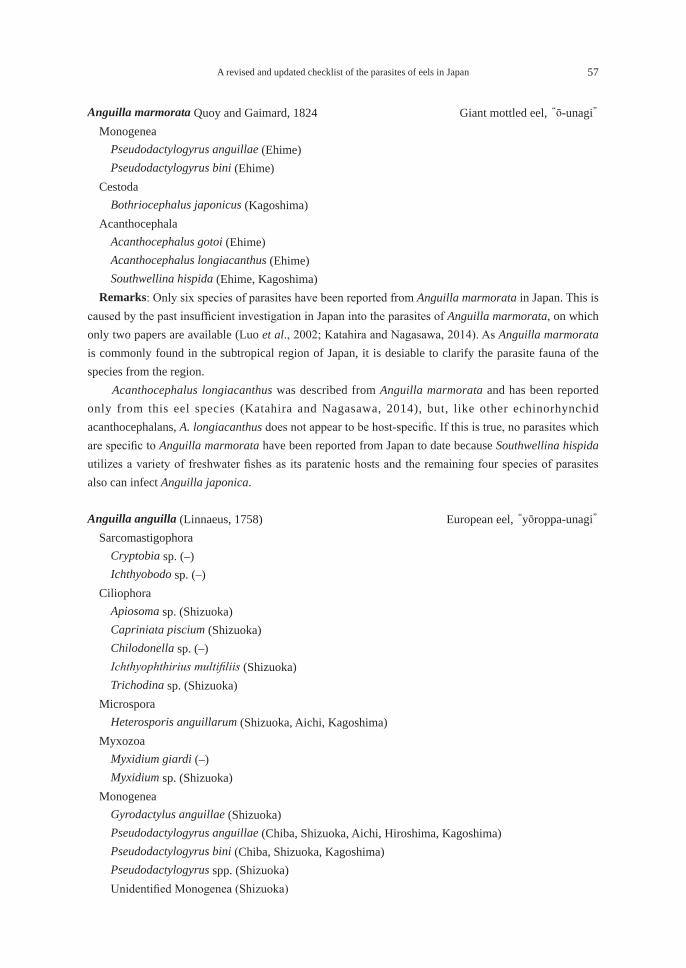
A revised and updated checklist of the parasites of eels in Japan 57
Anguilla marmorata Quoy and Gaimard, 1824 Giant mottled eel, “ō-unagi” Monogenea
Pseudodactylogyrus anguillae (Ehime)Pseudodactylogyrus bini (Ehime)
CestodaBothriocephalus japonicus (Kagoshima)
AcanthocephalaAcanthocephalus gotoi (Ehime)Acanthocephalus longiacanthus (Ehime)Southwellina hispida (Ehime, Kagoshima)
Remarks: Only six species of parasites have been reported from Anguilla marmorata in Japan. This is caused by the past insufficient investigation in Japan into the parasites of Anguilla marmorata, on which only two papers are available (Luo et al., 2002; Katahira and Nagasawa, 2014). As Anguilla marmorata is commonly found in the subtropical region of Japan, it is desiable to clarify the parasite fauna of the species from the region. Acanthocephalus longiacanthus was described from Anguilla marmorata and has been reported only from this eel species (Katahira and Nagasawa, 2014), but, like other echinorhynchid acanthocephalans, A. longiacanthus does not appear to be host-specific. If this is true, no parasites which are specific to Anguilla marmorata have been reported from Japan to date because Southwellina hispida utilizes a variety of freshwater fishes as its paratenic hosts and the remaining four species of parasites also can infect Anguilla japonica.
Anguilla anguilla (Linnaeus, 1758) European eel, “yōroppa-unagi” Sarcomastigophora
Cryptobia sp. (–)Ichthyobodo sp. (–)
CiliophoraApiosoma sp. (Shizuoka)Capriniata piscium (Shizuoka)Chilodonella sp. (–)Ichthyophthirius multifiliis (Shizuoka)Trichodina sp. (Shizuoka)
MicrosporaHeterosporis anguillarum (Shizuoka, Aichi, Kagoshima)
MyxozoaMyxidium giardi (–)Myxidium sp. (Shizuoka)
MonogeneaGyrodactylus anguillae (Shizuoka)Pseudodactylogyrus anguillae (Chiba, Shizuoka, Aichi, Hiroshima, Kagoshima)Pseudodactylogyrus bini (Chiba, Shizuoka, Kagoshima)Pseudodactylogyrus spp. (Shizuoka)Unidentified Monogenea (Shizuoka)
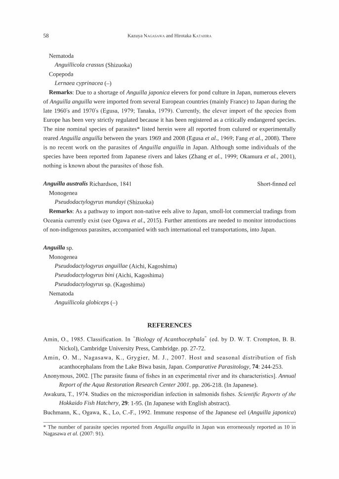
58 Kazuya NAGASAWA and Hirotaka KATAHIRA
NematodaAnguillicola crassus (Shizuoka)
CopepodaLernaea cyprinacea (–)
Remarks: Due to a shortage of Anguilla japonica elevers for pond culture in Japan, numerous elevers of Anguilla anguilla were imported from several European countries (mainly France) to Japan during the late 1960’s and 1970’s (Egusa, 1979; Tanaka, 1979). Currently, the elever import of the species from Europe has been very strictly regulated because it has been registered as a critically endangered species. The nine nominal species of parasites* listed herein were all reported from culured or experimentally reared Anguilla anguilla between the years 1969 and 2008 (Egusa et al., 1969; Fang et al., 2008). There is no recent work on the parasites of Anguilla anguilla in Japan. Although some individuals of the species have been reported from Japanese rivers and lakes (Zhang et al., 1999; Okamura et al., 2001), nothing is known about the parasites of those fish.
Anguilla australis Richardson, 1841 Short-finned eel Monogenea
Pseudodactylogyrus mundayi (Shizuoka) Remarks: As a pathway to import non-native eels alive to Japan, smoll-lot commercial tradings from Oceania currently exist (see Ogawa et al., 2015). Further attentions are needed to monitor introductions of non-indigenous parasites, accompanied with such international eel transportations, into Japan.
Anguilla sp. Monogenea
Pseudodactylogyrus anguillae (Aichi, Kagoshima)Pseudodactylogyrus bini (Aichi, Kagoshima)Pseudodactylogyrus sp. (Kagoshima)
NematodaAnguillicola globiceps (–)
REFERENCES
Amin, O., 1985. Classification. In “Biology of Acanthocephala” (ed. by D. W. T. Crompton, B. B. Nickol), Cambridge University Press, Cambridge. pp. 27-72.
Amin, O. M., Nagasawa, K., Grygier, M. J., 2007. Host and seasonal distribution of fish acanthocephalans from the Lake Biwa basin, Japan. Comparative Parasitology, 74: 244-253.
Anonymous, 2002. [The parasite fauna of fishes in an experimental river and its characteristics]. Annual Report of the Aqua Restoration Research Center 2001. pp. 206-218. (In Japanese).
Awakura, T., 1974. Studies on the microsporidian infection in salmonids fishes. Scientific Reports of the Hokkaido Fish Hatchery, 29: 1-95. (In Japanese with English abstract).
Buchmann, K., Ogawa, K., Lo, C.-F., 1992. Immune response of the Japanese eel (Anguilla japonica)
* The number of parasite species reported from Anguilla anguilla in Japan was errorneously reported as 10 in Nagasawa et al. (2007: 91).
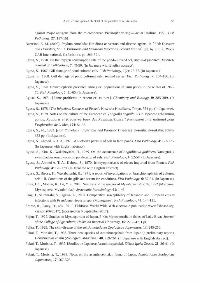
A revised and updated checklist of the parasites of eels in Japan 59
against major antigens from the microsporean Pleistophora anguillarum Hoshina, 1951. Fish Pathology, 27: 157-161.
Burreson, E. M. (2006): Phylum Annelida: Hirudinea as vectors and disease agents. In “Fish Diseases and Disorders, Vol. 1. Protozoan and Metazoan Infections. Second Edition” (ed. by P. T. K. Woo), CAB International, Oxfordshire. pp. 566-591.
Egusa, S., 1958. On the oxygen consumption rate of the pond-cultured eel, Anguilla japonica. Japanese Journal of Ichthtyology, 7: 49-56. (In Japanese with English abstract).
Egusa, S., 1967. Gill damage of pond cultured eels. Fish Pathology, 1(2): 72-77. (In Japanese).Egusa, S., 1968. Gill damage of pond cultured eels, second series. Fish Pathology, 2: 184-186. (In
Japanese).Egusa, S., 1970. Branchiophritis prevailed among eel populations in farm ponds in the winter of 1969-
70. Fish Pathology, 5: 51-66. (In Japanese).Egusa, S., 1971. [Some problems in recent eel culture]. Chemistry and Biology, 9: 385-389. (In
Japanese).Egusa, S., 1978. [The Infectious Diseases of Fishes]. Koseisha Koseikaku, Tokyo. 554 pp. (In Japanese).Egusa, S., 1979. Notes on the culture of the European eel (Anguilla anguilla L.) in Japanese eel-farming
ponds. Rapports et Proces-verbaux des Reunions/Conseil Permanent International pour l’exploration de la Mer, 174: 51-58.
Egusa, S., ed., 1983. [Fish Pathology-Infectious and Parasitic Diseases]. Koseisha Koseikaku, Tokyo. 352 pp. (In Japanese).
Egusa, S., Ahmed, A. T. A., 1970. A suctorian parasite of eels in farm ponds. Fish Pathology, 4: 172-175. (In Japanese with English abstract).
Egusa, S., Kira, K., Wakabayashi, H., 1969. On the occurrence of Anguillicola globiceps Yamaguti, a swimbladder roundworm, in pond-cultured eels. Fish Pathology, 4: 52-58. (In Japanese).
Egusa, S., Ahmed A. T. A., Kubota, S., 1970. Ichthyophthriasis of elvers imported from France. Fish Pathology, 4: 176-179. (In Japanese with English abstract).
Egusa, S., Hirose, H., Wakabayashi, H., 1971. A report of investigations on branchionephritis of cultured eels-II. Conditions of the gills and serum ion conditions. Fish Pathology, 6: 57-61. (In Japanese).
Eiras, J. C., Molnar, K., Lu, Y. S., 2005. Synopsis of the species of Myxobolus Bütschli, 1882 (Myxozoa: Myxosporea: Myxobolidae). Systematic Parasitology, 61: 1-46.
Fang, J., Shirakashi, S., Ogawa, K., 2008. Comparative susceptibility of Japanese and European eels to infections with Pseudodactylogyrus spp. (Monogenea). Fish Pathology, 43: 144-151.
Froese, R., Pauly, D., eds., 2017. FishBase. World Wide Web electronic publication.www.fishbase.org, version (06/2017). (accessed on 6 September 2017).
Fujita, T., 1927. Studies on Myxosporidia of Japan. 5. On Myxosporidia in fishes of Lake Biwa. Journal of the College of Agriculture, Hokkaido Imperial University, 16: 229-247, 1 pl.
Fujita, T., 1929. The skin-disease of the eel. Annotationes Zoologicae Japonenses, 12: 245-250.Fukui, T., Morisita, T., 1936. Three new species of Acanthocepahala from Japan (a preliminary report).
Dobutsugaku Zasshi (Zoological Magazine), 48: 759-764. (In Japanese with English abstract).Fukui, T., Morisita, T., 1937. [Studies on Japanese Acanthocepahala]. Zikken Igaku Zasshi, 21: 36-41. (In
Japanese).Fukui, T., Morisita, T., 1938. Notes on the acanthocephalan fauna of Japan. Annotationes Zoologicae
Japonenses, 17: 567-576.
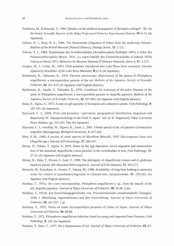
60 Kazuya NAGASAWA and Hirotaka KATAHIRA
Furukawa, M., Kobayashi, Y., 1966. [Studies on the artificial propagation of Hyriopsis schlegeli-III. On the hosts]. Scientific Reports of the Shiga Prefectural Fisheries Experimental Station, 19: 6-13. (In Japanese).
Gibson, D. I., Bray, R. A., 1986. The Hemiuridae (Digenea) of fishes from the north-east Atlantic. Bulletin of the British Museum (Natural History), Zoology Series, 51: 1-111.
Golvan, Y. J., 1969. Sytématique des Acanthocéphales (Acanthocephala Rudolphi 1801), L’ordre des Palaeacanthocephala Meyer, 1931, La super-famille des Echinorhynchidea (Cobbold 1876) Golvan et Houin 1973. Mémoires du Muséum National D’Histoire Naturelle, Série A, 47: 5-373.
Grygier, M. J., Urabe, M., 2003. [Fish parasites introduced into Lake Biwa from overseas]. Umindo (Quarterly Newsletter of the Lake Biwa Museum), 8(1): 6. (In Japanese).
Hashimoto, K., Takinami, K., 1976. Electron microscopic observations of the spores of Plistophora anguillarum, a microsporidian parasite of the eel. Bulletin of the Japanese Society of Scientific Fisheries, 42: 411-419. (In Japanese with English abstract).
Hashimoto, K., Sasaki, Y., Takinami, K., 1976. Conditions for extrusion of the polar filament of the spore of Plistophora anguillarum, a microsporidian parasite in Anguilla japonica. Bulletin of the Japanese Society of Scientific Fisheries, 42: 837-845. (In Japanese with English abstract).
Hatai, K., Egusa, S., 1973. A note on gill parasites of European eels cultured in ponds. Fish Pathology, 8: 102-105. (In Japanese).
Hayward, C. J., 2004. [Fish and parasites-speciation, geographical distribution, migration and dispersion]. In “Aquaparasitology in the Field in Japan” (ed. by K. Nagasawa), Tokai University Press, Hadano. pp. 313-325, 344. (In Japanese).
Hayward, C. J., Iwashita, M., Ogawa, K., Ernst, I., 2001. Global spread of the eel parasite Gyrodactylus anguillae (Monogenea). Biological Invasions, 3: 417-424.
Hine, P. M., 1980. A review of some species of Myxidium Bütschli, 1882 (Myxosporea) from eels (Anguilla spp.) Journal of Protozoology, 27: 260-267.
Hirose, H., Sekino, T., Egusa, S., 1976. Notes on the egg deposition, larval migration and intermediate host of the nematode Anguillicola crassa parasitic in the swimbladder of eels. Fish Pathology, 11: 27-31. (In Japanese with English abstract).
Hirose, H., Yabu, T., Hirono, I., Aoki, T., 1998. The phylogeny of Anguillicola crassus and A. globiceps based on partial 18S ribosomal RNA sequences. Journal of Fish Diseases, 21: 265-271.
Horiuchi, M., Kuwahara, A., Souma, T., Nakata, M., 1988. Availability of long-hour bathing in ammonia water for control of pseudodactylogyrosis in cultured eels. Suisanzoshoku, 35: 259-263. (In Japanese with English abstract).
Hoshina, T., 1951a. On a new microsporidian, Plistophora anguillarum n. sp., from the muscle of the eel, Anguilla japonica. Journal of Tokyo University of Fisheries, 38: 35-46, 2 pls.
Hoshina, T., 1951b. Zur Entwicklungsgeschichte von, Proctotrematoides pisodontophidis Yamaguti, 1938. I. Mitteilung, Agamodistoma und ihre Entwicklung. Journal of Tokyo University of Fisheries, 38: 247-257, 1 pl.
Hoshina, T., 1952. Notes on some myxosporidian parasites of fishes of Japan. Journal of Tokyo University of Fisheries, 39: 69-89.
Hoshina, T., 1972. Plistophora anguillarum infection found in young eels imported from Formosa. Fish Pathology, 6: 120. (In Japanese).
Hoshina, T., Sano, T., 1957. On a trypanosome of eel. Journal of Tokyo University of Fisheries, 43: 67-
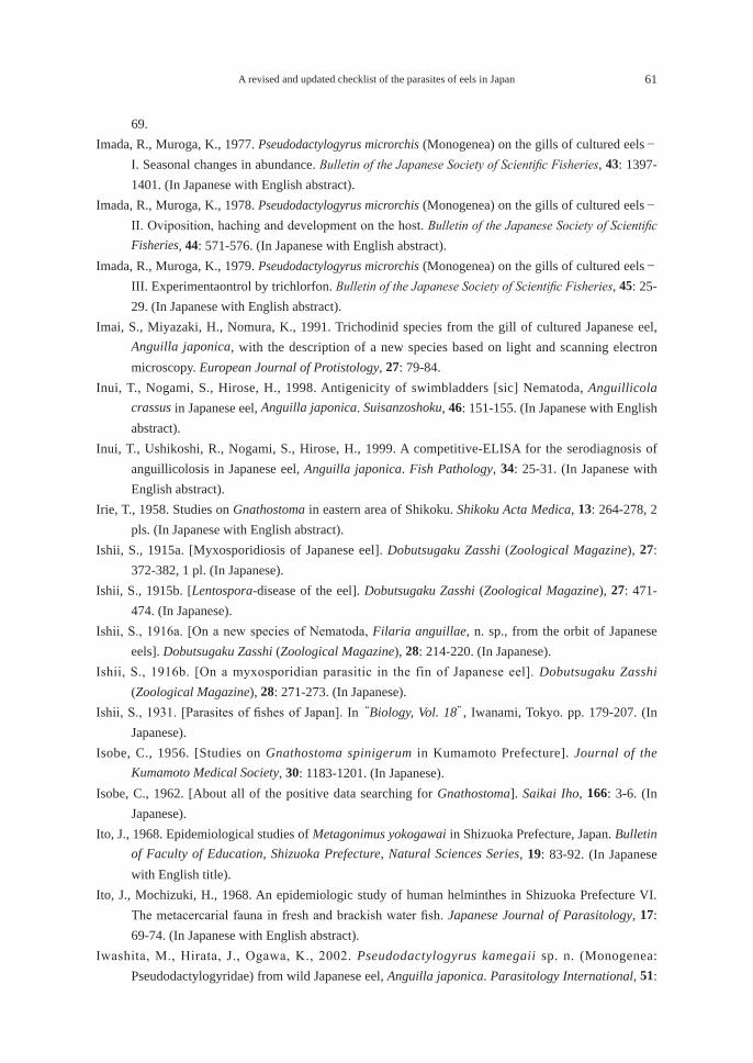
A revised and updated checklist of the parasites of eels in Japan 61
69.Imada, R., Muroga, K., 1977. Pseudodactylogyrus microrchis (Monogenea) on the gills of cultured eels-
I. Seasonal changes in abundance. Bulletin of the Japanese Society of Scientific Fisheries, 43: 1397-1401. (In Japanese with English abstract).
Imada, R., Muroga, K., 1978. Pseudodactylogyrus microrchis (Monogenea) on the gills of cultured eels-II. Oviposition, haching and development on the host. Bulletin of the Japanese Society of Scientific Fisheries, 44: 571-576. (In Japanese with English abstract).
Imada, R., Muroga, K., 1979. Pseudodactylogyrus microrchis (Monogenea) on the gills of cultured eels-III. Experimentaontrol by trichlorfon. Bulletin of the Japanese Society of Scientific Fisheries, 45: 25-29. (In Japanese with English abstract).
Imai, S., Miyazaki, H., Nomura, K., 1991. Trichodinid species from the gill of cultured Japanese eel, Anguilla japonica, with the description of a new species based on light and scanning electron microscopy. European Journal of Protistology, 27: 79-84.
Inui, T., Nogami, S., Hirose, H., 1998. Antigenicity of swimbladders [sic] Nematoda, Anguillicola crassus in Japanese eel, Anguilla japonica. Suisanzoshoku, 46: 151-155. (In Japanese with English abstract).
Inui, T., Ushikoshi, R., Nogami, S., Hirose, H., 1999. A competitive-ELISA for the serodiagnosis of anguillicolosis in Japanese eel, Anguilla japonica. Fish Pathology, 34: 25-31. (In Japanese with English abstract).
Irie, T., 1958. Studies on Gnathostoma in eastern area of Shikoku. Shikoku Acta Medica, 13: 264-278, 2 pls. (In Japanese with English abstract).
Ishii, S., 1915a. [Myxosporidiosis of Japanese eel]. Dobutsugaku Zasshi (Zoological Magazine), 27: 372-382, 1 pl. (In Japanese).
Ishii, S., 1915b. [Lentospora-disease of the eel]. Dobutsugaku Zasshi (Zoological Magazine), 27: 471-474. (In Japanese).
Ishii, S., 1916a. [On a new species of Nematoda, Filaria anguillae, n. sp., from the orbit of Japanese eels]. Dobutsugaku Zasshi (Zoological Magazine), 28: 214-220. (In Japanese).
Ishii, S., 1916b. [On a myxosporidian parasitic in the fin of Japanese eel]. Dobutsugaku Zasshi (Zoological Magazine), 28: 271-273. (In Japanese).
Ishii, S., 1931. [Parasites of fishes of Japan]. In “Biology, Vol. 18”, Iwanami, Tokyo. pp. 179-207. (In Japanese).
Isobe, C., 1956. [Studies on Gnathostoma spinigerum in Kumamoto Prefecture]. Journal of the Kumamoto Medical Society, 30: 1183-1201. (In Japanese).
Isobe, C., 1962. [About all of the positive data searching for Gnathostoma]. Saikai Iho, 166: 3-6. (In Japanese).
Ito, J., 1968. Epidemiological studies of Metagonimus yokogawai in Shizuoka Prefecture, Japan. Bulletin of Faculty of Education, Shizuoka Prefecture, Natural Sciences Series, 19: 83-92. (In Japanese with English title).
Ito, J., Mochizuki, H., 1968. An epidemiologic study of human helminthes in Shizuoka Prefecture VI. The metacercarial fauna in fresh and brackish water fish. Japanese Journal of Parasitology, 17: 69-74. (In Japanese with English abstract).
Iwashita, M., Hirata, J., Ogawa, K., 2002. Pseudodactylogyrus kamegaii sp. n. (Monogenea: Pseudodactylogyridae) from wild Japanese eel, Anguilla japonica. Parasitology International, 51:
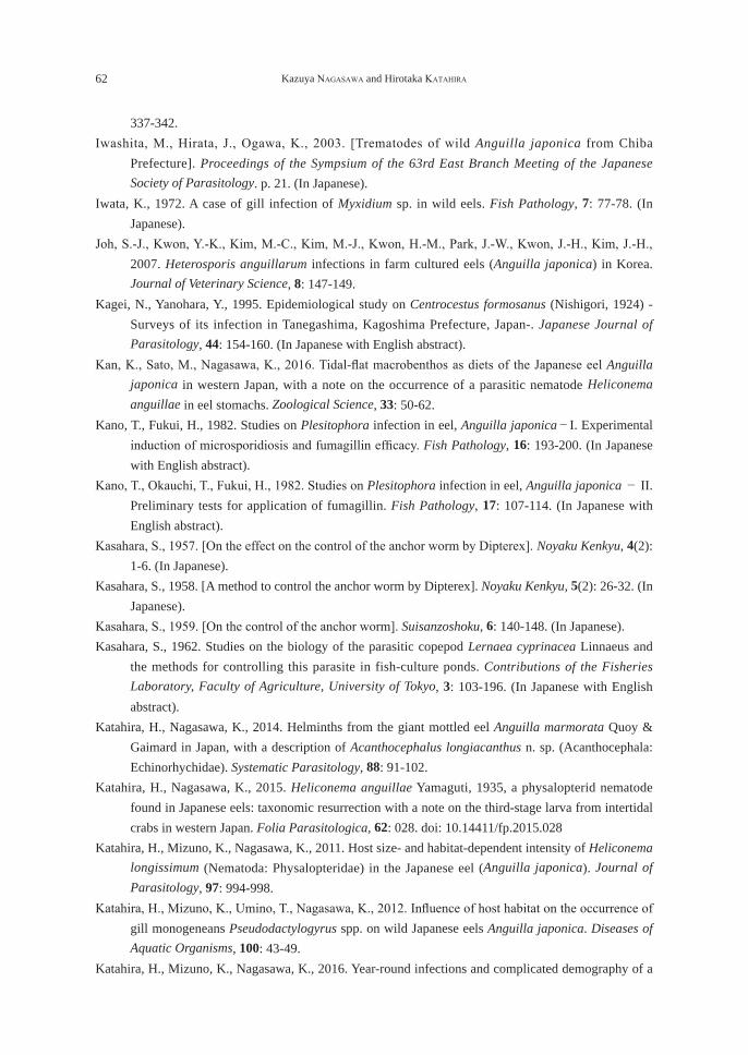
62 Kazuya NAGASAWA and Hirotaka KATAHIRA
337-342.Iwashita, M., Hirata, J., Ogawa, K., 2003. [Trematodes of wild Anguilla japonica from Chiba
Prefecture]. Proceedings of the Sympsium of the 63rd East Branch Meeting of the Japanese Society of Parasitology. p. 21. (In Japanese).
Iwata, K., 1972. A case of gill infection of Myxidium sp. in wild eels. Fish Pathology, 7: 77-78. (In Japanese).
Joh, S.-J., Kwon, Y.-K., Kim, M.-C., Kim, M.-J., Kwon, H.-M., Park, J.-W., Kwon, J.-H., Kim, J.-H., 2007. Heterosporis anguillarum infections in farm cultured eels (Anguilla japonica) in Korea. Journal of Veterinary Science, 8: 147-149.
Kagei, N., Yanohara, Y., 1995. Epidemiological study on Centrocestus formosanus (Nishigori, 1924) - Surveys of its infection in Tanegashima, Kagoshima Prefecture, Japan-. Japanese Journal of Parasitology, 44: 154-160. (In Japanese with English abstract).
Kan, K., Sato, M., Nagasawa, K., 2016. Tidal-flat macrobenthos as diets of the Japanese eel Anguilla japonica in western Japan, with a note on the occurrence of a parasitic nematode Heliconema anguillae in eel stomachs. Zoological Science, 33: 50-62.
Kano, T., Fukui, H., 1982. Studies on Plesitophora infection in eel, Anguilla japonica-I. Experimental induction of microsporidiosis and fumagillin efficacy. Fish Pathology, 16: 193-200. (In Japanese with English abstract).
Kano, T., Okauchi, T., Fukui, H., 1982. Studies on Plesitophora infection in eel, Anguilla japonica- II. Preliminary tests for application of fumagillin. Fish Pathology, 17: 107-114. (In Japanese with English abstract).
Kasahara, S., 1957. [On the effect on the control of the anchor worm by Dipterex]. Noyaku Kenkyu, 4(2): 1-6. (In Japanese).
Kasahara, S., 1958. [A method to control the anchor worm by Dipterex]. Noyaku Kenkyu, 5(2): 26-32. (In Japanese).
Kasahara, S., 1959. [On the control of the anchor worm]. Suisanzoshoku, 6: 140-148. (In Japanese).Kasahara, S., 1962. Studies on the biology of the parasitic copepod Lernaea cyprinacea Linnaeus and
the methods for controlling this parasite in fish-culture ponds. Contributions of the Fisheries Laboratory, Faculty of Agriculture, University of Tokyo, 3: 103-196. (In Japanese with English abstract).
Katahira, H., Nagasawa, K., 2014. Helminths from the giant mottled eel Anguilla marmorata Quoy & Gaimard in Japan, with a description of Acanthocephalus longiacanthus n. sp. (Acanthocephala: Echinorhychidae). Systematic Parasitology, 88: 91-102.
Katahira, H., Nagasawa, K., 2015. Heliconema anguillae Yamaguti, 1935, a physalopterid nematode found in Japanese eels: taxonomic resurrection with a note on the third-stage larva from intertidal crabs in western Japan. Folia Parasitologica, 62: 028. doi: 10.14411/fp.2015.028
Katahira, H., Mizuno, K., Nagasawa, K., 2011. Host size- and habitat-dependent intensity of Heliconema longissimum (Nematoda: Physalopteridae) in the Japanese eel (Anguilla japonica). Journal of Parasitology, 97: 994-998.
Katahira, H., Mizuno, K., Umino, T., Nagasawa, K., 2012. Influence of host habitat on the occurrence of gill monogeneans Pseudodactylogyrus spp. on wild Japanese eels Anguilla japonica. Diseases of Aquatic Organisms, 100: 43-49.
Katahira, H., Mizuno, K., Nagasawa, K., 2016. Year-round infections and complicated demography of a
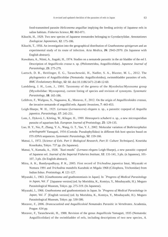
A revised and updated checklist of the parasites of eels in Japan 63
food-transmitted parasite Heliconema anguillae implying the feeding activity of Japanese eels in saline habitats. Fisheries Science, 82: 863-871.
Kikuchi, H., 1929. Two new species of Japanese trematodes belonging to Gyrodactylidae. Annotationes Zoologicae Japonenses, 12: 175-186.
Kikuchi, T., 1956. An investigation into the geographical distribution of Gnathostoma spinigerum and an experimental study of its route of infection. Acta Medica, 26: 2943-2970. (In Japanese with English abstract).
Kuwahara, A., Niimi, A., Itagaki, H., 1974. Studies on a nematode parasitic in the air bladder of the eel I. Description of Anguillicola crassa n. sp. (Philometridea, Anguillicolidae). Japanese Journal of Parasitology, 23: 275-279.
Laetsch, D. R., Heitlinger, E. G., Taraschewski, H., Nadler, S. A., Blaxter, M. L., 2012. The phylogenetics of Anguillicolidae (Nematoda: Angullicoloidea), swimmbladder parasites of eels. BMC Evolutionary Biology, 12: 60. doi:10.1186/1471-2148-12-60.
Landsberg, J. H., Lom, J., 1991. Taxonomy of the genera of the Myxobolus/Myxosoma group (Myxobolidae: Myxosporea), current listing of species and revision of synonyms. Systematic Parasitology, 18: 165-186.
Lefebvre, F., Wielgoss, S., Nagasawa, K., Moravec, F., 2012. On the origin of Anguillicoloides crassus, the invasive nematode of anguillid eels. Aquatic Invasions, 7: 443-453.
Leigh-Sharpe, W. H., 1925. Lernaea (Lernaeocera) elegans n. sp., a parasitic copepod of Anguilla japonica. Parasitology, 17: 245-251.
Lom, J., Dyková, I., Körting, W., Klinger, H., 1989. Heteosporis schuberti n. sp., a new microsporidian parasite of aquarium fish. European Journal of Protistology, 25: 129-135.
Luo, H. Y., Nie, P., Zhang, Y. A., Wang, G. T., Yao, Y. J., 2002. Molecular variation of Bothriocephalus acheilognathi Yamaguti, 1934 (Cestoda: Pseudophyllidea) in different fish host species based on ITS rDNA sequences. Systematic Parasitology, 52: 159-166.
Matsui, I., 1972. [Science of Eels. Part I: Biological Research, Part II: Culture Techniques]. Koseisha Koseikaku, Tokyo. 737 pp. (In Japanese).
Matsui, Y., Kumada, A., 1928. “Ikari-mushi” (Lernaea elegans Leigh-Sharpe), a new parasitic copepod of Japanese eel. Journal of the Imperial Fisheries Institute, 12: 131-141, 3 pls. (n Japanese), 101-107, 3 pls. (In English abstract).
Mitra, A. K., Bandyopadhyay, P. K., 2005. First record of Trichodina japonica Imai, Miyazaki et Nomura 1991 and Trichodina mutabilis Kazubski et Migala 1968 (Ciliophora, Trichonidae) from Indian fishes. Protistology, 4: 121-127.
Miyazaki, I., 1963. [Gnathostoma and gnathostomiasis in Japan]. In “Progress of Medical Parasitology in Japan, Vol. 3” [Japanese version] (ed. by Morishita, K., Komiya, Y., Mtsubayashi, H.). Meguro Parasitological Museum, Tokyo. pp. 275-319. (In Japanese).
Miyazaki, I., 1966. Gnathostoma and gnathostomiasis in Japan. In “Progress of Medical Parasitology in Japan, Vol. 3” [English version] (ed. by Morishita, K., Komiya, Y., Mtsubayashi, H.). Meguro Parasitological Museum, Tokyo. pp. 530-586.
Moravec, F., 2006. Drancucunloid and Anguillicoloid Nematodes Parasitic in Vertebrates. Academia, Prague. 634 pp.
Moravec, F., Taraschewski, H., 1988. Revision of the genus Anguillicola Yamaguti, 1935 (Nematoda: Anguillicolidae) of the swimbladder of eels, including descriptions of two new species, A.
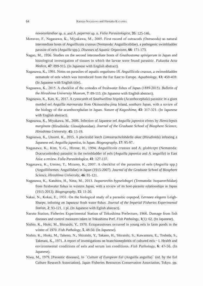
64 Kazuya NAGASAWA and Hirotaka KATAHIRA
novaezelandiae sp. n. and A. papernai sp. n. Folia Parasitologica, 35: 125-146.Moravec, F., Nagasawa, K., Miyakawa, M., 2005. First record of ostracods (Ostracoda) as natural
intermediate hosts of Anguillicola crassus (Nematoda: Anguillicolidae), a pathogenic swimbladder parasite of eels (Anguilla spp.). Diseases of Aquatic Organisms, 66: 171-173.
Nagao, M., 1956. Studies on the second intermediate hosts of Gnathostoma spinigerum in Japan and histological investigation of tissues in which the larvae were found parasitic. Fukuoka Acta Medica, 47: 899-915. (In Japanese with English abstract).
Nagasawa, K., 1991. Notes on parasites of aquatic organisms-18. Anguillicola crassus, a swimmbladder nematode of eels which was introduced from the Far East to Europe. Aquabiology, 13: 458-459. (In Japanese with English title).
Nagasawa, K., 2015. A checklist of the cestodes of freshwater fishes of Japan (1889-2015). Bulletin of the Hiroshima University Museum, 7: 89-115. (In Japanese with English abstract).
Nagasawa, K., Kan, K., 2017. A cystacanth of Southwellina hispida (Acanthocephala) parasitic in a giant mottled eel Anguilla marmorata from Okinoerabu-jima Island, southern Japan, with a review of the biology of the acanthocephalan in Japan. Nature of Kagoshima, 43: 317-321. (In Japanese with English abstract).
Nagasawa, K., Miyakawa, M., 2006. Infection of Japanese eel Anguilla japonica elvers by Hemiclepsis marginata (Hirudinida: Glossiphonidae). Journal of the Graduate School of Biosphere Science, Hiroshima University, 45: 15-19.
Nagasawa, K., Utsumi, K., 2015. A piscicolid leech Limnotrachelobdella okae (Hirudinida) infesting a Japanese eel, Anguilla japonica, in Japan. Biogeography, 17: 95-97.
Nagasawa, K., Kim, Y.-G., Hirose, H., 1994. Anguillicola crassus and A. globiceps (Nematoda: Dracucunloidea) parasitic in the swimbladder of eels (Anguilla japonica and A. anguilla) in East Asia: a review. Folia Parasitologica, 41: 127-137.
Nagasawa, K., Umino, T., Mizuno, K., 2007. A checklist of the parasites of eels (Anguilla spp.) (Anguilliformes: Anguillidae) in Japan (1915-2007). Journal of the Graduate School of Biosphere Science, Hiroshima University, 46: 91-121.
Nagasawa, K., Katahira, H., Nitta, M., 2013. Isoparorchis hypselobagri (Trematoda: Isoparorchiidae) from freshwater fishes in western Japan, with a review of its host-parasite relationships in Japan (1915-2013). Biogeography, 15: 11-20.
Nakai, N., Kokai, E., 1931. On the biological study of a parasitic copepod, Lernaea elegans Leigh-Sharpe, infesting on Japanese fresh water fishes. Journal of the Imperial Fisheries Experimental Station, 2: 93-121, 1 pl. (In Japanese with Eglish abstract).
Naruto Station, Fisheries Experimental Station of Tokushima Prefecture, 1966. Damage from fish diseases and control measures taken in Tokushima Pref. Fish Pathology, 1(1): 62. (In Japanese).
Nishio, K., Hioki, M., Shiraishi, Y., 1970. Ectoparasitoses occurred in young eels in farm ponds in the winter of 1970. Fish Pathology, 5, 48-50. (In Japanese).
Nishio, K., Hioki, M., Takeno, N., Shiraishi, Y., Takano, H., Shiraishi, S., Kawamura, E., Toshida, S., Taketani, K., 1971. A report of investigations on branchionephritis of cultured eels-I. Health and environmental conditions of eels and serum ion conditions. Fish Pathology, 6: 47-56. (In Japanese).
Niwa, M., 1979. [Parasitic diseases]. In “Culture of European Eel (Anguilla anguilla)” (ed. by the Eel Culture Research Association), Japan Fisheries Resources Conservation Association, Tokyo. pp.
A revised and updated checklist of the parasites of eels in Japan 65
109-112. (In Japanese). Ogawa, K., Egusa, S., 1976. Studies on eel pseudodactylogyrosis- I. Morphology and classification of
three eel dactylogyrids with a proposal of a new species, Pseudodactylogyrus microrchis. Bulletin of the Japanese Society of Scientific Fisheries, 42: 395-404.
Ogawa, K., Egusa, S., 1978. Seven species of Gyrodactylus (Monogenea: Gyrodactylidae) from Plecoglossus altivelis (Plecoglossidae), Cyprinus carpio (Cyprinidae) and Anguilla spp. (Anguillidae). Bulletin of the Japanese Society of Scientific Fisheries, 44: 613-618.
Ogawa, K., Egusa, S., 1980. Gyrodactylus infestations of cultured eels (Anguilla japonica and A. anguilla) in Japan. Fish Pathology, 15: 95-99. (In Japanese with English abstract).
Ogawa, K., Hioki, M., 1986. Two new species of Gyrodactylus (Monogenea: Gyrodactylidae) of eel, Anguilla japonica, with some data on the occurrence of gyrodactylids in greenhouse culture at Yoshida, Shizuoka Prefecture, Japan. Fish Pathology, 21: 89-94.
Ogawa, K., Chung, H.-Y., Kou, G.-H., Imada, R., 1985a. On the validity of an eel monogenean Pseudodactylogyrus microrchis Ogawa et Egusa, 1976. Bulletin of the Japanese Society of Scientific Fisheries, 51: 381-385.
Ogawa, K., Uno, S., Ito, S., 1985b. Infection of cultured eel with Bartacobdella smaragdina (Hirudinea: Glossiphonidae). Fish Pathology, 20: 67-68. (In Japanese).
Ogawa, K., Iwashita, M., Hayward, C. J. , Kurashima, A., 2015. Three new species of Pseudodactylogyrus (Monogenea: Pseudodactylogyridae) from Australian eels. Folia Parasitologica, 62: 046. doi:10.14411/fp.2015.046
Oka, H. P., 1973a. Observations on white spot disease in youngs of the European eel reared in ponds. Fish Pathology, 8: 32-36. (In Japanese with English abstract).
Oka, H. P., 1973b. A note on the spores of Myxidium sp. observed in an “Ich”. Fish Pathology, 8: 37-40. (In Japanese with English abstract).
Oka, H. P., Egusa, S., 1983. On the Mixidium parasitic in fins and the skin of a Japanese eel, Anguilla japonica. In “Proceedings of the ROC-Japan Cooperative Scientific Seminar on Fish Diseases” (ed. by G.-H. Kou and S. Egusa), Taipei. pp. 68-72.
Okamura, A., Zhang, H., Yamada, Y., Utoh, T., Mikawa, N., Horie, N., Tanaka, S., Motonobu, T., 2001. Identification of two eel species, Anguilla japonica and A. anguilla by discriminant function analysis. Nippon Suisan Gakkaishi, 67: 1056-1060. (In Japanese with English abstract).
Ozaki, Y., 1924. [On a new species of trematode of the genus Azygia]. Dobutsugaku Zasshi (Zoological Magazine), 36: 426-435. (In Japanese).
Petrochenko, V. I., 1956. Acanthocephala of Domestic and Wild Animals. Volume I. Izdatel’stvo Akademii Nauk SSSR, Moscow. [English translation by Israel Program for Scientific Translations, Keter Press, Jerusalem, 1971, 465 pp.]
Rahhou, I., Morand, S., Lecomte-Finiger, R., Sasal, P., 2005. Biogeographical relationships of the eel parasite Anguillicola crassus revealed by random amplified polymorphic DNA markers (RAPD). Bulletin Français de la Pêche et de la Pisciculture, 378-379: 87-98.
Rasheed, S., 1963. A revision of the genus Philometra Costa, 1845. Journal of Helminthology, 37: 89-130.
Salati, F., 1987. La parassitosi da Anguillicola sp. in Giappone. Rivista Italiana Piscicoltura e Ittiopatologia, 22: 115-117.
Satoh, T., Tanaka, M., 2007. [Control of pseudodactylogyrids in glass eels-II. Occurrence and treatment
66 Kazuya NAGASAWA and Hirotaka KATAHIRA
by temperature change in rearing water]. Hamana, 518: 1-4. (In Japanese).Scholz, T., Škeříková, A., Shimazu, T., Grygier, M. J., 2004. A taxonomic study of species of
Bothriocephalus Rudolphi, 1808 (Cestoda: Pseudophyllidea) from eels in Japan: morphological and molecular evidence for the occurrence of B. claviceps (Goeze, 1782) and confirmation of the validity of B. japonicus Yamaguti, 1934. Systematic Parasitology, 57: 87-96.
Shimazu, T., 1979. Developmental stages of Azygia gotoi (Digenea, Azygiidae). Bulletin of the National Science Museum, Series A (Zoology), 5: 225-234.
Shimazu, T., 1995. Trematodes of the genus Genarchopsis (Digenea, Derogenidae, Halipeginae) from freshwater fishes of Japan. Proceedings of the Japanese Society of Systematic Zoology, 54: 1-18.
Shimazu, T., 1997. Cestodes of freshwater fishes in Japan: a review. Journal of Nagano Prefectural College, 52: 9-17. (In Japanese with English abstract).
Shimazu, T., 1998. Nematodes of freshwater fishes in Japan: a review. Journal of Nagano Prefectural College, 53: 1-19. (In Japanese with English abstract).
Shimazu, T., 1999a. [Turbellarians and trematodes of freshwater animals in Japan]. In “Progress of Medical Parasitology in Japan” [Japanese version] (ed. by M. Otsuru, S. Kamegai, S. Hayashi), Meguro Parasitological Museum, Tokyo, 6: 65-86. (In Japanese).
Shimazu, T., 1999b. Acanthocephalans of freshwater fishes in Japan: a review. Journal of Nagano Prefectural College, 54: 21-29. (In Japanese with English abstract).
Shimazu, T., 2003. Turbellarians and trematodes of freshwater animals in Japan. In “Progress of Medical Parasitology in Japan” [English version] (ed. by M. Otsuru, S. Kamegai, S. Hayashi), Meguro Parasitological Museum, Tokyo, 7: 63-86.
Shimazu, T., 2005. Digeneans found in fresh- and brackish-water fishes of Lake Ogawara in Aomori Prefecture, Japan. Bulletin of the National Science Museum, Series A (Zoology), 31: 137-150.
Shimazu, T., 2007. Digeneans (Trematoda) of freshwater fishes from Nagano Prefecture, central Japan. Bulletin of the National Museum of Nature and Science, Series A, 33: 1-30.
Shimazu, T., 2008. Digeneans (Trematoda) found in freshwater fishes of Wakayama, Tokushima, and Kochi Prefectures, Japan. Bulletin of the National Museum of Nature and Science, Series A, 34: 41-61.
Shimazu, T., 2014a. Digeneans parasitic in freshwater fishes (Osteichthyes) of Japan. II. Gorgoderidae and Orientocreadiidae. Bulletin of the National Museum of Nature and Science, Series A, 40: 53-78.
Shimazu, T., 2014b. Digeneans parasitic in freshwater fishes (Osteichthyes) of Japan. III. Azygiidae and Bucephalidae. Bulletin of the National Museum of Nature and Science, Series A, 40: 167-190.
Shimazu, T., 2015. Digeneans parasitic in freshwater fishes (Osteichthyes) of Japan. IV. Derogenidae. Bulletin of the National Museum of Nature and Science, Series A, 41: 77-103.
Shimazu, T., 2016a. Digeneans parasitic in freshwater fishes (Osteichthyes) of Japan. VI. Lissorchiidae. Bulletin of the National Museum of Nature and Science, Series A, 42: 1-22.
Shimazu, T., 2016b. Digeneans parasitic in freshwater fishes (Osteichthyes) of Japan. IX. Opecoelidae, Opecoelinae. Bulletin of the National Museum of Nature and Science, Series A, 42: 163-180.
Shimazu, T., Araki, J., 2006. A list of the helminth parasite specimens deposited in the Department of Zoology, the University Museum, the University of Tokyo. In “Catalogue of Invertebrate Collection Deposited in the Department of Zoology, the University Museum, the University of Tokyo” (ed. by R. Ueshima), The University Museum, The University of Tokyo Material Report,
A revised and updated checklist of the parasites of eels in Japan 67
62: 151-161. Shimazu, T., Urabe, M., 2005. Digeneans found freshwater fishes of the Uji River at Uji, Kyoto
Prefecture, and the Takami River at Higashiyoshino, Nara Prefecture, Japan. Journal of Nagano Prefectural College, 60: 1-14.
Shimazu, T., Urabe, M., Grygier, M. J., 2011. Digeneans (Trematoda) parasitic in freshwater fishes (Osteichthyes) of the Lake Biwa basin in Shiga Prefecture, central Japan. National Museum of Nature and Science Monographs, 43: 1-105.
Suh, J.-W., Chun, S.-K., 1988. The infection experiment of Pleistophora to eels, Anguilla japonica and the histopathological investigation of the infection development. Bulletin of the Korean Society of Fish Pathology, 1: 51-57. (In Korean with English abstract).
Suyehiro, Y., 1957. [On the ecology of parasites of Japanese eel]. In “Suisangaku Shusei” (ed. by Suyehiro, Y., Oshima, Y., Hiyama, Y.), Tokyo University Press, Tokyo. pp. 415-418. (In Japanese).
Tanaka, S., 1979. [Status of imports of Anguilla anguilla elevers]. In “Culture of European Eel (Anguilla anguilla)” (ed. by the Eel Culture Research Association), Japan Fisheries Resources Conservation Association, Tokyo. pp. 135-140. (In Japanese).
Tanaka, M., Satoh, T., 2007. [Control of pseudodactylogyrids in glass eels]. Hamana, 517: 1-3. (In Japanese).
Tanaka, M., Satoh, T., Matsuyama, H., 2009. Efficacy of high water temperature treatment against Pseudodactylogyrus spp. infection in Japanese eel. Fish Pathology, 44: 133-138. (In Japanese with English abstract).
Tsai, S.-J., Kou, G.-H., Lo, C.-F., Wang, C.-H., 2002. Complete sequence and structure of ribosomal RNA gene of Heterosporis anguillarum. Diseases of Aquatic Organisms, 49: 199-206.
T’sui, W.-H., Wang, C.-H., 1988. On the Plistophora infection in eel. I. Histopathology, ultrastructure, and development of Plistophora anguillarum in eel, Anguilla japonica. Bulletin of the Institute of Zoology, Academia Sinica, 27: 159-166.
T’sui, W.-H., Wang, C.-H., Lo, C.-F., 1988. On the Plistophora infection in eel. II. The development of Plistophora anguillarum in experimentally infected elvers, Anguilla japonica. Bulletin of the Institute of Zoology, Academia Sinica, 27: 249-258.
Tsukamoto, K, Arai, T., 2001. Facultive catadromy of the eel Anguilla japonica between freshwater and seawater habitats. Marine Ecology Progress Series, 220: 265-276.
Tsukamoto, K., Nakai, I., Tesch, F. W., 1998. Do all freshwater eels migrate? Nature, 396: 635-636.Tsutsumi, T., 1978. [Treatment of fish diseases at aquaria. Part 7. The anchor worm, a parasite of
freshwater fishes, and its control]. Noyaku Kenkyu, 12: 21-22. (In Japanese).Umeda, N., Nibe, H., Hara, T., Hirazawa, N., 2006: Effects of various treatments on hatching of eggs
and viability of oncomiracidia of the monogenean Pseudodactylogyrus anguillae and Pseudodactylogyrus bini. Aquaculture, 253: 148-153.
Ushikoshi, R., Inui, T., Mano, N., Hirose, H., 1999. A method for specific antibody detection from Japanese eel by indirect ELISA using cuticular antigen of Anguillicola crassus. Fish Pathology, 34: 81-82.
Ushiyama, M., Misaki, S., 1977. Seasonal change of physiological conditions in the eel of a typical culture pond. Bulletin of the Shizuoka Prefectural Fisheries Experimental Station, 11: 25-32. (In Japanese).
Van Cleave, H. J., 1925. Acanthocephala from Japan. Parasitology, 17: 149-156.
68 Kazuya NAGASAWA and Hirotaka KATAHIRA
Wielgoss, S., Taraschewski, H., Meyer, A., Wirth, T., 2008. Population structure of the parasitic nematode Anguillicola crassus, an invader of declining North Atlantic eel stocks. Molecular Ecology, 17: 3478-3495.
Xu, K., Song, W., Warren, A., 1999. Trichodinid ectoparasites (Ciliophora: Petritrichida) from the gills of cultured marine fishes in China, with the description of Trichodina lomi n. sp. Systematic Parasitology, 42: 219-227.
Xu, K., Song, W., Warren, A., Choi, J. K., 2001. Trichodinid ectoparasites (Ciliophora: Petritrichida) of some marine fishes from coastal regions of the Yellow Sea and Bohai Sea. Systematic Parasitology, 50: 69-79.
Yamaguti, S., 1934a. Studies on the helminth fauna of Japan. Part 2. Trematodes of fishes, I. Japanese Journal of Zoology, 5: 249-541.
Yamaguti, S., 1934b. Studies on the helminth fauna of Japan. Part 4. Cestodes of fishes. Japanese Journal of Zoology, 6: 1-112.
Yamaguti, S., 1935a. Studies on the helminth fauna of Japan. Part 8. Acanthocephala. Japanese Journal of Zoology, 6: 247-278.
Yamaguti, S., 1935b. Studies on the helminth fauna of Japan. Part 9. Nematodes of fishes, 1. Japanese Journal of Zoology, 6: 337-386.
Yamaguti, S., 1938. Studies on the helminth fauna of Japan. Part 21. Trematodes of fishes, IV. Published by the author. 139 pp., 1 pl.
Yamaguti, S., 1939. Parasitic copepods from fishes of Japan. Part 5. Caligoida, III. Volumen Jubilare pro Professore Sadao Yoshida, 2: 443-487, 33 pls.
Yamaguti, S., 1941. Studies on the helminth fauna of Japan. Part 33. Nematodes of fishes, II. Japanese Journal of Zoology, 9: 343-396.
Yamaguti, S., 1952. Studies on the helminth fauna of Japan. Part 49. Cestodes of fishes, II. Acta Medicinae Okayama, 8: 1-76, 22 pls.
Yamaguti, S., 1963. Systema Helminthum. Volume V. Acanthocephala. Interscience Publishers, New York and London. 423 pp.
Yanohara, Y., Kagei, N., 1983. Studies on metacercariae of Centrocestus formosanus (Nishigori, 1924)-I. Parasitism of metacercariae in gills of young rearing eels, and abnormal deaths of the hosts. Fish Pathology, 17: 237-241. (In Japanese with English abstract).
Yoshikawa, M., 2005. [Test of control drugs for pseudodactylogyrids of eels]. Hamana, 509: 1-5. (In Japanese).
Yoshikawa, M., Tanaka, M., Iinuma, N., Uemura, N., Suzuki, K., 2006. Rearing, and male and female of European eel Anguilla anguilla. Bulletin of the Shizuoka Prefectural Fisheries Experiment Station, 41: 63-76. (In Japanese).
Zhang, H., Mikawa, N., Yamada, Y., Horie, N., Okamura, A., Utoh, T., Tanaka, S., Motonobu, T., 1999. Foreign eel species in the natural waters of Japan detected by polymerase chain reaction of mitochondrial cytochrome b region. Fisheries Science, 65: 684-686.
A revised and updated checklist of the parasites of eels in Japan 69
日本産ウナギ類の寄生虫目録:追補改定版(1915-2017年)
長澤和也1)・片平浩孝2)
1)広島大学大学院生物圏科学研究科 〒739-8528 東広島市鏡山1-4-42)三重大学生物資源科学研究科 〒514-8507 津市栗真町屋町1577
要 旨 1915-2017年の103年間に出版された文献に基づき,日本産ウナギ属魚類3種(ニホンウナギAnguilla japonica,オオウナギ Anguilla marmorata,ヨーロッパウナギ Anguilla anguilla)と日本に輸入された Anguilla australisの寄生虫に関する情報を2つのリスト(寄生虫-宿主リスト,宿主-寄生虫リスト)に整理して目録を作成した。宿主のニホンウナギとオオウナギは在来種であり,ヨーロッパウナギはシラスウナギとして輸入され養殖された個体,Anguilla australisはオーストラリアから輸入された個体である。本目録は2007年に出版した同名目録の追補改定版である。本目録には,54名義種の寄生虫(繊毛虫類6種,微胞子虫類1種,ミクソゾア類6種,吸虫類12種,単生類8種,条虫類3種,線虫類7種,鉤頭虫類6種,ヒル類3種,二枚貝類1種,カイアシ類1種)に加えて,学名がまだ決定していない寄生虫の情報が含まれる。寄生虫-宿主リストでは,各寄生虫は高位分類群ごとに配列され,最新の学名,シノニム,寄生部位,地理的分布および報告者の情報が示されている。上記54名義種のうち,ニホンウナギから50種,オオウナギから6種,ヨーロッパウナギから9種,Anguilla australis から1種の寄生虫が報告されていた。単生類の Gyrodactylus anguillae,Gyrodactylus nipponensisおよびPseudodactylorgyrus mundayi,条虫類の Bothriocephalus claviceps,線虫類の Raphidascaris acusは海外から持ち込まれたと推察されており,残りの49名義種が日本にもともと分布するものである。ニホンウナギから報告された寄生虫のうち,9名義種(Lecithochrium musculus, Proctotrematoides pisodontophidis, Tubulovesicula anguillae [吸虫類], Gyrodactylus nipponensis, Pseudodactylogyrus kamegaii [単生類], Nybelinia angullicola [条虫類], Cucullanus filiformis, Heliconema anguillae [線虫類], Limnotrachelobdella okae [ヒル類])は海産または汽水産であり,海ウナギや河口ウナギとして知られる個体がそれら寄生虫の宿主になっていると考えられる。キーワード:オオウナギ,寄生虫,ニホンウナギ,目録,ヨーロッパウナギ,Anguilla australis

REVIEW
A synopsis of the parasites of medaka (Oryzias latipes) of Japan (1929-2017)
Kazuya Nagasawa*
Graduate School of Biosphere Science, Hiroshima University, 1-4-4 Kagamiyama, Higashi-Hiroshima, Hiroshima 739-8528, Japan
Abstract Information on the protistan and metazoan parasites of medaka, Oryzias latipes (Temminck and Schlegel, 1846), from Japan is summarized based on the literature published for 89 years between 1929 and 2017. This is a revised and updated checklist of the parasites of medaka published in Japanese in 2012. The parasites, including 27 nominal species and those not identified to species level, are listed by higher taxa as follows: Ciliophora (no. of nominal species: 6), Cestoda (1), Monogenea (1), Trematoda (9), Nematoda (3), Bivalvia (5), Acari (0), Copepoda (1), and Branchiura (1). For each parasite species listed, the following information is given: its currently recognized scientific name, any original combination, synonym(s), or other previous identification used for the parasite from medaka; site(s) of infection within or on the host; known geographical distribution in Japanese waters; and the published source of each record. A skin monogenean, Gyrodatylus sp., has been encountered in research facilities and can be regarded as one of the most important parasites of laboratory-reared medaka in Japan.Key words: bibliography, medaka, Oryzias latipes, parasites, synopsis
INTRODUCTION
Medaka, also known as Japanese rice fish, Oryzias latipes (Temminck and Schlegel, 1846), belongs to the family Adrianichthyidae (order Beloniformes) and is a small freshwater fish which is distributed in Asian Far East including Japan, Taiwan, Korea, and China (Kinoshita et al., 2009). In Japan, this species was commonly found in inland waters, but, recently, its wild populations have declined markedly and it has thus been designated as “vulnerable” (Hosoya, 2015). Medaka is also reared in many research facilities in Japan and other countries because the species is an important model animal for biomedical research (e.g., Yamamoto, 1975; Egami et al., 1990; Iwamatsu, 2006; Kinoshita et al., 2009). A checklist of the parasites of medaka of Japan was published in 2012 based on the literature published between 1929 and 2012 (Nagasawa et al., 2012). This checklist contained the information on both protistan and metazoan parasites reported from medaka in Japan, and 18 nominal species of parasites were listed by higher taxa as follows: Ciliophora (5 spp.), Cestoda (1 sp.), Trematoda (7 spp.), Nematoda (2 spp.), Bivalvia (1 sp.), Copepoda (1 sp.), and Branchiura (1 sp.). The checklist also contained the information on unidentified species of Ciliophora, Tematoda, Monogenea, and Acari. Since the checklist was published in Japanese, it is almost impossible for scientists in countries other than Japan to use it. The checklist is revised and updated herein based on three sources of the literature: 1) the papers
生物圏科学Biosphere Sci. 56:71-85 (2017)
Accepted on September 19, 2017 *E-mail: [email protected]
72 Kazuya NAGASAWA
and books cited by Nagasawa et al. (2012); 2) 14 papers overlooked by these authors (Ichioka, 1930; Matsumura, 1933; Okabe, 1936; Mizumoto and Kobayashi, 1956; Ikuyama, 1960a, 1960b; Furukawa et al., 1965; Furukawa and Kobayashi, 1966; Suhama, 1968; Saito and Moriyama, 1993; Ponpornpisit et al., 2000; Miyabe et al., 2007; Beatte et al., 2008; Baba and Urabe, 2011); and 3) five currently published papers (Nagasawa et al., 2012; Ito et al., 2014, 2016a, 2016b; Nitta and Nagasawa, 2017). In this synopsis, the following eight species of parasites are newly added: 1. Tetrahymena pyriformis (Ehrenberg, 1830) (Ciliophora); 2. Dactylogyrus oryziasi Nitta and Nagasawa, 2017 (Monogenea); 3. Centrocestus nycticoracis Izumi, 1935 (Trematoda); 4. Parabucephalopsis parasiluri Wang, 1985 (Trematoda); 5. Cristaria plicata (Leach, 1815) (Bivalvia); 6. Hyriopsis schlegeli (Martens, 1861) (Bivalvia); 7. Pletholophus tenuis (Gray in Griffin and Pidgeon, 1833) (Bivalvia); and 8. Pronodularia japanensis (Lea, 1859) (Bivalvia). Information is herein assembled in a List of the Parasites of Medaka of Japan. In this list, parasites are arranged by higher taxa in the following order: Ciliophora, Cestoda, Monogenea, Trematoda, Nematoda, Bivalvia, Acari, Copepoda, and Branchiura. Within each higher taxa, genera and species are listed alphabetically. For each species of parasite, the following information is provided: 1) The current scientific name, including author(s) and date(s), followed by any previous or other identifications that have been used in establishing records from medaka in Japan. The scientific names of unionid bivalves used in this paper follow those recommended by Kondo (2015). 2) The Site(s) of infection of the parasite in or on its host. When the site was not given in the original record, the likely site was determined from other records and is enclosed in square brackets. 3) The Distribution of the parasite is indicated by prefecture (boundaries shown in Fig. 1), in geographical order from northeast to southwest in Japan. When no prefectural name was given in the original reports, the distribution is shown by a dash (-). 4) The Record(s). The authors responsible for the records are listed in chronological order. Each reference is followed by the locality or localities given in two parts, first the prefecture and then the detailed collection locality or localities from which the parasite was reported. When no locality record was given, the geographical locality is shown by a dash (-). When all records are from the same prefecture, only the detailed collection locality or localities are listed. 5) Under Remarks, comments are given on nomenclature and the infection of the parasite on wild-caught or laboratory-reared medaka. The References section includes works directly cited in a List of the Parasites of Medaka of Japan. If only a Japanese title was given by the original author(s), my translation of the title into English is provided in square brackets. In Japan, Oryzias sakaizumii Asai, Senou and Hosoya, 2011, also occurs (Asai et al., 2011) and was previously recognized as medaka of the “Northern Population” occurring along the Sea of Japan coast of the northern half of Honshu Island (Sakaizumi, 1986). A trematode, Exorchis oviformis, has been reported from this population (= O. sakaizumii) (Saito et al., 1964). Also, glochidia of a unionid bivalve, Pletholophus tenuis (reported as Cristaia tenuis) can experimentally infect O. sakaizumii in tanks (Itoh et al., 2016a). No further published information is available on the parasites of O. sakaizumii.
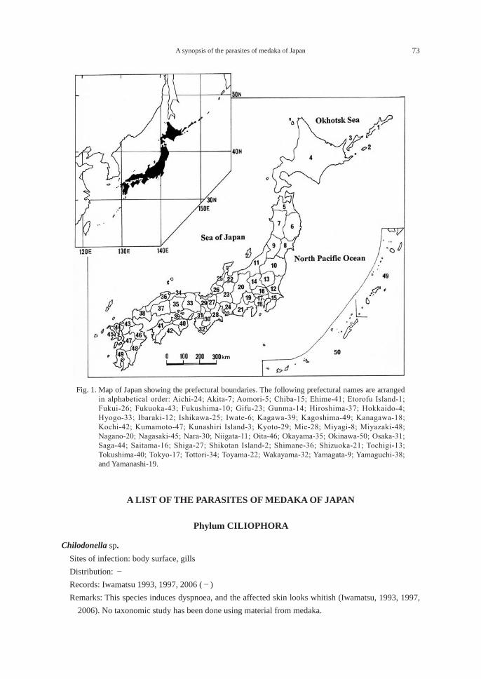
A synopsis of the parasites of medaka of Japan 73
A LIST OF THE PARASITES OF MEDAKA OF JAPAN
Phylum CILIOPHORA
Chilodonella sp. Sites of infection: body surface, gills Distribution: - Records: Iwamatsu 1993, 1997, 2006 (-) Remarks: This species induces dyspnoea, and the affected skin looks whitish (Iwamatsu, 1993, 1997,
2006). No taxonomic study has been done using material from medaka.
Fig. 1. Map of Japan showing the prefectural boundaries. The following prefectural names are arranged in alphabetical order: Aichi-24; Akita-7; Aomori-5; Chiba-15; Ehime-41; Etorofu Island-1; Fukui-26; Fukuoka-43; Fukushima-10; Gifu-23; Gunma-14; Hiroshima-37; Hokkaido-4; Hyogo-33; Ibaraki-12; Ishikawa-25; Iwate-6; Kagawa-39; Kagoshima-49; Kanagawa-18; Kochi-42; Kumamoto-47; Kunashiri Island-3; Kyoto-29; Mie-28; Miyagi-8; Miyazaki-48; Nagano-20; Nagasaki-45; Nara-30; Niigata-11; Oita-46; Okayama-35; Okinawa-50; Osaka-31; Saga-44; Saitama-16; Shiga-27; Shikotan Island-2; Shimane-36; Shizuoka-21; Tochigi-13; Tokushima-40; Tokyo-17; Tottori-34; Toyama-22; Wakayama-32; Yamagata-9; Yamaguchi-38; and Yamanashi-19.
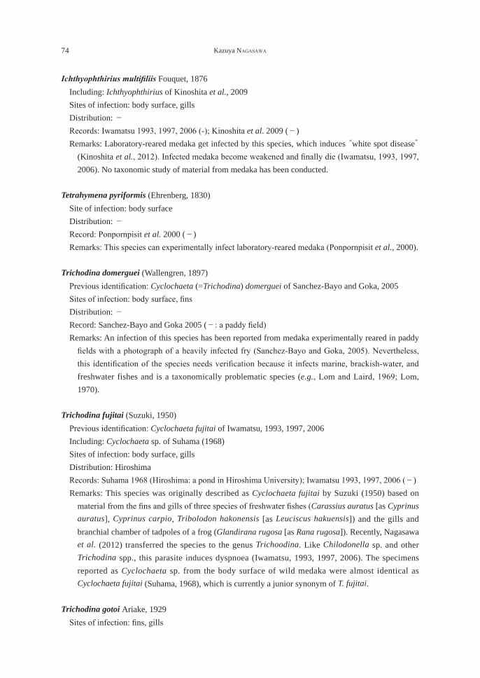
74 Kazuya NAGASAWA
Ichthyophthirius multifiliis Fouquet, 1876 Including: Ichthyophthirius of Kinoshita et al., 2009 Sites of infection: body surface, gills Distribution: - Records: Iwamatsu 1993, 1997, 2006 (-); Kinoshita et al. 2009 (-) Remarks: Laboratory-reared medaka get infected by this species, which induces “white spot disease”
(Kinoshita et al., 2012). Infected medaka become weakened and finally die (Iwamatsu, 1993, 1997, 2006). No taxonomic study of material from medaka has been conducted.
Tetrahymena pyriformis (Ehrenberg, 1830) Site of infection: body surface Distribution: - Record: Ponpornpisit et al. 2000 (-) Remarks: This species can experimentally infect laboratory-reared medaka (Ponpornpisit et al., 2000).
Trichodina domerguei (Wallengren, 1897) Previous identification: Cyclochaeta (=Trichodina) domerguei of Sanchez-Bayo and Goka, 2005 Sites of infection: body surface, fins Distribution: - Record: Sanchez-Bayo and Goka 2005 (-: a paddy field) Remarks: An infection of this species has been reported from medaka experimentally reared in paddy
fields with a photograph of a heavily infected fry (Sanchez-Bayo and Goka, 2005). Nevertheless, this identification of the species needs verification because it infects marine, brackish-water, and freshwater fishes and is a taxonomically problematic species (e.g., Lom and Laird, 1969; Lom, 1970).
Trichodina fujitai (Suzuki, 1950) Previous identification: Cyclochaeta fujitai of Iwamatsu, 1993, 1997, 2006 Including: Cyclochaeta sp. of Suhama (1968) Sites of infection: body surface, gills Distribution: Hiroshima Records: Suhama 1968 (Hiroshima: a pond in Hiroshima University); Iwamatsu 1993, 1997, 2006 (-) Remarks: This species was originally described as Cyclochaeta fujitai by Suzuki (1950) based on
material from the fins and gills of three species of freshwater fishes (Carassius auratus [as Cyprinus auratus], Cyprinus carpio, Tribolodon hakonensis [as Leuciscus hakuensis]) and the gills and branchial chamber of tadpoles of a frog (Glandirana rugosa [as Rana rugosa]). Recently, Nagasawa et al. (2012) transferred the species to the genus Trichoodina. Like Chilodonella sp. and other Trichodina spp., this parasite induces dyspnoea (Iwamatsu, 1993, 1997, 2006). The specimens reported as Cyclochaeta sp. from the body surface of wild medaka were almost identical as Cyclochaeta fujitai (Suhama, 1968), which is currently a junior synonym of T. fujitai.
Trichodina gotoi Ariake, 1929 Sites of infection: fins, gills
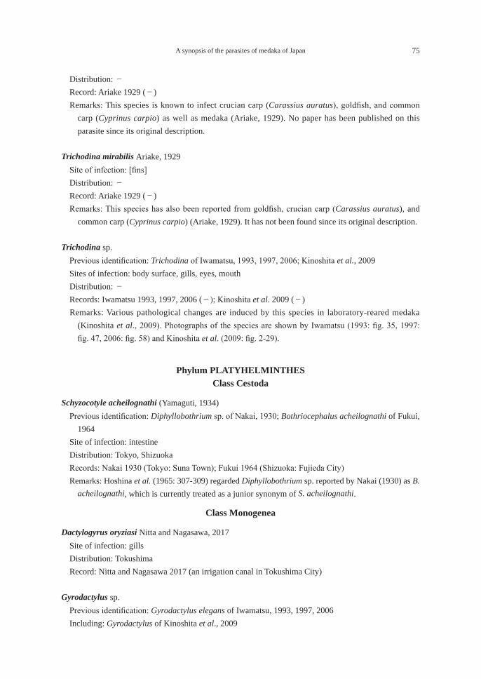
A synopsis of the parasites of medaka of Japan 75
Distribution: - Record: Ariake 1929 (-) Remarks: This species is known to infect crucian carp (Carassius auratus), goldfish, and common
carp (Cyprinus carpio) as well as medaka (Ariake, 1929). No paper has been published on this parasite since its original description.
Trichodina mirabilis Ariake, 1929 Site of infection: [fins] Distribution: - Record: Ariake 1929 (-) Remarks: This species has also been reported from goldfish, crucian carp (Carassius auratus), and
common carp (Cyprinus carpio) (Ariake, 1929). It has not been found since its original description.
Trichodina sp. Previous identification: Trichodina of Iwamatsu, 1993, 1997, 2006; Kinoshita et al., 2009 Sites of infection: body surface, gills, eyes, mouth Distribution: - Records: Iwamatsu 1993, 1997, 2006 (-); Kinoshita et al. 2009 (-) Remarks: Various pathological changes are induced by this species in laboratory-reared medaka
(Kinoshita et al., 2009). Photographs of the species are shown by Iwamatsu (1993: fig. 35, 1997: fig. 47, 2006: fig. 58) and Kinoshita et al. (2009: fig. 2-29).
Phylum PLATYHELMINTHESClass Cestoda
Schyzocotyle acheilognathi (Yamaguti, 1934) Previous identification: Diphyllobothrium sp. of Nakai, 1930; Bothriocephalus acheilognathi of Fukui,
1964 Site of infection: intestine Distribution: Tokyo, Shizuoka Records: Nakai 1930 (Tokyo: Suna Town); Fukui 1964 (Shizuoka: Fujieda City) Remarks: Hoshina et al. (1965: 307-309) regarded Diphyllobothrium sp. reported by Nakai (1930) as B.
acheilognathi, which is currently treated as a junior synonym of S. acheilognathi.
Class Monogenea
Dactylogyrus oryziasi Nitta and Nagasawa, 2017 Site of infection: gills Distribution: Tokushima Record: Nitta and Nagasawa 2017 (an irrigation canal in Tokushima City)
Gyrodactylus sp. Previous identification: Gyrodactylus elegans of Iwamatsu, 1993, 1997, 2006 Including: Gyrodactylus of Kinoshita et al., 2009
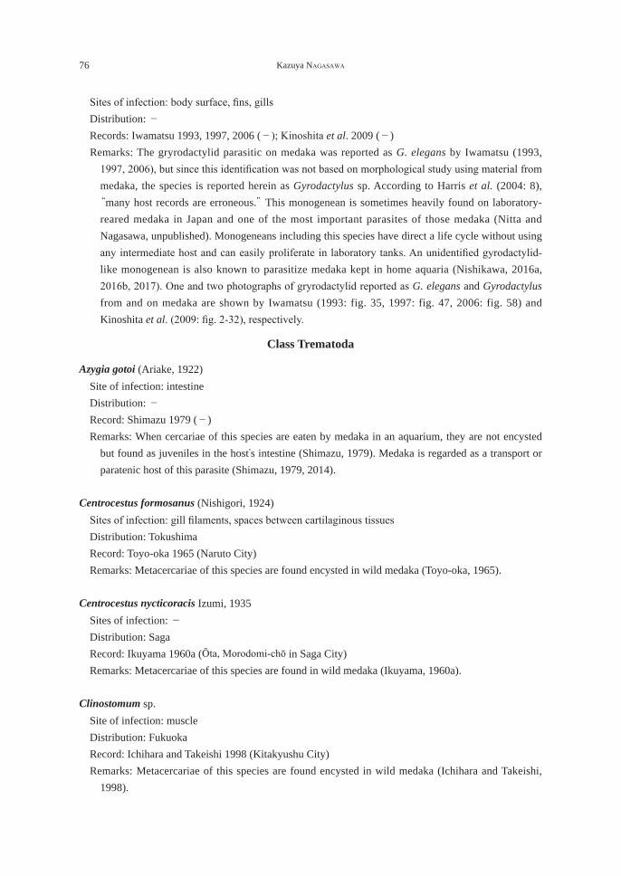
76 Kazuya NAGASAWA
Sites of infection: body surface, fins, gills Distribution: - Records: Iwamatsu 1993, 1997, 2006 (-); Kinoshita et al. 2009 (-) Remarks: The gryrodactylid parasitic on medaka was reported as G. elegans by Iwamatsu (1993,
1997, 2006), but since this identification was not based on morphological study using material from medaka, the species is reported herein as Gyrodactylus sp. According to Harris et al. (2004: 8), “many host records are erroneous.” This monogenean is sometimes heavily found on laboratory-reared medaka in Japan and one of the most important parasites of those medaka (Nitta and Nagasawa, unpublished). Monogeneans including this species have direct a life cycle without using any intermediate host and can easily proliferate in laboratory tanks. An unidentified gyrodactylid-like monogenean is also known to parasitize medaka kept in home aquaria (Nishikawa, 2016a, 2016b, 2017). One and two photographs of gryrodactylid reported as G. elegans and Gyrodactylus from and on medaka are shown by Iwamatsu (1993: fig. 35, 1997: fig. 47, 2006: fig. 58) and Kinoshita et al. (2009: fig. 2-32), respectively.
Class Trematoda
Azygia gotoi (Ariake, 1922) Site of infection: intestine Distribution: - Record: Shimazu 1979 (-) Remarks: When cercariae of this species are eaten by medaka in an aquarium, they are not encysted
but found as juveniles in the host’s intestine (Shimazu, 1979). Medaka is regarded as a transport or paratenic host of this parasite (Shimazu, 1979, 2014).
Centrocestus formosanus (Nishigori, 1924) Sites of infection: gill filaments, spaces between cartilaginous tissues Distribution: Tokushima Record: Toyo-oka 1965 (Naruto City) Remarks: Metacercariae of this species are found encysted in wild medaka (Toyo-oka, 1965). Centrocestus nycticoracis Izumi, 1935 Sites of infection: - Distribution: Saga Record: Ikuyama 1960a (Ōta, Morodomi-chō in Saga City) Remarks: Metacercariae of this species are found in wild medaka (Ikuyama, 1960a).
Clinostomum sp. Site of infection: muscle Distribution: Fukuoka Record: Ichihara and Takeishi 1998 (Kitakyushu City) Remarks: Metacercariae of this species are found encysted in wild medaka (Ichihara and Takeishi,
1998).
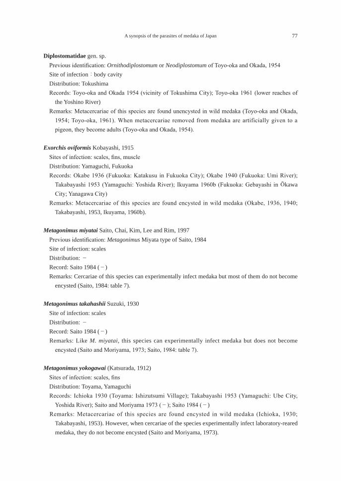
A synopsis of the parasites of medaka of Japan 77
Diplostomatidae gen. sp. Previous identification: Ornithodiplostomum or Neodiplostomum of Toyo-oka and Okada, 1954 Site of infection:body cavity Distribution: Tokushima Records: Toyo-oka and Okada 1954 (vicinity of Tokushima City); Toyo-oka 1961 (lower reaches of
the Yoshino River) Remarks: Metacercariae of this species are found unencysted in wild medaka (Toyo-oka and Okada,
1954; Toyo-oka, 1961). When metacercariae removed from medaka are artificially given to a pigeon, they become adults (Toyo-oka and Okada, 1954).
Exorchis oviformis Kobayashi, 1915 Sites of infection: scales, fins, muscle Distribution: Yamaguchi, Fukuoka Records: Okabe 1936 (Fukuoka: Katakusu in Fukuoka City); Okabe 1940 (Fukuoka: Umi River);
Takabayashi 1953 (Yamaguchi: Yoshida River); Ikuyama 1960b (Fukuoka: Gebayashi in Ōkawa City; Yanagawa City)
Remarks: Metacercariae of this species are found encysted in wild medaka (Okabe, 1936, 1940; Takabayashi, 1953, Ikuyama, 1960b).
Metagonimus miyatai Saito, Chai, Kim, Lee and Rim, 1997 Previous identification: Metagonimus Miyata type of Saito, 1984 Site of infection: scales Distribution: - Record: Saito 1984 (-) Remarks: Cercariae of this species can experimentally infect medaka but most of them do not become
encysted (Saito, 1984: table 7).
Metagonimus takahashii Suzuki, 1930 Site of infection: scales Distribution: - Record: Saito 1984 (-) Remarks: Like M. miyatai, this species can experimentally infect medaka but does not become
encysted (Saito and Moriyama, 1973; Saito, 1984: table 7).
Metagonimus yokogawai (Katsurada, 1912) Sites of infection: scales, fins Distribution: Toyama, Yamaguchi Records: Ichioka 1930 (Toyama: Ishizutsumi Village); Takabayashi 1953 (Yamaguchi: Ube City,
Yoshida River); Saito and Moriyama 1973 (-); Saito 1984 (-) Remarks: Metacercariae of this species are found encysted in wild medaka (Ichioka, 1930;
Takabayashi, 1953). However, when cercariae of the species experimentally infect laboratory-reared medaka, they do not become encysted (Saito and Moriyama, 1973).
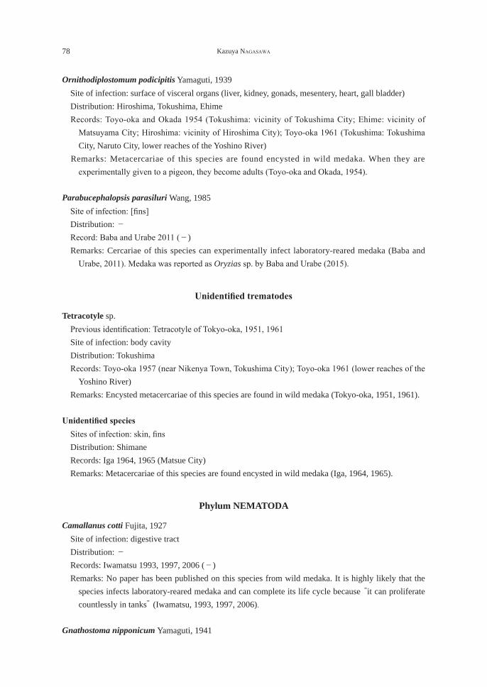
78 Kazuya NAGASAWA
Ornithodiplostomum podicipitis Yamaguti, 1939 Site of infection: surface of visceral organs (liver, kidney, gonads, mesentery, heart, gall bladder) Distribution: Hiroshima, Tokushima, Ehime Records: Toyo-oka and Okada 1954 (Tokushima: vicinity of Tokushima City; Ehime: vicinity of
Matsuyama City; Hiroshima: vicinity of Hiroshima City); Toyo-oka 1961 (Tokushima: Tokushima City, Naruto City, lower reaches of the Yoshino River)
Remarks: Metacercariae of this species are found encysted in wild medaka. When they are experimentally given to a pigeon, they become adults (Toyo-oka and Okada, 1954).
Parabucephalopsis parasiluri Wang, 1985 Site of infection: [fins] Distribution: - Record: Baba and Urabe 2011 (-) Remarks: Cercariae of this species can experimentally infect laboratory-reared medaka (Baba and
Urabe, 2011). Medaka was reported as Oryzias sp. by Baba and Urabe (2015).
Unidentified trematodes
Tetracotyle sp. Previous identification: Tetracotyle of Tokyo-oka, 1951, 1961 Site of infection: body cavity Distribution: Tokushima Records: Toyo-oka 1957 (near Nikenya Town, Tokushima City); Toyo-oka 1961 (lower reaches of the
Yoshino River) Remarks: Encysted metacercariae of this species are found in wild medaka (Tokyo-oka, 1951, 1961).
Unidentified species Sites of infection: skin, fins Distribution: Shimane Records: Iga 1964, 1965 (Matsue City) Remarks: Metacercariae of this species are found encysted in wild medaka (Iga, 1964, 1965).
Phylum NEMATODA
Camallanus cotti Fujita, 1927 Site of infection: digestive tract Distribution: - Records: Iwamatsu 1993, 1997, 2006 (-) Remarks: No paper has been published on this species from wild medaka. It is highly likely that the
species infects laboratory-reared medaka and can complete its life cycle because “it can proliferate countlessly in tanks” (Iwamatsu, 1993, 1997, 2006).
Gnathostoma nipponicum Yamaguti, 1941
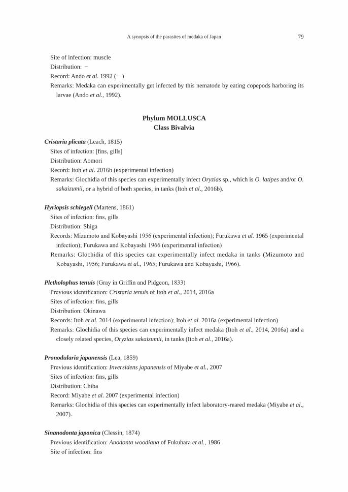
A synopsis of the parasites of medaka of Japan 79
Site of infection: muscle Distribution: - Record: Ando et al. 1992 (-) Remarks: Medaka can experimentally get infected by this nematode by eating copepods harboring its
larvae (Ando et al., 1992).
Phylum MOLLUSCAClass Bivalvia
Cristaria plicata (Leach, 1815) Sites of infection: [fins, gills] Distribution: Aomori Record: Itoh et al. 2016b (experimental infection) Remarks: Glochidia of this species can experimentally infect Oryzias sp., which is O. latipes and/or O.
sakaizumii, or a hybrid of both species, in tanks (Itoh et al., 2016b).
Hyriopsis schlegeli (Martens, 1861) Sites of infection: fins, gills Distribution: Shiga Records: Mizumoto and Kobayashi 1956 (experimental infection); Furukawa et al. 1965 (experimental
infection); Furukawa and Kobayashi 1966 (experimental infection) Remarks: Glochidia of this species can experimentally infect medaka in tanks (Mizumoto and
Kobayashi, 1956; Furukawa et al., 1965; Furukawa and Kobayashi, 1966).
Pletholophus tenuis (Gray in Griffin and Pidgeon, 1833) Previous identification: Cristaria tenuis of Itoh et al., 2014, 2016a Sites of infection: fins, gills Distribution: Okinawa Records: Itoh et al. 2014 (experimental infection); Itoh et al. 2016a (experimental infection) Remarks: Glochidia of this species can experimentally infect medaka (Itoh et al., 2014, 2016a) and a
closely related species, Oryzias sakaizumii, in tanks (Itoh et al., 2016a).
Pronodularia japanensis (Lea, 1859) Previous identification: Inversidens japanensis of Miyabe et al., 2007 Sites of infection: fins, gills Distribution: Chiba Record: Miyabe et al. 2007 (experimental infection) Remarks: Glochidia of this species can experimentally infect laboratory-reared medaka (Miyabe et al.,
2007).
Sinanodonta japonica (Clessin, 1874) Previous identification: Anodonta woodiana of Fukuhara et al., 1986 Site of infection: fins
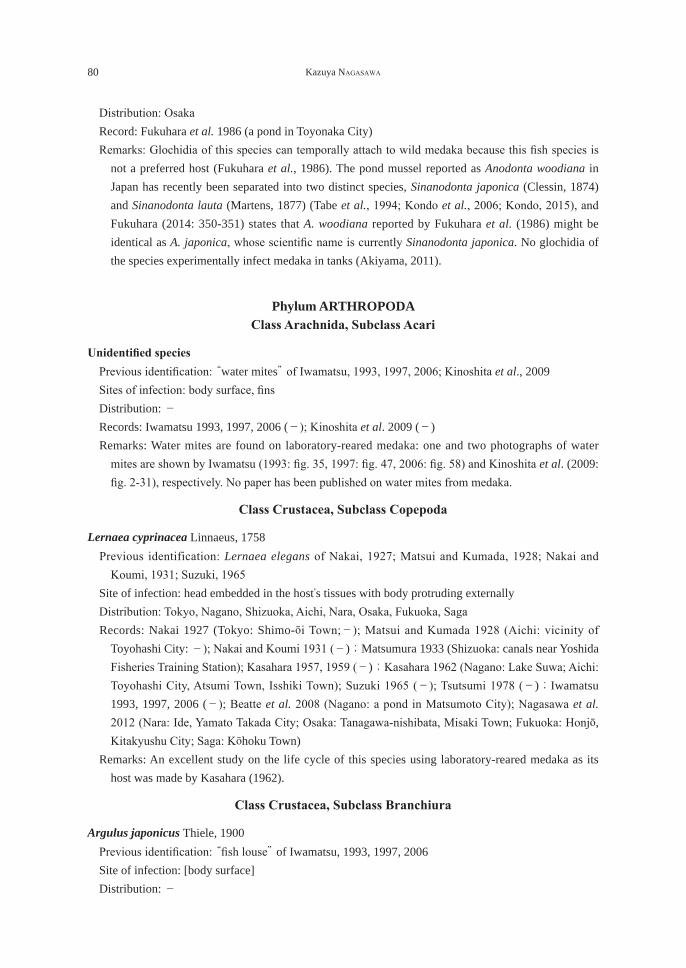
80 Kazuya NAGASAWA
Distribution: Osaka Record: Fukuhara et al. 1986 (a pond in Toyonaka City) Remarks: Glochidia of this species can temporally attach to wild medaka because this fish species is
not a preferred host (Fukuhara et al., 1986). The pond mussel reported as Anodonta woodiana in Japan has recently been separated into two distinct species, Sinanodonta japonica (Clessin, 1874) and Sinanodonta lauta (Martens, 1877) (Tabe et al., 1994; Kondo et al., 2006; Kondo, 2015), and Fukuhara (2014: 350-351) states that A. woodiana reported by Fukuhara et al. (1986) might be identical as A. japonica, whose scientific name is currently Sinanodonta japonica. No glochidia of the species experimentally infect medaka in tanks (Akiyama, 2011).
Phylum ARTHROPODAClass Arachnida, Subclass Acari
Unidentified species Previous identification: “water mites” of Iwamatsu, 1993, 1997, 2006; Kinoshita et al., 2009 Sites of infection: body surface, fins Distribution: - Records: Iwamatsu 1993, 1997, 2006 (-); Kinoshita et al. 2009 (-) Remarks: Water mites are found on laboratory-reared medaka: one and two photographs of water
mites are shown by Iwamatsu (1993: fig. 35, 1997: fig. 47, 2006: fig. 58) and Kinoshita et al. (2009: fig. 2-31), respectively. No paper has been published on water mites from medaka.
Class Crustacea, Subclass Copepoda
Lernaea cyprinacea Linnaeus, 1758 Previous identification: Lernaea elegans of Nakai, 1927; Matsui and Kumada, 1928; Nakai and
Koumi, 1931; Suzuki, 1965 Site of infection: head embedded in the host’s tissues with body protruding externally Distribution: Tokyo, Nagano, Shizuoka, Aichi, Nara, Osaka, Fukuoka, Saga Records: Nakai 1927 (Tokyo: Shimo-ōi Town;-); Matsui and Kumada 1928 (Aichi: vicinity of
Toyohashi City: -); Nakai and Koumi 1931 (-);Matsumura 1933 (Shizuoka: canals near Yoshida Fisheries Training Station); Kasahara 1957, 1959 (-);Kasahara 1962 (Nagano: Lake Suwa; Aichi: Toyohashi City, Atsumi Town, Isshiki Town); Suzuki 1965 (-); Tsutsumi 1978 (-);Iwamatsu 1993, 1997, 2006 (-); Beatte et al. 2008 (Nagano: a pond in Matsumoto City); Nagasawa et al. 2012 (Nara: Ide, Yamato Takada City; Osaka: Tanagawa-nishibata, Misaki Town; Fukuoka: Honjō, Kitakyushu City; Saga: Kōhoku Town)
Remarks: An excellent study on the life cycle of this species using laboratory-reared medaka as its host was made by Kasahara (1962).
Class Crustacea, Subclass Branchiura
Argulus japonicus Thiele, 1900 Previous identification: “fish louse” of Iwamatsu, 1993, 1997, 2006 Site of infection: [body surface] Distribution: -
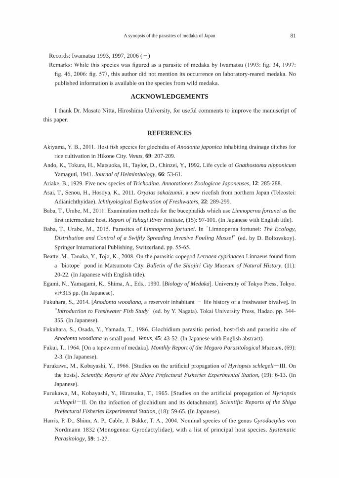
A synopsis of the parasites of medaka of Japan 81
Records: Iwamatsu 1993, 1997, 2006 (-) Remarks: While this species was figured as a parasite of medaka by Iwamatsu (1993: fig. 34, 1997:
fig. 46, 2006: fig. 57), this author did not mention its occurrence on laboratory-reared medaka. No published information is available on the species from wild medaka.
ACKNOWLEDGEMENTS
I thank Dr. Masato Nitta, Hiroshima University, for useful comments to improve the manuscript of this paper.
REFERENCES
Akiyama, Y. B., 2011. Host fish species for glochidia of Anodonta japonica inhabiting drainage ditches for rice cultivation in Hikone City. Venus, 69: 207-209.
Ando, K., Tokura, H., Matsuoka, H., Taylor, D., Chinzei, Y., 1992. Life cycle of Gnathostoma nipponicum Yamaguti, 1941. Journal of Helminthology, 66: 53-61.
Ariake, B., 1929. Five new species of Trichodina. Annotationes Zoologicae Japonenses, 12: 285-288. Asai, T., Senou, H., Hosoya, K., 2011. Oryzias sakaizumii, a new ricefish from northern Japan (Teleostei:
Adianichthyidae). Ichthyological Exploration of Freshwaters, 22: 289-299.Baba, T., Urabe, M., 2011. Examination methods for the bucephalids which use Limnoperna fortunei as the
first intermediate host. Report of Yahagi River Institute, (15): 97-101. (In Japanese with English title).Baba, T., Urabe, M., 2015. Parasites of Limnoperna fortunei. In “Limnoperna fortunei: The Ecology,
Distribution and Control of a Swiftly Spreading Invasive Fouling Mussel” (ed. by D. Boltovskoy). Springer International Publishing, Switzerland. pp. 55-65.
Beatte, M., Tanaka, Y., Tojo, K., 2008. On the parasitic copepod Lernaea cyprinacea Linnaeus found from a ‘biotope’ pond in Matsumoto City. Bulletin of the Shiojiri City Museum of Natural History, (11): 20-22. (In Japanese with English title).
Egami, N., Yamagami, K., Shima, A., Eds., 1990. [Biology of Medaka]. University of Tokyo Press, Tokyo. vi+315 pp. (In Japanese).
Fukuhara, S., 2014. [Anodonta woodiana, a reservoir inhabitant - life history of a freshwater bivalve]. In “Introduction to Freshwater Fish Study” (ed. by Y. Nagata). Tokai University Press, Hadao. pp. 344-355. (In Japanese).
Fukuhara, S., Osada, Y., Yamada, T., 1986. Glochidium parasitic period, host-fish and parasitic site of Anodonta woodiana in small pond. Venus, 45: 43-52. (In Japanese with English abstract).
Fukui, T., 1964. [On a tapeworm of medaka]. Monthly Report of the Meguro Parasitological Museum, (69): 2-3. (In Japanese).
Furukawa, M., Kobayashi, Y., 1966. [Studies on the artificial propagation of Hyriopsis schlegeli-III. On the hosts]. Scientific Reports of the Shiga Prefectural Fisheries Experimental Station, (19): 6-13. (In Japanese).
Furukawa, M., Kobayashi, Y., Hiratsuka, T., 1965. [Studies on the artificial propagation of Hyriopsis schlegeli-II. On the infection of glochidium and its detachment]. Scientific Reports of the Shiga Prefectural Fisheries Experimental Station, (18): 59-65. (In Japanese).
Harris, P. D., Shinn, A. P., Cable, J. Bakke, T. A., 2004. Nominal species of the genus Gyrodactylus von Nordmann 1832 (Monogenea: Gyrodactylidae), with a list of principal host species. Systematic Parasitology, 59: 1-27.
82 Kazuya NAGASAWA
Hoshina, T., Shikama, Y., Egusa, S., 1965. [Fish diseases]. In “[Introduction to Fish Culture]” (ed. by N. Kawamoto). Koseisha-Koseikaku, Tokyo. pp. 186-363. (In Japanese).
Hosoya, K., 2015. Oryzias latipes (Temminck & Schlegel, 1846). In “Red Data Book 2014. Threatened Wildlife of Japan - Volume 4, Pisces - Brackish and Fresh Water Fishes” (ed. by the Ministry of the Environment). Gyosei Corporation, Tokyo. pp. 166-167. (In Japanese).
Ichihara, A., Takeishi, M., 1998. [On Clinostomum sp. (Clinostomidae) parasitic in the muscle of medaka from Kitakyushu]. Abstracts of 58th East Japan Meeting of the Japanese Society of Parasitology. p. 46. (In Japanese).
Ichioka, S., 1930. [On the distributions of human parasites and Ligula-like diphyllobothriid larvae using freshwater fishes as intermediate hosts in Ishizutsumi Village, a newly recognized prevalent area of rickets, Nishi-tonami County, Toyama Prefecture]. Byorigaku Kiyo, 6: 742-760. (In Japanese).
Iga, T., 1964. Studies on melanomas induced by treamatode [sic] infection on the skin of fresh-water teleosts. I. Physiological properties of the melanoma cells. Bulletin of Shimane University (Natural Science), (14): 85-91.
Iga, T., 1965. Studies on melanomas induced by trematode infection on the skin of fresh-water teleosts. II. Formation of the dark spot. Bulletin of Shimane University (Natural Science), (15): 89-95.
Ikuyama, T., 1960a. Studies on Clonorchis sinensis in the delta of the Chikugo River. I. The results of Clonorchis sinensis - investigation in Saga Prefecture. Journal of the Kurume Medical Association, 23: 2730-2753. (In Japanese with English abstract).
Ikuyama, T., 1960b. Studies on Clonorchis sinensis in the delta of the Chikugo River. II. The results of Clonorchis sinensis investigation in Fukuoka Prefecture. Journal of the Kurume Medical Association, 23: 2754-2776. (In Japanese with English abstract).
Itoh, T., Kitano, T., Toma, M., Fujimoto, H., Sakihara, K., Kohno, H., 2014. Host species for glochidia of the freshwater unionid mussel Cristaria tenuis in [sic] Ishigakijima Island, Japan. Venus, 72: 77-87.
Itoh, T., Kitano, T., Fujimoto, H., Sakihara, K., Kohno, H., 2016a. Record of five new host species for the glochidia of the freshwater unionid mussel Cristaria tenuis. Venus, 74: 41-44.
Itoh, T., Uesugi, S., Kitano, W., 2016b. Host fish species for glochidia of the freshwater unionid mussel Cristaria plicata in tanks. Venus, 74: 79-88. (In Japanese with English abstract).
Iwamatsu, T., 1993. [The Biology of the Medaka]. Scientist Press, Tokyo. vii+324 pp. (In Japanese). Iwamatsu, T., 1997. The Integrated Book for the Biology of the Medaka. University Education Press,
Okayama. vi+360 pp. (In Japanese). Iwamatsu, T., 2006. The Integrated Book for the Biology of the Medaka. University Education Press,
Okayama. x+473 pp. (In Japanese). Kasahara, S. 1957. [On control effects of dipterex on anchor worm]. Noyaku Kenkyu, 4: 1-6. (In Japanese).Kasahara, S., 1959. [On control of anchor worm]. Suisan Zoshoku, 6: 140-148. (In Japanese).Kasahara, S., 1962. Studies on the biology of the parasitic copepod Lernaea cyprinacea Linnaeus and the
methods for controlling this parasite in fish-culture ponds. Contributions of the Fisheries Laboratory, Faculty of Agriculture, University of Tokyo, (3): 103-196. (In Japanese with English abstract).
Kinoshita, M., Murata, K., Naruse, K., Tanaka, M., eds., 2009. Medaka: Biology, Management, and Experimental Protocols. Willey-Blackwell, Singapore. xiv+419 pp.
Kondo, T., 2015. [Illustrations of unionid mussels of Japan]. http://www.osaka-kyoiku.ac.jp/~kondo/unio/unio.html (accessed on 20 August 2017).
Kondo, T., Tabe, M., Fukuhara, S., 2006. Morphological differences of glochidia between two genetic types
A synopsis of the parasites of medaka of Japan 83
of Anodonta “woodiana” (Bivalvia: Unionidae). Venus, 65: 241-245. (In Japanese with English abstract).
Lom, J., 1970. Observations on trichodinid ciliates from freshwater fishes. Archive für Protistenkunde, 112: 153-177.
Lom, J., Laird, M., 1969. Parasitic Protozoa from marine and euryhaline fish of Newfoundland and New Brunswick. I. Peritrichous ciliates. Canadian Journal of Zoology, 47: 1367-1380.
Matsui, Y., Kumada, A., 1928. Studies on fish-diseases. I. “Ikari-mushi” (Lernaea elegans Leigh-Sharpe), a new parasitic copepod of Japanese eel. Journal of the Imperial Fisheries Institute, 23: 101-107 (in English), 131-141, 3 pls. (In Japanese).
Matsumura, A., 1933. [Anchor worm parasitic on fishes in canals of a fisheries training station]. Yoshoku Kaishi, 3: 180-181. (In Japanese).
Miyabe, T., Takahashi, K., Inoue, M., 2007. Basic research related to artificial multiplication of Inversidens japanensis. Bulletin of the Chiba Prefectural Fisheries Research Center, (2): 53-60. (In Japanese with English title).
Mizumoto, S., Kobayashi, Y., 1956. [Studies on the propagation of Hyriopsis schlegeli. Part 5]. Scientific Reports of the Shiga Prefectural Fisheries Experimental Station, (6): 9-13. (In Japanese).
Nagasawa, K., Morimoto, S., Asai, T., Kitagawa, T., Hosoya, K., 2012. A checklist of the parasites of medaka (Oryzias latipes) of Japan (1929-2012), with new records of Lernaea cyprinacea (Copepoda: Lernaeidae) in wild populations of medaka in Japan. Bulletin of the Biogeographical Society of Japan, 67: 1-13. (In Japanese with English abstract).
Nakai, N., 1927. On the development of a parasitic copepod, Lernaea elegans Leigh-Sharpe, infesting on [sic] Cyprinus carpio L. Journal of the Imperial Fisheries Institute, 23: 39-59 (in English), 69-88, 3 pls. (In Japanese).
Nakai, N., 1930. [On Diphyllobothrium sp., a tapeworm of medaka (preliminary report)]. Suisan Butsuri Danwakai Kaiho, (17): 217-222. (In Japanese).
Nakai, N., Koumi, E., 1931. On the biological study of a parasitic copepod, Lernaea elegans Leigh-Sharpe, infesting on [sic] Japanese fresh water fishes. Journal of the Imperial Fisheries Experimental Station, (2): 93-128. (In Japanese with English abstract).
Nishikawa, H., 2016a. [Disease control of ornamental fishes 126. Monogenean infection of medaka]. Aqualife, (442): 72-73. (In Japanese).
Nishikawa, H., 2016b. [Disease control of ornamental fishes 130. Monogenean infection of medaka and treatment with SUNACE]. Aqualife, (446): 116-117. (In Japanese).
Nishikawa, H., 2017. [Disease control of ornamental fishes 140. Monogenean infection of medaka]. Aqualife, (456): 72-73. (In Japanese).
Nitta, M., Nagasawa, K., 2017. Dactylogyrus oryziasi n. sp. (Monogenea: Dactylogyridae) from Oryzias latipes (Beloniformes: Adrianichthyidae) in Japan. Species Diversity, 22: 1-5.
Okabe, K., 1936. [On the life history of Exorchis ovifomis [sic] Kobayashi (Trematoda)]. Fukuoka Acta Medica, 29: 211-220. (In Japanese).
Okabe, K., 1940. [Overview of encysted metacercariae of trematodes using freshwater fishes as intermediate hosts in Fukuoka Prefecture]. Fukuoka Acta Medica, 33: 309-335. (In Japanese).
Ponpornpisit, A., Endo, M., Murata, H., 2000. Experimental infections of a ciliate Tetrahymena pyriformis on ornamental fishes. Fisheries Science, 66: 1026-1031.
Saito, S., 1984. [On differences between species in the genus Metagonimus]. Japanese Society for
84 Kazuya NAGASAWA
Systematic Parasitology Circular, (2): 1-4. (In Japanese).Saito, S., Moriyama, N., 1993. Studies on genus Metagonimus XIII. The experimental infection to Oryzias
latipes with both cercariae of M. takahashii and M. yokogawai. Japanese Journal of Parasitology, 22 (supplement): 70. (In Japanese with English title).
Saito, S., Otsuru, M., Hasegawa, K., Hori, M., 1964. Some observations on metacercariae of trematodes encysted in fresh-water fishes in Niigata Prefecture, Japan. Niigata Medical Journal, 78: 376-386. (In Japanese with English title).
Sakaizumi, M., 1986. Genetic divergence in wild populations of Medaka, Oryzias latipes (Pisces: Oryziatidae) from Japan and China. Genetica, 69: 119-125.
Sanchez-Bayo, F., Goka, K., 2005. Unexpected effects of zinc pyrithione and imidacloprid on Japanese medaka fish (Oryzias latipes). Aquatic Toxicology, 74: 285-293.
Shimazu, T., 1979. Developmental stages of Azygia gotoi (Digenea, Azygiidae). Bulletin of the National Science Museum, Tokyo, Series A (Zoology), 5: 225-234.
Shimazu, T., 2014. Digeneans parasitic in freshwater fishes (Osteichthyes) of Japan. III. Azygiidae and Bucephalidae. Bulletin of the National Museum of Nature and Science, Series A, 40: 167-190.
Suhama, M., 1968. The fine structure of adhesive disc in urceolarid ciliate, Cyclochaeta sp. from Oryzias latipes. Bulletin of the Biological Society of Hiroshima University, (34): 6-12. (In Japanese with English abstract).
Suzuki, H., 1965. Lernaea elegans Leigh-Sharpe, a common parasitic copepoda [sic] found in the skin of the fresh-water fish, Oryzias latipes (Temminck et Schlegel). Researches on Crustacea, (2): 67-76. (In Japanese with English abstract).
Suzuki, S., 1950. Studies on the uroceolarid ciliates of Japan. Bulletin of Yamagata University (Natural Sciences), (2): 181-218, 6 pls.
Tabe, M., Fkuhara, S., Nagata, Y., 1994. Genetic differentiation between two types of swan mussel, Anodonta woodiana, in Japan. Venus, 53: 29-35. (In Japanese with English abstract).
Takabayashi, Y., 1953. [Studies on trematodes utilizing fishes as intermediate hosts, with a survey in Yamaguchi Prefecture]. Acta Scholae Medicinalis in Gifu, 1: 219-226. (In Japanese).
Toyo-oka, R., 1957. [On Tetracotyle infecting medaka]. Jikken Seibutsu Gappo (Tokushima Biological Society), 7: 122. (In Japanese).
Toyo-oka, R., 1961. [Metacercariae encysted in freshwater fishes in Tokushima Prefecture]. Kyodo Kenkyu Happyokai Kiyo, (6-8): 15-26. (In Japanese).
Toyo-oka, R., 1965. [Human parasites in Naruto City and the distribution of their intermediate hosts]. Kyodo Kenkyu Happyokai Kiyo, (11): 120-141. (In Japanese).
Toyo-oka, R., Okada, K., 1954. Studies on the development of two diplostomatid metacercariae, found in Oryzias laticeps, a freshwater fish. Journal of Gakugei, Tokushima University, 4: 55-64.
Tsusumi, T., 1978. [Treatment of fish diseases at aquaria 7. Anchor worm infecting freshwater fishes and its control]. Doyaku Kenkyu, (12): 21-22. (In Japanese).
Yamamoto, T., 1975. Medaka (Killifish): Biology and Strains. Keigaku Publishing, Tokyo. iv+365 pp. 18 pls.
A synopsis of the parasites of medaka of Japan 85
日本産メダカの寄生虫目録(1929-2017年)
長澤 和也
広島大学大学院生物圏科学研究科,〒739-8528 広島県東広島市鏡山1-4-4
要 旨 1929-2017年に出版された文献を用いて,日本産メダカの寄生虫目録を作成した。本目録は2012年に日本語で出版した同名目録の追補改定版である。本目録では,メダカから報告された各寄生虫の情報を最新の学名,異名,寄生部位,地理的分布,記録(報告書と報告年)に整理した。その結果,2017年までに日本産メダカから27名義種の寄生虫が次の分類群からで報告されていたことが明らかになった:繊毛虫類(6種),条虫類(1種),単生類(1種),吸虫類(9種),線形動物(3種),二枚貝類(5種),カイアシ類(1種),エラオ類(1種)。また本目録では,それらに加えて,種まで同定されなかった繊毛虫類,単生類,吸虫類,ダニ類の各種に関する情報も含めた。日本のメダカ研究施設で問題になる寄生虫として,体表に寄生する単生類の1種 Gyrodatylus sp.があり,注意を要する。キーワード:寄生虫,文献集,メダカ,目録

総 説
日本産魚類に寄生するサメジラミ科カイアシ類の目録(1898-2017年)
長澤和也1)*・上野大輔2)
1)広島大学大学院生物圏科学研究科,〒739-8528 広島県東広島市鏡山1-4-42)鹿児島大学大学院理工学研究科,〒890-0065 鹿児島市郡元1-21-35
要 旨 1898-2017年に出版された文献に基づき,日本産魚類から記録されたサメジラミ科カイアシ類の16属24種と未同定種に関する情報(宿主,寄生部位,地理的分布,文献)を種ごとに整理した。Achtheinus, Cecropus, Demoleus, Dinemoura, Gangliopus, Nesippus, Orthagoriscicola, Parnnosus,Paranesippus,Philorthagoriscus,Pseudopandarusの11属に対して,それぞれフトバラサメジラミ,マンボウノチョウ,ドウナガサメジラミ,ツバササメジラミ,マルミサメジラミ,ホソミサメジラミ,マンボウノシラミ,ヤマトサメジラミ,ヘラツノザメジラミ,マンボウノヒフヤドリ,サメジラミモドキの新標準和名を提唱した。キーワード:カイアシ類,サメジラミ科,魚類寄生虫,板鰓類,文献目録
緒 言
本目録で扱うサメジラミ科 Pandaridaeはカイアシ亜綱 Copepodaの管口目 Siphonostomatoidaに属する甲殻類である。本科に属する種はすべて海水魚に寄生し,淡水魚に寄生する種はない。その名前が示すように,本科の多くの種はサメ類に寄生するが,近年の分類学的研究(Tang et al., 2012)によって,マンボウなどに寄生するマンボウノシラミ科 Cercropidaeカイアシ類も本科に含まれることになった。統合されたサメジラミ科に含まれるカイアシ類の種数は世界で63種であり(Boxshall, 2009),魚類寄生性カイアシ類としては大きな科ではない。ただ,マンボウの体表に寄生するマンボウノシラミ Orthagoriscicola muricatusは体長が18 mmもあるため(椎野,1965b),わが国ではその際立った存在が19世紀末から知られており(宍戸,1898b),本科に関する研究の歴史は長い。 本目録は,日本産サメジラミ科の16属24種と未同定種について,1898-2017年の120年間に出版された文献をもとに,各種の情報を整理したものである。1898年は,帝国理科大学の宍戸一郎がサメジラミ科カイアシ類をわが国で最初に報告した年である(宍戸,1998a, 1898b)。その後,本科に関する研究は,石井重美(石井,1916a, 1916b),C. B. Wilson(Wilson, 1922),丸川久俊(丸川,1927)に引き継がれ,京都大学(後に岡山大学)の山口左仲と三重県立大学の椎野季雄が分類学的研究で極めて大きな成果を挙げた(Yamaguti, 1936;Yamaguti and Yamasu, 1959, 1960;Shiino, 1954a, 1954b, 1955, 1957, 1959, 1960)。わが国と世界各地で得られた知見は,山口が1963年に『Parasitic Copepoda and Branchiura of fishes』(Yamaguti, 1963)に収録して当時の集大成を図った。近年は,三重大学を退職後,伊澤海洋生物学研究所を主宰する伊澤邦彦が分類学的研究を精力的に実施している(Izawa, 2010a, 2010b, 2010c, 2012)。また,広島大学の長澤和也と博士研究員であった D. Tang(Nagasawa and Nakaya, 1997;Tang et al., 2010, 2012;Nagasawa and Senou, 2012),鹿児島大学の上野大輔(Uyeno et al., 2016)も研究を行っている。長澤は板鰓類研究会の機
2017年9月19日受理 *E-mail: [email protected]
生物圏科学Biosphere Sci. 56:87-104 (2017)
88 長澤和也・上野大輔
関誌『板鰓類研究会報』でサメジラミ科カイアシ類の解説を行っている(長澤,2009,2014,2017;長澤ら,2013)。 本目録では,サメジラミ科内の各属をアルファベット順に並べた後,各属では種小名のアルファベット順に各種を記述した。標準和名と最新の学名をまず記し,異名リストと宿主,寄生部位を示したあと,地理的分布を示した。異名リストに示した学名はわが国で用いられたものに限り,これを欠くものは異名での報告がわが国にないことを示す。各異名の直後には,それらを報告した著者名と出版年を示した。宿主の魚類は,中坊(2013)が示した分類体系に従って配列し,標準和名と学名を記した。この際,過去の論文で現在の学名や和名と異なるものが使われた場合には括弧内にそれらを記した。地理的分布に関する情報は,海域(北太平洋,日本海,東シナ海)ごとに整理し,都道府県名を含む詳細な採集地と出典情報(著者名と出版年)を示した。都道府県名は北から南に順に配列した。原典に詳細な採集地の情報を欠く場合には「-」で示した。水族館飼育魚からの採集記録は分けて示した。備考では,当該種の生物学的情報(分類や他国での分布等に関する事項)や新標準和名の提案根拠などを記した。 各寄生虫の記録を上記のように纏めたあと,さらに宿主-寄生虫リストとして整理した。このリストでは,宿主として報告された魚類を中坊(2013)の分類体系に従って並べ,各魚種から記録されたカイアシ類を示した。各科において宿主魚類は五十音順に並べた。 サメジラミ科カイアシ類の分類体系に関しては Yamaguti(1963),Kabata(1979),Boxshall and Halsey(2004)が参考になる。東アジアにおける本科に関する知見は韓国(Kim, 1998),中国(Yu, 1933;Song and Kuang, 1980),台湾(Ho, 1963;Cressey, 1967;Ho et al., 2012)を除いて極めて限られている。本目録は,筆者らによる日本産寄生性カイアシ類目録の第12報である(ニセエラジラミ科[長澤ら,2007];イカリムシ科[Nagasawa et al., 2007];ウオジラミ属[長澤ら,2010];瀬戸内海産寄生性カイアシ類(2報)[Nagasawa, 2011, 2015];ヒトガタムシ科[長澤・上野,2011];ツツウオジラミ科,エラノミ科,ニセエラノミ科[長澤・上野,2012];ツブムシ科[長澤ら,2013a];ヒジキムシ科[長澤・上野,2014];ナガクビムシ科[長澤・上野,2015];カクレムシ科[長澤・上野,2016])。
日本産サメジラミ科カイアシ類の目録
カイアシ亜綱
Subclass Copepoda Milne Edwards, 1830
新カイアシ下綱
Infraclass Neocopepoda Huys and Boxshall, 1991
管口目(シフォノストム目)
Order Siphonostomatoida Burmeister, 1835
本目を管口目(シフォノストム目)と呼ぶ経緯については長澤ら(2010)が記している。
サメジラミ科Family Pandaridae Mile Edwards, 1840
サメジラミ科と近縁科に関して,近年,広島大学の博士研究員であった D. Tangらの研究(Tang et al., 2012)によって大きな変更が行われた。それは,19世紀半ば以降,長年にわたって独立した科であったマンボウノシラミ科 Cecropidae Dana, 1849と2008年に新設されたアマテラス科 Amaterasidae Izawa, 2008の2
サメジラミ科カイアシ類の目録 89
科がサメジラミ科の異名とみなされ,サメジラミ科に統合されたことである。サメジラミ科はその名前が示すようにサメ類(板鰓亜綱)に寄生するのに対し,他二者はほとんどが硬骨魚類(条鰭亜綱)に寄生する(Kabata, 1979;Boxshall and Halsey, 2004;Izawa, 2008)。このため,Tang et al.(2012)の研究によって,サメジラミ科は分類学的に大きく異なる魚類(板鰓類と条鰭類)に寄生するカイアシ類を含むことになった。 本科の標準和名として,古くは「パンダルス科」(丸川,1947)が用いられたが,椎野(1957:138)が「サメジラミ科」 を使用して以来, この和名が用いられている (椎野, 1965a, 1979;長澤, 2009, 2014, 2017;長澤ら, 2013)。 なお,今はサメジラミ科の異名であるマンボウノシラミ科に関連して,Yamaguti(1963: 86, 336)は新科(Cecropidae Yamaguti, 1963)を設けたが,これは受け入れられておらず,サメジラミ科の異名とされている(Boxshall, 2009)。
フトバラサメジラミ属(新称)
Genus Achtheinus C. B. Wilson, 1908
新標準和名は,本属に含まれるフトバラサメジラミ(新称)Achtheinus pingiusの標準和名に従う。
フトバラサメジラミ(新称)
Achtheinus pingius C. B. Wilson, 1912
異名リスト:Achtheinus japonicus (cf. Wilson, 1922), Achtheinus galeorhini (cf. Yamaguti, 1936), Perissopus oblongatus (cf. Yamaguchi et al., 2003)宿主:ホシザメMustelus manazo (= Galeorhinus megalops), ツマリツノザメ Squalus brevirostris (= Squalus
megalops), ツノザメ属の1種 Squalus sp. (= Acanthias sp.)寄生部位:鰭,鰓,鼻腔地理的分布:北太平洋(東京湾:Yamaguchi et al., 2003;Izawa, 2010c;神奈川県三崎:Wilson, 1922;静岡県伊豆近海:Izawa, 2010c;静岡県沼津:Yamaguti, 1936)備考:本種の詳細な分類学的研究が Izawa(2010c)によって行われた。本種は大西洋,インド洋,北西太平洋から報告されている(Izawa, 2010c)。近隣では中国(Yu, 1933;Song and Kuang, 1980)と韓国(Kim, 1998)にも分布する.新標準和名は,本種の体後部が前部より太いことに因る。「フトバラ」は「太腹」である。
マンボウノチョウ属(新称)
Genus Cecropus Leach, 1816
本属は以前マンボウノシラミ科 Cecropidaeに収められていたが(例えば Yamaguti, 1936;Boxshall and Halsey, 2004),近年この科はサメジラミ科の異名とされている(Tang et al., 2012)。新標準和名は,本属に含まれるマンボウノチョウ Cecropus latreilliiの標準和名に従う。
マンボウノチョウ
Cecropus latreillii Leach, 1816
宿主:マンボウMola sp. B,アカマンボウ Lampris guttatus
90 長澤和也・上野大輔
寄生部位:鰓,口腔地理的分布:北太平洋(神奈川県三崎:宍戸,1898a;和歌山県白浜:Shiino, 1959;-:Yamaguti, 1936),東シナ海(熊本県富岡:Shiino, 1959)備考:本種はマンボウ属魚類の鰓寄生虫として,それらの分布域の多くの場所から記録されている(Kabata,
1979: 232)。韓国からも記録がある(Kim, 1998)。標準和名は宍戸(1898a)に従う:「マンボウノテフ」と記した。
ドウナガサメジラミ属(新称)
Genus Demoleus Heller, 1865
新標準和名は,本属に含まれるドウナガサメジラミ(新称)Demoleus latusの標準和名に従う。
ドウナガサメジラミ(新称)
Demoleus latus Shiino, 1954
宿主:ヘラツノザメ Deania calcea(= Acanthidium elantina)寄生部位:体表地理的分布:北太平洋(高知県[徳島県と誤記]甲浦:Shiino, 1954a)備考:新標準和名は,本種の生殖節が長いことに因る。「ドウナガ」は「胴長」である。
メガマウスザメジラミ属
Genus Dinemoleus Cressey and Boyle, 1978
標準和名は長澤(2009)に従う。
メガマウスザメジラミ
Dinemoleus indeprensus Cressey and Boyle, 1978
宿主:メガマウスザメMegachasma pelagios寄生部位:体表地理的分布:北太平洋(神奈川県相模湾:Nagasawa and Senou, 2012),日本海(福岡県博多湾:高田,
1994;Nagasawa and Nakaya, 1997)備考:本種の生物学的知見を長澤(2009)が整理している。標準和名は長澤(2003:95)に従う。本種を高田(1997)は「チョウのような寄生虫」,長澤・仲谷(1997)は「メガマウスジラミ」と呼んだ。
ツバササメジラミ属(新称)
Genus Dinemoura Latreille, 1829
新標準和名は,本属に含まれるツバササメジラミ(新称)Dinemoura latifoliaの標準和名に従う。
サメジラミ科カイアシ類の目録 91
ツバササメジラミ(新称)
Dinemoura latifolia (Steenstrup and Lütken, 1861)
異名リスト:Dinematura latifolia(cf. Shiino, 1954a, 1957)宿主:アオザメ Isurus oxyrinchus(= Isuropsis glauca, Isurus glaucus),ネズミザメ Lamna ditropis,ヨシキリザメ Prionace glauca
寄生部位:体表(腹面),鰭地理的分布:北太平洋(宮城県気仙沼沖:Shiino, 1957;静岡県石廊崎沖:Shiino, 1954a;和歌山県紀伊勝浦町魚市場:Izawa, 2010)備考:本種はアオザメ属,ネズミザメ属,ホホジロザメ属の3属に頻繁に寄生する(Cressey, 1967)。新標準和名は,第2胸節の後側方に伸びる1対の背板が翼のように見えることに因る。
オオツバササメジラミ(新称)
Dinemoura producta (Müller, 1785)
異名リスト:Dinematura affinis(cf. Shiino, 1957)宿主:ネズミザメ Lamna ditropis寄生部位:体表,鰭地理的分布:北太平洋(宮城県気仙沼沖:Shiino, 1957)備考:本種はネズミザメ属やアオザメ属のサメ類に頻繁に寄生し,分布は汎世界的と考えられている(Cressey, 1967)。韓国からも記録がある(Kim, 1998)。新標準和名は,本種がツバササメジラミDinemoura latifoliaより大きいことに因る。
ヨシキリジラミ属
Genus Echthrogaleus Steenstrup and Lütken, 1861
標準和名は椎野(1964:125)に従う。
ヨシキリジラミ
Echthrogaleus coleoptratus (Guérin-Méneville, 1837)
宿主:アオザメ Isurus oxyrinchus (= Isuropsis glauca), ネズミザメLamna ditropsis, ヒラガシラRhizoprionodon acutus,ヨシキリザメ Prionace glauca(= Glyphis glauca)
寄生部位:体表,鰭,口腔底地理的分布:北太平洋(宮城県気仙沼港:Shiino, 1957;愛知県渥美半島沖[遠州灘]:Izawa, 2010a;三重県尾鷲市魚市場:Shiino, 1954a;三重県尾鷲市久喜:Izawa, 2010a;和歌山県紀伊勝浦町魚市場:Izawa, 2010a;-:Yamaguti, 1936)備考:わが国では丸川(1927)が『日本動物圖鑑』のなかで本種を初めて記述した(採集地は不明)。雌成体の形態が正確に記載された。同様の記述(丸川,1947)は『改訂増補 日本動物圖鑑』でも見られる。雌成体は他の研究者によっても記載され(Yamaguti, 1936;Shiino, 1954a, 1957;Izawa, 2010a),雌コペポディド5期も記載された(Izawa, 2010a)。本種は北太平洋,北大西洋,インド洋に広く分布する(Cressey,
92 長澤和也・上野大輔
1967)。近隣では韓国(Kim, 1998)と台湾(Ho et al., 2012)から記録がある。わが国における本種の知見を長澤(2017)が整理している。標準和名は丸川(1927)に従う。
ヨシキリジラミモドキ
Echthrogaleus denticulatus Smith, 1873
宿主:アオザメ Isurus oxyrinchus(= Isuropsis glauca),ニタリ Alopias pelagicus,ヨシキリザメ Prionace glauca(= Glyphis glauca),シロシュモクザメ Sphyrna zygaena
寄生部位:体表,口腔底地理的分布:北太平洋(三重県尾鷲市魚市場:Shiino, 1954a;三重県尾鷲・浜島:Shiino, 1959;和歌山県紀伊勝浦町魚市場:Izawa, 2010a)備考:本種は太平洋,大西洋,インド洋に広く分布し,オナガザメ属サメ類によく見られる(Cressey,
1967)。わが国における本種の知見を長澤(2017)が整理している。標準和名は長澤(2017)に従う。
トウヨウサメジラミ
Echthrogaleus asiaticus Ho, Liu and Lin, 2012
異名リスト:Echthrogaleus denticulatus(cf. Ho and Kim, 1996)宿主:ホホジロザメ Carcharodon carcharias寄生部位:[鰭]地理的分布:北太平洋(北海道渡島半島沖:Ho and Kim, 1996)備考:本種は,わが国では当初,ヨシキリジラミモドキ Echthrogaleus denticulatusとして報告された(Ho
and Kim, 1996)。しかし,台湾で採集された標本に基づいて,日本産標本も本種と判明した(Ho et al., 2012)。わが国における本種の知見を長澤(2017)が整理している。標準和名は長澤(2017)に従う。
ミツクリザメジラミ
Echthrogaleus mitsukurinae Izawa, 2012
宿主:ミツクリザメMitsukurina owstoni寄生部位:体表地理的分布:北太平洋(千葉県勝浦市沖の東京海谷斜面:Izawa, 2012)備考:わが国における本種の知見を長澤(2017)が整理している。標準和名は長澤(2017)に従う。
マルミサメジラミ属(新称)
Genus Gangliopus Gerstaecker, 1854
新標準和名は,下記のマルミサメジラミ Gangliopus pyriformisの備考を参照のこと。
マルミサメジラミ(新称)
Gangliopus pyriformis Gerstaecker, 1854
サメジラミ科カイアシ類の目録 93
宿主:マオナガ Alopias vulpinus寄生部位:鰓地理的分布:北太平洋(和歌山県江住沖:Izawa, 2010a, 2010b)備考:本種は大洋に広く分布し,ヨシキリザメの鰓に普通に見られる(Cressey, 1967)。Yamaguti and
Yamasu(1960)は台湾産マカジキ Kajikia audax(= Tetrapturus mitsukurii)の鰓から本種(=Gangliopus tetrapturi)を得た。新標準和名は本種の生殖節が丸い形状(=丸身)を呈することに因る。
マヨイサメジラミ属
Genus Luetkenia Claus, 1864
本属は以前マンボウノシラミ科 Cecropidaeに収められていたが(例えば Boxshall and Halsey, 2004),近年,この科はサメジラミ科の異名とされている(Tang et al., 2012)。標準和名は Uyeno et al.(2016)に従う。
マヨイサメジラミ
Luetkenia elongata Shiino, 1963
宿主:アマシイラ Luvarus imperials寄生部位:体表 地理的分布:北太平洋(神奈川県江の島:Uyeno et al., 2016)備考:本種は日本以外では米国カリフォルニア沖から記録がある(Shiino, 1963)。本種が寄生していたアマシイラは江の島海岸に漂着したものである(Uyeno et al., 2016)。標準和名は Uyeno et al.(2016)に従う。
ホソミサメジラミ属(新称)
Genus Nesippus Heller, 1868
新標準和名は,下記のホソミサメジラミ Nesippus orientalisの備考を参照のこと。
ホソミサメジラミ(新称)
Nesippus orientalis Heller, 1868
宿主:シロシュモクザメ Sphyrna zygaena寄生部位:口腔,鰓弓地理的分布:北太平洋(三重県浜島:Izawa, 2010)備考:本種は多くのサメ類から報告があり,汎世界的に分布すると考えられている(Cressey, 1967)。新標準和名は,本種の生殖節が頭胸甲に比して狭くて細い(=細身)ことに因る。
ホソミサメジラミ属の1種
Nesippus sp.
宿主:ヨゴレ Carcharhinus longimanus(= Pterolamiops longimanus)寄生部位:体表地理的分布:北太平洋(Kabata and Gusev, 1966)

94 長澤和也・上野大輔
マンボウノシラミ属(新称)
Genus Orthagoriscicola Poche, 1902
本属は以前マンボウノシラミ科 Cecropidaeに収められていたが(例えば Shiino, 1954b;Boxshall and Halsey, 2004),近年,この科はサメジラミ科の異名とされている(Tang et al., 2012)。新標準和名は,本属に含まれる唯一の種,マンボウノシラミ Orthagoriscicola muricatusの標準和名に従う。
マンボウノシラミ
Orthagoriscicola muricatus (Krøyer, 1837)
異名リスト:Laemargus muricatus (cf. 宍戸, 1898b), Orthagoriscicola sp. (cf. 石井, 1916a), Orthagoriscicola muricata (cf. 石井,1916b;Shiino, 1954)宿主:マンボウMola sp. B(= Mola mola),不明宿主 unknown host寄生部位:口腔,体表地理的分布:北太平洋(北海道様似沖:石井,1916a,1916b;神奈川県三崎:宍戸,1898b;和歌山県沖:
Shiino, 1954b)備考:本種はマンボウの体表に時に集合して寄生し,大西洋・地中海・太平洋に分布する(Kabata, 1979:
234)。本種に関する解説がある(椎野,1965b)。標準和名は宍戸(1898b)に従う。
サメジラミ属
Genus Pandarus Leach, 1816
標準和名は椎野(1964:125)に従う。
シイノサメジラミ
Pandarus cranchii Leach, 1819
異名リスト:Pandarus satyrus(cf. Shiino, 1954a, 1957, 1959, 1960)宿主:アオザメ Isurus oxyrinchus(= Isuropsis glauca, Isuruss glaucus),ニタリ Alopias pelagicus,ヨシキリザメ Prionace glauca(= Glyphis glauca),シロシュモクザメ Sphyrna zygaena,シノノメサカタザメRhina ancylostoma,種不明サメ類 unspecified shark,マカジキ Kajikia audax(= Makaira mitsukurii)
寄生部位:体表地理的分布:北太平洋(宮城県気仙沼沖:Shiino, 1957;愛知県渥美半島沖[遠州灘]:Izawa, 2010a;三重県浜島・尾鷲魚市場・大王崎:Shiino, 1954a, 1959, 1960;和歌山県紀伊勝浦:Izawa, 2010a)備考:本種は太平洋,大西洋,インド洋に広く分布する(Cressey, 1967)。Shiino(1957)が Pandarus
satyrusとして報告した種名について Cressey(1967)も Izawa(2010a)も言及していないが本種に含めておく。わが国で広く用いられている図鑑において「サメジラミ」(椎野,1957, 1965a, 1970)として解説されている種は本種である。標準和名は長澤(2014)に従う。
サメジラミ科カイアシ類の目録 95
ジンベエザメジラミ(新称)
Pandarus rhincodonicus Norman, Newbound and Knott, 2000
宿主:ジンベエザメ Rhincodon typus寄生部位:体表地理的分布:水族館(海遊館:田中ほか,2015)備考:本種はオーストラリア沖の標本に基づいて記載された(Norman et al., 2000)。新標準和名は,本種がジンベエザメに寄生することに因る。
サメジラミ
Pandarus satyrus Dana, 1852
宿主:アオザメ Isurus oxyrinchus,ヨシキリザメ Prionace glauca,ヨゴレ Carcharhinus longimanus(= Pterolamiops longimanus)寄生部位:体表,口腔地理的分布:北太平洋(-:Yamaguti, 1936;Kabata and Gusev, 1966; Ho and Nagasawa, 2001)備考:本種はヨシキリザメによく寄生し,太平洋,大西洋,インド洋に分布する(Cressey, 1967)。標準和名は椎野(1957)に従う。
イザワサメジラミ
Pandarus smithii Rathbun, 1886
宿主:ヨシキリザメ Prionace glauca(= Galeus glaucus),シロシュモクザメ Sphyrna zygaena,シノノメサカタザメ Rhina ancylostoma寄生部位:体表地理的分布:北太平洋(愛知県渥美半島沖[遠州灘]:Izawa, 2010a;三重県浜島・尾鷲:Shiino, 1959)備考:本種は太平洋,大西洋,インド洋に広く分布する(Cressey, 1967)。標準和名は長澤(2014)に従う。
ヤマトサメジラミ属(新称)
Genus Pannosus Cressey, 1967
新標準和名は,本属に含まれる唯一の種,ヤマトサメジラミ(新称)Pannosus japonicusの標準和名に従う。
ヤマトサメジラミ(新称)
Pannosus japonicus (Shiino, 1960)
異名リスト:Gangliopus japonicus(cf. Shiino, 1960)宿主:シロシュモクザメ Sphyrna zygaena寄生部位:体表地理的分布:北太平洋(三重県浜島:Shiino, 1960)
96 長澤和也・上野大輔
備考:Shiino(1960)によって記載された本種は Cressey(1967)によって設けられた新属ヤマトサメジラミ属に含められた。新標準和名は,本種が日本(古称は大和)から採集・記載されたことに因る。
ヘラツノザメジラミ属(新称)
Genus Paranesippus Shiino, 1955
新標準和名は,本属に含まれるヘラツノザメジラミ(新称)Paranesippus incisusの標準和名に従う。
ヘラツノザメジラミ(新称)
Paranesippus incisus Shiino, 1955
宿主:ヘラツノザメ Deania calcea(= Acanthidium elantina)寄生部位:体表地理的分布:北太平洋(高知県甲浦沖:Shiino, 1955)備考:新標準和名は本種の宿主名に因む。
マンボウノヒフヤドリ属(新称)
Genus Philorthagoriscus Horst, 1897
本属は以前マンボウノシラミ科 Cecropidaeに収められていたが(例えば Shiino, 1954b;Boxshall and Halsey, 2004),近年,この科はサメジラミ科の異名とされている(Tang et al., 2012)。新標準和名は,本属に含まれる唯一の種,マンボウノヒフヤドリ(新称)Philorthagoriscus serratusの標準和名に従う。
マンボウノヒフヤドリ(新称)
Philorthagoriscus serratus (Krøyer, 1863)
異名リスト:Philorthagoriscus sp.(cf. 石井,1916a)宿主:マンボウMola sp. B(= Mola mola),不明宿主 unknown host寄生部位:体表地理的分布:北太平洋(北海道様似沖:石井,1916a;和歌山県沖:Shiino, 1954b;和歌山県白浜:Shiino,
1959)備考:本種はマンボウの体表に寄生し,しばしばマンボウノシラミ Orthagoriscicola muricatusやマンボウノチョウ Cecropus latreilliiとともに見られ,大西洋や太平洋に分布する(Kabata, 1979: 235)。新標準和名は,本種がマンボウの体表(皮膚)に寄生していることに因る。
ジンベエザメエラジラミ属
Genus Prosaetes C. B. Wilson, 1907
標準和名は長澤ほか(2013)に従う。
サメジラミ科カイアシ類の目録 97
ジンベエザメエラジラミ
Prosaetes rhinodontis (Wright, 1876)
異名リスト:Dysgamus atlanticus(cf. Williams and Williams, 1986)宿主:ジンベエザメ Rhincodon typus寄生部位:鰓耙,口腔地理的分布:東シナ海(沖縄県本部町山川沖の生簀:Tang et al., 2010, 2012),水族館(国営沖縄記念公園水族館[沖縄美ら海水族館の前身]:Williams and Williams, 1986;海遊館:城戸ら,2016)備考:本種はインド洋や大西洋のジンベエザメにも寄生している(Tang et al., 2010;長澤ら,2013b)。本種の分類学的混乱とその解決,わが国における本種の最発見とその意義,外部形態や他の生物学的知見が長澤ら(2013b)によって詳しく紹介されている。標準和名は長澤ら(2013b)に従う。
サメジラミモドキ属(新称)
Genus Pseudopandarus Kirtisinghe, 1950
新標準和名は,本属の属名が類似を意味する接頭語 Pseudoとサケジラミ属を示す Pandarusの組み合わせであることに因る。
シロザメジラミ(新称)
Pseudopandarus gracilis Kirtisinghe, 1950
宿主:シロザメMustelus griseus寄生部位:体表地理的分布:北太平洋(和歌山県瀬戸:Izawa, 2010)備考:ここでは Izawa(2010)の同定結果に基づいて本種を掲載するが,報文中で下記のサメジラミモドキ
Pseudopandarus scylliiはその異名として扱われた。一方,ごく最近,Bernot and Boxshall(2017)はサメジラミモドキを有効種として認めたが,Izawa(2010)の同定結果には言及しなかった。このため,わが国における本種の同定には曖昧さが残っており,更なる検討が必要である。本種は大洋に広く分布し,ヨシキリザメの鰓に普通に見られる(Cressey, 1967)。新標準和名は宿主名に因む。
サメジラミモドキ(新称)
Pseudopandarus scyllii Yamaguti and Yamasu, 1959
宿主:ドチザメ Triakis scyllium寄生部位:鰭地理的分布:水族館(玉野海洋博物館付属水族館:Yamaguti and Yamasu, 1959)備考:本種は Izawa(2010)ではシロザメジラミ Pseudopandarus gracilisの異名として扱われた。しかし,
Bernot and Boxshall(2017)は本種を有効種として認めているので,本目録もそれに従う。これら両種の同定に関する問題は,シロザメジラミの備考を参照のこと。本種が寄生していたドチザメは瀬戸内海で採集されたと推測される。新標準和名は属名に倣う。
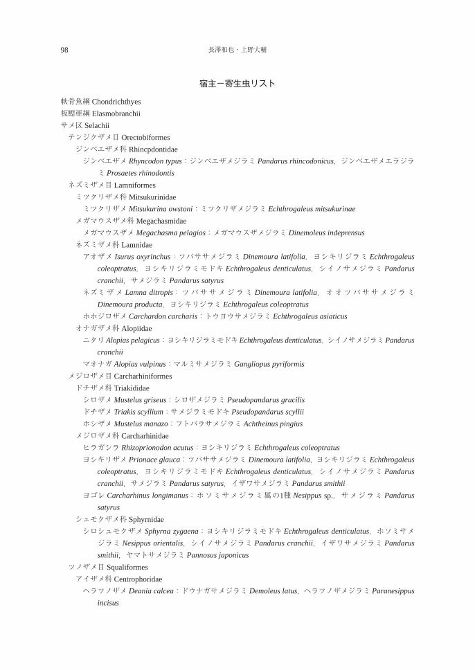
98 長澤和也・上野大輔
宿主-寄生虫リスト
軟骨魚綱 Chondrichthyes板鰓亜綱 Elasmobranchiiサメ区 Selachii テンジクザメ目 Orectobiformes ジンベエザメ科 Rhincpdontidae ジン ベエザメ Rhyncodon typus:ジンベエザメジラミ Pandarus rhincodonicus,ジンベエザメエラジラ
ミ Prosaetes rhinodontis ネズミザメ目 Lamniformes ミツクリザメ科Mitsukurinidae ミツクリザメMitsukurina owstoni:ミツクリザメジラミ Echthrogaleus mitsukurinae メガマウスザメ科Megachasmidae メガマウスザメMegachasma pelagios:メガマウスザメジラミ Dinemoleus indeprensus ネズミザメ科 Lamnidae アオ ザメ Isurus oxyrinchus:ツバササメジラミ Dinemoura latifolia,ヨシキリジラミ Echthrogaleus
coleoptratus,ヨシキリジラミモドキ Echthrogaleus denticulatus,シイノサメジラミ Pandarus cranchii,サメジラミ Pandarus satyrus
ネズ ミザメ Lamna ditropis:ツバササメジラミ Dinemoura latifolia,オオツバササメジラミDinemoura producta,ヨシキリジラミ Echthrogaleus coleoptratus
ホホ ジロザメ Carchardon carcharis:トウヨウサメジラミ Echthrogaleus asiaticus オナガザメ科 Alopiidae ニタ リAlopias pelagicus:ヨシキリジラミモドキEchthrogaleus denticulatus, シイノサメジラミPandarus
cranchii マオ ナガ Alopias vulpinus:マルミサメジラミ Gangliopus pyriformis メジロザメ目 Carcharhiniformes ドチザメ科 Triakididae シロザメMustelus griseus:シロザメジラミ Pseudopandarus gracilis ドチザメ Triakis scyllium:サメジラミモドキ Pseudopandarus scyllii ホシザメMustelus manazo:フトバラサメジラミ Achtheinus pingius メジロザメ科 Carcharhinidae ヒラガシラ Rhizoprionodon acutus:ヨシキリジラミ Echthrogaleus coleoptratus ヨシ キリザメ Prionace glauca:ツバササメジラミ Dinemoura latifolia,ヨシキリジラミ Echthrogaleus
coleoptratus,ヨシキリジラミモドキ Echthrogaleus denticulatus,シイノサメジラミ Pandarus cranchii,サメジラミ Pandarus satyrus,イザワサメジラミ Pandarus smithii
ヨゴ レ Carcharhinus longimanus:ホソミサメジラミ属の1種 Nesippus sp.,サメジラミ Pandarus satyrus
シュモクザメ科 Sphyrnidae シロ シュモクザメ Sphyrna zygaena:ヨシキリジラミモドキ Echthrogaleus denticulatus,ホソミサメ
ジラミ Nesippus orientalis,シイノサメジラミ Pandarus cranchii,イザワサメジラミ Pandarus smithii,ヤマトサメジラミ Pannosus japonicus
ツノザメ目 Squaliformes アイザメ科 Centrophoridae ヘラ ツノザメ Deania calcea:ドウナガサメジラミ Demoleus latus,ヘラツノザメジラミ Paranesippus
incisus
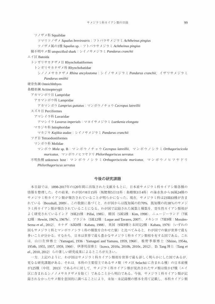
サメジラミ科カイアシ類の目録 99
ツノザメ科 Squalidae ツマリツノザメ Squalus brevirostris:フトバラサメジラミ Achtheinus pingius ツノザメ属の1種 Squalus sp.:フトバラサメジラミ Achtheinus pingius 種不明サメ類 unspecified shark:シイノサメジラミ Pandarus cranchiiエイ区 Batoida トンガリサカタザメ目 Rhynchobatiformes トンガリサカタザメ科 Rhynchobatidae シノ ノメサカタザメ Rhina ancylostoma:シイノサメジラミ Pandarus cranchii,イザワサメジラミ
Pandarus smithii硬骨魚綱 Osteichhthyes条鰭亜綱 Actinopterygii アカマンボウ目 Lampridae アカマンボウ科 Lampridae アカマンボウ Lamprius guttatus:マンボウノチョウ Cecropus latreillii スズキ目 Perciformes アマシイラ科 Luvaridae アマシイラ Luvarus imperials:マヨイサメジラミ Luetkenia elongata マカジキ科 Istiophoridae マカジキ Kajikia audax:シイノサメジラミ Pandarus cranchii フグ目 Tetraodontiformes マンボウ科Molidae マン ボウ Mola sp. B:マンボウノチョウ Cecropus latreillii,マンボウノシラミ Orthagoriscicola
muricatus,マンボウノヒフヤドリ Philorthagoriscus serratus 不明 魚種 unknown host:マンボウノシラミ Orthagoriscicola muricatus,マンボウノヒフヤドリ
Philorthagoriscus serratus
今後の研究課題
本目録では,1898-2017年の120年間に出版された文献をもとに,日本産サメジラミ科カイアシ類各種の情報を整理した。その結果,わが国の8目15科(板鰓類5目11科;条鰭類3目4科)の海水魚から16属24種のサメジラミ科カイアシ類が報告されていることが明らかになった。現在,サメジラミ科は23属63種が含まれている(Boxshall, 2009)。この数値に基づくと,わが国からは既知属の約70%,既知種の約38%のサメジラミ科カイアシ類が報告されていることになる。わが国で記録された属数と種数を,寄生性カイアシ類相がよく研究されているインド(9属21種:Pillai, 1985),韓国(5属5種:Kim, 1998),ニュージーランド(7属12種:Hewitt, 1967a, 1967b),ブラジル(5属12種:Luque and Tavares, 2007),メキシコ(7属9種:Morales-Serna et al., 2012),カナダ(6属8種:Kabata, 1988),英国(9属9種と未同定2種:Kabata, 1979)(いずれの国もサメジラミ科とマンボウノシラミ科の種数を合わせた値)と比べてみると,わが国での値が世界で最も多いことが分かる。すなわち,日本は世界で最も豊かなサメジラミ科カイアシ類相を有する国である。これは,山口左仲博士(Yamaguti, 1936;Yamaguti and Yamasu, 1959, 1960),椎野季雄博士(Shiino, 1954a, 1954b, 1955, 1957, 1959, 1960),伊澤邦彦博士(Izawa, 2010a, 2010b, 2010c, 2012),D. Tang博士(Tang et al., 2010, 2012)らの著しい研究成果によるところが大きい。 一方,上記のように,わが国はサメジラミ科カイアシ類相を世界で最も詳しく明らかにした国であるが,更なる研究課題がある。それは,本科の主要宿主であるサメ類(サメ区 Selachiiに含まれる種)の日本産種が125種(中坊,2013)であるのに対して,サメジラミ科カイアシ類が見出されたサメ類は僅か17種(エイ区に含まれるシノノメサカタザメを除く)であることから明白である。今後,サメジラミ科カイアシ類が記録されなかったサメ類を意図的に調べることにより,未知・未記録種の標本を得て記載し,本科カイアシ類
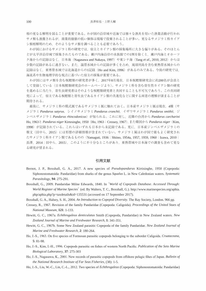
100 長澤和也・上野大輔
相の更なる解明を図ることが重要である。わが国の沿岸域や近海では様々な漁具を用いた漁業活動が行われサメ類も漁獲されるが,商業的価値の低い個体は現場で投棄されることが多い。更なるサメジラミ科カイアシ類相解明のため,そのようなサメ類を調べることも必要であろう。 わが国におけるサメジラミ科の研究では,宿主とカイアシ類の採集場所に大きな偏りがある。そのほとんどが太平洋沿岸域で採集されたものである。瀬戸内海沿岸の水族館での1例を除くと,瀬戸内海とオホーツク海からの記録はなく,日本海(Nagasawa and Nakaya, 1997)や東シナ海(Tang et al., 2010, 2012)からは少数の記録があるに過ぎない。また,温帯水域からの記録が多くを占め,琉球列島を含む亜熱帯水域からの記録はなく,亜寒帯水域では北海道からの1記録(Ho and Kim, 1996)があるのみである。今後の研究では,海流系や生物地理学的な視点に基づいた宿主の採集が重要である。 わが国にはサメ類を含む板鰓類の研究者が多く,2017年8月現在,日本板鰓類研究会に約200名が会員として登録している(日本板鰓類研究会のホームページより)。サメジラミ科を含む寄生性カイアシ類の研究を進めるに当たり,寄生虫研究者はそのような板鰓類研究者と共同することも不可欠であろう。この共同研究によって,宿主である板鰓類と寄生虫であるカイアシ類の共進化などに関する両者の理解が深まることが期待される。 最後に,サメジラミ科の模式属であるサメジラミ属に触れておく。日本産サメジラミ属は現在,4種(サメジラミ Pandarus satyrus,シイノサメジラミ Pandarus cranchii,イザワサメジラミ Pandarus smithii,ジンベエザメジラミ Pandarus rhincodonicus)が知られる。これに対し,近隣の台湾から Pandarus carcharini Ho, 1963と Pandarus niger Kirtesinghe, 1950(Ho, 1963;Cressey, 1967),また韓国から Pandarus niger(Kim, 1998)が記録されている。これらはいずれも日本から未記録である。更に,日本産ジンベエザメジラミの報文(田中ら,2015)には形態の詳細情報が含まれていない。サメジラミ属はわが国で最もよく研究されたサメジラミ科カイアシ類であるものの(Yamaguti, 1936;Shiino, 1954a, 1957, 1959, 1960;Izawa, 2010;長澤,2014;田中ら,2015),このように不十分なところがあり,亜熱帯域や日本海での調査も含めて更なる研究が望まれる。
引用文献
Bernot, J. P., Boxshall, G. A., 2017. A new species of Pseudopandarus Kirtisinghe, 1950 (Copepoda: Siphonostomatoida: Pandaridae) from sharks of the genus Squalus L. in New Caledonian waters. Systematic Parasitology, 94: 275-291.
Boxshall, G., 2009. Pandaridae Milne Edwards, 1840. In “World of Copepods Database. Accessed Through: World Register of Marine Species” (ed. By Walters, T. C., Boxshall, G.). http://www.marinespecies.org/aphia.php/aphia.php?p=taxdetails&id=135531 (accessed on 17 September 2017).
Boxshall, G. A., Halsey, S. H., 2004. An Introduction to Copepod Diversity. The Ray Society, London. 966 pp.Cressey, R., 1967. Revision of the family Pandaridae (Copepoda: Caligoida). Proceedings of the United Staes of
National Museum, 121: 1-133.Hewitt, G. C., 1967a. Echthrogaleus denticulatus Smith (Copepoda, Pandaridae) in New Zealand waters. New
Zealand Journal of Marine and Freshwater Research, 1: 341-351.Hewitt, G. C., 1967b. Some New Zealand parasitic Copepoda of the family Pandaridae. New Zealand Journal of
Marine and Freshwater Research, 2: 180-264.Ho, J.-S., 1963. On five species of Formosan parasitic copepods belonging to the suborder Caligoida. Cruataceana,
5: 81-98.Ho, J.-S., Kim, I.-H., 1996. Copepods parasitic on fishes of western North Pacific. Publication of the Seto Marine
Biological Laboratory, 37: 275-303Ho, J.-S., Nagasawa, K., 2001. New records of parasitic copepods from offshore pelagic fihes of Japan. Bulletin of
the National Research Institute of Far Seas Fisheries, (38): 1-5.Ho, J.-S., Liu, W.-C., Lin, C.-L., 2012. Two species of Echthrogaleus (Copepoda: Siphonostomatoida: Pandaridae)
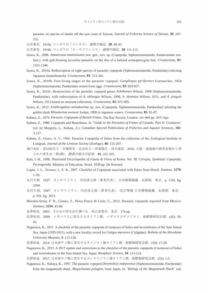
サメジラミ科カイアシ類の目録 101
parasitic on species of sharks off the east coast of Taiwan. Journal of Fisheries Science of Taiwan, 39: 247-255.
石井重美,1916a. マンボウの「ペンネラ」.動物学雑誌, 28: 40-43.石井重美,1916b. マンボウの「オーサゴリシコラ」.動物学雑誌, 28: 111-113.Izawa, K., 2008. Amaterasia amanoiwatoi nov. gen., nov. sp. (Copepoda, Siphonostomatoida, Amaterasidae nov.
fam.), with gall-forming juveniles parasitic on the fins of a balistid actinopterygian fish. Crustaceana, 81: 1331-1346.
Izawa, K., 2010a. Redsecription of eight species of parasitic copepods (Siphonostomatoida, Pandaridae) infecting Japanese elasmobranchs. Crustaceana, 83: 313-341.
Izawa, K., 2010b. Free-living stages of the parasitic copepod, Gangliopus pyriformes Gerstaecker, 1854 (Siphonostomatoida, Pandaridae) reared from eggs. Crustaceana, 83: 829-837.
Izawa, K., 2010c. Resurrection of the parasitic copepod genus Achtheinus Wilson, 1908 (Siphonostomatoida, Pandaridae), with redescription of A. oblongus Wilson, 1908, A. dentatus Wilson, 1911, and A. pinguis Wilson, 1912 based on museum collections. Crustaceana, 83: 971-995.
Izawa, K., 2012. Echthrogaleus mitsukurinae sp. nov. (Copepoda, Siphonostomatoida, Pandaridae) infesting the goblin shark Mitsukurina owstoni Jordan, 1898 in Japanese waters. Crustaceana, 85: 81-87.
Kabata, Z., 1979. Parasitic Copepoda of British Fishes. The Ray Society, London. xii+468 pp. 2031 figs.Kabata, Z., 1988. Copepoda and Branchiura. In “Guide to the Parasites of Fishes of Canada. Part II. Crustacea”
(ed. by Margolis, L., Kabata, Z.). Canadian Special Publication of Fisheries and Aquatic Sciences, 101: 3-127.
Kabata, Z., Gusev, A. V., 1966. Parasitic Copepoda of fishes from the collection of the Zoological Institute in Lenigrad. Journal of the Linnean Society (Zoology), 46: 155-207.
城戸美紅・恩田紀代子・宮側賀美・北谷佳万・伊東隆臣・浅川満彦,2016. 大阪・海遊館の飼育魚類から得られた寄生虫(第3報).酪農学園大学紀要,41: 101-105.
Kim, I.-H., 1998. Illustrated Encyclopedia of Fauna & Flora of Korea. Vol. 38. Cirrepia, Symbiotic Copepoda, Pycnogonida. Ministry of Education, Seuol. 1038 pp. [In Korean].
Luque, J. L., Tavares, L. E. R., 2007. Checklist of Copepoda associated with fishes from Brazil. Zootaxa, 1579: 1-39.
丸川久俊,1927.ヨシキリジラミ.内田淸之助(著者代表),日本動物圖鑑,北隆館,東京.p. 1242, fig. 2394.
丸川久俊,1947.ヨシキリジラミ.内田淸之助(著者代表),改訂增補 日本動物圖鑑,北隆館,東京.p. 926, fig. 2652.
Morales-Serna, F. N., Gomez, S., Pérez-Ponce de León, G., 2012. Parasitic copepods reported from Mexico. Zootaxa, 3234: 43-68.
長澤和也,2003.さかなの寄生虫を調べる.成山堂書店,東京.176 pp.長澤和也,2009.メガマウスに寄生するカイアシ類,メガマウスザメジラミ.板鰓類研究会報,(45): 39-
43.Nagasawa, K., 2011. A checklist of the parasitic copepods (Crustacea) of fishes and invertebrates of the Seto Inland
Sea, Japan (1935-2011), with a new locality record for Caligus macarovi (Caligidae). Bulletin of the Hiroshima University Museum, 3: 113-128.
長澤和也,2014. 日本産サメ類に寄生するサメジラミ属カイアシ類.板鰓類研究会報,(50): 17-20.Nagasawa, K., 2015. A 2015 update and corrections to the checklist of the parasitic copepods (Crustacea) of fishes
and invertebrates of the Seto Inland Sea, Japan. Biosphere Science, 54: 113-124.長澤和也,2017. 日本産サメ類に寄生するヨシキリジラミ属カイアシ類.板鰓類研究会報,(53): 1-5.Nagasawa, K., Nakaya, K., 1997. The parasitic copepod Dinemoleus indeprensus (Siphonostomatoida: Pandaridae)
from the megamouth shark, Megachasma pelagios, from Japan. In “Biology of the Megamouth Shark” (ed.
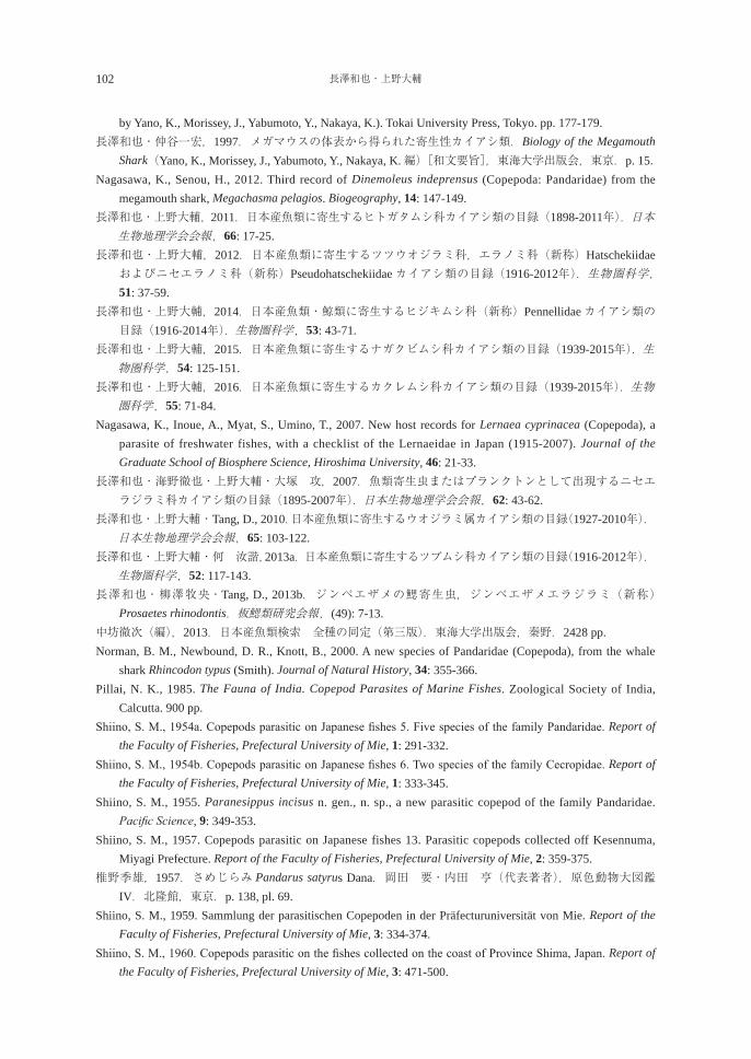
102 長澤和也・上野大輔
by Yano, K., Morissey, J., Yabumoto, Y., Nakaya, K.). Tokai University Press, Tokyo. pp. 177-179.長澤和也・仲谷一宏,1997.メガマウスの体表から得られた寄生性カイアシ類.Biology of the Megamouth
Shark(Yano, K., Morissey, J., Yabumoto, Y., Nakaya, K.編)[和文要旨],東海大学出版会,東京.p. 15.Nagasawa, K., Senou, H., 2012. Third record of Dinemoleus indeprensus (Copepoda: Pandaridae) from the
megamouth shark, Megachasma pelagios. Biogeography, 14: 147-149.長澤和也・上野大輔, 2011.日本産魚類に寄生するヒトガタムシ科カイアシ類の目録(1898-2011年).日本生物地理学会会報,66: 17-25.
長澤和也・上野大輔,2012.日本産魚類に寄生するツツウオジラミ科,エラノミ科(新称)Hatschekiidaeおよびニセエラノミ科(新称)Pseudohatschekiidaeカイアシ類の目録(1916-2012年).生物圏科学,51: 37-59.
長澤和也・上野大輔,2014.日本産魚類・鯨類に寄生するヒジキムシ科(新称)Pennellidaeカイアシ類の目録(1916-2014年).生物圏科学,53: 43-71.
長澤和也・上野大輔,2015.日本産魚類に寄生するナガクビムシ科カイアシ類の目録(1939-2015年).生物圏科学,54: 125-151.
長澤和也・上野大輔,2016.日本産魚類に寄生するカクレムシ科カイアシ類の目録(1939-2015年).生物圏科学,55: 71-84.
Nagasawa, K., Inoue, A., Myat, S., Umino, T., 2007. New host records for Lernaea cyprinacea (Copepoda), a parasite of freshwater fishes, with a checklist of the Lernaeidae in Japan (1915-2007). Journal of the Graduate School of Biosphere Science, Hiroshima University, 46: 21-33.
長澤和也・海野徹也・上野大輔・大塚 攻,2007.魚類寄生虫またはプランクトンとして出現するニセエラジラミ科カイアシ類の目録(1895-2007年).日本生物地理学会会報,62: 43-62.
長澤和也・上野大輔・Tang, D., 2010.日本産魚類に寄生するウオジラミ属カイアシ類の目録(1927-2010年). 日本生物地理学会会報,65: 103-122.
長澤和也・上野大輔・何 汝諧,2013a.日本産魚類に寄生するツブムシ科カイアシ類の目録(1916-2012年). 生物圏科学,52: 117-143.
長澤和也・柳澤牧央・Tang, D., 2013b.ジンベエザメの鰓寄生虫,ジンベエザメエラジラミ(新称)Prosaetes rhinodontis.板鰓類研究会報,(49): 7-13.
中坊徹次(編),2013.日本産魚類検索 全種の同定(第三版).東海大学出版会,秦野.2428 pp.Norman, B. M., Newbound, D. R., Knott, B., 2000. A new species of Pandaridae (Copepoda), from the whale
shark Rhincodon typus (Smith). Journal of Natural History, 34: 355-366.Pillai, N. K., 1985. The Fauna of India. Copepod Parasites of Marine Fishes. Zoological Society of India,
Calcutta. 900 pp. Shiino, S. M., 1954a. Copepods parasitic on Japanese fishes 5. Five species of the family Pandaridae. Report of
the Faculty of Fisheries, Prefectural University of Mie, 1: 291-332. Shiino, S. M., 1954b. Copepods parasitic on Japanese fishes 6. Two species of the family Cecropidae. Report of
the Faculty of Fisheries, Prefectural University of Mie, 1: 333-345. Shiino, S. M., 1955. Paranesippus incisus n. gen., n. sp., a new parasitic copepod of the family Pandaridae.
Pacific Science, 9: 349-353.Shiino, S. M., 1957. Copepods parasitic on Japanese fishes 13. Parasitic copepods collected off Kesennuma,
Miyagi Prefecture. Report of the Faculty of Fisheries, Prefectural University of Mie, 2: 359-375. 椎野季雄,1957.さめじらみ Pandarus satyrus Dana.岡田 要・内田 亨(代表著者),原色動物大図鑑
IV.北隆館,東京.p. 138, pl. 69. Shiino, S. M., 1959. Sammlung der parasitischen Copepoden in der Präfecturuniversität von Mie. Report of the
Faculty of Fisheries, Prefectural University of Mie, 3: 334-374. Shiino, S. M., 1960. Copepods parasitic on the fishes collected on the coast of Province Shima, Japan. Report of
the Faculty of Fisheries, Prefectural University of Mie, 3: 471-500.
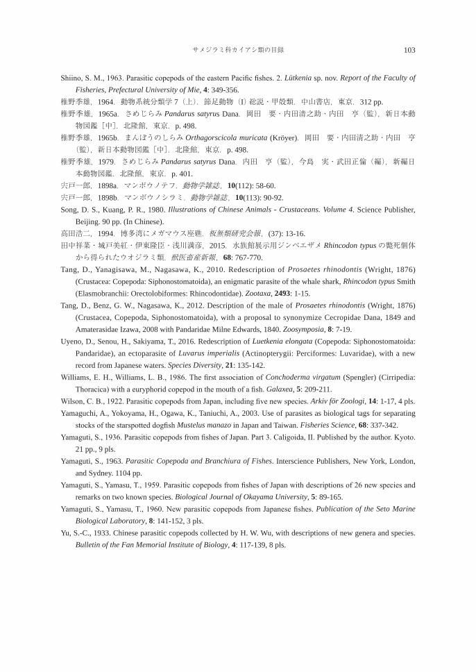
サメジラミ科カイアシ類の目録 103
Shiino, S. M., 1963. Parasitic copepods of the eastern Pacific fishes. 2. Lütkenia sp. nov. Report of the Faculty of Fisheries, Prefectural University of Mie, 4: 349-356.
椎野季雄,1964.動物系統分類学 7(上).節足動物(I) 総説・甲殻類.中山書店,東京.312 pp.椎野季雄,1965a.さめじらみ Pandarus satyrus Dana.岡田 要・内田清之助・内田 亨(監),新日本動
物図鑑[中].北隆館,東京.p. 498.椎野季雄,1965b.まんぼうのしらみ Orthagorscicola muricata (Kröyer).岡田 要・内田清之助・内田 亨(監),新日本動物図鑑[中].北隆館,東京.p. 498.
椎野季雄,1979.さめじらみ Pandarus satyrus Dana.内田 亨(監),今島 実・武田正倫(編),新編日本動物図鑑.北隆館,東京.p. 401.
宍戸一郎,1898a.マンボウノテフ.動物学雑誌,10(112): 58-60. 宍戸一郎,1898b.マンボウノシラミ.動物学雑誌,10(113): 90-92. Song, D. S., Kuang, P. R., 1980. Illustrations of Chinese Animals - Crustaceans. Volume 4. Science Publisher,
Beijing. 90 pp. (In Chinese).高田浩二,1994.博多湾にメガマウス座礁.板無類研究会報,(37): 13-16.田中祥菜・城戸美紅・伊東隆臣・浅川満彦,2015.水族館展示用ジンベエザメ Rhincodon typusの斃死個体から得られたウオジラミ類.獣医畜産新報,68: 767-770.
Tang, D., Yanagisawa, M., Nagasawa, K., 2010. Redescription of Prosaetes rhinodontis (Wright, 1876) (Crustacea: Copepoda: Siphonostomatoida), an enigmatic parasite of the whale shark, Rhincodon typus Smith (Elasmobranchii: Orectolobiformes: Rhincodontidae). Zootaxa, 2493: 1-15.
Tang, D., Benz, G. W., Nagasawa, K., 2012. Description of the male of Prosaetes rhinodontis (Wright, 1876) (Crustacea, Copepoda, Siphonostomatoida), with a proposal to synonymize Cecropidae Dana, 1849 and Amaterasidae Izawa, 2008 with Pandaridae Milne Edwards, 1840. Zoosymposia, 8: 7-19.
Uyeno, D., Senou, H., Sakiyama, T., 2016. Redescription of Luetkenia elongata (Copepoda: Siphonostomatoida: Pandaridae), an ectoparasite of Luvarus imperialis (Actinopterygii: Perciformes: Luvaridae), with a new record from Japanese waters. Species Diversity, 21: 135-142.
Williams, E. H., Williams, L. B., 1986. The first association of Conchoderma virgatum (Spengler) (Cirripedia: Thoracica) with a euryphorid copepod in the mouth of a fish. Galaxea, 5: 209-211.
Wilson, C. B., 1922. Parasitic copepods from Japan, including five new species. Arkiv för Zoologi, 14: 1-17, 4 pls.Yamaguchi, A., Yokoyama, H., Ogawa, K., Taniuchi, A., 2003. Use of parasites as biological tags for separating
stocks of the starspotted dogfish Mustelus manazo in Japan and Taiwan. Fisheries Science, 68: 337-342.Yamaguti, S., 1936. Parasitic copepods from fishes of Japan. Part 3. Caligoida, II. Published by the author. Kyoto.
21 pp., 9 pls.Yamaguti, S., 1963. Parasitic Copepoda and Branchiura of Fishes. Interscience Publishers, New York, London,
and Sydney. 1104 pp. Yamaguti, S., Yamasu, T., 1959. Parasitic copepods from fishes of Japan with descriptions of 26 new species and
remarks on two known species. Biological Journal of Okayama University, 5: 89-165. Yamaguti, S., Yamasu, T., 1960. New parasitic copepods from Japanese fishes. Publication of the Seto Marine
Biological Laboratory, 8: 141-152, 3 pls.Yu, S.-C., 1933. Chinese parasitic copepods collected by H. W. Wu, with descriptions of new genera and species.
Bulletin of the Fan Memorial Institute of Biology, 4: 117-139, 8 pls.
104 長澤和也・上野大輔
A checklist of copepods of the family Pandaridae (Siphonostomatoida)from fishes in Japanese waters (1898-2017)
Kazuya Nagasawa1) and Daisuke Uyeno
2)
1) Graduate School of Biosphere Science, Hiroshima University,1-4-4 Kagamiyama, Higashi-Hiroshima, Hiroshima 739-8528, Japan
2) Graduate School of Science and Engineering, Kagoshima University,1-21-35 Korimoto, Kagoshima, Kagoshima 890-0065, Japan
Abstract Based on the literature published between 1898 and 2017, a checklist is compiled for the 24 nominal species in 16 genera (Achtheinus [1 sp.], Cecropus [1 sp.], Demoleus [1 sp.], Dinemoleus [1 sp.], Dinemoura [2 spp.], Echthrogaleus [4 spp.], Gangliopus [1 sp.], Luetkenia [1 sp.], Nesippus [1 sp.], Orthagoriscicola [1 sp.], Pandarus [4 spp.], Parnnosus [1 sp.], Paranesippus [1 sp.], Philorthagoriscus [1 sp.], Prosaetes [1 sp.], and Pseudopandarus [2 spp.]) of the copepod family Pandaridae from Japanese fishes. This checklist contains information for each taxon regarding its host(s), attachment site(s), known geographical distribution in Japanese waters, and references. A host-parasite list is also given.Key words: bibliography, checklist, Copepoda, elasmobranchs, fish parasites, Pandaridae
総 説
日本に定着したサンフィッシュ科魚類3種(ブルーギル,オオクチバス,コクチバス)の寄生虫目録(1962-2017年)
長澤和也*
広島大学大学院生物圏科学研究科 〒739-8528 東広島市鏡山1-4-4
要 旨 1962-2017年の56年間に出版された報文に基づき,日本に定着したサンフィッシュ科魚類3種(ブルーギル,オオクチバス,コクチバス)から報告された寄生虫の情報を整理した。その結果,21名義種・亜種と未同定の寄生虫が次の分類群からが報告されていた:ミクソゾア類[名義種数:0種],条虫類[2種],単生類[6種],吸虫類[5種],線形動物[2種],鉤頭動物[1種],二枚貝類[3種・亜種],カイアシ類[2種])。ブルーギルからは12名義種,オオクチバスからは13名義種・亜種,コクチバスからは3名義種の寄生虫が記録されていた。これら寄生虫は,日本の在来種と外国から宿主とともに持ち込まれた外来種からなり,条虫類1種(オオクチバスとコクチバス寄生のバスハイトウジョウチュウ Proteocephalus fluviatilis)と単生類6種(ブルーギル寄生のActinocleidus fergusoni,Onchocleidus dispar,Onchocleidus ferox;オオクチバス寄生の Onchocleidus furcatus,Onchocleidus helicus,Syncleithrium fusiformes)は北米に起源を有する種である。人体に寄生する線虫,ドロレス顎口虫 Gnathostoma doloresiと日本顎口虫 Gnathostoma nipponicumがそれぞれブルーギルとオオクチバスから報告され,後者の人体寄生例もあった。キーワード: オオクチバス,魚類寄生虫,コクチバス,サンフィッシュ科魚類,人体寄生虫,ブルー
ギル,目録
緒 言
ブルーギル Lepomis macrochirus Rafinesque, 1819,オオクチバスMicropterus salmoides (Lacepède, 1802),コクチバスMicropterus dolomieu Lacepède, 1802の3種はいずれもスズキ目サンフィッシュ科に属し,北米原産の淡水魚である。それぞれの種は異なった年にわが国に導入され,現在,河川・湖沼など内水面で繁殖して定着している。特に前二者の生息域は全国に及び,在来淡水魚への悪影響など,様々な問題を起こしている(日本魚類学会自然保護委員会,2002;環境省自然環境局野生生物課,2004;淀・井口,2004;細谷・高橋,2006)。 外国から魚類を導入する際,特に注意すべきはウイルスや細菌,寄生虫などの病原生物を持ち込まないことである。しかし,わが国は,かつて検疫体制が不十分だったことなどにより,外国から導入した淡水魚とともに多くの病原生物(例えばサケ科魚類の疾病原因ウイルスなど[若林・室賀,2004を参照])の侵入を許し,水産業で大きな被害を被ってきた歴史を有する。本報で扱うサンフィッシュ科魚類に言及すれば,後述するように,日本在来の寄生虫のほかに,原産地の北米に起源をもつ寄生虫が見出されており,それらは宿主とともに日本に持ち込まれたものと考えられている。本報では,わが国に定着したサンフィッシュ科魚類の寄生虫相を概観するため,これまでに日本で本科3種から報告された寄生虫の情報を整理して,目録として示した。わが国のサンフィッシュ科魚類から最初に見出された寄生虫は,笠原(1962: 130)によって報じられた「ブラックバス」(=オオクチバス)に寄生していたカイアシ類のイカリムシである。本目録には,
2017年9月19日受理 *E-mail: [email protected]
生物圏科学Biosphere Sci. 56:105-120 (2017)
106 長澤和也
それ以後,2017年までの56年間に公表された寄生虫の情報が整理されている。 本目録は,2つのリスト(寄生虫-宿主リスト,宿主-寄生虫リスト)とそれに続く3節(日本産サンフィッシュ科魚類の寄生虫相の総括,日本産サンフィッシュ科魚類に見られる人体寄生虫,今後の研究課題)から構成される。 寄生虫-宿主リストでは,サンフィッシュ科魚類3種から報告された寄生虫を高次分類群(刺胞動物[ミクソゾア類],扁形動物[条虫類,単生類,吸虫類],線形動物,鉤頭動物,軟体動物[二枚貝類],節足動物[カイアシ類])で仕分けし,各分類群において各寄生虫を学名のアルファベット順に掲載した。各寄生虫の情報は,最新の学名,わが国における過去の同定結果,宿主,寄生部位,地理的分布,記録を記し,必要に応じて備考を含む。寄生虫に標準和名がある場合は,それを学名の前に記した。魚類に幼虫が寄生する場合には,学名のあとにそのステージを括弧内に示した。当該寄生虫が外国から持ち込まれた場合には外来種,国内にもともと分布する場合には在来種として,学名のあとに角括弧内に示した。どちらとも判断できない場合は不明とした。過去の同定結果の項には,わが国で記録された寄生虫の学名や名称が最新のものと異なる場合に,その学名と名称とともに,それらを報告した文献情報(報告者名と発表年)を示した。寄生部位の情報を欠く場合には不明としたが,他文献に基づいて適切な寄生部位を示すことができる場合には角括弧内に記した。宿主は,ブルーギル,オオクチバス,コクチバスの順に並べた。地理的分布の項では,寄生虫が見出された府県名を記し,北から南に向かう順番で並べた。記録の項では,各寄生虫を記録した文献情報(報告者名と発表年)を記したあと,括弧内に原著で示された産地を示した。原著に産地の情報が記されていない場合には-で示した。また,ある寄生虫が複数の報文で報告された場合には,古い報文から番号を付けるとともに,宿主の項において各報文が報告した宿主を区別するため,宿主名の直後の括弧内に番号を付した。備考では,当該寄生虫に関わる情報を記した。なお,二枚貝類の幼生がサンフィッシュ科魚類に寄生することが知られているが,二枚貝類の学名はまだ統一されていない状況に鑑み,本目録では近藤(2015)に従った。 宿主-寄生虫リストでは,ブルーギル,オオクチバス,コクチバスの順に,各魚種から記録された寄生虫を高次分類群ごとに示した。寄生虫-宿主リストに基づき,各寄生虫の学名直後に外来種か在来種(判断できない場合は不明)であるかを記すとともに,各寄生虫が見出された府県名(不明の場合は-)を示した。
日本産サンフィッシュ科魚類の寄生虫目録
A checklist of the parasites of three species of centrarchids in Japan
寄生虫-宿主リスト Parasite-Host List
刺胞動物 Cnidariaミクソゾア類 Myxozoa
Myxozoa sp.[不明] 宿主:ブルーギル,オオクチバス 寄生部位:不明 地理的分布:岐阜県 記録:無記名(2002:木曽川[実験河川])
扁形動物 Platyhelminthes条虫類 Cestoda
バスハイトウジョウチュウ(バス杯頭条虫)Proteocephalus fluviatilis Bangham, 1925[外来種] 宿主:オオクチバス(1, 2),コクチバス(3)
日本産サンフィッシュ科3種の寄生虫目録 107
寄生部位:腸 地理的分布:長野県 記録:1. Shimazu(1990)(木崎湖);2. Hypša et al.(2005)(野尻湖);3. Scholz et al.(2007)(野尻湖) 備考:本種は,北米から偶然に持ち込まれたと考えられている(Shimazu, 1990)。Hypša et al.(2005)は
野尻湖を誤って「Lake Noiri」と報告した。福島県秋元湖産コクチバスの胃から「リグラ条虫」が得られている(成田ほか,2002)。長澤(2015)は,この「リグラ条虫」が本種である可能性を検討する必要があるとしている。
アユハイトウジョウチュウ(鮎杯頭条虫)Proteocephalus plecoglossi Yamaguti, 1934[在来種] 宿主:ブルーギル 寄生部位:[腸] 地理的分布:滋賀県 記録:Shimazu(1993)(琵琶湖[尾松])
単生類 Monogenea
Actinocleidus fergusoni Mizelle, 1938[外来種] 過去の同定結果:another member of Ancyrocephalinae(cf. Muroga et al., 1980: 30) 宿主:ブルーギル 寄生部位:鰓 地理的分布:広島県 記録:Muroga et al.(1980)(福山市千塚池);Maneepitaksanti and Nagasawa(2012)(東広島市[広島大学構内ぶどう池])
備考:本種は,ブルーギルとともに北米から持ち込まれたと考えられている(Maneepitaksanti and Nagasawa, 2012)。
Ancyrocephalus sp.[不明] 宿主:オオクチバス 寄生部位:鰓 地理的分布:神奈川県 記録:堤・三浦(2003)(横浜・八景島シーパラダイス) 備考:本種がオオクチバスとともに日本に持ち込まれたかは不明とされている(堤・三浦,2003)。本種
の重度寄生を受けた飼育オオクチバスが斃死した(堤・三浦,2003)。
Onchocleidus dispar Mueller, 1936[外来種] 宿主:ブルーギル 寄生部位:鰓 地理的分布:広島県 記録:Maneepitaksanti and Nagasawa(2013)(東広島市[広島大学構内ぶどう池]) 備考:本種は,ブルーギルとともに北米から持ち込まれたと考えられている(Maneepitaksanti and
Nagasawa, 2013)。
Onchocleidus ferox (Mueller, 1934)[外来種] 過去の同定結果:Urocleidus ferox (cf. Muroga et al., 1980) 宿主:ブルーギル 寄生部位:鰓
108 長澤和也
地理的分布:滋賀県,広島県 記録:Muroga et al.(1980)(広島県:福山市[千塚池]);グライガー・浦部(2003)(滋賀県:琵琶湖);
Ogawa(2011: 1496)(滋賀県:琵琶湖);Maneepitaksanti and Nagasawa(2012)(東広島市[広島大学構内ぶどう池])
備考:本種は,ブルーギルとともに北米から持ち込まれたと考えられている(Muroga et al., 1980)。
Onchocleidus furcatus (Mueller, 1937)[外来種] 過去の同定結果:Haplocleidus furcatus(cf. グライガー・浦部,2003),Haplocleidus furcatus(cf. Ogawa,
2011: 1496) 宿主:オオクチバス 寄生部位:[鰓] 地理的分布:滋賀県 記録:グライガー・浦部(2003)(琵琶湖);Ogawa(2011: 1496)(琵琶湖) 備考:本種は,オオクチバスとともに北米から日本に持ち込まれたと考えられている(グライガー・浦部,
2003)。日本産標本に基づく本種の形態・分類学的研究はまだ行われていない。Wheeler and Beverley-Burton(1989)は Haplocleidus furcatusを本種の異名としており,新田(2017:表3)もそれに従っている。
Onchocleidus helicus Mueller, 1936[外来種] 宿主:オオクチバス 寄生部位:[鰓] 地理的分布:滋賀県 記録:グライガー・浦部(2003)(琵琶湖) 備考:本種は,オオクチバスとともに北米から日本に持ち込まれたと考えられている(グライガー・浦部,
2003)。日本産標本に基づく本種の形態・分類学的研究はまだ行われていない。Wheeler and Beverley-Burton(1989)は本種を未確定種(species inquirenda)としているが,新田(2017:表3)は上記学名を用いている。
Syncleithrium fusiformes (Mueller, 1934)[外来種] 過去の同定結果:Actinocleidus fusiformisの類似種(cf. グライガー・浦部 , 2003),Actinocleidus sp.,
similar to A. fusiformis(cf. Ogawa, 2011: 1496) 宿主:オオクチバス 寄生部位:[鰓] 地理的分布:滋賀県 記録:グライガー・浦部(2003)(琵琶湖);Ogawa(2011: 1496)(琵琶湖) 備考:本種は,オオクチバスとともに北米から日本に持ち込まれたと考えられている(グライガー・浦部,
2003)。Ogawa(2011)が採集した標本を新田(2017:表3)は本種に同定した(広島大学新田理人博士からの私信)。ただし,日本産標本に基づく本種の形態に関する報告はまだない。本種の学名はBeverley-Burton(1986)に従う。
Monopisthocotylea spp.[不明] 宿主:ブルーギル,オオクチバス 寄生部位:[鰓] 地理的分布:岐阜県 記録:無記名(2002)(木曽川[実験河川]) 備考:単生類は宿主特異性が強いので,これらの単生類は宿主とともに北米から持ち込まれた可能性が高い。分類学的研究が望まれる。
日本産サンフィッシュ科3種の寄生虫目録 109
吸虫類 Trematoda
Azygia gotoi (Ariake, 1922)[在来種] 過去の同定結果:Azygia aguillae(cf. Shimazu, 2007) 宿主:コクチバス 寄生部位:胃 地理的分布:長野県 記録:Shimazu(2007)(野尻湖);Shimazu(2014)(野尻湖)
チュウブゴッポキュウチュウ(中部ゴッポ吸虫)Genarchopsis chubuensis Shimazu, 2015 [在来種] 過去の同定結果:Genarchopsis goppo(cf. Shimazu, 1995) 宿主:オオクチバス 寄生部位:胃 地理的分布:長野県 記録:1. Shimazu(1995)(木崎湖,諏訪湖);2. Shimazu(2015)(木崎湖,諏訪湖)
Genarchopsis sp.[在来種] 過去の同定結果:Genarchopsis goppo(cf. Shimazu, 1995; Shimazu et al., 2001) 宿主:ブルーギル 寄生部位:胃,鰓または鰭 地理的分布:滋賀県 記録:1. Shimazu(1995)(琵琶湖[尾松]);2. Shimazu et al.(2011)(滋賀県:琵琶湖[尾松],かや池) 備考:滋賀県産ブルーギルに寄生していた本種は,ゴッポキュウチュウ(ゴッポ吸虫)Genearchopsis
goppo Ozaki, 1925かギギキュウチュウ(ギギ吸虫)Genarchopsis gigi Yamaguti, 1939のいずれかに同定されると推測される(Urabe et al., 2013;Shimazu, 2015;浦部,2016を参照)。しかし,同県産ブルーギルからの本種標本(Shimazu, 1995;Shimazu et al., 2011)は最新知見に基づいて同定されていないため(Shimazu, 2015),種小名を保留しておく。
ナマズキノウキュウチュウ(鯰気嚢吸虫)Isoparorchis eurytremum (Kobayashi, 1915)[在来種] 過去の同定結果:Isoparorchis hypselobagri (cf. グライガー,2004;Shimazu et al., 2011) 宿主:ブルーギル(1),オオクチバス(1, 2) 寄生部位:筋肉 地理的分布:滋賀県 記録:1. グライガー(2004)(琵琶湖);2. Shimazu et al.(2011)(琵琶湖)
Metagonimus spp.(メタセルカリア)[在来種] 宿主:ブルーギル 寄生部位:[鱗] 地理的分布:岐阜県 記録:無記名(2002)(木曽川[新境川]) 備考:採集地周辺のタヌキとアオサギからミヤタキュウチュウ Metagonimus miyataiとタカハシキュウ
チュウMetagonimus takahashiiの成虫が得られている(無記名,2002)。
ナマズフッコウキュウチュウ(鯰腹口吸虫)Parabucephalopsis parasiluri Wang, 1985(セルカリア,未成熟虫)[外来種]
過去の同定結果:腹口類(cf. 浦部ほか,2001)
110 長澤和也
宿主:ブルーギル(2),オオクチバス(1) 寄生部位:直腸(オオクチバス),尾鰭(ブルーギル) 地理的分布:京都府,大阪府 記録:1. 浦部ほか(2001)(京都府:宇治川);2. Urabe et al.(2007)(大阪府:淀川) 備考:本種は外来種であるが,サンフィッシュ科魚類とともに北米から持ち込まれたものではない。第一中間宿主のカワヒバリガイとともにアジア大陸から日本に持ち込まれたと考えられている(浦部,2002;浦部,2016: 44)。淡水魚類が第二中間宿主で,ブルーギルにはメタセルカリアが寄生する(Urabe et al., 2007)。本種の可能性が高いメタセルカリアがオオクチバスに見られるという(馬場・浦部,2011)。なお,浦部ほか(2001)は宇治川産オオクチバスから本種と思われる腹口類の未成熟虫を得ているが,続報(Urabe et al., 2007;馬場・浦部,2011;Baba and Urabe, 2015)ではこのことに触れていない。
オザキフッコウキュウチュウ(尾崎腹口吸虫)Prosorhynchoides ozakii (Nagaty, 1937)(メタセルカリア)[外来種]
宿主:ブルーギル 寄生部位:尾鰭 地理的分布:大阪府 文献:Urabe et al.(2007)(淀川) 備考:本種も,上記のナマズフッコウキュウチュウと同様に,第一中間宿主のカワヒバリガイとともに日本に侵入したと考えられている(Urabe et al., 2007;馬場・浦部,2011)。
線形動物 Nematoda
Contracaecum sp.(幼虫)[在来種] 宿主:ブルーギル 寄生部位:不明 地理的分布:岐阜県 記録:無記名(2002)(木曽川[実験河川]) 備考:本種の形態・分類学的研究はまだ行われていない。本種の終宿主はアオサギであるという(無記名 ,
2002)。
Eustrongylides sp.(幼虫)[在来種] 宿主:ブルーギル(1, 2),オオクチバス(1) 寄生部位:内臓,腹腔,筋肉 地理的分布:茨城県,滋賀県 記録:1. グライガー(2004)(滋賀県:琵琶湖);2. 荒木ほか(2011)(茨城県:潮来地区)
ドロレスガッコウチュウ(ドロレス顎口虫)Gnathostoma doloresi Tubangui, 1925(幼虫)[在来種] 宿主:ブルーギル 寄生部位:筋肉 地理的分布:宮崎県 記録:Nawa et al.(1993)(一ツ瀬ダム湖) 備考:本種は人体に寄生する(赤羽,1999)。本論文の別節(日本産サンフィッシュ科魚類に見られる人体寄生虫)で本種のことを解説する。
日本産サンフィッシュ科3種の寄生虫目録 111
ニッポンガッコウチュウ(日本顎口虫)Gnathostoma nipponicum Yamaguti, 1941(幼虫)[在来種] 宿主:オオクチバス 寄生部位:頭部,内臓,筋肉 地理的分布:秋田県 記録:Ishida et al.(2003)(秋田市[郊外溜池]) 備考:本種は人体に寄生する(安藤,1999)。上記のドロレスガッコウチュウ Gnathostoma doloresiと同
様に,本論文の別節(日本産サンフィッシュ科魚類に見られる人体寄生虫)で本種のことを解説する。
鉤頭動物 Acanthocephala
Polymorphidae sp.(被嚢幼虫)[在来種] 宿主:オオクチバス 寄生部位:腹腔 地理的分布:兵庫県 記録:正垣(1984)(円山川) 備考:本種は,下記のサウスウエルコウトウチュウの可能性がある。
サウスウエルコウトウチュウ(サウスウエル鉤頭虫)Southwellina hispida (Van Cleave, 1925) Witenberg, 1932(シストアカント幼虫)[在来種]
宿主:オオクチバス 寄生部位:腹腔 地理的分布:滋賀県 記録:Amin et al.(2007)(琵琶湖および流入河川)
軟体動物 Mollusca二枚貝類 Bivalvia
イケチョウガイ Hyriopsis schlegeli (Martens, 1861)(グロキディウム幼生)[在来種] 宿主:ブルーギル 寄生部位:鰓,鰭 地理的分布:滋賀県 記録:古川・小林(1966)(実験感染)
タテボシガイ Nodularia douglasiae biwae (Kobelt, 1879)(グロキディウム幼生)[在来種] 宿主:オオクチバス 寄生部位:[鰓,鰭] 地理的分布:滋賀県 記録:近藤(1997)(琵琶湖[京都大学生態学研究センター前])
イシガイ Nodularia douglasiae nipponensis (Martens, 1877)(グロキディウム幼生)[在来種] 宿主:ブルーギル(1, 2),オオクチバス(1, 2, 3) 寄生部位:鰓,鰭等 地理的分布:大阪府 記録:1. 木邑(2009)(淀川[城北ワンド群]);2. 石田ほか(2010)(淀川[城北ワンド群]);3. 伊藤(2013)(実験感染)
112 長澤和也
Sinanodonta sp.(グロキディウム幼生)[在来種] 過去の同定結果:ドブガイ Anodonta woodiana (cf. 福原ほか,1986) 宿主:ブルーギル 寄生部位:鰓,鰭等 地理的分布:大阪府 記録:福原ほか(1986)(豊中市[灌漑用溜池]) 備考:かつてドブガイ Anodonta woodianaとして報告された種は,近年,タガイ Sinanodonta japonicaとヌマガイ Sinanodonta lautaの2種に識別されているため(近藤,2015),本種がどの種に該当するかを記すことは難しく,ここでは未同定種として示す。
Sinanodonta spp.(グロキディウム幼生)[在来種] 過去の同定結果:ドブガイ属(cf. 木邑 , 2009);ドブガイ属 Anodonta spp.(cf. 石田ほか,2010) 宿主:ブルーギル,オオクチバス 寄生部位:鰓,鰭等 地理的分布:大阪府 記録:木邑(2009)(淀川[城北ワンド群]);石田ほか(2010)(淀川[城北ワンド群])
節足動物 Arthropodaカイアシ類 Copepoda
イカリムシ Lernaea cyprinacea Linnaeus, 1758[在来種] 宿主:ブルーギル(2, 3, 4),オオクチバス(1) 寄生部位:体前部を宿主の体内に潜入させ,体後部を水中に露出する 地理的分布:滋賀県,愛媛県 記 録:1. 笠原(1962)(-);2. 浦和(2004)(-);3. グライガー(2004)(滋賀県:堅田内湖);4.
Nagasawa(2013)(愛媛県:増田川)
ヤマトニセエラジラミ Neoergasilus japonicus (Harada, 1930)[在来種] 過去の同定結果:Ergasilus japonicus (cf. 室賀ほか , 1974) 宿主:ブルーギル(1, 2, 3, 4, 5, 6, 7, 8, 9, 10, 12, 13),オオクチバス(8, 12),コクチバス(11) 寄生部位:鰭,体表 地理的分布:群馬県,長野県,広島県,島根県,長崎県,沖縄県 記録:1. 室賀ほか(1974)(広島県:福山市[千田大池]);2. Urawa et al.(1980)(広島県:福山市[千塚池]);3. Urawa et al.(1991)(広島県:千塚池);4. Yoshikoshi and Kô(1991a)(長崎県:-);5. Yoshikoshi and Kô(1991b)(長崎県:-);6. 浦和(2004)(広島県:千塚池);7. Nagasawa and Uyeno(2012)(沖縄県:倉敷ダム湖);8. Nagasawa and Inoue(2012)(広島県:芦田川,高屋川);9. Nagasawa and Obe(2013)(広島県:東広島市[広島大学構内ぶどう池]);10. 長澤・新田(2013)(島根県:江津市[菰沢池]);11. Nagasawa and Obe(2015)(長野県:信濃町[針ノ木池]);12. Nagasawa and Sato(2015)(群馬県:高崎市[東谷川の砂防ダム池]);13. 長澤・佐藤(2016)(群馬県:館林市[城沼])
宿主-寄生虫リスト Host-Parasite List
各寄生虫の学名直後の角括弧内に,その種が外来種か在来種かを示した。また,当該宿主によって北米から日本に持ち込まれた寄生虫には星印(*)を付けた。
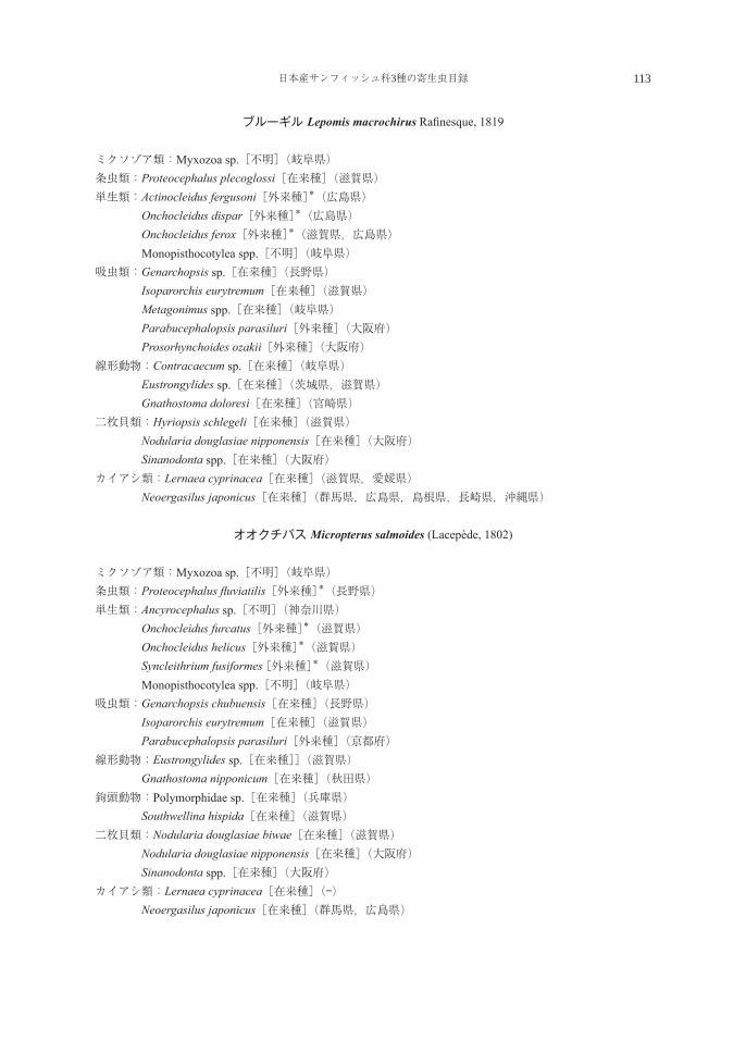
日本産サンフィッシュ科3種の寄生虫目録 113
ブルーギル Lepomis macrochirus Rafinesque, 1819
ミクソゾア類:Myxozoa sp.[不明](岐阜県)条虫類:Proteocephalus plecoglossi[在来種](滋賀県)単生類:Actinocleidus fergusoni[外来種]*(広島県) Onchocleidus dispar[外来種]*(広島県) Onchocleidus ferox[外来種]*(滋賀県,広島県) Monopisthocotylea spp.[不明](岐阜県)吸虫類:Genarchopsis sp.[在来種](長野県) Isoparorchis eurytremum[在来種](滋賀県) Metagonimus spp.[在来種](岐阜県) Parabucephalopsis parasiluri[外来種](大阪府) Prosorhynchoides ozakii[外来種](大阪府)線形動物:Contracaecum sp.[在来種](岐阜県) Eustrongylides sp.[在来種](茨城県,滋賀県) Gnathostoma doloresi[在来種](宮崎県)二枚貝類:Hyriopsis schlegeli[在来種](滋賀県) Nodularia douglasiae nipponensis[在来種](大阪府) Sinanodonta spp.[在来種](大阪府)カイアシ類:Lernaea cyprinacea[在来種](滋賀県,愛媛県) Neoergasilus japonicus[在来種](群馬県,広島県,島根県,長崎県,沖縄県)
オオクチバス Micropterus salmoides (Lacepède, 1802)
ミクソゾア類:Myxozoa sp.[不明](岐阜県)条虫類:Proteocephalus fluviatilis[外来種]*(長野県)単生類:Ancyrocephalus sp.[不明](神奈川県) Onchocleidus furcatus[外来種]*(滋賀県) Onchocleidus helicus[外来種]*(滋賀県) Syncleithrium fusiformes [外来種]*(滋賀県) Monopisthocotylea spp.[不明](岐阜県)吸虫類:Genarchopsis chubuensis[在来種](長野県) Isoparorchis eurytremum[在来種](滋賀県) Parabucephalopsis parasiluri[外来種](京都府)線形動物:Eustrongylides sp.[在来種]](滋賀県) Gnathostoma nipponicum[在来種](秋田県)鉤頭動物:Polymorphidae sp.[在来種](兵庫県) Southwellina hispida[在来種](滋賀県)二枚貝類:Nodularia douglasiae biwae[在来種](滋賀県) Nodularia douglasiae nipponensis[在来種](大阪府) Sinanodonta spp.[在来種](大阪府)カイアシ類:Lernaea cyprinacea[在来種](-) Neoergasilus japonicus[在来種](群馬県,広島県)
114 長澤和也
コクチバス Micropterus dolomieu Lacepéde, 1802
条虫類:Proteocephalus fluviatilis[外来種]*(長野県)吸虫類:Azygia gotoi[在来種](長野県)カイアシ類:Neoergasilus japonicus[在来種](長野県)
日本産サンフィッシュ科魚類の寄生虫相の総括
今回の目録作成によって,日本に定着したサンフィッシュ科魚類から1962-2017年の56年間に21名義種・亜種の寄生虫(条虫類2種,単生類6種,吸虫類5種,線形動物2種,鉤頭動物1種,二枚貝類3種・亜種,カイアシ類2種)が報告されていることが明らかになった。ブルーギルからは12名義種(条虫類1種,単生類3種,吸虫類3種,線形動物1種,二枚貝類2種,カイアシ類2種),オオクチバスからは13名義種・亜種(条虫類1種,単生類3種,吸虫類3種,線形動物1種,鉤頭動物1種,二枚貝類2種・亜種,カイアシ類2種),コクチバスからは3名義種(条虫類1種,吸虫類1種,カイアシ類1種)である。 今回の整理によって明らかになった日本産サンフィッシュ科魚類3種の寄生虫相の特徴のひとつとして,これら魚種は北米原産であるにもかかわらず,わが国の在来寄生虫の感染を受けていることが挙げられる。寄生虫は,特定の宿主にのみ寄生する宿主特異性が強いのが普通であるが,なかには宿主特異性が弱く宿主範囲の広い種も知られている。この範疇に含まれる日本の在来寄生虫は,北米に起源を有するサンフィッシュ科魚類であっても,日本の在来魚に寄生するのと同じように,宿主として利用することができると考えられる。今回確認された寄生虫のなかで,典型的な種を挙げると,吸虫類のナマズキノウキュウチュウIsoparorchis eurytremumは5目10科に属する26種(Nagasawa et al., 2013),カイアシ類のイカリムシ Lernaea cyprinaceaは10目17科に属する30種以上(Nagasawa et al., 2007)の日本産淡水魚から報告されている。 一方,今回確認された寄生虫のなかで,サンフィッシュ科魚類に宿主特異的と考えられるのは単生類である。ブルーギルから3種(Actinocleidus fergusoni,Onchocleidus dispar,Onchocleidus ferox),オオクチバスから4種(Ancyrocephalus sp.,Onchocleidus furcatus,Onchocleidus helicus,Syncleithrium fusiformes)が報告されている。このうち,オオクチバスから報告された Ancyrocephalus sp.は外来種か在来種かは不明とされている(堤・三浦,2003)。わが国にはサンフィッシュ科魚類はもともと生息しないため,それらに特異的に寄生する単生類は日本の在来魚から感染したものではなく,北米からサンフィッシュ科魚類を導入した際に宿主とともに日本に侵入したものと考えられる。したがって,上記の単生類のうち,Ancyrocephalus sp.を除いた6種は,宿主であるブルーギルやオオクチバスとともに日本に侵入した外来寄生虫と言える。 また,オオクチバスとコクチバスに寄生する条虫類のバスハイトウジョウチュウ Proteocephalus fluviatilisも,これら魚種に特異的な寄生虫であり,北米から宿主を導入した際に持ち込まれた外来寄生虫である。
日本産サンフィッシュ科魚類に見られる人体寄生虫
わが国に定着したサンフィッシュ科魚類には人体に感染する寄生虫が見つかることがある。特に重要なのはガッコウチュウ(顎口虫)属に属する線虫類の幼虫で,宮崎県産ブルーギルの筋肉からドロレスガッコウチュウ Gnathostoma doloresi,また秋田県産オオクチバスの筋肉からニッポンガッコウチュウ Gnathostoma nipponicumが発見されている(Nawa et al., 1993;Ishida et al., 2003)。特に後者に関しては,オオクチバスを生食したことによる人体寄生例が報告されている(Ishida et al., 2003)。その詳細を以下に記述する。 ドロレスガッコウチュウは,宮崎県一之瀬ダム湖で1992年9月に採集されたブルーギル51尾より11個体の幼虫が見出された(Nawa et al., 1993)。この幼虫は,形態学的特徴から第3後期幼虫に同定され,宿主の筋肉中に被嚢していた。幼虫の体長は1.5-2.4(平均1.95)mmであった。宮崎県の山村では本線虫による人体寄生例が確認されており,渓流魚を含む魚類の生食がその原因と考えられている(名和ほか,1993)。 一方,2001年にオオクチバスの生食によるニッポンガッコウチュウのヒトへの寄生が秋田県で明らかになり,秋田市郊外の溜池で採集した淡水魚3種(オオクチバス9尾,ゲンゴロウブナ3尾,ウグイ5尾)を検
日本産サンフィッシュ科3種の寄生虫目録 115
査し,オオクチバス6尾(寄生率66.7%)に本線虫の第3後期幼虫の寄生を認めた(Ishida et al., 2003)。オオクチバス各尾における寄生数は1-4個体で,幼虫はオオクチバスの頭部や内臓,体側筋肉などの寄生していた。幼虫の体長は0.97-1.685mmであった。患者は,ガッコウチュウ類の寄生による典型的な症状である,腹部の線状爬行疹の症状を呈した(Ishida et al., 2003)。これは本線虫の幼虫がヒトの皮下の浅いところを移動していることに因る。 このように,サンフィッシュ科魚類をゴッコウチュウ類の寄生が知られることから,同科魚類を刺身など生食することは厳に慎むべきである。それら魚類を食べる際は,適切な加熱・冷凍処理を行うことが重要である。両寄生虫に関しては,生物学的知見に加えて,両種に起因する疾病や疫学的知見が赤羽(1999)と安藤(1999)によって整理されており,公益財団法人目黒寄生虫館のホームページで公開されている。 なお,上記2種の線虫類に加えて,岐阜県産ブルーギルに寄生が認められた吸虫類のMetagonimus spp.(無記名,2002)も,魚類の生食によって人体に寄生する可能性がある。しかし,それら寄生虫の感染状況に関する詳しい情報は出版されていない。
今後の研究課題
今回の整理によって,これまでに知られている日本産サンフィッシュ科魚類の寄生虫相の概要を明らかにすることができた。しかし,この整理の過程で,今後取り組むべき研究課題が明確になったことも事実である。ここに,その幾つかを記しておく。 何と言っても,わが国におけるサンフィッシュ科魚類の寄生虫相に関する更なる研究を行うことが重要である。近年,私たちが有する知識は増えつつあるものの,知見はまだ極めて限られている。例えば,コクチバスの寄生虫相に関する研究はほとんど行われておらず,報告された寄生虫は僅か3種に過ぎない。本魚種にはブルーギルやオオクチバスと同じように単生類が寄生していると推察されるが,知見は皆無であり,分類学的研究が強く望まれる。また,オオクチバスに寄生する単生類として知られる3名義種は,いずれも学名のみが報告された過ぎない。同定に至った形態分類学的な研究成果の報告が待たれる。いずれにしても,寄生虫各種の分類学的研究を基礎に寄生虫相の研究を推進することにより,北米から宿主とともに持ち込まれた寄生虫全種を明らかにするとともに,どのような在来寄生虫が外来のサンフィッシュ科魚類を宿主として利用しているかを示すことが期待される。 日本産サンフィッシュ科魚類の寄生虫相研究に関連して,過去に調査に行われたのは滋賀県や長野県,広島県など幾つかの県に限られている。しかし,ブルーギルやオオクチバスは全国に分布域を広げていることから,これらの魚種の寄生虫相解明には出来るだけ広い地域から標本を得て,寄生虫の地理的分布の情報も併せて収集することが肝要である。特に,上記したように,サンフィッシュ科魚類に寄生する単生類は宿主特異的で北米から持ち込まれた種であるため,本科魚類の全国的な寄生虫相研究は,それら外来寄生虫がわが国で分布域をどのように広げているのかを解明する大きな助けになると考えられる。 一方,サンフィッシュ科魚類を材料に用いて,寄生虫の生態研究を行うことにも挑戦すべきである。それはブルーギルの釣獲が容易であることに起因している。寄生虫の生態研究の成否に関わる要因のひとつに宿主の安定した確保があるが,ブルーギルは多くの水域で極めて簡単に釣獲できるため,寄生虫の生態研究には好都合である(Nagasawa and Inoue, 2012: 114)。ブルーギルの鰓に寄生する単生類各種,鰭に寄生するヤマトニセエラジラミ Neoergasilus japonicusなどが研究対象になると考えられる。こうした寄生虫の生態研究により,北米原産の寄生虫が日本でどのようにして生き残って定着してきたのかを明らかにできるほか,在来寄生虫がブルーギルのような外来魚を宿主として利用する戦略等を示すことも可能になるであろう。また,亜寒帯域から亜熱帯域にまで国土が及ぶ日本では,それぞれの気候帯で寄生虫の生活史や生態が異なっている可能性があり,それらを解明することも重要な研究テーマと言える。加えて,極東アジア原産のヤマトニセエラジラミは近年,欧米等に外来寄生虫として侵入している(Nagasawa and Uyeno, 2012)。このため,日本におけるヤマトニセエラジラミの生態学的知見は,他国での本種の生活様式を比較する際に重要な役割を果たすと考えられる。
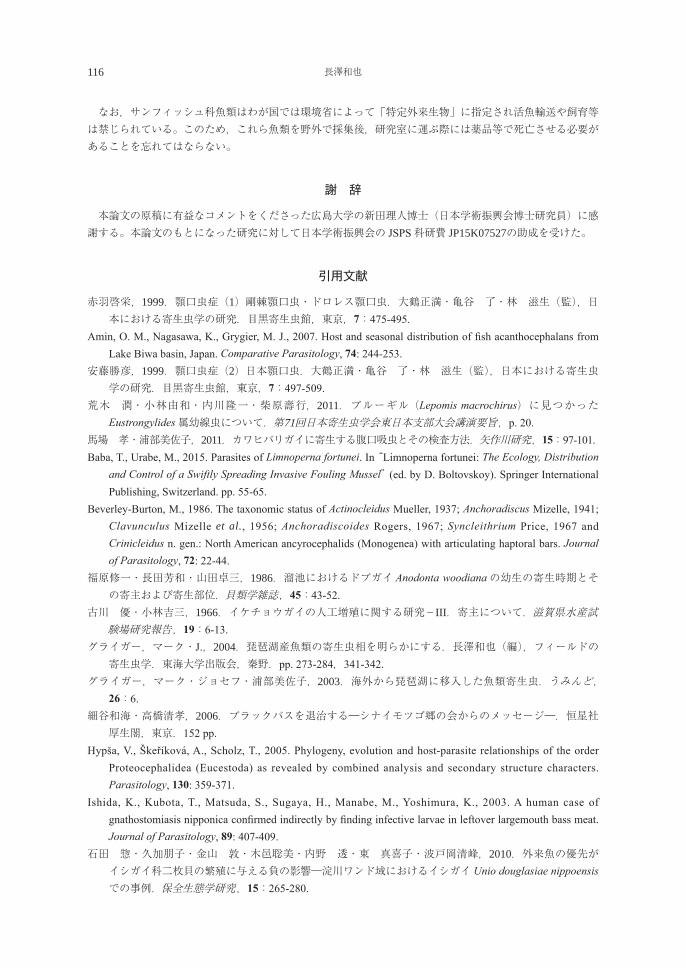
116 長澤和也
なお,サンフィッシュ科魚類はわが国では環境省によって「特定外来生物」に指定され活魚輸送や飼育等は禁じられている。このため,これら魚類を野外で採集後,研究室に運ぶ際には薬品等で死亡させる必要があることを忘れてはならない。
謝 辞
本論文の原稿に有益なコメントをくださった広島大学の新田理人博士(日本学術振興会博士研究員)に感謝する。本論文のもとになった研究に対して日本学術振興会の JSPS科研費 JP15K07527の助成を受けた。
引用文献
赤羽啓栄,1999.顎口虫症(1)剛棘顎口虫・ドロレス顎口虫.大鶴正満・亀谷 了・林 滋生(監),日本における寄生虫学の研究.目黒寄生虫館,東京,7:475-495.
Amin, O. M., Nagasawa, K., Grygier, M. J., 2007. Host and seasonal distribution of fish acanthocephalans from Lake Biwa basin, Japan. Comparative Parasitology, 74: 244-253.
安藤勝彦,1999.顎口虫症(2)日本顎口虫.大鶴正満・亀谷 了・林 滋生(監),日本における寄生虫学の研究.目黒寄生虫館,東京,7:497-509.
荒木 潤・小林由和・内川隆一・柴原壽行,2011.ブルーギル(Lepomis macrochirus)に見つかったEustrongylides属幼線虫について.第71回日本寄生虫学会東日本支部大会講演要旨,p. 20.
馬場 孝・浦部美佐子,2011.カワヒバリガイに寄生する腹口吸虫とその検査方法.矢作川研究,15:97-101.Baba, T., Urabe, M., 2015. Parasites of Limnoperna fortunei. In “Limnoperna fortunei: The Ecology, Distribution
and Control of a Swiftly Spreading Invasive Fouling Mussel” (ed. by D. Boltovskoy). Springer International Publishing, Switzerland. pp. 55-65.
Beverley-Burton, M., 1986. The taxonomic status of Actinocleidus Mueller, 1937; Anchoradiscus Mizelle, 1941; Clavunculus Mizelle et al., 1956; Anchoradiscoides Rogers, 1967; Syncleithrium Price, 1967 and Crinicleidus n. gen.: North American ancyrocephalids (Monogenea) with articulating haptoral bars. Journal of Parasitology, 72: 22-44.
福原修一・長田芳和・山田卓三,1986.溜池におけるドブガイ Anodonta woodianaの幼生の寄生時期とその寄主および寄生部位.貝類学雑誌,45:43-52.
古川 優・小林吉三,1966.イケチョウガイの人工増殖に関する研究-III.寄主について.滋賀県水産試験場研究報告,19:6-13.
グライガー,マーク・J.,2004.琵琶湖産魚類の寄生虫相を明らかにする.長澤和也(編),フィールドの寄生虫学.東海大学出版会,秦野.pp. 273-284,341-342.
グライガー,マーク・ジョセフ・浦部美佐子,2003.海外から琵琶湖に移入した魚類寄生虫.うみんど, 26:6.
細谷和海・高橋清孝,2006.ブラックバスを退治する―シナイモツゴ郷の会からのメッセージ―.恒星社厚生閣,東京.152 pp.
Hypša, V., Škeříková, A., Scholz, T., 2005. Phylogeny, evolution and host-parasite relationships of the order Proteocephalidea (Eucestoda) as revealed by combined analysis and secondary structure characters. Parasitology, 130: 359-371.
Ishida, K., Kubota, T., Matsuda, S., Sugaya, H., Manabe, M., Yoshimura, K., 2003. A human case of gnathostomiasis nipponica confirmed indirectly by finding infective larvae in leftover largemouth bass meat. Journal of Parasitology, 89: 407-409.
石田 惣・久加朋子・金山 敦・木邑聡美・内野 透・東 真喜子・波戸岡清峰,2010.外来魚の優先がイシガイ科二枚貝の繁殖に与える負の影響―淀川ワンド域におけるイシガイ Unio douglasiae nippoensisでの事例.保全生態学研究,15:265-280.
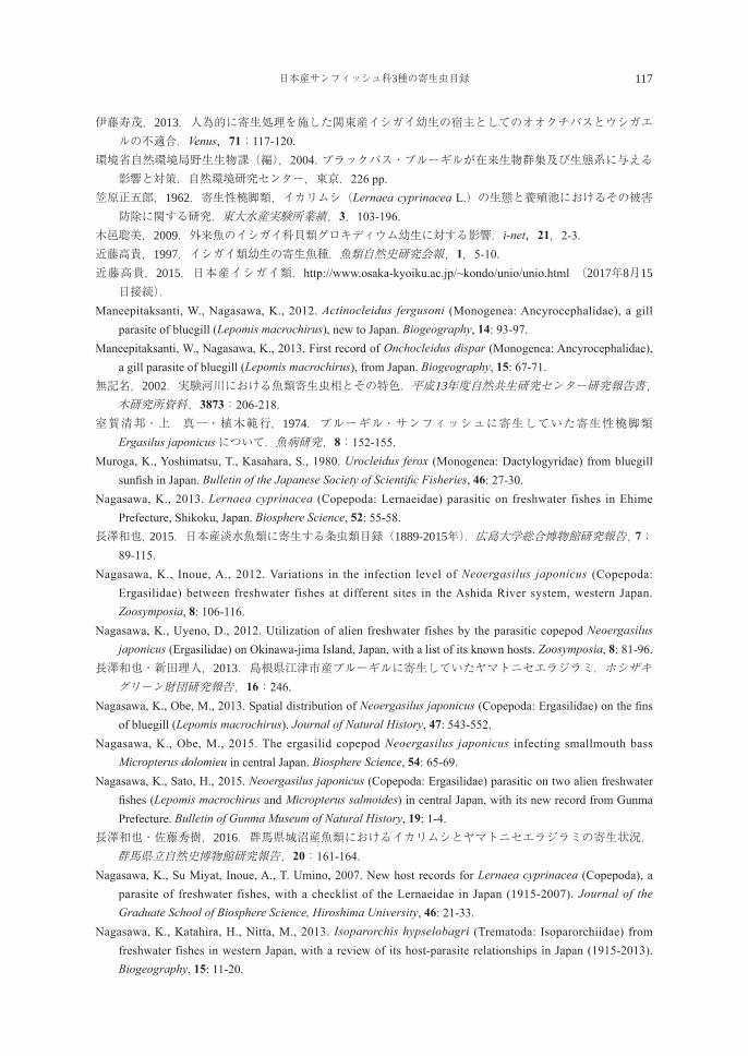
日本産サンフィッシュ科3種の寄生虫目録 117
伊藤寿茂,2013.人為的に寄生処理を施した関東産イシガイ幼生の宿主としてのオオクチバスとウシガエルの不適合.Venus,71:117-120.
環境省自然環境局野生生物課(編),2004. ブラックバス・ブルーギルが在来生物群集及び生態系に与える影響と対策.自然環境研究センター,東京.226 pp.
笠原正五郎,1962.寄生性橈脚類,イカリムシ(Lernaea cyprinacea L.)の生態と養殖池におけるその被害防除に関する研究.東大水産実験所業績,3,103-196.
木邑聡美,2009.外来魚のイシガイ科貝類グロキディウム幼生に対する影響.i-net,21,2-3.近藤高貴,1997.イシガイ類幼生の寄生魚種.魚類自然史研究会報,1,5-10.近藤高貴,2015.日本産イシガイ類.http://www.osaka-kyoiku.ac.jp/~kondo/unio/unio.html (2017年8月15日接続).
Maneepitaksanti, W., Nagasawa, K., 2012. Actinocleidus fergusoni (Monogenea: Ancyrocephalidae), a gill parasite of bluegill (Lepomis macrochirus), new to Japan. Biogeography, 14: 93-97.
Maneepitaksanti, W., Nagasawa, K., 2013. First record of Onchocleidus dispar (Monogenea: Ancyrocephalidae), a gill parasite of bluegill (Lepomis macrochirus), from Japan. Biogeography, 15: 67-71.
無記名,2002.実験河川における魚類寄生虫相とその特色.平成13年度自然共生研究センター研究報告書,木研究所資料,3873:206-218.
室賀清邦・上 真一・植木範行,1974.ブルーギル・サンフィッシュに寄生していた寄生性橈脚類Ergasilus japonicusについて.魚病研究,8:152-155.
Muroga, K., Yoshimatsu, T., Kasahara, S., 1980. Urocleidus ferox (Monogenea: Dactylogyridae) from bluegill sunfish in Japan. Bulletin of the Japanese Society of Scientific Fisheries, 46: 27-30.
Nagasawa, K., 2013. Lernaea cyprinacea (Copepoda: Lernaeidae) parasitic on freshwater fishes in Ehime Prefecture, Shikoku, Japan. Biosphere Science, 52: 55-58.
長澤和也,2015.日本産淡水魚類に寄生する条虫類目録(1889-2015年).広島大学総合博物館研究報告,7:89-115.
Nagasawa, K., Inoue, A., 2012. Variations in the infection level of Neoergasilus japonicus (Copepoda: Ergasilidae) between freshwater fishes at different sites in the Ashida River system, western Japan. Zoosymposia, 8: 106-116.
Nagasawa, K., Uyeno, D., 2012. Utilization of alien freshwater fishes by the parasitic copepod Neoergasilus japonicus (Ergasilidae) on Okinawa-jima Island, Japan, with a list of its known hosts. Zoosymposia, 8: 81-96.
長澤和也・新田理人,2013.島根県江津市産ブルーギルに寄生していたヤマトニセエラジラミ.ホシザキグリーン財団研究報告,16:246.
Nagasawa, K., Obe, M., 2013. Spatial distribution of Neoergasilus japonicus (Copepoda: Ergasilidae) on the fins of bluegill (Lepomis macrochirus). Journal of Natural History, 47: 543-552.
Nagasawa, K., Obe, M., 2015. The ergasilid copepod Neoergasilus japonicus infecting smallmouth bass Micropterus dolomieu in central Japan. Biosphere Science, 54: 65-69.
Nagasawa, K., Sato, H., 2015. Neoergasilus japonicus (Copepoda: Ergasilidae) parasitic on two alien freshwater fishes (Lepomis macrochirus and Micropterus salmoides) in central Japan, with its new record from Gunma Prefecture. Bulletin of Gunma Museum of Natural History, 19: 1-4.
長澤和也・佐藤秀樹,2016.群馬県城沼産魚類におけるイカリムシとヤマトニセエラジラミの寄生状況.群馬県立自然史博物館研究報告,20:161-164.
Nagasawa, K., Su Miyat, Inoue, A., T. Umino, 2007. New host records for Lernaea cyprinacea (Copepoda), a parasite of freshwater fishes, with a checklist of the Lernaeidae in Japan (1915-2007). Journal of the Graduate School of Biosphere Science, Hiroshima University, 46: 21-33.
Nagasawa, K., Katahira, H., Nitta, M., 2013. Isoparorchis hypselobagri (Trematoda: Isoparorchiidae) from freshwater fishes in western Japan, with a review of its host-parasite relationships in Japan (1915-2013). Biogeography, 15: 11-20.
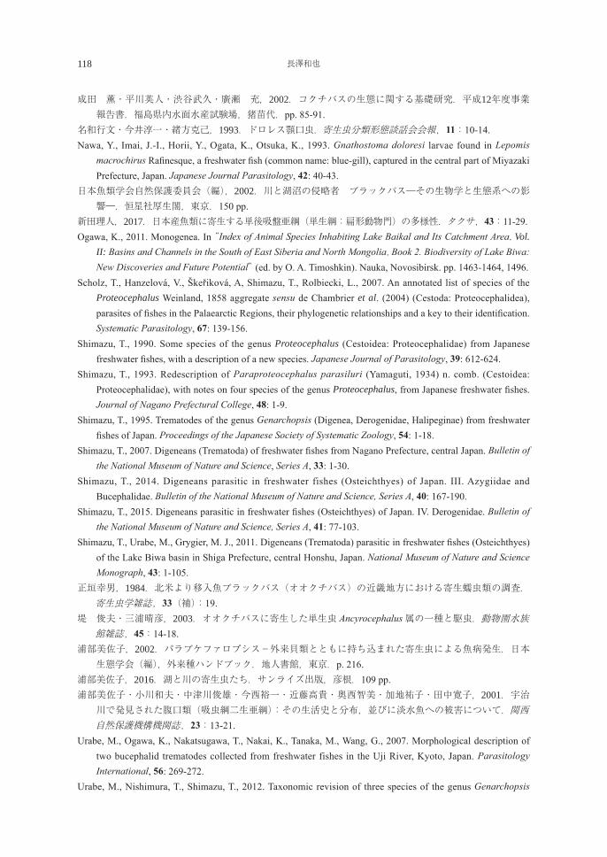
118 長澤和也
成田 薫・平川英人・渋谷武久・廣瀬 充,2002.コクチバスの生態に関する基礎研究.平成12年度事業報告書.福島県内水面水産試験場,猪苗代.pp. 85-91.
名和行文・今井淳一・緒方克己,1993.ドロレス顎口虫.寄生虫分類形態談話会会報,11:10-14. Nawa, Y., Imai, J.-I., Horii, Y., Ogata, K., Otsuka, K., 1993. Gnathostoma doloresi larvae found in Lepomis
macrochirus Rafinesque, a freshwater fish (common name: blue-gill), captured in the central part of Miyazaki Prefecture, Japan. Japanese Journal Parasitology, 42: 40-43.
日本魚類学会自然保護委員会(編),2002.川と湖沼の侵略者 ブラックバス―その生物学と生態系への影響―.恒星社厚生閣,東京.150 pp.
新田理人,2017.日本産魚類に寄生する単後吸盤亜綱(単生綱:扁形動物門)の多様性.タクサ,43:11-29.Ogawa, K., 2011. Monogenea. In “Index of Animal Species Inhabiting Lake Baikal and Its Catchment Area. Vol.
II: Basins and Channels in the South of East Siberia and North Mongolia. Book 2. Biodiversity of Lake Biwa: New Discoveries and Future Potential” (ed. by O. A. Timoshkin). Nauka, Novosibirsk. pp. 1463-1464, 1496.
Scholz, T., Hanzelová, V., Škeřiková, A, Shimazu, T., Rolbiecki, L., 2007. An annotated list of species of the Proteocephalus Weinland, 1858 aggregate sensu de Chambrier et al. (2004) (Cestoda: Proteocephalidea), parasites of fishes in the Palaearctic Regions, their phylogenetic relationships and a key to their identification. Systematic Parasitology, 67: 139-156.
Shimazu, T., 1990. Some species of the genus Proteocephalus (Cestoidea: Proteocephalidae) from Japanese freshwater fishes, with a description of a new species. Japanese Journal of Parasitology, 39: 612-624.
Shimazu, T., 1993. Redescription of Paraproteocephalus parasiluri (Yamaguti, 1934) n. comb. (Cestoidea: Proteocephalidae), with notes on four species of the genus Proteocephalus, from Japanese freshwater fishes. Journal of Nagano Prefectural College, 48: 1-9.
Shimazu, T., 1995. Trematodes of the genus Genarchopsis (Digenea, Derogenidae, Halipeginae) from freshwater fishes of Japan. Proceedings of the Japanese Society of Systematic Zoology, 54: 1-18.
Shimazu, T., 2007. Digeneans (Trematoda) of freshwater fishes from Nagano Prefecture, central Japan. Bulletin of the National Museum of Nature and Science, Series A, 33: 1-30.
Shimazu, T., 2014. Digeneans parasitic in freshwater fishes (Osteichthyes) of Japan. III. Azygiidae and Bucephalidae. Bulletin of the National Museum of Nature and Science, Series A, 40: 167-190.
Shimazu, T., 2015. Digeneans parasitic in freshwater fishes (Osteichthyes) of Japan. IV. Derogenidae. Bulletin of the National Museum of Nature and Science, Series A, 41: 77-103.
Shimazu, T., Urabe, M., Grygier, M. J., 2011. Digeneans (Trematoda) parasitic in freshwater fishes (Osteichthyes) of the Lake Biwa basin in Shiga Prefecture, central Honshu, Japan. National Museum of Nature and Science Monograph, 43: 1-105.
正垣幸男,1984.北米より移入魚ブラックバス(オオクチバス)の近畿地方における寄生蠕虫類の調査.寄生虫学雑誌,33(補):19.
堤 俊夫・三浦晴彦,2003.オオクチバスに寄生した単生虫 Ancyrocephalus属の一種と駆虫.動物園水族館雑誌,45:14-18.
浦部美佐子,2002.パラブケファロプシス-外来貝類とともに持ち込まれた寄生虫による魚病発生.日本生態学会(編),外来種ハンドブック.地人書館,東京.p. 216.
浦部美佐子,2016.湖と川の寄生虫たち.サンライズ出版,彦根.109 pp.浦部美佐子・小川和夫・中津川俊雄・今西裕一・近藤高貴・奥西智美・加地祐子・田中寛子,2001.宇治川で発見された腹口類(吸虫綱二生亜綱):その生活史と分布,並びに淡水魚への被害について.関西自然保護機構機関誌,23:13-21.
Urabe, M., Ogawa, K., Nakatsugawa, T., Nakai, K., Tanaka, M., Wang, G., 2007. Morphological description of two bucephalid trematodes collected from freshwater fishes in the Uji River, Kyoto, Japan. Parasitology International, 56: 269-272.
Urabe, M., Nishimura, T., Shimazu, T., 2012. Taxonomic revision of three species of the genus Genarchopsis
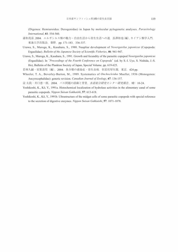
日本産サンフィッシュ科3種の寄生虫目録 119
(Digenea: Hemiuroidea: Derogenidae) in Japan by molecular pylogenetic analyses. Parasitology International, 61: 554-560.
浦和茂彦,2004.エルガシルス類の魅力-自由生活から寄生生活への道.長澤和也(編),カイアシ類学入門.東海大学出版会,秦野.pp. 171-183,336-337.
Urawa, S., Muroga, K., Kasahara, S., 1980. Naupliar development of Neoergasilus japonicus (Copepoda: Ergasilidae). Bulletin of the Japanese Society of Scientific Fisheries, 46: 941-947.
Urawa, S., Muroga, K., Kasahara, S., 1991. Growth and fecundity of the parasitic copepod Neoergasilus japonicus (Ergasilidae). In “Proceedings of the Fourth Conference on Copepoda” (ed. by S.-I. Uye, S. Nishida, J.-S. Ho), Bulletin of the Plankton Society of Japan, Special Volume. pp. 619-625.
若林久嗣・室賀清邦(編),2004.魚介類の感染症・寄生虫病.恒星社厚生閣,東京.424 pp.Wheeler, T. A., Beverley-Burton, M., 1989. Systematics of Onchocleidus Mueller, 1936 (Monogenea:
Ancyrocephalidae): generic revision. Canadian Journal of Zoology, 67: 136-157.淀 太我・井口恵一郎,2004.バス問題の経緯と背景.水産総合研究センター研究報告,12:10-24.Yoshikoshi, K., Kô, Y., 1991a. Histochemical localization of hydrolase activities in the alimentary canal of some
parasitic copepods. Nippon Suisan Gakkaishi, 57: 613-618.Yoshikoshi, K., Kô, Y., 1991b. Ultrastructure of the midgut cells of some parasitic copepods with special reference
to the secretion of digestive enzymes. Nippon Suisan Gakkaishi, 57: 1071-1078.
120 長澤和也
A checklist of the parasites of three species of centrarchids (bluegill, largemouth bass, and smallmouth bass) in Japan (1962-2017)
Kazuya Nagasawa
Graduate School of Biosphere Science, Hiroshima University 1-4-4 Kagamiyama, Higashi-Hiroshima, Hiroshima 739-8528, Japan
Abstract: Information on the metazoan parasites of three species of the family Centrarchidae (bluegill Lepomis macrochirus, largemouth bass Micropterus salmoides, and smallmouth bass Micropterus dolomieu) in Japan is summarized in the Parasite-Host and Host-Parasite lists, based on the literature published for 56 years between 1962 and 2017. These fish species were introduced from the U. S. A. into Japan, where they have widely established. The parasites, including 21 nominal species and subspecies and those not identified to species level, are listed by higher taxa as follows: Myxozoa (no. of nominal and subspecies species: 0), Cestoda (2), Monogenea (6), Trematoda (5), Nematoda (2), Acanthocephala (1), Bivalvia (3), and Copepoda (2). For each parasite species, the following information is given: its currently recognized scientific name; previous identification used for the parasite from centrarchids in Japan; site(s) of infection within or on the host; known geographical distribution in Japanese waters; and the published source of each record. Of the 21 nominal species and subspecies of parasites listed, 12 species are from bluegill, 13 species and subspecies from largemouth bass, and three species from smallmouth bass. The parasites listed consist of both those native to Japan and those introduced from overseas. One species of Cestoda (Proteocephalus fluviatilis) and six species of Monogenea (Actinocleidus fergusoni, Onchocleidus dispar, and Onchocleidus ferox from bluegill; Onchocleidus furcatus, Onchocleidus helicus, and Syncleithrium fusiformes from largemouth bass) are the parasites of North American origin. Two species of Nematoda (Gnathostoma doloresi and Gnathostoma nipponicum) are among human parasites and have been reported from bluegill and largemouth bass, respectively. A human infection by the latter species occurred by eating the raw flesh of a largemouth bass from Japan. Key words: bluegill, Centrarchidae, checklist, fish parasites, human parasites, largemouth bass,
smallmouth bass
Information
Hiroshima University has granted the Doctor’s degree to the following researchers.The list is only concerned with the Graduate School of Biosphere Science.
DEPARTMENT OF BIORESOURCE SCIENCEMarch 2, 2017 Doctor of Agriculture Yuichiro OCHI
March 2, 2017 Doctor of Agriculture Yusuke KONDO
March 2, 2017 Doctor of Philosophy Aira SEO
March 2, 2017 Doctor of Agriculture Satoshi TOMANO
March 2, 2017 Doctor of Philosophy Kanokon SeemanonMarch 2, 2017 Doctor of Agriculture Renlong LV
March 2, 2017 Doctor of Philosophy KHUSHDIL MAROOF
March 2, 2017 Doctor of Agriculture Masato NITTA
September 4, 2017 Doctor of Agriculture Huang ANQI
DEPARTMENT OF BIOFUNCTIONAL SCIENCE AND TECHNOLOGYMarch 2, 2017 Doctor of Agriculture Da TENG
March 2, 2017 Doctor of Agriculture Junki MIYAMOTO
March 2, 2017 Doctor of Agriculture Takayuki KONDO
March 2, 2017 Doctor of Agriculture Sotaro FUJII
March 2, 2017 Doctor of Agriculture Yukari YABUKI
September 4, 2017 Doctor of Philosophy REHAB MARRY ABDELATY NSRELDEN
September 4, 2017 Doctor of Agriculture Tran Van HUNG
生物圏科学Biosphere Sci. 56:121-122 (2017)
122
DEPARTMENT OF ENVIRONMENTAL DYNAMICS AND MANAGEMENTMarch 2, 2017 Doctor of Agriculture MAUNG SAW HTOO THAW March 2, 2017 Doctor of Agriculture Youji HAMANA
March 2, 2017 Doctor of Agriculture Shuichi WATANABE
September 4, 2017 Doctor of Agriculture Naoto KAJIHARA
DISSERTATION PhDMarch 2, 2017 Doctor of Agriculture Toyohiro NISHIOKA
March 2, 2017 Doctor of Agriculture Mitsugu MIYAMOTO
March 2, 2017 Doctor of Agriculture Ryuki MIYAUCHI
June 26, 2017 Doctor of Agriculture Yojiro ISHIDA
September 4, 2017 Doctor of Agriculture Shinnosuke IWASAKI
September 4, 2017 Doctor of Agriculture Natsumi TSUJITA
September 4, 2017 Doctor of Agriculture Takao HASHIMOTO
123
Ecological study on the Asian sheephead wrasseSemicossyphus reticulatus (Labridae) in the western Seto Inland Sea
Yuichiro Ochi
Graduate School of Biosphere Science, Hiroshima University,Higashi-Hiroshima 739-8528, Japan
瀬戸内海西部におけるコブダイ Semicossyphus reticulatusの生活史に関する生態学的研究
越智 雄一郎広島大学大学院生物圏科学研究科,739-8528 東広島市
The Asian sheephead wrasse Semicossyphus reticulatus is well known as the largest labrid in temperate waters around Japan including the Seto Inland Sea and for the well-developing humphead. Only three Pacific temperate water species constitute the genus Semicossyphus, i.e., S. pulcher in the eastern Pacific, S. darwini in the southern Pacific, and S. reticulatus in the western Pacific, and all three species attain close to a total length of 1 m. There are few ecological study about S. reticulatus in contrast to the other two congeners are attribute from their local importance of fisheries and recreational fishing.
The aim of this study is to reveal ecological and biological characteristics of S. reticulatus with comparing those of congeneric wrasses, which have geographically far isolated distributions each other. I conducted sampling study focused on the following aspects, 1) age and growth, 2) reproduction and sexuality, and 3) head morphology. All fish samples were obtained from fish markets and from bait fishing by the author in the western Seto Inland Sea around Hiroshima, Ehime and Yamaguchi Prefectures.
For the age and growth (Chapter 2), I conducted the age determination by reading annuli of sectioned otolith extract from the fish. By Adapting von Bertalanffy growth equation model, I calculated the relationship between the estimated age and standard length in the following equation: Lt = 489 (1-e-0.12 (t+1.75)). The calculated growth model indicated that the wrasse maintains considerably slow growth particularly after 10 yrs age, which is similar to the previous reports in a congener the California sheephead wrasse S. pulcher. The estimated maximum age of S. reticulatus was 31 yrs and which individual was male, indicating considerable long life of the wrasse even in Seto Inland Sea.
For the reproduction and sexuality (Chapter 3), I found that the spawning season occurred during April - June in Seto Inland Sea, based on the gonadosomatic index (GSI) analysis. Histological observation of gonads revealed that all males maintained secondary testis that possess ovariform structures inside the testis, e.g., ovarian cavity and remnant of ovarian cells. This result strongly suggests the occurrence of the protogynous sex change (female to male sex change) in this wrasse. Sex ratio of sampled specimens was strongly biased to females. In addition, males appeared only in larger size class over 400 mm in standard length (SL), and were significantly larger than females. No small males possessing the primary testis (gonochoristic form testis) were confirmed in the samples specimens.
生物圏科学Biosphere Sci. 56:123-124 (2017)
124
These results suggest that the fish has a life history of monandric protogyny; all males are derived from sex change of females. Of females, not all female individuals were smaller than males. Conversely, a few fish reaching male SL size class remained to be females. This implies that the sex change of female S. reticulatus is controlled not by the age or size automatically but by their social structures probably dominance orders among individuals in the local groups.
For the humphead morphology (Chapter 4), I analyzed the elevation angle of the humps on forehead of S. reticulatus as an indicator of the development. In general, it has long been believed that individuals of S. reticulatus having well-developed humpheads could be males, namely considered as a sex-specific characteristic without any evidential data. However, the results of my analysis led the conclusion as the hump of the fish gradually enlarged with body growth, namely the size-associated development. The growth pattern resulted in the often occurrence of the well-developed humpheads even in large females.
In the present Ph D thesis, I first revealed the basic ecology of S. reticulatus. The fish shows considerable ecological similarity to congeneric wrasses regardless of the world-scale geographical separation in the Pacific Ocean. Considering a slow growth speed, a long life-span, a strongly biased sex ratio and sex-related body size imply their potential weakness against to the fishery activities and environmental change on the habitats. I am fortunate to conduct the present study on this large reef fish S. reticulatus in this area. This is partly because the fishery pressure for this fish is not so strong in the western Seto Inland Sea, in contrast to most of large reef fishes have be faced exploitations by fisheries. I hope our data of S. reticulatus would contribute to further scientific understanding of a valued bioresource in the Seto Inland Sea and the conservative control of the Sato Umi coastal ecosystem.
Key words: Semicossyphus reticulatus, Growth pattern, Protogyny, Humphead, Labridae
125
Ecological studies on symbiotic relationships between large-sized jellyfish and otheranimals in Asian waters
Yusuke Kondo
Graduate School of Biosphere Science, Hiroshima University,Higashi-Hiroshima 739-8528, Japan
アジア海域における大型クラゲ類と他動物との共生に関する生態学的研究
近藤 裕介広島大学大学院生物圏科学研究科,739-8528 東広島市
1. In recent years, various aspects of jellyfish have been actively studied, such as their mass occurrence, application of useful compounds extracted from them, and their use as aquatic resources. Accordingly, the role of jellyfishes in the marine ecosystem has also been reviewed. It is known that various organisms associate with jellyfish. However, the information on the interaction between jellyfish and symbionts is not enough. Therefore, I investigated the fauna that associates symbiotically with the jellyfish species and their interspecific relationships in Japan, Korea, Thailand, Philippines, and Malaysia. 2. Seasonal changes in the prevalence and intensity of metacercariae of Lepotrema clavatum, Cephalolepidapedon saba, and Opechona olssoni in three species of host jellyfish, Aurelia aurita s.l., Chrysaora pacifica, and Cyanea nozakii, were examined. The prevalence and mean intensity of metacercariae in C. nozakii were higher than in A. aurita s.l. and C. pacifica. It is presumed that metacercariae were accumulated in C. nozakii due to their predation by other infected jellyfish. Cyanea nozakii plays a role as a paratenic host rather than a second intermediate host. The adults and metacercariae of trematodes were found together with nematocysts in the guts of the Japanese butterfish, Psenopsis anomala, and juveniles of the black scraper, Thamnaconus modestus. In contrast, these were not found in the guts of the juveniles of the Japanese jack mackerel, Trachurus japonicus, indicating that it does not use jellyfish as a food source. The transmission of trematodes into a definitive host fish occurs via predation of infected jellyfish. 3. In Japan and Korea, associations of two species of fish with jellyfish were common. The juveniles of T. japonicus were associated with five species of jellyfish (Aequorea macrodactyla, A. aurita s.l., C. nozakii, Netrostoma setouchianum, Morbakka virulenta), which were found only in Japan. The juveniles of P. anomala were associated with three species of jellyfish in Japan (C. pacifica, C. nozakii, M. virulenta) and Korea (A. aurita s.l., Nemopilema nomurai, Sandria malayensis). The host jellyfishes of the shrimp scads, Alepes djedaba, were variable, with four species being the hosts in Thailand (Acromitus flagellatus, Catostylus townsendi, Lobonemoides robustus, Rhopilema hispidum), two in the Philippines (A. maculosus, L. robustus), and one in Malaysia (Chrysaora chinensis). All the juveniles of these three species of fish occurring in East and Southeast Asian waters were 0-year in age. 4. The megalopa larvae to juveniles of the Christ crab, Charybdis feriata, were associated with jellyfish in Thailand and the Philippines. The juveniles of the ophiuroid, Ophiocnemis marmorata, occurred on jellyfish in Thailand and Malaysia. The final stages of planktonic larvae of these organisms appeared to settle on the host jellyfish directly, and then grow during the early stages of their life cycle
生物圏科学Biosphere Sci. 56:125-126 (2017)
126
on the hosts. The caridean shrimp, Latreutes anoplonyx, was found on R. hispidum, L. robustus, and A. flagellatus collected from Thailand, the Philippines, and Malaysia, and seemed to breed on the host. The host jellyfish likely function in the settling, feeding, and growing spots of the crab and ophiuroids, and in the breeding spot of the shrimp. 5. The nematocysts were detected in the Foods of all the symbionts, but the frequency of their occurrence was low (14.3% in T. japonicus) to high (100% in P. anomala and C. feriata), depending on the taxa. It is evident that the symbionts directly fed on the hosts or stole the prey captured by them. The juveniles of C. feriata were powerful predators and devoured not only the host jellyfish but also the other symbionts. 6. In Thailand, two species of rhizostomes, R. hispidum and L. robustus, were commercially harvested. These jellyfishes harbored A. djedaba, C. feriata, L. anoplonyx, and O. marmorata. Especially, because the ophiuroids are firmly attached to the host with specialized attachment organs, almost all the individuals are probably killed by jellyfish fishery. The negative impact of jellyfish fishery on these symbionts was estimated based on my original data and the statistics from the FAO fisheries. The worst case, was determined to be for O. marmorata, which was estimated to be killed at the rate of 126-165 million individuals per year by the jellyfish fisheries in Thailand. It is likely that such contaminations greatly influence the benthic communities. For sustainable societies, new alternative collection methods should be proposed.
Key words: Asian water, Jellyfish, Jellyfish fishery, Predation, Symbiont
127
Study on the symbiotic relation of free-roaming cats and humans in old town Onomichi, Hiroshima prefecture, Japan
Aira Seo
Graduate School of Biosphere Science, Hiroshima University, Higashi-Hiroshima 739-8528, Japan
広島県尾道市旧市街地における自由徘徊ネコとヒトとの共生に関する研究
妹尾 あいら広島大学大学院生物圏科学研究科,739-8528 東広島市
第Ⅰ章 序論 近年全国でネコの街が話題となり,自由徘徊ネコとのふれあいを求める観光客が数多く訪れるようになっている。しかしその一方で,ネコの糞尿や人獣共通感染症など公衆衛生の悪化が問題となっている。ヒトと自由徘徊ネコとの関係は,観光資源としてのネコの経済効果やネコとのふれあいを通して得られる癒しの効果などヒトにとって有益な関わりと,ネコによる糞尿被害,人獣共通感染症,ダニやノミの発生などヒトにとって有害な関わりについて,受益者と被害者の間の利害対立として取り上げられてきたが,動物福祉など,ネコ側の視点からヒトと自由徘徊ネコとの関係が注目されることはほとんどなかった。そこで本研究は,尾道市旧市街地に生息する自由徘徊ネコを4年間にわたって調査することで,今後のヒトとネコの共生のあり方について考えることを目的とした。
第Ⅱ章 尾道市旧市街地の自由徘徊ネコの福祉について個体数の経年変化を通して考える 近年わが国では,自由徘徊ネコを観光資源として地域活性に利用する動きがあるが,その一方でネコの福祉の現状については明らかになっていない。そこで本研究は,自由徘徊ネコが多く生息する尾道市旧市街地において,3年間にわたりネコの個体数の変化を調べることで,ネコの福祉の状況を明らかにすることを目的とした。旧市街地を山手地区と商店地区に分け,ルートセンサス法を用いて月に8回調査を行った。調査1年目に,山手地区に124頭,商店地区に80頭ものネコが生息していることが明らかとなった。またその多くが野良ネコであった。2年後に生息を確認できた個体は25頭と14頭だけであった。観察されなくなったネコの多くは,病気や怪我によって地区内で死亡したものと考えられた。以上の結果から,旧市街地に生息する野良ネコの福祉の状況はかなり深刻であることが明らかとなったので,今後は繁殖の制限とともに健康管理の必要性が指摘された。
第Ⅲ章 尾道市旧市街地の自由徘徊ネコの福祉について給餌行為を通して考える 都市部の住宅街や観光地に生息する野良ネコは,一部住民や観光客が与える餌に依存していると考えられている。そこで本研究は,山手地区の5つの給餌場所で餌を与えられている給餌個体と,給餌場所には来ない非給餌個体の間で福祉の状態と行動を比較することを目的とした。その結果,給餌個体は43頭で非給餌個体は144頭であった。給餌個体の方が非給餌個体よりも健康に問題のある個体の割合が有意に低かった。一方で,給餌個体の方が非給餌個体よりも人馴れしている個体の割合が有意に高く,給餌行為によって野良ネコが居着くことが示唆された。以上の結果から,健康管理と不妊去勢手術を伴った継続的な給餌は,ネコの健康と福祉の状態を良好にすることが示唆されたが,その一方で給餌行為は特定の地域に多数のネコを棲み着かせてしまうので,糞尿被害などの地域の問題に発展することが危惧された。
生物圏科学Biosphere Sci. 56:127-128 (2017)
128
第Ⅳ章 �尾道市旧市街地の自由徘徊ネコによる糞尿被害の軽減について酢酸及びイソ吉草酸を含有した忌避剤の効果の検証を通して考える
近年,自由徘徊ネコによる糞尿被害が深刻化し,社会問題にまで発展している地域もある。その解決の一助として,効果的なネコ用忌避剤を開発することが考えられる。そこで本研究は,自由徘徊ネコが多く生息している山手地区の4つの寺院において,酢酸及びイソ吉草酸を含有する忌避剤が自由徘徊ネコの侵入行動と排糞行動を抑制する効果を検証することを目的とした。対照期間を経て,忌避剤の試験期間を約5か月間設けた。その結果,忌避剤は侵入回数を有意に低下させることができた。また墓地に設置した忌避剤はネコの排糞量を有意に減少させた。しかし,忌避剤の臭いがヒトにも不快感を与えることから,臭いの改善が必要であることが指摘された。
第Ⅴ章 �尾道市旧市街地の自由徘徊ネコによる糞尿被害の軽減について酢酸,イソ吉草酸及びシトラールを含有した忌避剤の効果の検証を通して考える
第Ⅳ章において効果の認められたネコ用忌避剤にシトラールを添加することでヒトに対する不快臭の低減を試みることを目的とした。実験は第Ⅳ章の4つの寺院で実施した。対照期間(忌避剤設置前期間),ダミー期間(忌避成分を含まないダミーの忌避剤設置期間)を経て,シトラール添加忌避剤設置期間を約4か月間設けた。その結果,シトラール添加忌避剤は,添加しない忌避剤と同様にネコの侵入行動に対して一定の抑制効果を示したが,排糞行動に対する効果は若干不安定であった。以上の結果から,ヒトが頻繁に立ち寄る場所ではシトラール添加忌避剤を設置することで,ある程度の効果があるものと考えられた。
第Ⅵ章 総括 以上の研究結果を踏まえて,今後の旧市街地の自由徘徊ネコ対策を提案する。まず自由徘徊ネコの個体識別と生息個体数の調査を行い,ヒトに対する馴れの程度と外貌による健康状態の評価(福祉の評価)を行う。人馴れし不妊去勢手術を受けていない個体は,手術とともに血液検査を行う。また病気や怪我をしている人馴れ個体は治療する。人馴れした健康個体は動物愛護センターを通して里親募集を行う。人馴れしておらず不妊去勢手術を受けていない個体は,動物愛護センターの協力を得て捕獲して血液検査をする。伝染性の病気に罹患している個体は動物愛護センターで安楽死処分する。一方で人馴れしておらず伝染病に罹患していない個体は,手術後に元いた場所に戻し,その後は地域猫活動によって飼育管理する。その結果,人馴れした個体には里親を募集する。観光客と一部住民によって行われてきた野良ネコへの給餌を規制し,無人の餌販売所を設置し,餌から得た収入は活動に還元する。ネコ用忌避剤とネコ用公衆トイレを旧市街地の公園や公共施設に設置し,トイレへの誘導訓練をする。以上の活動の結果,ネコの個体数は減少すると予想されるので,観光資源としてのネコは自由に徘徊している飼いネコを対象とする。以上の活動を実施するためには,地域住民,尾道市,尾道観光協会,動物愛護団体,広島県動物愛護センター,大学の研究者などの連携による協働型運営が必須条件である。
キーワード:自由徘徊ネコ,動物福祉,共生,地域猫活動
129
A study on stock structure of the oval squid Sepioteuthis spp. around Japan
Satoshi Tomano
Graduate School of Biosphere Science, Hiroshima University,Higashi-Hiroshima, 739-8528, Japan
日本沿岸におけるアオリイカ属の資源構造に関する研究
笘野 哲史広島大学大学院生物圏科学研究科,739-8528 東広島市
漁業に代表される海産生物資源は人類の食料保障を支えているが,世界の海面漁獲量は過去30年間で減少傾向である。それに反してイカ類は,近年の急激な需要増加に伴い,過去40年間で漁獲量は約4倍まで増えた。日本は世界有数のイカ消費国であり,イカ類は国民の重要なタンパク源となっている。本研究対象種であるアオリイカ Sepioteuthis cf. lessonianaは美味であり,「イカの王様」として愛されている。漁業においては商品価値がイカ類の中で最も高く,1kgあたり2,000円以上で取引されている。ただし,「アオリイカ」と呼ばれるイカには,別種レベルの遺伝的差異を有するアカイカ Sepioteuthis sp. 1,シロイカ Sepioteuthis sp. 2およびクアイカ Sepioteuthis sp. 3の3種類の存在が報告されている。 アオリイカ3種は分類形質に乏しいため,種判別にはタンパク質の電気泳動多型であるアロザイム分析が用いられてきた。しかし,アロザイム分析はサンプルの保存状態や発生段階により再現性が低下する。その点,DNAマーカーは組織や発生段階による制約がないため,イカ類を含む多くの生物の種同定に用いられている。このように DNAマーカーは本邦産アオリイカ属3種の種判別に有効と考えられるが,現在まで報告されていない。さらに,アオリイカ3種の分布や漁獲物の種組成といった資源保全に必須な基本情報は南西諸島に限られており,本州,四国,九州では不明であった。加えて,野生集団の保全や管理を行う上で,遺伝的な交流がある繁殖集団を把握することは極めて重要である。そのため,繁殖単位を構成する集団の数や集団間の遺伝的差異を調べ,集団構造を明らかにすることが必要である。 本博士論文では,アオリイカ属の遺伝学的解析ツールを新規開発する事によって,日本沿岸における資源貢献および遺伝学的集団構造を定量化し,日本沿岸におけるアオリイカ属の資源構造を明らかにすることを目的とした。研究対象として,特に,本州沿岸で優占すると期待されるアオリイカ属アカイカとシロイカを対象とした。
第1章では,再現性の高く,かつ高感度な DNAマーカーを新規開発した。ミトコンドリア(mt)DNAでは,DNAバーコーディングに用いられるシトクロームオキシダーゼサブユニット I(COI)領域の部分配列において,両種間での塩基置換が多く見られ,種判別に有効であることが示された。アカイカおよびシロイカの各ゲノム DNAから単離されたマイクロサテライト DNA(ms)マーカーはいずれも多型に富み,遺伝的多様性の評価に有用であることが示された。さらにシロイカから単離されたマーカー座をアカイカで増幅させたところ,アリルレンジに差異がみられ,種判別に有効であることが示唆された。上記の DNAマーカーはいずれも,卵および筋肉組織で使用可能であり,発生段階や保存方法に影響されなかった。
第2章では,日本沿岸19海域から採集された約1700個体のアオリイカ属を,第1章で開発した DNAマーカーによって種判別し,アカイカとシロイカの分布と資源貢献を調べた。その結果,日本沿岸ではシロイカが主な漁業対象種であるが,種子島と屋久島(大隅諸島)ではアカイカが大きく資源に貢献していた。またクアイカは種子島,屋久島および和歌山から発見されたが,個体数が非常に少ないため資源への貢献度は低いと
生物圏科学Biosphere Sci. 56:129-130 (2017)
130
いえる。さらに,ゲノム msマーカーにて雑種が検出されなかったため,アカイカとシロイカは同所的に生息しながらも生殖隔離していることが追認された。大隅諸島では,アカイカ成体は全水深帯で採集された一方,シロイカの成体は30m以浅のみ採集され,両種は生息水深が異なることが示唆された。さらに,両種の性成熟サイズ(外套背長)にも違いがみられ,雌雄ともにアカイカの方が大型になることが明らかになった。
第3章では,第1章にて開発した msマーカーを使用し,アカイカおよびシロイカの遺伝的多様性と集団構造の定量を行った。まずシロイカでは,7マーカー座を用いて本州,四国,九州から採集された12海域840個体を解析した。その結果,日本沿岸のシロイカ集団が持つ遺伝的多様性は平均ヘテロ接合度の観測値(HO)で0.68,平均アリル数(NA)で10.0となり,地域集団間で同等であった。また,有意な遺伝分化が認められなかったことから,日本沿岸のシロイカは移動回遊によって集団間での遺伝子流動が活発であることが示唆された。アカイカでは,13マーカー座を用いて台湾から和歌山まで6海域から採集された274個体を解析した。アカイカが示した遺伝的多様性は HO = 0.742および NA = 8.7となり,集団間で同等の値となった。一方,台湾から本州にかけて有意なアカイカの集団構造が示され,大きく4つのグループ(和歌山,屋久島と種子島,沖縄と石垣島,台湾)に大別されることが示唆された。ただし,すべてのペア集団間においても有意な遺伝的差異が認められたため,独立性の高いローカルな繁殖集団が維持されていると考えられた。
本研究では,遺伝学的アプローチによって,アオリイカ属資源の現状の把握が可能となり,有効的な繁殖サポートが提示された。シロイカは集団構造を持たないため,日本沿岸で単一の資源として保全管理するのが妥当である。アカイカでは,日本沿岸では石垣,沖縄,種子島,屋久島,和歌山の集団を保全管理の単位とするべきである。両種は産卵水深に違いがみられたため,人工産卵床の設置水深を,優占種の産卵水深に合わせることで,より効率的な産卵促進が期待できる。アカイカでは,島嶼間の移動回遊は制限されることで地域ごとにローカル集団を形成していると考えられる。ローカルな繁殖集団において,人工的な産卵床設置などの繁殖サポートは,資源の維持増大に効果的であろう。特に,太平洋岸では人工産卵礁を水深20m以浅と40m付近の両方に設置することで,両種の産卵サポートを図ることが可能と思われる。 将来的には,本研究で開発された msマーカーを用いて天然の産出卵から間接的に親イカの数や遺伝的多様性を定量化することも可能であろう。さらに,本研究で得られた地域集団の遺伝的多様性の情報を合わせることで,アオリイカ属の遺伝資源モニタリングへ応用できる可能性も秘めている。アオリイカ属は日本以外でも重要な漁業対象種であるため,本研究で明らかとなったアオリイカ属の資源構造が,他海域での資源構造の解明へ波及する可能性を秘めている。さらには,本研究が他のイカ類の資源構造解析への応用へと発展することを切に願う。
キーワード:アオリイカ属,マイクロサテライト DNA,ミトコンドリア DNA,遺伝的多様性,集団構造
131
Improvement of Thai Farmers’ Livelihood through Alternative Rice Farming: A Case Study of Japonica Rice in the Northern Thailand
Kanokon Seemanon
Graduate School of Biosphere Science, Hiroshima University,Higashi-Hiroshima 739-8528, Japan
代替稲作によるタイ農民の生計向上―北部のジャポニカ米生産の事例研究―
カノオン シーマノン広島大学大学院生物圏科学研究科,739-8528 東広島市
Thai farmers still have faced the lower yield of rice production, paddy price and high production cost, thereby suffering from low income. Therefore, private and government sectors have so far encouraged farmers to cultivate top-quality rice or alternative rice for specialty markets such as organic rice and Japonica rice in order to increase farmers’ income. These rice planting are handled by rice mills, which are produced through contract farming (CF) systems. The production through CF system may bring an increase of farmers’ income because its contract secures price and high yield. This research focused on Japonica rice variety in the northern Thailand, especially Chiang Rai Province. The research objectives are to explore the characteristics of Thailand’s Japonica rice production through CF systems, analyze costs and earnings of growers, examine the current domestic marketing system of Thailand’s Japonica rice, and evaluate the perspectives of Thai consumers towards Japonica rice consumption. The results of each objective are to provide the answer toward the actual benefits for farmers from the current Japonica rice production and marketing. In Japonica rice production side in Chiang Rai Province, its planting through CF systems were based on the intermediate model. The collectors who acted as a local coordinator and consultant for the contract farmers played the significant role in CF systems. They could reduce the management trouble of rice millers. In the system, contract rice millers guaranteed the purchase price of paddy, and provided extension officers and agricultural inputs, especially Japonica rice seeds for growers. The seeds were produced by the Chiang Rai Rice Research Center (CRI). Each rice mill adopted different management policy on signing a contact, setting up purchase price, and commission and transportation fees for their collector. The rice miller who established a good strategy of pricing could attract farmers to participate in this contract. The high contract price, assured market and high yield were advantages of Japonica rice production in the selected areas. The fertilizer was the main cost of inputs for planting Japonica rice in the selected areas. Some growers, particularly the collectors who also planted Japonica rice would mainly buy fertilizers from their contract rice mills, in order to stock and sell such inputs to their farmers, and used for their Japonica rice planting. However, some rice mills allowed the contract Japonica rice growers to purchase fertilizers and agricultural chemicals from any other suppliers whose prices were cheaper than the contract rice mills. Therefore, the collectors had higher production cost than contract farmers leading to lower earning. This indicated that the use of fertilizers provided from extension service of contract rice mills had a significant impact on the economic structure of growers. However, the systems of Japonica rice contract farming could help farmers realize a higher price and high yields which brought more income. Moreover, Japonica rice cultivation was more attractive because the growers had lower
生物圏科学Biosphere Sci. 56:131-132 (2017)
132
production costs and got higher income, compared to the results achieved by non-contract glutinous rice farmers in the same region. In Thailand’s Japonica rice marketing side, rice mills directly distributed polished Japonica rice to distributors who are located mainly in Bangkok, retailers and Japanese restaurants. In this chain, the main users of such Japonica rice were Japanese restaurants. In part of the positive aspects for Thailand’s Japonica rice marketing, the buyers indicated that the government and private sectors should promote the planted area and consumption of Thailand’s Japonica rice more. The consumers were the final party involved in the process of milled Thailand’s Japonica rice distribution. The consumers in Bangkok who were the respondents in this research were mostly a salaried employee. They enjoyed Japanese cuisine consumption at restaurants especially in the shopping malls. These restaurants mostly cooked and served Japanese cuisine using Thailand’s Japonica rice. The respondents are increasing Japanese cuisine consumption, influencing the increase of demand for Japonica rice as well. Analysis on their preference for different Japonica rice choices found that their first preference was Japanese rice imported from Japan. Thailand’s Japonica rice was chosen as the second best in all attributes, which its flavor, smell and soft sticky texture were similarly to the original Japanese rice from Japan. Therefore, the respondents agreed that the northern region had a great potential to produce Japonica rice for consumption in the domestic market. According to the all results, selecting Japonica rice cultivation under the CF system make more benefit for farmers, especially in Chiang Rai Province including a high yield, high contract price and more income, as well as a certain market. The contract farmers received the better knowledge of cultivation practices and other support services from extension officers of contract rice mill, except supply of fertilizers. Moreover, consumers had a positive attitude toward Thailand’s Japonica rice consumption. However, the CRI should more improve and develop a quality of Japonica rice seed continuously, in order to be more suitable for the environment in the northern region and more resist to disease.
Keywords: Japonica rice, Contract farming, Economic characteristic, Marketing system, Consumer perspective
133
Studies on the utilization of phytol in forages for ruminant production
Renlong Lv
Graduate School of Biosphere Science, Hiroshima University, Higashi-Hiroshima 739-8528, Japan
粗飼料中フィトールの反芻家畜生産への利用に関する研究
呂 仁龍広島大学大学院生物圏科学研究科,739-8528 東広島市
Ruminants produce milk and meet through utilizing forages. Therefore, it is necessary to explore the potential values of forages and to maximize their utilization of forages. Recently, consumers have shown increased concern for livestock products enriched with bioactive compounds that impact on human health. The ruminal degradation of chlorophyll in ingested forages liberates phytol moiety which is metabolized by rumen microbes to phytanic acid. This phytanic acid, a natural ligand of peroxisome proliferator-activated receptor, appears in meat and milk produced by ruminants, and presumably has positive effect on human health. The study aims to investigate the factors affecting phytol contents in Italian ryegrass (IR) herbages and extent of phytanic acid production in ruminants.
In Chapter 2, changes of photosynthetic pigments and phytol content in herbage were investigated through 4 experiments. In experiment 1, three rates of nitrogen (N) fertilization levels (0, 60 and 120 kg/N ha) were applied for IR and the contents of chemical components and photosynthetic pigments (β-carotene, lutein and chlorophylls) in fresh herbages and hay were measured. The crude protein (CP), ether extract (EE), photosynthetic pigments and phytol in IR (fresh herbages and hay) linearly increased with increasing N fertilization levels, and depressed with hay preparation. In experimental 2, time course changes of the photosynthetic pigments in IR during ensiling were determined. The IR harvested at the heading stage was ensiled using a small scale pouch for 5-weeks. β-carotene content decreased at 2 weeks after ensiling. Although the chlorophyll content decreased rapidly in the first week of ensiling, phytol content did not changed over the five weeks. In experiment 3, the effect of N fertilization level and harvesting stage on the content of photosynthetic pigments in IR silage were investigated. Three rates of N fertilization as experiment 1 were applied for IR. The herbage harvested at the booting stage or heading stage was ensiled for 60 days using a small scale pouch. In silage, increasing N fertilizer application increased the content of CP, EE and photosynthetic pigments and their derivatives. Lutein and phytol contents in silages were higher at the booting stage or grown under higher N fertilizer treatment. In the pre-ensiled herbages, the molar content of phytol was higher than those of the chlorophyll content. N fertilizer application and early harvesting of herbage increased carotenoids and phytol contents in IR silage. Lutein and phytol in IR herbages were indicated to be well preserved during ensiling. In experiment 4, the effect of adding lactic acid bacteria (LAB) on the photosynthetic pigments and phytol content in ensiled IR were investigated. The IR herbages grown with three fertilization levels as experiment 1 were harvested at the heading stage. The chopped herbages were ensiled for 60 days with or without LAB addition (5 mg/kg fresh grass). After ensiling, the LAB added silage showed lower pH
生物圏科学Biosphere Sci. 56:133-134 (2017)
134
and higher β-carotene content compared with the silage without LAB. However, LAB addition did not affect phytol content in silages.
In Chapter 3, the ruminal phytanic acid production from herbage phytol was explored by in vitro incubation experiments with fresh herbage (Experiment 5) and silage (Experiment 6 and 7). The IR herbages grown at three levels of fertilizer as experiment 1 and harvested at booting and heading stages were used. Two wethers fed basal diets of 50% hay and 50% concentrate at maintenance energy level were used as donors of rumen fluid for the incubation. After 48 hours incubation of herbages, the phytanic acid production was higher for both fresh herbages and silages at the higher N fertilization levels and at the booting stage. The ratio of phytanic acid production to total phytol was found to be higher for silage (15-36%) compared with those for fresh herbages (12-17%).
In Chapter 4, phytanic acid content in milk was investigated for cows fed total mixed ration (TMR) containing IR silage or corn silage. Phytol content was higher for IR silage than for corn silage. The feeding experiment was conducted with 17 lactating dairy cows for three 21 days periods. In the first and third period, cows were fed the corn silage TMR while the cows were fed the IR silage TMR in the second period. Milk yield and major component content in milk were not different between cows fed the IR and corn silage TMR. However, phytanic acid content in milk was higher for cows fed the IR silage TMR compared with those fed the corn silage TMR. Conversion ratio from dietary phytol to milk phytanic acid was estimated to be only 2.6%.
In summary, the result of this study indicates that higher N fertilizer levels or harvested at early stages are an effective way to increase the phytol content in the herbages. Ensiling of herbages effectively preserves phytol compared with hay. The forages containing higher phytol could produce higher phytanic acid in the rumen as well as in milk, although the conversion ratio of phytanic acid production from dietary phytol is relatively low in dairy cows.
Key words: phytol, phytanic acid, silage, rumen, milk, dairy cow
135
Studies on Utilization of Japanese Pepper Seeds as Feed Additives in Broiler Chicks
KHUSHDIL maroof
Graduate School of Biosphere Science, Hiroshima University,Higashi-Hiroshima 739-8528, Japan
ブロイラーヒナ用飼料添加剤としての山椒種子の利用に関する研究
クシュディル マルーフ広島大学大学院生物圏科学研究科,739-8528 東広島市
Many efforts have been paid to investigate the utilization of the agro-industrial by-products such as crop residues, and these include soybean cake, cereal straws and bran, cottonseeds, root crop tops and vines, and bagasse. It is not only used for reduction of feed cost, but also these by-products can expect the advantage effects which they content. The present study was therefore designed to investigate the effect of supplemental Japanese pepper seeds (JPS) on performance, physiological parameters, and heat stress in broilers. This dissertation describes four studies that were designed to evaluate Japanese pepper seeds as feed additives in broiler chicks.
1. Acute Effects of Supplemental Japanese Pepper Seed on Feed Intake and Physiological Parameters in Broiler Chicks Acute effect of Japanese pepper seed (JPS) on feed and water intake, and physiological parameters using 5-20% JPS supplemental feeds was investigated. Feed intake in 5% group chicks did not differ from that in control chicks, but higher levels (10 and 20%) of JPS suppressed feed intake in chicks at 2 h post-feeding. Although the main effect of JPS level was slightly significant, an interaction between JPS and time was not significant. Similar to feed intake, higher levels (10 and 20%) of JPS inhibited water intake in chicks. However, a repeated measures analysis of variance for JPS and time revealed both the effect for JPS and a JPS × time interaction were not significant. High levels of supplemental JPS adversely affects starting of feeding behavior by its fragrance ingredient, but the effect disappears five hours later.
2. Effects of Supplemental Japanese Pepper Seed on Growth Performance and Physiological Parameters in Broiler Chicks
Effects of supplemental JPS on growth and physiological parameters were investigated. Supplemental JPS did not affect feed intake and BW gain but feed conversion ratio in 5.0% JPS chicks was slightly lower than that in control chicks. No significant differences were detected in liver glycogen level and most blood parameters among the groups while the level of plasma triglyceride in 5.0% JPS chicks tended to be lower than that in control chicks. JPS as feed additives can be included in broiler starter diets without adversely affecting the growth performance, but that it may affect the lipid metabolism in broilers.
3. Effects of Supplemental Japanese Pepper Seed on Muscles and Gastrointestinal Tracts in Broiler Chicks Effect of JPS on weights of muscle, lengths of gastrointestinal tracts, or fat contents was investigated. Although no significant differences were detected in weight and percent per body weight
生物圏科学Biosphere Sci. 56:135-136 (2017)
136
of pectoralis major and sartorius among the groups, the percent per body weight of pectoralis deep in 5% JPS chicks tended to be higher than that in control ones. However, all intestinal tract lengths were not significantly different between the control and the JPS supplement groups. Moreover, each “total body electrical conductivity” value, which indicates fat content levels in animals was not significantly different between the control and the JPS supplement groups. Supplemental JPS affect the development of slow motor muscles, but that it does not may affect the development of gastrointestinal tracts or lipoprotein metabolism in broilers.
4. Effects of Supplemental Japanese Pepper Seed on Thermoregulation and Blood Monoamines in Heat Exposed Broiler Chicks
Effect of supplemental JPS on thermoregulation and plasma monoamines was investigated. After 6-day feedings, body weight gain and feed intake were not significantly different between the control and the JPS supplement groups. However, feed conversion ratio significantly decreased in chicks fed with 1% JP feed compared to control and 2% JPS chicks. Thereafter, all groups were exposed to high temperature at 38°C for 3 h with water but not feed. The latency of panting or wing-droop during heat challenge test were not significantly different between the control and the JPS supplement groups. The temperatures of all groups were elevated by acute heat stress. The effects of JPS and time were significant in heat exposed chicks. An interaction between JPS and time was considered to reflect a trend towards significance. There were tendencies for rectal temperatures of control and 2.0% JP chicks to decrease after the 2 hours while that of 1% JPS kept advancing. In the levels of plasma monoamines, there were no significant differences in NA, Ad and 5-HT among the groups while the level of plasma DA in 2% JPS chicks was lower than that in control ones. JPS affect thermoregulation via the catecholaminergic system in chicks but it may become the adverse effect under the long term heat stress in broilers.
Conclusion The present findings suggest that (1) high levels (more than 10%) of supplemental JPS adversely affects starting of feeding behavior by its fragrance ingredient, but the effect disappears five hours later and have no adverse effect on the blood parameters, (2) supplemental JPS affect protein deposition, but that it does not may affect the development of gastrointestinal tracts, (3) JPS affect thermoregulation via the catecholaminergic system in chicks but it may become the adverse effect under the long term heat stress. In conclusion, JPS can be a useful feedstuff as sources of fat and protein in poultry.
Key words: Japanese pepper, Growth, Palatability, Lipid metabolism, Heat stress, Broiler
137
Taxonomic studies on monogeneans parasitic on cyprinids and alien freshwater fishes in Japan
Masato NiTTa
Graduate School of Biosphere Science, Hiroshima University, Higashi-Hiroshima 739-8528, Japan
日本産コイ科魚類および外来魚に寄生する単生類の分類学的研究
新田 理人広島大学大学院生物圏科学研究科,739-8528 東広島市
The class Monogenea (Platyhelminthes) is commonly parasitic on or in aquatic or amphibious vertebrates, mainly fishes, but rarely on aquatic invertebrates. In Japan, 227 monogenean species have been reported from only about 169 species of fishes, 3 species of reptiles, 9 species of amphibians, and 3 species of invertebrates. Of these monogeneans, 76 nominal species have been reported from freshwater fishes, and most of these fishes are commercially important species and have been examined from the viewpoints of fish diseases.
This doctoral thesis deals with taxonomy of monogeneans from Japanese cyprinids and alien freshwater fishes in central Honshū to the Ryūkyū Islands, Japan, and 10 monogenean species are included. They are six introduced species (Ligictaluridus pricei, Unilatus unilatus, Unilatus brittani, Trinigyrus peregrinus, Heteropriapulus heterotylus, and Salsuginus seculus) and four native species (Dactylogyrus squameus, Bivaginogyrus obscurus, Ancyrocephalus pseudorasborae, and Dactylogyrus bicorniculus), containing one endemic species (D. bicorniculus).
Dactylogyrus squameus, Bivaginogyrus obscurus and Ancyrocephalus pseudorasborae were collected from the gills of topmouth gudgeon Pseudorasbora parva and shinai topmouth gudgeon Pseudorasbora pumila in Ibaraki, Nagano, Okayama, Tottori and Saga prefectures. Dactylogyrus squameus and B. obscurus are known as alien parasites in Europe, and all of the three monogeneans found in this study are considered to be native to Japan. However, they might have become established outside of their original range on these fishes in Japan as domestic alien parasites.
Dactylogyrus bicorniculus is described from the gills of kazetoge bittering Rhodeus atremius atremius, an endemic species in Japan, from Saga Prefecture, northern Kyūshū. A phylogenetic analysis of 28S rDNA shows that D. bicorniculus is a basal species with the T-shaped ventral bar in the genus. This species has strict host-specificity to R. a. atremius, one of the endangered freshwater fishes in Japan, and may face the danger of co-extinction with its host.
The alien monogenean Ligictaluridus pricei from the gills of channel catfish Ictalurus punctatus, is described from Lake Kasumigaura, Ibaraki Prefecture, central Honshū. This monogenean is native to North America and is known as an introduced parasite in Eurasia. As it is not strictly host-specific to ictalurids, native freshwater fishes in Japan have a risk of infection by this monogenean species.
生物圏科学Biosphere Sci. 56:137-138 (2017)
138
Four alien monogeneans, Unilatus unilatus, U. brittani, Trinigyrus peregrinus, and Heteropriapulus heterotylus, were collected from the gills of vermiculated sailfin catfish Pterygoplichthys disjunctivus in inland waters of Okinawa-jima island, Okinawa Prefecture. These monogeneans are all considered to be native to South America and to have been co-introduced with the host fish into the inland waters of the island by release of ornamental pet fish.
Salsuginus seculus was found infecting the gills of mosquitofish Gambusia affinis from Okinawa, Aichi, Tokushima, and Kyōto prefectures. This parasite was most likely introduced along with mosquitofish from Texas (USA) through Hawaii and Taiwan into Japan in the late 1910s. It appears to have low salinity tolerance.
A number of freshwater fishes occur as endemic and have been currently listed in the Red Data Book in Japan, where endemic monogeneans also may occur. Extinction and sudden decrease of wild hosts and alteration of local ecosystems have been suggested to cause co-extinction of their parasites, and host-specific monogeneans may be under the same situation. In Japan, many fishes also have been introduced into their non-native range and affected the endemic species, which has caused the verge of extinction of the latter animals. The parasite fauna of those endemic fishes is poorly understood. Moreover, despite the fact that some of them are regarded as endangered, only 10 monogenean species have been reported from such endangered fishes. One species of Japanese freshwater fish is estimated to harbor 1.3-1.8 species of monogenean. As about 500 species of freshwater fish occur, 650-900 monogenean species may be found in Japanese inland waters. It is highly desirable to clarify the parasite fauna of the freshwater fishes being on the verge of co-extinction and to conserve biological diversity including the parasitic species in Japan.
Based on the previous and present studies, a total of 31 nominal species of monogeneans have been described in Okinawa Prefecture, the Ryūkyū Islands, southern Japan. Subtropical fishes have been suggested to shift their distribution northward to the Japanese main islands with global warming, and research on the monogeneans should be more intensively conducted in southern Japan to monitor their distributional change. Eight species of monogeneans have been identified to species level from freshwater fishes in Okinawa Prefecture, but all of them are alien species. About 20 species of ornamental fishes have been recorded from the inland waters of Okinawa-jima island, and it is most likely that ornamental fish trade is one of the major invasion routes of alien fish monogeneans to Okinawa Prefecture. As about 500 fish species occur in the inland waters of Okinawa Prefecture, more study is needed to clarify the monogenean fauna of the fresh- and brackish-water fishes of the prefecture.
Several reports have described high negative impacts of alien monogeneans on certain wild fishes, and dramatic decreases in wild fish stocks due to heavy and uncontrolled infections by introduced monogeneans are known. In addition, the monogeneans can establish more readily together with their hosts than other groups of parasites because of their simple life cycle. There are several comprehensive studies on the monogenean fauna of introduced fishes in terms of dangerousness of alien parasites. Based on this and previous studies, 15 species of alien monogeneans have been reported from nine species of introduced freshwater fishes in Japan. In Japan, there are records of about 50 species of introduced fishes from other countries, and almost all of those introduced live fishes are considered to bring foreign monogeneans to Japanese waters. Therefore, the equivalent or more number of species of monogeneans may have already established in Japan. Moreover, no information is available about parasites of Japanese domestic alien fishes. The risk of introduced monogeneans is poorly understood in Japan, and it is necessary to clarify the monogenean fauna of such domestic alien fishes to take necessary actions.
Key words: Monogenea, fish parasites, cyprinids, alien freshwater fishes, taxonomy
139
Studies on the Sperm Storage Mechanism in the Hen Oviduct
Huang Anqi
Graduate School of Biosphere Science, Hiroshima University,Higashi-Hiroshima 739-8528, Japan
ニワトリ卵管における精子貯蔵機構に関する研究
コウ アンキ広島大学大学院生物圏科学研究科,739-8528 東広島市
The unique function of sperm storage tubules (SST) in the utero-vaginal junction (UVJ) to store sperm for a prolonged period in oviduct enables hens to lay a series of fertilized eggs, after single dose of artificial insemination (AI) or natural mating. The survivability of sperm in SST is related to hen fertility. The aim of this study was to determine the factors involved in sperm survivability in SST and their significance in hen fertility.
1. Protein and gene expression of carbonic anhydrase 2 (CA2) in the UVJ of oviduct: comparison between before and after AI, and the correlation with aging and fertility
The CA2 plays a major role in pH regulation of the hen oviduct. The localization of CA2 protein, the density of CA2 protein, and the expression of CA2 gene was determined in the mucosal tissues of UVJ and SST cells in the hens of different age and fertility, without (not-inseminated) or with AI. The results showed CA2 was localized in the UVJ mucosal epithelium and the SST cells. The RT-PCR products of CA2 were identified in SST cells. However, no significant differences were found in the localization, or the expression level of CA2 protein and gene between not-inseminated and AI hens, or among different age hens. No correlation was found between CA2 expression level and hen fertility.
2. Expression of lipases and lipid receptors in sperm storage tubules and the possible role of fatty acids in sperm survivability in the hen oviduct
Generally, cells accumulate lipids through receptors and store in lipid droplets, lipases hydrolyze lipid droplets and release free fatty acids. In Experiment 1, the localization of lipid droplets in SST from hens without or with AI was examined. In Experiment 2, the gene expression of lipid receptors and lipases in SST cells were examined. In Experiment 3, lipids were extracted from UVJ mucosa and the predominant fatty acid composition were analyzed. In Experiment 4, viability of sperm cultured for 24 h with different concentration of fatty acids identified in the UVJ mucosa were examined. In Experiment 5, the effect of oleic acid reagent or olive pomace supplementation on hen fertility was examined. The results show the SST in contained dense lipid droplets. The PCR products of lipid receptors including FAT/CD36, VLDLR and LDLR, and lipase ATGL were identified in SST cells. The relative expression levels of ATGL were significantly higher in AI hens than not-inseminated hens. Saturated fatty acids including myristic acid, palmitic acid and stearic acid, and unsaturated fatty acids including oleic acid and linoleic acid were predominant in UVJ mucosa. The viability of sperm cultured with saturated fatty acids was not different from the control sperm. However, the viability of sperm cultured with 1 mM unsaturated fatty acids was significantly higher than control sperm. The oleic acid reagent
生物圏科学Biosphere Sci. 56:139-140 (2017)
140
supplementation did not affect the egg production. However, the hens fed with 2.5% or 5% oleic acid reagent supplementation showed a higher fertility rate and a longer duration of fertile egg laying than hens without the supplementation. No significant differences were found between hens fed with or without olive pomace supplementation.
3. Changes in the localization and density of CD63-positive exosome-like substances in the hen oviduct with artificial insemination and the effect of oviduct-exosomes on sperm viability
Exosomes are small membrane vesicles that play a role in intercellular communication. The CD63 is known as one of the exosome markers. The localization of CD63 protein in oviduct and the change of localization and expression level of CD63 protein in mucosal tissues of UVJ with AI was examined. The protein composition and the expression of CD63 protein were examined in UVJ- and vagina-exosomes isolated from the medium of cell cultures. The viability and motility of sperm incubated for 36 h with or without different concentration of UVJ- or vagina-exosomes were analyzed. The results show the CD63 was present in the mucosal epithelium, lamina propria cells and SST cells in UVJ mucosa, as well as in the mucosal epithelium of the vagina. The localization of CD63 was decreased in SST cells surrounding the sperm and tended to transfer into the SST lumen. The density of CD63 protein in the AI hens was significantly higher than not-inseminated hens. Four different molecular weight protein bands, and specific CD63 bands were identified in UVJ- and vagina-exosomes. Compared with sperm incubated without exosomes or with UVJ-exosomes, the viability of sperm incubated with 1 μg/μL vagina-exosomes was significantly lower. No significant differences were found in the motility of sperm incubated with or without UVJ- or vagina-exosomes.
Conclusion The results of these studies suggest that SST cells may accumulate lipids through lipid receptors. Lipid droplets of SST may be hydrolyzed by ATGL. Unsaturated fatty acids such as oleic acid may be released from SST and utilized by resident sperm for their survivability in SST. The supplementation of oleic acid in chicken feed may facilitate SST function and ultimately improve hen fertility. Exosomes may contribute to sperm storage function by delivering sperm key substances. These knowledges may help us to establish a feeding strategy which aims to increase the SST function and hen fertility not only in poultry but also in endangered avian species.
Key words: SST, Sperm survivability, Fertility, Fatty acids, Exosomes
141
Stabilization of freeze-dried Lactobacillus paracasei subsp. paracasei JCM 8130T
Da Teng
Graduate School of Biosphere Science, Hiroshima University, Higashi-Hiroshima 739-8528, Japan
凍結乾燥 Lactobacillus paracasei subsp. paracasei JCM 8130Tの常温安定化に関する研究
滕 達広島大学大学院生物圏科学研究科,739-8528 東広島市
乳酸菌は凍結乾燥によって常温安定化できるが,一部の菌株にとっては凍結乾燥並びに保存過程で生じる様々なストレスが致命傷となり,生菌数が大幅に低下する.このストレスから乳酸菌を保護するために様々な保護物質の利用が検討されているが,その作用機構は十分に理解されていなかった。本論文の目的は凍結乾燥並びにその後の保存過程で生菌数が低下する乳酸菌(Lactobacillus paracasei subsp. paracasei JCM 8130T)を試料とし,各種保護物質の単独並びに混合添加による保護効果を検討することであった。 1章では研究背景および乳酸菌の乾燥保護に関する既往の研究報告を説明した。 2章では本研究で用いた乳酸菌,各種保護物質,実験操作を説明した。 3章では各種保護物質の単独並びに混合添加が凍結乾燥乳酸菌の生存率に及ぼす影響を調べた。各凍結乾燥試料の水分活性(aw)は0.36以下にあることを確認した。また,各試料のガラス転移温度(Tg)を示差走査熱量計によって調べた。無添加試料は凍結乾燥直後では比較的高い生存率を示したが,その後の保存 (37 ºC,4週間)によって検出限界以下となった。本研究で用いた保護剤の中では特に二糖(スクロースおよびトレハロース)および牛血清アルブミン(BSA)の保護効果が高かった。また,二糖と BSAとを混合して使用した場合,相乗的な保護効果が認められた。スクロース -BSA混合物は保存過程においてラバー状態(Tg > 37 ºC)であったにもかかわらず,最も高い乳酸菌の生存率を示したことから,本乳酸菌の乾燥保護に対してはガラス転移効果よりも水置換効果の方が支配的であると結論付けた。 4章では本研究において最も高い生存率を示したスクロース -BSA試料について,保存条件が乳酸菌の生存率に及ぼす影響について調べた。スクロース -BSA試料を様々な温度で保存し,1次反応速度として死滅速度を決定した。また,死滅速度の温度依存性はアレニウスの式によって解析した。得られた活性化エネルギーから乳酸菌の死滅要因を推定すると共に,その妥当性を実験的に確認した。また,スクロース -BSA試料を様々な awで保存(25 ºC,4週間)したときの生存率より,凍結乾燥乳酸菌を非晶質固体として捉えることによって導かれた新たな常温安定化機構を提唱した。 5章では以上の研究成果を総括し,今後の展望について説明した。
キーワード:凍結乾燥,乳酸菌,ガラス転移温度,スクロース,牛血清アルブミン
生物圏科学Biosphere Sci. 56:141 (2017)
Study on the intestinal barrier recovering effects by a gut microbial metabolite
Junki miyamoTo
Graduate School of Biosphere Science, Hiroshima University,Higashi-Hiroshima 739-8528, Japan
腸内細菌代謝産物の腸管保護作用に関する研究
宮本 潤基広島大学大学院生物圏科学研究科,739-8528 東広島市
我々の腸管は「内なる外」とも呼ばれ,食品の消化吸収を司ると同時に食品抗原や細菌などに常に暴露されていることから,生体最大の免疫器官としても機能している。また,近年では,「腸内細菌学」の発展によって,腸管内での腸内細菌叢の全容とその機能性が明らかとなり,腸内細菌叢の変化が種々の疾患の発症・増悪(炎症性疾患,代謝性疾患や自己免疫疾患など)に寄与することが明らかになりつつある。しかしながら,このような腸内細菌叢の破綻が宿主側にどのように寄与しているのか,すなわち,宿主と腸内細菌との相互作用の分子メカニズムまでは不明なままであった。 本研究では,食事脂質中のリノール酸由来腸内細菌代謝産物群の一つである HYA(10-hydroxy-cis-12-octadecenoic acid)が腸内環境と宿主を繋ぐ分子実体として,GPR40-MEK-ERK経路を介した腸管バリア保護作用に寄与することを明らかにした。腸内細菌代謝産物の腸管バリア保護作用の解析 腸内細菌代謝産物群の腸管バリア保護作用を検討するためにヒト腸管上皮様細胞株 Caco-2細胞をTranswell systemに培養することで検討した。Caco-2細胞を炎症性サイトカインである interferon-γ(IFN-γ)と tumot necrosis factor-α(TNF-α)で刺激することでバリア損傷を誘導した。腸管バリア機能の指標は,経上皮電気抵抗(TER),FITC-dextran透過量および培養上清中の interleukin-8(IL-8)の濃度で評価した。IFN-γおよび TNF-α刺激により TERの減少,FITC-dextranの透過量亢進および IL-8産生の亢進が見られたが,HYAの添加によりそれぞれ有意に抑制された。一方,他の腸内細菌代謝産物群には,改善作用は確認されなかった。HYAの腸管バリア作用のメカニズムを解明するため,タイトジャンクション関連因子の発現量を解析した。その結果,炎症性サイトカインによって Occludin, Claudin-1およびMLCK発現に変化が確認されたが,HYAはそれらを正常レベルにまで改善した。従って,HYAは腸管バリア保護作用を有することが示された。HYAの腸管バリア保護作用におけるシグナル解析 HYAの腸管バリア保護作用のメカニズムをさらに検討するために,炎症促進シグナルである NF-κBに着目した。その結果,炎症性サイトカインによって,NF-κB p65の発現量と IκBα(NF-κBの阻害タンパク質)のリン酸化がそれぞれ亢進したが,HYAの添加によってそれらが正常レベルにまで改善した。また,IFN-γを Caco-2細胞に作用させることで,TNF-αの受容体である TNF受容体(TNF receptors; TNFRs)の発現増加を誘導することが知られており,実際に,TNFR1と TNFR2の発現の増加を確認した。一方,HYAはTNFRs(特に,TNFR2)の発現を有意に改善した。従って,HYAは腸管上皮細胞の TNFR2発現を制御し,NF-κBの活性化を抑制することで腸管バリア保護作用を発揮することが示唆された。 次なる検討として,HYAの受容体を明らかにすることを目的に解析を行った。長鎖脂肪酸受容体として同定されている GPR40,GPR120や PPARγ,あるいは短鎖脂肪酸や中鎖脂肪酸の受容体の発現を解析した結果,HYAを処理することで GPR40の発現量が亢進することが明らかとなった。GPR40はヘテロ3量体のGタンパク質の αサブユニットとして Gqに結合し,細胞内カルシウム [Ca2+]iを流入するため,Caco-2細
生物圏科学Biosphere Sci. 56:142-143 (2017)
143
胞にリノール酸,HYAあるいは HYBを作用さようさせることで [Ca2+]iを確認した。その結果,リノール酸と HYAで [Ca2+]iの流入が確認され,その作用は HYAの強いことが示された。そこで,GPR40強制発現HEK293細胞を用いて [Ca2+]iアッセイを検討結果,HYAは GPR40に対して高い親和性を示し,その作用は内因性リガンドであるリノール酸よりも顕著であった。また,GPR40の腸管バリアへの影響を検討するために,GPR40 antagonistである GW1100を用いて検討した。その結果,GW1100存在下では,HYAの腸管バリア機能改善作用が消失し,TNFR2発現制御作用も消失した。さらに,GPR40シグナルの腸管バリアへの寄与を明らかにするために,MEK-ERK経路に着目した。Gqタンパク質と共役した GPCRsは,[Ca2+]iの流入によるMEK-ERK経路を活性化する。HYAは濃度依存的,処理時間依存的に ERKのリン酸化を促進しており,そのリン酸化レベルは,GW1100あるいはMEK inhibitorである U0126存在下で消失した。加えて,U0126存在下でも,HYAの腸管バリア保護作用および TNFR2発現制御作用も消失した。すなわち,HYAは腸管上皮細胞における GPR40-MEK-ERK経路を介して,TNFR2発現を制御することで,腸管バリア保護作用を発揮することが示された。HYAの腸炎改善作用 BALB/cマウスに3.5%デキストラン硫酸ナトリウム(DSS)を5日間自由飲水させ,腸炎モデルマウスを作製した。DSS投与開始5日前から解剖までの全10日間,毎日 HYA(100 nmol/mouse/day)を経口投与で与えた。腸炎症状は,体重減少,糞便スコア,大腸萎縮および組織学的スコアにより評価した。DSS投与群では,マウスの体重減少,糞便スコアの悪化,大腸の顕著な萎縮および大腸上皮の損傷が確認された。一方,HYA投与群ではこれらの症状を有意に改善した。また,タイトジャンクション関連因子の発現を検討した結果,DSS投与群で観察された Occludin, Claudin-1およびMLCKの発現異常を,HYA投与群は有意に改善した。また,腸管における TNF受容体の発現を解析した結果,DSS投与群で増加した TNF受容体の発現を,HYA投与群は有意に正常レベルにまで改善した(特に TNFR2)。さらに,フローサイトメトリーにて腸管上皮細胞に発現する TNFR2の割合を検討した結果,HYA投与群で TNFR2陽性腸管上皮細胞の割合も減少していた。さらに,免疫蛍光染色で,腸管組織の NF-κB p65陽性細胞を検出した結果,DSS投与群で増加した NF-κB p65陽性細胞の割合を,HYA投与群は有意に改善した。すなわち,HYAは腸管上皮細胞のTNFR2発現を制御することで,腸管バリア保護作用を発揮し,DSS誘導性腸炎モデルマウスの症状を改善することが示された。本研究では,新規の腸内細菌代謝産物の腸管バリア保護作用とそのメカニズムを明らかにした。本成果は,腸炎疾患のみならず,腸管バリア破綻に起因する様々な疾患の予防・緩和に寄与する有用な機能性食品の開発に繋がると考えられる。また,GPR40は腸管ホルモン分泌やインスリン分泌などの代謝改善作用に関する報告が主であったが,腸管バリアに寄与する新たな知見を提供した。以上のように,本研究は腸内細菌代謝産物の腸炎抑制メカニズムを解明したのみにとどまらず,将来的には腸管バリアの破綻に起因する疾患の予防・治療に寄与すると考えられる。
Key words:腸内細菌代謝産物,タイトジャンクション,腸管バリア
生物圏科学Biosphere Sci. 56:143-143 (2017)
Elucidation of high accumulation mechanism of ascorbic acid in tropical plants
Takayuki Kondo
Graduate School of Biosphere Science, Hiroshima University, Higashi-Hiroshima 739-8528, Japan
熱帯植物のアスコルビン酸高集積機構の解明
近藤 隆之広島大学大学院生物圏科学研究科,739-8528 東広島市
Ascorbic acid (AsA) plays an important role of scavenging reactive oxygen species (ROS) through its antioxidant properties. In higher plants, a plurality of AsA biosynthesis pathways, the mannose pathway, the uronic acid pathway, the gulose pathway, the galacturonic acid pathway and the myo-inositol pathway, have been proposed, and one major pathway is the mannose pathway, in which AsA is synthesized via D-mannose and L-galactose. Two tropical plants, moringa (Moringa oleifera) and acerola (Malpighia glabra), which are originated from northwest India and Latin America respectively, contain large amount of AsA. However, details of the high accumulation mechanism of AsA in tropical plants remain unclear. Thus, this study focuses on mRNA expression and promoter activity of the AsA biosynthesis enzymes of the mannose pathway, and aims to elucidate the high accumulation mechanism of AsA.
Chapter 1. Structural analysis and mRNA expression of AsA biosynthesis enzyme genes in moringa. At the first, cDNA cloning of AsA biosynthesis enzyme genes in the mannose pathway was performed. The deduced amino acid sequences of AsA biosynthesis enzymes of moringa show high homology with those of Arabidopsis (Arabidopsis thaliana), suggesting that the primary structures of AsA biosynthesis enzymes are highly conserved even in moringa. To evaluate AsA biosynthesis at transcriptional levels in moringa leaves, mRNA levels of AsA biosynthesis enzymes were measured using quantitative RT-PCR. As a result, the mRNAs encoding all six AsA biosynthesis enzymes showed a different expression pattern between moringa and Arabidopsis. Among them, the mRNAs encoding GDP-D-mannose pyrophosphorylase (GMP) and GDP-L-galactose phosphorylase (GGP) were higher expressed in moringa than those of Arabidopsis, containing 1/3 volume of AsA compared to moringa. The effects of light on AsA biosynthesis in moringa was analyzed by measuring mRNA levels in leaf discs treated with continuous light exposure. As a result, mRNA expression levels of AsA biosynthesis enzymes excluding GMP, as well as the amount of AsA, tended to increase by light stimulation, and in particular, GGP expression level increased greatly. These results suggest that AsA is mainly biosynthesized by the mannose pathway, and in particular, GGP may play an important role in AsA biosynthesis in moringa.
Chapter 2. Cloning and promoter analysis of 5’-upstream region of GMP and GGP genes in moringa and acerola.
To evaluate the promoter activities of the moringa and acerola GGP genes, initially, 813 and 1723 bp of 5’-upstream regions from initiation codon were respectively cloned. As a result of searching transcription factors in the 5’-upstream regions using the PLACE program, several consensus elements
生物圏科学Biosphere Sci. 56:144-145 (2017)
145
involved in response to light and phytohormones were found in the 5’-upstream regions of both genes, it suggests that the AsA biosynthesis may be regulated by phytohormones as well as light. Promoter analysis by transient expression assay using Arabidopsis mesophyll protoplasts showed that these 5’-upstream regions have promoter activities. In a previous study, the aceola GMP (MgGMP) promoter had only a 2.6-fold higher activities than the Arabidopsis GMP (AtGMP) promoter in tobacco BY-2 cells, whereas quantitative RT-PCR showed that acerola leaves had 100-fold higher GMP expression levels than Arabidopsis leaves. Then, MgGMP promoter activity was re-evaluated using Arabidopsis mesophyll protoplasts, and 1185 bp 5'-upstream region from the transcription start site of the MgGMP gene showed higher promoter activities than that shown by previous studies.
Chapter 3. Analysis of transcriptional activating factor of acerola GMP gene. To clarify cis-elements contributing to high promoter activity, promoter analysis using Arabidopsis mesophyll protoplasts with several deletion constructs was performed. The results suggest that potent enhancer(s) are present between -1100 and -1080 bp of the MgGMP promoter. From sequence analysis of the MgGMP promoter using the PLACE program, a sequence similar to the MYB1AT element (WAACCA)) was found to be located between -1097 and -1092 bp of the MgGMP promoter. In addition, a palindrome-like sequence (ACCTCGAAGT at -1092 to -1083 bp) was found in the region. Further analysis using deleted or mutated constructs of these elements suggests that he MYB1AT and the palindrome-like sequences do not serve as regulators of MgGMP gene expression, whereas the sequence (GAAGT) from -1087 to -1083 bp functions as an enhancer of MgGMP expression. After careful review, an abscisic acid response element (ABRE)-like sequence was found. However, a replacement of the sequence from AAGT to ACGT, a typical ABRE core sequence, was showed the significant reduction in luciferase activity. Taking into consideration of the replacement of the sequence (GAAGT; -1087 to -1083 bp) with its complementary sequence (CTTCA), it seems that the sequence dose not acts as an ABRE cis-element. To further analyze the transcriptional activation ability of the MgGMP promoter, AtGMP promoter was partially replaced by MgGMP promoter based on the position from initiation codons. The results showed that the -1100 to -600 bp sequence of the MgGMP promoter is necessary for high promoter activity, suggesting the presence of unknown cis-element(s), which function as enhancers of MgGMP expression, present in the -1080 to -600 bp sequence of the MgGMP promoter.
Key words: Tropical plants, Moringa, Acerola, Ascorbic acid biosynthesis, Gene expression
Adaptation mechanisms of heme proteins from extremophiles
Sotaro Fujii
Graduate School of Biosphere Science, Hiroshima University,Higashi-Hiroshima 739-8528, Japan
極限環境生物由来ヘム蛋白質の環境適応機構の解明
藤井 創太郎広島大学大学院生物圏科学研究科,739-8528 東広島市
【序論】 極限環境とは,ヒトが住めない環境である等の定義が為されており,温泉源などの高温酸性環境がこれに該当する。そのような環境中にも生命は確認でき,好熱菌などの原核生物や,酸耐性ユスリカなどの真核生物がこのような環境適応して生育することが報告されている。高温や酸性の条件は蛋白質を変性させる要因となるため,温泉環境に生育する生物は熱や酸に対して安定な蛋白質を有することが期待できる。本研究では,好熱菌や酸耐性ユスリカを由来とするヘム蛋白質に着目し,熱や酸に対してどのように適応しているか,それらの分子レベルの安定化機構を探る。本研究結果は,安定な機能性ヘム蛋白質の人工的デザインの基盤となりうる。
【好熱菌 Hydrogenophilus thermoluteolus由来シトクロム c′の高温適応機構の解明】 シトクロム c′は細菌から見出されるヘム蛋白質の1種であり,CO・NO結合能を有する。当研究室では,至適生育温度52 ºCの好熱菌 Hydrogenophilus thermoluteolusから新規のシトクロム c′を見出し,PHCPと名付けている。本項目では,PHCPが高温環境に適応するために,どのような構造的特徴やガス結合能を有するかを調べた。比較対象として,至適生育温度25 ºCの常温菌 Allochromatium vinosum由来シトクロム c′(AVCP)を用いた。 CDスペクトルの222 nmを指標とした昇温実験を行うことで,熱安定性を測定した。その結果,PHCPの方が AVCPよりも安定性が高いことを見出した。PHCPの X線結晶構造解析の結果から,ヘム周辺での相互作用や,サブユニット間での相互作用がその安定化に寄与することが示唆された。PHCPのアミノ酸残基を AVCPに近づけた変異体を作製して安定性を測定した結果,それらヘム周辺およびサブユニット界面の側鎖を AVCPに近づけた際に安定性が大きく低下し,それらの相互作用が安定化に寄与することが明らかになった。 PHCP変異体の熱安定性は AVCPの安定性に大きく近づいたが,AVCPの変性温度には達しなかった。さらなる PHCPの安定化要因を探るため,ヘムを除いたアポ型の蛋白質を作製し,アポ型でも構造を維持できるかどうかを調べた。すると,アポ型 PHCPは,CDスペクトルの測定により特徴的な負のピークが観測された。このピークは,AVCPでは観測されなかった。すなわち,PHCPはアポ型でも構造を有し,ヘムに依存しないようなサブユニット内部の安定化機構もまた有する可能性が示唆された。 PHCPの立体構造から,チャネルおよび5配位型ヘムを有することが分かり,COや NOに対して結合できることが示唆された。PHCP蛋白質溶液に対して COや NOを作用させると,その吸収スペクトルが特徴的なピークを示すことが分かり,PHCPがこれらのリガンドに対する結合能を有することが示唆された。COについてその親和性を測定すると,25ºCにおける PHCPの COに対する親和性は,AVCPよりも低いという結果が得られた。これは,ヘム周辺の疎水性残基の違いが関与していると考察した。さらに,PHCPでは52ºCまで昇温した際にも CO結合能を有するが,AVCPでは温度を上げると結合能を示さなかった。よっ
生物圏科学Biosphere Sci. 56:146-147 (2017)

147
て,PHCPは安定化することによって,高温環境でもガスを結合して機能していることが示唆された。
【酸耐性ユスリカ Chironomus surfurosus由来Hbの酸適応機構の解明】 ヘモグロビン(Hb)は,生体内で O2を運搬して機能するヘム蛋白質である。Hbの O2の親和性には,温度や pHなどが関与しており,特に低い pHでは O2を結合しにくくなる(Bohr効果)。霧島山の河川から採集された酸耐性ユスリカ Chironomus sulfurosusの幼虫は pH 3.1の酸性水域に生息する。すなわち C. sulfurosusは,Bohr効果に対して適応するような O2親和性の高い Hbを有することが考えられる。本項目では,C. sulfurosus由来 Hbに着目し,その立体構造からの酸性環境への適応機構を調べた。 C. sulfurosusの成虫を形態から同定し,その成虫から受精卵を得た。この卵塊から幼虫を孵化させ,酸性(pH 2.0)および中性(pH 7.0)の飼育環境中で,第4齢まで飼育した。それぞれの幼虫体液を用いて Native-PAGEをすることにより,酸性と中性環境で発現する Hbの量や種類に違いがあることを見出した。それらHbが遺伝子レベルでも発現量に違いがあるかどうかを調べるため,C. sulfurosus幼虫から全 RNAを抽出し,mRNA次世代シーケンス(RNA-seq)によって全遺伝子の発現量を比較した。その結果,Hb遺伝子を21種類同定することができ,そのうち約11種の Hb遺伝子の発現量が酸性条件で上昇した。そして6種類の Hbの発現量が低下し,残り4種類の Hbの発現量は変化しなかった。Hbの蛋白質の三次構造を予測することにより,酸性環境で発現が誘導される Hbはヘム周辺がより疎水的である特徴があり,より安定性の高い Hb成分が発現していることが明らかとなった。以上の結果から,C. sulfurosus幼虫は,酸性環境に適応するために,ヘム周辺の疎水性度を上げた安定性の高い Hb有しており,さらにそれらを転写レベルで制御していることが明らかとなった。これら酸性条件で発現量が上昇した Hbがボーア効果に対して耐性を有することが期待できるため,今後の展望としてこれら Hbを用いた O2親和性の解析が望まれる。
【総括・展望】 好熱菌由来のシトクロム c′である PHCPは,常温菌由来の AVCPよりも高い安定性を有しており,その安定化にはヘム周辺環境とサブユニット間にある相互作用が寄与していた。また,PHCPが COや NOなどのリガンドに対する結合能を有することを明らかにし,その結合能は高温でも維持されていることが分かった。そして,酸耐性ユスリカ C. sulfurosusは21種のアミノ酸配列の異なる Hbを有しており,酸性条件によって発現誘導された Hbがボーア効果に耐性を持って機能している可能性が示唆された。当研究による蛋白質の安定化および高機能化の知見を元に,安定性の高いバイオセンサーヘム蛋白質のデザインが望まれる。
キーワード:好熱菌,シトクロム c′,安定化,酸耐性ユスリカ,ヘモグロビン
Studies on regulation mechanism of ribosome biosynthesis in response to stress.
Yukari yabuKi
Graduate School of Biosphere Science, Hiroshima University,Higashi-Hiroshima 739-8528, Japan
ストレスに応答したリボソーム生合成調節機構に関する研究
矢吹 友佳理広島大学大学院生物圏科学研究科,739-8528 東広島市
序論 リボソームは生体内において蛋白質を合成する唯一の翻訳装置であるため,あらゆる生物が有している細胞内小器官である。細胞はリボソーム生合成に莫大なエネルギーを費やしていることから,その生合成量は他の細胞内制御機構と連携し,厳密に制御されていると考えられる。 出芽酵母において,出芽時の膜合成に必須な分泌経路が遮断されると,rRNA遺伝子,ribosomal protein(RP)遺伝子群および tRNA遺伝子の転写が特異的に,かつ顕著に抑制される。この応答は分泌経路のどの段階が遮断されても誘導されること,さらに,unfolded protein response(UPR)の主要な制御因子である Ire1の遺伝子破壊によって影響を受けないことから,小胞体ストレス応答とは異なる制御であると考えられる。分泌経路の異常は,細胞膜のセンサー蛋白質であるWsc(Wsc1,Wsc2およびWsc3)によって細胞膜ストレスとして感知され,そのシグナルがプロテインキナーゼ C(Pkc1)依存的に核内へと伝達されていると考えられているが,その詳細な機構は未だ明らかにされていない。 本研究では,分泌経路遮断による RP遺伝子の転写抑制に関与する因子を探索することにより,分泌経路遮断時のシグナル伝達経路を解明することを目的とした。
1.分泌経路遮断時のシグナル伝達におけるスフィンゴ脂質の機能解析 スフィンゴ脂質は主要な膜脂質の一つであり,その合成と代謝は分泌経路に依存する。本シグナル伝達において,分泌経路の停止によって膜成分が供給されなくなることが引き金となるならば,細胞膜におけるスフィンゴ脂質のバランスの維持が重要である可能性が考えられる。この可能性を検証するために,スフィンゴ脂質合成の各ステップにおいて機能する因子の遺伝子変異株を用いて,分泌経路遮断時のシグナル伝達への影響を調べた。その結果,スフィンゴイド塩基(LCB)の合成を触媒する Lcb1の遺伝子変異によって分泌経路遮断による RP遺伝子の転写抑制に欠陥が生じたのに対し,下流の複合スフィンゴ脂質合成を触媒する因子の遺伝子変異は分泌経路遮断による RP遺伝子の転写抑制に影響を及ぼさなかった。このことから,分泌経路遮断時のシグナル伝達において,Lcb1によって合成される LCBあるいはセラミドが重要な機能をもつことが示唆された。 LCBは,セリン /スレオニンキナーゼである Pkh1/2を介して(1)target of rapamycin complex(TORC)1の下流エフェクター Sch9,(2)TORC2の下流エフェクター Slm1/2および Ypk1/2,(3)Pkc1を制御することが知られている。Pkc1は前述のとおり,シグナル伝達に関与することが報告されている。そこで,他のLCB-Pkh1/2経路の下流因子に着目した結果,Sch9および Slm1/2が分泌経路遮断時のシグナル伝達に関与することが示された。 Sch9は TORC1によって直接リン酸化され,その活性が制御されており,リボソーム生合成,寿命およびストレス応答に機能する。TORC1依存的なリン酸化部位に変異を持つ Sch9発現株を用いた解析により,分泌経路遮断時のシグナル伝達には TORC1-Sch9経路の活性が必要であることが示唆された。
生物圏科学Biosphere Sci. 56:148-149 (2017)
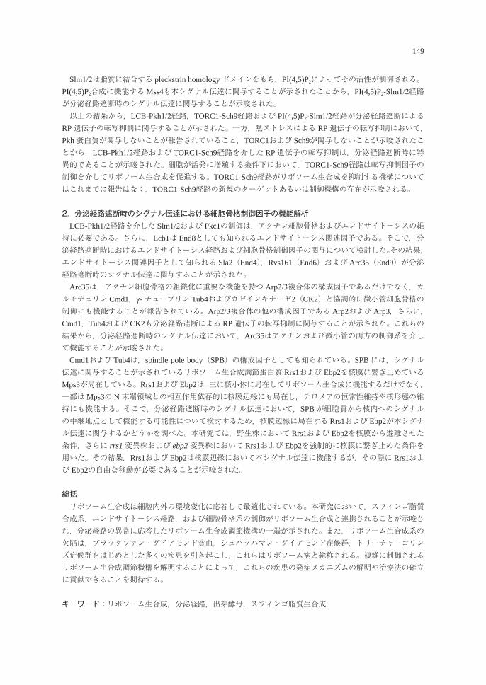
149
Slm1/2は脂質に結合する pleckstrin homologyドメインをもち,PI(4,5)P2によってその活性が制御される。PI(4,5)P2合成に機能するMss4も本シグナル伝達に関与することが示されたことから,PI(4,5)P2-Slm1/2経路が分泌経路遮断時のシグナル伝達に関与することが示唆された。 以上の結果から,LCB-Pkh1/2経路,TORC1-Sch9経路および PI(4,5)P2-Slm1/2経路が分泌経路遮断によるRP遺伝子の転写抑制に関与することが示された。一方,熱ストレスによる RP遺伝子の転写抑制において,Pkh蛋白質が関与しないことが報告されていること,TORC1および Sch9が関与しないことが示唆されたことから,LCB-Pkh1/2経路および TORC1-Sch9経路を介した RP遺伝子の転写抑制は,分泌経路遮断時に特異的であることが示唆された。細胞が活発に増殖する条件下において,TORC1-Sch9経路は転写抑制因子の制御を介してリボソーム生合成を促進する。TORC1-Sch9経路がリボソーム生合成を抑制する機構についてはこれまでに報告はなく,TORC1-Sch9経路の新規のターゲットあるいは制御機構の存在が示唆される。
2.分泌経路遮断時のシグナル伝達における細胞骨格制御因子の機能解析 LCB-Pkh1/2経路を介した Slm1/2および Pkc1の制御は,アクチン細胞骨格およびエンドサイトーシスの維持に必要である。さらに,Lcb1は End8としても知られるエンドサイトーシス関連因子である。そこで,分泌経路遮断時におけるエンドサイトーシス経路および細胞骨格制御因子の関与について検討した。その結果,エンドサイトーシス関連因子として知られる Sla2(End4),Rvs161(End6)および Arc35(End9)が分泌経路遮断時のシグナル伝達に関与することが示された。 Arc35は,アクチン細胞骨格の組織化に重要な機能を持つ Arp2/3複合体の構成因子であるだけでなく,カルモデュリン Cmd1,γ-チューブリン Tub4およびカゼインキナーゼ2(CK2)と協調的に微小管細胞骨格の制御にも機能することが報告されている。Arp2/3複合体の他の構成因子である Arp2および Arp3,さらに,Cmd1,Tub4および CK2も分泌経路遮断による RP遺伝子の転写抑制に関与することが示された。これらの結果から,分泌経路遮断時のシグナル伝達において,Arc35はアクチンおよび微小管の両方の制御系を介して機能することが示唆された。 Cmd1および Tub4は,spindle pole body(SPB)の構成因子としても知られている。SPBには,シグナル伝達に関与することが示されているリボソーム生合成調節蛋白質 Rrs1および Ebp2を核膜に繋ぎ止めているMps3が局在している。Rrs1および Ebp2は,主に核小体に局在してリボソーム生合成に機能するだけでなく,一部はMps3の N末端領域との相互作用依存的に核膜辺縁にも局在し,テロメアの恒常性維持や核形態の維持にも機能する。そこで,分泌経路遮断時のシグナル伝達において,SPBが細胞質から核内へのシグナルの中継地点として機能する可能性について検討するため,核膜辺縁に局在する Rrs1および Ebp2が本シグナル伝達に関与するかどうかを調べた。本研究では,野生株において Rrs1および Ebp2を核膜から遊離させた条件,さらに rrs1変異株および ebp2変異株において Rrs1および Ebp2を強制的に核膜に繋ぎ止めた条件を用いた。その結果,Rrs1および Ebp2は核膜辺縁において本シグナル伝達に機能するが,その際に Rrs1および Ebp2の自由な移動が必要であることが示唆された。
総括 リボソーム生合成は細胞内外の環境変化に応答して最適化されている。本研究において,スフィンゴ脂質合成系,エンドサイトーシス経路,および細胞骨格系の制御がリボソーム生合成と連携されることが示唆され,分泌経路の異常に応答したリボソーム生合成調節機構の一端が示された。また,リボソーム生合成系の欠陥は,ブラックファン・ダイアモンド貧血,シュバッハマン・ダイアモンド症候群,トリーチャーコリンズ症候群をはじめとした多くの疾患を引き起こし,これらはリボソーム病と総称される。複雑に制御されるリボソーム生合成調節機構を解明することによって,これらの疾患の発症メカニズムの解明や治療法の確立に貢献できることを期待する。
キーワード:リボソーム生合成,分泌経路,出芽酵母,スフィンゴ脂質生合成
Gene expression analysis of antimicrobial peptides in ayu stimulated with LPS
REHAB MARRY ABDELATY nSrelden
Graduate School of Biosphere Science, Hiroshima University, Higashi-Hiroshima 739-8528, Japan
LPS刺激後のアユにおける抗菌性ペプチドの mRNA発現
リハブ・マリー・アブラティ・ノスレディン広島大学大学院生物圏科学研究科,739-8528 東広島市
Fish supply from capture will be static over the next 30 years. A growing percent of world aquatic production derives from aquaculture, whose importance is set to increase dramatically as a result of overfishing of the world’s waters and an increasing demand for sea food. Fish is free-living organism exposed to stress problems such as diseases and deterioration of environmental conditions often results in economic losses. Most of this causative agents in fish ponds is bacterial in source. The most ancient and efficient line for defenses the fish against this microbes is the innate immune responses. They respond in short time scale and efficient manner with one of its alarm arms represented in my study by the antimicrobial peptides stimulated with LPS (lipopolysaccharides). The profile of the ayu antimicrobial peptides stimulated with LPS was investigated. Fisrt of all, analysis of cathelicidin gene expression from liver tissues which stimulated with LPS different doses in different life stages. Next, analysis of hepcidin-1 gene expression from liver tissues which stimulated with LPS different doses in different life stages. Finally, analysis of cathelicidin gene expression from various tissues at portal of pathogens entrance (mucosal surfaces). The second chapter included the first item, I have studied the relative transcriptional level of the cathelicidin gene in vivo stimulated with LPS of different doses LPS. LPS was injected intraperitoneal to ayu at different ages; young immature, mature and sexual mature adults. Liver tissues were collected three times per season. First time (group 1), at the mid of April, internal observation showed complete absence of the sexual organs. The second time (group 2), at the end of May. The third sample collection (group 3), at the mid of October, they were sexual mature adults, internal sexual organs observed full raipned in both sex just before the spawning. The relative expression level of the cathelicidin was measured using semi quantitative RT-PCR and Image-J software for normalization against β -actin gene expression level. The results showed a direct association between the cathelicidin mRNA expression and the LPS used for the induction. Young fish showed significant up regulation in a time, dose dependent manner while mature and sexual mature fish showed non-significant change. That I concluded that young fish may relay mainly on it is innate immune response than adults. The third chapter included the second item, we have studied the relative transcriptional level of the hepcidin-1 gene using cDNA samples synthesized from liver tissues for the first experiment. The relative expression level of the hepcidin was measured as same as semi quantitative RT-PCR. The results showed a direct association between the hepcidin mRNA expression and the LPS used for the induction. Young, mature and sexual mature adults showed up regulation mainly in age dependent manner. As the young and mature fish showed up regulation in a dose and time dependent manner. In the other hand, sexual matured fish showed significant down regulation. I could concluded from the first and second experiment that the hepcidin may be involved more in the ayu defenses against stressors
生物圏科学Biosphere Sci. 56:150-151 (2017)
151
such as pathogens. In the fourth chapter, the relative quantitation of cathelicidin expression level was analyzed. Tissues sample were collected from various organs (liver, gill, skin and intestine) at time point 0 hr, 6 hr and 24 hr post injection. The expression of cathelicidin mRNA were analyzed in the three various tissues (gill, skin and intestine) by RT-qPCR. And normalized to the β- actin gene expression level. ΔCt and ΔΔCt
value were determined using the auto setting of the system. The date shoed that the constitutive cathelicidin expression from the mucosal surfaces were higher than that of liver tissues. While the induced expression of cathelicidin with LPS showed only significant decrease in gill and skin at 24 hr after stimulation. The results clear out that the constitutive and LPS inducible expression of the cathelicidin is under developmental control and the recognition of LPS may be tissue specific although the mechanism of LPS recognition still unclear and also how this mechanisms affect in organs set distal to site of the immunostimulant administration. In conclusion the ayu antimicrobial peptides seems to play important role in ayu immune defenses against pathogens although it is age, time, dose and tissue-specific dependent production. Further investigation is required for analysis both hepcidin isoforms and production of monoclonal antibodies to clear the post translation regulation of ayu antimicrobial peptides.
Key words: ayu fish, innate immune response, antimicrobial peptides, LPS
Studies on novel roles of dietary fibers for intestinal homeostasis
Tran Van hung
Graduate School of Biosphere Science, Hiroshima University,Higashi-Hiroshima 739-8528, Japan
消化管の恒常性維持における食物繊維の新たな役割に関する研究
トラン ヴァン フン広島大学大学院生物圏科学研究科,739-8528 東広島市
1. General Introduction Human health is critically dependent on the maintenance of intestinal homeostasis. Inflammatory bowel diseases (IBDs) are a group of gastrointestinal disorders including Crohn's disease (CD) and ulcerative colitis (UC) and characterized by chronic inflammation. The number of individuals diagnosed with both UC and CD has steadily increased in the past several decades. IBD patients experience chronic and relapsing inflammation in intestines and suffer from diarrhea, abdominal pain and rectal bleeding. Although the etiology for IBD is unknown, it is believed that the impaired intestinal barrier resulting in hyperpermeability to luminal noxious molecules and robust chronic activation of immune system contribute to the development of intestinal inflammation. Accordingly, it is meaningful for us to develop the novel preventive and/or therapeutic approaches in the maintenance of intestinal homeostasis. Dietary fiber (DF) is the edible parts of plants or analogous carbohydrates that are resistant to digestion and absorption in the human small intestine with complete or partial fermentation in the large intestine. Accumulating evidence shows that supplemental feeding with DFs provides various beneficial effects for our health. Inflammatory status of intestines also seems to be regulated by feeding DFs and subsequent modification of intestinal microbiota. Intestinal fermentation of DFs produces different metabolites including short chain fatty acids (SCFAs). However, precise roles of DFs for regulation of intestinal inflammation are still unclear. The objective of the present study was to understand novel roles of DFs for the maintenance of intestinal homeostasis. I used the murine models of experimental colitis and CKD and the intestinal epithelial models under inflammatory conditions.
2. Fermentable and viscous DFs reduce intestinal barrier defects and inflammation in colitic mice In Chapter 2, I aimed to investigate the preventive effect of guar gum (GG) fiber on colonic inflammation and barrier defects in dextran sodium sulfate (DSS)-induced colitis mice. GG fiber, a soluble DF, is characterized by high fermentability and high viscosity. DSS administration caused severe colon damage and inflammation, as indicated by body weight loss, increased clinical scores, colon shortening, increased plasma lipopolysaccharide binding protein (LBP), elevated myeloperoxidase activity, and decreased TJ protein expression in the colon. Supplemental feeding with GG fiber partially or totally reversed these symptoms, suggesting that GG fiber ameliorates the DSS-induced colitis at least partially through protection of the TJ barrier.
3. Fermentable DFs reduce intestinal barrier defects and inflammation in colitic mice In Chapter 3, I aimed to examine the physicochemical properties of DFs contributed to protection of colitis against DSS. Along with GG, mice were fed with partially enzymatic hydrolyzed GG (PHGG), which
生物圏科学Biosphere Sci. 56:152-153 (2017)
153
shares high fermentability with GG, but presents low viscosity due to the low molecular mass. The results found that feeding PHGG and GG reversed the colitic symptoms, suggesting that high fermentability, rather than viscosity, is important for the DF-mediated protection of colons against DSS. In addition, feeding GG and PHGG suppressed the increased colonic cytokine expression by DSS and increased the production of SCFAs through the microbial fermentation. These observations suggest that SCFAs at least in part contribute to the anti-inflammatory effects of PHGG and GG through the suppression of inflammatory cytokines.
4. SCFAs suppress inflammatory reactions in Caco-2 cells and mouse colons In Chapter 4, I aimed to examine the roles of SCFAs on the regulation of inflammatory reactions in the colonic epithelium using human intestinal Caco-2 cells and mouse colons. Stimulation of Caco-2 cells with tumor necrosis factor (TNF)-α increased interleukin (IL)-8 and IL-6 expression through the inflammatory cellular signaling, whereas pre-treatment of cells with acetate, propionate and butyrate suppressed these inflammatory reactions by TNF-α. Pharmacological inhibition of monocarboxylate transporter (MCT)-1 attenuated the SCFAs-mediated suppression of the TNF-α-induced inflammatory responses. Administration of DSS to mice increased the CXC motif chemokine ligand 2 (an IL-8 homologue) and IL-6 expression in the colonic organ culture, whereas treatments with SCFAs mixtures composed of acetate, propionate and butyrate decreased them. These results indicate that the SCFAs acetate, propionate, and butyrate suppressed up-regulation of cellular signaling and expression of IL-8 and IL-6 in TNF-α-stimulated Caco-2 cells and in colons of colitic mice. Activity of MCT-1, located on the apical membranes, was essential for SCFA effects.
5. GG fiber suppresses inflammatory response in small intestinal epithelial cells In Chapter 5, I examined the anti-inflammatory effect of intact GG fiber in small intestinal epithelium. Because ingested DFs such as GG pass through the small intestines without any degradation, they directly interact with small intestinal epithelium. Although the SCFAs, microbial metabolites of DFs, show the anti-inflammatory regulation in colons, the intact DFs may also present the biological functions. I hypothesized that the intact GG has a role for the regulation of inflammatory responses in small intestinal epithelium based on the observation that GG suppressed the inflammatory cytokine expressions in the small intestines of DSS-administered mice. Pre-treatment of cells with GG suppressed the production of the IL-8 in intestinal Caco-2 cells stimulated by TNF-α. Interestingly, the pre-incubation of cells with anti-TLR-2 or anti-dectin-1 reduced the suppressive effects of GG fiber. In addition, the reporter cells confirmed the direct interaction and stimulation of TLR-2 and dectin-1 with GG fiber. Taken together, GG suppresses the inflammatory response in intestinal Caco-2 cells through the activation of TLR-2 and dectin-1.
6. General Discussion (written in Chapter 7) The present results demonstrated that fermentable DFs, such as GG and PHGG, had ameliorative effects on intestinal barrier defects and inflammation in a murine model of colitis. Fermentable DFs reach the colons and are metabolized to SCFAs by microbial activity without any degradation in small intestines. My results show that both the intact DF and the SCFAs have roles for the regulation of intestinal inflammation with the distinct molecular mechanisms. The present study suggested that supplemental feeding with fermentable DFs might be beneficial for prevention and/or management of different disorders associated with intestinal inflammation and barrier defect.
キーワード:食物繊維,短鎖脂肪酸,腸管バリア,タイトジャンクション,炎症
Seasonal dynamics influencing coastal primary production andphytoplankton communities along the southern Myanmar coast
maung Saw hToo Thaw
Graduate School of Biosphere Science, Hiroshima University, Higashi-Hiroshima 739-8528, Japan
季節性動的要因がミャンマー南部沿岸の海洋基礎生産と植物プランクトン群集に与える影響
マウ ソー トゥー ソー広島大学大学院生物圏科学研究科,739-8528 東広島市
Myanmar (The Republic of the Union of Myanmar) stands as the 10th position among the world fish producing countries in 2010 and the 3rd position in ASEAN, with the production of over 2.3 million tons in marine capture fisheries. The Myanmar coastline stretches about 3,000 km and is characterized by wide and numerous rivers, forming large and small deltas, estuaries, and extensive mangrove creeks. Myanmar has diverse tropical monsoon climate and its coastal areas are influenced by strong monsoon regimes; southwest monsoon (rainy season) and northeast monsoon (dry season). For the sustainable capture fisheries, the key mechanisms and the characteristic environments supporting such high fish catches should be investigated, however, very few survey have been conducted in the Myanmar coastal areas so far and this fact leads to the lack of appropriate conservation or regulation in the coastal fisheries. In this study, seasonal primary productivity off the foremost fisheries ground, Tanintharyi coast, was investigated for the first time in the Myanmar coasts. In the surveys, instead of using conventional bottle incubation methodologies using carbon isotopes, primary production estimation was performed using a principle of the pulse amplitude modulation (PAM) fluorometry. By applying this new PAM fluorometry, and with other conventional oceanographic surveys, the mechanisms of the coastal primary production off Myeik City was surveyed in three distinct seasons; at the onset of the dry season (December, 2014), the end of the dry season (March, 2015) and the rainy season (September, 2015), and at 13 sampling stations around Kadan Island covering characteristic coastal environments of the region (e.g. estuaries, mangrove channels or creeks, and offshore region facing the Andaman Sea). The lowest surface salinity value (7.11) in the rainy season indicated this area was largely affected by river inflow and also throughout the seasons, as a result, nutrient concentrations were high especially SiO2-Si and DIN-N. However, PO4-P concentration show different trend which might be supplied by different mechanisms rather than river inflow. The most notable feature of the ocean production was the well-defined seasonality, which has not previously been recognized as a typical model in a tropical ocean system, regardless to the seasonal nutrient variation. According to the estimations employing the PAM fluorometry, the primary productivity was highest in the dry season, 2.59 ± 1.56 g C m-2 d-1, while the productivities were low at the onset of the dry season and the rainy season (1.36 ± 0.77 and 0.17 ± 0.11 g C m-2 d-1, respectively). However, in account for the possible over estimation in the PAM fluorometry, the overall primary productivities may decrease; when the productivity values were recalculated by incorporating observed O2/ETR ratios (0.117 under PFD<500 μ mol photon m-2 sec-1, or 0.073 under PFD>500) in other literature, the annual primary productivity value was 129.6 g C m-2 yr-1, which is
生物圏科学Biosphere Sci. 56:154-155 (2017)
155
unexpectedly lower than the candidate values (300 - 500 g C m-2 yr-1) obtained from the world eutrophic estuarine and coastal ocean ecosystems. Based on the principal component analysis (PCA), the primary productivity was related with the extensive river inflows, especially in the rainy season where turbid river water inflow deterred the primary productivity. High primary productivity at the end of dry season related with the increase of euphotic layer depth, probably due to approach of oceanic water as well as decrease of the river water inflows and this was the main factor promoting primary productivity in this season. Interestingly, low primary productivities might be compensated by microbial food chain at the onset of the dry season. High DOC concentrations (average 7.48 ± 4.22 mg L-1) of this season probably led to abundant bacterial populations (average bacterial density = 1.1 × 10⁷ cells mL-1), and the estimated bacterial biomass of this season was 3 times higher than the estimated phytoplankton biomass. These DOC originated bacterial cells may be incorporated to a so-called microbial food chain and may support further production at the coast. Through this study, the seasonal trend of primary productivity and controlling environmental factors near the Myeik City were clarified for the first time. The main primary producers were the chain-forming diatoms associated with euhaline and eutrophic natures. Their production was primarily driven by the characteristic monsoon climates and showed well-defined seasonality; the end of the dry season was the most productive period, while the onset of the dry season and the rainy season remained in significantly lower productivities, 52.5% and 6.6% of that at the end of the dry season. These drops in the productivities for more than half of a year led unexpected low annual primary productivity (129.6 g C m-2 yr-1). The long rainy season that delivered heavy precipitation and extensive river runoff brought terrestrial nutrients to the coast, but as a trade-off, the coasts were largely affected by turbid waters, which decreased light penetration into the water column and thus reduced primary production. This highly turbid water was probably originated from soil erosion due to deforestations by industrial plantations along the Tanintharyi River Basin. Especially, deforestation of mangrove trees along the riverbanks and the estuaries seemed to be serious and was considered to lead to problems of soil erosion, because mangrove forests could reserve fluvial sediments as buffer areas for ocean- land interaction. Together with the findings that suggesting DOC derived from the mangrove sediment might enhance microbial food chain and supplement the primary production, conservation of mangrove forests is needed to sustain the coastal productivity.
Key words: Myanmar, primary production, diatoms, deforestation, mangrove
Studies on the uniformity of fruit characteristics and tree vigor, labor-saving,early achievement of mature orchard in the tree joint training system
for peach (Prunus persica (L.) Batsch)
Youji Hamana
Graduate School of Biosphere Science, Hiroshima University, Higashi-Hiroshima 739-8528, Japan
モモの樹体ジョイント仕立てによる早期成園化,作業の省力化および果実形質と樹勢の均質化効果に関する研究
浜名 洋司広島大学大学院生物圏科学研究科,739-8528 東広島市
モモの栽培において従来から一般的に普及している立木仕立ての開心自然形整枝などは,樹高が5 m以上となるため,せん定や収穫作業に大きな脚立が必要となり,危険性が高い上に作業効率が悪い。また,樹内での新梢の勢力や日照条件の違いなどにより果実形質にばらつきが生じるなどの問題も発生している。立木仕立てのこうした作業効率の悪さ,危険性,樹勢や果実形質を均一にするためのせん定等の管理技術の難しさは,新規参入,後継者の確保および規模拡大の障害となっている。こうした要因が重なり,近年,モモの生産者や栽培面積は減少している。従って,高齢の生産者でも身体的な負担が少なく,かつ栽培が長期に継続でき,また,新規に参入した生産者でも安定した生産が早期に実現できる栽培技術の確立が求められている。 一方,これまでに開発されてきた栽植密度の高い仕立ておよび大苗育成法などでは早期成園化,また,低樹高の立木仕立ておよび平棚仕立てや一文字形整枝などの棚仕立てにおいて作業の省力化が確認されている。しかし,これらの栽培技術においても,主枝の基部と先端部の新梢の勢力や果実形質の不均質化が課題となっている。 そこで,本論文では,モモの生産現場で重要な課題となっている早期成園化,管理作業の省力化,勢力および果実形質の均質化を同時に解決することを目的に,モモの苗木を列状に定植し,すべての樹の主枝を同一の列方向に水平に誘引し,主枝先端部を隣接樹の主枝基部に連続的に接ぎ木を行い連結する樹体ジョイント仕立てを新たに開発し,その有効性を評価した。
第1章の緒論では,モモ栽培の現状,モモの仕立て法,モモ生産農家で起こっている諸課題をまとめた上で,モモの早期成園化,省力化,均質化を達成する仕立て法の開発の重要性等,本研究の意義・目的を述べた。
第2章では,樹体ジョイント仕立てに適した苗木を育成することを目的に,苗木の切り返し,栽培用の培養土,副梢の摘心方法および枝の伸長促進効果のあるジベレリンペースト剤の塗布処理が,苗木の生育に及ぼす影響を調査した。その結果,地上1.0 mの高さでの苗木の切り返し,赤玉土での育苗,2葉摘心および頂芽基部へのジベレリンペースト剤2回塗布が良質な苗木を生産する上で重要であることを明らかにした。
第3章では,樹体ジョイント仕立てと省力的な仕立て法の一つである一文字形整枝との樹体生育,果実生産および作業の省力性の比較を行った。その結果,樹体ジョイント仕立ては,一文字形整枝より1年早く面積当たりの目標収量に達し,また,せん定時間が短く,かつ収穫作業時の心拍数が低かった。さらに,樹内での新梢長および果実重は,一文字形整枝に比べて差が見られなかった。以上の結果から,樹体ジョイント仕立ては,一文字形整枝よりも早期成園化,作業の省力化および果実形質と樹勢の均質化効果が高いことが明らかとなった。さらに,収量性および作業の省力化効果を検討した結果,樹体ジョイント仕立ての主枝高は1.55mが最適であると結論付けた。
生物圏科学Biosphere Sci. 56:156-157 (2017)
157
第4章では,樹体ジョイント仕立てと最も一般的な仕立て法である開心自然形との樹体生育,果実生産および作業の省力性の比較を行った。その結果,開心自然形では,着果位置により果実糖度にばらつきがあったが,樹体ジョイント仕立てではばらつきが見られなかった。また,樹体ジョイント仕立では冬季のせん定作業時間が開心自然形に比べて長くなったものの,夏季の繁忙期の収穫作業の時間が大幅に減少した。開心自然形では作業に脚立を必要とするが,樹体ジョイント仕立てでは脚立が不要となった。以上の結果から,樹体ジョイント仕立ては,開心自然形に比べて,果実形質の均質化および作業の省力化効果が大きいことが示唆された。
第5章の総合考察では,本研究で開発した樹体ジョイント仕立てが従来のモモの仕立て法に比べて実用性の高い栽培管理技術であることを述べた。さらに,樹体ジョイント仕立ては,早期成園化が可能となり,従来の仕立て法に比べて生産者の所得向上が図れることを示した。
以上のように,本論文で確立した樹体ジョイント仕立ては,従来の仕立て法と比較して,早期成園化,樹勢,果実形質の均質化および作業の省力化効果が高いことから,新規生産者や高齢の生産者の支援につながり,モモ生産の維持および拡大に寄与する新しい栽培技術として期待できる。本論文で確立した樹体ジョイント仕立てはモモ生産の維持および拡大に寄与する新しい栽培技術として期待できる.
キーワード:果実形質の均質化,作業の省力化,樹勢,樹体ジョイント仕立て,早期成園化,モモ
Study on effective fertilization by drip irrigation in open field cultivation
Shuichi WaTanabe
Graduate School of Biosphere Science, Hiroshima University, Higashi-Hiroshima 739-8528, Japan
点滴灌水を導入した露地栽培における効率的施肥に関する研究
渡邊 修一広島大学大学院生物圏科学研究科,739-8528 東広島市
緒言 施肥は作物生産に欠かせない活動である。しかし,必要以上に施肥を行うことは,溶脱窒素による水質汚染や農地への過剰リン酸蓄積などの環境負荷の増大につながるため,効率的な施肥が求められている。そこで,本研究では,作物の株元に水と肥料を同時に供給できる点滴灌水に着目し,これにより効率的施肥を達成することを目的とした。
第1章 点滴灌水がナスの根分布に及ぼす影響 作物の株元に水と肥料を同時に供給できる点滴灌水栽培では,根の生育状況を把握することは重要である。しかし,畝内の根分布のような根の広がり方を調査することは,従来の掘り取り法では時間と労力がかかるため困難である。そこで,「根の簡易調査法」を開発し,栽培現場での適用性を検証するために従来法である掘り取り法と比較した。その結果,簡易調査法と掘り取り法による調査結果は,高い正の相関関係にあり,両者の結果は概ね一致することが認められた。また,栽培現場での調査は複数の担当者が行うこともあるため,観察者による測定値のばらつきを調べた結果,おおむね一致する測定値が得られることが認められた。このことから,本手法が,現地試験地などの栽培現場で簡便に適用可能な手法と判断した。最後に,3箇所の露地ナス栽培圃場の調査事例から,地下水や降雨の影響のある露地栽培においても,点滴灌水による灌水管理を行った際のナスの根は,点滴灌水チューブ敷設位置の直下で多くなることが認められた。
第2章 カンキツ園における周年マルチ点滴灌水同時施肥による窒素施肥量の削減 環境省が定めた地下水および公共用水域の水質汚濁に係る人の健康保護に関する環境基準の健康項目において,硝酸性窒素および亜硝酸性窒素(以下,硝酸態窒素)に関する環境基準超過率は他の調査項目に比べ最も高い状況にある。広島県の島しょ部のカンキツ園が多く分布する流域において施肥由来の高濃度の硝酸態窒素を含む浅層地下水が存在することが報告されているなど,長期にわたる施肥が地下水に悪影響を及ぼした事例が報告されている。 そこで,広島県島しょ部に位置する温州ミカンの圃場において,周年マルチ点滴灌水同時施肥(マルドリ方式)の導入が窒素溶脱に及ぼす影響を調査した。現地施肥基準をもとに窒素24 kgを固形肥料で表面散布した対照区と,対照区の60%に減肥した窒素14.4 kgを灌水同時施肥したマルドリ区の比較では,栽培後の土壌無機態窒素量は対照区よりもマルドリ区の方が低い傾向を示した。試験前後の土壌無機態窒素量,施肥量,果実収穫量から推定した窒素溶脱量は対照区10.17 kg,マルドリ区2.75 kgでマルドリ区の方が少なかった。さらに,マルドリ区の推定窒素溶脱量と灌漑水から圃場に還元された窒素との差し引きはほぼ一致しており,正味の溶脱はほとんど生じなかったと考えられる。以上より,マルドリ方式の導入はカンキツ園での窒素溶脱を低減する効果があることが明らかとなった。
生物圏科学Biosphere Sci. 56:158-159 (2017)
159
第3章 露地ナス栽培における点滴灌水とリン酸施肥量がリン酸利用効率に及ぼす影響 全国の土壌モニタリング調査結果により,日本の畑地の可給態リン酸は蓄積傾向にあることが報告されている。リン酸施肥量を減らし,植物によるリン酸の利用効率を高めるためには,施肥管理の改善が不可欠である。特に,ナスは日本の主要な露地野菜の中でもリン酸施肥量が多く,リン酸施肥技術について改善の余地が大きい。 そこで,畝間灌水による水管理を実施する対照区とリン酸施肥量の異なる点滴灌水区をもうけて,2ヵ年の栽培試験を行った。リン酸施肥量は,収量目標値を設定し,1作あたりの養分要求量を考慮して3水準を設定した。なお,供試圃場のリン酸肥沃度は土壌診断基準値の上限に近いレベルであった。 その結果,生育・収量については,点滴灌水区のほうが,対照区より高かった。また,リン酸施用量を減じて点滴灌水を行った場合にも,対照区より高い生育・収量が得られた。また,植物体の乾物重量とリン含量から求めた作物体へのリン酸蓄積量は,点滴灌水区のほうが,対照区よりも多かった。また,リン酸施肥量を減じた場合でも単位果実収量を得るためのリン酸要求量はほとんど変わらず平均で1.12 gP2O5 kg-1であった。このため,リン酸施肥量に対する植物体地上部へのリン酸蓄積量の割合は,リン施肥量を減じた区では100%を上回り,土壌からの供給割合が多いことが示唆された。以上より,点滴灌水導入によりナスの生育・収量が優れ,リン酸の利用も効率的に利用される可能性が示された。
総合考察 以上の結果,点滴灌水は,カンキツ園での窒素溶脱を低減する効果があることや,ナスの生育・収量を向上させ,リン酸も効率的に利用できるなど,農業生産現場での利用価値が高いことが示された。ただし,優れた栽培技術であっても経済性が伴わなければ生産現場への導入は進まない。そこで,各章で得られた結果をもとに,点滴灌水技術の経済性について考察した。第1章では,点滴灌水を細根の発達していた位置が局所的に発達するため,灌水の影響の小さい畝肩付近では,肥料の吸い残しが発生する可能性が示唆された。したがって,点滴灌水チューブ敷設位置を考慮した部分施肥等と組み合わせることで,さらに施肥コストを削減しうる可能性が示唆された。第2章で検討したマルドリ方式では施肥効率が上がるため,大幅な減肥が可能であるが,液肥の単価が高いため,肥料コストとして増加の懸念が認められた。しかし,灌漑水に含まれる硝酸態窒素を肥料の一部として循環利用することも考慮すれば,経営費のさらなる削減に貢献できることが示唆された。また,第3章では,減肥をしつつも,収量を確保できる技術として,肥料価格の高騰などのリスクに対して点滴灌水は有効であると判断された。 今後,点滴灌水が露地栽培における効率的な施肥法として広く普及し,農耕地からの窒素溶脱低減や,限りあるリン資源の有効活用といった持続的農業生産を推進するとともに,生産者の収益向上を達成するために,本研究で得られた知見が活用されることが期待される。
キーワード:点滴灌水,露地栽培,カンキツ,ナス,リン酸,窒素溶脱
生物圏科学Biosphere Sci. 56:160-161 (2017)
Hardness measurements of marine bottom sediments to reveal specific distribution pattern of benthic organisms.
Naoto Kajihara
National Fisheries Research Institute of Fisheries and Environment of Inland Sea, Fisheries Research Agency, Hatsukaichi, Hiroshima 739-0452, Japan
堆積物底における底質硬度の測定と潜砂性底生生物の分布への応用
梶原 直人国立研究開発法人 水産研究・教育機構 瀬戸内海区水産研究所,739-0452 廿日市市丸石2-17-5
堆積物中に潜砂あるいは管棲する底生生物にとって,その性状は非常に重要な生息要因の一つとなる。これまで堆積物底の物理指標としては,底質の粒度組成や粒径分布と生息する底生生物の能力との関係で解析されてきたが,この方法だけでは底生生物の分布は明確に説明できなかった。いっぽう,底質の粒径等に硬度を加える事によって,底質の粒度組成や粒径分布による解析が適用できない領域において底生生物の帯状分布を解析できる可能性があり,分布域の成立をより合理的に説明することができるのではないかと考えられる。そこで,本研究では従来の堆積物底の調査手法に硬度を指標として加え,潜砂 ・営巣する底生生物の好適生息環境を評価することができるかを検討した。 第1章では,既存の文献類を整理して本研究を進めるに際し必要となる,海底堆積物に関する一般的な情報や認識及び由来 ・起源や分布と堆積プロセスを検証した。特に研究の対象となる小型底生生物の生物学的特性や移動分散スケ-ルに即した生息環境の特徴や,それらを評価するためのデータの取り扱い等も含め,水圏環境における様々な物理環境との比較から,底質硬度という物理的環境指標を抽出し,本研究における有用性を検討した。 第2章では,底質硬度を底生生物の物理的環境指標として活用するためには,海洋環境調査時に簡便に測定しうるかどうか,について検証する必要がある。そこで,硬度が広く活用されている土木分野で使用されている底質硬度の測定 ・調査法を検証し,これまでに底質硬度の調査研究に用いられた測定機器類が海洋環境に適用可能かどうかを検討した。底質硬度の評価は,従来は底質硬度計を使って計測されてきたが,本研究ではそれ以外に硬度と関連の深いサクション,飽和水面,飽和度,粘着力などの各物理指標について着目し,それらの原理や測定する意味合いについて解説した。それぞれの測定機器については,その測定原理毎に大まかな分類を行い,測定範囲や改造の有無等によって細分化して記述した。また,各々の測定機器についてそれらの特徴や底質毎の海洋環境における底生生物の環境評価への有効性を考察した。さらに,各々の測定機器を海洋環境で使用する際の注意点や測定の最適化のため留意する諸問題も記述し,底生生物の生息環境としての底質硬度の測定方法を検証した。 第3章では,実海域での新たな底質硬度の評価事例として,まず,砂浜海岸における複数の底質硬度の各々の底質硬度指標の特性を検討し,このような環境で有効な飽和水位やサクションとの関係を明らかにした。また,調査現場での簡易な測定が困難なサクション測定の代替法の検討並びに試行をおこなった。その結果,少なくとも飽和状態の推定については,サクションを測定しなくても代替法による評価が可能と考えられた。これについて,実験室内で再評価し,本手法は汎用性も精度も高いことを明らかにした。いっぽうで,砂浜海岸汀線域においては,飽和状態でサクションが殆ど変動しない潮下帯最上部でも小型底生生物の帯状分布が認められることに着目し,指標生物として用いたナミノリソコエビの分布下限の成立要因を土砂環境,特に漂砂動態に着目した解析による解明を試みた。その結果,砂漣やステップが特徴の漂流漂砂域においては,三次元的な漂砂動態により,ナミノリソコエビ特有の半潜砂状態での摂餌行動が阻害されることや,魚類等捕食者によるトップダウン効果により分布の下限が決定されているということを明らかにした。また,
161
漂流漂砂域とそれ以浅の掃流漂砂域は,岸沖方向の流速や底層の漂砂量の大幅な変化から判別出来ることも明らかとなった。 第4章では,第3章の調査が潮位の変動が殆ど無い日本海側の砂浜海岸だけであったので,そこで得られた結果を,我が国の太平洋側のように潮位変動を伴う砂浜海岸において再検証することによって,その普遍性を検討した。調査では,潮位毎に土砂環境と潜砂性小型甲殻類の分布を調べた。その結果,潮位毎の土砂環境の変動にナミノリソコエビの分布域が上限・下限とも連動していた。また,海底勾配が変動することによって帯状分布域の幅が増減することや漂流漂砂域の始点が固定されるなど,新たな知見が得られた。しかし,第3章で得られたナミノリソコエビ等の分布の上・下限は変化しなかった。これらの結果から,底質の硬度や土砂環境に起因する潜砂性小型甲殻類の帯状分布域の成立は,極めて普遍性が高いことを証明した。 第5章では,瀬戸内海の島嶼部を中心に分布する自然干潟は砂浜より礫浜であることが多い。そのため,このような海岸では前章までに得られた結果とは異なる可能性がある。そこで,砂よりも粒径の大きい礫に関して,実験的手法を用いて硬度と関連する物理的性質を把握することを試みた。その結果,礫は砂と物理的性質は全く異なり,サクションや底質硬度の重要性は低く,飽和・不飽和の区別と粒径によって殆どの物理的性質が決定されていた。いっぽう,土木工学で決められている砂相当の粒径においても,粒径毎の物理的性質が少しずつ異なることを明らかにし,測定した全ての物理的指標が砂の性質を示すのは粒径が0.5㎜以下であることを明らかにした。 第6章では,前章までの調査や研究結果をとりまとめ,底質硬度を用いた現状の問題点や今後の展開について,測定原理・測定方法・測定器具等多岐にわたる観点から検討と考察を行った。本研究で重要な位置を占めるサクションについては,安価で,簡便な代替測定法を提案するとともにその有効性について記述した。以上より,底質の硬度は潜砂性小型甲殻類にとって帯状分布域を成立させる極めて重要な環境要因の一つであることを明らかにした。さらに,これらの技術を社会や海浜の生態系保全に活用する方法についても検証し,本研究結果が広く活用できる可能性についても提唱した。
Studies on the control of viral nervous necrosis in seed production process of marine fish
Toyohiro niShioKa
Graduate School of Biosphere Science, Hiroshima University, Higashi-Hiroshima 739-8528, Japan
海産魚の種苗生産過程に発生するウイルス性神経壊死症の防除に関する研究
西岡 豊弘広島大学大学院生物圏科学研究科,東広島市 739-8528
本研究では,種苗生産過程で発生する疾病のうち,最も被害が大きいベータノダウイルスを原因とするウイルス性神経壊死症(viral nervous necrosis: VNN)を取りあげ,栽培漁業や養殖業の新規対象魚種として注目されているキジハタ,アカアマダイ,クロマグロについて,本疾病の防除法を検討した。
第1章 緒論 研究の背景となる栽培漁業と養殖業について詳述し,それらの種苗生産において多発する疾病が種苗の安定供給に支障を来していることを示した。特に,魚類では VNNの発生が大きな問題であることを指摘し,本病についての既報の知見を整理した。
第2章 種苗生産対象種および種苗生産過程における疾病の発生状況 1984年から2009年までの種苗生産対象種について調査した。この間,2000年以降に総種苗生産数は減少したが,魚類16種,甲殻類8種,介類18種で100万個体以上の種苗が生産され対象種の種類数に大きな変化はなかった。2000~2009年度の疾病や大量死亡の発生状況について取りまとめた結果,ウイルス病が26%,細菌病が37%,真菌病が8%,寄生虫病が12%,不明が17%であった。各原因別の発生状況を1989~1999年度の発生状況と比べると,ウイルス病と真菌病がやや減少した一方で,新たな魚種での発生が報告された細菌病と寄生虫病が増加した。VNNでは1989~2009年度までに,5目21魚種で発生があり,キジハタ,クエ,シマアジ,ヒラメでは10年以上にわたって発生が継いたことから,VNNが種苗の安定生産を妨げる最も重要な疾病であると位置付けた。
第3章 キジハタにおけるベータノダウイルスの感染状況 移動範囲が限定的であることから栽培漁業の対象種として重要なキジハタを対象とし VNN防除対策を検討した。VNNに感染耐過したキジハタ稚魚からは3年後も PCRによりベータノダウイルス(RGNNV)遺伝子が検出されることから,これらの耐過魚は不顕性感染の状態にあることが明らかとなった。また ,日本近海4海域で採取した132個体の天然キジハタを検査した結果,RT-PCRで4.5%,nested PCRで33.3%の魚が RGNNV陽性となり,親魚候補となる天然キジハタのウイルス感染を確認した。分離した天然キジハタ由来 RGNNVはキジハタ病魚由来ウイルスと同等の病原性を示した。これらの結果から,キジハタの VNNは不顕性感染した親魚からの垂直伝播によって起こると考えられた。瀬戸内海の4か所で漁獲された天然キジハタの nested PCRによるウイルス検出率は5.6%~72.9%と漁獲海域により差が認められた。以上の結果から ,防除対策として,感染率が低い海域から親魚を搬入し,養成期間が短い親魚群から受精卵を得て卵消毒には電解海水を用いることを提案した。
生物圏科学Biosphere Sci. 56:162-163 (2017)
163
第4章 アカアマダイにおけるウイルス性神経壊死症の防除対策 沿岸漁業の重要な水産資源で商品価値が高いアカアマダイを対象とした。2004年冬季に種苗生産した稚魚に異常行動を示して死亡する個体が認められた。病魚の脳に空胞が認められ,PCR法で RGNNV遺伝子型のウイルス,また抗ベータノダウイルス血清を用いた蛍光抗体法でベータノダウイルス抗原が検出されたことから,VNNによる死亡であると判断した。疫学調査において,親魚として用いる天然アカアマダイがPCR陽性となり,一部の個体からウイルスが分離されたことから,VNNの発生は垂直伝播によると推定した。実験感染により ,天然アカアマダイ由来ウイルスの病原性が確認された。PCR陰性の親魚を用い,受精卵を電解海水で消毒し,かつ電解処理海水を用いて飼育することが防除対策として有効であることを明らかにした。
第5章 クロマグロにおけるウイルス性神経壊死症の防除対策 養殖種として世界的に注目されているクロマグロを対象とした。本種の種苗生産過程における仔稚魚期の大量死亡事例について検討した結果,死亡魚に VNNに特徴的な脳や網膜の空胞変性が認められ,蛍光抗体法により病変部に特異蛍光が確認された。E-11細胞により ,死亡魚1個体当たり105.3~109.6 TCID50/gのウイルスが分離され,RGNNV遺伝子型のウイルスが PCR法により検出されたことから,VNNが大量死亡の一要因であると推定した。天然幼魚や養成親魚,受精卵およびふ化仔魚からベータノダウイルスが検出されたことから,本病は親魚からの垂直伝播によると結論づけた。電解海水で卵消毒し ,電解処理海水で卵管理や仔魚飼育を行うと,VNN発生数は低下して稚魚の生産数が増加した。
第6章 ウイルス感染源としての餌料魚の重要性 既報の VNN防除対策を実施したシマアジ種苗生産において,ベータノダウイルス(遺伝子型 SJNNV)による VNNが発生した。養成親魚の餌料用に冷凍保管していた天然マアジからウイルスが検出され ,シマアジ病仔魚から検出されたものと RNA2の塩基配列が一致したことから,天然マアジがシマアジ仔魚のウイルス感染源であると推定した。また,天然マアジから分離したウイルスをマアジ仔魚およびシマアジ仔魚に浸漬感染させた結果,シマアジ病魚由来 SJNNVと同等の強い病原性を示す株が確認された。日本とヨーロッパの病魚および天然マアジ由来の SJNNVについてそれらの RNA2の塩基配列を比較した結果,強い病原性を示す株は,日本でのシマアジ病魚由来株の配列に近いクラスターに分類された。
第7章 総合考察 第1章から第6章において得られた主要な知見に基づき ,種苗期のVNN防除法について総合的に考察した。
キーワード: ウイルス性神経壊死症,VNN,ベータノダウイルス,海産魚の種苗生産,キジハタ,アカアマダイ,クロマグロ,シマアジ
Study on the influence of metabolic factors on bioconcentration of chemicalsin aquatic organisms
Mitsugu miyamoTo
Environmental Health and Science Laboratory, Sumitomo Chemical Co., Ld.,Takarazuka, 665-8555, Japan
水生生物における化学物質の濃縮性への代謝要因の影響に関する研究
宮本 貢住友化学株式会社生物環境科学研究所,665-8555 宝塚市
第1章の緒論では,環境汚染・生物濃縮性懸念化学物質規制の国際動向,生物濃縮性とその主な支配・影響要因と予測手法,生物多様性と関連した生物濃縮性の多様性などを通じて,各種生物濃縮性評価の簡便法や補足技法の蓄積の必要性を説明し,本論文の目的を明らかにした。 すなわち,過去の生物濃縮性に関連した大規模環境汚染を端緒に環境保護,持続的・安定な地球環境との共生に向けて世界各地域において化学物質の危険性を優先して是非判断がなされる時代となってきており,事前の様々な安全性関連項目の調査が求められ,その中で生物濃縮性は重要な評価項目の1つである。特にEUでは微量不純物や環境代謝物までも評価が要求されている。他方,生物濃縮性の支配要因としては化学物質の脂溶性や分子サイズなどあり,様々な予測手法が作成されているものの,その他の生物側の大きな影響要因である化学物質代謝分解能の予測手法への組み込みは不十分である。有機化学物質の多様性などから適用性の幅広い手法の実現には色々な困難・制約があると予想される。更に標準試験生物としては魚が用いられているものの,実環境では種数や多様性から節足動物の評価も重要と考えられる一方で生物多様性に関連し生物濃縮性の予測性や標準試験法作成に多くの課題があり,現状,節足動物での標準試験法確立に向けての明確な取り組みは殆ど見られない。国際的な第一標準である魚の生物濃縮性試験が複雑,高額である点や近年の動物愛護,更に評価対象化学物質が多いことなど踏まえると,代謝要因を踏まえた微量成分・分解物等の類剤の評価や節足動物の多様性も踏まえた簡便評価などの補足技法の充実が望まれる。 本論文ではそれを踏まえて生物濃縮性に影響を及ぼし得る主な課題の1つと考えられている代謝要因に関連した簡便手法の構築について検討した。
第2章,「生物・代謝多様性を踏まえ,体内致死濃度を活用した節足動物の簡便濃縮性評価手法の検討(殺虫剤ピリダリルのセスジユスリカ,ヨコエビの一種 Hyalella. aztecaの体内致死濃度を用いた BCF推算の検討)」では,先ず,殺虫剤ピリダリルのセスジユスリカ幼虫およびヨコエビの一種 H. azteca成体に対する急性毒性および代謝ならびに体内致死濃度 LBRについて14C標識化合物を用いて調べた。その結果,平均実測水中濃度に基づく急性 LC50値はそれぞれ1.1 mg/L(48時間),0.015 mg/L(96時間)であり,いずれの生物種においても主な代謝様式はジクロロアリルオキシ基のエーテル結合の脱アルキル化および抱合化であった。より高い代謝能がセスジユスリカでは認められ,ピリダリルの生物中濃度に基づく LBRは両生物種で同レベルであった事から毒性ポテンシャルに顕著な違いはなく見かけの毒性は主に代謝能に影響されているものと考えられた。このラボでの急性毒性・代謝試験において得られた毒性値 LC50と体内致死濃度 LBRから生物濃縮係数 BCF=LBR/ LC50の関係式に基づいて推算されたセスジユスリカおよび H. aztecaの BCFはそれぞれ72,1200であり17倍の違いが認められた。他方,既存のピリダリル野外池試験における水中および各種水生生物中のピリダリル濃度推移データから推算された BCFのうち,水生昆虫のヤゴ,甲殻類のミジンコの値はそれぞれ180±120,3100±2300であり,ラボでの毒性試験データからの昆虫類と甲殻類での推算値は野外の類似分類生物の値と良く類似した。このことから,生物間の比較などにおいて毒性値と体
生物圏科学Biosphere Sci. 56:164-165 (2017)
165
内致死濃度からの BCF推算の活用性が示唆された。また,ラボ標準種,野外生物の何れにおいても BCFの低い生物群では代謝物量の割合が高く,各種無脊椎動物においても代謝が BCFに及ぼす影響の大きいことが示唆された。
第3章,「一次代謝物に着目した速度論的解析や in vitro試験活用の類縁体 BCF簡易評価手法の検討(in vivoおよび in vitroの魚濃縮性/代謝試験によるピレスロイド系殺虫剤テトラメトリンのトランスおよびシス異性体の生物濃縮性の検討)」では,殺虫剤テトラメトリンの構成成分であるシス,トランス異性体のうち,主成分であるトランス異性体についてブルーギル稚魚における濃縮および代謝,排泄挙動を2種類の14C標識化合物を用いて,連続流水試験系(設定濃度1 ppb,28日曝露 /14日排泄)において評価した。トランステトラメトリンは魚体内でまずエステル開裂を受け,その後,アルコール側,酸側部位とも引き続いて各種代謝を受けた(N脱アルキル化,二重結合の還元,イミノ環開裂,オメガトランスメチル基の酸化,エポキシを経由した水酸化,酸側代謝物のグルクロン酸やタウリン抱合化)。生物濃縮係数 BCFおよび魚体からの消失半減期はそれぞれ180-310,0.54-0.72日であった。加えて,代謝経路を踏まえて濃縮平衡時および排泄期間の一次代謝物の動態に着目し,その生成,消失挙動の速度論的解析から,トランステトラメトリンの代謝,排泄速度をそれぞれ0.41,0.55 /dayと算出し代謝の寄与の大きさを定量的に明らかにした。更に,エステラーゼ活性に着目し,魚全身ホモジネートを用いて in vitro分解速度のシス,トランス異性体の比較を行い,それぞれ1.8,3.7 /dayとトランス異性体のより速やかな分解が確認された。トランス異性体の in vivoの BCF値ならびにシスおよびトランス異性体の in vitro分解速度を用いてシス異性体の BCF値の推算を行い230-400と算出された。
第4章,「混合物微量分析や代謝阻害剤を活用した濃縮性への代謝要因の明確化簡便法の検討(シス,トランス幾何異性体混合物であるピレスロイド系殺虫剤 d-フェノトリンの各異性体の魚濃縮性に及ぼす代謝影響の分別定量分析及び代謝阻害剤を用いた検討)」では,コイ当歳魚をトランス,シス異性体の混合物(4:1)である殺虫剤 d-フェノトリンに連続流水下で暴露し各異性体の生物濃縮係数 BCFを検討したところ,トランス体よりもシス体の BCFは1.1-2.2倍有意に高値であった。異性体間の物理化学的性質の類似性からこのBCF値の違いは各異性体の魚における代謝能の違いによると考えられた。更に酸化酵素阻害剤であるピペロニルブトキサイド共存下で各異性体の BCFを評価したところ,トランス体では阻害剤の有無による顕著な変化は無かったものの,シス体では BCF値の顕著な上昇が認められ,これは代謝分解要因の排除によると考えられシス異性体の酸化代謝が大きいことがうかがえた。また,この BCF値の違いから代謝速度と排泄速度の比は最大2.3-11倍と見積もられ,代謝分解要因が d-フェノトリン幾何異性体の生物濃縮性に大きく寄与していることが示された。加えて,魚体重に基づく取込み速度推算式と代謝阻害時のシス体 BCFから算出された d-フェノトリンの取込み,排泄速度はそれぞれ200 L/kg/day,0.064 /day推算され,更に,BCF値からシス体,トランス体の代謝速度はそれぞれ0.33,0.49 /dayと推算された。
第5章の「総合考察」では,本研究において検討・構築した代謝要因を考慮した生物濃縮性に関連する以下の評価・解析手法について,生物濃縮性の予測や評価の更なる改良,精緻化や既存手法校正,より正確なパラメータ取得への活用,或いは実際の化合物の評価,特に類縁化合物群の効率的・簡便評価への活用性について考察した。 1.毒性値および体内致死濃度からの BCF推算と生物間の簡便比較 2.代謝様式を踏まえた1次代謝物動態からの速度論的解析による代謝速度評価 3.in vivo試験とブリッジング in vitro代謝試験を組み合せた類縁体の BCF推測 4.代謝阻害剤を用いた代謝寄与度検証や速度パラメータ推算
キーワード:生物濃縮性,BCF,水生生物,体内致死濃度,速度論的解析,代謝阻害
Elucidation of nectrisine biosynthesis pathway and its application for production
Ryuki miyauchi
Daiich Sankyo Co., Limited,Shinagawa-ku 140-8710, Japan
ネクトリシンの生合成機構の解明と製法構築への応用に関する研究
宮内 隆記第一三共株式会社,140-8710 品川区
第1章ではイミノ糖の発見と医薬への応用とその生合成について紹介するとともに,本研究を開始した経緯に付いて述べた。 微生物や植物などが産生する糖のアナログ,イミノ糖は,一般にグリコシダーゼ阻害活性を有するため,糖尿病やウイルス感染,ライソゾーム病などの様々な治療薬への応用が報告されている。もっとも基本的なイミノ糖は,ピラノース環やフラノース環中の酸素が窒素に置換された構造を有する。イミノ糖の生合成に関しては実質的には6員環のデオキシノジリマイシンについて報告があるのみで,知見が乏しい。デオキシノジリマイシンについては,グルコースを中間体とする推定生合成経路が報告されている。 ネクトリシンは,糸状菌が産生する5員環のイミノ糖で,α-,β-グリコシダーゼ,α-,β-マンノシダーゼ阻害活性を示す。ネクトリシンの化学構造は,糖尿病治療薬として三共株式会社(現第一三共株式会社)にて創製された化合物である CS-1036の部分構造(Cユニット)と類似している。CS-1036は,イミノ糖である Cユニットの化学合成コストが高いことが課題となっていた。そこで,醗酵生産で得たネクトリシンをCユニットの原料とすることで CS-1036の製造原価低減することを企図した。菌株のスクリーニングによって糸状菌である Thelonectria discophora SANK 18292株をネクトリシン高生産株として見出したが,工業的に本菌株を利用するにはその生産性は非常に低かった。製造原価低減のためには,大量生産に適した遺伝子組み換え微生物によるネクトリシンの生産が魅力的と考えられたが,ネクトリシンの生合成遺伝子や生合成経路に関する情報は報告されていなかった。そこで本研究ではネクトリシン生合成機構を解明してその製法構築に応用することを目的とした。
第2章では T. discophoraにおけるネクトリシン生合成経路を解析した。 はじめに,デオキシノジリマイシンに関する先行研究を踏まえて,ネクトリシンの生合成中間体としてペントース類を推測した。そして,ペントース6種と糖アルコール1種を添加して T. discophoraを培養したところ,本菌はこれらの中で D-キシロースを良く資化した。また,D-リボースを添加した場合にネクトリシンの生産量が増加した。そこで,D-キシロースと D-リボースの各種13C同位体を添加し,産生されたネクトリシンへの同位体の取り込みを NMRと質量分析で確認した結果,これらの基質はネクトリシンの中間体であり,その取り込みパターンはペントースリン酸回路を経る経路で矛盾なく説明できた。さらに,T. discophora菌体抽出液中に4-アミノ -4-デオキシアラビニトールが含まれていることを精製物の X線結晶構造解析及び各種分光スペクトル解析,元素分析により明らかにした。この化合物に菌体抽出液を添加するとネクトリシンに変換されたことから本化合物がネクトリシンの前駆体であることが示された。以上より,ネクトリシンの T. discophoraにおける推定生合成経路を提案した。
第3章では,4-アミノ -4-デオキシアラビニトールをネクトリシンへ変換する酵素(NecC)について解析した。 まず,菌体抽出液より硫安沈殿と陰イオン交換クロマトグラフィーにより NecCを精製した。そしてNecCをトリプシン消化し LC-MS/MS分析した結果,いくつかのアミノ酸配列断片を同定できた。それら
生物圏科学Biosphere Sci. 56:166-167 (2017)
167
はデータベース検索によりグルコース -メタノール -コリンオキシダーゼと相同性を示した。また,NecCはフラビン骨格を有する補因子を含有し,オリゴマーを形成していた。NecCの活性については,至適 pHが7.0,至適温度が30°Cであり,EDTAにより阻害され,MnCl2により若干上昇した。さらに,4-アミノ -4-デオキシアラビニトールからネクトリシンへの変換反応は菌体から抽出後に in vitroで主に起こっていることを示した。そして,ネクトリシンの醗酵生産にあたっては,基質だけでなく NecCも活性を維持した状態で抽出されなければならないことを提言した。
第4章では,ネクトリシン生合成遺伝子を取得し,その機能を解析するとともに,遺伝子組換え大腸菌によるネクトリシン生産を検討した。 NecC部分アミノ酸配列から設計した縮重プライマーで necC遺伝子断片を取得,クローニングし,さらに T. discophoraのゲノムライブラリーをスクリーニングすることで necC遺伝子全長のクローニングに成功した。 残りのネクトリシン生合成遺伝子もゲノム上で necC遺伝子座周辺にあると予想し,解析した結果,アミノトランスフェラーゼとコリンキナーゼに相同性を示す配列(necAと necB)が見つかった。上記で推定された生合成経路がアミノ化と脱リン酸化反応を含むことから,これら2つの遺伝子がネクトリシンの生合成遺伝子の候補と考え,その機能を検証した。necA遺伝子を破壊すると4-アミノ -4-デオキシアラビニトールとネクトリシンの生産が観察されなくなり,necA破壊株に necAを相補するとその生産が回復したことから,necAはネクトリシン生合成遺伝子であることが示された。NecB遺伝子を破壊すると,ネクトリシンの生産は親株より顕著に減少したものの認められた。また,necAと necCを共に発現する組み換え大腸菌は,ネクトリシンを生産したことから,necBはネクトリシンの生合成に関係しているが必須ではないと推測され,NecBを代替し得る酵素の存在が考えられた。なお,necC遺伝子についても破壊と相補によりその機能を検証したところ,necCが4-アミノ -4-デオキシアラビニトールをネクトリシンへ変換する反応を担っていることが確認できた。 以上より,NecA,NecBにより4-アミノ -4-デオキシアラビニトールが生じ,NecCによりネクトリシンが生成すると推定された。この推定生合成経路は,アミノ化,脱リン酸化,酸化の各反応を含んでいる点がデオキシノジリマイシンの推定生合成経路と類似していた。しかし,これら3つのネクトリシン生合成酵素は,対応するデオキシノジリマイシンの推定生合成酵素との相同性がいずれも低かったため,新規性が高い酵素であることが示唆された。 そして,necA/necB/necCまたは necA/necCの共発現ベクターを導入した組み換え大腸菌は,ネクトリシンを生産できることを示した。
第5章では,総括と今後の展望について述べた。 今回得られたネクトリシンの生合成遺伝子の情報を活用することで異種発現によるネクトリシンの直接生産が可能になった。今後,遺伝子工学や代謝工学の手法を用いてその生産性を向上させることが期待される。また,本研究で得られた遺伝子情報を利用して天然物またはデータベース上の遺伝子を探索することで,他のイミノ糖の生合成機構の解明にも役立つのではないかと期待している。また,元株を利用することもできる。検討の結果,元株の菌株改良と培地改良によりネクトリシン生産量を当初の65倍に増加させることができた。そして,上記で得られた基本的知見を生かして6 kLスケールでの大量製造プロセスを確立し,CS-1036の製造原価低減を達成した。
Genetic engineering of Escherichia coli for protein productionfor functional and NMR structural study
Yojiro iShida
Center for Advanced Biotechnology and Medicine, Rutgers University, NJ 08854, USA
蛋白質の機能解析 , NMR構造解析への遺伝子改変を用いた大腸菌発現系の構築
石田 洋二郎ラトガース大学,08854, New Jersey, USA
Antimicrobial peptide Bac7(1-35) production using the PST-SPP system in E. coli MazF, one of the toxins from E. coli, functions as an ACA-specific endoribonuclease in cells. Taking advantage of MazF endoribonuclease activity in E. coli, a new expression system, the Single-Protein Production (SPP) system was previously developed. In the SPP system, all E. coli cellular mRNAs are eliminated when MazF is induced. However, the mRNA of a target protein is engineered without ACA sequences while conserving the amino acid sequence so that it remains singularly intact when MazF is induced. Therefore E. coli is converted into a bioreactor producing only target proteins, making it especially effective for toxic proteins. One of the difficult to express proteins in E. coli is an antimicrobial peptide (AMP) because of its toxicity to bacteria. Since the first discovery of the defensing peptide, a number of AMPs have been isolated. AMPs do not create drug-resistant bacteria because there are multiple intra-cellular targets for AMPs. In addition, it is relatively easy to modify the peptide sequences and possibly enhance the selectivity of its activity to bacterial cells with such changes. Bac7(1-35) is a bovine AMP of 35 amino acid residues and is a Pro- and Arg rich peptide. Bac7(1-35) inhibits protein synthesis by binding to 70S ribosome, thus it is difficult to express in E. coli in its functional form. Here I developed a novel expression system in E. coli, combining Protein S, a spore coat protein from Myxococcus xanthus with the SPP system for antimicrobial peptide production. This system produces a fusion protein which functions the same as the protein without the Protein S tag (PST). After overexpression of Bac7(1-35) using the PST-SPP system, PST-Bac7(1-35) is not only soluble, but also it functions as an antimicrobial peptide without cleaving the protein S tag from the fusion protein. This technology enables us to obtain a large amount of antimicrobial peptide in E. coli in a cost effective way
Replacement of Arg residues in MazFbs with canavanine alters its specificity I also explored expanding the capabilities of the SPP system to incorporate amino acid analogues, in particular the toxic arginine analogue, canavanine, in order to observe how protein function is altered with such substitutions. Canavanine is originally extracted from jack beans, and well known as a toxic amino acid analogue to cells. The mechanism of the toxicity is possibly due to the pKa of guanidino group in canavanine being 7 while that of arginine is 11. Here, the SPP system is combined with an arginine auxotrophic strain, allowing canavanine to be incorporated into protein efficiently without showing its toxicity to the cells. This is because when MazF
生物圏科学Biosphere Sci. 56:168-169 (2017)
169
is expressed and cell growth is arrested, the cells use canavanine for only target protein production. As an example, MazFbs, a MazF homologue from Bacillus subtilis and a UACAU specific endoribonuclease, is used as a model protein. Incorporating canavanine into MazFbs caused MazFbs(can) to become more helical in structure but less stable in comparison to MazFbs because the conformation of the protein was changed. This is considered to be due to the change in the pI of MazFbs(can), altering the recognition sequence for cleavage to UACAUA rather than the original MazFbs UACAU recognition site. This is the first example of alteration of the RNA restriction enzyme recognition site by incorporating a toxic amino acid analogue.
Construction of a residue- and stereo-specific methyl labeling method by engineering E. coli Thirdly, since the auxotrophic strain is highly useful for specific amino acid labeling, I established a cost effective labeling system for NMR structural studies. Large molecular weight proteins have some dynamics, and their function and dynamics have been characterized by NMR spectrometry. However, deuteration of proteins larger than 20-kD proteins is necessary and methyl specific protonation of Ile, Leu and Val residues is commonly used to study its dynamics. For 80 kDa or larger proteins, Stereo Array Isotope Labeling (SAIL) amino acids, in which the amino acid is stereo-specifically labeled, are used as it can dramatically reduce proton density compared to proteins labeled with common labeling precursors such as α -ketoisovalerate and α -ketoisobutyrate. However, since SAIL amino acids are extremely expensive, they have not been widely used in the NMR community. Here, I engineered the E. coli strain for residue-, stereo- and methyl-specific labeling systems, to use minimal SAIL amino acids. As a result, I was able to reduce the usage of SAIL amino acids up to 10% compared to the standard method while maintaining protein production efficiency. Lastly, I developed an alternative expression/labeling system for residuestereo- methyl-specific labeled sample preparation for NMR using the common precursor, 2-acetolactate. In this system, the stereo specifically isotope-labeled 2-acetolactate is combined with genetically engineered E. coli, which allows proteins to be labeled in residue specific manners. Using a standard strain, Val specific labeling is possible but Leu-specific labeling is difficult when using 2-acetolactate. To circumvent this, I engineered a biosynthetic pathway in E. coli to allow the cells to use 2-acetolactate for either Leu or Val synthesis so that either Leu or Val in a target protein can be labeled in a residue- and stereo- specific manner.
key words: Single-Protein Production (SPP) system, E. coli, antimicrobial peptides, amino acid analogues, auxotrophic strains, methyl labeling, solution NMR
An Empirical Research about Japanese Protected Horticulture under Labor Scarcity Conditions in Agricultural Sector
Shinnosuke iwaSaKi
Japan-Cooperative General Research InstituteTokyo, Shinjuku 162-0826, Japan
労働力稀少条件下の施設園芸農業に関する実証的研究― イチゴ作経営の革新と農協の経営支援 ―
岩﨑 真之介一般社団法人 JC総研,162-0826 東京都新宿区
本論文の研究目的は,労働力稀少条件下における施設園芸農業の構造的縮小の要因と今後の展開方向を明らかにすることである。そのため,九州北部地域のイチゴ作経営を事例として,施設園芸の技術的特質と,施設園芸の主たる生産主体であった家族経営の特質との関係性を重視しつつ,現段階における日本農業発展の制約要因とそれを克服するための方策について検討している。 本論文は,序章,第1章~第6章,および終章の全8章で構成されている。 序章では,問題背景,既存研究のサーベイ,目的と課題および研究方法を示した。 第1章では,統計データ等の分析により,施設園芸部門は中小規模の生業的家族経営が中心的で,家計収入における農業依存度は高いが家族労働力の保有状況は脆弱であること,また,農業労働力の外部調達も困難であることを確認した。 第2章では,統計データ等の分析により,施設園芸の技術的特質と構造変動の実相を明らかにし,それらをふまえ施設園芸の問題状況を示した。施設園芸は規模の経済性が小さく,経営成果が発揮されにくい部門であること,また施設園芸経営全体の8~9割が農業所得360万円を下回っていることを確認した。 第3章では,中小規模家族経営層に該当する施設イチゴ作の生業的家族経営の経営実態について,福岡県A農協組合員のイチゴ作経営6戸の事例調査より農業専従労働力の調達および経営の存続が困難になっている要因を明らかにした。 第4章および第5章は産地レベルの検討として,イチゴのパッケージセンター(施設イチゴ作経営の労働時間の4分の1以上を占める選別・包装作業を受託する施設。以下「PC」)を研究対象としている。 第4章では,一般的なイチゴ PCである手選別 PC を取り上げ,福岡県 A農協の事例調査結果から,PCを利用することでイチゴ作経営が労働時間の大きな割合を占め激しい労働ピークを発生させる選別・包装作業から解放され,個別経営の品質向上,経営成果の増大,規模拡大の可能性をもたらしていることを確認した。 第5章では,佐賀県 B農協の事例調査結果から,同農協の機械選別 PCは A農協の手選別 PCに比べ作業員労働が3分の1に省力化できており,多額の投資を必要とするものの,作業員労働力の調達すら厳しいという手選別 PCの弱点を補完する存在であることを確認した。 第6章では,施設園芸の大規模経営 Oファームの事例調査結果から,費用対効果を考慮した製品政策,従業員満足度を考慮した人的資源管理など,他産業では一般的な経営管理手法を農業部門に導入することで組織経営体としての経営持続性が高まる可能性を確認した。 終章では以上を総括し,労働力希少条件下の施設園芸農業においては,農業従事者を持続的に確保するために,とりわけ収穫・選別労働における省力化,生業的家族経営の持続性を担保するための農協の役割を明確にし,一方でその経営手法に持続性の高さが見込まれる組織経営体を担い手として積極的に位置づけることを示唆している。
生物圏科学Biosphere Sci. 56:170-171 (2017)
171
施設園芸ではこれまで,生業的家族経営の資本節約的な経営行動から,個々の経営の作付規模はほとんど与件として捉えられてきた。そのため,集約的な栽培技術による土地生産性の最大化が精力的に追求されてきた。施設園芸経営におけるこのような土地生産性偏重の考え方は,合理性の低い労働投入,すなわち労働投入の限界生産力が低い局面(収穫逓減が強く作用している局面)においても追加的な労働投入が行われる状況を常態化させた可能性が高い。したがって個々の経営と産地の長期的存続を図るには,イチゴ作においては,パッケージセンター導入による労働ピークの緩和を好機と捉え,相対的な粗放化と1人当たり作付規模の拡大による経営改善を進めることが重要である。そこでは農協が主導する形で,生産部会において,従来の集約的な栽培方法を継続し高単収・高品質を追求する高齢農家などの現状維持指向経営とは別に,規模拡大指向の経営を組織化し,粗放的な栽培技術の確立・普及とそれに適合的な販売先の確保を進めていくことが必要となる。 本論文がもたらした新たな知見は,①日本農業の制約要因がもはや農地ではなく,労働力であること, ②日本農業の大多数の担い手である生業的家族経営を引き続き産地連帯によって維持することの重要性, ③持続的な農業従事者確保の面での組織経営体の可能性,④農業経営の持続性を担保するための商品作物の品質至上主義・厳選主義からの脱却の必要性,などを明確に示したことである。
キーワード:労働力希少条件,施設園芸,イチゴ作経営,生業的家族経営,経営革新
The molecular analysis of strobilation in the moon jellyfish, Aurelia aurita.
Natsumi TSujiTa
Graduate School of Biosphere Science, Hiroshima University,Higashi-Hiroshima 739-8528, Japan
刺胞動物ミズクラゲのストロビレーションに関する分子生物学的研究
辻田 菜摘広島大学大学院生物圏科学研究科,739-8528 東広島市
刺胞動物ミズクラゲ(Aurelia aurita)の生活環は,有性生殖をおこなう浮遊性のクラゲ世代と,無性的に増殖する底生性のポリプ世代とを交互に繰り返す。ポリプ世代からクラゲ世代の転換期においては,ポリプの胴体部が伸び,数個から十数個の分節が形成されたストロビラとなる。分節のそれぞれは稚クラゲであるエフィラとなって海水中に遊離し,その後成長して成体クラゲとなる。ポリプから分節の形成を経てエフィラを遊離するまでの一連の過程はストロビレーションと呼ばれ,成体クラゲの個体数決定の上で非常に重要なステップである。しかしながら,ストロビレーションの分子機構はいまだ解明されていない点が多いのが現状である。 本研究では,ストロビレーションの分子機構を解明するため,ポリプとストロビラの間で発現量に差がある遺伝子群(Differentially-expressed Genes; DEG)を探索し,その一部について詳細な解析をおこなった。
第1章 瀬戸内海産ミズクラゲのポリプクローン系統の確立と特徴 実験に用いるサンプルの遺伝的背景を揃えるため,瀬戸内海で採集された5個体の雌クラゲ由来のポリプクローン系統を15系統作出した。さらに,これらのポリプクローン株について,ストロビレーションの条件検討をおこなった。その結果,いずれの系統も25 °Cから10 °Cへの海水温の低下によりストロビレーションを開始した。また,最初のくびれが出現した時点でエフィラへの発生運命が決定されていることを明らかにした。 ところで,近年の分子系統学的な分析によって,ミズクラゲ Aurelia auritaには隠蔽種が含まれていることが指摘されている。そこで,Internal Spacer 1/5.8S rDNA遺伝子の配列を用いて分子系統解析をおこなった結果,瀬戸内海産ミズクラゲは,宮津湾やカリフォルニアなど,世界の広い範囲に分布する Aurelia sp. 1と同種である可能性が示唆された。
第2章 differential�display 法による変態関連遺伝子の探索� DEGを探索するため,differential displayをおこなった。その結果,ポリプ特異的遺伝子が1個(P3),ストロビラ特異的遺伝子が4個(S1, S2, S3, S4)見出された。このうち,特徴的なアミノ酸配列を有していたP3と S2について,さらに解析を進めた。 ポリプ特異的遺伝子 P3については,第3章の次世代シーケンサーを用いた transcriptome解析の結果,アミノ酸配列が類似した別の遺伝子の存在が明らかとなったため,3章で詳しく触れる。 ストロビラ特異的遺伝子 S2は,リソソーム加水分解酵素遺伝子の一種である aspartylglucosaminidase(AGA)のオーソログと考えられた。AGAは,リソソーム内での N結合型糖タンパク質の分解の最終段階を担う酵素である。S2について,ストロビレーション中の各ステージにおける発現量を定量した結果,ストロビラで発現量が有意に高かった。 ストロビラにおいて AGAの発現量が上昇していることから,ストロビレーションとリソソームの活動に何らかの関連があると考え,リソソーム酸性化阻害剤の投与実験をおこなった。その結果,触手側の末端に
生物圏科学Biosphere Sci. 56:172-173 (2017)
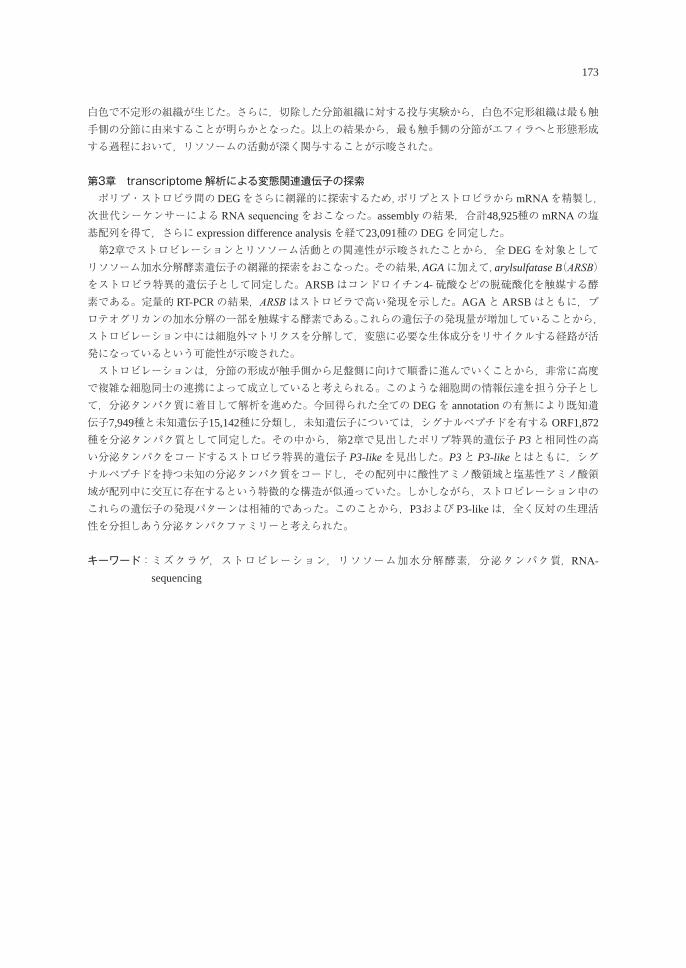
173
白色で不定形の組織が生じた。さらに,切除した分節組織に対する投与実験から,白色不定形組織は最も触手側の分節に由来することが明らかとなった。以上の結果から,最も触手側の分節がエフィラへと形態形成する過程において,リソソームの活動が深く関与することが示唆された。
第3章 transcriptome 解析による変態関連遺伝子の探索� ポリプ・ストロビラ間のDEGをさらに網羅的に探索するため,ポリプとストロビラからmRNAを精製し,次世代シーケンサーによる RNA sequencingをおこなった。assemblyの結果,合計48,925種の mRNAの塩基配列を得て,さらに expression difference analysisを経て23,091種の DEGを同定した。 第2章でストロビレーションとリソソーム活動との関連性が示唆されたことから,全 DEGを対象としてリソソーム加水分解酵素遺伝子の網羅的探索をおこなった。その結果,AGAに加えて,arylsulfatase B(ARSB)をストロビラ特異的遺伝子として同定した。ARSBはコンドロイチン4-硫酸などの脱硫酸化を触媒する酵素である。定量的 RT-PCRの結果,ARSBはストロビラで高い発現を示した。AGAと ARSBはともに,プロテオグリカンの加水分解の一部を触媒する酵素である。これらの遺伝子の発現量が増加していることから,ストロビレーション中には細胞外マトリクスを分解して,変態に必要な生体成分をリサイクルする経路が活発になっているという可能性が示唆された。 ストロビレーションは,分節の形成が触手側から足盤側に向けて順番に進んでいくことから,非常に高度で複雑な細胞同士の連携によって成立していると考えられる。このような細胞間の情報伝達を担う分子として,分泌タンパク質に着目して解析を進めた。今回得られた全ての DEGを annotationの有無により既知遺伝子7,949種と未知遺伝子15,142種に分類し,未知遺伝子については,シグナルペプチドを有する ORF1,872種を分泌タンパク質として同定した。その中から,第2章で見出したポリプ特異的遺伝子 P3と相同性の高い分泌タンパクをコードするストロビラ特異的遺伝子 P3-likeを見出した。P3と P3-likeとはともに,シグナルペプチドを持つ未知の分泌タンパク質をコードし,その配列中に酸性アミノ酸領域と塩基性アミノ酸領域が配列中に交互に存在するという特徴的な構造が似通っていた。しかしながら,ストロビレーション中のこれらの遺伝子の発現パターンは相補的であった。このことから,P3および P3-likeは,全く反対の生理活性を分担しあう分泌タンパクファミリーと考えられた。
キーワード: ミズクラゲ,ストロビレーション,リソソーム加水分解酵素,分泌タンパク質,RNA-sequencing
Pathophysiological analysis of transgenic mice overexpressing GDE5 in skeletal muscles
Takao haShimoTo
Graduate School of Biosphere Science, Hiroshima University,Higashi-Hiroshima 739-8528, Japan
骨格筋特異的 GDE5過剰発現マウスの病態解析
橋本 貴生
広島大学大学院生物圏科学研究科,739-8528 東広島市
【序論】 骨格筋萎縮は運動機能の低下だけでなく,肥満や糖尿病リスクの上昇をさせるなど,人々の生活において様々な問題をもたらす。骨格筋萎縮発症の要因は多様であり,特に,加齢に伴って骨格筋重量と筋力が低下する sarcopeniaは,加齢による生理機能の変化の他に神経伝達やエネルギー代謝の低下などが絡み合い,発症過程は極めて複雑である。Sarcopeniaの動物試験は長期飼育が必要であり,除神経や固定などの処置による骨格筋萎縮モデルは短期に筋萎縮を発症するものの,ヒトの病態を再現しない問題がある。 Glycerophosphodiester phosphodiesterase(GDE)ファミリーの一つである GDE5は,骨格筋を含むエネルギー消費量の多い組織に強く発現する。先行研究によって,その酵素活性の欠損変異体である GDE5dc471を骨格筋特異的に過剰発現させたマウス(GDE5Tgマウス)を作出した結果,わずか12週齢で解糖系の酵素活性が高い Type2筋繊維を多く含む大腿筋や腓腹筋の優先的な萎縮が観察された。これは sarcopeniaの表現型と類似しており,GDE5Tgマウスは sarcopeniaの短期自然発症モデルとして期待されてきた。しかし,骨格筋病態の発症要因が明らかでない上,12週齢以降の病態の進展など詳細なメカニズムは不明な点が多い。そこで本研究では,4週齢(幼若期),8週齢(若齢期)および1年齢(中年期)の GDE5Tgマウスを用いて骨格筋病態あるいは生理機構を経時的に解析し,病態進展メカニズムを明らかにした。さらに,幼若期の GDE5Tgマウスを用いて骨格筋病態の発症要因を探索した。
【GDE5Tgマウスにおける骨格筋病態の進展メカニズムの解明】 4週齢の GDE5Tgマウスは筋委縮を示さなかったが,8週齢の GDE5Tgマウスは野生型マウスに比べてType2筋繊維が豊富な骨格筋のみが選択的に萎縮し,運動機能が低下した。さらに,血中脂質濃度と白色脂肪組織重量も増加しており,以上の形質は1年齢まで不可逆的に引き続いた。これらは sarcopeniaで報告されている現象であり,GDE5Tgマウスは sarcopeniaと多くの共通の形質を示した。また筋病理標本の経時評価によって,8週齢から1年齢の GDE5Tgマウス腓腹筋において,筋細胞質内に異常タンパク質が高頻度に蓄積することを明らかにした。さらに GDE5Tgマウスの筋委縮の進展に伴って,アポトーシスによる細胞死を示唆する,断片化核が増加した。 これまで sarcopeniaの進展には,neuromuscular junction(NMJ)の形態変化による神経伝達の低下が密接に関連すると考えられているが,GDE5Tgマウスの大腿筋において,NMJの形態と筋単位面積当たりの個数は中年期においても野生型マウスとの差異がなかった。一方,NMJを構成する筋特異的な nicotinic acetylcholine receptor(nAchR)サブユニット mRNAは,筋委縮を示さない4週齢においても発現上昇し,1年齢まで持続していた。したがって,GDE5Tgマウスにおける NMJ関連 mRNAは,筋委縮や NMJの変性による代償的な発現上昇と異なる,別の発現機構により誘導されたと考えられた。
生物圏科学Biosphere Sci. 56:174-175 (2017)
175
【GDE5Tgマウスにおける骨格筋病態の発症メカニズムの探索】 筋委縮を呈さない幼弱期における,異常タンパク質の蓄積に関連したストレス応答の有無を検証するため,4週齢の GDE5Tgマウスの腓腹筋を用いて HSP70タンパク質の発現をウエスタンブロッティング法により解析した。その結果,GDE5Tgマウスは野生型に比べ HSP70の発現量が著しく増加した。さらに,この発現増加は生後間もない2週齢の GDE5Tgマウスも示したことから,幼若期により何らかの細胞ストレスが誘導されている可能性が示唆された。 次に,GDE5Tgマウスの筋委縮発症要因を網羅的に探索することを目的とし,2週齢の GDE5Tgマウスの大腿筋を用いて DNA microarray解析を行ったところ,ストレス応答に関連した様々な遺伝子群が発現上昇した。HSPファミリーにおいては,HSP70の他にも Hspa4l(HSP40),および Hsp110(HSP110)の発現上昇が顕著であった。さらに,炎症性サイトカイン IL-1と結合する Il1rap,mammalian target of rapamycin(mTOR)阻害作用を持つ sesn2の顕著な発現上昇が認められた。
【考察および結論】 GDE5Tgマウスの経時的な骨格筋病態の解析によって明らかになった,Type2筋繊維の選択的かつ緩やかに継続する筋萎縮は,除神経や固定などの処置による骨格筋萎縮モデルの形質と明らかに異なっており,sarcopeniaとの類似性が示唆された。また,GDE5Tgマウスの骨格筋病態の進展に異常タンパク質の蓄積とアポトーシスを伴うことを初めて見出し,筋委縮を呈さない幼少期においても様々な細胞ストレスに対する応答が示唆された。今後,GDE5Tgマウスを用いた研究を通して,蓄積した異常タンパク質と筋萎縮との関連性,特に骨格筋細胞に対するストレスなどの作用機序を明らかにすることで sarcopeniaの発症と進展機構に新たな知見を提供できるものと期待される。 GDE5Tgマウスは筋委縮を示さない幼若時から NMJを構成する筋特異的 nAchRサブユニットの mRNAが発現上昇しており,NMJの形態を維持するための代償的な生理作用が考えられた。事実,GDE5Tgマウスは出生間もない時期から複数のストレス関連遺伝子が発現上昇しており,ストレス誘導による NMJ構成因子の発現誘導の可能性を示唆するものである。NMJ構成因子の増加による NMJの形態維持は骨格筋老化に対する防御的方策として有望であることから,GDE5Tgマウスは新規の骨格筋委縮モデルとしての有用性のみならず,筋特異的 nAchRサブユニットの発現誘導機構の新たな概念を提供できる可能性も示唆された。 以上,sarcopeniaの病態を極めて早期に,かつ長期的に示す自然発症するモデルは前例がなく,GDE5Tgマウスの評価系としての有用性が考えられるとともに,新たな NMJ維持機構の解明においても極めて有用である。本研究成果を基にした医薬品ならびに機能性食品の発掘,および評価を通して,人々の健康寿命・QOLの向上に貢献することが期待される。
キーワード:骨格筋萎縮,細胞ストレス,GDE5,NMJ,nAchR,sarcopenia
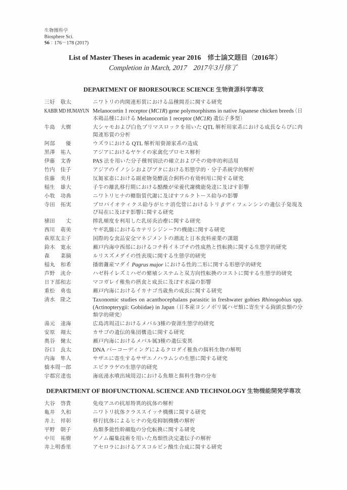
List of Master Theses in academic year 2016 修士論文題目(2016年)Completion in March, 2017 2017年3月修了
DEPARTMENT OF BIORESOURCE SCIENCE 生物資源科学専攻
三好 敬太 ニワトリの肉関連形質における品種間差に関する研究KABIR MD HUMAYUN Melanocortin 1 receptor (MC1R) gene polymorphisms in native Japanese chicken breeds(日
本鶏品種におけるMelanocortin 1 receptor (MC1R) 遺伝子多型)牛島 大樹 大シャモおよび白色プリマスロックを用いた QTL解析用家系における成長ならびに肉
関連形質の分析阿部 優 ウズラにおける QTL解析用資源家系の造成黒澤 祐人 アジアにおけるヤケイの家禽化プロセス解析伊藤 文香 PAS法を用いた分子種判別法の確立およびその効率的利活用竹内 佳子 アジアのイノシシおよびブタにおける形態学的・分子系統学的解析佐藤 美月 反芻家畜における副産物発酵混合飼料の有効利用に関する研究稲生 雄大 子牛の離乳移行期における酪酸が栄養代謝機能発達に及ぼす影響小牧 功典 ニワトリヒナの糖脂質代謝に及ぼすフルクトース給与の影響寺田 拓実 プロバイオティクス給与がヒナ消化管におけるトリβディフェンシンの遺伝子発現及
び局在に及ぼす影響に関する研究植田 丈 搾乳頻度を利用した乳房炎治療に関する研究西川 萌美 ヤギ乳腺におけるカテリシジン-7の機能に関する研究萩原友圭子 国際的な食品安全マネジメントの潮流と日本食料産業の課題鈴木 寛永 瀬戸内海中西部におけるコチ科イネゴチの性成熟と性転換に関する生態学的研究森 菜摘 ルリスズメダイの性表現に関する生態学的研究稲丸 和希 播磨灘産マダイ Pagrus major における性的二形に関する形態学的研究芦野 洸介 ハゼ科イレズミハゼの繁殖システムと双方向性転換のコストに関する生態学的研究日下部和志 マコガレイ稚魚の摂食と成長に及ぼす水温の影響重松 勇也 瀬戸内海におけるイカナゴ当歳魚の成長に関する研究清水 隆之 Taxonomic studies on acanthocephalans parasitic in freshwater gobies Rhinogobius spp.
(Actinopterygii: Gobiidae) in Japan (日本産ヨシノボリ属ハゼ類に寄生する鉤頭虫類の分類学的研究)
湯元 達海 広島湾周辺におけるメバル3種の資源生態学的研究安原 翔太 カサゴの遺伝的集団構造に関する研究奥谷 健太 瀬戸内海におけるメバル属3種の遺伝変異谷口 良太 DNAバーコーディングによるクロダイ稚魚の餌料生物の解明内海 隼人 サザエに寄生するサザエノハラムシの生態に関する研究橋本周一郎 エビクラゲの生態学的研究宇都宮達也 海底湧水噴出域周辺における魚類と餌料生物の分布
DEPARTMENT OF BIOFUNCTIONAL SCIENCE AND TECHNOLOGY 生物機能開発学専攻
大谷 啓貴 免疫アユの抗原特異的抗体の解析亀井 久和 ニワトリ抗体クラススイッチ機構に関する研究井上 祥彰 移行抗体によるヒナの免疫抑制機構の解析平野 朝子 鳥類多能性幹細胞の分化転換に関する研究中川 祐樹 ゲノム編集技術を用いた鳥類性決定遺伝子の解析井上明香里 アセロラにおけるアスコルビン酸生合成に関する研究
生物圏科学Biosphere Sci. 56:176-178 (2017)
177
石濱 諒 イネにおけるキシラナーゼインヒビターの発現機構に関する研究深見洋次郎 ペルオキシソーム輸送におけるシロイヌナズナカタラーゼの構造に関する研究坂尾 理恵 トマトホスホリパーゼ A2の構造と遺伝子発現に関する研究栗林 貴明 2種 Shewanella属細菌由来の 5’-ヌクレオチダーゼの比較研究加藤 雄基 Shewanella属細菌由来シトクロム c’の安定化機構の解明小林 伸弥 変性剤の疎水基が蛋白質変性に及ぼす影響池田 拓真 スフィンゴ脂質合成遺伝子と遺伝学的相互作用する遺伝子の探索岡野 晃 脂質代謝異常が酵母のアミノ酸量に及ぼす影響湯浅 康智 アシタバ種子由来の新規セスキテルペノイド類
椿 侑也 ジャノヒゲ種皮の生理活性成分紙川小百合 ジャケツイバラとマメゾウムシの生物間相互作用に係わる生理活性物質福間 美樹 染色体外因子を介した細胞内遺伝子増幅系で,蛋白質発現を制御するための研究宇都宮 健 チオレドキシンのペルオキシレドキシン Q還元反応に関する研究川上 貴大 乳酸菌代謝産物による免疫調節作用に関する研究溝口 菜美 ポリフェノールによる消化管上皮バリア保護作用に関する研究小方 美幸 食物繊維による消化管の恒常性維持に関する研究畑堀 翔 乳酸菌による皮膚疾患抑制効果に関する研究佐藤 有紗 トリラウリンの複数のβ型多形の結晶化下田 康平 油脂移行速度測定によるチョコレートの構造予測田添 由真 単軸圧縮動的粘弾性計測によるレトルトパウチ内食品の力学的物性の未開封・非破壊評価曽我部知史 乾燥食品の力学的ガラス転移特性に関する研究加藤 義啓 植物抽出液を用いた Bacillus属細菌の制御付 楚然 Study on the beneficial effect of low dose of ethanol
(少量エタノール摂取の効能に関する研究)岩本 茜 腸内環境を改善する食事因子に関する研究浦上 真治 Study on the physiological function of vitamin B6(ビタミン B6の生理作用に関する研究)髙原 貫 Study on the effect of low dose of ethanol on colon carcinogenesis(大腸癌発現に及ぼす少
量エタノールの効果に関する研究)武田 崇登 グリセロホスホコリン分解酵素の基礎的研究とその応用宮地佑希野 choline代謝における性差の解明金井すみれ 異所性脂肪組織の生理的役割と発生に関する研究渡部 里奈 choline欠乏による NASH発症機序の解明宇原 諒 清酒老香の主要成分であるジメチルトリスルフィド関連酵母遺伝子に関する研究水呉 太一 酵母由来カルバミン酸エチル分解酵素の研究原田 春佳 清酒粕の長期保存における葉酸と α-グリセロホスホコリンの安定化に関する研究下田 隆正 各種初発条件による生もと酒母の成分及び微生物叢解析に関する研究嶋本 孝平 麴菌の総合オミックス解析による清酒メタボローム形成機構の解析平尾 凌 清酒醸造における原料米無機成分に関する研究
DEPARTMENT OF ENVIROMENTAL DYNAMICS AND MANAGEMENT 環境循環系制御学専攻
藤原 笙子 外来草本マルバルコウの発芽と成長に関する研究李 陽 Growth and photosynthetic activity of the invasive aquatic plant Egeria densa in an
irrigation pond(溜池における侵略的外来水草オオカナダモの成長と光合成活性)
辻 浩明 瀬戸内海,伊勢湾,三河湾における多環芳香族炭化水素の分布
178
藤澤 克成 紫外線 LEDを用いたヒドロキシルラジカル反応速度定数の計測法の開発と環境中の物質動態評価への応用
野嶋 仁志 フェントン反応を利用した海水中の過酸化水素の高感度測定法の開発伊藤 友也 施肥材によるアサリ増殖に関する研究脇山 拓也 極短穂型ホールクロップサイレージ用飼料イネの子実生産に及ぼす窒素施肥の影響北里 薫平 Na+蓄積能が異なるイネ品種の選抜KARTHIKA SRISKANTHARAJAH
Molecular Physiological Study of Salt Acclimation in Rice(イネの塩馴化能に関する分子生理学的研究)
渡邉 湧也 植物根圏での定着性における細菌バイオフィルム形成の役割川下 真奈 耐塩性ジャポニカイネ品種の耐塩性機構に関する分子生理学的研究大隅 彰太 ナトリウム施肥により生育が促進されるイネ品種の選抜関藤 怜志 瀬戸内海における粒子径を加味した光減衰モデルの構築および有色溶存有機物の動態
と起源GAU JING JE 生物農薬による生態系撹乱評価のための分子マーカー探索小原 静夏 広島県東部海域における海洋環境と基礎生産藤原 俊樹 原材料の異なる堆肥から分離した微生物のフィチン酸分解能の評価高柳 テラ ワケギの生育と成分に及ぼす栽培条件の影響大西 美央 シロイヌナズナの根毛形成に関わるMYB遺伝子の機能解析花城 清俊 低リン環境下におけるシロバナルーピンの適応特性
Completion in September, 2017 2017年9月修了DEPARTMENT OF BIORESOURCE SCIENCE 生物資源科学専攻
FATSI PATRICKSENAM KOFI
Taxonomic study on microorganisms passable through sterilizing-grade filters(除菌フィルター通過菌の分類学的研究)
SUCI ANDIEWATI Taxonomic studies on the parasitic crustaceans of the flathead grey mullet (Mugil cephalus) and the red seabream (Pagrus major) in Hiroshima Bay,Japan(広島湾産ボラとマダイに寄生する甲殻類の分類学的研究)
鈴木 将司 ミズクラゲの内在性変態誘導物質の検討OKTA PRINGGAPAKPAHAN
The role of local agricultural cooperatives on implement food safety activities in food chain: A case study of mandarin orange in Saga prefecture, Japan(フードチェーンにおける食品安全活動の実施に向けた農協の役割:佐賀県におけるみかんの事例研究。)
DEPARTMENT OF BIOFUNCTIONAL SCIENCE AND TECHNOLOGY 生物機能開発学専攻
AHMED MAHROUS SOLIMAN
Molecular Characterization of Antimicrobial Resistance in Gram-negative Bacteria Isolated from Clinical Samples(臨床検体より分離したグラム陰性細菌の薬剤耐性化の分子レベル解析)
DYAH AYU SAVITRI
The melting phenomena of chocolate : Investigation by artificial mouth model system and sensory analysis(チョコレートの融解現象:人工口腔モデルおよび官能分析による研究)
QIN DONG Study on physiological characteristics of germination of plant differing in phytate content(フィチン酸含有量の異なる植物における発芽時の生理学的特性に関する研究)
AYU LANA NAFISYAH
Microphytobenthos flora on mangrove sediment in the East Java coastal area, Indonesia: Their ecological and physiological characters(インドネシア・ジャワ島東部沿岸におけるマングローブ底泥上の底生微細藻類の生物相:その生態学的・生理学的特性について)
179
平成28年度研究科長裁量経費による助成研究報告Reports of Studies supported by Grant-in-Aid for Research from the Graduate
School of Biosphere Science, Hiroshima University
助成区分 研究課題名 研究代表者
基盤研究サポートGrant-in-Aid for Fundamental Research
流域圏スケールでの水質および水量の管理に関する国際比較研究International Comparative Study on Management of Basin-scale Water Quality and Quantity
山本民次Tamiji
YAMAMOTO
育成前期乳用牛の夏季暑熱への順応に関する研究Research on acclimazation of prepubertal Holstein heifers for hot summer season
沖田美紀Miki OKITA
生物圏科学Biosphere Sci. 56:179 (2017)
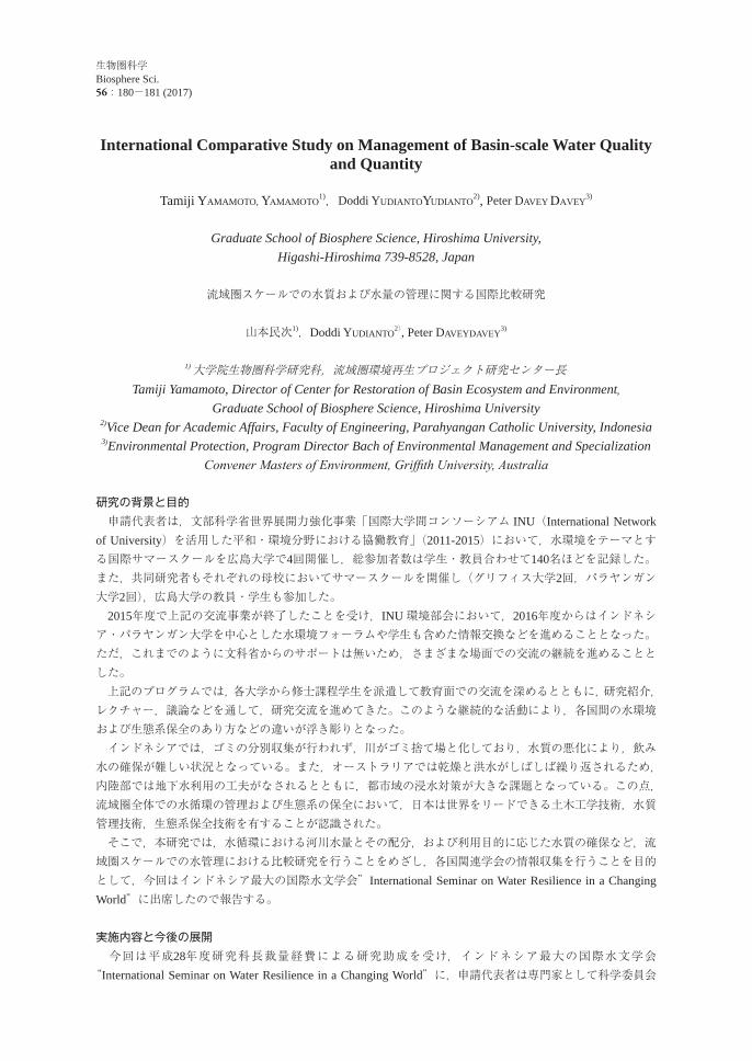
International Comparative Study on Management of Basin-scale Water Quality and Quantity
Tamiji YAMAMOTO, YAMAMOTO1),Doddi YUDIANTOYUDIANTO
2), Peter DAVEY DAVEY3)
Graduate School of Biosphere Science, Hiroshima University,Higashi-Hiroshima 739-8528, Japan
流域圏スケールでの水質および水量の管理に関する国際比較研究
山本民次1),Doddi YUDIANTO2), Peter DAVEYDAVEY3)
1)大学院生物圏科学研究科,流域圏環境再生プロジェクト研究センター長Tamiji Yamamoto, Director of Center for Restoration of Basin Ecosystem and Environment,
Graduate School of Biosphere Science, Hiroshima University2)Vice Dean for Academic Affairs, Faculty of Engineering, Parahyangan Catholic University, Indonesia3)Environmental Protection, Program Director Bach of Environmental Management and Specialization
Convener Masters of Environment, Griffith University, Australia
研究の背景と目的 申請代表者は,文部科学省世界展開力強化事業「国際大学間コンソーシアム INU(International Network of University)を活用した平和・環境分野における協働教育」(2011-2015)において,水環境をテーマとする国際サマースクールを広島大学で4回開催し,総参加者数は学生・教員合わせて140名ほどを記録した。また,共同研究者もそれぞれの母校においてサマースクールを開催し(グリフィス大学2回,パラヤンガン大学2回),広島大学の教員・学生も参加した。 2015年度で上記の交流事業が終了したことを受け,INU環境部会において,2016年度からはインドネシア・パラヤンガン大学を中心とした水環境フォーラムや学生も含めた情報交換などを進めることとなった。ただ,これまでのように文科省からのサポートは無いため,さまざまな場面での交流の継続を進めることとした。 上記のプログラムでは,各大学から修士課程学生を派遣して教育面での交流を深めるとともに,研究紹介,レクチャー,議論などを通して,研究交流を進めてきた。このような継続的な活動により,各国間の水環境および生態系保全のあり方などの違いが浮き彫りとなった。 インドネシアでは,ゴミの分別収集が行われず,川がゴミ捨て場と化しており,水質の悪化により,飲み水の確保が難しい状況となっている。また,オーストラリアでは乾燥と洪水がしばしば繰り返されるため,内陸部では地下水利用の工夫がなされるとともに,都市域の浸水対策が大きな課題となっている。この点,流域圏全体での水循環の管理および生態系の保全において,日本は世界をリードできる土木工学技術,水質管理技術,生態系保全技術を有することが認識された。 そこで,本研究では,水循環における河川水量とその配分,および利用目的に応じた水質の確保など,流域圏スケールでの水管理における比較研究を行うことをめざし,各国関連学会の情報収集を行うことを目的として,今回はインドネシア最大の国際水文学会”International Seminar on Water Resilience in a Changing World”に出席したので報告する。
実施内容と今後の展開 今回は平成28年度研究科長裁量経費による研究助成を受け,インドネシア最大の国際水文学会“International Seminar on Water Resilience in a Changing World”に,申請代表者は専門家として科学委員会
生物圏科学Biosphere Sci. 56:180-181 (2017)
181
メンバーの一人として出席した。サブテーマが,“Water Conservation and Risk and Impact of Extreme Event”, “Water Security for All”, “Water Governance and Partnership”の3つあり,インドネシアおよび周辺国から合計95課題の発表があった。 水循環や水環境に関する多くの発表を聞き,発表者とのディスカッションを通じて,情報交換ができた。INUのメンバー校であるパラヤンガン大学以外に,本学と大学間協定を結んでいる,ハサヌディン大学,ガジャ・マダ大学,バンドン工科大学,ブラウィジャヤ大学,ディポネゴロ大学,インドネシア大学,北スマトラ大学などからの発表もあった。その他,オランダ,韓国の発表,日本からも水資源機構の発表があった。流域圏スケールでの水質および水量の管理に関する国際比較研究を進めるには,このような情報交流を重ねることが重要であり,共同で取り組める具体的課題を洗い出す方向性が見えてきた。 所期の思いに違わず,我が国の水処理,ゴミ処理,環境保全などに関する技術と知識,それらに付随するソフト的なシステムも含めて世界トップレベルにあることを実感した。今後,我が国で培われてきたそれらの高度な技術やシステムは,本共同研究をさらに進めることで,今後,具体的な技術移転などにつながる可能性がある。 申請代表者は,広島市産業振興アドバイザー,広島県循環型社会推進機構理事,中国経済産業局水ビジネス推進協議会アドバイザーなどの経験を有する。今後は,これらのつながりを生かし,同分野に関する国際セミナーを東広島市あるいは広島市で開催することを計画しているところである。
生物圏科学Biosphere Sci. 56:182-183 (2017)
Research on acclimazation of prepubertal Holstein heifers for hot summer season
Miki OKITA, Yuzo KUROKAWA, Takashi BUNGO
Graduate School of Biosphere Science, Hiroshima University,Higashi-Hiroshima 739-8528, Japan
育成前期乳用牛の夏季暑熱への順応に関する研究
沖田美紀,黒川勇三,豊後貴嗣広島大学大学院生物圏科学研究科,739-8528 東広島市
【背景・目的】 野中ら(2012)は,育成前期乳用牛に及ぼす高温高湿の影響として,呼吸数・体温の上昇や血液性状の変化等を明らかにしているが,それらは環境制御室においての結果であり,暑熱に暴露されている期間は短い。著者らは搾乳牛に対し夏季暑熱時に試験を行い,剪毛が乳生産と生理指標に及ぼす影響を調べた結果,剪毛による熱放散改善の乳量への効果が示唆された(沖田ら,2014)。その中で,夏季暑熱のもとであっても試験期の経過とともに対照区において,呼吸数,直腸温が低くなる傾向を認め,長期暑熱暴露により熱放散が高まる等の順応が生じた可能性が考えられた。 本研究はホルスタイン種育成前期乳牛を用いて,長期の夏季暑熱暴露により順応が生じる過程を生理生産面から明らかにすることを目的として行った。【材料と方法】 試験は2016年6月から9月までの4か月間にわたって実施し,供試牛にはホルスタイン種育成前期雌牛5頭を用いた。 測定は1期(6月28日-7月3日),2期(7月25-30日),3期(8月23-28日)および4期(9月20-25日)の計4回実施した。測定項目は,飼料摂取量・体重・心拍数・呼吸数・皮膚温・直腸温(15時),および血液性状であった。皮膚温は,左側のき甲・肩中央・肩関節・前膊の4か所を測定した。 血液は頸静脈から採取し,生化学分析装置により,血漿中アラニンアミノトランスフェラーゼ(ALT),グルコース(GLU),総コレステロール(T-Cho),アルカリフォスファターゼ(ALP)等を,高速微量ヘマトクリット用遠心機によりヘマトクリット(Ht)を測定した。統計処理ソフト Rを用い,試験期を要因とした一元配置分散分析を行なった。【結果】 測定した牛舎内の相対湿度,気温から,Maderら (2006)の式を用いて各期の温湿度指数(THI)の平均値を求めた(1期:73.9,2期:77.0,3期:75.7および4期:69.6)。野中らの報告では,育成前期乳牛への高温の影響は,相対湿度80%,28℃から認められ(THI換算で79.7),本試験の1~3期が近い環境にあったと考えられた。 乾物摂取量は,1,2期にくらべ,3,4期と進行するに従い有意に低下した。平均日増体量,心拍数に,試験期の間に有意差は認められなかった。呼吸数および直腸温は2,3期にくらべ4期で有意に低かった。皮膚温は,計測した4か所すべての部位においてすべての試験期の間に有意差が認められ,2期に最も高かった。血漿中 ALT,T-Choおよび Ht値は1,2期にくらべ3期で有意に低かった。ALPは4期にくらべ2,3期で有意に低かった。【考察】 直腸温と呼吸数の結果から,1~3期は暑熱の影響を受けた期であり,4期はその影響がやや低下していたと示唆される。しかし皮膚温は1期より2~4期で有意に高くなっており,2期以降皮膚からの熱放散が高まることで体熱放散が促進された可能性がある。一方ALPは迅速かつ正確に反応する暑熱ストレス指標(Abeni
183
ら,2007)との報告があり,ALT,T-Choおよび Ht値の結果も考慮すると,3期の暑熱の影響が大きかった可能性がある。乾物摂取量は,1期,2期より3期のほうが低下しており,この3期に2期より皮膚温が低かったことは皮膚からの熱放散の低下を示唆し,体温調節機能の一部を,乾物摂取量の低下による熱生産量の低下へとシフトさせていた可能性がある。 以上より,季節の進行に伴って THIでは大差がなくとも暑熱の影響のあらわれ方が変化することが示唆され,長期にわたる変化を追うことにより,短期の暑熱暴露では見られなかった変化を捉えることができたと考えられる。この変化が暑熱に対する順応につながるものであるかどうかは,今後この観点からの試験を継続することにより明らかにしていきたい。

List of Papers by the staff of the Graduate School of Biosphere Science,Hiroshima University, published during January 2016 to March, 2017
広島大学大学院生物圏科学研究科教員業績目録
(2016年 1月- 2017年 3月)
Animal Science Division 陸域動物生産学講座
Nishibori, M., Osman, S.A.M., Ghanem, M.E., Kazymbet, P., Bakhtin, M., Meldelokov, A., Suleimenov, M.Z., Kazhmrat, A.M., Bibigul, S.B., Mannen, H., Yamamoto, Y., 2017. Mitochondrial genetic diversity of Saiga tatarica tatarica (Saiga antelope) in Kazakhstan. Rep.Soc.Res.Native Livestock, 28: 27-35.
Mannen, H., Takahashi, Y., Yamagata, T., Yamamoto, Y., Tsunoda, K., Nomura, K., Kunieda, T., Onishi, R., Ito, F., Takeuchi, K., Meldelokov, A., Suleimenov, M.Z., Bakhtin, M., Kazymbet, P., Nishibori, M., 2017. Outline of sampling and coat-color variations of the native cattle in Kazakhstan. Rep.Soc.Res.Native Livestock, 28: 37-43.
Nomura, K., Takahashi, Y., Tahara, G., Kurosawa, Y., Mannen, H., Yamagata, T., Kunieda, T., Tsunoda, K., Yamamoto, Y., Onishi, R., Ito, F., Takeuchi, K., Nishibori, M., Meldelokov, A., Suleimenov, M.Z., Bakhtin, M., Kazymbet. P., 2017. Microsatellite DNA polymorphisms observed in the indigenous cattle of Kazakhstan. Rep.Soc.Res.Native Livestock, 28: 45-51.
Kunieda, T., Ezoe, H., Okuda, Y., Mannen, H., Takahashi, Y., Nomura, K., Yamagata, T., Yamamoto, Y., Tsunoda, K., Bakhtin, M., Kazymbet, P., Meldelokov, A., Suleimenov, M.Z., Safronova, O., Nishibori, M., 2017. Coat color variation and allelic frequency of the genes associated with body composition and locomotion traits in Kazakhstan native horse. Rep.Soc.Res.Native Livestock, 28: 53-67.
Mannen, H., Tabata, R., Takahashi, Y., Yamagata, T., Yamamoto, Y., Tsunoda, K., Nomura, K., Kunieda, T., Onishi, R., Ito, F., Takeuchi, K., Meldelokov, A., Suleimenov, M.Z., Bakhtin, M., Kazymbet, P., Nishibori, M., 2017. Outline of sampling and mitochondrial DNA polymorphisms in Kazakhstan domestic goats. Rep.Soc.Res.Native Livestock, 28: 65-73.
Nomura, K., Tahara, G., Suzuki, S., Takahashi, Y., Kurosawa, Y., Mannen, H., Yamagata, T., Kunieda, T., Tsunoda, K., Yamamoto, Y., Onishi, R., Ito, F., Takeuchi, K., Nishibori, M., Meldelokov, A., Suleimenov, M.Z., Bakhtin, M., Kazymbet, P., 2017. Microsatellite DNA polymorphisms observed in the indigenous goats of Kazakhstan. Rep.Soc.Res.Native Livestock, 28: 75-82.
Yamagata, T., Tsunoda, K., Takahashi, Y., Nomura, K., Mannen, H., Kunieda, T., Onishi, R., Ito, F., Takeuchi, K., Yamamoto, Y., Nishibori, M., Meldelokov, A., Suleimenov, M.Z., Bakhtin, M., Kazymbet, P., 2017. External morphological characters of local sheep breeds and populations in Kazakhstan. Rep.Soc.Res.Native Livestock, 28: 83-96.
Tsunoda, K., Yamagata, T., Takahashi, Y., Nomura, K., Mannen, H., Kunieda, T., Sato, K., Onishi, R., Ito, F., Takeuchi, K., Yamamoto, Y., Nishibori, M., Meldelokov, A., Suleimenov, M.Z., Bakhtin, M., Kazymbet, P., 2017. Phylogenetic research on several local sheep breeds and populations in Kazakhstan. Rep.Soc.Res.Native Livestock, 28: 97-113.
Yamagata, T., Yamamoto, Y., Meldelokov, A., Suleimenov, M.Z., Bakhtin, M, Kazymbet, P., Nishibori, M.,
生物圏科学Biosphere Sci. 56:185-196 (2017)
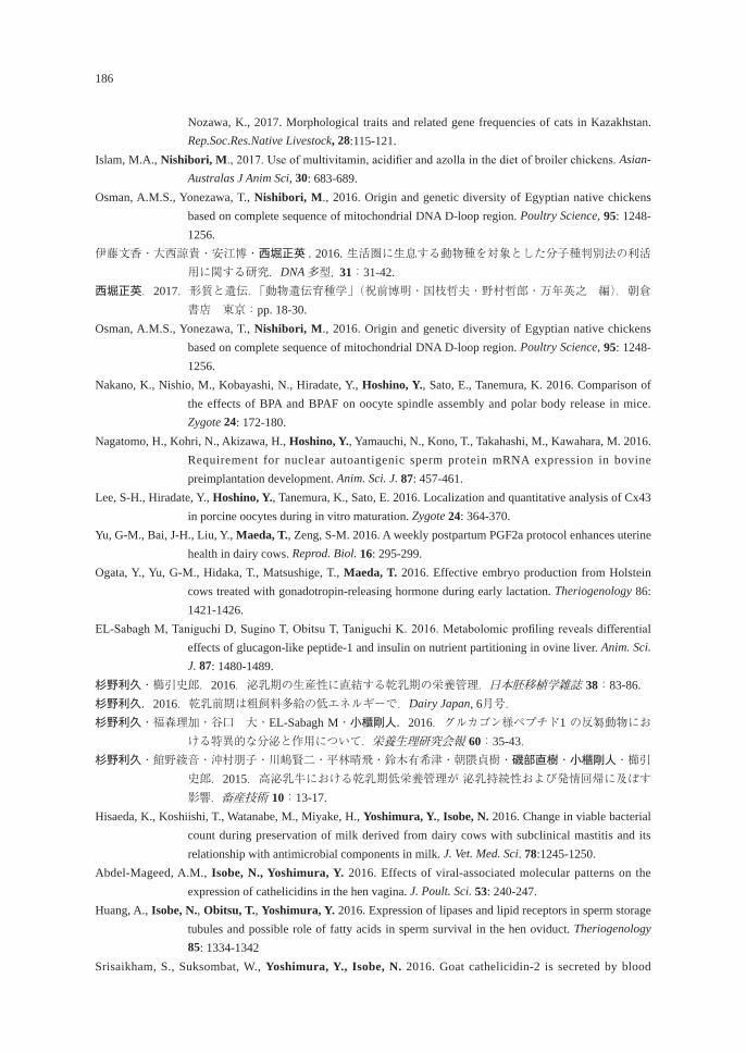
186
Nozawa, K., 2017. Morphological traits and related gene frequencies of cats in Kazakhstan. Rep.Soc.Res.Native Livestock, 28:115-121.
Islam, M.A., Nishibori, M., 2017. Use of multivitamin, acidifier and azolla in the diet of broiler chickens. Asian-Australas J Anim Sci, 30: 683-689.
Osman, A.M.S., Yonezawa, T., Nishibori, M., 2016. Origin and genetic diversity of Egyptian native chickens based on complete sequence of mitochondrial DNA D-loop region. Poultry Science, 95: 1248-1256.
伊藤文香・大西諒貴・安江博・西堀正英 . 2016. 生活圏に生息する動物種を対象とした分子種判別法の利活用に関する研究.DNA 多型, 31:31-42.
西堀正英.2017.形質と遺伝.「動物遺伝育種学」(祝前博明・国枝哲夫・野村哲郎・万年英之 編).朝倉書店 東京:pp. 18-30.
Osman, A.M.S., Yonezawa, T., Nishibori, M., 2016. Origin and genetic diversity of Egyptian native chickens based on complete sequence of mitochondrial DNA D-loop region. Poultry Science, 95: 1248-1256.
Nakano, K., Nishio, M., Kobayashi, N., Hiradate, Y., Hoshino, Y., Sato, E., Tanemura, K. 2016. Comparison of the effects of BPA and BPAF on oocyte spindle assembly and polar body release in mice. Zygote 24: 172-180.
Nagatomo, H., Kohri, N., Akizawa, H., Hoshino, Y., Yamauchi, N., Kono, T., Takahashi, M., Kawahara, M. 2016. Requirement for nuclear autoantigenic sperm protein mRNA expression in bovine preimplantation development. Anim. Sci. J. 87: 457-461.
Lee, S-H., Hiradate, Y., Hoshino, Y., Tanemura, K., Sato, E. 2016. Localization and quantitative analysis of Cx43 in porcine oocytes during in vitro maturation. Zygote 24: 364-370.
Yu, G-M., Bai, J-H., Liu, Y., Maeda, T., Zeng, S-M. 2016. A weekly postpartum PGF2a protocol enhances uterine health in dairy cows. Reprod. Biol. 16: 295-299.
Ogata, Y., Yu, G-M., Hidaka, T., Matsushige, T., Maeda, T. 2016. Effective embryo production from Holstein cows treated with gonadotropin-releasing hormone during early lactation. Theriogenology 86: 1421-1426.
EL-Sabagh M, Taniguchi D, Sugino T, Obitsu T, Taniguchi K. 2016. Metabolomic profiling reveals differential effects of glucagon-like peptide-1 and insulin on nutrient partitioning in ovine liver. Anim. Sci. J. 87: 1480-1489.
杉野利久・櫛引史郎.2016.泌乳期の生産性に直結する乾乳期の栄養管理.日本胚移植学雑誌 38:83-86.杉野利久.2016.乾乳前期は粗飼料多給の低エネルギーで.Dairy Japan, 6月号.杉野利久・福森理加・谷口 大・EL-Sabagh M・小櫃剛人.2016.グルカゴン様ペプチド1 の反芻動物にお
ける特異的な分泌と作用について.栄養生理研究会報 60:35-43.杉野利久・館野綾音・沖村朋子・川嶋賢二・平林晴飛・鈴木有希津・朝隈貞樹・磯部直樹・小櫃剛人・櫛引
史郎.2015.高泌乳牛における乾乳期低栄養管理が 泌乳持続性および発情回帰に及ぼす影響.畜産技術 10:13-17.
Hisaeda, K., Koshiishi, T., Watanabe, M., Miyake, H., Yoshimura, Y., Isobe, N. 2016. Change in viable bacterial count during preservation of milk derived from dairy cows with subclinical mastitis and its relationship with antimicrobial components in milk. J. Vet. Med. Sci. 78:1245-1250.
Abdel-Mageed, A.M., Isobe, N., Yoshimura, Y. 2016. Effects of viral-associated molecular patterns on the expression of cathelicidins in the hen vagina. J. Poult. Sci. 53: 240-247.
Huang, A., Isobe, N., Obitsu, T., Yoshimura, Y. 2016. Expression of lipases and lipid receptors in sperm storage tubules and possible role of fatty acids in sperm survival in the hen oviduct. Theriogenology 85: 1334-1342
Srisaikham, S., Suksombat, W., Yoshimura, Y., Isobe, N. 2016. Goat cathelicidin-2 is secreted by blood
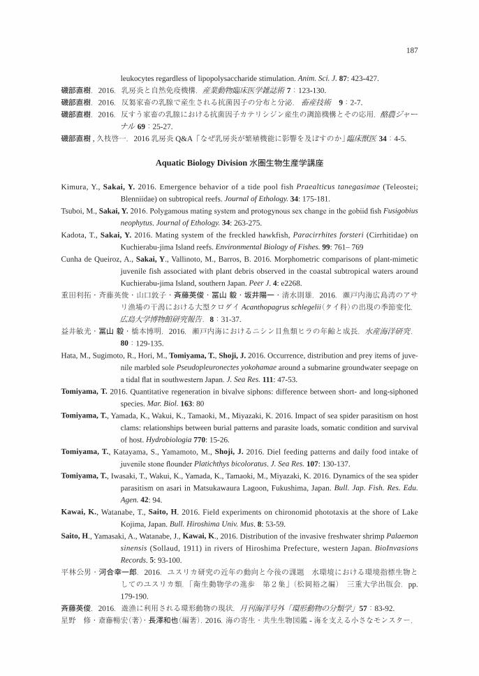
187
leukocytes regardless of lipopolysaccharide stimulation. Anim. Sci. J. 87: 423-427.磯部直樹.2016.乳房炎と自然免疫機構.産業動物臨床医学雑誌術 7:123-130.磯部直樹.2016.反芻家畜の乳腺で産生される抗菌因子の分布と分泌. 畜産技術 9:2-7.磯部直樹.2016.反すう家畜の乳腺における抗菌因子カテリシジン産生の調節機構とその応用.酪農ジャー
ナル 69:25-27.磯部直樹 ,久枝啓一.2016 乳房炎 Q&A「なぜ乳房炎が繁殖機能に影響を及ぼすのか」 臨床獣医 34:4-5.
Aquatic Biology Division 水圏生物生産学講座
Kimura, Y., Sakai, Y. 2016. Emergence behavior of a tide pool fish Praealticus tanegasimae (Teleostei; Blenniidae) on subtropical reefs. Journal of Ethology. 34: 175-181.
Tsuboi, M., Sakai, Y. 2016. Polygamous mating system and protogynous sex change in the gobiid fish Fusigobius neophytus. Journal of Ethology. 34: 263-275.
Kadota, T., Sakai, Y. 2016. Mating system of the freckled hawkfish, Paracirrhites forsteri (Cirrhitidae) on Kuchierabu-jima Island reefs. Environmental Biology of Fishes. 99: 761– 769
Cunha de Queiroz, A., Sakai, Y., Vallinoto, M., Barros, B. 2016. Morphometric comparisons of plant-mimetic juvenile fish associated with plant debris observed in the coastal subtropical waters around Kuchierabu-jima Island, southern Japan. Peer J. 4: e2268.
重田利拓・斉藤英俊・山口敦子・斉藤英俊・冨山 毅・坂井陽一・清水則雄.2016.瀬戸内海広島湾のアサリ漁場の干潟における大型クロダイ Acanthopagrus schlegelii(タイ科)の出現の季節変化.広島大学博物館研究報告.8:31-37.
益井敏光・冨山 毅・橋本博明.2016.瀬戸内海におけるニシン目魚類ヒラの年齢と成長.水産海洋研究. 80:129-135.
Hata, M., Sugimoto, R., Hori, M., Tomiyama, T., Shoji, J. 2016. Occurrence, distribution and prey items of juve-nile marbled sole Pseudopleuronectes yokohamae around a submarine groundwater seepage on a tidal flat in southwestern Japan. J. Sea Res. 111: 47-53.
Tomiyama, T. 2016. Quantitative regeneration in bivalve siphons: difference between short- and long-siphoned species. Mar. Biol. 163: 80
Tomiyama, T., Yamada, K., Wakui, K., Tamaoki, M., Miyazaki, K. 2016. Impact of sea spider parasitism on host clams: relationships between burial patterns and parasite loads, somatic condition and survival of host. Hydrobiologia 770: 15-26.
Tomiyama, T., Katayama, S., Yamamoto, M., Shoji, J. 2016. Diel feeding patterns and daily food intake of juvenile stone flounder Platichthys bicoloratus. J. Sea Res. 107: 130-137.
Tomiyama, T., Iwasaki, T., Wakui, K., Yamada, K., Tamaoki, M., Miyazaki, K. 2016. Dynamics of the sea spider parasitism on asari in Matsukawaura Lagoon, Fukushima, Japan. Bull. Jap. Fish. Res. Edu. Agen. 42: 94.
Kawai, K., Watanabe, T., Saito, H. 2016. Field experiments on chironomid phototaxis at the shore of Lake Kojima, Japan. Bull. Hiroshima Univ. Mus. 8: 53-59.
Saito, H., Yamasaki, A., Watanabe, J., Kawai, K., 2016. Distribution of the invasive freshwater shrimp Palaemon sinensis (Sollaud, 1911) in rivers of Hiroshima Prefecture, western Japan. BioInvasions Records. 5: 93-100.
平林公男・河合幸一郎.2016.ユスリカ研究の近年の動向と今後の課題 水環境における環境指標生物としてのユスリカ類.「衛生動物学の進歩 第2集」(松岡裕之編) 三重大学出版会.pp. 179-190.
斉藤英俊.2016.遊漁に利用される環形動物の現状.月刊海洋号外「環形動物の分類学」57:83-92.星野 修・斎藤暢宏(著)・長澤和也(編著).2016.海の寄生・共生生物図鑑 - 海を支える小さなモンスター.
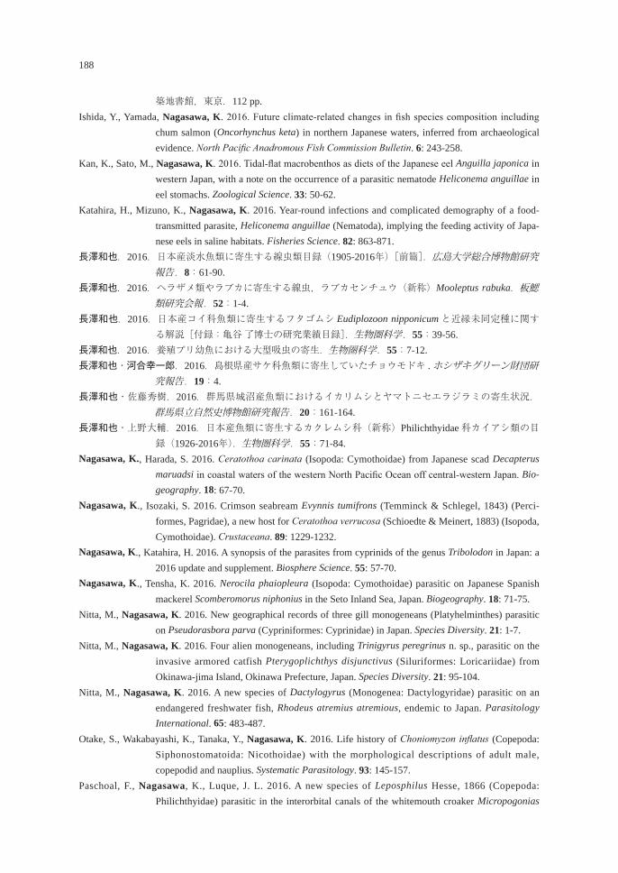
188
築地書館,東京.112 pp.Ishida, Y., Yamada, Nagasawa, K. 2016. Future climate-related changes in fish species composition including
chum salmon (Oncorhynchus keta) in northern Japanese waters, inferred from archaeological evidence. North Pacific Anadromous Fish Commission Bulletin. 6: 243-258.
Kan, K., Sato, M., Nagasawa, K. 2016. Tidal-flat macrobenthos as diets of the Japanese eel Anguilla japonica in western Japan, with a note on the occurrence of a parasitic nematode Heliconema anguillae in eel stomachs. Zoological Science. 33: 50-62.
Katahira, H., Mizuno, K., Nagasawa, K. 2016. Year-round infections and complicated demography of a food-transmitted parasite, Heliconema anguillae (Nematoda), implying the feeding activity of Japa-nese eels in saline habitats. Fisheries Science. 82: 863-871.
長澤和也.2016.日本産淡水魚類に寄生する線虫類目録(1905-2016年)[前篇].広島大学総合博物館研究報告.8:61-90.
長澤和也.2016.ヘラザメ類やラブカに寄生する線虫,ラブカセンチュウ(新称)Mooleptus rabuka.板鰓類研究会報.52:1-4.
長澤和也.2016.日本産コイ科魚類に寄生するフタゴムシ Eudiplozoon nipponicumと近縁未同定種に関する解説[付録:亀谷 了博士の研究業績目録].生物圏科学.55:39-56.
長澤和也.2016.養殖ブリ幼魚における大型吸虫の寄生.生物圏科学.55:7-12.長澤和也・河合幸一郎.2016.島根県産サケ科魚類に寄生していたチョウモドキ . ホシザキグリーン財団研
究報告.19:4.長澤和也・佐藤秀樹.2016.群馬県城沼産魚類におけるイカリムシとヤマトニセエラジラミの寄生状況.
群馬県立自然史博物館研究報告.20:161-164.長澤和也・上野大輔.2016.日本産魚類に寄生するカクレムシ科(新称)Philichthyidae科カイアシ類の目
録(1926-2016年).生物圏科学.55:71-84.Nagasawa, K., Harada, S. 2016. Ceratothoa carinata (Isopoda: Cymothoidae) from Japanese scad Decapterus
maruadsi in coastal waters of the western North Pacific Ocean off central-western Japan. Bio-geography. 18: 67-70.
Nagasawa, K., Isozaki, S. 2016. Crimson seabream Evynnis tumifrons (Temminck & Schlegel, 1843) (Perci-formes, Pagridae), a new host for Ceratothoa verrucosa (Schioedte & Meinert, 1883) (Isopoda, Cymothoidae). Crustaceana. 89: 1229-1232.
Nagasawa, K., Katahira, H. 2016. A synopsis of the parasites from cyprinids of the genus Tribolodon in Japan: a 2016 update and supplement. Biosphere Science. 55: 57-70.
Nagasawa, K., Tensha, K. 2016. Nerocila phaiopleura (Isopoda: Cymothoidae) parasitic on Japanese Spanish mackerel Scomberomorus niphonius in the Seto Inland Sea, Japan. Biogeography. 18: 71-75.
Nitta, M., Nagasawa, K. 2016. New geographical records of three gill monogeneans (Platyhelminthes) parasitic on Pseudorasbora parva (Cypriniformes: Cyprinidae) in Japan. Species Diversity. 21: 1-7.
Nitta, M., Nagasawa, K. 2016. Four alien monogeneans, including Trinigyrus peregrinus n. sp., parasitic on the invasive armored catfish Pterygoplichthys disjunctivus (Siluriformes: Loricariidae) from Okinawa-jima Island, Okinawa Prefecture, Japan. Species Diversity. 21: 95-104.
Nitta, M., Nagasawa, K. 2016. A new species of Dactylogyrus (Monogenea: Dactylogyridae) parasitic on an endangered freshwater fish, Rhodeus atremius atremious, endemic to Japan. Parasitology International. 65: 483-487.
Otake, S., Wakabayashi, K., Tanaka, Y., Nagasawa, K. 2016. Life history of Choniomyzon inflatus (Copepoda: Siphonostomatoida: Nicothoidae) with the morphological descriptions of adult male, copepodid and nauplius. Systematic Parasitology. 93: 145-157.
Paschoal, F., Nagasawa, K., Luque, J. L. 2016. A new species of Leposphilus Hesse, 1866 (Copepoda: Philichthyidae) parasitic in the interorbital canals of the whitemouth croaker Micropogonias
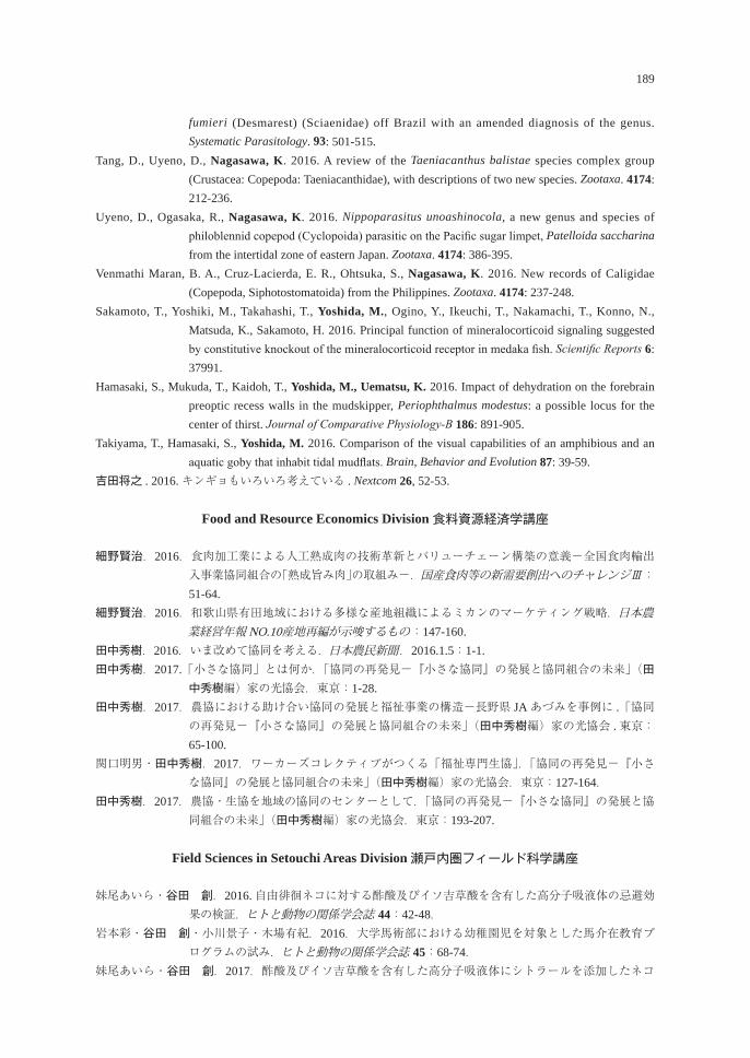
189
fumieri (Desmarest) (Sciaenidae) off Brazil with an amended diagnosis of the genus. Systematic Parasitology. 93: 501-515.
Tang, D., Uyeno, D., Nagasawa, K. 2016. A review of the Taeniacanthus balistae species complex group (Crustacea: Copepoda: Taeniacanthidae), with descriptions of two new species. Zootaxa. 4174: 212-236.
Uyeno, D., Ogasaka, R., Nagasawa, K. 2016. Nippoparasitus unoashinocola, a new genus and species of philoblennid copepod (Cyclopoida) parasitic on the Pacific sugar limpet, Patelloida saccharina from the intertidal zone of eastern Japan. Zootaxa. 4174: 386-395.
Venmathi Maran, B. A., Cruz-Lacierda, E. R., Ohtsuka, S., Nagasawa, K. 2016. New records of Caligidae (Copepoda, Siphotostomatoida) from the Philippines. Zootaxa. 4174: 237-248.
Sakamoto, T., Yoshiki, M., Takahashi, T., Yoshida, M., Ogino, Y., Ikeuchi, T., Nakamachi, T., Konno, N., Matsuda, K., Sakamoto, H. 2016. Principal function of mineralocorticoid signaling suggested by constitutive knockout of the mineralocorticoid receptor in medaka fish. Scientific Reports 6: 37991.
Hamasaki, S., Mukuda, T., Kaidoh, T., Yoshida, M., Uematsu, K. 2016. Impact of dehydration on the forebrain preoptic recess walls in the mudskipper, Periophthalmus modestus: a possible locus for the center of thirst. Journal of Comparative Physiology-B 186: 891-905.
Takiyama, T., Hamasaki, S., Yoshida, M. 2016. Comparison of the visual capabilities of an amphibious and an aquatic goby that inhabit tidal mudflats. Brain, Behavior and Evolution 87: 39-59.
吉田将之 . 2016. キンギョもいろいろ考えている . Nextcom 26, 52-53.
Food and Resource Economics Division 食料資源経済学講座
細野賢治.2016.食肉加工業による人工熟成肉の技術革新とバリューチェーン構築の意義-全国食肉輸出入事業協同組合の「熟成旨み肉」の取組み-.国産食肉等の新需要創出へのチャレンジⅢ:51-64.
細野賢治.2016.和歌山県有田地域における多様な産地組織によるミカンのマーケティング戦略.日本農業経営年報 NO.10産地再編が示唆するもの:147-160.
田中秀樹.2016.いま改めて協同を考える.日本農民新聞.2016.1.5:1-1.田中秀樹.2017.「小さな協同」とは何か.「協同の再発見-『小さな協同』の発展と協同組合の未来」(田
中秀樹編)家の光協会.東京:1-28.田中秀樹.2017.農協における助け合い協同の発展と福祉事業の構造-長野県 JAあづみを事例に .「協同
の再発見-『小さな協同』の発展と協同組合の未来」(田中秀樹編)家の光協会 .東京:65-100.
関口明男・田中秀樹.2017.ワーカーズコレクティブがつくる「福祉専門生協」.「協同の再発見-『小さな協同』の発展と協同組合の未来」(田中秀樹編)家の光協会.東京:127-164.
田中秀樹.2017.農協・生協を地域の協同のセンターとして.「協同の再発見-『小さな協同』の発展と協同組合の未来」(田中秀樹編)家の光協会.東京:193-207.
Field Sciences in Setouchi Areas Division 瀬戸内圏フィールド科学講座
妹尾あいら・谷田 創.2016.自由徘徊ネコに対する酢酸及びイソ吉草酸を含有した高分子吸液体の忌避効果の検証.ヒトと動物の関係学会誌 44:42-48.
岩本彩・谷田 創・小川景子・木場有紀.2016.大学馬術部における幼稚園児を対象とした馬介在教育プログラムの試み.ヒトと動物の関係学会誌 45:68-74.
妹尾あいら・谷田 創.2017.酢酸及びイソ吉草酸を含有した高分子吸液体にシトラールを添加したネコ
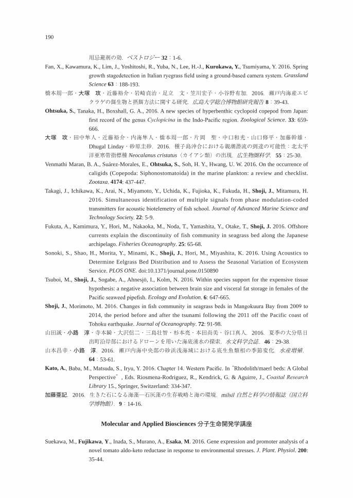
190
用忌避剤の効.ペストロジー 32:1-6.Fan, X., Kawamura, K., Lim, J., Yoshitoshi, R., Yuba, N., Lee, H.-J., Kurokawa, Y., Tsumiyama, Y. 2016. Spring
growth stagedetection in Italian ryegrass field using a ground-based camera system. Grassland Science 63:188-193.
橋本周一郎・大塚 攻・近藤裕介・岩崎貞治・足立 文・笠川宏子・小谷野有加.2016.瀬戸内海産エビクラゲの餌生物と摂餌方法に関する研究.広島大学総合博物館研究報告 8:39-43.
Ohtsuka, S., Tanaka, H., Boxshall, G. A., 2016. A new species of hyperbenthic cyclopoid copepod from Japan: first record of the genus Cyclopicina in the Indo-Pacific region. Zoological Science. 33: 659-666.
大塚 攻・田中隼人・近藤裕介・内海隼人・橋本周一郎・片岡 聖・中口和光・山口修平・加藤幹雄・Dhugal Linday・砂原圭砂.2016.種子島沖合における親潮潜流の到達の可能性:北太平洋亜寒帯指標種 Neocalanus cristatus(カイアシ類)の出現.広生物圏科学.55:25-30.
Venmathi Maran, B. A., Suárez-Morales, E., Ohtsuka, S., Soh, H. Y., Hwang, U. W. 2016. On the occurrence of caligids (Copepoda: Siphonostomatoida) in the marine plankton: a review and checklist. Zootaxa. 4174: 437-447.
Takagi, J., Ichikawa, K., Arai, N., Miyamoto, Y., Uchida, K., Fujioka, K., Fukuda, H., Shoji, J., Mitamura, H. 2016. Simultaneous identification of multiple signals from phase modulation-coded transmitters for acoustic biotelemetry of fish school. Journal of Advanced Marine Science and Technology Society. 22: 5-9.
Fukuta, A., Kamimura, Y., Hori, M., Nakaoka, M., Noda, T., Yamashita, Y., Otake, T., Shoji, J. 2016. Offshore currents explain the discontinuity of fish community in seagrass bed along the Japanese archipelago. Fisheries Oceanography. 25: 65-68.
Sonoki, S., Shao, H., Morita, Y., Minami, K., Shoji, J., Hori, M., Miyashita, K. 2016. Using Acoustics to Determine Eelgrass Bed Distribution and to Assess the Seasonal Variation of Ecosystem Service. PLOS ONE. doi:10.1371/journal.pone.0150890
Tsuboi, M., Shoji, J., Sogabe, A., Ahnesjö, I., Kolm, N. 2016. Within species support for the expensive tissue hypothesis: a negative association between brain size and visceral fat storage in females of the Pacific seaweed pipefish. Ecology and Evolution. 6: 647-665.
Shoji, J., Morimoto, M. 2016. Changes in fish community in seagrass beds in Mangokuura Bay from 2009 to 2014, the period before and after the tsunami following the 2011 off the Pacific coast of Tohoku earthquake. Journal of Oceanography. 72: 91-98.
山田誠・小路 淳・寺本瞬・大沢信二・三島壮智・杉本亮・本田尚美・谷口真人.2016.夏季の大分県日出町沿岸部におけるドローンを用いた海底湧水の探索.水文科学会誌.46:29-38.
山本昌幸・小路 淳.2016.瀬戸内海中央部の砂浜浅海域における底生魚類相の季節変化.水産増殖.64:53-61.
Kato, A., Baba, M., Matsuda, S., Iryu, Y. 2016. Chapter 14. Western Pacific. In “Rhodolith/maerl beds: A Global Perspective”, Eds. Riosmena-Rodriguez, R., Kendrick, G. & Aguirre, J., Coastal Research Library 15., Springer, Switzerland: 334-347.
加藤亜記.2016.生きた石になる海藻─石灰藻の生存戦略と海の環境.milsil 自然と科学の情報誌(国立科学博物館).9:14-16.
Molecular and Applied Biosciences 分子生命開発学講座
Suekawa, M., Fujikawa, Y., Inada, S., Murano, A., Esaka, M. 2016. Gene expression and promoter analysis of a novel tomato aldo-keto reductase in response to environmental stresses. J. Plant. Physiol. 200: 35-44.
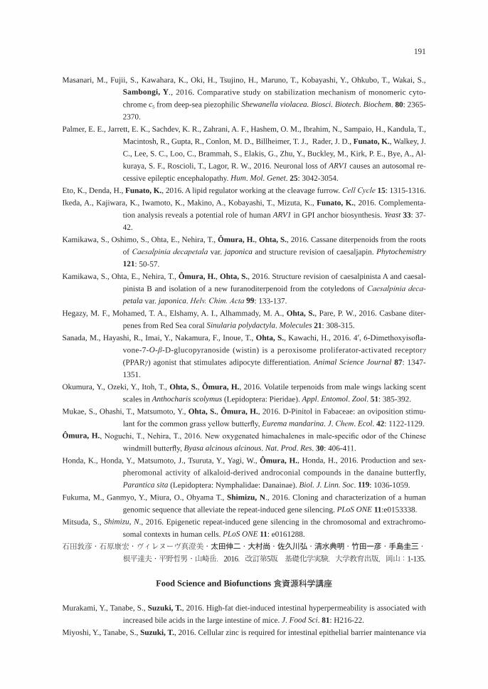
191
Masanari, M., Fujii, S., Kawahara, K., Oki, H., Tsujino, H., Maruno, T., Kobayashi, Y., Ohkubo, T., Wakai, S., Sambongi, Y., 2016. Comparative study on stabilization mechanism of monomeric cyto-chrome c5 from deep-sea piezophilic Shewanella violacea. Biosci. Biotech. Biochem. 80: 2365-2370.
Palmer, E. E., Jarrett, E. K., Sachdev, K. R., Zahrani, A. F., Hashem, O. M., Ibrahim, N., Sampaio, H., Kandula, T., Macintosh, R., Gupta, R., Conlon, M. D., Billheimer, T. J., Rader, J. D., Funato, K., Walkey, J. C., Lee, S. C., Loo, C., Brammah, S., Elakis, G., Zhu, Y., Buckley, M., Kirk, P. E., Bye, A., Al-kuraya, S. F., Roscioli, T., Lagor, R. W., 2016. Neuronal loss of ARV1 causes an autosomal re-cessive epileptic encephalopathy. Hum. Mol. Genet. 25: 3042-3054.
Eto, K., Denda, H., Funato, K., 2016. A lipid regulator working at the cleavage furrow. Cell Cycle 15: 1315-1316.Ikeda, A., Kajiwara, K., Iwamoto, K., Makino, A., Kobayashi, T., Mizuta, K., Funato, K., 2016. Complementa-
tion analysis reveals a potential role of human ARV1 in GPI anchor biosynthesis. Yeast 33: 37-42.
Kamikawa, S., Oshimo, S., Ohta, E., Nehira, T., Ômura, H., Ohta, S., 2016. Cassane diterpenoids from the roots of Caesalpinia decapetala var. japonica and structure revision of caesaljapin. Phytochemistry 121: 50-57.
Kamikawa, S., Ohta, E., Nehira, T., Ômura, H., Ohta, S., 2016. Structure revision of caesalpinista A and caesal-pinista B and isolation of a new furanoditerpenoid from the cotyledons of Caesalpinia deca-petala var. japonica. Helv. Chim. Acta 99: 133-137.
Hegazy, M. F., Mohamed, T. A., Elshamy, A. I., Alhammady, M. A., Ohta, S., Pare, P. W., 2016. Casbane diter-penes from Red Sea coral Sinularia polydactyla. Molecules 21: 308-315.
Sanada, M., Hayashi, R., Imai, Y., Nakamura, F., Inoue, T., Ohta, S., Kawachi, H., 2016. 4′, 6-Dimethoxyisofla-vone-7-O-β-D-glucopyranoside (wistin) is a peroxisome proliferator-activated receptorγ (PPARγ) agonist that stimulates adipocyte differentiation. Animal Science Journal 87: 1347-1351.
Okumura, Y., Ozeki, Y., Itoh, T., Ohta, S., Ômura, H., 2016. Volatile terpenoids from male wings lacking scent scales in Anthocharis scolymus (Lepidoptera: Pieridae). Appl. Entomol. Zool. 51: 385-392.
Mukae, S., Ohashi, T., Matsumoto, Y., Ohta, S., Ômura, H., 2016. D-Pinitol in Fabaceae: an oviposition stimu-lant for the common grass yellow butterfly, Eurema mandarina. J. Chem. Ecol. 42: 1122-1129.
Ômura, H., Noguchi, T., Nehira, T., 2016. New oxygenated himachalenes in male-specific odor of the Chinese windmill butterfly, Byasa alcinous alcinous. Nat. Prod. Res. 30: 406-411.
Honda, K., Honda, Y., Matsumoto, J., Tsuruta, Y., Yagi, W., Ômura, H., Honda, H., 2016. Production and sex-pheromonal activity of alkaloid-derived androconial compounds in the danaine butterfly, Parantica sita (Lepidoptera: Nymphalidae: Danainae). Biol. J. Linn. Soc. 119: 1036-1059.
Fukuma, M., Ganmyo, Y., Miura, O., Ohyama T., Shimizu, N., 2016. Cloning and characterization of a human genomic sequence that alleviate the repeat-induced gene silencing. PLoS ONE 11:e0153338.
Mitsuda, S., Shimizu, N., 2016. Epigenetic repeat-induced gene silencing in the chromosomal and extrachromo-somal contexts in human cells. PLoS ONE 11: e0161288.
石田敦彦・石原康宏・ヴィレヌーヴ真澄美・太田伸二・大村尚・佐久川弘・清水典明・竹田一彦・手島圭三・ 根平達夫・平野哲男・山崎岳.2016.改訂第5版 基礎化学実験.大学教育出版,岡山:1-135.
Food Science and Biofunctions 食資源科学講座
Murakami, Y., Tanabe, S., Suzuki, T., 2016. High-fat diet-induced intestinal hyperpermeability is associated with increased bile acids in the large intestine of mice. J. Food Sci. 81: H216-22.
Miyoshi, Y., Tanabe, S., Suzuki, T., 2016. Cellular zinc is required for intestinal epithelial barrier maintenance via
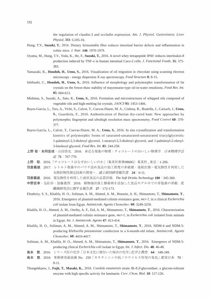
192
the regulation of claudin-3 and occludin expression. Am. J. Physiol. Gastrointest. Liver Physiol. 311: G105-16.
Hung, T.V., Suzuki, T., 2016. Dietary fermentable fiber reduces intestinal barrier defects and inflammation in colitic mice. J. Nutr. 146: 1970-1979.
Oyama, M., Hung, T.V., Yoda, K., He, F., Suzuki, T., 2016. A novel whey tetrapeptide IPAV reduces interleukin-8 production induced by TNF-α in human intestinal Caco-2 cells. J. Functional Foods. 35: 375-383.
Yamazaki, K., Hondoh, H., Ueno, S., 2016. Visualization of oil migration in chocolate using scanning electron microscopy - energy dispersion X-ray spectroscopy. Food Structure 8: 8-15.
Ishibashi, C., Hondoh, H., Ueno, S., 2016. Influence of morphology and polymorphic transformation of fat crystals on the freeze-thaw stability of mayonnaise-type oil-in-water emulsions. Food Res. Int. 85: 604-613.
Mishima, S., Suzuki, A., Sato, K., Ueno, S., 2016. Formation and microstructures of whipped oils composed of vegetable oils and high-melting fat crystals. JAOCS 93: 1453-1466.
Bayes-García, L., Tres, A., Vichi, S., Calvet, T., Cuevas-Diarte, M. A., Codony, R., Boatella, J., Caixach, J., Ueno, S., Guardiola, F., 2016. Authentication of Iberian dry-cured ham: New approaches by polymorphic fingerprint and ultrahigh resolution mass spectrometry. Food Control 60: 370-377.
Bayes-García, L., Calvet, T., Cuevas-Diarte, M. A., Ueno, S., 2016. In situ crystallization and transformation kinetics of polymorphic forms of saturated-unsaturated-unsaturated triacylglycerols: 1-palmitoyl-2,3-dioleoyl glycerol, 1-stearoyl-2,3-dioleoyl glycerol, and 1-palmitoyl-2-oleoyl-3-linoleoyl glycerol. Food Res. Int. 85: 244-258.
上野 聡・本同宏成・山田悟史.2016.身近な現象の物理:チョコレートのおいしい物理学.日本物理学会誌 71:767-770.
上野 聡.2016.「チョコレートはなぜおいしいのか」(集英社新書0860G)集英社 , 東京:1-206.羽倉義雄.2017.レトルト殺菌中のパウチ詰め食品の加工程度の非破壊・連続計測~電気物性を利用した
未開封物性測定技術の開発~.浦上財団研究報告書.24:6-11.羽倉義雄.2016.電気物性を利用した液状食品の品質評価.The Soft Drinks Technology 180:345-360.中野宏幸・包紅杉・加藤義啓.2016.植物抽出液と静菌剤を添加した食品モデル中での芽胞菌の挙動.乳
酸菌研究会に関する報告書.27:172-173.Elnahriry, S. S., Khalifa, H. O., Soliman, A. M., Ahmed, A. M., Hussein, A. H., Shimamoto, T., Shimamoto, T.,
2016. Emergence of plasmid-mediated colistin resistance gene, mcr-1, in a clinical Escherichia coli isolate from Egypt. Antimicrob. Agents Chemother. 60: 3249-3250.
Khalifa, H. O., Ahmed, A. M., Oreiby, A. F., Eid, A. M., Shimamoto, T., Shimamoto, T., 2016. Characterization of plasmid-mediated colistin resistance gene, mcr-1, in Escherichia coli isolated from animals in Egypt. Int. J. Antimicrob. Agents 47: 413-414.
Khalifa, H. O., Soliman, A. M., Ahmed, A. M., Shimamoto, T., Shimamoto, T., 2016. NDM-4 and NDM-5-producing Klebseilla pneumoniae coinfection in a 6-month-old infant. Antimicrob. Agents Chemother. 60: 4416-4417.
Soliman, A. M., Khalifa, H. O., Ahmed, A. M., Shimamoto, T., Shimamoto, T., 2016. Emergence of NDM-5-producing clinical Escherichia coli isolate in Egypt. Int. J. Infect. Dis. 48: 46-48.
島本 整.2016.シリーズ匠の化学「日本文化に根付いた柿渋の化学」 化学と教育.64:348-349.島本 整.2016.果樹研究最前線 No.250「カキタンニンの抗ノロウイルス作用の発見」 果実日本.71:
8-11.Thongekkaew, J., Fujii, T., Masaki, K., 2016. Candida easanensis strain JK-8 β-glucosidase: a glucose-tolerant
enzyme with high specific activity for laminarin. Curr. Chem. Biol. 10: 117-126.
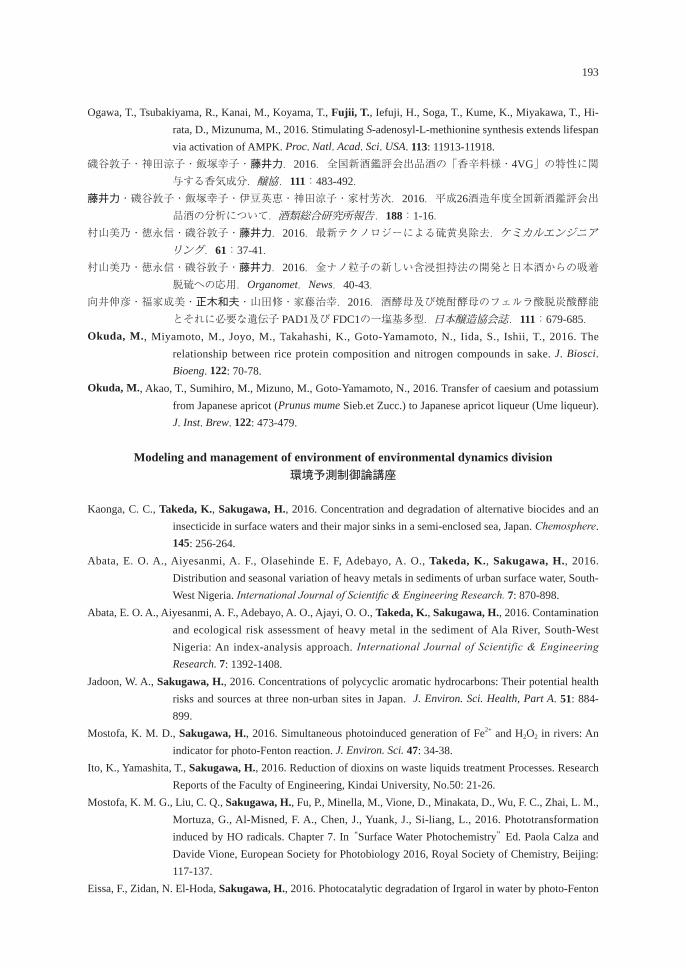
193
Ogawa, T., Tsubakiyama, R., Kanai, M., Koyama, T., Fujii, T., Iefuji, H., Soga, T., Kume, K., Miyakawa, T., Hi-rata, D., Mizunuma, M., 2016. Stimulating S-adenosyl-L-methionine synthesis extends lifespan via activation of AMPK. Proc. Natl. Acad. Sci. USA. 113: 11913-11918.
磯谷敦子・神田涼子・飯塚幸子・藤井力.2016.全国新酒鑑評会出品酒の「香辛料様・4VG」の特性に関与する香気成分.醸協.111:483-492.
藤井力・磯谷敦子・飯塚幸子・伊豆英恵・神田涼子・家村芳次.2016.平成26酒造年度全国新酒鑑評会出品酒の分析について.酒類総合研究所報告.188:1-16.
村山美乃・徳永信・磯谷敦子・藤井力.2016.最新テクノロジーによる硫黄臭除去.ケミカルエンジニアリング.61:37-41.
村山美乃・徳永信・磯谷敦子・藤井力.2016.金ナノ粒子の新しい含浸担持法の開発と日本酒からの吸着脱硫への応用.Organomet.News.40-43.
向井伸彦・福家成美・正木和夫・山田修・家藤治幸.2016.酒酵母及び焼酎酵母のフェルラ酸脱炭酸酵能とそれに必要な遺伝子 PAD1及び FDC1の一塩基多型.日本醸造協会誌.111:679-685.
Okuda, M., Miyamoto, M., Joyo, M., Takahashi, K., Goto-Yamamoto, N., Iida, S., Ishii, T., 2016. The relationship between rice protein composition and nitrogen compounds in sake. J. Biosci. Bioeng. 122: 70-78.
Okuda, M., Akao, T., Sumihiro, M., Mizuno, M., Goto-Yamamoto, N., 2016. Transfer of caesium and potassium from Japanese apricot (Prunus mume Sieb.et Zucc.) to Japanese apricot liqueur (Ume liqueur). J. Inst. Brew. 122: 473-479.
Modeling and management of environment of environmental dynamics division環境予測制御論講座
Kaonga, C. C., Takeda, K., Sakugawa, H., 2016. Concentration and degradation of alternative biocides and an insecticide in surface waters and their major sinks in a semi-enclosed sea, Japan. Chemosphere. 145: 256-264.
Abata, E. O. A., Aiyesanmi, A. F., Olasehinde E. F, Adebayo, A. O., Takeda, K., Sakugawa, H., 2016. Distribution and seasonal variation of heavy metals in sediments of urban surface water, South-West Nigeria. International Journal of Scientific & Engineering Research. 7: 870-898.
Abata, E. O. A., Aiyesanmi, A. F., Adebayo, A. O., Ajayi, O. O., Takeda, K., Sakugawa, H., 2016. Contamination and ecological risk assessment of heavy metal in the sediment of Ala River, South-West Nigeria: An index-analysis approach. International Journal of Scientific & Engineering Research. 7: 1392-1408.
Jadoon, W. A., Sakugawa, H., 2016. Concentrations of polycyclic aromatic hydrocarbons: Their potential health risks and sources at three non-urban sites in Japan. J. Environ. Sci. Health, Part A. 51: 884-899.
Mostofa, K. M. D., Sakugawa, H., 2016. Simultaneous photoinduced generation of Fe2+ and H2O2 in rivers: An indicator for photo-Fenton reaction. J. Environ. Sci. 47: 34-38.
Ito, K., Yamashita, T., Sakugawa, H., 2016. Reduction of dioxins on waste liquids treatment Processes. Research Reports of the Faculty of Engineering, Kindai University, No.50: 21-26.
Mostofa, K. M. G., Liu, C. Q., Sakugawa, H., Fu, P., Minella, M., Vione, D., Minakata, D., Wu, F. C., Zhai, L. M., Mortuza, G., Al-Misned, F. A., Chen, J., Yuank, J., Si-liang, L., 2016. Phototransformation induced by HO radicals. Chapter 7. In “Surface Water Photochemistry” Ed. Paola Calza and Davide Vione, European Society for Photobiology 2016, Royal Society of Chemistry, Beijing: 117-137.
Eissa, F., Zidan, N. El-Hoda, Sakugawa, H., 2016. Photocatalytic degradation of Irgarol in water by photo-Fenton
194
reactions. Chapter 14. In “Surface Water Photochemistry” Ed. Paola Calza and Davide Vione, European Society for Photobiology 2016, Royal Society of Chemistry, Beijing: 275-289.
Mostofa, K. M. G., Liu, C. Q., Zhai, W., Minella, M., Vione, D., Gao, K., Minakata, D., Arakaki, T., Yoshioka, T., Hayakawa, K., Konohira, E., Tanoue, E., Akhand, A., Chanda, A., Wang, B., Sakugawa, H., 2016. Reviews and Syntheses: Ocean acidification and its potential impacts on marine ecosystems. Biogeosciences. 13: 1767–1786.
佐久川弘 , 2016.高大連携に関する日本化学会中国四国支部の活動紹介.尚志会誌 , 一般社団法人尚志会 , 広島市.No.21:7-9.
Assaha, D. V. M., Liu, L., Mekawy, A. M. M., Ueda, A., Nagaoka, T., Saneoka, H., 2016. Effects of drought stress on growth, solute accumulation and membrane stability of leafy vegetable, huckleberry (Solanum scabrum Mill.). J. Environ. Biol. 37: 107-114.
Barutçular, C., Yıldırım, M., Koç, M., Dizlek, H., Akıncı, C., El Sabagh, A., Saneoka, H., Ueda, A., Islam, M. S., Toptaş, I., Albayrak, O., Tanrıkulu, A., 2016. Quality of spring wheat in Mediterranean environments: Grain quality characterization under drought and heat stress. Sylwan. 160: 43-56.
Barutçular, C., El Sabagh, A., Islam, M. S., Ueda, A., Saneoka, H., 2016. Identification of drought tolerance indices associated with grain weight in maize at grain filling stage. Sylwan. 160: 32-42.
El Sabagh, A., Sorour, S., Morsi, A., Islam, M. S., Ueda, A., Barutcular, C., Arioglu, H., Saneoka, H., 2016. Role of osmoprotectants and compost application in improving water stress tolerance in soybean (Glycine max L.). Int. J. Curr. Res. 8: 25949-25954.
Barutcular, C., El Sabagh, A., Konuskan, O., Saneoka, H., Yoldash, K.M. 2016. Evaluation of maize hybrids to terminal drought stress tolerance by defining drought indices. J. Exp. Biol. Agric. Sci. 4: 610-616.
Barutcular, C., Yildirim, M., Kocl, M., Dizlek, H., Akinci, C., Sabagh, A., Saneoka, H., Ueda. A., Islam, M.S., Toptas, I. 2016. Quality traits performance of bread wheat genotypes under drought and heat stress condition. Fresenius Environ. Bull. 25: 6159-6165.
浜名洋司・須川瞬・平尾晃・中元勝彦・柴田健一郎・実岡寛文.2016.モモの樹体ジョイント仕立てと一文字形整枝における定植4年目までの樹体生育,果実生産および栽培管理省力性の比較.園芸学研究.15:153-159.
Kittiwanich, J., Yamamoto, T., Kawaguchi, O., Madinabeitia, I. 2016. Assessing responses of the Hiroshima Bay ecosystem to increasing or decreasing phosphorus and nitrogen inputs. Mar. Poll. Bull., 102: 256-264.
Yamamoto, T., Osawa, K., Asaoka, S., Madinabeitia, I., Liao, L. M., Hirata, S. 2016. Enhancement of marine phytoplankton growth by steel-making slag as a promising component for the development of algal biofuels. ISIJ International. 56: 708-713.
Miyata, Y., Takahashi, K., Watanabe, K., Yamamoto, T., Hayashi, A., Urabe, N. 2016. Field experiments on suppression of hydrogen sulfide in silty sediments using steelmaking slag. Proceedings of the 5th International Conference on Process Development in Iron and Steelmaking (SCANMET V), CD-ROM.
Yamamoto, H., Yamamoto, T., Mito, Y., Asaoka, S. 2016. Numerical evaluation of the use of granulated coal ash to reduce an oxygen-deficient water mass. Mar. Poll. Bull. 107: 188-205.
Miyata, Y., Hayashi, A., Kuwayama, M., Yamamoto, T., Tanishiki, K., Urabe, N. 2016. A field experiment of sulfide reduction in silty sediment using steel-making slag. ISIJ International. 56: 2100-2106.
山本民次・清田忠志・河尻義隆・中原真哉・竹田一彦・中川 光.2016.鉄溶出施肥材適用によるカキ成長促進の試み.水産工学.53:81-85.
山本民次.2016.アサリ復活漁業者模索.フロントライン/備後,中国新聞 .
195
山本民次・大嶋雄治・田中丈裕・西川哲也.2016.栄養塩添加による漁場生産力の向上.日本水産学会誌.82:834-837.
山本民次.2016.貧栄養化に対する局所的施肥の提案.瀬戸内海.72:24-26.山本民次・宮田康人.2016.製鋼スラグ施工時の持続的な環境改善性の検証.平成27年度 JFE と広島大学
の包括協定に基づく報告書.17 pp.山本民次・中川 光.2016.実海域における Hi-ビーズの環境改善メカニズムに関する研究.平成 27 年度
共同研究報告書.30 pp.山本民次.2016.二枚貝増産のための革新的環境改善技術体系の確立.攻めの農林水産業の実現に向けた
革新的技術緊急展開事業研究成果報告書.47 pp.山田 寛・森岡竜二・大塚善久・西村和之・崎田省吾・山本民次・米田輝隆・谷迫淳二・中山雄三.2016.
カキ殻リサイクル材のアサリ育成資材としての検討・評価.平成 27 年度特定非営利活動法人広島循環型社会推進機構研究課題成果報告書.pp.145-158..
Assessment of environmental dynamics division 環境評価論講座
Tominaga-Wada, R., Wada, T., 2016. The Arabidopsis CAPRICE protein fused to the VP16 transcriptional acti-vation domain alters root hair and trichome development. Plant Biotechnol. 33: 129-132.
Tominaga-Wada, T., Wada, T., 2016. The ectopic localization of CAPRICE LIKE MYB3 protein in Arabidopsis root epidermis. J. Plant Physiol. 199: 111-115.
Tominaga-Wada, T., Wada, T., 2016. Analysis of TTG1 and CPC-like MYB genes during Arabidopsis epidermal cell differentiation. Plant Biotechnol. 33: 201-206.

NOTICEAll communication relating to this journal should be addressed to:
The Committee of the Journal,Graduate School of Biosphere Science,
Hiroshima University,Kagamiyama 1-4-4, Higashi-Hiroshima, 739-8528, Japan
Committee of the Journal for 2017Yoshio HAGURA (Food Science and Biofunctions, Professor)*Hideki TANAKA (Food and Resource Economics, Professor)Takashi BUNGO (Animal Science, Professor)Hisashi OMURA (Molecular and Applied Biosciences, Associate Professor)Toshiya HASHIMOTO (Modeling and Management of Environmental Dynamics, Associate Professor)*Chairman of the committee ([email protected])
平成29年12月15日発行
� 非 売 品�
編集発行者 広島大学大学院生物圏科学研究科 〠739-8528 東広島市鏡山1丁目4の4 電話(082)422-7111(代表)
印 刷 所 株式会社ニシキプリント 印 刷 者 宮 㟢 真 〒733-0833 広島市西区商工センター7丁目5-33 電話(082)277-6954㈹
Biosphere ScienceJournal of the Graduate School of Biosphere Science,
Hiroshima University, Vol. 56, 2017
Published by the Graduate School of Biosphere Science,Hiroshima University, Higashi-Hiroshima, Japan
December 2017
CONTENTSORIGINAL ARTICLES
Kazuya Nagasawa and Shinji TaNaka
1 A rare infection of Ceratothoa verrucosa (Isopoda: Cymothoidae) on red seabream, Pagrus major, cultured in central Japan
Kazuya Nagasawa and Hiroki Nakao
7 Chub mackerel, Scomber japonicus (Perciformes: Scombridae), a new host record for Nerocila phaiopleura (Isopoda: Cymothoidae)
Kazuya Nagasawa 13 Two species of copepods, Lernanthropus atrox and Hatschekia pagrosomi, parasitic on crimson seabream, Evynnis tumifrons, in Hiroshima Bay, western Japan
Masato NiTTa 23 A new record of Chaetopsylla mikado from Higashi-Hiroshima city, Hiroshima Prefecture
Mari YoNeTaNi, Ken iida, Taiki Fuji, Katsushi HiraNo, Yusuke koNdo, Susumu oHTsuka, Kazumitsu NakagucHi, Shuhei YamagucHi, Mikio kaTo, Masato Hirose and Toshihiko FujiTa
27 An observation of the walking behavior of Podosphaeraster toyoshiomaruae collected from the bank Oshima-shinsone, Kagoshima Prefecture, Japan
REVIEWKazuya Nagasawa and Hirotaka kaTaHira
33 A revised and updated checklist of the parasites of eels (Anguilla spp.)(Anguilliformes: Anguillidae) in Japan (1915-2017)
Kazuya Nagasawa 71 A synopsis of the parasites of medaka (Oryzias latipes) of Japan (1929-2017)
Kazuya Nagasawa and Daisuke uYeNo
87 A checklist of copepods of the family Pandaridae (Siphonostomatoida)from fishes in Japanese waters (1898-2017)
Kazuya Nagasawa 105 A checklist of the parasites of three species of centrarchids (bluegill, largemouth bass, and smallmouth bass) in Japan (1962-2017)
INFORMATION123 Doctoral dissertation abstracts
176 List of master theses
179 Reports of studies supported by Grant-in-Aid for Research from the Graduate School of Biosphere science, Hiroshima University
185 List of papers by the faculty staff (2016-2017)
56 2017


![グローバルビジネス学科 / Department of Global …...グローバルビジネス学科 2020年度[春 学 ] - 開講表/Course List - Version AY2020 科 番号 開講学年](https://img.pdfslide.us/doc/110x75/5fddb00459d6be56116721d9/ffffffc-department-of-global-ffffffc.jpg)