Embed Size (px)
Citation preview

Shankaracharya *et al. /International Journal Of Pharmacy&Technology
IJPT | Sep-2010 | Vol. 2 | Issue No.3 | 513-527 Page 513
ISSN: 0975-766X Available Online through Research Article
www.ijptonline.com HOMOLOGY MODELING OF TUBULIN ββββ-1 CHAIN AND ITS DOCKING
STUDY WITH COLCHICINE ANALOGUES Shankaracharya*, Priyanka Sharma and Ambarish Sharan Vidyarthi.
Department of Biotechnology, Biral Institute of Technology, MESRA, Ranchi – 835215. E-Mail : [email protected]
Received on 18-06-2010 Accepted on 05-07-2010
ABSTRACT
Present study involves the homology modeling of human tubulin β-1 chain(TUBB1) using known
protein structure of common pig tubulin. Its 3-D structure was evaluated and validated using PROCHECK
comprising 96.8% amino acid residues in favored and allowed region of Ramachandran plot. Stability of the
structure was confirmed by the program ERRAT having the overall quality factor as 83.476 and Verify_3D
result with 83.85% residues having 3D-1D score > 0.2. With the structure of TUBB1 generated, flexible
docking was performed using GLIDE. Results indicate that among 123 colchicine analogues, CID 5370577
was found as best interacting with TUBB1 having lowest dope score of -6.45. Particularly residues
TYR208, SER172 and GLU181 of TUBB1 at bond length 2.272, 1.997 and 2.249 respectively form
hydrogen bonds and showed strong nonbonding interaction with analogue (CID 5370577). Further the
generation of different derivatives can be made by the modification in the moieties of CID 5370577. These
derivatives can be used to develop effective drugs against acute gouty arthritis as well as cancer.
KeyWords: Colchicine, docking, drug design, molecular modeling, TUBB1.
Shankaracharya *et al. /International Journal Of Pharmacy&Technology
IJPT | Sep-2010 | Vol. 2 | Issue No.3 | 513-527 Page 514
INTRODUCTION
Identification of ligand-receptor interactions is important for drug design and the treatment of
diseases. The formation and dissociation of specific noncovalent interactions between varieties of
macromolecules play a crucial role in the function of biological systems.
Tubulin β-1 chain (TUBB1) is the major constituent of microtubules. It binds two moles of GTP, one
at an exchangeable site on the β chain and one at a non-exchangeable site on the α-chain. It is the complete
sequence of about 451 residues having the molecular weight of 50328. Its major function is macromolecule
(eg: protein etc) metabolism, organelle organization and biogensis, protein polymerization. It also shows
GTPase activity, hydrolase activity (acting on acid anhydrides, in phosphorus Containing anhydrides),
nucleoside triphosphatase activity and pyrophosphatase activity. (http://www.drugbank.ca/drugs/DB01394).
Colchicine is a plant derivative, originally extracted from the bulb of Colchicum autumnale. It is an anti-
inflammatory drug that has been in continuous use for more than 3000 years [1]. Its extract was first
described as a treatment for gout in De Materia Medica by Pedanius Dioscorides in the first century. In
August 2009, colchicine won Food and Drug Administration (FDA) approval in the United States as a
stand-alone drug for the treatment of acute flares of gout and familial Mediterranean fever [2]. Colchicine
has been demonstrated to dramatically abrogate the inflammatory response to urate crystals in humans.
Colchicine by it for the treatment of acute attacks of gouty arthritis, before prophylaxis during the
intercritical phase and in the late chronic tophaceous phase of the disease, before prophylaxis of Familial
Mediterranean Fever attacks, and prevention of the development of amyloidosis in this disease. Colchicine
has been showed to be effective in the treatment of articular, cutaneous and mucosal symptoms and
sarcoidosis. Colchicine by it is a neutral and highly lipid-soluble compound at physiologic pH (measure of
acid strength [pKa] = 12.8, molecular weight [MW] = 398), which enables its rapid passage into body
tissues.

Shankaracharya *et al. /International Journal Of Pharmacy&Technology
IJPT | Sep-2010 | Vol. 2 | Issue No.3 | 513-527 Page 515
Tubulin-colchicine interaction has attracted a great deal of attention, probably because of its unusual
chemical characteristics. The drug binds exceptionally slowly but noncovalently to tubulin and once formed
the drug-protein complex is highly stable. [3]
In the present study structure modeling was performed by the program Modeler 9v3. Homology
modeling relies on the identification of one or more known protein structures likely to resemble the structure
of the query sequence, and on the production of an alignment that maps residues in the query sequence to
residues in the template sequence [4]. The predicted model was validated with Procheck, What_Check,
Verify_3d, Errat etc (http://nihserver.mbi.ucla.edu/SAVES/). These programs give the different information
about the structural properties of modeled protein.
Schrodinger Inc docking software Glide 5.0 is frequently used to predict the binding orientation of
small molecule drug to their protein targets in order to predict the affinity and activity of the small molecule.
Docking plays an important role in the rational design of drugs. [5]
MATERIALS AND METHODS
Collection of sequences and Selection of template
Information about Colchicine, its various targets and the protein sequence of the target protein were
retrieved from Drug bank, UniProt/SwissProt database (http://www.uniprot.org/uniprot/Q9H4B7). Template
was selected by homology search of query protein (TUBB1) sequence against the databases available on
PDB (http://www.rcsb.org) through BlastP [6]. Homologous structure of sequence having the lowest E-
value, 50% and above identity, lower resolution was selected as template.
Generation of 3-D structure through homology modeling
Homology modeling was done using Modeler 9v3 [7, 8, 9]. This requires one sequence of known 3D
structure with significant similarity with the target sequence and Python 2.5 script files containing Modeler
commands. The co-ordinate file of template from PDB was used as such.
Shankaracharya *et al. /International Journal Of Pharmacy&Technology
IJPT | Sep-2010 | Vol. 2 | Issue No.3 | 513-527 Page 516
Evaluation and validation of the 3-D structure
All predicted 5 models were evaluated by Procheck [10], Errat [11] and Verify_3D [12].
Ramachandran plot statistics was used to evaluate the stability of the model. Gnuplot was finally used to
plot the profiles generated by Modeler (http://www.gnuplot.info) to validate substantially the structure.
Virtual screening of flavopiridol analogues through molecular docking
Colchicine and its analogues were taken from NCBI Pub-chem in SDF format and converted to 3-D
structure in PDB file format using weblab viewer lite program. Total 123 analogues were used in the study.
The 3-D structure of tubulin beta-1 chain and colchicine 3-D analogues were used for molecular docking
using GLIDE program [13].
Protein structure accession number
The refined homology model of 3D structure of human TUBB1 chain protein was submitted to
PMDB [14] and the same was assigned the identifier PM0076390.
RESULTS AND DISCUSSION
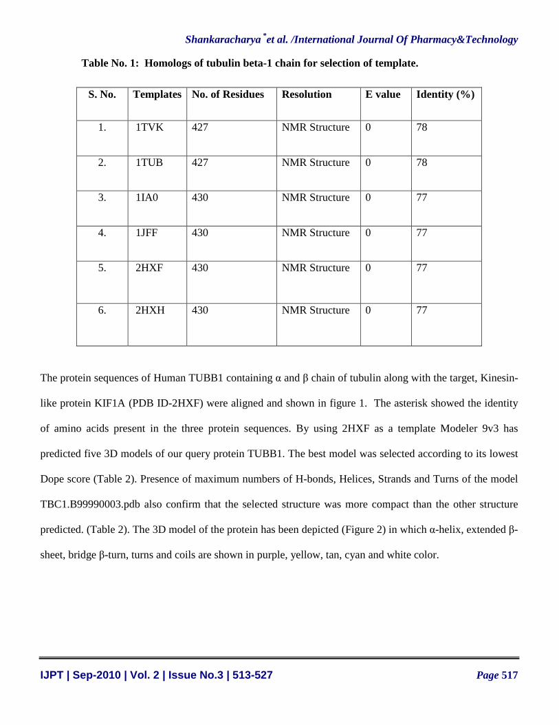
Search for template for the protein sequence of human TUBB1 in Protein Data Bank through RCSB
BLAST has generated 49 homologous structures. Most probable homologous proteins are listed in table-1.
Among them NMR structure of tubulin protein of Sus scrofa, also known as common pig (PDB ID - 2HXF)
was selected on the basis of its NMR structure, high identity (77%) and lowest E-Value (0) (Table-1).
Shankaracharya *et al. /International Journal Of Pharmacy&Technology
IJPT | Sep-2010 | Vol. 2 | Issue No.3 | 513-527 Page 517
Table No. 1: Homologs of tubulin beta-1 chain for selection of template.
S. No. Templates No. of Residues Resolution E value Identity (%)
1. 1TVK 427 NMR Structure 0 78
2. 1TUB 427 NMR Structure 0 78
3. 1IA0 430 NMR Structure 0 77
4. 1JFF 430 NMR Structure 0 77
5. 2HXF 430 NMR Structure 0 77
6. 2HXH 430 NMR Structure 0 77
The protein sequences of Human TUBB1 containing α and β chain of tubulin along with the target, Kinesin-
like protein KIF1A (PDB ID-2HXF) were aligned and shown in figure 1. The asterisk showed the identity
of amino acids present in the three protein sequences. By using 2HXF as a template Modeler 9v3 has
predicted five 3D models of our query protein TUBB1. The best model was selected according to its lowest
Dope score (Table 2). Presence of maximum numbers of H-bonds, Helices, Strands and Turns of the model
TBC1.B99990003.pdb also confirm that the selected structure was more compact than the other structure
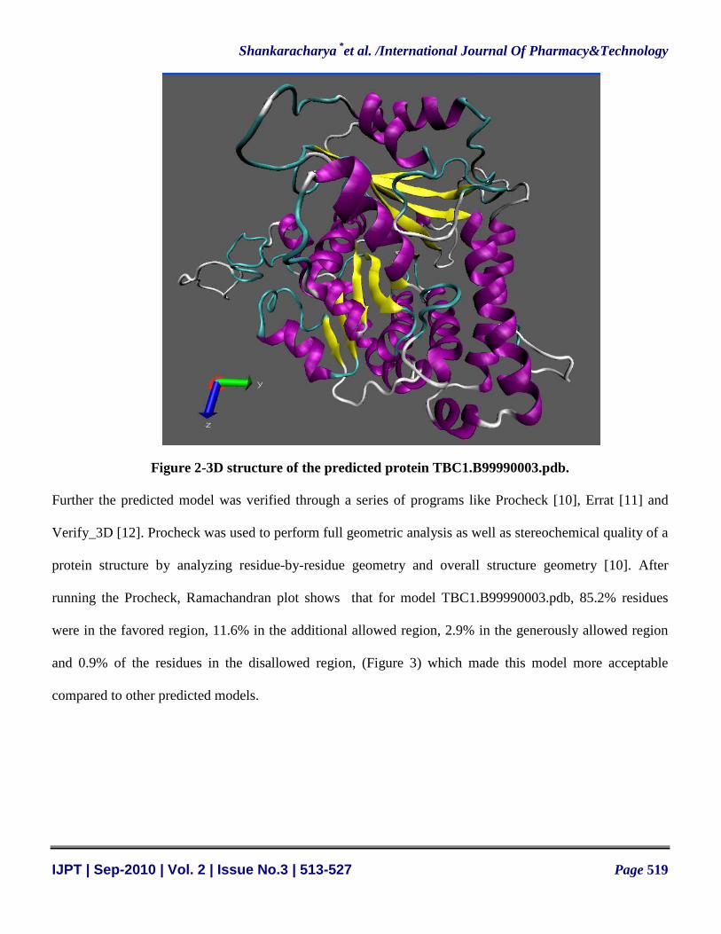
predicted. (Table 2). The 3D model of the protein has been depicted (Figure 2) in which α-helix, extended β-
sheet, bridge β-turn, turns and coils are shown in purple, yellow, tan, cyan and white color.

Shankaracharya *et al. /International Journal Of Pharmacy&Technology
IJPT | Sep-2010 | Vol. 2 | Issue No.3 | 513-527 Page 518
Figure 1 – MSA Result of human tubulin beta-1 chain with template sequence
Table No 2: Dope score and information about H-Bonds, Helices, Strands and Turns of the five
models generated.
S. No. Models Dope score H-Bonds Helices Strands Turns
1. TBC1.B99990001.pdb -47431.36328 311 18 13 46
2. TBC1.B99990002.pdb -47310.61719 310 18 13 42
3. TBC1.B99990003.pdb -47660.28516 313 18 13 46
4. TBC1.B99990004.pdb -47345.20312 313 17 15 46
5. TBC1.B99990005.pdb -47410.92188 305 18 13 45
Shankaracharya *et al. /International Journal Of Pharmacy&Technology
IJPT | Sep-2010 | Vol. 2 | Issue No.3 | 513-527 Page 519
Figure 2-3D structure of the predicted protein TBC1.B99990003.pdb.
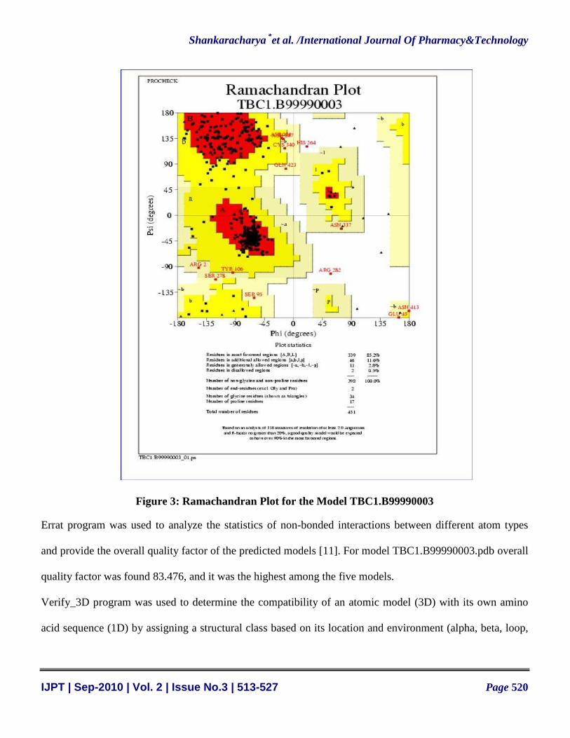
Further the predicted model was verified through a series of programs like Procheck [10], Errat [11] and
Verify_3D [12]. Procheck was used to perform full geometric analysis as well as stereochemical quality of a
protein structure by analyzing residue-by-residue geometry and overall structure geometry [10]. After
running the Procheck, Ramachandran plot shows that for model TBC1.B99990003.pdb, 85.2% residues
were in the favored region, 11.6% in the additional allowed region, 2.9% in the generously allowed region
and 0.9% of the residues in the disallowed region, (Figure 3) which made this model more acceptable
compared to other predicted models.
Shankaracharya *et al. /International Journal Of Pharmacy&Technology
IJPT | Sep-2010 | Vol. 2 | Issue No.3 | 513-527 Page 520
Figure 3: Ramachandran Plot for the Model TBC1.B99990003
Errat program was used to analyze the statistics of non-bonded interactions between different atom types
and provide the overall quality factor of the predicted models [11]. For model TBC1.B99990003.pdb overall
quality factor was found 83.476, and it was the highest among the five models.
Verify_3D program was used to determine the compatibility of an atomic model (3D) with its own amino
acid sequence (1D) by assigning a structural class based on its location and environment (alpha, beta, loop,
Shankaracharya *et al. /International Journal Of Pharmacy&Technology
IJPT | Sep-2010 | Vol. 2 | Issue No.3 | 513-527 Page 521
polar, non-polar etc) and compare the results to good structures [12]. The results showed that model-3 has
passed this verification and 83.85% of the residues have an averaged 3D-1D score > 0.2. Further process is
proceeded by taking the model generated by the modeler with lowest dope score i.e. TBC1.B99990003.pdb.
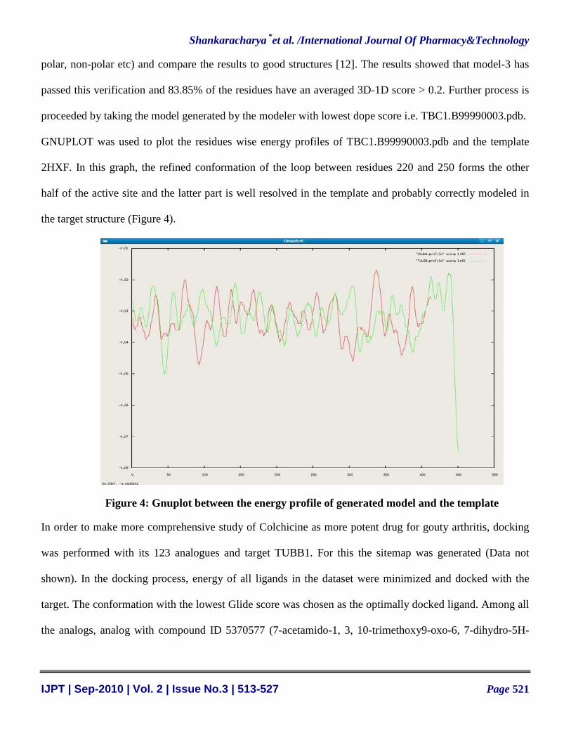
GNUPLOT was used to plot the residues wise energy profiles of TBC1.B99990003.pdb and the template
2HXF. In this graph, the refined conformation of the loop between residues 220 and 250 forms the other
half of the active site and the latter part is well resolved in the template and probably correctly modeled in
the target structure (Figure 4).
Figure 4: Gnuplot between the energy profile of generated model and the template
In order to make more comprehensive study of Colchicine as more potent drug for gouty arthritis, docking
was performed with its 123 analogues and target TUBB1. For this the sitemap was generated (Data not
shown). In the docking process, energy of all ligands in the dataset were minimized and docked with the
target. The conformation with the lowest Glide score was chosen as the optimally docked ligand. Among all
the analogs, analog with compound ID 5370577 (7-acetamido-1, 3, 10-trimethoxy9-oxo-6, 7-dihydro-5H-

Shankaracharya *et al. /International Journal Of Pharmacy&Technology
IJPT | Sep-2010 | Vol. 2 | Issue No.3 | 513-527 Page 522
benzo[a]heptalen-2-yl) (E)-3-(4-methylphenyl) prop-2-enoate) (figure 5) showed the lowest glide score -
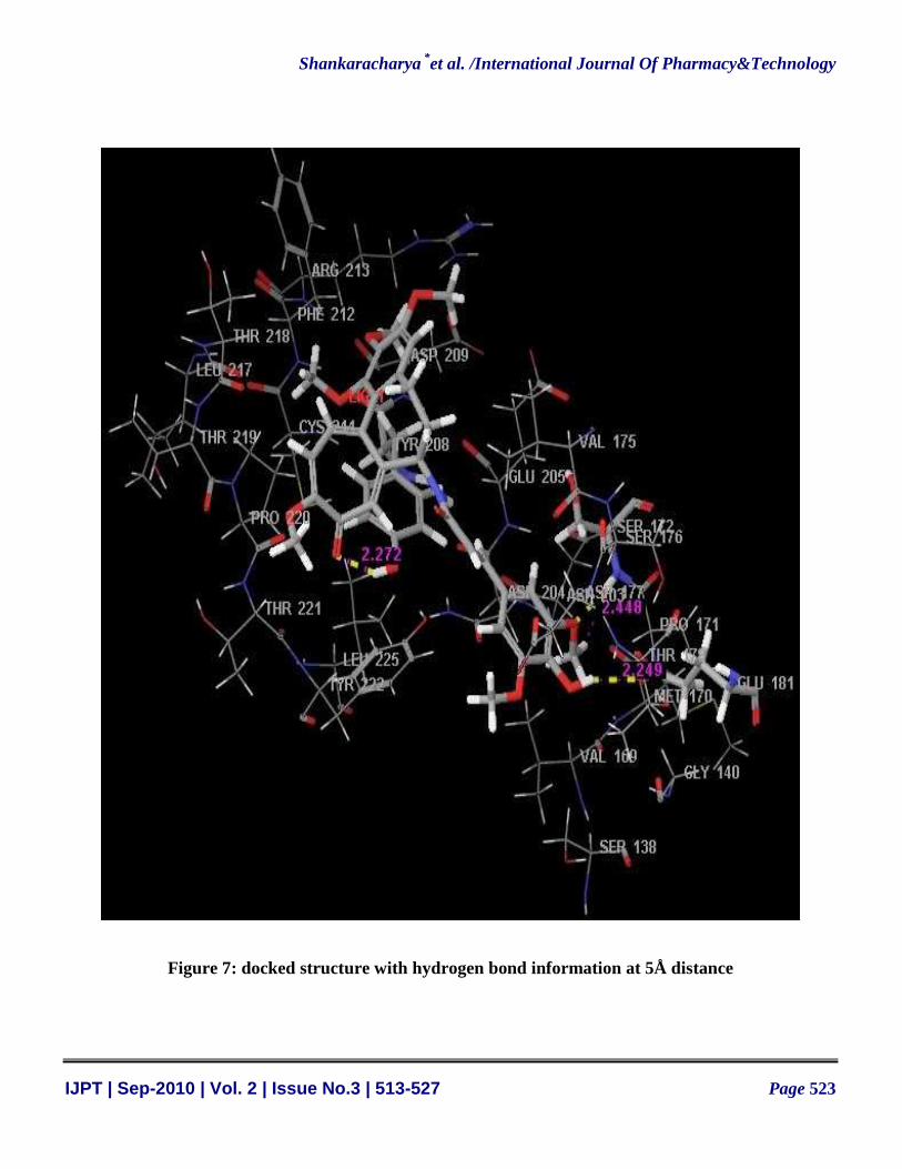
6.45 (figure 6) and minimal RMSD (RMSD=1.015 Å). Various residues like ASP209, CYS244, TYR208,
VAL175, GLU205, SER 172, SER176, PRO220, THR221, LEU225, ASN204, THR178, GLU181 and
MET170 were found in the interaction region of the protein (figure 7). Various physical and chemical
characteristics of this analog have been tabulated in table 3 for the understanding of the nature of the analog.
Figure 5: Analog with compound ID 5370577
Figure 6: Screen-shot of the docking report generated by GLIDE 5.0
Shankaracharya *et al. /International Journal Of Pharmacy&Technology
IJPT | Sep-2010 | Vol. 2 | Issue No.3 | 513-527 Page 523
Figure 7: docked structure with hydrogen bond information at 5Å distance
Shankaracharya *et al. /International Journal Of Pharmacy&Technology
IJPT | Sep-2010 | Vol. 2 | Issue No.3 | 513-527 Page 524
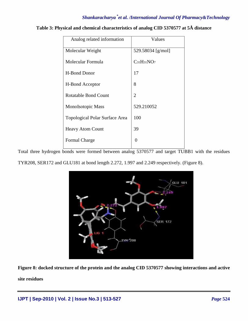
Table 3: Physical and chemical characteristics of analog CID 5370577 at 5Å distance
Analog related information Values
Molecular Weight
Molecular Formula
H-Bond Donor
H-Bond Acceptor
Rotatable Bond Count
MonoIsotopic Mass
Topological Polar Surface Area
Heavy Atom Count
Formal Charge
529.58034 [g/mol]
C31H31NO7
17
8
2
529.210052
100
39
0
Total three hydrogen bonds were formed between analog 5370577 and target TUBB1 with the residues
TYR208, SER172 and GLU181 at bond length 2.272, 1.997 and 2.249 respectively. (Figure 8).
Figure 8: docked structure of the protein and the analog CID 5370577 showing interactions and active
site residues
Shankaracharya *et al. /International Journal Of Pharmacy&Technology
IJPT | Sep-2010 | Vol. 2 | Issue No.3 | 513-527 Page 525
Predicting energetics of protein-ligand binding and searching space of possible poses and
conformations are the major challenges in finding new drugs. Further several variants of the analog CID
5370577 can be designed by modifying its moieties at different positions in the compound to find out better
drug than the existing one. This ligand has diverse and interesting biological activities, and it should be
particularly useful as lead compound for drug development. Better understanding of the interactions
between tubulin β-1 chain and ligand CID 5370577 will be crucial for the treatment of acute gouty arthritis,
familial Mediterranean fever and cancer as well as useful for pharmaceutical and functional food industries.
Hence the selected analogue CID_5370577 can be used as a replacement of the existing drug Colchicine and
may be subject to laboratory confirmation and clinical trial.
CONCLUSION
3-D structure of TUBB1 protein of human was constructed with modeler9v3 and the virtual
screening of drug was performed to search suitable analog among 123 analogs of drug Colchicine.
Compound ID_5370577 was found more potent one comprising highest binding affinity with modeled
TUBB1 protein. The present investigation may provide new insight to control superfluous use of drugs in
vitro. The 3-D structure of tubulin β-1 chain of human and Colchicine analogue CID_5370577 would
needed to be verified experimentally. Further various variants of this analog can be designed and docked to
find out more potent drug than the suggested Colchicine (CID_5370577).
ACKNOWLEDGEMENT
The authors acknowledge BTISnet, Department of Biotechnology, Government of India, New Delhi for
providing Infrastructure facility for Bioinformatics and Department of Biotechnology, B. I. T. MESRA,
Ranchi for providing Docking software.
Shankaracharya *et al. /International Journal Of Pharmacy&Technology
IJPT | Sep-2010 | Vol. 2 | Issue No.3 | 513-527 Page 526
REFERENCES
1. E.F. Hartung, 1954, Ann Rheum Dis., Vol. 13, pp190–200.
2. C. Petrochko, S. Writer, 2009, FDA Approves Gout Treatment After Long Years of use,MedPage
Today. (http://www.medpagetoday.com/ProductAlert/Prescriptions/15358).
3. R. Bai, D. G. Covell, X. Pei, J. B. Ewell, 2000, J Biol Chem, Vol 275(51), pp40443-40452.
4. M.A. Marti-Renom, A.C. Stuart, A. Fiser, R. Sanchez, F. Melo, A. Sali, 2000, Annu Rev Biophys
Biomol Struct., Vol 29, pp291-325.
5. D.B. Kitchen, H. Decornez, J.R. Furr, J. Bajorath, 2004, Nature reviews. Drug discovery, Vol 3(11),
pp935–949.
6. S.F. Altschul, W. Gish, W. Miller, E.W. Myers, D. J. Lipman, 1990, J Mol Biol., Vol 215(3),
pp403-410.
7. R. Sánchez, A. Sali, 1997, Curr Opin Struct Biol., Vol 7 pp206-214.
8. A. Fiser, A. Sali., 2003, Methods Enzymol., Vol 374, pp461-469.
9. A. Sali, T.L. Blundell, 1993, J Mol Biol.,Vol. 234, pp779-815.
10. R.A. Laskowski, M.W. MacArthur, D.S. Moss, J.M Thornton., 1993, J Appl Crystallography, Vol26,
pp283-291.
11. C. Colovos, T.O. Yeates, 1993, Protein Sci., Vol 2, pp1511-1519.
12. R. Luthy, J.U. Bowie, D. Eisenberg, 1992, Nature, Vol 356, pp83-85.
13. R.A. Friesner, J.L. Banks, R.B. Murphy, T.A. Halgren, J.J. Klicic, D.T. Mainz, M.P. Repasky, E.H.
Knoll, M. Shelley, J.K. Perry, D.E. Shaw, P. Francis, P.S. Shenkin., J. Med. Chem., 2004, Vol 47 (7),
pp1739–49.
14. T. Castrignano, P.D. De Meo, D. Cozzetto, I.G. Talamo, A. Tramontano, 2006, Nucleic Acids Res.,
Vol 34(1), ppD306 - D309.

Shankaracharya *et al. /International Journal Of Pharmacy&Technology
IJPT | Sep-2010 | Vol. 2 | Issue No.3 | 513-527 Page 527
CORRESPONDING AUTHOR’S INFORMATION
SHANKARACHARYA
DEPARTMENT OF BIOTECHNOLOGY,
BIRLA INSTITUTE OF TECHNOLOGY,
MESRA, RANCHI – 835215
E-Mail : [email protected]
Tel : + 91 651 2276223 (O), + 91 9431978640 (M)