Embed Size (px)
Citation preview
International Journal of Phytomedicine 3 (2011) 204-215 http://www.arjournals.org/index.php/ijpm/index
Original Research Article
Identification of β-carotene and β-sitosterol in methanolic extract of
Dipteracanthus patulus (Jacq) nees and their role in antimicrobial and antioxidant activity.
Shrinivas B Bumrela*1 and Suresh R Naik1
*Corresponding author: Shrinivas B. Bumrela Sinhgad Technical Education Society’s, Sinhgad Institute of Pharmaceutical Sciences, Off Mumbai Pune. Expressway, Kusgaon (Bk), Lonavala, Dist-Pune, Pin: 410401, MS, India. Telephone: +919822628406, +912114-3043024, Fax-+912114270258. [email protected]
Abstract The Dipteracanthus patulus (Jacq) nees is undershrub belonging to the family acantheaceae. Antimicrobial activity (disc diffusion method) and antioxidant activity by different in-vitro methods (DPPH, hydrogen peroxide, nitric oxide radical scavenging and reducing power) of methanolic extract of Dipteracanthus patulus (MEDP) was evaluated. The qualitative and quantitative estimation of β-carotene and β-sitosterol in MEDP was carried out by high performance thin layer chromatography (HPTLC). The total phenolic content of was determined by Folin-Ciocalteu method. Experimental findings indicate promising antimicrobial activity (antibacterial and antifungal) and potent antioxidant activity of MEDP. In addition, phytochemical analysis and spectral studies of MEDP were also performed. It is presumed that antimicrobial and antioxidant activity observed with MEDP may largely be attributed to the presence of major phytoconstituents (β-carotene, β-sitosterol and iridoid glycosides) and other minor components may participate as promoters. Keywords Dipteracanthus patulus; HPTLC; β-carotene; β-sitosterol; iridoid glycosides; Antioxidant activity; Antimicrobial activity.
Introduction It is well known that plants are rich source of variety of chemicals with nutritive and therapeutic properties. In modern era, herbs are seen as potential medicine for a variety of diseases often viewed to supersede the pharmacological efficacy of allopathic drugs. Medicinal plants have long been used as a resource of therapeutic agents worldwide. Herbal medicines have progressively been used to treat many infectious and metabolic diseases. All plants produce chemical compounds as a part of their normal metabolic activity viz. primary and secondary metabolites. Many secondary metabolites have shown therapeutic activity [1].
Infectious diseases are the world’s leading cause of premature deaths and killing thousands of people every day. In recent years, drug resistance to human pathogenic bacteria has been commonly reported from all over the world [2]. There is an increasing demand from pharmaceutical industry for new lead molecules which can inhibit the growth or kill pathogenic microbes. Plants are rich in wide variety of secondary metabolites, such as tannins, terpenoids, alkaloids and flavonoids have exhibited antimicrobial activity in-vitro [3]. Free radicals are generated in our body as a result of oxidation of biomolecules. Formations of such free radicals are known to cause injury to cells
ISSN: 0975-0185
This work is licensed under a Creative Commons Attribution 3.0 License.
Bumrela et al. International Journal of Phytomedicine 3 (2011) 204-215
205
and also responsible for many ailments such as cancer, Alzheimer’s disease, atherosclerosis, inflammatory disorders and others. Natural antioxidants are the chemical agents which have the ability to quench these hazardous free radicals and to neutralize their potential to attack the cells. Herbs are known to elicit antioxidant activities due to the presence of various phytoconstituents like terpenoids, lycopene, β-carotene, α-carotene, lutein, polyphenolics, catechins, isoflavone and many other secondary metabolites [4]. The Dipteracanthus patulus (Jacq) nees (DP) is undershrub belonging to the family acantheaceae. In folklore medicine it has been documented that DP is used for curing the eyesore by introducing the extract into eyelid. Furthermore, the leaves of various species of Dipteracanthus were used in various infectious diseases [5]. DP showed the presence of lyoniresinol-9’-O-β-D-glucoside, 5,5-dimethoxy-lariciresinol-9-O-β glucopyranoside, β-sitosterol, lupeol, α-ethyl galactose, apiginin-7-O-rutinoside, α-D-glucose, β-D-glucose and β-D-fructose [6]. Different phytoconstituents viz ascorbic acid, phenolic compounds, tannin, lycopene, carotenoid and α-tocopherol in DP have also been reported [7]. Scanty studies on DP pertaining to wound healing [8], antidiabetic [9] and cardiovascular system [10] have been reported. However, no studies on the antimicrobial and antioxidant activity of DP have been reported. Keeping in mind, wide usage of DP in folklore medicine and therapeutically important phytochemical constituents present in this plant, it was felt necessary to evaluate in-vitro antimicrobial and antioxidant activity of methanolic extract of DP leaves. The levels of phenolics, and high performance thin layer chromatography (HPTLC) fingerprinting with quantification (β-carotene and β-sitosterol) of MEDP were also performed to find out the phytoconstituents responsible for such activity.
Experimental Chemicals 1,1-Diphenyl-2-picrylhydrazyl (DPPH), β-carotene, β-sitosterol (Sigma Aldrich, USA), methanol (Qualigens, Mumbai), 2,4,6-tris(2-pyridyl)-s-triazine (TPTZ) (Sisco Research Lab., Mumbai), trichloroacetic acid (TCA), ascorbic acid, butylated hydroxytoluene (BHT), gallic acid, potassium ferricyanide, ferrous and ferric chloride and other reagents of analytical grade were purchased from Fine Chemicals, Pune. Plant Material The plant material was collected in the month of June 2010 from the nearby villages of Solapur district (MS), India [It is located between 17.10 to 18.32 degrees to the north latitude while it is about 74.42 to 76.15 degrees to the east longitude]. The soil is black and of fertile quality and rain fall is scanty. The collected plant material was botanically authenticated (Certificate No. BSI/WC/Tech/2007/460 dated: 03/08/2007) by Botanical Survey of India (BSI), Pune Division, Maharashtra State, India and the voucher specimen is deposited in BSI, Pune, MS (India). Preparation of extract The methanolic extract of DP leaves (MEDP) was prepared by extracting 20gm of DP leaves in Soxhlet apparatus with methanol (2x500ml) for 6h. The combined extract was then evaporated on a rotary vacuum evaporator to obtain semi solid mass. Concentrated MEDP extract was stored in amber color glass containers at 4oC in refrigerator. The extract was re-dissolved in methanol for determining antimicrobial and antioxidant activities (in-vitro). Phytochemical screening The qualitative phytochemical analysis were performed for MEDP extracts to determine the presence/ absence of various chemical constituents viz carbohydrate, tannins, phenolics; flavonoids, steroids, carotinoids and iridoid glycoside [11 & 12].
Bumrela et al. International Journal of Phytomedicine 3 (2011) 204-215
206
Determination of total phenolic content The total phenolic content in MEDP was determined by the method of Slinkard and Singleton [13] using Folin Ciocalteu (FC) reagent. The standard curve was drawn using gallic acid (50-500μg/ml) in ethanol. Briefly, 1.0ml of extract containing 1.0gm extract was diluted to 46ml and 1.0ml of FC reagent was added and allowed to stand for 3min. Aliquot of 3ml of 2% (w/v) sodium carbonate was added and 2hr later the absorbance was measured at 760nm. The total phenolic content was expressed as microgram of gallic acid equivalent (GAE) per gram dry weight of the sample. The results were average of triplicate reading. Analytical profiling of MEDP UV-visible absorption spectra Ultraviolet-visible absorption spectra and λ-maximum for MEDP was studied using Jasco V520 UV-visible spectrophotometer (Japan). Qualitative analysis of β-carotene and β-sitosterol by HPTLC HPTLC was performed on 2 separate precoated silica gel aluminium TLC plates 60F254 (E-Merk, Germany) for qualitative evaluation of β-carotene and β-sitosterol in MEDP. In brief, concentrated MEDP (10μL) and standard markers (5μL) were loaded on TLC plates with Camag Linomat 5 applicator with nitrogen supply. The mobile phase used for β-carotene and β-sitosterol was n-hexane: acetone (70:30 v/v) and toluene: ethyl acetate (80:20 v/v) respectively. The plates were developed to a distance of 80mm in a Camag twin-trough chamber previously equilibrated with mobile phase for 20min. After development of β-sitosterol plate, derivatization was carried out with 5% sulphuric acid in methanol and heated at 110oC on Camag TLC plate platform heater for 5min. Camag TLC visualizer-150503 was used for photodocumentation of β-sitosterol and β-carotene at 254nm, 366nm and White R. The β-carotene HPTLC chromatogram was obtained using Camag Scanner-170422 in conjunction with WinCATS-5 software.
Assay of β-carotene by HPTLC For the quantitative estimation of β-carotene, 2000μg of MEDP and 2μg of standard β-carotene were loaded on precoated silica gel aluminium TLC plates 60F254 (E-Merk, Germany) and plates were developed to a distance of 80mm with n-hexane: acetone (70:30 v/v). Densitometric analysis was carried out using Camag TLC Scanner-170422 in absorbance mode at 455nm. The chromatogram was integrated using WinCATS software for area calculations. Assay of β-sitosterol by HPTLC For the quantitative estimation of β-sitosterol, 500μg of MEDP and 2μg of standard β-sitosterol were loaded on precoated silica gel aluminium TLC plates 60F254 (E-Merk, Germany) and plates were developed to a distance of 80mm with toluene: ethyl acetate (80:20 v/v). Densitometric analysis was carried out using Camag TLC Scanner-170422 in absorbance mode at 580nm. The chromatogram was integrated using WinCATS software for area calculations. Evaluation of antioxidant activity (in-vitro) Antioxidant activity (in-vitro) of MEDP was determined using well documented analytical methods. All the experiments are performed in triplicate. DPPH radical scavenging activity DPPH is a free radical compound and has been used to evaluate the free radical scavenging ability in-vitro. Free radical scavenging capacity of MEDP was determined using method of Chen et al [14] and compared with ascorbic acid and BHT. Briefly MEDP, ascorbic acid or BHT (0.3125-10mg/ml) concentration prepared in methanol was mixed with 1ml of 0.1mM DPPH methanolic solution. The reaction mixture was incubated at 37oC for 30min and absorbance was measured at 517nm on Jasco V-520 UV-visible spectrophotometer (Japan). The percent inhibition was calculated by the formula [A1-A2/A2] x 100 (A1=absorbance without extract; A2=absorbance with extract).
Bumrela et al. International Journal of Phytomedicine 3 (2011) 204-215
207
Nitric oxide scavenging activity Nitric oxide scavenging activity was measured by Griess reaction described by Green LC et al [15]. The reaction mixture containing 4ml of 10mM sodium nitropruside, 1ml of saline phosphate buffer (pH 7.4) and 1ml (0.1875-3mg/ml) of MEDP, ascorbic acid or BHT were incubated at 25oC for 150min. After incubation, 0.5ml of Griess reagent (1% sulphanilamide, 2% O-phosphoric acid and 0.1% NEDD) was added into 0.5ml of reaction mixture, allowed to stand in diffused light for 30min. The absorbance was measured at 535nm against corresponding blank solutions. The percent inhibition was calculated by the formula [A1-A2/A2] x 100 (A1=absorbance without extract; A2=absorbance with extract). Hydrogen peroxide scavenging activity Hydrogen peroxide scavenging activity was determined by the method of Ruch et al [16]. Hydrogen peroxide is a non-radical derivative of oxygen and found to be toxic to cells as they give rise to hydroxyl radical formation inside the cell. Hydrogen peroxide solution 2mM in 50mM phosphate buffer (pH 7.4) was prepared. The concentration of H2O2 was determined spectrophotometrically by measuring absorbance at 230nm and using the molar extinction coefficient for H2O2 of 81 mol-1cm-1. Aliquot of 0.5ml (0.1563-10mg/ml) of MEDP or ascorbic acid was transferred into a test tube and volume made up to 2ml with 50mM phosphate buffer or solvent. A solution of hydrogen peroxide 3ml was added to above mixture, vortexed and absorbance was measured at 230nm after 10min, against 50mM phosphate buffer as blank. The percent hydrogen peroxide scavenging was calculated by the formula [A1-A2/A2] x 100 (A1=absorbance without extract; A2=absorbance with extract). Reducing Power The reducing power of MEDP in comparison with ascorbic acid and BHT was determined
using the modified method of Oyaizu [17]. Aliquot of 1 ml of MEDP (0.625-10mg/ml), ascorbic acid or BHT was mixed with 2.5 ml of 0.2M phosphate buffer (pH 6.6) and 2.5ml of 1% potassium ferricyanide and then incubated at 50oC for 20min. After incubation 2.5 ml of 10% (w/v) tricholroacetic acid was added to the mixture to stop the reaction. The mixture was centrifuged for 10min at 2790g, and 2.5ml of supernatant was mixed with 2.5ml distilled water and 0.5ml of 0.1% FeCl3. The absorbance of the resulting solution was measured at 700nm. Increased absorbance of the reaction mixture indicates an increase in reduction capacity. Ferric reducing (FRAP) assay Ferric reducing activity of MEDP was determined according to the method of Benzie and Strain [18] with slight modification. The antioxidant potential of extracts was determined from the standard curve of ferrous sulphate (100-2000mM) in methanol. The FRAP reagent was prepared fresh daily by mixing 100ml of acetate buffer (300mM, pH 3.6), 10ml TPTZ solution (10mM TPTZ in 40mM HCl), 10ml FeCl3 (20mM) in a ratio of 10:1:1 (v/v). FRAP reagent was warmed to 37oC on a water bath prior to use. Aliquot 100µl of MEDP (5mg/ml), ascorbic acid or BHT (1mg/ml) was added to 1.5ml of the FRAP reagent. The absorbance of the reaction mixture was measured at 593nm after 4min. The results were expressed as µM Fe(II)/g dry weight of plant material [19]. Antimicrobial activity screening The stock solution of MEDP (1000µg/ml) was prepared by dissolving in methanol. Standard Microbial Cultures The authentic cultures of test microorganisms have been procured from National Chemical Laboratory (NCL), Pune, MS, India. Gram (+Ve) bacteria, Bacillus subtilis (ATCC 633), Bacillus cereus (ATCC 10703), Micrococcus luteus (ATCC 4698), Streptococcus pyogenes (ATCC 8668), Streptococcus faecalis (ATCC 14506), Staphylococcus aureus (ATCC 9144) and the
Bumrela et al. International Journal of Phytomedicine 3 (2011) 204-215
208
gram (-Ve) bacteria, Escherichia coli (ATCC 8739), Pseudomonas aeruginosa (ATCC 19429) and Klebsiella pneumoniae (NCIM 2957) were used as test microorganisms. The fungal organisms tested were Aspergillus niger (NCIM 549) and Candida albicans (NCIM 3471). Kirby-Bauer method The antimicrobial activity of MEDP was screened using the disc diffusion method described by Kirby and Bauer [20]. The freshly incubated cultures of bacteria and fungi in physiological saline were used for inoculation. The Muller-Hinton agar and sabouraud dextrose agar (Hi-media) were used for bacteria and fungi respectively. Inoculum density of each microbial suspension was adjusted to reach an optical comparison to that of a 0.5 McFarland standard, resulting in a suspension and approximately containing 1 to 2 x 10⁸ CFU/ml. The agar plates were inoculated by streaking the swab over the entire sterile agar surface. This procedure was repeated by streaking 2 more times, rotating the plate approximately 60o each time to ensure even
distribution of the microorganisms. The plates were allowed to dry at room temperature. The MEDP 250, 500, 1000µg per disc was loaded on 6-mm sterile discs in an aseptic area. The discs with the help of sterile forceps were impregnated on labeled culture plates. The plates were allowed to stand at room temperature for 30min to diffuse the extract into the agar and then incubated at 37oC for 24h for bacteria and for 48h for fungi. Subsequently, the plates were examined for growth inhibition of microorganism measuring inhibition zone diameter (IZD) in millimeter (mm). Ciprofloxacin 15μg and Fluconazole 10μg discs were used as standard for bacteria and fungi respectively. All the experiments were performed in triplicate. Statistical analysis The results were expressed as mean ± standard deviation. Data were analyzed using student t-tests and ANOVA. Linear regression analysis was used to calculate the IC50 values. P values less than 0.05 (p < 0.05) were considered significant.
Table 1 Standardization of DP leaves and MEDP.
Particulars Description DP leaves Colour of dried leaves Dull green Surface Hairy Stomata Anisocytic (Cruciferous) stomata LOD % NMT 3.2% Total Asha 344 ± 10.24 Extractive valuea 199 ± 08.64 Total phenolics 351.09 ± 01.89 μgGAE per gram dried sample β-carotene content 0.08 gm% β-sitosterol content 0.19 gm% MEDP Colour Green semisolid mass Solubility Soluble in chloroform, acetone, DMSO, ethyl acetate, methanol pH of 1% w/v solution 6.22 ± 0.02 UV spectra (λmax) 422nm (β-carotene) Colour in UV 254nm Reddish orange fluorescence HPTLC analysis β-carotene Rf 0.77 and β-sitosterol Rf 0.4
a values are mg per gram ± SD of dried leaves sample.
Bumrela et al. International Journal of Phytomedicine 3 (2011) 204-215
209
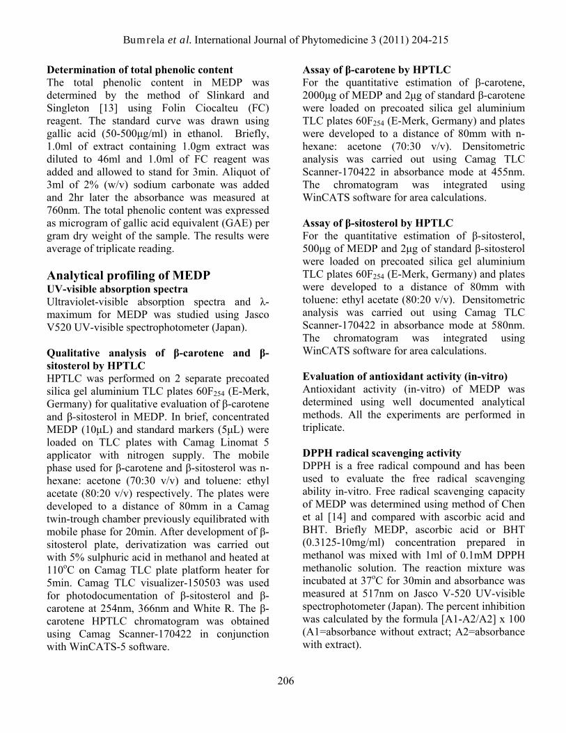
Fig 1 UV-visible spectra of MEDP.
Results and discussion Extractive value and phytochemical screening The extractive value with methanol was found to be 199±8.54 mg/g dry weight of DP leaves. The appearance of extract was dark green in color and showed intense orange colored fluorescence in UV light at 254nm. Phytochemical analysis showed the presence of carbohydrate, iridoid glycosides, phenolics, tannins, flavonoids, carotinoids and steroids in MEDP. Standardization parameters of DP leaves and MEDP are summarized in Table 1. Total phenolic content Total phenolic content was determined by FC method from the regression equation of gallic acid calibration curve (y = 0.001x + 0.033, r² = 0.994) and expressed in microgram gallic acid equivalents (GAE) per gram of dried sample. The total phenolic content obtained in MEDP was 300.08 ± 2.06 µg GAE/g.
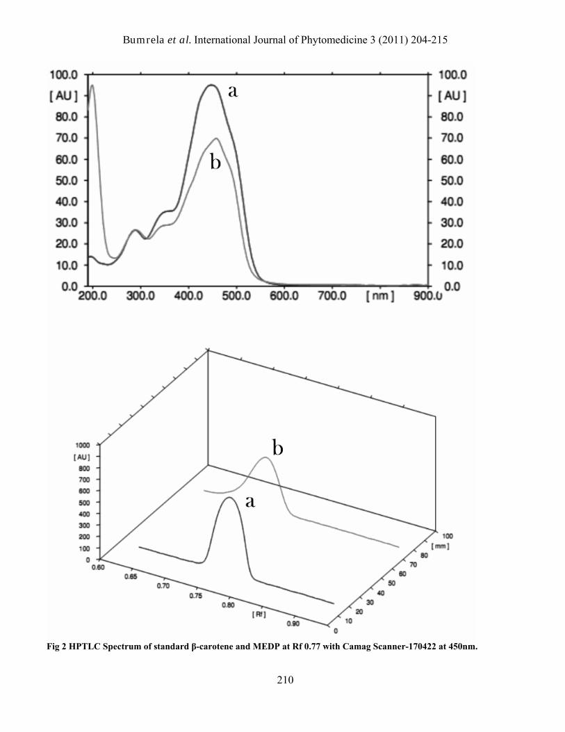
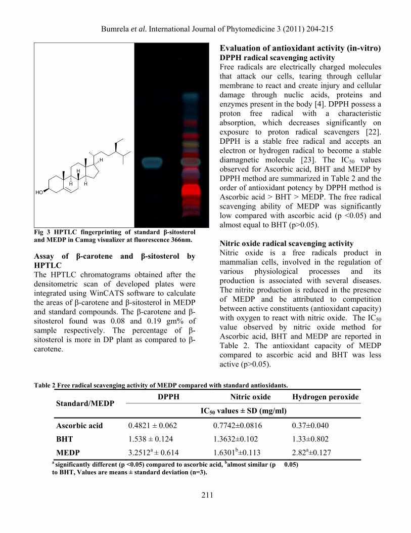
Analytical profiling UV-visible absorption spectra The λmax values observed in UV-visible absorption spectrum of MEDP were at 422nm and 664nm (Fig 1). The documented reports on β-carotene [21] showed maximum absorbance at 422nm thereby suggesting the presence of this phytochemical constituents in MEDP. Qualitative analysis of β-carotene and β-sitosterol by HPTLC HPTLC analysis of MEDP confirmed the presence of β-carotene and β-sitosterol at Rf values 0.77 and 0.40 respectively. HPTLC spectrum of β-carotene and MEDP recorded in Camag TLC scanner-170422 at 450nm showed complete overlapping at start, middle and end position of spectrum (Fig 2). The bands of β-carotene showed yellow-orange coloration in WhiteR light. The photo-documentation β-sitosterol TLC plate after derivatization with 5% sulphuric acid in methanol showed gray color bands in Camag visualizer at fluorescence 366nm (Fig 3).
Bumrela et al. International Journal of Phytomedicine 3 (2011) 204-215
210
Fig 2 HPTLC Spectrum of standard β-carotene and MEDP at Rf 0.77 with Camag Scanner-170422 at 450nm.
Bumrela et al. International Journal of Phytomedicine 3 (2011) 204-215
211
Fig 3 HPTLC fingerprinting of standard β-sitosterol and MEDP in Camag visualizer at fluorescence 366nm. Assay of β-carotene and β-sitosterol by HPTLC The HPTLC chromatograms obtained after the densitometric scan of developed plates were integrated using WinCATS software to calculate the areas of β-carotene and β-sitosterol in MEDP and standard compounds. The β-carotene and β-sitosterol found was 0.08 and 0.19 gm% of sample respectively. The percentage of β-sitosterol is more in DP plant as compared to β-carotene.
Evaluation of antioxidant activity (in-vitro) DPPH radical scavenging activity Free radicals are electrically charged molecules that attack our cells, tearing through cellular membrane to react and create injury and cellular damage through nuclic acids, proteins and enzymes present in the body [4]. DPPH possess a proton free radical with a characteristic absorption, which decreases significantly on exposure to proton radical scavengers [22]. DPPH is a stable free radical and accepts an electron or hydrogen radical to become a stable diamagnetic molecule [23]. The IC50 values observed for Ascorbic acid, BHT and MEDP by DPPH method are summarized in Table 2 and the order of antioxidant potency by DPPH method is Ascorbic acid > BHT > MEDP. The free radical scavenging ability of MEDP was significantly low compared with ascorbic acid (p <0.05) and almost equal to BHT (p>0.05). Nitric oxide radical scavenging activity Nitric oxide is a free radicals product in mammalian cells, involved in the regulation of various physiological processes and its production is associated with several diseases. The nitrite production is reduced in the presence of MEDP and be attributed to competition between active constituents (antioxidant capacity) with oxygen to react with nitric oxide. The IC50 value observed by nitric oxide method for Ascorbic acid, BHT and MEDP are reported in Table 2. The antioxidant capacity of MEDP compared to ascorbic acid and BHT was less active (p>0.05).
Table 2 Free radical scavenging activity of MEDP compared with standard antioxidants.
DPPH Nitric oxide Hydrogen peroxideStandard/MEDP
IC50 values ± SD (mg/ml)
Ascorbic acid 0.4821 ± 0.062 0.7742±0.0816 0.37±0.040
BHT 1.538 ± 0.124 1.3632±0.102 1.33±0.802
MEDP 3.2512a ± 0.614 1.6301b±0.113 2.82a±0.127 a significantly different (p <0.05) compared to ascorbic acid, balmost similar (p � 0.05) to BHT, Values are means ± standard deviation (n=3).
Bumrela et al. International Journal of Phytomedicine 3 (2011) 204-215
212
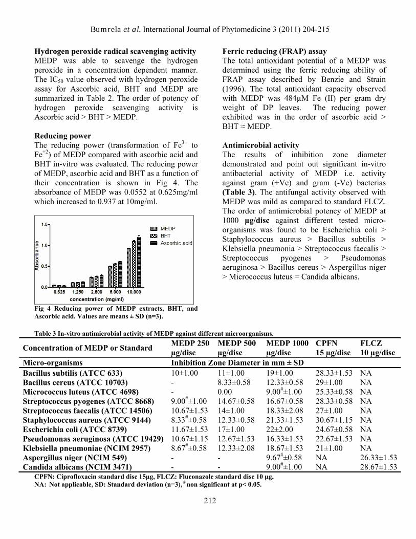
Hydrogen peroxide radical scavenging activity MEDP was able to scavenge the hydrogen peroxide in a concentration dependent manner. The IC50 value observed with hydrogen peroxide assay for Ascorbic acid, BHT and MEDP are summarized in Table 2. The order of potency of hydrogen peroxide scavenging activity is Ascorbic acid > BHT > MEDP. Reducing power The reducing power (transformation of Fe3+ to Fe+2) of MEDP compared with ascorbic acid and BHT in-vitro was evaluated. The reducing power of MEDP, ascorbic acid and BHT as a function of their concentration is shown in Fig 4. The absorbance of MEDP was 0.0552 at 0.625mg/ml which increased to 0.937 at 10mg/ml.
Fig 4 Reducing power of MEDP extracts, BHT, and Ascorbic acid. Values are means ± SD (n=3).
Ferric reducing (FRAP) assay The total antioxidant potential of a MEDP was determined using the ferric reducing ability of FRAP assay described by Benzie and Strain (1996). The total antioxidant capacity observed with MEDP was 484µM Fe (II) per gram dry weight of DP leaves. The reducing power exhibited was in the order of ascorbic acid > BHT ≈ MEDP. Antimicrobial activity The results of inhibition zone diameter demonstrated and point out significant in-vitro antibacterial activity of MEDP i.e. activity against gram (+Ve) and gram (-Ve) bacterias (Table 3). The antifungal activity observed with MEDP was mild as compared to standard FLCZ. The order of antimicrobial potency of MEDP at 1000 µg/disc against different tested micro-organisms was found to be Escherichia coli > Staphylococcus aureus > Bacillus subtilis > Klebsiella pneumonia > Streptococcus faecalis > Streptococcus pyogenes > Pseudomonas aeruginosa > Bacillus cereus > Aspergillus niger > Micrococcus luteus = Candida albicans.
Table 3 In-vitro antimicrobial activity of MEDP against different microorganisms.
Concentration of MEDP or Standard MEDP 250 µg/disc
MEDP 500 µg/disc
MEDP 1000 µg/disc
CPFN 15 µg/disc
FLCZ 10 µg/disc
Micro-organisms Inhibition Zone Diameter in mm ± SD Bacillus subtilis (ATCC 633) 10±1.00 11±1.00 19±1.00 28.33±1.53 NA Bacillus cereus (ATCC 10703) - 8.33±0.58 12.33±0.58 29±1.00 NA Micrococcus luteus (ATCC 4698) - 0.00 9.00#±1.00 25.33±0.58 NA Streptococcus pyogenes (ATCC 8668) 9.00#±1.00 14.67±0.58 16.67±0.58 28.33±0.58 NA Streptococcus faecalis (ATCC 14506) 10.67±1.53 14±1.00 18.33±2.08 27±1.00 NA Staphylococcus aureus (ATCC 9144) 8.33#±0.58 12.33±0.58 21.33±1.53 30.67±1.15 NA Escherichia coli (ATCC 8739) 11.67±1.53 17±1.00 22±2.00 24.67±0.58 NA Pseudomonas aeruginosa (ATCC 19429) 10.67±1.15 12.67±1.53 16.33±1.53 22.67±1.53 NA Klebsiella pneumoniae (NCIM 2957) 8.67#±0.58 12.33±2.08 18.67±1.53 21±1.00 NA Aspergillus niger (NCIM 549) - - 9.67#±0.58 NA 26.33±1.53Candida albicans (NCIM 3471) - - 9.00#±1.00 NA 28.67±1.53
CPFN: Ciprofloxacin standard disc 15µg, FLCZ: Fluconazole standard disc 10 µg, NA: Not applicable, SD: Standard deviation (n=3), # non significant at p< 0.05.
Bumrela et al. International Journal of Phytomedicine 3 (2011) 204-215
213
Possible role of phytochemical constituents of MEDP in antimicrobial and antioxidant activity Other researchers have demonstrated that DP contains various phytochemicals viz. lyoniresinol-9´-O-β-D-glucoside, 5,5-dimethoxy-lariciresinol-9-0-β glucopyranoside, β-sitosterol, lupeol, α-ethyl galactose, apiginin-7-O-rutinoside, α-D-glucose, β-D-glucose and β-D-fructose [6]. Furthermore, Manikandan et al [7] have reported various phytochemical constituents in DP viz. ascorbic acid, phenolic compounds, tannins, lycopene, carotenoid and tocopherol. The relationship between the chemical structure of flavonoids and their ability for radical-scavenging activities was analyzed by Bors, et al. [24] Djeridane et al. [25] and demonstrated a linear correlation between the content of total phenolic compounds and their antioxidant capacity. Lupeol [26] and lignin-glycoside, (+)-lyoniresinol-3α-O-β-glucopyranoside [27] found to exert antioxidant activity. Present findings are in agreement with the earlier reports which indicate β-carotene is known to elicit antioxidant activity in-vitro and in-vivo systems. Mixtures of carotenoids with others antioxidants enhanced their antioxidant activity against free radicals. Chemical abilities of β-carotene to quench singlet oxygen and also neutralize peroxyl free radicals are well documented [28]. The antimicrobial properties of tannins have been documented by Scalbert [29] and suggested that they are toxic to filamentous fungi, yeast and also bacterias. Tannins are known to form irreversible complexes with proline-rich proteins [30] resulting in the inhibition of cell wall protein synthesis. Polyphenolic compounds can form heavy soluble complexes with proteins or bind to bacterial adhesions and thereby impairing the availability of receptors on the cell surface [31]. Antimicrobial property of β-sitosterol and β-D-glucopyranoside has already been reported [32]. The above mentioned findings support the
antimicrobial activity of MEDP observed in the present study. In addition, iridoid glycosides (IGs) are known to have antimicrobial activity against Bacillus subtilis, Escherichia coli, Pseudomonas aeruginosa, Staphylococcus aureus and Candida albicans [33]. The toxic and antimicrobial effect of IGs has been correlated to the presence of aglycone moiety. Hence it is believed that the phytochemical constituents present in MEDP might be participating individually or collectively in the observed antimicrobial and antioxidant activity which may warrant further in-vivo studies. It is further presumed that the glucopyranosides an active phytoconstituent of MEDP might be an ideal lead molecule for the development of new generation of antibacterial agents as suggested by Lee et al. [34]. Conclusion MEDP has shown promising antimicrobial and antioxidant activity (in-vitro) and probably the first report on Dipteracanthus patulus (Jacq) nees plant. The recent research reports envisage that glucopyranosides might be an ideal lead molecule for antibacterial activity. It is presumed that the iridoid glycosides, beta-carotene along with beta-sitosterol and lupeol may be a good combination observed in MEDP for potential antioxidant activity. Acknowledgement The authors are thankful to the management of Sinhgad Technical Education Society (STES) and Principal, STES’s, Sinhgad Institute of Pharmaceutical Sciences, Lonavala, Pune, MS, India for providing the research facilities to conduct the present research work. Authors are grateful to Anchrom Laboratory, Mumbai, MS, India for providing HPTLC facilities.
Bumrela et al. International Journal of Phytomedicine 3 (2011) 204-215
214
References 1. Chirangini P, Amit K, Pandey BN,
Sharma GJ, Mishra KP. Herbals for human health and disease: prospects and challenges in 21st century. In: Sharma RK, Arora R, editors. Herbal Drugs - A Twenty First Century Perspective. New Delhi: Jaypee Brothers Medical Publishers; 2006;p. 1-7.
2. Piddock LJV, Wise R. Mechanisms of resistance to quinolones and clinical perspectives. J. Antimicro. Chemother. 1989;23(4),475-480.
3. Parida MM, Dash PK, Saxena P, Jana AM, Rao PVL. Perspectives on antimicrobial activity of natural plant products. In: Sharma RK, Arora R, editors. Herbal Drugs - A Twenty First Century Perspective. New Delhi: Jaypee Brothers Medical Publishers; 2006;p. 484-496.
4. Narasimhan S, Shobana R, Sathya TN. Antioxidants- natural rejuvenators that heal, detoxify and provide nourishment. In: Sharma RK, Arora R, editors. Herbal Drugs - A Twenty First Century Perspective. New Delhi: Jaypee Brothers Medical Publishers; 2006;p.548-558.
5. Chopra RN, Nayar SL, Chopra IC. Glossary of Indian Medicinal Plants. New Delhi: Council of Scientific and Industrial Research. 1986;p.99.
6. Akthar MF. Chemical and biological investigations of medicinal herbs phyla nodiflora, ruellia patula and ruellia brittoniana. Dissertation submitted to University of Karachi, Department of Pharmacognosy, Faculty of Pharmacy, Pakistan. 1993;p. 59.
7. Manikandan A, Doss DVA. Evaluation of biochemical contents, nutritional value, trace elements, SDS-PAGE and HPTLC profiling in the leaves of Ruellia tuberosa L. and Dipteracanthus patulus (Jacq.). J. Chem. Pharm. Res. 2010;2(3):295-303.
8. Saroja K, Elizabeth JD, Gopalkrishnan S. Wound healing activity of leaves of
Dipteracanthus patulus (Jacq.). Pharmacologyonline. 2009;2:462-469.
9. Manikandan A, Doss DVA. Effect of 50% hydroethanolic leaf extracts of Ruellia tuberosa L. and Dipteracanthus patulus (Jacq.) on AST, ALT, ACP and ALP levels in serum, liver and kidney of alloxan induced diabetic rats. Anna. Pharm. Pharm. Sci. 2010;1(2):142-146.
10. Akhtar MF, Rashid S, Ahmad M, Usmanghani K. Cardiovascular evaluation of Ruellia patula and Ruellia brittoniana. J. Islamic. Acad. Sci. 1992;5(1):67-71.
11. Ansari SH. Essentials of Pharmacognosy. Delhi: Birla Publication; 2006;p.357-383.
12. Harborne JB. Phytochemical methods: A guide to modern techniques of plant analysis. London: Chapman and Hall Publication; 1998;p. 119.
13. Slinkard K, Singleton VL. Total phenol analysis: automation and comparison with manual methods. Am. J. Enol. Viticult. 1977;28:49–55.
14. Chen Y, Wang MF, Rosen RT, Ho CT. 2, 2-Diphenyl -1 -picrylhydrazyl radical-scavenging active components from Polygonum multiflorum Thunb. J. Agric. Food Chem. 1999;47:2226–2228.
15. Green LC, Wagner DA, Glogowiski J, Skipper PL, Wishnok JS, Tannenbaum SR. Analysis of nitrate, nitrite and 15N nitrate in biological fluids. Anal. Biochem. 1982;126:131-138.
16. Ruch RJ, Cheng SJ, Klaunig JE. Prevention of cytotoxicity and inhibition of intercellular communication by antioxidant catechins isolated from Chinese green tea. Carcinogenesis. 1989;10:1003–1008.
17. Oyaizu M. Studies on products of browning reactions: antioxidative activities of products of browning reaction prepared from glucosamine. Jpn. J. Nutr. 1986;44:307–315.
18. Benzie IFF, Strain JJ. The ferric reducing ability of plasma (FRAP) as a measure of
Bumrela et al. International Journal of Phytomedicine 3 (2011) 204-215
215
antioxidant power: The FRAP assay. Anal. Biochem. 1996;239:70–76.
19. Wong SP, Leong LP, Koh JHW. Antioxidant activities of aqueous extracts of selected plants. Food Chem. 2006;99:775-783.
20. Kirby MDK, Bauer RW, Sherris JC, Turck M. Antibiotic susceptibility testing by standard single disc diffusion method. Am. J. Clin. Pathol. 1966;45:493-496.
21. Godinho A, Bhosale S. Carotenes produced by alkaliphilic orange-pigmented strain of microbacterium arborescens-AGSB isolated from coastal sand dunes. Indian J. Mar. Sci. 2008;37(3):307-312.
22. Yamaguchi T, Takamura H, Matoba T, Terao J. HPLC method for evaluation of the free radical-scavenging activity of foods by using 1,1,-diphenyl-2-picrylhydrazyl. Biosci. Biotech. Biochem. 1998;62:1201–1204.
23. Chen CW, Ho CT. Antioxidant properties of polyphenols extracted from green and black tea. J. Food Lipids. 1995;2:35–46.
24. Bors W, Heller W, Michael C, Saran M. Radical chemistry of flavonoids antioxidants. Adv. Exp. Med. Biol. 1990;264:165–170.
25. Djeridane A, Yousfi M, Nadjemi B, Boutassouna D, Stocker P, Vidal N. Antioxidant activity of some Algerian medicinal plants extracts containing phenolic compounds. Food Chem. 2006;97:654–660.
26. Senthilkumar N, Badami S, Dongre SH, Bhojraj S. Antioxidant and hepatoprotective activity of methanolic extract of Careya arborea bark in Ehrlich
ascites carcinoma bearing mice. J. Nat. Remedies. 2008;62:336-339.
27. Da Silva VS, Silvab GH, Bolzani VDS, Lopes MN. Isolation of lignin glycoside from Alibertia sessilis (Vell) K. Schum (Rubiaceae) by preparative HPLC. Ecletica Quimica 2006;31(4):55-58.
28. Sies H, Stahl W. Vitamin E and C, beta carotene and other carotenoids as antioxidants. Am. J. Clin. Nutr. 1995;62:15S-21S.
29. Scalbert A. Antimicrobial properties of tannins. Phytochemistry. 1991;30:3875-3883.
30. Hagerman AE, Butler LG. The specificity of proanthocyanidin-protein interactions. The J. Biol. Chem. 1981;256:4494–4497.
31. Haslam E. Natural polyphenols (vegetable tannins) as drugs: possible modes of action. J. Nat. Prod. 1996;59:205–215.
32. Beltrame FL, Pessini GL, Doro DL, Filho BPD, Bazotte RB, Cortez DAG. Evaluation of the antidiabetic and antibacterial activity of Cissus sicyoides. Braz. Arch. Biol. Tech. 2002;45(1):21- 25.
33. Akunyili DN, Houghton PJ, Raman A. Antimicrobial activities of the stembark of Kigelia pinnata. J. Ethonopharmacol. 1991;35(2):173-177.
34. Lee DG, Jung HJ, Woo ER. Antimicrobial property of (+)-lyoniresinol-3alpha-O-beta-D-glucopyranoside isolated from the root bark of Lycium chinense Miller against human pathogenic microorganisms. Arch. Pharm. Res. 2005;28(9):1031-1036.