Embed Size (px)
Citation preview
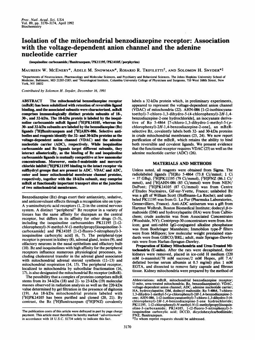
Proc. Natl. Acad. Sci. USAVol. 89, pp. 3170-3174, April 1992Biochemistry
Isolation of the mitochondrial benzodiazepine receptor: Associationwith the voltage-dependent anion channel and the adeninenucleotide carrier
(isoquinoline carboxamide/flunitrazepam/PK11195/PK14105/porphyrins)
MAUREEN W. MCENERY*, ADELE M. SNOWMAN*, ROSARIO R. TRIFILETTIt, AND SOLOMON H. SNYDER*t*Departments of Neuroscience, Pharmacology and Molecular Sciences, and Psychiatry and Behavioral Sciences, The Johns Hopkins University School ofMedicine, Baltimore, MD 21205-2185; and tNeurological Institute, Columbia University College of Physicians and Surgeons, 710 West 168th Street, NewYork, NY 10032
Contributed by Solomon H. Snyder, December 16, 1991
ABSTRACT The mitochondrial benzodiazepine receptor(mBzR) has been solubilized with retention of reversible ligandbinding, and the associated subunits were characterized. mBzRcomprises immunologically distinct protein subunits of 18-,30-, and 32-kDa. The 18-kDa protein is labeled by the isoqui-noline carboxamide mBzR ligand PHJPK14105, whereas the30- and 324kDa subunits are labeled by the benzodiazepine (Bz)l~igands (3Hflunitrazepam and 3HIAHN-086. Selective anti-bodies and reagents identify the 32- and 30-kDa proteins as thevoltage-dependent anion channel (VDAC) and the adeninenucleotide carrier (ADC), respectively. While isoquinolinecarboxamide and Bz ligands target different subunits, theyinteract allosterically, as the binding of Bz and isoquinolinecarboxamide ligands is mutually competitive at low nanomolarconcentrations. Moreover, eosin-5-maleimide and mercuricchloride inhibit [3H]PK11195 binding to the intact receptor viasulfbydryl groups that are present in ADC. VDAC and ADC,outer and inner mitochondrial membrane channel proteins,respectively, together with the 18-kDa subunit, may comprisemlzR at functionally important transport sites at the junctionof two mitochondrial membranes.
Benzodiazepine (Bz) drugs exert their antianxiety, sedative,and anticonvulsant effects through a recognition site on type-A 'y-aminobutyric acid receptors (1, 2) in the central nervoussystem. A distinct "peripheral" Bz receptor in a variety oftissues has the same affinity for diazepam as the centralreceptor, but differs in its affinity for other drugs (3-5),including the isoquinoline carboxamides PK11195 [1-(2-chlorophenyl)-N-methyl-N-(1-methylpropyl)isoquinoline-3-carboxamide] and PK14105 [1-(2-fluoro-5-nitrophenyl)-3-isoquinoline carboxylic acid] (6, 7). The peripheral-typereceptor is present in kidney (8), adrenal gland, testes (9), andolfactory neurons in the nasal epithelium and olfactory bulb(10). Bz and isoquinolines with high affinity for the peripheralreceptors influence numerous physiological processes, in-cluding cholesterol transfer in the adrenal gland associatedwith mitochondrial adrenal steroid synthesis (11-13) andmitochondrial respiration (14, 15). The peripheral receptor,localized to mitochondria by subcellular fractionation (16,17), is also designated the mitochondrial Bz receptor (mBzR).The possibility that a complex of proteins comprises mBzR
stems from its 34-kDa (18) and 21- to 23-kDa (19) molecularmasses observed in radiation analysis as well as the 220-kDavalue determined by gel filtration in the presence of digitonin(19). 'An 18-kDa mitochondrial protein 'labeled with[3H]PK14105 has been purified and cloned (20, 21). Bycontrast, the Bz [3H]flunitrazepam ([3HJFNZ) covalently
labels a 32-kDa protein which, in preliminary experiments,appeared to represent the voltage-dependent anion channel(VDAC) of mitochondria (22). AHN-086 [1-(2-isothiocyana-toethyl)-7-chloro-1,3-dihydro-5-(4-chlorophenyl)-2H-1 ,4-benzodiazepine-2-one hydrochloride], an isocyanate deriva-tive of Ro 5-4864 [7-chloro-1,3-dihydro-1-methyl-5-(p-chlorophenyl)-2H-1,4-benzodiazepine-2-one], an mBzR-selective Bz, covalently labels both 32- and 30-kDa proteinsin crude mitochondrial membranes (23, 24). We now reportpurification of the mBzR, which retains the ability to bindboth reversible and covalent ligands. We present evidencethat the functional receptor requires VDAC (25) as well as theadenine nucleotide carrier (ADC) (26).
MATERIALS AND METHODSUnless noted, all reagents were obtained from Sigma. Theradiolabeled ligands [3H]Ro 5-4864 (73.8 Ci/mmol; 1 Ci= 37 GBq), [3H]PK11195 (79 Ci/mmol), [3H]FNZ (86.1 Ci/mmol), and [3H]AHN-086 (85 Ci/mmol) were from NEN/DuPont; [3H]PK14105 (87 Ci/mmol) was from Centred'Etudes Nucleaires, Gif-sur-Yvette, France; unlabeled Bzwas a gift of William Scott (Hoffmann-La Roche); and unla-beled PK11195 was from G. Le Fur (Pharmuka Laboratories,Gennevilliers, France). Anti-ADC antiserum was a gift fromHartmut Wolhrab, Boston Biomedical Institute; dodecyl f-D-maltoside (DM) and hydroxylapatite (HA) were from Calbio-chem; crude asolectin was from Associated Concentrates(Woodside, NY); Centriprep-30 concentrators were from Am-icon; goat anti-rabbit IgG-conjugated alkaline phosphatasewas from Boehringer Mannheim; Immobilon type-P filterswere from Millipore; low molecular weight prestained stan-dards were from GIBCO/BRL; adult, male Sprague-Dawleyrats were from Harlan-Sprague-Dawley.
Preparation of Kidney Mitochondria and Urea-Treated MN-tochondria (U-mito). After the rats were decapitated, theirkidneys were removed, placed in ice-cold H medium (220mM D-mannitol/70 mM sucrose/2 mM Hepes, pH 7.4/defatted bovine serum albumin at 0.5 mg/ml) plus 1 mMEGTA, and dissected to remove fatty capsule and fibroustissue. Kidney mitochondria were prepared by the method of
Abbreviations: mBzR, mitochondrial benzodiazepine receptor;U-mito, urea-treated mitochondria; Bz, benzodiazepine(s); VDAC,voltage-dependent anion channel; ADC, adenine nucleotide carrier;HA, hydroxylapatite; DM, dodecyl maltoside; Ro 54864, 7-chloro-1,3-dihydro-1-methyl-5-(p-chlorophenyl)-2H-1,4-benzodiazepine-2-one; AHN-086, 1-(2-isothiocyanatoethyl)-7-chloro-1,3-dihydro-5-(4-chlorophenyl)-2H-1,4-benzodiazepine-2-one hydrochloride;PK11195, 1-(2-chlorophenyl)-N-methyl-N-(1-methylpropyl)isoquin-oline-3-carboxamide; PK14105, 1-(2-fluoro-5-nitrophepyl)-3-isoquinoline carboxylic acid; DCCD, dicyclohexylcarboduimide;FNZ, flunitrazepam.*To whom reprint requests should be addressed.
3170
The publication costs of this article were defrayed in part by page chargepayment. This article must therefore be hereby marked "advertisement"in accordance with 18 U.S.C. §1734 solely to indicate this fact.
Proc. Natl. Acad. Sci. USA 89 (1992) 3171
Wehrle et al. (27). Mitochondria were either used immedi-ately or stored at a protein concentration of 50 mg/ml at-700C. To prepare U-mito, mitochondria were diluted to aprotein concentration of 2 mg/ml in 20 mM potassiumphosphate, pH 6.5/20 mM potassium chloride/4 M urea in a
final volume of 3 ml and incubated on ice for 30 min,whereupon 21 ml of 20mM potassium phosphate, pH 6.5/200mM potassium chloride was added. Samples were centri-fuged at 210,000 X g for 75 min. The resulting U-mitomembrane pellet was resuspended in half its original volumein buffer B (25 mM Tris, pH 7.7/5 mM EDTA/40 potassiumchloride/2.5% ethylene glycol) and used immediately.
Solubilization and Fractionation of mBzR. U-mito werediluted to 2.5 mg of protein per ml in buffer B plus 0.5% DM,solubilized for 15 min on ice, and centrifuged for 60 min at230,000 x g. The soluble fraction was removed, diluted byimmediately adding a 4-fold volume of buffer B, and purifiedby adsorption to HA. Soluble mBzR was diluted with water(1:1, vol/vol) and poured over multiple HA columns (previ-ously washed with 5 ml of 0.5 x buffer B) at a ratio of 4 ml per
1.6 g of HA, and the columns were washed in a batch-wisefashion with increasing concentrations (0.5-1.5 M) of potas-sium phosphate, pH 6.5, in 0.5 x buffer B. The fraction withthe highest specific activity, corresponding to 1.2 M potas-sium phosphate, was concentrated 10-fold by Centriprep-30concentration and stored at -70°C until use. The concen-trated mBzR was chromatographed on a FPLC Superose-12Bsizing column in the presence of 0.5x buffer B.
Photoaffinity and Covalent Labeling of mBzR. Rat kidneymitochondria and other mBzR fractions were incubated with 20nM [3H]PK14105 or 100 nM [3H]FNZ (see figure legends forprotein concentration) for 30 min in the dark on ice. Sampleswere then exposed to UV light (254 nm) for 45 min and thenquenched with 5 x Laemmli sample buffer (15% SDS/0.825 Msucrose/0.325 M Tris-HCI, pH 6.8/5% 2-mercaptoethanol/0.002% bromophenol blue). After covalent labeling with 10 nM[3H]AHN-086 on ice for 60 min, the samples were quenched as
above. Nonspecific binding was determined in the presence of10 ,uM unlabeled drug. ['4C]Dicyclohexylcarbodiimide (["'C]DCCD) labeling (28) and covalent modification with eosin-5-maleimide (29) were as reported.Bands incorporating the radiolabeled drugs were deter-
mined by two procedures. In the first procedure, afterresolution on SDS/PAGE (30), gel strips were cut into 2-mmslices, dissolved overnight at 60°C in 0.5 ml of H202 (37%stock), and assayed directly in 4 ml of formula 963 plus 5%acetic acid. The second procedure involved the fluorographyof Immobilon filters previously developed for antigenic iden-tification, permitting accelerated detection of tritiated pro-
teins and establishing the coidentity of antigen and radiola-beled protein. Dried Immobilon filters were soaked in 100%methanol for 10 sec, transferred to a glass tray, soaked for 60sec in 100% dimethyl sulfoxide, and incubated with a solutionof 20% 2,5-diphenyloxazole in 100% dimethyl sulfoxide (31)for 1 min with constant shaking. The opaque filters were thensubmerged twice in 3 liters of water to precipitate the2,5-diphenyloxazole, dried, and exposed to film.
Antibodies Against the 32-kDa Protein (VDAC). Highlypurified mBzR (1.2 M potassium phosphate fraction from theHA column) was used to immunize rabbits. The immune andpreimmune sera were characterized for their reactivityagainst total rat kidney mitochondria, purified mBzR, and ratkidney VDAC prepared by standard procedures (32). Forimmunological detection of transferred proteins, antiserawere diluted in TTBS (20 mM Tris, pH 7.4/500 mM NaCl/0.05% Tween 20) plus 3% bovine serum albumin and incu-bated with protein transferred to Immobilon P filters for 2 hrat room temperature. After extensive washing in TABS andincubation with anti-rabbit alkaline phosphatase secondaryantiserum (1:1500) for 1 hr at room temperature, the blot was
developed by using nitroblue tetrazolium and 5-bromo-4-chloro-3-indolyl phosphate or 125I-labeled protein A (33).Other Methods. One-dimensional gel electrophoresis was
as described (26), and protein was visualized by Coomassieor silver staining (34). Two-dimensional, nonequilibrium gelelectrophoresis was as described (35). Membrane (36) andsoluble (37) proteins were determined with bovine serumalbumin as a standard. Asolectin was prepared in 0.05% DM(38). Radioligand binding was carried out on ice in a totalvolume of 200 pd, which contained 50 mM Tris and thetritium-labeled drug at 1 nM (with the exception of [3H]FNZ,which was at 100 nM). Nonspecific binding was determinedin the presence of 10 gM unlabeled ligand. The final con-centration of detergent was <0.05% in all cases, becausehigher concentrations of DM potently inhibited ligand bind-ing. Samples were filtered over glass-fiber filters (WhatmanGF/B) as described (39).
RESULTSPurification ofmBzR and Asignment ofLigand Binding Sites.
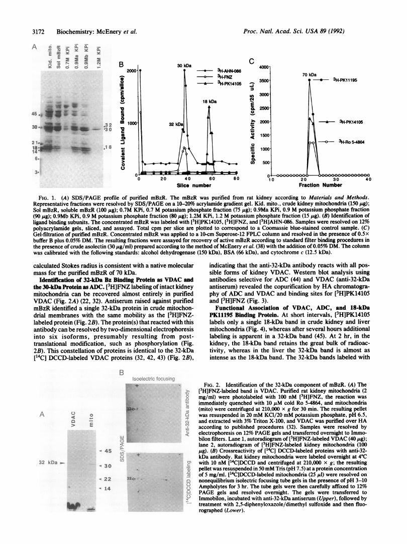
The ability ofmBzR to reversibly bind ligands is lost followingtreatment with numerous detergents, but is retained to a limitedextent following solubilization with digitonin (39). We havesuccessfully solubilized mBzR with DM, which solubilizes, inactive form, membrane proteins containing multiple subunits(40, 41). Whole mitochondria were treated with urea to removeperipheral proteins, providing about a 3-fold enhancement inspecific [3H]PK11195 binding (Table 1). Solubilization withDMyielded a further 2-fold purification with retention of 80o of[3H]PK11195 binding of the intact mitochondria. Fractionationof the solubilized extract on HA columns provided a further10-fold enhanced specific activity in the fraction eluted with 1.2M salt. This fraction is enriched 60-fold in [3H]PK11195 bindingcompared to the original mitochondria, with >50% recovery oftotal binding activity of the original mitochondria, and com-prises major proteins of 32 and 30 kDa and a faintly stained18-kDa protein (Fig. 1A). The elution pattern of mitochondrialmembrane proteins from HA in the presence of DM differsmarkedly from methods that employ Triton X-100 (32) or otherdetergents (42).To examine the role of the protein subunits of the mBzR
complex in ligand recognition, we photolabeled the 1.2 Mpotassium phosphate HA eluate with specific Bz ligands (Fig.1B). [3H]PK14105 labeled an 18-kDa band. [3H]FNZ wasassociated with the 32-kDa band, whereas [3H]AHN-086 la-beled both the 32- and 30-kDa bands, with more label onthe 30-kDa protein. Chromatography of the purified prepara-tion on Superose-12 revealed a single sharp peak of protein(data not shown) coincident with the peak binding of[3H]PK11195, [3H]PK14105, and [3H]Ro 5-4864 (Fig. 1C). The
Table 1. Purification of mBzR from rat kidney mitochondria
SpecificProtein, % total activity, Fold
Fraction mg activity pmol/mg purification
Mitochondria 330 100 18U-mito 119 100 50 2.8Soluble Bz 49 91 110 6.1HA (1.2 M salt)* 3.2 59 1100 60
This procedure was carried out more than 20 times with <5%variation. Crude asolectin (15:1, wt/wt), which markedly stabilizedligand binding to the purified samples (data not shown), was routinelyincluded.*Kd values of the highly purified mBzR for [3H]PK11195, [3H]Ro5-4864, and [3H]PK14105 were 15, 25, and 10 nM, respectively. Thebinding of [3H]PK11195 and [3 H]Ro 5-4684 to mBzR was inhibitedby protoporphyrin IX with an IC50 of 250 nM.
Biochemistry: McEnery et al.
3172 Biochemistry: McEnery et al.
A o cc _ X X ._,- N CE Do*E
.. Cl) co _
CL
45
30@*** *iF a s - '3 02 X
la: _ -1 814 e6._
to
3.
C
-i
u0N
00
-
E
0
C
0.Co
40 60Slice number
FIG. 1. (A) SDS/PAGE profile of purified mBzR. The mBzR was purified from rat kidney according to Materials and Methods.Representative fractions were resolved by SDS/PAGE on a 10-20o acrylamide gradient gel. Kid. mito., crude kidney mitochondria (150 ,ug);Sol mBzR, soluble mBzR (100 pug); 0.7M KPi, 0.7 M potassium phosphate fraction (75 pug); 0.9Ma KPi, 0.9 M potassium phosphate fraction(90 ,Ag); 0.9Mb KPi, 0.9 M potassium phosphate fraction (80 j&g); 1.2M KPi, 1.2 M potassium phosphate fraction (15 ,ug). (B) Identification ofligand binding subunits. The concentrated mBzR was labeled with [3H]PK14105, [3H]FNZ, and [3H]AHN-086. Samples were resolved on 12%polyacrylamide gels, sliced, and assayed. Total cpm per slice are plotted to correspond to a Coomassie blue-stained control sample. (C)Gel-filtration of purified mBzR. Concentrated mBzR was applied to a 10-cm Superose-12 FPLC column and resolved in the presence of 0.5xbuffer B plus 0.05% DM. The resulting fractions were assayed for recovery of active mBzR according to standard filter binding procedures inthe presence of crude asolectin (30 ,Lg/ml) prepared according to the method of McEnery et al. (38) with the addition of 0.05% DM. The columnwas calibrated with the following standards: alcohol dehydrogenase (150 kDa), BSA (66 kDa), and cytochrome c (12.5 kDa).
calculated Stokes radius is consistent with a native molecularmass for the purified mBzR of 70 kDa.
Identification of 32-kDa Bz Binding Protein as VDAC andthe 30-kDa Protein as ADC. [3H]FNZ labeling of intact kidneymitochondria can be recovered almost entirely in purifiedVDAC (Fig. 2A) (22, 32). Antiserum raised against purifiedmBzR identified a single 32-kDa protein in crude mitochon-drial membranes with the same mobility as the [3H]FNZ-labeled protein (Fig. 2B). The protein(s) that reacted with thisantibody can be resolved by two-dimensional electrophoresisinto six isoforms, presumably resulting from post-translational modification, such as phosphorylation (Fig.2B). This constellation of proteins is identical to the 32-kDa[14C] DCCD-labeled VDAC proteins (32, 42, 43) (Fig. 2B),
indicating that the anti-32-kDa antibody reacts with all pos-sible forms of kidney VDAC. Western blot analysis usingantibodies selective for ADC (44) and VDAC (anti-32-kDaantiserum) revealed the copurification by HA chromatogra-phy of ADC and VDAC and binding sites for [3HJPK14105and [3H]FNZ (Fig. 3).
Functional Association of VDAC, ADC, and 18-kDaPK11195 Binding Protein. At short intervals, [3H]PK14105labels only a single 18-kDa band in crude kidney and livermitochondria (Fig. 4), whereas after several hours additionallabeling is apparent in a 32-kDa band (45). At 2 hr, in thekidney, the 18-kDa band retains the great bulk of radioac-tivity, whereas in the liver the 32-kDa band is almost asintense as the 18-kDa band. The 32-kDa bands labeled with
A 0 0:
-> E
32 kDa .
BIsoelectric foc
.:+
IC>
-S4Cl)c3Cn
- 30
musingFIG. 2. Identification of the 32-kDa component of mBzR. (A) The
U [3H]FNZ-labeled band is VDAC. Purified rat kidney mitochondria (2mg/ml) were photolabeled with 100 nM [3H]FNZ, the reaction wasimmediately quenched with 10 ,uM cold Ro 5-4864, and mitochondria
. (mito) were centrifuged at 210,000 x g for 30 min. The resulting pelletwas resuspended in 20 mM KCl/20 mM potassium phosphate, pH 6.5,
N and extracted with 3% Triton X-100, and VDAC was purified over HAaccording to published procedures (32). Samples were resolved by
c electrophoresis on 12% PAGE gels and transferred overnight to Immo-bilon filters. Lane 1, autoradiogram of [3H]FNZ-labeled VDAC (40 ,ug);lane 2, autoradiogram of [3H]FNZ-labeled kidney mitochondria (100pig). (B) Crossreactivity of [14C] DCCD-labeled proteins with anti-32-kDa antibody. Rat kidney mitochondria were labeled overnight at 40C
: with 10 nM [14C]DCCD and centrifuged at 210,000 x g; the resultingC pellet was resuspended in 50mM Tris (pH 7.5) at a protein concentrationCu of 5 mg/ml. [14C]DCCD-labeled mitochondria (25 Al) were resolved onq nonequilibrium isolectric focusing tube gels in the presence of pH 3-10U Ampholytes for 3 hr. The tube gels were then carefully affixed to 12%2 PAGE gels and resolved overnight. The gels were transferred to
Immobilon, incubated with anti-32-kDa antiserum (Upper), followed bytreatment with 2,5-diphenyloxazole/dimethyl sulfoxide and then fluo-rographed (Lower).
20 30Fraction Number
22 32.>y
A 14
p .-.
Proc. Natl. Acad. Sci. USA 89 (1992)
Proc. Natl. Acad. Sci. USA 89 (1992) 3173
1 f-
E 8000-cLu
'aC
m 6000-0
m
_ 4000-
co
0
cuI= 20001
a 1251-Prot. A - anti-VDAC AbE 1251-Prot. A + anti-ADC Ab
E 3H-PK14105{ 3H-FNZ
F;T 0.5M 0.7M O.9MA O.9MB 1.2M
KPi [M]
FIG. 3. Copurification of VDAC, ADC, and ligand binding sitesby HA chromatography. Soluble mBzR were photolabeled with[3H]PK14105 and [3H]FNZ and then resolved by HA chromatogra-phy. Aliquots (20 Al) were assayed after filtration to remove unboundligand. Also, resulting HA fractions were resolved by SDS/PAGE,transferred to Immobilon filters, and probed with anti-VDAC andanti-ADC antibodies (Ab), followed by reaction with 125I-labeledprotein A (125I-Prot. A) secondary antibody. The filters were slicedand assayed directly in a GammaRIA counter, and the quantity of125I-labeled protein A bound per lane was plotted per HA fraction.
[3H]PK14105 in kidney and liver correspond closely to the32-kDa band labeled with [3H]FNZ and that stains with theanti-VDAC antibody. The 65-kDa band is believed to be adimer of VDAC. The transfer of radiolabel from the 18-kDato the 32-kDa band suggests that these two protein subunitsare closely associated in mBzR, permitting migration of thelabel between the two subunits.
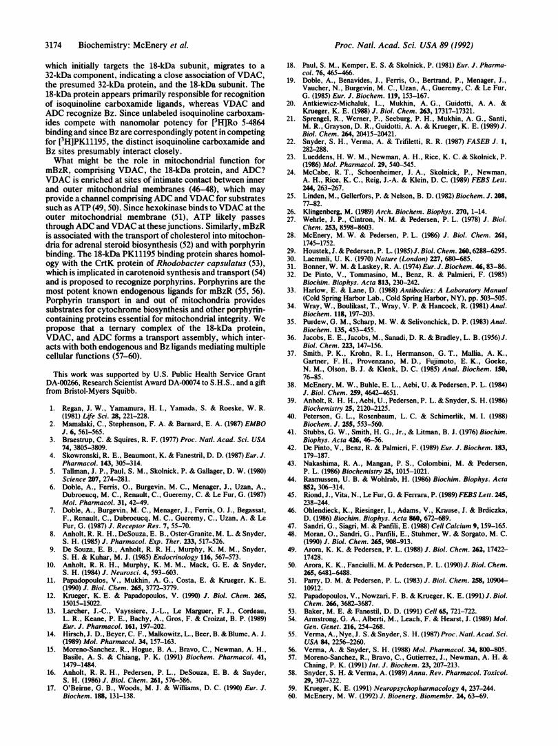
Evidence that the 18-kDa protein cannot of itself provideligand binding comes from an examination of the role of sulfhy-dryl groups. The cloned 18-kDa protein contains no cysteine (20).Eosin-5-maleimide and mercury are protein-modifying reagentsthat react with reduced sulihydryl groups of cysteine. [3H]Ro5-4864 binding is abolished by both reagents, whereas eosin-5-maleimide and mercury maximally reduce [3H]PK11195 binding80%o and 40%o, respectively (Fig. 5A). Eosin itselfhas no influenceon ligand binding (data not shown). In crude mitochondria,eosin-5-maleimide covalently labels the 30-kDaADC, the 31-kDaphosphate carrier, and larger molecular mass proteins (Fig. 5B,lane 1; ref. 29). In the purified mBzR preparation, only the ADC
Kidney1 2 3
Liver
4 5 6
S 32 kDa.
_ * -18 kDa'
FIG. 4. [3H]PK14105 photolabels both the 18-kDa isoquinolinecarboxamide binding protein and VDAC. Purified rat kidney andliver mitochondria were photolabeled with 20 nM [3H]PK14105 and100 nM [3H]FNZ at a protein concentration of 1 mg/ml for statedlengths of time and then quenched with 5 x Laemmli sample buffer.The samples (100 ,u1) were resolved on 12% PAGE gels, transferredto Immobilon filters, fluorographed, and exposed to film. Kidneymitochondria (lanes 1-3) and liver mitochondria (lanes 4-6) werelabeled with [3H]PK14105 for 1 hr (lanes 1 and 4) or for 2 hr (lanes2 and 5) or were labeled with [3H]FNZ for 1 hr (lanes 3 and 6). Thebands at the top of lanes 3 and 6 are aggregates or dimers of VDAC.
protein is modified (Fig. 5B, lane 2). The pronounced effects ofmercury and eosin-5-maleimide at concentrations that interactwith cysteine indicate that the 18-kDa protein cannot of itselfaccount for ligand binding to mBzR in kidney or adrenal mito-chondria (data not shown) and that ADC is required for ligandbinding.
DISCUSSIONHere, we provide evidence for a functional requirement ofVDAC, ADC, and the 18-kDa PK11195 binding subunit in theintact mBzR: (i) The purified mBzR contains VDAC andADC along with the 18-kDa component; and all three pro-teins, despite differing molecular masses, copurify and mi-grate as a single sharp peak upon gel chromatography. (ii)The 32-kDa FNZ binding site is identical to VDAC; further-more, binding of the Bz AHN-086 involves both VDAC andADC, which either contains Bz binding sites or is in closeproximity to a Bz binding protein. (iii) Sulfhydryl reagentspotently inhibit ligand binding to mBzR; the Bz binding siteis more sensitive than the PK11195 binding site. Eosin-5-maleimide, a potent inhibitor of ligand binding, interactsselectively with ADC in purified mBzR. (iv) [3H]PK14105,
-'---_ 3H-PK11 195----o 3H-Ro 5-4864
1 .01 .1 1 1 0
Eosin-5-maleimide [mM].1 1
Mercuric chloride [mM]
B 1 2
32 130
1 0
FIG. 5. Sulfhydryls in mBzR. (A) Ef-fect of sulfhydryl reagents on ligand bind-ing to mBzR. Purified rat kidney mito-chondria were incubated for 30 min on icewith stated concentrations of eosin-5-maleimide and mercuric chloride and as-sayed for ligand binding. (B) Eosin-5-maleimide labels ADC in mBzR. Rat kid-ney mitochondria (100 jig; lane 1) andpurified mBzR (20 ,ug; lane 2) were incu-bated, respectively, with 10,uM eosin-5-maleimide in the dark on ice according topublished procedures (29). The reactionwas terminated by the addition of 5xLaemmli sample buffer, and the sampleswere resolved on 12% PAGE gels. Thegels were then visualized with a 260/280nm transilluminator and photographed.The labeling was completely preventedby 1 mM dithiothreitol (data not shown).
A
04-
0)
-1
Fn'OC
,)
im
n._
C.)._
cn
Biochemistry: McEnery et al.
3174 Biochemistry: McEnery et al.
which initially targets the 18-kDa subunit, migrates to a32-kDa component, indicating a close association of VDAC,the presumed 32-kDa protein, and the 18-kDa subunit. The18-kDa protein appears primarily responsible for recognitionof isoquinoline carboxamide ligands, whereas VDAC andADC recognize Bz. Since unlabeled isoquinoline carboxam-ides compete with nanomolar potency for [3H]Ro 5-4864binding and since Bz are correspondingly potent in competingfor [3H]PK11195, the distinct isoquinoline carboxamide andBz sites presumably interact closely.What might be the role in mitochondrial function for
mBzR, comprising VDAC, the 18-kDa protein, and ADC?VDAC is enriched at sites of intimate contact between innerand outer mitochondrial membranes (46-48), which mayprovide a channel comprising ADC and VDAC for substratessuch as ATP (49, 50). Since hexokinase binds to VDAC at theouter mitochondrial membrane (51), ATP likely passesthrough ADC and VDAC at thesejunctions. Similarly, mBzRis associated with the transport of cholesterol into mitochon-dria for adrenal steroid biosynthesis (52) and with porphyrinbinding. The 18-kDa PK11195 binding protein shares homol-ogy with the CrtK protein of Rhodobacter capsulatus (53),which is implicated in carotenoid synthesis and transport (54)and is proposed to recognize porphyrins. Porphyrins are themost potent known endogenous ligands for mBzR (55, 56).Porphyrin transport in and out of mitochondria providessubstrates for cytochrome biosynthesis and other porphyrin-containing proteins essential for mitochondrial integrity. Wepropose that a ternary complex of the 18-kDa protein,VDAC, and ADC forms a transport assembly, which inter-acts with both endogenous and Bz ligands mediating multiplecellular functions (57-60).
This work was supported by U.S. Public Health Service GrantDA-00266, Research Scientist Award DA-00074 to S.H.S., and a giftfrom Bristol-Myers Squibb.
1. Regan, J. W., Yamamura, H. I., Yamada, S. & Roeske, W. R.(1981) Life Sci. 28, 221-228.
2. Mamalaki, C., Stephenson, F. A. & Barnard, E. A. (1987) EMBOJ. 6, 561-565.
3. Braestrup, C. & Squires, R. F. (1977) Proc. Natl. Acad. Sci. USA74, 3805-3809.
4. Skowronski, R. E., Beaumont, K. & Fanestril, D. D. (1987) Eur. J.Pharmacol. 143, 305-314.
5. Tallman, J. P., Paul, S. M., Skolnick, P. & Gallager, D. W. (1980)Science 207, 274-281.
6. Doble, A., Ferris, O., Burgevin, M. C., Menager, J., Uzan, A.,Dubroeucq, M. C., Renault, C., Gueremy, C. & Le Fur, G. (1987)Mol. Pharmacol. 31, 42-49.
7. Doble, A., Burgevin, M. C., Menager, J., Ferris, 0. J., Begassat,F., Renault, C., Dubroeucq, M. C., Gueremy, C., Uzan, A. & LeFur, G. (1987) J. Receptor Res. 7, 55-70.
8. Anholt, R. R. H., DeSouza, E. B., Oster-Granite, M. L. & Snyder,S. H. (1985) J. Pharmacol. Exp. Ther. 233, 517-526.
9. De Souza, E. B., Anholt, R. R. H., Murphy, K. M. M., Snyder,S. H. & Kuhar, M. J. (1985) Endocrinology 116, 567-573.
10. Anholt, R. R. H., Murphy, K. M. M., Mack, G. E. & Snyder,S. H. (1984) J. Neurosci. 4, 593-603.
11. Papadopoulos, V., Mukhin, A. G., Costa, E. & Krueger, K. E.(1990) J. Biol. Chem. 265, 3772-3779.
12. Krueger, K. E. & Papadopoulos, V. (1990) J. Biol. Chem. 265,15015-15022.
13. Larcher, J.-C., Vayssiere, J.-L., Le Marguer, F. J., Cordeau,L. R., Keane, P. E., Bachy, A., Gros, F. & Croizat, B. P. (1989)Eur. J. Pharmacol. 161, 197-202.
14. Hirsch, J. D., Beyer, C. F., Malkowitz, L., Beer, B. & Blume, A. J.(1989) Mol. Pharmacol. 34, 157-163.
15. Moreno-Sanchez, R., Hogue, B. A., Bravo, C., Newman, A. H.,Basile, A. S. & Chiang, P. K. (1991) Biochem. Pharmacol. 41,1479-1484.
16. Anholt, R. R. H., Pedersen, P. L., DeSouza, E. B. & Snyder,S. H. (1986) J. Biol. Chem. 261, 576-586.
17. O'Beirne, G. B., Woods, M. J. & Williams, D. C. (1990) Eur. J.Biochem. 188, 131-138.
18. Paul, S. M., Kemper, E. S. & Skolnick, P. (1981) Eur. J. Pharma-col. 76, 465-466.
19. Doble, A., Benavides, J., Ferris, O., Bertrand, P., Menager, J.,Vaucher, N., Burgevin, M. C., Uzan, A., Gueremy, C. & Le Fur,G. (1985) Eur. J. Biochem. 119, 153-167.
20. Antkiewicz-Michaluk, L., Mukhin, A. G., Guidotti, A. A. &Krueger, K. E. (1988) J. Biol. Chem. 263, 17317-17321.
21. Sprengel, R., Werner, P., Seeburg, P. H., Mukhin, A. G., Santi,M. R., Grayson, D. R., Guidotti, A. A. & Krueger, K. E. (1989) J.Biol. Chem. 264, 20415-20421.
22. Snyder, S. H., Verma, A. & Trifiletti, R. R. (1987) FASEB J. 1,282-288.
23. Lueddens, H. W. M., Newman, A. H., Rice, K. C. & Skolnick, P.(1986) Mol. Pharmacol. 29, 540-545.
24. McCabe, R. T., Schoenheimer, J. A., Skolnick, P., Newman,A. H., Rice, K. C., Reig, J.-A. & Klein, D. C. (1989) FEBS Lett.244, 263-267.
25. Linden, M., Gellerfors, P. & Nelson, B. D. (1982) Biochem. J. 208,77-82.
26. Klingenberg, M. (1989) Arch. Biochem. Biophys. 270, 1-14.27. Wehrle, J. P., Cintron, N. M. & Pedersen, P. L. (1978) J. Biol.
Chem. 253, 8598-8603.28. McEnery, M. W. & Pedersen, P. L. (1986) J. Biol. Chem. 261,
1745-1752.29. Houstek, J. & Pedersen, P. L. (1985)J. Biol. Chem. 260, 6288-6295.30. Laemmli, U. K. (1970) Nature (London) 227, 680-685.31. Bonner, W. M. & Laskey, R. A. (1974) Eur. J. Biochem. 46, 83-86.32. De Pinto, V., Tommasino, M., Benz, R. & Palmieri, F. (1985)
Biochim. Biophys. Acta 813, 230-242.33. Harlow, E. & Lane, D. (1988) Antibodies: A Laboratory Manual
(Cold Spring Harbor Lab., Cold Spring Harbor, NY), pp. 503-505.34. Wray, W., Boulikast, T., Wray, V. P. & Hancock, R. (1981) Anal.
Biochem. 118, 197-203.35. Purdew, G. M., Scharp, M. W. & Selivonchick, D. P. (1983) Anal.
Biochem. 135, 453-455.36. Jacobs, E. E., Jacobs, M., Sanadi, D. R. & Bradley, L. B. (1956) J.
Biol. Chem. 223, 147-156.37. Smith, P. K., Krohn, R. I., Hermanson, G. T., Mallia, A. K.,
Gartner, F. H., Provenzano, M. D., Fujimoto, E. K., Goeke,N. M., Olson, B. J. & Klenk, D. C. (1985) Anal. Biochem. 150,76-85.
38. McEnery, M. W., Buhle, E. L., Aebi, U. & Pedersen, P. L. (1984)J. Biol. Chem. 259, 4642-4651.
39. Anholt, R. H. H., Aebi, U., Pedersen, P. L. & Snyder, S. H. (1986)Biochemistry 25, 2120-2125.
40. Peterson, G. L., Rosenbaum, L. C. & Schimerlik, M. I. (1988)Biochem. J. 255, 553-560.
41. Stubbs, G. W., Smith, H. G., Jr., & Litman, B. J. (1976) Biochim.Biophys. Acta 426, 46-56.
42. De Pinto, V., Benz, R. & Palmieri, F. (1989) Eur. J. Biochem. 183,179-187.
43. Nakashima, R. A., Mangan, P. S., Colombini, M. & Pedersen,P. L. (1986) Biochemistry 25, 1015-1021.
44. Rasmussen, U. B. & Wohlrab, H. (1986) Biochim. Biophys. Acta852, 306-314.
45. Riond, J., Vita, N., Le Fur, G. & Ferrara, P. (1989) FEBS Lett. 245,238-244.
46. Ohlendieck, K., Riesinger, I., Adams, V., Krause, J. & Brdiczka,D. (1986) Biochim. Biophys. Acta 860, 672-689.
47. Sandri, G., Siagri, M. & Panfili, E. (1988) Cell Calcium 9, 159-165.48. Moran, O., Sandri, G., Panfili, E., Stuhmer, W. & Sorgato, M. C.
(1990) J. Biol. Chem. 265, 908-913.49. Arora, K. K. & Pedersen, P. L. (1988) J. Biol. Chem. 262, 17422-
17428.50. Arora, K. K., Fanciulli, M. & Pedersen, P. L. (1990) J. Biol. Chem.
265, 6481-6488.51. Parry, D. M. & Pedersen, P. L. (1983) J. Biol. Chem. 258, 10904-
10912.52. Papadopoulos, V., Nowzari, F. B. & Krueger, K. E. (1991) J. Biol.
Chem. 266, 3682-3687.53. Baker, M. E. & Fanestil, D. D. (1991) Cell 65, 721-722.54. Armstrong, G. A., Alberti, M., Leach, F. & Hearst, J. (1989) Mol.
Gen. Genet. 216, 254-268.55. Verma, A., Nye, J. S. & Snyder, S. H. (1987) Proc. Natl. Acad. Sci.
USA 84, 2256-2260.56. Verma, A. & Snyder, S. H. (1988) Mol. Pharmacol. 34, 800-805.57. Moreno-Sanchez, R., Bravo, C., Gutierrez, J., Newman, A. H. &
Chaing, P. K. (1991) Int. J. Biochem. 23, 207-213.58. Snyder, S. . & Verma, A. (1989) Annu. Rev. Pharmacol. Toxicol.
29, 307-322.59. Krueger, K. E. (1991) Neuropsychopharmacology 4, 237-244.60. McEnery, M. W. (1992) J. Bioenerg. Biomembr. 24, 63-69.
Proc. Natl. Acad. Sci. USA 89 (1992)