Embed Size (px)
Citation preview

ISOLATION OF CRYSTALLINE TOBACCO MOSAIC VIRUS PROTEIN FROM TOMATO PLANTS*
BY HUBERT S. LORING AND W. M. STANLEY
(From the Department of Animal and Plant Pathology of The Rockefeller Institute for Medical Research, Princeton)
(Received for publication, December 15, 1936)
The isolation of a crystalline protein possessing the properties of tobacco mosaic virus has been described (1, 2). This crystalline protein was obtained from the globulin fraction of extracts of diseased Turkish tobacco plants, and was found to be over 100 times as active as the crude juice from the diseased plants used as
starting material. The chemical composition, optical rotation, and infectivity of the crystalline protein remained unchanged after ten successive recrystallizations. These facts have suggested that the protein is essentially pure and is the agent responsible for the tobacco mosaic disease.
The tobacco mosaic disease, as is well known, occurs in plants belonging to many different genera of the solanaceous family. The disease was first described in tomato plants by Clinton (3), who showed that the infectious material obtained from tomato plants infected with tobacco mosaic virus produced the same symptom complex on healthy tobacco plants as that caused by infectious material from diseased tobacco plants. As the juice obtained from diseased tomato plants is highly infectious, such plants offered the possibility of providing a new source of material for the isolation of the tobacco mosaic virus protein. If the pro- tein is the infectious agent, then it would necessarily be present in mosaic-diseased tomato plants. The isolation of a similar highly infectious, crystalline protein from diseased tomato plants would prove that this protein is associated with the tobacco mosaic disease in tomato plants and would provide additional evidence
* A preliminary announcement of this work was published in Science, 83, 85 (1936).
733
by guest on April 10, 2019
http://ww
w.jbc.org/
Dow
nloaded from
by guest on April 10, 2019
http://ww
w.jbc.org/
Dow
nloaded from
by guest on April 10, 2019
http://ww
w.jbc.org/
Dow
nloaded from

734 Mosaic Virus Protein from Tomato Plants
for the identity of the protein with the infectious agent. The present paper describes the isolation of a crystalline protein from tobacco mosaic-diseased tomato plants and presents the results of comparative studies on the properties of this protein and that isolated from mosaic-diseased tobacco plants.
EXPERIMENTAL
Method of Isolation-The same experimental procedure used in the improved method for the isolation of the crystalline protein from tobacco plants (4) was applied to diseased tomato plants, Lycopersicon esculentum, Miller, var. Bonny Best, which had been grown in the field. 2 months after inoculation with the ordinary strain of tobacco mosaic virus (Johnson’s tobacco Virus l), the plants were harvested. They were frozen, ground, and extracted. The first extract from 200 tomato plants gave 105 liters of juice, containing 2.6 mg. of total nitrogen per cc. (by Kjeldahl) and 0.65 mg. of protein nitrogen per cc., while a second extract gave 105 liters of juice containing 1.0 mg. of total nitrogen per cc. and 0.2 mg. of protein nitrogen per cc. The globulin fraction in each extract was precipitated with (NH&S04 and filtered. The com- bined yield of crude globulin amounted to 350 gm. or approxi- mately 0.2 per cent of the weight of the freshly cut plants.
Protein nitrogen was determined by precipitation of the protein with hot 5 per cent trichloroacetic acid. The suspension was immediately cooled and the denatured protein was centrifuged and dissolved in about 1 cc. of 0.2 N NaOH. It was then reprecipi- tated with about 5 cc. of 10 per cent trichloroacetic acid and again centrifuged. The protein residue was mixed with about 5 cc. of 5 per cent trichloroacetic acid and again centrifuged. It was then transferred to a micro-Kjeldahl flask with 0.2 N NaOH and di- gested, etc., as for a Kjeldahl analysis.
According to the improved method, the crude globulin was purified by two precipitations with ammonium sulfate, by adsorp- tion on celite at pH 4.5, and by treatment with 0.1 per cent CaO. The application of this procedure to the crude globulin first obtained from mosaic-diseased tomato plants resulted in a yellow pigmented preparation which failed to crystallize. However, it was found that if the procedure described above was repeated several times, the yellow color was gradually lost and the protein
by guest on April 10, 2019
http://ww
w.jbc.org/
Dow
nloaded from

H. S. Loring and W. M. Stanley 735
could then be crystallized. The first crystalline material obtained from tomato plants still contained an appreciable amount of yellow color. This could be removed by further fractionation with ammonium sulfate, celite, and CaO.
The application of the improved method to diseased tobacco plants grown under the same conditions in the field as were the tomato plants mentioned above also led finally to white crystalline preparations, but, as in the case of the tomato plants, the crude material had to be fractionated with ammonium sulfate, celite, and CaO more often than when greenhouse plants were used. The amount of treatment necessary to remove the colored impuri- ties from the tobacco protein was, however, considerably less than that required for the tomato protein. Whereas, in the case of the crude tobacco globulin, four to six precipitations with (NH&SO4 and two or three precipitations on celite were sufficient to give a white opalescent preparation, the crude tomato globulin required approximately twice as much treatment with (NH&S04 and celite.
Measurement of Infectivity-The relative infectivity of the crys- talline virus protein obtained from tomato and tobacco plants, as described above, was determined by the local lesion method of Holmes (5) as modified by Samuel and Bald (6). If infectivity is a characteristic of the crystalline protein, then one would expect it to possess the same infectivity regardless of the plant source. The experiments were, therefore, planned to detect whatever difference, if any, there might be in the infectivity of the different preparations. Samples of virus protein from tobacco plants were compared with samples of virus protein from tomato plants. Solutions to be tested were prepared in dilution series representing concentrations of 10k4, 10w5, lo-“, and 1O-7 gm. of protein per cc. In each test one solution was rubbed on the right halves of all the leaves of half the plants and on the left halves of all the leaves of the remaining half of the plants, while the solution with which it was compared was rubbed on the remaining un- inoculated right and left halves of the leaves. Before infectivity measurements were made, the protein solutions were dialyzed until free of salt. Dilutions were then made with 0.1 M phosphate at pH 7. At least twenty, and in some cases from twenty-eight to forty-one, half leaves of Nicotiana glutinosa, L., or Phaseolus vulgaris, L., var. Early Golden Cluster were used for each concen-
by guest on April 10, 2019
http://ww
w.jbc.org/
Dow
nloaded from

736 Mosaic Virus Protein from Tomato Plants
tration in each test. The relative infectivity of two solutions was then determined by the average number of necrotic lesions pro- duced on the half leaves on which each solution had been rubbed.
In order to evaluate the data obtained, the results of each test at each dilution were treated statistically by ‘Student’s” method (7) on the basis of half leaf units. In this way the mean difference in the number of lesions between the half leaves at one dilution and the standard deviation of the mean difference at that same dilution were obtained. The ratio of the mean difference to the standard deviation was calculated and the odds for significance were determined from Fisher’s table of t (7). The infectivities of two samples were considered significantly different if ratios greater than 2.1, corresponding to odds of about 2O:l or more, were obtained consistently for two or more dilutions in each of two or more tests.
Relative Infectivity of Virus Protein from Tomato and Tobacco Plants Grown in Field-The results of a few typical tests on the infectivity of different samples of virus protein from the tomato and tobacco plants described above are shown in Table I. The samples of virus protein from tomato plants, Tomato 1 and 2, as well as those from tobacco plants, Tobacco 1 and 2, were prepared from different batches of crude globulin obtained from plants harvested on different dates. All four samples were purified by the same general procedure. The tomato samples, as mentioned above, required more extensive fractionation than the tobacco samples before they could be crystallized. The preparation, Tomato 3, was obtained from that labeled Tomato 2 by further fractionation with celite and (NH&,S04.
The results show that the crystalline virus protein obtained from the tomato plants was deiinitely less infectious than that isolated from the tobacco plants. The data presented also show that repeated and prolonged fractionation of the virus protein resulted in a gradual loss of activity. The crude globulin obtained from old tomato plants grown in the field contained much dark colored pigment and the virus protein could not be crystallized without the prolonged fractionation mentioned above. Several other attempts to obtain more active preparations from this crude globulin likewise led to crystalline but comparatively in- active products.
by guest on April 10, 2019
http://ww
w.jbc.org/
Dow
nloaded from

H. S. Loring and W. M. Stanley 737
Isolation of Virus Protein from Young Greenhouse Plants-The results obtained from old tomato plants grown in the field showed that an active crystalline protein similar in properties to that present in mosaic-diseased tobacco plants, but of lower infectivity,
TABLE I
Relative Infectivity of Crystalline Virus Protein from Tomato and Tobacco Plants Grown in Field
Preparation Test plant
Tomato 1 Tobacco 1
No. of half leaves
M.DJS.D.1.
Tomato 2 Tobacco 2
No. of half leaves
M.D./S.D.
Tomato 2 I(
31 No. of half
leaves M.D./S.D.
Nicotiana glutinosa
Phaseolus vulgaris
“
Concentration km. protein per cc.)
lo-4 10-s 10-6 --__
68.2* 6.7 2.6 Tomato 1, slightly yellow 92.7 14.3 8.45 crystalline preparation 24 24 23 first obtained from plants
grown in field. Tobacco 2.92 6.19 4.13 1, sample comparable to
Tomato 1 but prepared by less drastic procedure
69.9 23.7 7 Tomato 2, second prepara- .06.3 37.3 12 tion from tomato plants. 22 22 20 Tobacco 2, second prepa-
ration from tobacco 4.80 3.02 3.48 plants
83.3 38.8 7.5 Tomato 3, sample obtained 67.3 24.3 3.7 from Tomato 2 after pro- 24 22 24 longed fractionation, as
described in text 2.04 4.42 4.15
* Numbers opposite a particular preparation represent the average number of necrotic lesions per half leaf obtained on inoculation with the designated preparation and concentration.
t To show a significant difference between the mean number of lesions in any one experiment, the ratio of the difference of the mean (M.D.) to the standard deviation of the difference (S.D.) should not be less than 2.1.
1 The concentration of Tomato 3 in these tests was 4 times as much as indicated.
could be obtained from mosaic-diseased tomato plants. Because of the difficulties encountered in purifying the protein, however, it could not be determined from the above samples whether the preparations from the two sources represented the same protein, one with a lowered activity due to the more drastic treatment, or
by guest on April 10, 2019
http://ww
w.jbc.org/
Dow
nloaded from

738 Mosaic Virus Protein from Tomato Plants
whether they represented different proteins with different activi- ties. It was also possible that the tomato virus protein differed from the tobacco virus protein, because the tomato plants had been infected for a longer time previous to cutting than the tobacco plants. As shown by Wyckoff, Biscoe, and Stanley (S), there is some variation in the sedimentation constants of the virus proteins obtained from tobacco plants of different ages at different intervals after inoculation. Virus protein obtained from plants 2 weeks after inoculation gave a higher sedimentation constant than that obtained at 4 or 5 weeks after inoculation.
It was desirable, therefore, in comparing the virus protein from diseased tomato and tobacco plants to use plants of approximately the same size, to inoculate them at the same time, and later to harvest the plants after the same period of infection. When young plants grown under greenhouse conditions were used as the source of virus protein, it was found that the purification procedure used for old plants could be simplified and the crystalline protein could be readily obtained from either tobacco or tomato plants. The procedure used in this case was similar to the improved method (4) but differed in several respects. The detailed pro- cedure is described below.
Two flats each of young tobacco and tomato plants, planted twenty-five to a flat, were inoculated when about 3 inches high with 5 cc. of diluted, filtered juice from a mosaic-diseased tobacco plant. The juice contained 0.22 mg. of protein nitrogen per cc. Both the tobacco and tomato plants were cut 4 weeks later, after they had shown symptoms of tobacco mosaic for about 3 weeks. The plants were placed in burlap bags and frozen in a refrigerator at -8”. Each group of plants was ground and extracted as previously described (2). The extracts from the tomato plants after filtration through Hyflo Super-Cel contained a total of 0.82 gm. of protein nitrogen, while those from the tobacco plants con- tained 1.05 gm. of protein nitrogen. The globulin present in each extract was then precipitated by the addition of 40 gm. of (NH&S04 per 100 cc. of extract. The globulin was removed by filtration with the aid of suction on a layer of Hyflo Super-Cel, and the precipitate was washed with about 200 cc. of 40 per cent (NH&Sod. The celite was extracted with 1 liter of 1 per cent NatHPOd, u.s.P., and the extract was filtered through a second
by guest on April 10, 2019
http://ww
w.jbc.org/
Dow
nloaded from

H. S. Loring and W. M. Stanley 739
layer of celite. The globulin in the filtrate was next precipitated with 20 gm. of (NH&S04 per 100 cc. of extract and removed by filtrationonStandard Super-Cel (celite). The filter cakewaswashed with about 100 cc. of 20 per cent (NH&S04 and extracted twice with 300 cc. and 200 cc., respectively, of 0.1 M phosphate buffer at pH 7.0. The suspension was filtered each time through a thin layer of celite. The protein in the combined extracts was next precipitated with 15 gm. of (NH&S04 per 100 cc. of extract and filtered as before on celite. The filter cake was washed with 15 per cent (NH&S04 solution and extracted again with 0.1 M
phosphate buffer. The protein was next precipitated with 13 gm. of (NH&S04 per 100 cc. and filtered as before on celite, and the celite was washed with a small amount of 13 per cent ammonium sulfate solution. The phosphate extract from the 13 per cent (NH&S04 precipitation gave a white opalescent solution of protein with only a trace of yellow color. As the protein was freed from brown pigment, it became less soluble and could be precipitated with a lower concentration of (NH&S04. The use of lower concentrations of ammonium sulfate also aided in the removal of the remaining brown pigment, for the latter was more soluble in the dilute salt solution.
The protein was next precipitated with 20 per cent (NH&S04 and was removed by gravity filtration on filter paper. It was scraped from the paper, dissolved in water at pH 7 to 8, and precipitated on an equal weight of celite by adjustment of the hydrogen ion concentration of the solution to pH 4.5. The celite and protein were filtered out on a layer of celite on a Buchner funnel and suspended in 400 cc. of water at pH 7 to 8. The suspension after being thoroughly mixed was filtered, and the virus protein in the filtrate was crystallized by the addition of enough saturated (NH&S04 dropwise to cause a slight silkiness in the appearance of the solution, and then by the addition of enough 10 per cent acetic acid in one-half saturated (NH&S04 to lower the hydrogen ion concentration to about pH 5.5, Crystal- lization was completed by the addition of sufficient saturated (NH&SO4 to bring the salt concentration to approximately 20 per cent by weight. The virus protein from either tomato or tobacco plants may be recrystallized repeatedly without change of activity if the solution is kept cold and the hydrogen ion concentration is
by guest on April 10, 2019
http://ww
w.jbc.org/
Dow
nloaded from

740 Mosaic Virus Protein from Tomato Plants
maintained between pH 5 and 7.5. In fractionation experiments in which virus protein from tobacco plants was recrystallized fifteen times, the final sample proved to have the same infectivity as the original material. The results of the infectivity compari-
TABLE II _
Relative Infectivity of Virus Protein from Tobacco Plants after One Crystallization and Fifteen Recrystallizations
Experi- ment No.
1*
2*
Test No.
-
-
Preparation
Concentration (pm. protein per cc.)
10-s 5 x 10-s
Crystallized once 66.6t 43.3 I‘ 15 times 68.4 49.0
No. of half leaves 42 42 M.D./S.D.l 0.44 1.4 Crystallized once 38.6 34.5
‘I 15 times 35.5 32.9 No. of half leaves 44 44 M.D./S.D. 1.05 0.47 Crystallized once 79.8 38.4
“ 15 times 72.5 47.0 No. of half leaves 44 44 M.D./S.D. 1.59 2.57 Crystallized once 51.1 42.4
‘I 15 times 55.3 41.9 No. of half leaves 34 36 M.D./S.D. 0.98 0.12
* In Experiment 1, 30 per cent and in Experiment 2, 81 per cent of the original amount of virus protein was lost in the mother liquor during recrystallization.
t Numbers opposite a particular preparation represent the average num- ber of necrotic lesions per half leaf obtained on Phaseolus vulgaris on in- oculation with the designated preparation and concentration.
$ To show a significant difference between the mean number of lesions in any one experiment, the ratio of the difference of the mean (M.D.) to the standard deviation of the difference (s.D.) should be not less than 2.1.
sons for two such samples are shown in Table II. In one experi- ment 30 per cent and in the other 81 per cent of the original weight of protein was lost during recrystallization. The recrystalliza- tions were carried out by the same procedure used for the original crystallization, with the exception that the solutions were kept at
by guest on April 10, 2019
http://ww
w.jbc.org/
Dow
nloaded from

H. S. Loring and W. M. Stanley 741
about 10’. The crystallized protein was removed by centrifuga- tion and the fifteen recrystallizations in each case were completed in 2 days. It may be seen that in both experiments the fifteen times recrystallized sample had the same infectivity as the original, once crystallized material.
The yield of crystalline virus.protein obtained from 1.8 kilos of tomato plants by the procedure described above was 1.7 gm., or about 35 per cent of the protein nitrogen present in the extracts. In the case of the tobacco plants, a yield of 3.4 gm.of crystalline virus protein, or approximately 54 per cent of the protein nitrogen
FIQ. 1, a FIG. 1, b
FIG. 1. (a) Tobacco mosaic virus protein from tobacco plants; (b) tobacco mosaic virus protein from tomato plants. X 520. by J. A. Carlile.)
(Photographed
present in the extracts, was obtained from 1.6 kilos of plant mate- rial. Photomicrographs of several times recrystallized samples from these young tomato and tobacco plants are shown in Fig. 1.
Relative Inject&dies of Mosaic Virus Protein from Young Tomato and Tobacco Plants-The relative infectivities of the crystalline protein obtained from the young tomato and tobacco plants were determined by the method outlined above. The results of four separate tests on Phaseolus vulgaris and Nicotiana glutinosa are shown in Table III. In one experiment, in which from thirty- seven to forty-one half leaves of Phaseolus vulgaris were used for
by guest on April 10, 2019
http://ww
w.jbc.org/
Dow
nloaded from

742 Mosaic Virus Protein from Tomato Plants
TABLE III
Relative Infectivity oJ Cr?ystalline Virus Protein Jrom Greenhouse Tomato and Tobacco Plants
Preparation
Tomato 5 Tobacco 3
No. of half leaves
M.D./S.D.t
Tomato 5 Tobacco 3
No. of half leaves
M.D./S.D.
Tomato 5 Tobacco 3
No. of half leaves
M.D./S.D.
Tomato 5 Tobacco 3
No. of half leaves
M.D./S.D.
Tomato 5 Tobacco 2
No. of half leaves
M.D./S.D.
Tomato 6 ‘< 7
No. of half leaves
M.D./S.D.
Test plant
Phnseolus vulgaris
“
Nicotiana glutinosc
“
Phaseolus vulgaris
‘i
concentration (gm. protein per cc.)
10-4 10-s 10-s 10-7
96 1*161.7 46.1 17.6 74.8 146 9 36.7 14.9 37 40 38 41
1.73 1.96 2.11 1.28 133.0 36.3 5.4 123 8 38.4 3.5
28 36 41
1.01 0.48 2.62 60.0 95.4 43.1 50.6 103.6 41.7 23 30 33
1.95 1.40 0.55 60 79.8 18.7
1.08 0.27 2.86 Q4.6 150.5 97 5 39.4 18.0 81 6 35.1 10.2 19 24 22 20
7 87 7.55 9.55 6.0; 51.7 167.7 93.9 61.7 150 1 53.2 35 37 40
0.85 2 81 7.58
Remarks
Tomato 5 and To- bacco 3, samples from young green- house plants
Tobacco 2, same sam- ple from old tobacco plants described in Table I
Tomato 6, once crys- tallized sample. Tomato 7, same sample after re- peated treatment with celite
* Numbers opposite a particular preparation represent average number of necrotic lesions per half leaf obtained on inoculation with the designated preparation and concentration.
t To show a significant difference between the mean number of lesions in any one experiment, the ratio of the difference of the mean (M.D.) to the standard deviation of the difference (s.D.) should be not less than 2.1.
by guest on April 10, 2019
http://ww
w.jbc.org/
Dow
nloaded from

H. S. Loring and W. M. Stanley 743
each of the four dilutions tested, no significant odds for differences in their infectivities were found in three of the four dilutions compared. In another test, in which from twenty-eight to forty- one half leaves of Phaseolus vulgaris were used for each of three dilutions tested, only one of the three dilutions gave significant odds for difference. Similar results were found when Nicotiana glutinosa was used as the test plant. In two separate tests, in which from twenty-one to thirty-three half leaves of Nicotiana glutinosa were used for each of three dilutions tested, only one of the six dilutions gave significant odds for difference. In this and in the other two instances in which differences were found, the odds, however, were not highly significant. As shown in Table III, Tomato 5 and Tobacco 2, the infectivity of the virus prepara- tion from young tomato plants grown in the greenhouse was also compared with that of the protein obtained from old tobacco plants grown in the field. The data show that the virus protein obtained from young plants was significantly more active than that obtained after more prolonged treatment from old plants. In experiments in which the virus protein from young plants was repeatedly treated with celite, it was found that this protein, like that from old plants, became partially inactivated. The infectiv- ity of one such preparation, Tomato 7, which had been recrystal- lized twelve times and filtered on celite each time, compared to the original sample, Tomato 6, is shown in Table III.
Relative Infectivity of Juice from Greenhouse Tomato and Tobacco Plants-The percentage yield of crystalline protein from the young tomato plants was appreciably smaller than that from the young tobacco plants. The yields as mentioned previously were 35 and 54 per cent, respectively, of the total protein nitrogen in the tomato and tobacco extracts. The lower yield in the case of the tomato plants may have been due to a greater loss of the tomato virus protein during fractionation or to its lower relative concentration in the extract. The relative infectivities of extracts from tomato and tobacco plants were, therefore, determined to compare by infectivity measurements the relative concentration of virus present. Both crude freshly expressed juice, obtained by pressing the juice from the ground plants through bandage gauze, and the same after filtration through Hyflo Super-Cel were compared. The extracts from tomato and tobacco plants were analyzed for
by guest on April 10, 2019
http://ww
w.jbc.org/
Dow
nloaded from

744 Mosaic Virus Protein from Tomato Plants
protein and were diluted so that all contained the same protein concentration before they were rubbed on the test plants. The concentrations tested and the method of testing were the same as for the crystalline preparation described above. The results of several tests are shown in Table IV. It may be seen that the crude tobacco extract was significantly more infectious than the
TABLE IV
Relative Infectivity of Juice from Greenhouse Tomato and Tobacco Plants
Preparation
Tomato S.J. 1 Tobacco S.J. 1
No. of half leaves
M.D./SD.7
Tomato S.J. 2 Tobacco S.J. 2
No. of half leaves
M.D./&D.
Tomato F.J. Tobacco F.J.
No. of half leaves
M.D./SD.
Concentration km. protein per cc.)
Remarks
Freshly expressed samples of juice strained through bandage gauze
10-4 10-s 10-s -___~
lOl.l* 60.2 19.3 140.2 80.3 37.3
20 22 20
4.29 2.15 3.94 191.7 50.6 30.6 233.0 85.3 34.3
22 22 24
2.65 3.85 0.97 148.8 53.4 19.6 244.6 88.0 37.5
22 22 20
5.68 4.27 4.26
Same as Tomato S.J. 2 and To- bacco S.J.2 after filtration through Hyflo Super-ccl
Different from Tomato S.J. 1 and Tobacco S.J. 1, but prepared in same way
* Numbers opposite a particular preparation represent the average number of necrotic lesions per half leaf obtained on Phaseolus vulgaris on inoculation with the designated preparation and concentration.
i To show a significant difference between the mean number of lesions in any one experiment, the ratio of the difference of the mean (M.D.) to the standard deviation of the difference (s.D.) should be not less than 2.1.
crude tomato extract on a total protein basis. Since crystalline protein of the same infectivity was obtained from both these extracts, it appears that the difference in infectivity was due to a difference in concentration of the infectious protein. The total protein in the extract of 1.8 kilos of tomato plants was not only less than that in the extract of 1.6 kilos of tobacco plants, but the relative amount of infectious protein was also less. Thus it
by guest on April 10, 2019
http://ww
w.jbc.org/
Dow
nloaded from
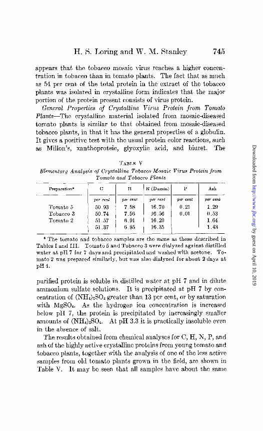
H. S. Loring and W. M. Stanley 745
appears that the tobacco mosaic virus reaches a higher concen- tration in tobacco than in tomato plants. The fact that as much as 54 per cent of the total protein in the extract of the tobacco plants was isolated in crystalline form indicates that the major portion of the protein present consists of virus protein.
General Properties of Crystalline Virus Protein from Tomato Plants-The crystalline material isolated from mosaic-diseased tomato plants is similar to that obtained from mosaic-diseased tobacco plants, in that it has the general properties of a globulin. It gives a positive test with the usual protein color reactions, such as Millon’s, xanthoproteic, glyoxylic acid, and biuret. The
Elementary Analysis of Crystalline Tobacco Mosaic Virus Protein from Tomato and Tobacco Plan.ts
Preparation*
Tomato 5 Tobacco 3 Tomato 2
C
per cent
50.93 50.74 51.57 51.37
H
per cent
7.58 7.56 6.91 6.95
N (Dumas)
per cent
16.70 16.56 16.20 16.35
P
per cent
0.21 0.01
Ash
per cent
1.29 0.53 1.64 1.43
* The tomato and tobacco samples are the same as those described in Tables I and III. Tomato 5 and Tobacco 3 were dialyzed against distilled water at pH 7 for 7 days and precipitated and washed with acetone. To- mato 2 was prepared similarly, but was also dialyzed for about 2 days at pH4.
purified protein is soluble in distilled water at pH 7 and in dilute ammonium sulfate solutions. It is precipitated at pH 7 by con- centration of (NH&S04 greater than 13 per cent, or by saturation with MgSOd. As the hydrogen ion concentration is increased below pH 7, the protein is precipitated by increasingly smaller amounts of (NH&S04. At pH 3.3 it is practically insoluble even in the absence of salt.
The results obtained from chemical analyses for C, H, N, P, and ash of the highly active crystalline proteins from young tomato and tobacco plants, together with the analysis of one of the less active samples from old tomato plants grown in the field, are shown in Table V. It may be seen that all samples have about the same
by guest on April 10, 2019
http://ww
w.jbc.org/
Dow
nloaded from

746 Mosaic Virus Protein from Tomato Plants
chemical composition. The values for C, H, and N of the highly active tomato sample are in good agreement with those obtained for the tobacco sample of comparable activity.
Opticab Activity-The results of several comparative determina- tions of optical activity for preparations of quite different infectivi- ties from both old and young plants are shown in Table VI. They were obtained by determining the rotation of a solution of the purified protein prepared by the addition of 2 drops of 2 N NaOH to 5 cc. of approximately a 0.5 per cent dialyzed suspension of the protein. These likewise show all the samples to have about the same specific rotation, regardless of their infectivity or whether they were prepared from tomato or tobacco plants. It is interest- ing to note that completely inactive tobacco mosaic virus protein
TABLE VI
Optical Activity of Different Samples of Crystalline Tobacco Mosaic Virus Protein
Preparation*
Tomato 1 “ 2 I‘ 3 L‘ 5
Tobacco 3 -
[a]: (per mg. N) Conoentration pH of solution
degrees
-0.43 -0.42 -0.44 -0.42 -0.45
gm. per 100 cc.
0.57 0.88 0.46 0.49 0.40
11.2 11.45 11.65
-!- * The preparations are the same as those described in Tables I and III.
prepared by treatment with ultraviolet light, formaldehyde, or nitrous acid (9) likewise has a specific rotation of -0.42” per mg. of nitrogen.
Isoelectric Point-Cataphoresis experiments were carried out by means of the Northrop-Kunite apparatus on dilute suspensions of crystals of virus protein. The suspensions were prepared by the addition of 2 drops of saturated (NH&S04 solution and 3 to 4 drops of 0.2 N HCl to 10 cc. of a 0.4 per cent solution of the puri- fied protein dialyzed at pH 7 to 8. The suspensions were then diluted with 90 cc. of distilled water adjusted to pH 3.3 with 6 drops of 0.2 N HCl. Portions of this suspension were adjusted to various hydrogen ion concentrations with 0.02 N NaOH and 0.02 N HCl, and were examined in the cataphoresis cell. Under these
by guest on April 10, 2019
http://ww
w.jbc.org/
Dow
nloaded from

H. S. Loring and W. M. Stanley 747
conditions, dilute suspensions of tiny crystals easily visible under the microscope were obtained. The hydrogen ion concentration was measured with a MacInnes type glass electrode.
The values found for the isoelectric points of different prepara- tions from both tomato and tobacco plants have varied from about pH 3.2 to about pH 3.35. All the samples examined from either tomato or tobacco plants showed consistent migration towards the negative pole below pH 3.2 and towards the positive pole above pH 3.35. The less active samples obtained from old plants gave isoe- lectric points in the lower range, while the more active samples ob- tained from young plants gave values towards the higher range. The most active samples isolated from young tomato and tobacco plants grown in the greenhouse gave the same value of about 3.3. The isoelectric point of an individual preparation was found to vary depending on the salt concentration of the buffer mixture and the nature of the salt present. In experiments in which acetic acid, as well as (NH&S04 and NaCl, was present, the isoelectric point was raised from 0.1 to 0.3 of a pH unit depending on the concen- tration of (NH&S04 and acetic acid. Eriksson-Quensel and Svedberg (10) have found the isoelectric point of a many times recrystallized sample in (NH&S04-acetate buffer to be pH 3.49. Best (ll), from precipitation studies, has reported the isoelectric point of a purified but not crystalline tobacco mosaic preparation to be pH 3.4. Takahashi and Rawlins (12), in experiments in which the migration of virus particles at various hydrogen ion concentrations was determined by infectivity measurements, found the virus to be isoelectric below pH 4.
Sedimentation Constants-The sedimentation constants and molecular weights of samples of crystalline tobacco mosaic virus protein from both the old and young tomato plants described above have been determined by Dr. Wyckoff and Mr. Biscoe (8). The sedimentation constant for the crystalline protein isolated from old tomato plants is slightly larger than that found for the protein from tobacco plants. However, the sedimentation con- stants of the virus proteins prepared under comparable conditions from young tobacco and tomato plants were found to be exactly the same.
Samples of the virus proteins from young tomato and tobacco plants were submitted to Dr. Svedberg for an ultracentrifugal
by guest on April 10, 2019
http://ww
w.jbc.org/
Dow
nloaded from

748 Mosaic Virus Protein from Tomato Plants
analysis. During their preparation these samples were main- tained between pH 6 and 8 in an effort to retain their molecular homogeneity. The samples, however, arrived too late to be included in the report by Eriksson-Quensel and Svedberg (10). Dr. Svedberg has kindly consented to have the results of the ultracentrifugal study, which was carried out by Mrs. Eriksson- Quensel, presented here. Although the new samples of virus protein were found to be somewhat inhomogeneous with respect to molecular weight, they were more homogeneous than the virus protein previously studied. The sedimentation constant,, Xzo’ X 1013, of virus protein from tobacco plants in solution at pH 6.8 was found to be 197 for the sample submitted in solution and 190 for the sample submitted in the form of a paste. That of virus protein from tomato plants submitted and tested in solution at pH 6.8 was found to be 202. These constants correspond to a molecular weight of about 17,000,OOO. At pH 9.8 the sample from tomato @ants was found to have formed two components having constants of 185 and 125, respectively, and at pH 11.7 it was found to have been disintegrated and split into low molecular weight components having constants of 8.1 and 3.8, respectively. The ultracentrifugal analyses of Dr. Svedberg and Dr. Wyckoff indicate that the sedimentation constant of virus protein obtained from young tomato plants is probably the same as that of virus protein obtained under comparable conditions from young tobacco plants.
Serological Experiments-Both precipitation and absorption experiments were performed to compare the serological properties of the crystalline virus protein preparations obtained from tobacco and tomato plants. Antisera were obtained from rabbits which had been injected with 200 mg. of each preparation, in weekly doses of 50 mg. each, dissolved in physiological saline solution at pH 7 to 8. The antiserum from the tobacco virus protein was then tested for precipitating antibodies both with tobacco virus protein and with that obtained from tomato plants, according to the precipitation technique used by Chester (13). The converse experiment was also performed for the antiserum to the tomato virus protein. The results of a typical experiment are shown in Table VII. As previously found (2), the antiserum to tobacco virus protein gave a precipitate with 2 X 10e6 gm. of the homol-
by guest on April 10, 2019
http://ww
w.jbc.org/
Dow
nloaded from

H. S. Loring and W. M. Stanley 749
ogous antigen. Likewise, the antiserum to tomato virus protein gave a precipitate with 2 X 1OW gm. of its homologous antigen. When antisera to either the tobacco virus protein or the tomato virus protein were mixed with the other antigen, a precipitate was likewise formed in each case with but 2 X lO-‘j gm. of the antigen.
In the absorption experiments, both antisera were completely absorbed with their heterologous antigens. The absorbed anti- serum was then tested with a small amount of its homologous antigen. In neither case was there an additional precipitate. One of two such experiments performed is outlined in Table VIII.
TABLE VII Results of Precipitin Tests with Crystalline Tomato and Tobacco
Coneentrat&;of protein
gm. Tomato virus protein
2 x 10-d 2 x 10-G 2 x 10-e
Tobacco virus protein 2 x 10-b 2 x 10-s 2 x 10⁻⁶
Virus Proteins
Antiserum to Antiserum to tomato virus tobacco virus
protein protein
++++ ++++ + + f f
++++ ++++ + + f f
. . . . i-i-+-/- indicate a very heavy precipitate; + a moderate precipitate;
A a very slight precipitate; - absence of a precipitate.
The results demonstrate that both the virus protein from tobacco and from tomato plants served equally well in absorbing the anti- bodies formed after the injection of either protein, and that the injection of virus protein from either plant host caused the forma- tion of no antibodies which were not precipitated by the virus protein from the other.
Solubility Experiments-The solubilities of the crystalline virus proteins obtained from tomato and tobacco plants were deter- mined to find whether the two samples would give the same solu- hility. Preliminary experiments, in which the virus proteins obtained from the old tomato and tobacco plants grown in the
by guest on April 10, 2019
http://ww
w.jbc.org/
Dow
nloaded from

TABL
E V
III
Abso
rptio
n Ex
perim
ent
on C
ryst
allin
e To
mat
o an
d To
bacc
o Vi
rus
Prot
ein
Proc
edur
e
(I)
I cc
. an
tiser
um
to
tom
ato
viru
s pr
otei
n +
2.33
cc
. (9
.3 m
g.)
toba
cco
viru
s pr
otei
n +
0.3
cc.
mer
thio
late
(p
rese
rvat
ive)
, *
mix
ed,
incu
bate
d at
37”
for
1 hr
., st
ored
in
ref
riger
ator
ov
erni
ght
and
cent
rifug
ed
(2)
Supe
rnat
ant
from
(1
) m
ixed
w
ith
1.0
cc.
(3.9
9 m
g.)
toba
cco
viru
s pr
otei
n,
incu
bate
d,
etc.
, as
in
(1
) (3
) Su
pern
atan
t fro
m
(2)
treat
ed
as i
n (2
)
(4)
“ ‘(
(3)
“ (‘
(‘ (2
)
(5)
“ “
(4)
mix
ed
with
1.
0 cc
. (3
.99
mg.
) to
mat
o vi
rus
prot
ein,
in
cuba
ted,
et
c.,
as
in
(1)
T
Res
ult
20+t
+t a t t
Proc
edur
e
(1)
1 cc
. an
tiser
um
to
toba
cco
viru
s pr
otei
n +
2.33
cc
. (9
.3 m
g.)
tom
ato
viru
s pr
otei
n +
0.3
cc.
mer
thio
late
(p
rese
rvat
ive)
, m
ixed
, in
cuba
ted
at
37”
for
1 hr
., st
ored
in
re
frige
rato
r ov
er-
nigh
t, an
d ce
ntrif
uged
(2
) Su
pern
atan
t fro
m
(1)
mix
ed
with
1.
0 cc
. (3
.99
mg.
) to
mat
o vi
rus
prot
ein,
in
cuba
ted,
et
c.,
as
in
(1)
(3)
Supe
rnat
ant
from
(2
) tre
ated
as
in
(2)
(4)
“ “
(3)
“ ((
“ (2
)
(5)
‘I “
(4)
mix
ed
with
1.
0 cc
. (3
.99
mg.
) to
bacc
o vi
rus
prot
ein,
in
cuba
ted,
et
c.,
as
in
(I)
Res
ult
2o+t
+t G
t t
* A
cont
rol
expe
rimen
t, in
w
hich
no
rmal
se
rum
w
as
mix
ed
with
th
e sa
me
conc
entra
tion
of
mer
thio
late
, fa
iled
to
give
a
prec
ipita
te.
t Th
e pr
ecip
itate
s fro
m
(1)
and
(2)
in
each
ex
perim
ent
wer
e co
mbi
ned,
w
ashe
d tw
ice
with
ph
ysio
logi
cal
salt
solu
tion,
an
d an
alyz
ed
for
nitro
gen.
Th
at
from
th
e an
tiser
um
to t
omat
o vi
rus
prot
ein
cont
aine
d 2.
14 m
g. o
f N
, w
hile
th
e ot
her
con-
ta
ined
2.
06 m
g.
of N
. 20
+ in
dica
tes
a la
rge
prec
ipita
te,
+ a
smal
l pr
ecip
itate
, an
d t a
tra
ce
of p
reci
pita
te.
$ Th
e tra
ce
of
prec
ipita
te
whi
ch
cont
inue
d to
be
for
med
af
ter
the
abso
rptio
n of
the
an
tibod
ies
prob
ably
co
nsis
ted
of
dena
ture
d pr
otei
n.
by guest on April 10, 2019
http://ww
w.jbc.org/
Dow
nloaded from

H. S. Loring and W. M. Stanley 751
field were compared, had shown the former to have a solubility about four times that of the latter. However, the tomato virus protein, as was mentioned before, carried more yellow pigment than did the tobacco sample. It was felt, therefore, that the difference might be due to the presence of the yellow impurity in the tomato sample or possibly to its lower infectivity. Subse- quent efforts to purify this tomato sample had resulted in prepara- tions that gave white solutions, but, as mentioned before, possessed still less virus activity than the original crystalline material.
Additional solubility determinations were, therefore, carried out on the virus proteins obtained from young tobacco and tomato plants. To 9.0 cc. of dialyzed solutions of the tomato and tobacco virus proteins, each containing 63.8 mg. of protein, was added 1 cc. of 1 M phosphate buffer at pH 5.6 and 625.0 mg. of (NH&S04. Kach solution was stirred during the addition of the (NH&S04, and in each case a part of the protein crystallized. Both suspen- sions were then stirred for 4 hour and centrifuged for a total of 4 hours. At the end of 2, 3, and 4 hours, respectively, samples were removed from each and analyzed for protein nitrogen.
The supernatant liquid from the tobacco protein contained 2.37 mg. of protein per cc. after centrifugation for 2 hours, while that from the tomato protein contained 1.84 mg. per cc. Additional centrifugation decreased the former value to 2.24 and finally to 1.47 mg. per cc., whereas the tomato decreased to 1.63 and 1.19 mg. per cc. Several experiments of this type in which the amount of ammonium sulfate was varied from 901 mg. to 590 mg. per 10 cc. were performed. In each case it was found that the protein concentration of the supernatant liquid decreased continually during 4 hours of centrifugation, and in all cases the values found for the tobacco protein were slightly higher than those found for the tomato protein. In some cases attempts were made to ap- proach equilibrium from the undersaturated side by extraction of the residue obtained after centrifugation with solvent of the same composition as the solution from which it had separated. In these experiments the protein concentration after 3 hours of centrifugation was always less than the lowest value found when equilibrium was approached from the supersaturated side.
The above experiments provide a rough comparison of the solu- bilities of the virus protein from tomato and tobacco plants and
by guest on April 10, 2019
http://ww
w.jbc.org/
Dow
nloaded from

752 Mosaic Virus Protein from Tomato Plants
show that they are of the same order. Additional data must be obtained, however, before it can be said with certainty whether the two proteins have identical or slightly different solubilities.
DISCUSSION
The highly active crystalline protein obtained from young tomato plants by careful fractionation agrees very closely in properties with that isolated from young tobacco plants grown under the same conditions. The general properties of both pro- teins are the same. They have the same elementary composition, the same optical activity, and the same isoelectric point. The properties of the virus proteins which are most likely to show differences between two preparations are their infectivities, sero- logical reactions, and solubilities. These have been compared for both the tomato and tobacco proteins. A number of infectiv- ity measurements at various concentrations have shown each preparation to have very much the same infectivity. The mean difference between the number of lesions obtained with the same concentration of tomato and tobacco virus protein was always, with but three exceptions, too small to indicate any significant difference in the infectivity of the two samples.
The serological experiments have, likewise, shown the two preparations to have identical serological properties. It is well known that antiserum to one strain of tobacco mosaic virus will give a precipitate when tested either with the same or with a different strain of mosaic virus. However, it has recently been shown by Chester (14) that some strains of tobacco mosaic virus may be differentiated by absorption experiments. It was found that antiserum to ordinary tobacco mosaic virus gave a precipi- tate with this virus after the serum had been completely absorbed with aucuba mosaic virus. Aucuba mosaic virus, therefore, does not completely absorb the antibodies present after the injection of ordinary tobacco mosaic virus. Absorption experiments on tomato and tobacco virus protein, however, have failed to show differences between these proteins. The antibodies formed after the injection of one protein were completely removed by absorption with the other.
The results of the solubility experiments suggest that the virus protein from tomato plants is slightly less soluble under the same
by guest on April 10, 2019
http://ww
w.jbc.org/
Dow
nloaded from
H. S. Loring and W. M. Stanley 753
conditions than virus protein from tobacco plants, However, the differences found were small and could be explained in a number of ways. More experimental data must be obtained before a definite conclusion can be drawn regarding the solubilities of the two proteins. It is entirely possible that the virus protein from tomato plants may differ slightly from that present in tobacco plants but still possess the same physiological properties. As shown by Northrop (15), crystalline pepsins prepared from differ- ent animal species possess the same physiological properties but have different solubilities and are therefore different proteins. It seems possible that slightly different proteins might also carry the same virus activity.
The isolation of crystalline protein preparations with varying degrees of infectivity from old tomato plants has shown that the loss of activity does not affect the ability of the protein to crystal- lize. This result is comparable to what has been found for aucuba mosaic virus (16) and even more strikingly for tobacco mosaic virus treat,ed with hydrogen peroxide or nitrous acid (9). Aucuba mosaic virus protein, like tobacco mosaic virus protein, becomes less active on repeated fractionation, while tobacco mosaic virus protein becomes entirely inactive when treated with hydrogen peroxide or nitrous acid. Both the less active aucuba and the entirely inactive hydrogen peroxide-treated proteins are well defined crystalline preparations. The fact that the changed protein obtained after hydrogen peroxide treatment resembles very closely the partially inactive samples suggests that the partial inactivation may be due to a similar type of reaction. Repeated treatment with celite as used in the course of fractiona- tion may well catalyze the oxidation by air of unstable groups in the molecule.
SUMMARY
The isolation from tobacco mosaic-diseased tomato plants of a crystalline protein possessing the properties of tobacco mosaic virus is described. It was found that the most active crystalline material could be best obtained from young rapidly growing green- house plants by a procedure involving a minimum amount of treatment with celite.
The properties of the proteins obtained from tomato and tobacco
by guest on April 10, 2019
http://ww
w.jbc.org/
Dow
nloaded from

754 Mosaic Virus Protein from Tomato Plants
plants grown under the same conditions and treated by the same procedure have been compared. The proteins have been shown to possess the same infectivities and to have identical serological properties and very nearly the same solubilities. They likewise have the same chemical composition, optical activity, and iso- electric point, and give the same sedimentation constant.
Repeated fractionation of the virus protein with celite at pH 4.5 and 8.0 results in a gradual inactivation of the protein, which remains soluble and may still be crystallized. It has been shown in other fractionation experiments, however, in which as much as 81 per cent of the original sample has been lost during the course of fifteen recrystallizations, that the crystals which remained pos- sessed the same infectivity as the original sample.
The comparison of the relative infectivities of the juices of diseased tomato and tobacco plants on a total protein basis indi- cates, in agreement with the percentage yields of crystalline virus protein isolated, that the tobacco mosaic virus reaches a higher concentration in tobacco than in tomato plants.
BIBLIOGRAPHY
1. Stanley, W. M., Science, 81, 644 (1935). 2. Stanley, W. M., Phytopathology, 26, 305 (1936). 3. Clinton, G. P., Connecticut Agric. Exp. Stat. Biennial Rep., pt. 12, 857
(190748). 4. Stanley, W. M., J. Biol. Chem., 116, 673 (1936). 5. Holmes, F. O., Bot. Gaz., 87, 39 (1929). 6. Samuel, G., and Bald, J. G., Ann. Appl. Biol., 20, 70 (1933). 7. Fisher, R. A., Statistical methods for research workers, Edinburgh
and London, 6th edition (1936). 8. Wyckoff, R. W. G., Biscoe, J., and Stanley, W. M., J. Bid. Chem., 117,
57 (1937). 9. Stanley, W. M., Science, 83, 626 (1936).
10. Eriksson-Quensel, I., and Svedberg, T., J. Am. Chem. Sot., 68, 1863 (1936).
11. Best, R. J., AustralianJ. Exp. Biol. andMe& SC., 14,l (1936). 12. Takahashi, W.‘N., and Rawlins, T. E., Hilgardia, 4, 441 (1930). 13. Chester, K. S., Phytopathology, 26, 686 (1935). 14. Chester, K. S., Phytopathology, 26, 778 (1936). 15. Northrop, J. H., J. Gen. Physiol., 16, 615 (1933). 16. Stanley, W. M., J. Biol. Chem., 117, 325 (1937).
by guest on April 10, 2019
http://ww
w.jbc.org/
Dow
nloaded from

Hubert S. Loring and W. M. StanleyFROM TOMATO PLANTS
TOBACCO MOSAIC VIRUS PROTEIN ISOLATION OF CRYSTALLINE
1937, 117:733-754.J. Biol. Chem.
http://www.jbc.org/content/117/2/733.citation
Access the most updated version of this article at
Alerts:
When a correction for this article is posted•
When this article is cited•
alerts to choose from all of JBC's e-mailClick here
tml#ref-list-1
http://www.jbc.org/content/117/2/733.citation.full.haccessed free atThis article cites 0 references, 0 of which can be
by guest on April 10, 2019
http://ww
w.jbc.org/
Dow
nloaded from

CORRECTION
On pages 737. 740, 742, 744, 763, and 765, Vol. 117, No. 2, February, 1937, the foot-note to the tables should read: To show a significant difference between the mean number of lesions in any one experiment, the ratio of the mean difference (M.D.) to the standard error of the mean difference (SD.) should not be less than 2.1.


![Review Article Earthworm Protease · fetida [27]. It has strong antiviral activities against cucumber mosaic virus and tomato mosaic virus. The protease (27,000 Da) is the most active](https://img.pdfslide.us/doc/110x75/5fd08da243d0e50fda5f4e1a/review-article-earthworm-fetida-27-it-has-strong-antiviral-activities-against.jpg)