Embed Size (px)
Citation preview
Downloaded from www.microbiologyresearch.org by
IP: 54.39.17.49
On: Wed, 04 Apr 2018 03:50:44
260 Journal of General Microbiology (1975), 87,260-272
Printed in Great Britain
Isolation of an Inducible Amidase from Pseudomonas acidovorans A E ~
By JEANETTE ALT A N D K. K R I S C H
Biochemisches Institut im Fachbereich Medizin der Universitat Kiel, Kiel23, Germany
AND P. HIRSCH
Institut fur Allgemeine Mikrobiologie der Universitat Kiel
(Received 9 September 1974)
SUMMARY
A bacterial strain, AEI, which hydrolysed acetanilide, was isolated from soil and identified as Pseudomonas acidovorans. Numerous amides, esters and enzyme inhibitors were tested as amidase inducers. Phenacetin was chosen as inducer for the large scale cultivation of these organisms because it was less toxic to the bacteria than acetanilide. The induction increased the enzymic activity 250-fold. In com- parison, the type culture strain of P. ucidovovans, ~ ~ ~ ~ 1 5 6 6 8 , had no amidase acti- vity which could be induced by phenacetin. Optimal growth conditions were established with respect to the concentration of carbon source and inducer so that about 10 % of the extractable bacterial protein consisted of the amidase. The organisms were lysed with lysozyme in the presence of EDTA and the enzyme was isolated mainly by column chromatography procedures. A preparation from 60 g (wet wt) bacteria yielded about IOO mg highly purified amidase with a specific activity of I 37 pmol substrate hydrolysed/min/mg protein. In addition to acetani- lide, the purified enzyme hydrolysed several other amides and esters. As standard substrate, p-nitroacetanilide was chosen.
I N T R O D U C T I O N
During a search for acetanilide-hydroxylating mono-oxygenases in micro-organisms, a bacterial strain was isolated from soil which catalysed the hydrolysis of acetanilide (F. Bernhardt, personal communication). An enzyme with the same activity had previously been isolated from pig liver microsomes (Krisch, 1963a), and this proved to be identical to the well-known unspecific carboxylesterase (EC. 3 . I . I . I ) (Krisch, 1963 b).
As part of a larger comparative project on the molecular and catalytic properties of esterases, it seemed worthwhile to include studies of a bacterial amidase/esterase.
We report the identification of the acetanilide-hydrolysing strain, its optimal growth con- ditions and large-scale cultivation. Results of induction studies and the isolation of the amidase to a homogeneous state are also described.
METHODS
Isolation of bacteria. The culture was obtained from F. Bernhardt (Giessen, Germany), who had previously isolated the strain from soil using a mineral salts medium (Ribbons & Evans, 1962) which contained 0.4 % (w/v) acetanilide as sole carbon and energy source.
Growth of cultures. Cultures were maintained on agar slants prepared from Standard
Downloaded from www.microbiologyresearch.org by
IP: 54.39.17.49
On: Wed, 04 Apr 2018 03:50:44
Isolation of amidase from Pseudomonas sp. 261 I-Nahragar (Merck, Darmstadt, Germany). Incubation was at 30 "C; fully grown cultures were stored at 4 "C. One litre of culture medium contained: I M-phosphate buffer pH 6.8 (40 ml); 'Hutner's vitamin-free mineral base' (Cohen-Bazire, Sistrom & Stanier, I 957) (20
ml); and 10 % (w/v) (NH4),S04 solution (10 ml). This comprised the 'standard mineral base' as described by Stanier, Palleroni & Doudoroff (1966). Carbon source, inducer and yeast extract (0.1 g/l), were added to this solution except in the tests for utilization of single organic carbon and energy sources. The pH was adjusted to 6.8 before autoclaving, and was found to be between 7.0 and 7.2 afterwards.
Measurement of bacterial growth. As an indicator of growth yield, the extinction at 660 nm (I cm light path) was measured with a PM4 spectrophotometer (Carl Zeiss, Oberkochen, Germany). Viable counts were obtained from plate counts, and the total number of micro- organisms was determined with a Thoma cell counting chamber. A total number of 108
bacteria/ml gave an El,, of 0.094. The relation was linear up to El,, = 0.8. Identification of species. For genus determination Bergey's Manual of Determinative
Bacteriology (I 957) was used. Species identification was carried out according to Stanier et al. (1966).
Chemicals. D-Fucose, D-tryptophan and valine were purchased from Serva, Heidelberg, Germany, sodium maleate, geraniol and baminovalerate from Fluka, Eschborn, Germany, and p-hydroxybenzoate from Riedel de Haen, Seelze-Hannover, Germany ; all other chemi- cals were from Merck.
Induction experiments. All substances to be tested as possible inducers were added at con- centrations of 0.01 to 0.18 % (w/v) to a culture medium containing 0.55 % (w/v) lactic acid. The pH was adjusted to 6-8 with 6 M-NaOH. The bacteria were grown for 72 h at 30 "C and then harvested by centrifuging (30 min, 12000 g). Sediments were stored frozen before testing. The bacterial sediment was disintegrated by lysozyme (see Disruption of bacteria, below) and the supernatant liquid was kept refrigerated until the assays of activity and protein could be carried out.
The following substances were tested as inducers. Amides : acetanilide, acetamide, p- ethoxyacetanilide (phenacetin), sulphanilamide, nicotinamide, xylocaine, L-leucineamide, L-leucine-/3-naphthylamide, L-tyrosyl-tyrosine, glycyl-L-proline, ~-leucyl-glycine, glycyl- tyrosine, leucyl-tyrosine, phenylalanyl-phenylalanine, benzoyl-L-arginineamide, monoethyl- glycine-xylidide. Esters : N-a-benzoyl-L-arginine-ethylester, N-benzoyl-L-tyrosine-ethylester, acetyl-L-tyrosine-ethylester, L-tyrosine-ethylester, L-phenylalanine-methylester, diethyl- aminoethyl-diphenylpropyl-acetate (SKF 525-A). Other substances: aniline, p-aminoben- zoic acid, p-hydroxybenzoic acid, naphthalene, /I-naphthylamine, xylidine, p-ethoxyaniline (phenetidine), 2,6-di methylanil i ne.
The phenacetin used for the induction was a product of Merck (No. 7221), and fulfilled the purity requirements of the British Pharmacopoeia I 968 and the Deutsche Arzneimittel- buch DAB 7. The melting point was I 35-5 to I 36 "C (I 37 to I 38 "C, Handbook of Chemistry and Physics, I 969-1970 ; 134-7 "C, D'Ans-Lax, 1964). A thin-layer chromatogram according to Bernhammer & Krisch (1965) showed only one spot.
Growth of large bacterial quantities. The inoculum was pre-grown for 24 h in a mineral salts medium with 0.22 % (w/v) lactic acid without phenacetin. The pH was adjusted to 6-8 with 6 M-NaOH. The main batch culture was carried out in 5 1 flasks containing 4 I of the same medium with I g phenacetin/l. These flasks were inoculated with 200 ml of the culture grown without phenacetin. After 72 h of vigorous aeration at 30 "C, the bacteria were harvested by continuous flow centrifuging (Sorvall KSB, I; Sorvall Inc., Norwalk, Connecticut, U.S.A.). The total yield was frozen at - 18 "C.
Downloaded from www.microbiologyresearch.org by
IP: 54.39.17.49
On: Wed, 04 Apr 2018 03:50:44
262 J. ALT, K. KRISCH AND P. HIRSCH
Disruption of bacteria. In preliminary experiments ultrasonic treatment with an 80 W sonifier Type B 12 (Branson Instruments, Inc., Stamford, Connecticut, U.S.A.) was used. The sonified solution was carefully cooled with ice water.
To obtain optimal conditions for lysis the EDTA concentration was varied. In such preliminary experiments the suspensions were incubated for 2 h at 30 "C. After centrifuging, the sediments were not washed. For standard enzyme preparations the bacteria were sus- pended in 4 ml o-05 M-phosphate buffer pH 7.5 (containing 0.025 M-EDTA) per gram bac- terial wet weight. Lysozyme (I 5 000 u/mg, Merck) was added to a concentration of 0.5 mg/ ml. This mixture was then stirred for 16 h at 4 "C and an extract obtained by centrifuging for 30 min at ~ooooog. The sediment was washed once with the same volume of distilled water. The pH was kept constant at 7-5. Distilled water for washing, instead of buffer, was necessary for the subsequent removal of nucleic acids. The supernatant liquid was combined with the extract and the resulting solution used for further enzyme purification.
Determination of amidase activity. The hydrolysis of p-nitroacetanilide was measured at 405 nm (I cm light path) in an Eppendorf photometer connected with an Eppendorf recorder (Netheler & Hinz, Hamburg, Germany). The cuvette contained I ml substrate solution ( I O - ~ M-p-nitroacetanilide dissolved in warm water and then cooled to room temperature) and I ml 0.1 M-tris buffer pH 8.6 containing the enzyme. The measurements were per- formed at 30 "C. The molar extinction coefficient of p-nitroaniline in 0.05 M-tris buffer pH 8.6 was 10000 I/mol/cm.
Protein determination. The protein concentration was determined using a micro version of the biuret method (Beisenherz et al. 1953). Water and protein solution were combined in Eppendorf micro test tubes (Netheler & Hinz) to a total volume of I ml. Then 0-2 ml of a 3 M-trichloroacetic acid solution was added. This mixture was shaken for 5 min with an Eppendorf rotation shaker. The denatured protein was then sedimented by centrifuging for I min in an Eppendorf microcentrifuge and the supernatant liquid discarded. The sediment was subsequently dissolved in 0.5 ml biuret reagent and 0.5 ml water and measured after 30 rnin in microcuvettes (2 cm path length) at 546 nm, with bovine serum albumin as standard. After column chromatography, the fractions were measured at 280 nm (0.5 cm light path) for protein determination.
Enzyme isolation. Step I : Removal of nucleic acids. Streptomycin sulphate was added to the crude extract to a final concentration of I g/Ioo ml. The sample was then centrifuged for 30 min at 12000g and the precipitate discarded.
Step 2 : Fractional precipitation by ammonium sulphate. To the supernatant liquid from step I, solid ammonium sulphate was slowly added until the solution was 30 % saturated. The sulphate concentration was checked by titration with BaCI, (Bergmeyer et al. 1961). The reaction mixture was centrifuged for 30 min at 120008, the sediment discarded, and further ammonium sulphate added to the supernatant liquid until 65 % saturation was reached. During the precipitation the pH was kept constant at 7-5. After centrifuging for 30 min at 12000 g the sediment was dissolved in 0.005 M-tris buffer pH 8.6 containing O-OOT
M-EDTA and 2 % (v/v) glycerol. This buffer will be designated as 'tris buffer for column chromatography '.
Step 3 : Column chromatography with DEAE-Sephadex A-50. The enzyme solution was either dialysed or desalted by gel filtration on a Sephadex G-50 column (Pharmacia, Uppsala, Sweden) and applied to a DEAE-Sephadex A-50 column (Pharmacia; 5 x 50 cm). The DEAE-Sephadex was equilibrated with tris buffer for column chromatography. A gradient was applied from a 2-vessel gradient system. The first vessel contained 1100 ml tris buffer for column chromatography, and the second vessel contained the same buffer with 0.35 M-
Downloaded from www.microbiologyresearch.org by
IP: 54.39.17.49
On: Wed, 04 Apr 2018 03:50:44
Isolation of amidase from Pseudomonas sp. 263 NaCI. The fractions containing amidase activity were combined and dialysed against tris buffer for column chromatography containing ammonium sulphate, the concentration of which had been calculated to yield a final concentration of 65 % saturation after dialysis.
Step 4: Gel filtration. The enzyme precipitate was centrifuged and dissolved in as little phosphate buffer (0.005 M, pH 8.0) as possible. This solution was applied to a column (2.5 x roo cm) filled with Sephadex G-IOO (Pharmacia) pretreated as specified by the manufacturer.
Step 5 : Hydroxyapatite. Bio-Gel HTP (Bio-RAD Laboratories, Richmond, California, U.S.A.) was equilibrated with 0.005 M-phosphate buffer pH 8.0. The pooled enzyme (after gel filtration) was applied to a hydroxyapatite column (2-5 x 13 cm). The adsorbed protein was eluted with a linear phosphate buffer gradient pH 8.0 from 0.005 to 0.06 M. Temperature and rate of elution were kept constant (Bernardi, 1971). If necessary the rate of elution was restricted to I ml/3 min.
Purity of the enzyme. Analytical disc electrophoresis. This was performed according to Hedrick & Smith (1968) with 4,6 or 8 % (w/v) polyacrylamide gels. The protein was stained with I % amidoblack in 7 % acetic acid and the gels were electrophoretically destained with 7 % acetic acid. Staining of esterase activity with a-naphthyl acetate was performed as described by Heymann, Junge & Krisch (1972).
Reaction with diethyl p-nitrophenylphosphate. A I o - ~ M stock solution of diethyl-p- nitrophenylphosphate (purity > 98 % ; Bayer, Leverkusen, Germany) was prepared accord- ing to Krisch (1966). The absorbance difference was measured in microcuvettes (light path 2 cm) at 405 nm with an Eppendorf photometer. The test tube contained 0.25 ml 0.2 M-tris buffer pH 8.0, 0.05 ml diethyl-p-nitrophenylphosphate stock solution and 0.2 ml of a highly purified enzyme solution (containing 0.5 to I mg protein/ml). The extinction of buffer, diethyl-p-nitrophenylphosphate and enzyme was subtracted from the ‘initial burst ’. The amount of p-nitrophenol was calculated using the extinction coefficient as determined by Krisch (1966): e = 16240 I/mol/cm at pH 8.0.
RESULTS
Species identijication A comparison of the isolate with Pseudoinonas acidovorans ATCCi 5668 (kindly provided
by Professor R. Y. Stanier) showed the same ‘general characters of diagnostic value for the differentiation of species of aerobic pseudomonads’ (Stanier et al. 1966). Our isolate and the reference strain differed in using phenacetin as the only source of carbon and energy. Strain ~ ~ ~ 1 5 6 6 8 did not grow at all, while strain AEI grew after a period of about two weeks. From this plate a second strain, A E ~ , was isolated which had the same properties as strain AEI except that it grew spontaneously with phenacetin as single carbon and energy source. These observations were reproducible. Strain ATCCI 5668 had no amidase activity which could be induced by phenacetin.
Induct ion The average amidase activity (substrate : p-nitroacetanilide) of the crude extract from
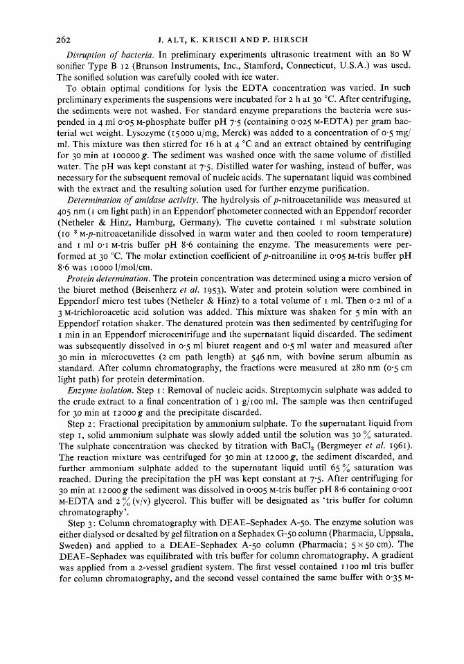
uninduced bacteria was 0.1 6 ,umol/min/mg (25 experiments). The extremes were 0.05 and 0.5 I ,umol/min/mg. Repeated measurements showed a large scattering of the values : the relative standard deviation was 75 % of the mean. Phenacetin was the best inducer (Figs. I and 2 and Table I). In some tests acetamide, xylocaine, acetanilide and aniline showed an induction too (Tables I and 2), but the induction with phenacetin was more intensive and reproducible.
Downloaded from www.microbiologyresearch.org by
IP: 54.39.17.49
On: Wed, 04 Apr 2018 03:50:44
264 J. ALT, K. K R I S C H AND P. HIRSCH
I I 1 1 1 I I I I
0.04 0.08 0.12 0.16 Phenacetin concn (".;I, w/v)
Fig. I . Total amidase activity as a function of the phenacetin concentration. Points indicate the mean values of I 5 experiments, vertical bars represent the standard deviation.
0.04 0.08 0.12 0-16 Phenacetin concn (%, w/v)
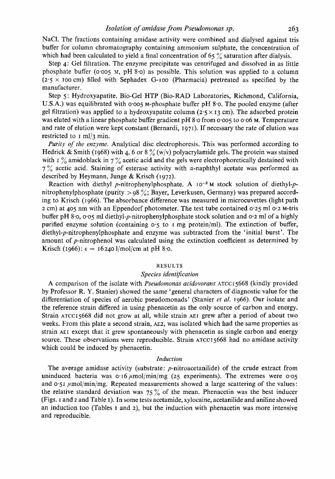
Fig. 2. Specific activity of the supernatant liquid from lysed bacteria as a function of the phenacetin concentration of the growth medium. Points indicate the mean values of I 5 experiments, vertical bars represent the standard deviation.
Growth of bacteria
We tested 0.5 % (w/v) succinate, p-hydroxbenzoate, lactate and adipate as carbon sources. Growth was best with lactate or swccinate, and lactate was therefore chosen as the standard carbon and energy source. The lactate concentration optimal for the induction was depen- dent on the phenacetin concentration, the highest activity being obtained with 0.22 % lactate at a phenacetin concentration of 0.1 %. Higher lactate concentrations increased growth but
Downloaded from www.microbiologyresearch.org by
IP: 54.39.17.49
On: Wed, 04 Apr 2018 03:50:44
Isolation of amidase from Pseudomonas sp. 265
Table I. Induction of the amidase from P. acidovorans AEI
The values are the results of individual experiments. They represent the relative increase of the specific activity as compared with controls without the inducer. Twenty-five other compounds were also tested (see Methods) but none had a significant inducing effect.
Increase in specific activity (pmol/min/mg) A < \
Concn of the inducer ( %). . . 0'0 I
Acetanilide 1.16, 2-42, 3.19 34'6 Acetamide 3.88, 1-35 1-36, 1-47 Aniline 1-56, 1-78 1 '49 Xylocaine 2.02, 1-07, 1-65 1'1
Phenacetin 6.63, 7-02, 2.62 47-29 34'6
0.03
Table 2 . Eflect of acetanilide concentration on amidase activity
Acetanilide concn (%, w/v) 0 0.03, 0.04, 0.06, 0.08, 0.1
Specific activity (pmol/min/mg) 0.029, 0.225, 0.145, 0-21 2, 0.80, o* Induction factor 1-0, 7'7, 5-0, 7.26, 27-4 Total activity/l medium (pmollmin) 4.0, 37.0, 10.7, 16.7, 15.7
* No measurable growth.
Table 3. Bacterial growth with diflerent phenacetin concentrations
The lactate concentration was 0-22 % (w/v). Bacteria were grown for 72 h at 30 "C.
I O - ~ x Total counts I O - ~ x Viable counts Phenacetin (I %, wlv) (bacteria/ml) (bacteria/ml)
- 0'02 0.06
0.14 0'10
20
19 I5 6 3
inhibited the induction, presumably due to ' catabolite repression' (Magasanik, I 96 I).
Although higher concentrations of phenacetin inhibited bacterial growth (Table 3), induc- tion of enzyme activity was enhanced even when some of the phenacetin remained undis- solved after autoclaving. After 72 h growth, undissolved phenacetin could no longer be detected. The lag phase of the growth curve was prolonged by the addition of phenacetin. With 0-1 % phenacetin the early exponential phase began after 20 h. Maximal growth yield was then reached after about three days instead of one day.
Disruption of bacteria
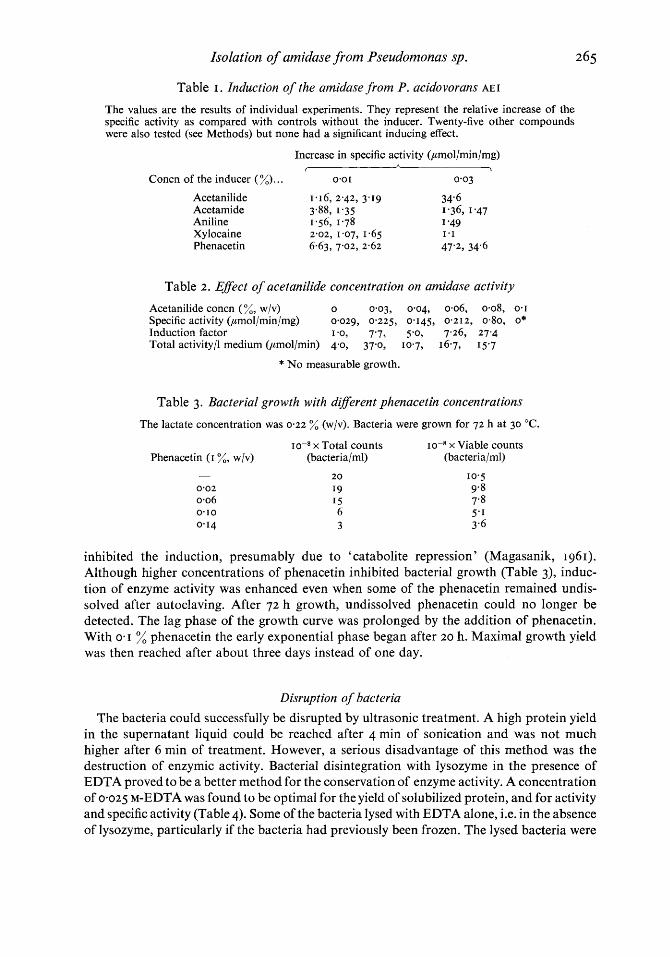
The bacteria could successfully be disrupted by ultrasonic treatment. A high protein yield in the supernatant liquid could be reached after 4 min of sonication and was not much higher after 6 min of treatment. However, a serious disadvantage of this method was the destruction of enzymic activity. Bacterial disintegration with lysozyme in the presence of EDTA proved to be a better method for the conservation of enzyme activity. A concentration of 0.025 M-EDTA was found to be optimal for the yield of solubilized protein, and for activity and specific activity (Table 4). Some of the bacteria lysed with EDTA alone, i.e. in the absence of lysozyme, particularly if the bacteria had previously been frozen. The lysed bacteria were
Downloaded from www.microbiologyresearch.org by
IP: 54.39.17.49
On: Wed, 04 Apr 2018 03:50:44
266 J. ALT, K. KRISCH A N D P. HIRSCH
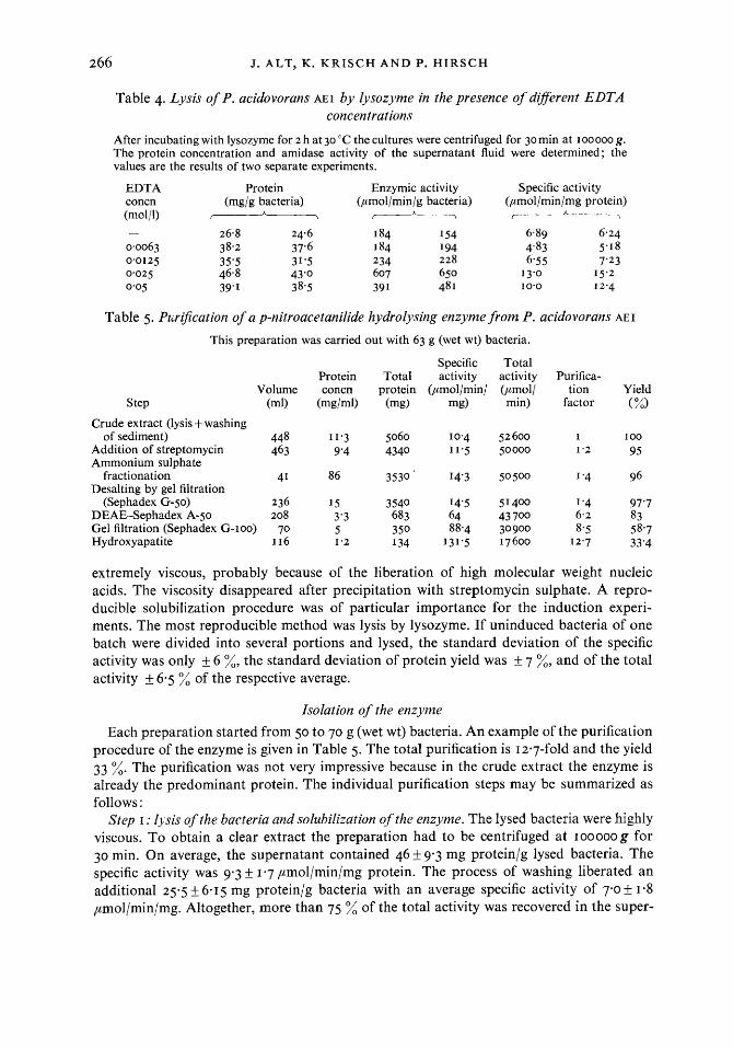
Table 4. Lysis of P. acidovorans AEI by lysozyme in the presence of diflerent EDTA concentrations
After incubating with lysozyme for 2 h at 30 "C the cultures were centrifuged for 30 min at IOOOOO g. The protein concentration and amidase activity of the supernatant fluid were determined ; the values are the results of two separate experiments.
EDTA Protein Enzymic activity Specific activity concn (mg/g bacteria) (pmol/min/g bacteria) (pmol/min/mg protein) (mol/l) - c--+ r------h----\
- 26.8 24.6 184 I54 6.89 6.24 0.0063 38.2 37.6 184 I94 4'83 5.1 8 0.0125 35'5 31.5 234 228 6-55 7'23 0.025 46.8 43'0 607 650 I 3.0 15.2 0.05 39.1 3 8.5 39 I 48 I 10.0 I 2.4
Table 5. Puri5cation of a p-nitroacetanilide hydrolysing enzyme from P. acidovorans AEI
This preparation was carried out with 63 g (wet wt) bacteria.
Step
Specific Total Protein Total activity activity Purifica-
Volume concn protein (pmol/min/ (pmoll tion Yield (m0 (mgh-4 (mg) mg) min) factor (%)
Crude extract (lysis + washing of sediment) 448 11.3 5060 10.4 52600 I I00
Addition of streptomycin 463 9'4 4340 11.5 50000 1-2 95
fractionation 41 86 3530 14'3 50500 I '4 96
(Sephadex G-50) 236 15 3540 14'5 51400 1 '4 97'7 DEAE-Sephadex A-50 208 3'3 683 64 43 700 6.2 83 Gel filtration (Sephadex G-100) 70 5 350 88.4 30900 8.5 58.7 Hydroxyapatite I 16 1.2 134 131.5 17600 I 2.7 33'4
Ammonium sulphate
Desalting by gel filtration
extremely viscous, probably because of the liberation of high molecular weight nucleic acids. The viscosity disappeared after precipitation with streptomycin sulphate. A repro- ducible solubilization procedure was of particular importance for the induction experi- ments. The most reproducible method was lysis by lysozyme. If uninduced bacteria of one batch were divided into several portions and lysed, the standard deviation of the specific activity was only & 6 %, the standard deviation of protein yield was & 7 %, and of the total activity & 6.5 % of the respective average.
Isolation of the enzyme
Each preparation started from 50 to 70 g (wet wt) bacteria. An example of the purification procedure of the enzyme is given in Table 5. The total purification is 12.7-fold and the yield 33 %. The purification was not very impressive because in the crude extract the enzyme is already the predominant protein. The individual purification steps may be summarized as follows :
Step I : ljsis of the bacteria and solubilization ofthe enzyme. The lysed bacteria were highly viscous. To obtain a clear extract the preparation had to be centrifuged at ~ o o o o o g for 30 min. On average, the supernatant contained 46 & 9.3 mg protein/g lysed bacteria. The specific activity was 9.3 & 1.7 ,umol/min/mg protein. The process of washing liberated an additional 25.5 5 6-15 mg protein/g bacteria with an average specific activity of 7.0 & 1.8 ,rcmol/min/mg. Altogether, more than 75 % of the total activity was recovered in the super-
Downloaded from www.microbiologyresearch.org by
IP: 54.39.17.49
On: Wed, 04 Apr 2018 03:50:44
Isolation of amidase jrom Pseudomonas sp. 267
x >
cd
* .- .- *
10 20 30 40 50 60 70 80
Amonium sulphate saturation (%)
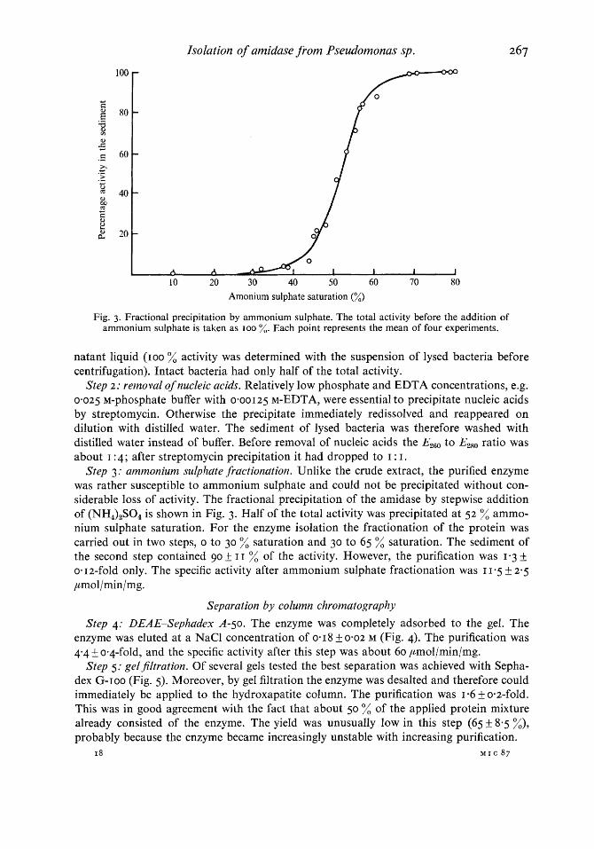
Fig. 3. Fractional precipitation by ammonium sulphate. The total activity before the addition of ammonium sulphate is taken as IOO %. Each point represents the mean of four experiments.
natant liquid (roo activity was determined with the suspension of lysed bacteria before centrifugation). Intact bacteria had only half of the total activity.
Step 2: removal of nucleic acids. Relatively low phosphate and EDTA concentrations, e.g. 0.025 M-phosphate buffer with 0-00125 M-EDTA, were essential to precipitate nucleic acids by streptomycin. Otherwise the precipitate immediately redissolved and reappeared on dilution with distilled water, The sediment of lysed bacteria was therefore washed with distilled water instead of buffer. Before removal of nucleic acids the EzG0 to Ezg0 ratio was about I : 4 ; after streptomycin precipitation it had dropped to I : I.
Step 3: ammonium sulphate fractionation. Unlike the crude extract, the purified enzyme was rather susceptible to ammonium sulphate and could not be precipitated without con- siderable loss of activity. The fractional precipitation of the amidase by stepwise addition of (NH,),S04 is shown in Fig. 3. Half of the total activity was precipitated at 52 % ammo- nium sulphate saturation. For the enzyme isolation the fractionation of the protein was carried out in two steps, o to 30 % saturation and 30 to 65 % saturation. The sediment of the second step contained go f I I % of the activity. However, the purification was 1.3 & om 12-fold only. The specific activity after ammonium sulphate fractionation was I 1-5 f 2-5
,umol/min/mg.
Separation by column chromatography
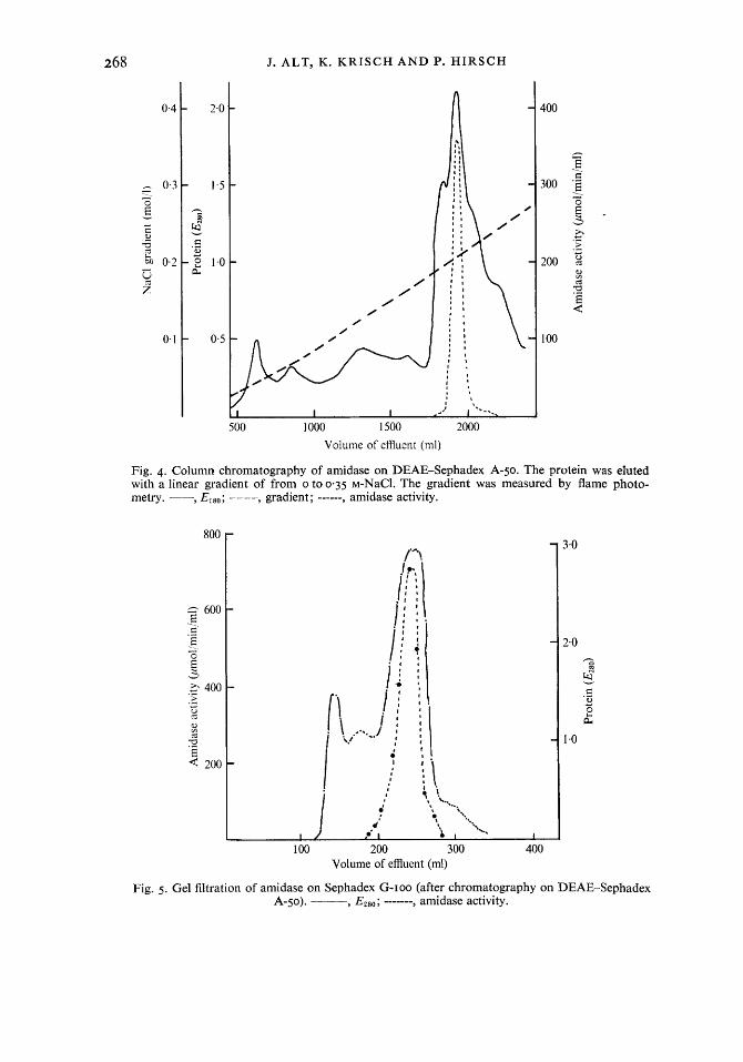
Step 4: DEAE-Sephadex A-50. The enzyme was completely adsorbed to the gel. The enzyme was eluted at a NaCl concentration of 0.18 5 0.02 M (Fig. 4). The purification was 4-4 f 0.4-f0ld, and the specific activity after this step was about 60 pmol/min/mg.
Step 5.- gelfiltration. Of several gels tested the best separation was achieved with Sepha- dex G-roo (Fig. 5). Moreover, by gel filtration the enzyme was desalted and therefore could immediately be applied to the hydroxapatite column. The purification was 1.6 f 0-2-fold. This was in good agreement with the fact that about 50 % of the applied protein mixture already consisted of the enzyme. The yield was unusually low in this step (65 8.5 %), probably because the enzyme became increasingly unstable with increasing purification.
18 M I C 87
Downloaded from www.microbiologyresearch.org by
IP: 54.39.17.49
On: Wed, 04 Apr 2018 03:50:44
268 J. ALT, K. K R I S C H AND P. H I R S C H
0.4
0 0
0 0
0 #
400
- 3 .
300 $ =.
W $ - x > * ._ .I
c
200 2 2 a E 6
100
500 1000 1500 2000
Volume of effluent (ml)
Fig. 4. Column chromatography of amidase on DEAE-Sephadex A-50. The protein was eluted with a linear gradient of from o to 0-35 M-NaCl. The gradient was measured by flame photo- metry. --, EZso; ---- gradient; ------, amidase activity.
800
z 1.. 6oo
v E,
G .- E .. I 0
3 400 .I > u .- *
x a E * 200
Fig. 5 . Gel filtration of amidase on Sephadex G-IOO (after chromatography on DEAE-Sephadex A-50). ---y E280; ------- , amidase activity.
Downloaded from www.microbiologyresearch.org by
IP: 54.39.17.49
On: Wed, 04 Apr 2018 03:50:44
0.5
0.4
h
0.3 6 v
c a, .- w
i 0.2
0.1
Isolation of amidase from Pseudomonas sp. 269
100 200 300 400 500 Volume of effluent (ml)
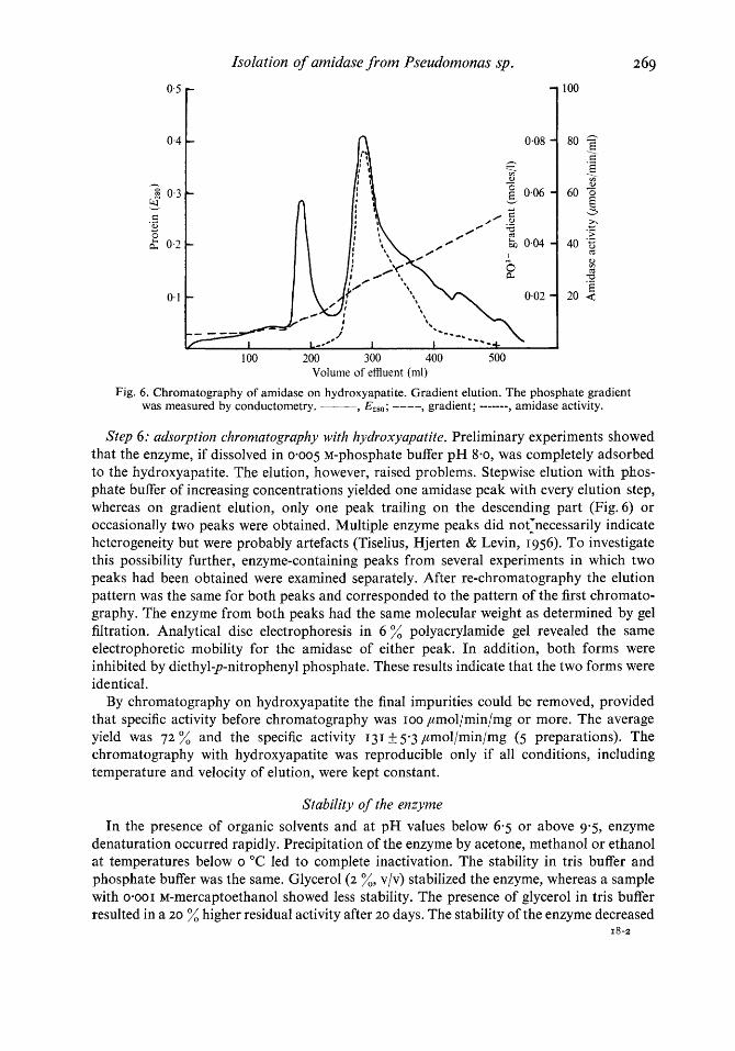
Fig. 6. Chromatography of amidase on hydroxyapatite. Gradient elution. The phosphate gradient was measured by conductometry. ___ Y E 4 8 0 ; ---- , gradient; ------- , amidase activity.
Step 6: adsorption chromatography with hydroxyapatite. Preliminary experiments showed that the enzyme, if dissolved in 0.005 M-phosphate buffer pH 8.0, was completely adsorbed to the hydroxyapatite. The elution, however, raised problems. Stepwise elution with phas- phate buffer of increasing concentrations yielded one amidase peak with every elution step, whereas on gradient elution, only one peak trailing on the descending part (Fig.6) or occasionally two peaks were obtained. Multiple enzyme peaks did not1necessarily indicate heterogeneity but were probably artefacts (Tiselius, Hjerten & Levin, I 956). To investigate this possibility further, enzyme-containing peaks from several experiments in which two peaks had been obtained were examined separately. After re-chromatography the elution pattern was the same for both peaks and corresponded to the pattern of the first chromato- graphy. The enzyme from both peaks had the same molecular weight as determined by gel filtration. Analytical disc electrophoresis in 6 % polyacrylamide gel revealed the same electrophoretic mobility for the amidase of either peak. In addition, both forms were inhibited by diethyl-p-nitrophenyl phosphate. These results indicate that the two forms were identical.
By chromatography on hydroxyapatite the final impurities could be removed, provided that specific activity before chromatography was roo ,umol/min/mg or more. The average yield was 7 2 % and the specific activity 131 If: 5-3 ,umol/min/mg (5 preparations). The chromatography with hydroxyapatite was reproducible only if all conditions, including temperature and velocity of elution, were kept constant.
Stability of the enzyme
In the presence of organic solvents and at pH values below 6.5 or above 93, enzyme denaturation occurred rapidly. Precipitation of the enzyme by acetone, methanol or ethanol at temperatures below o "C led to complete inactivation. The stability in tris buffer and phosphate buffer was the same. Glycerol ( 2 %, vlv) stabilized the enzyme, whereas a sample with 0.001 M-mercaptoethanol showed less stability. The presence of glycerol in tris buffer resulted in a 20 % higher residual activity after 20 days. The stability of the enzyme decreased
18-2
Downloaded from www.microbiologyresearch.org by
IP: 54.39.17.49
On: Wed, 04 Apr 2018 03:50:44
270 J. ALT, K. K R I S C H AND P. H I R S C H
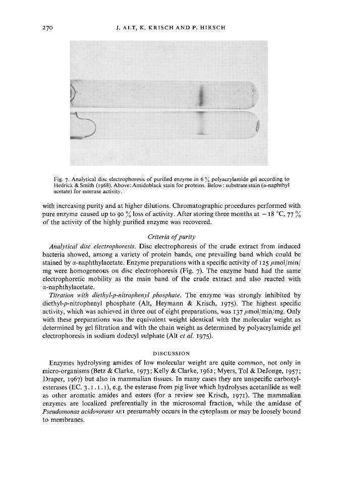
Fig. 7. Analytical disc electrophoresis of purified enzyme in 6 % polyacrylamide gel according to Hedrick & Smith (1968). Above: Amidoblack stain for proteins. Below: substrate stain (a-naphthyl acetate) for esterase activity.
with increasing purity and at higher dilutions. Chromatographic procedures performed with pure enzyme caused up to 90 % loss of activity. After storing three months at - 18 "C, 77 % of the activity of the highly purified enzyme was recovered.
Criteria of purity
Analytical disc electrophoresis. Disc electrophoresis of the crude extract from induced bacteria showed, among a variety of protein bands, one prevailing band which could be stained by a-naphthylacetate. Enzyme preparations with a specific activity of I 25 ,umol/min/ mg were homogeneous on disc electrophoresis (Fig. 7). The enzyme band had the same electrophoretic mobility as the main band of the crude extract and also reacted with a-naphthylacetate.
Titration with diethyl-p-nitrophenyl phosphate. The enzyme was strongly inhibited by diethyl-p-nitrophenyl phosphate (Alt, Heymann & Krisch, 1975). The highest specific activity, which was achieved in three out of eight preparations, was 137 pmol/min/mg. Only with these preparations was the equivalent weight identical with the molecular weight as determined by gel filtration and with the chain weight as determined by polyacrylamide gel electrophoresis in sodium dodecyl sulphate (Alt et al. 1975).
DISCUSSION
Enzymes hydrolysing amides of low molecular weight are quite common, not only in micro-organisms (Betz & Clarke, 1973; Kelly & Clarke, 1962; Myers, To1 & DeJonge, 1957; Draper, 1967) but also in mammalian tissues. In many cases they are unspecific carboxyl- esterases (EC. 3. I . I . I), e.g. the esterase from pig liver which hydrolyses acetanilide as well as other aromatic amides and esters (for a review see Krisch, 1971). The mammalian enzymes are localized preferentially in the microsomal fraction, while the amidase of Pseudomonas acidovorans AEI presumably occurs in the cytoplasm or may be loosely bound to membranes.
Downloaded from www.microbiologyresearch.org by
IP: 54.39.17.49
On: Wed, 04 Apr 2018 03:50:44
Isolation of amidase from Pseudornonas sp. 27 = The high initial activity after induction, the fast growth of P. acidovorans AEI as well as
the relatively convenient isolation procedure make this amidase a suitable object for a closer examination of its catalytic and molecular properties. The further characterization of the enzyme and its active site are reported elsewhere (Alt et al. 1975). Catabolite repression has also been observed with the acetamide hydrolysing amidase of Pseudomonas aeruginosa 8602/~ (Brammar & Clarke, 1964). However, with the enzyme of P. acidovorans AEI this cannot be overcome simply by adding more inducer, because phenacetin or a metabolite of it proved to be toxic for bacterial growth. For this reason it is important to keep the lactate concentration as low as possible.
The physiological function of mammalian esterases and amidases has not yet been eluci- dated. The liver esterase is probably important for the hydrolysis of certain drugs like acetanilide, phenacetin or procaine, and thus could contribute to the so-called detoxication of foreign compounds. The function of amidases or esterases in bacteria may be more easily explained. Most of them are inducible by substrates or analogous substances, for example, the enzymes of Mycobacterium smegmatis (Diaper, 1967) and of P. aerzdginosa (Kelly & Clarke, 1962; Betz & Clarke, 1973). It can be assumed that these inducible enzymes are functioning in the dissimilation of carbon and nitrogen sources.
The aliphatic amidase from P. aeruginosa, which preferentially hydrolyses short-chain aliphatic amides, has been extensively studied by Clarke and her collaborators. Recently, mutants grown on acetanilide as sole carbon source were isolated. They produced amidases with different substrate specificities and electrophoretic properties from the parent strain (Brown & Clarke, 1972). In another comparative study the biochemical and immunological properties of amidases from various Pseudomonas species were investigated. The amidases in extracts of cultures of acetamide-grown P. acidovorans strains 14 and ~ ~ 1 ~ 9 6 8 1 showed strong cross-reactivity with an antiserum against P. fferUginOSd A amidase (Clarke, I 972). They are possibly identical with the acetamide splitting activity found by us in crude extracts from P. acidovorans AEI. This activity, however, was lost during the purification procedure and the highly purified enzyme does not hydrolyse acetamide (Alt et al. 1975).
This work forms part of a doctoral thesis submitted by J.A. to the Kiel University. The culture of P. acidovorans was kindly provided by Dr F. Bernhardt. His advice was gratefully accepted.
REFERENCES
ALT, J., HEYMANN, E. & KRISCH, K. (1975). Characterization of an inducible amidase from Pseudomonas acidovorans. European Journal of Biochemistry (in the Press).
BEJSENHERZ, G., BOLTZE, H. H., BUCHER, T., CZOK, R., GARBADE, K. H., MEYER-ARENDT, E. &PFLEIDERER, G. (1953). Diphosphofructose-Aldolase, Phosphoglyceraldehyd-Dehydrogenase, Glycerophosphat- Dehydrogenase und Pyruvat-Kinase aus Kaninchenmuskulatur in einem Arbeitsgang. Zeitschrift firr Naturforschung 8b, 555-577.
BERGEY'S MANUAL OF DETERMINATIVE BACTERIOLOGY, 7th edn (1957). Edited by R. S. Breed, E. G. D. Murray and N. R. Smith. London: Baillikre, Tindall and Cox.
BERGMEYER, H.-U., HOLZ, G., KAUDER, E. M., MOLLERING, H. & WIELAND, 0. (1961). Kristallisierte Glycerokinase aus Candida mycoderma. Biochemische Zeitschrgt 333, 47 1-480.
BERNARDI, G. (1971). Chromatography of proteins on hydroxyapatite. In Methods in Enzymology, vol. 22,
pp. 325-339. New York and London: Academic Press. BERNHAMMER, E. & KRISCH, K. (1965). Deacetylation of phenacetin by liver esterase. Biochemical Pharma-
cology 14, 863-871. BETZ, J. L. & CLARKE, P. H. (1973). Growth of Pseudomonas species on phenylacetamide. Journalof General
Microbiology 75, 167-177. BRAMMAR, W. J. & CLARKE, P. H. (1964). Induction and repression of Pseudomonas aeruginosa amidase.
Journal of General Microbiology 37, 307-319.
Downloaded from www.microbiologyresearch.org by
IP: 54.39.17.49
On: Wed, 04 Apr 2018 03:50:44
272 J. ALT, K. KRISCH A N D P. HIRSCH
BROWN, P. R. & CLARKE, P. H. (1972). Amino acid substitution in an amidase produced by an acetanilide-
CLARKE, P. H. (1972). Biochemical and immunological comparison of aliphatic amidases produced by
COHEN-BAZIRE, G. W., SISTROM, R. & STANIER, R. Y . (1957). Kinetic studies of pigment synthesis by non-
DRAPER, P. (I 967). The aliphatic acylamide amidohydrolase of Mycobacterium smegmatis : its inducible
D’ANs-LAX (I 964). Taschenbuch fur Chemiker und Physiker. 2. Band: Organische Verbindungen. Edited by
HANDBOOK OF CHEMISTRY AND PHYSICS (1969-1970), 50th edn. Ohio: Chemical Rubber Co. HEDRICK, J. L. & SMITH, A. J. (1968). Size and charge isomer separation and estimation of molecular weights
of proteins by disc gel electrophoresis. Archives of Biochemistry and Biophysics 126, I 55-164. HEYMANN, E., JUNGE, W. & KRISCH, K. (I 972). Carboxylesterase aus Schweinelebermikrosomen, Reaktion
mit Phenylmethansulfonylfluorid und Nachweis von Isoenzymen. Hoppe-Seyler ’s Zeitschrift fiir Physiologische Chemie 353, 576-588.
KELLY, M. & CLARKE, P. H. (1962). An inducible amidase produced by a strain of Pseudomonas aeruginosa. Journal of General Microbiology 27, 305-3 I 6.
KRISCH, K. (I 963 a). Isolierung einer Esterase aus Schweinelebermikrosomen. Biochemische Zeitschrgt 337, 531-545.
KRISCH, K. ( I 963 6). Eigenschaften und Substratspezifitat einer Esterase aus Schweinelebermikrosomen . Biochemische Zeitschrift 337, 546-573.
KRISCH, K. ( I 966). Reaction of a microsomal esterase from hog-liver with diethyl-p-nitrophenyl-phosphate. Biochimica et biophysica acta 122, 265-280.
KRISCH, K. (1971). Carboxylic ester hydrolases. In The Enzymes, vol. 5, pp. 43-69. Edited by P. D. Boyer. New York and London: Academic Press.
MAGASANIK, B. (1961). Catabolite repression. Cold Spring Harbor Symposia on Quantitative Biology 26,
MYERS, D. K., TOL, J. W. & DEJONGE, M. H. T. (1957). Substrate specificity of the esterases of some sapro-
RIBBONS, D. W. & EVANS, W. C. (1962). Oxidative metabolism of protocatechuic acid by certain soil
STANIER, R. Y . , PALLERONI, N. J. & DOUDOROFF, M. (1966). The aerobic Pseudomonads: a taxonomic study.
TISELIUS, A., HJERTEN, S. & LEVIN, 0. (1956). Protein chromatography on calcium phosphate columns.
utilizing mutant of Pseudomonas aeruginosa. Journal of General Microbiology 70, 287-298.
Pseudomonas species. Journal of General Microbiology 71, 241-257.
sulfur purple bacteria. Journal of Cellular and Comparative Physiology 49, 25-36.
nature and relation to acyl-transfer to hydroxylamine. Journal of General Microbiology 46, I I 1-123.
E. Lax. Berlin, Gottingen and Heidelberg : Springer Verlag.
249-2 5 6.
phytic mycobacteria. Biochemical Journal 65, 223-232.
Pseudomonads : a new ring-fission mechanism. Biochemical Journal 83, 482-492.
Journal of General Microbiology 43, I 59-271.
Archives of Biochemistry and Biophysics 65, I 32-1 55.