Embed Size (px)
Citation preview

Proc. Nati. Acad. Sci. USAVol. 82, pp. 4-8, January 1985Biochemistry
Isolation and partial characterization of a fatty acid binding proteinin rat liver plasma membranes
(hepatocyte/hepatic uptake)
WOLFGANG STREMMEL*, GEORG STROHMEYER*, FRANZ BORCHARD*, SHAUL KOCHWAt, AND PAUL D. BERKtf
*Medizinische Klinik, Poliklinik D und Pathologisches Institut der Universitat Dusseldorf, Moorenstrasse 5, 4000 Dusseldorf, Federal Republic of Germany;and tThe Polly Annenberg Levee Hematology Center and the Hepatic Research Group, Department of Medicine, Mount Sinai School of Medicine,New York, NY 10029
Communicated by Hans Popper, June 21, 1984
ABSTRACT When [14C]oleate-bovine serum albumincomplexes were incubated in vitro with rat liver plasma mem-branes (LPM), specific, saturable binding of oleate to themembranes was observed. Maximal heat-sensitive (i.e., specif-ic) binding was 3.2 nmol/mg of membrane protein. Oleate-agarose affinity chromatography of Triton X-100-solubilizedLPM was used to isolate a single 40-kDa protein with highaffinity for oleate. On gel filtration, the protein comigratedwith various fatty acids but not with ['4C]bilirubin, [35S]sulfo-bromophthalein, [14C]taurocholate, [14C]phosphatidylcholine,or [14C]cholesteryloleate. A rabbit antibody to this membranefatty acid-binding protein gave a single precipitin line with theantigen but no reactivity with concentrated cytosolic proteins,LPM bilirubin/sulfobromophthalein-binding protein, or ratalbumin or other rat plasma proteins. The antibody selectivelyinhibited heat-sensitive binding of [14C]oleate to LPM. Immu-nofluorescence studies localized the antigen in liver-cell plas-ma membranes as well as in other major sites of fatty acidtransport. These data are compatible with the hypothesis thatthis protein may act as a receptor in a hepatocellular uptakemechanism for fatty acids.
Although the hepatic uptake of fatty acids is often describedas a passive, diffusional process (1), recent kinetic studieshave suggested that at least a portion of their cellular uptakemay be carrier mediated (2-6). However, others have ques-tioned carrier-mediated uptake of fatty acids and assertedthat the apparently saturable uptake component observed inprevious studies predominantly reflected fatty acid metabo-lism (7, 8). Previously, we presented evidence suggestive ofa component of saturable and reversible binding of fatty ac-ids to high-affinity membrane binding sites (9, 10). In thepresent study, we have further examined the binding of fattyacids to rat liver plasma membranes (LPM) and sought toisolate and characterize a putative membrane receptor pro-tein responsible for the binding.
MATERIALS AND METHODSMaterials. [1-'4C]Oleic acid, [1-14C]arachidonic acid, [1-
14C]palmitic acid, [1-14C]linoleic acid, [24-'4C]taurocholicacid, L-a-l-palmitoyl-2-[1_14C]oleoyl-phosphatidylcholine,cholesteryl[1- 4C]oleate, and Aquasol were from New En-gland Nuclear. [35S]sulfobromophthalein (disodium phenol-tetrabromophthalein di[35S]sulfonate; [35S]BSP) and[1',2,3',4,5,6' ,7',8-'4C]bilirubin were from Amersham. Theunconjugated [14C]bilirubin was repurified as previously de-scribed (11). Unlabeled oleate, taurocholate, BSP, bilirubin,bovine serum albumin (fraction V), 1-ethyl-3-(3-dimethyla-minopropyl)carbodiimide, and Triton X-100 were from Sig-ma; Sephadex G-100 and G-150, Sepharose 4B, and AH-
Sepharose 4B, from Pharmacia; Bio Beads SM-2, from Bio-Rad; rat serum albumin and rabbit antiserum against ratalbumin, from Cappel Laboratories (Cochranville, PA); goatanti-rat fibronectin (IgG fraction), from Calbiochem-Beh-ring; and anti-rat ligandin, by courtesy of A. W. Wolkoff(Department of Medicine and Liver Center, Albert EinsteinCollege of Medicine, New York, NY).
Isolation and Characterization of LPM. LPM enriched insinusoidal components were prepared from livers of maleSprague-Dawley rats by differential centrifugation (12, 13)and characterized by electron microscopy (14) and by analy-sis of enzymatic markers (13). Membrane protein contentwas determined by the method of Lowry et al. (15).
Binding of [14C]Oleate. To avoid exceeding the solubilityof oleate in the aqueous phase of incubation mixtures (10),membrane [14C]oleate binding was determined by incubationof LPM aliquots with 60 ,uM [14C]oleate bound to variousmeasured quantities of defatted (16) bovine serum albumin.Nonspecific binding was evaluated in parallel incubations ofLPM aliquots denatured by prior incubation for 4 hr at 70°C,avoiding agglutination of membrane particles. All incuba-tions were carried out in 1 ml of P,/NaCl (0.15 M NaCl/0.02M sodium phosphate, pH 7.6) for 30 min at 37°C, after pre-liminary studies had established that binding was completewithin 30 min. Binding was assessed by a centrifugation as-say (17, 18).Binding of Other Ligands. To examine the binding to LPM
of various organic anions under specified conditions, as abasis for antibody inhibition studies (see below), aliquotscontaining 5 mg of membrane protein were incubated at 37°Cfor 30 min in 1 ml of Pi/NaCl with various concentrations ofeither [14C]taurocholate, or 35S-BSP bound to defatted bo-vine serum albumin (1:2 molar ratio), or [14C]oleate bound todefatted bovine serum albumin (3:1 molar ratio). This systemhas been described in detail (18). In antibody inhibition stud-ies, resuspended aliquots of LPM were incubated for 30 minat 37°C with 0.2 mg of the IgG-fraction of either anti-fattyacid binding membrane protein antiserum or anti-fibronectin(control) antiserum before ligands were added.
Preparation of Affinity Columns. Oleate-agarose was pre-pared by incubating 40 ml of swollen, washed AH-Sepharose4B with 60 ml of 0.1 M sodium oleate, pH 10.0, and 2 g of 1-ethyl-3-(3-dimethylaminopropyl)carbodiimide for 3 days at37°C with gentle rotation (19). BSP was coupled to Sepha-rose 4B with epichlorhydrin (20) as described (21).
Solubilization and Affinity Chromatography of MembraneProteins. LPM proteins were solubilized with 1% (vol/vol)Triton X-100 (21). After centrifugation at 100,000 x g for 60min, residual detergent was removed with Bio-Beads SM-2(21). Aliquots containing 500 mg of the solubilized proteins
Abbreviations: LPM, liver plasma membrane(s); FABP, fatty acidbinding protein; BSP, sulfobronmophthalein.tTo whom reprint requests should be addressed.
4
The publication costs of this article were defrayed in part by page chargepayment. This article must therefore be hereby marked "advertisement"in accordance with 18 U.S.C. §1734 solely to indicate this fact.

Proc. Natl. Acad. Sci. USA 82 (1985) 5
were then loaded onto oleate-agarose and BSP-agarose col-umns and washed with P1/NaCl at 20 ml/hr until no furtherprotein (A280) appeared in the washes. Bound protein wasthen eluted from the BSP-agarose column with 50 AuM BSP(21) and from the oleate column with 8 M urea. The eluateswere concentrated by ultrafiltration (Diaflo PM-10 mem-branes, Amicon), and the proteins were characterized byNaDodSO4/PAGE (22) and analytical isoelectric focusing inpolyacrylamide gels (2117 Multiphor, LKB). The proteinbands were routinely stained with Coomassie blue; repre-sentative gels were also stained with Sudan black or periodicacid/Schiff reagent (23). Contamination of the isolated BSP-and oleate-binding proteins was examined by double-immunodiffusion in agar (24) with rabbit antisera against ratalbumin, rat hepatocyte BSP-binding protein (18), and rat li-gandin.
Further Characterization of the Oleate-Binding Protein.Aliquots (0.3 mg) of the purified oleate-binding protein weremixed with tracer amounts of [14C]oleate and subjected togel filtration through a Sephadex G-150 column, which wascalibrated with purified standard proteins spanning a rangeof 20-158 kDa. Other 0.3-mg aliquots of the purified oleate-binding protein were equilibrated with tracer amounts of var-ious 14C-labeled fatty acids, other organic anions, and hydro-phobic membrane constitutents and then subjected to gel fil-tration through a column of Sephadex G-100 equilibratedwith 0.1 M NaCl/1 mM NaHCO3, pH 7.6. Elution was car-ried out with the same solution at 14 ml/hr. Eluates, collect-ed in 1-ml fractions, were monitored for protein (A280) andfor 14C (liquid scintillation spectrometry) (18).
Preparation of Antibody to Oleate-Binding Protein. Out-bred New Zealand White rabbits were immunized with thepurified oleate-binding protein as described (18). Presenceand purity of the antibody was examined by radial double-immunodiffusion on agar plates (24) against the purifiedLPM oleate-binding protein, Triton X-100-solubilized LPMproteins, rat serum albumin, concentrated rat liver cytosolicproteins, rat LPM BSP-binding protein (18), and whole ratserum. IgG fractions of the antisera were prepared by stan-dard techniques (25).
Indirect Immunofluorescence Studies. Frozen 5-pAm sec-tions of various rat tissues were air-dried and then incubatedfor 30 min at room temperature with 1:50, 1:100, and 1:200dilutions of rabbit antibody against the LPM oleate-bindingprotein. Sections were then washed with Pi/NaCl for 30 min,incubated with fluorescein isothiocyante-conjugated swineanti-rabbit antiserum diluted 1:20 or 1:50, thoroughlywashed with P1/NaCl for 60-90 min, and finally mountedwith buffered glycerol. Controls consisted of incubationswith antiserum preabsorbed with the LPM oleate-bindingprotein in equal dilution, incubations without immune se-rum, and incubations with pre-immune serum. Within a fewhours after mounting, sections were examined with a fluo-rescence microscope (Zeiss), employing filter combinationBP485/FT510/LP520.
RESULTSCharacterization of LPM Fractions. The LPM fractions
were similar to those used previously (18) and showed homo-geneous membrane structures in numerous randomly select-ed electron micrographs. Very few tight junctions, typical ofcanalicular membranes, were observed. 5'-Nucleotidase wasenriched 25-fold, and glucagon-stimulated adenylate cy-clase, 12-fold compared to the initial homogenate. Contami-nation with microsomes or mitochondria was negligible, asassessed by enzymatic markers. Albumin or ligandin werenot detected by radial immunodiffusion against appropriateantibodies.
Binding of ["4C]Oleate to LPM. As the oleate/albumin mo-
lar ratio was increased from 0.25 to 5 in incubation mixturescontaining a total oleate concentration of 60 ,uM, the initialfree oleate concentration increased hyperbolically from0.018 to 6.2 ,uM (10). [These concentrations represent maxi-mal estimates, as they were calculated from the equilibriabetween oleate and albumin (10), ignoring the unknownbinding parameters of the incubated LPM.] Over the samerange of oleate/albumin ratios, total [14Cloleate bound to na-tive LPM increased nonlinearly (Fig. 1A). When the total-binding curve was corrected for heat-insensitive (i.e., non-specific) binding, an apparent saturation curve was observed(Fig. 1B). In the system employed, maximal heat-sensitiveoleate binding (16.04 ± 1.06 nmol/5 mg of membrane pro-tein) occurred at an oleate/albumin ratio of 3, at which point60% of the incubated oleate was still not membrane bound.Specific binding was not further increased by increasing theoleate/albumin ratio to 4 or 5, despite a progressive increasein the ratio of residual non-membrane-bound oleate to albu-min and a further increase of more than 2-fold in the calculat-ed free oleate concentration remaining in the incubation mix-ture.
Binding of Other Organic Anions to LPM. Incubation ofLPM with increasing concentrations of [14C]taurocholate,[35S]BSP (as a 1:2 complex with bovine serum albumin) and[14C]oleate (as a 3:1 complex with bovine serum albumin)resulted in saturable binding of each of these representativeligands, as previously described (18). In the presence of ex-cess unlabeled ligand, nonspecific binding of [35S]BSP and[14C]taurocholate to LPM was less than 2% and 6%, respec-tively, of total binding and was ignored in subsequent anti-body inhibition studies with these two ligands. Other than
c
o-
.0
Oa
O O
EL
30
20
lo1
2 3 4Oleate/albumin, molar ratio
FIG. 1. (A) Binding of ['4C]oleate to native and heat-denaturedLPM (mean + SD, n = 5). Each incubation contained 5 mg of LPMprotein and a total oleate concentration of 60 AM. The bovine serum
albumin concentration was varied so that the oleate/albumin ratioranged from 0.25-5. (B) Specific binding of [14C]oleate to LPM, cal-culated (from the data in A) as total binding to native membranesminus binding to heat-denatured LPM.
A
T", ~ T
T ative LPM
/ -
t~ ~~~~~~~~~,,I -~~~~~~~~~"-
, ,2~A Heat Denatured LPMIs ,,~~~~~~~~~~~~
II011'
,I ,
Biochemistry: Stremmel et aL
I

6 Biochemistry: Stremmel et al.
establishing conditions for maximal binding of each test sub-stance as a reference point for inhibition studies (see below),no attempt was made to further characterize ligand bindingin this system.
Affinity Chromatography Studies. In contrast to the multi-plicity of bands found for the initial solution of solubilizedmembrane proteins, NaDodSO4/PAGE revealed only a sin-gle band at -40 kDa in the concentrated eluate from the ole-ate-agarose column (Fig. 2). As previously reported (18), asomewhat larger protein, of -55 kDa, was recovered fromthe BSP-agarose column.
Further Characterization of the Oleate-Binding MembraneProtein. On cylindrical gels, the oleate-binding protein re-covered from oleate-agarose did not stain with either Sudanblack or periodic acid/Schiff reagent. Analytical isoelectricfocusing of the protein revealed only a single, sharp bandwith a pI of 9.0. The protein showed no immunologic reactiv-ity with rabbit antibodies against rat serum albumin, ligan-din, or the hepatocyte membrane BSP-binding protein. OnSephadex G-150 gel filtration, the oleate binding protein co-migrated with [14C]oleate as a discrete, sharp peak. By com-parison with other marker proteins, its molecular mass inthis nonreducing, nondetergent medium was again 40 kDa.When the purified protein was cochromatographed on Se-phadex G-100 with various organic anions and other hydro-phobic membrane constituents, binding to the protein wasonly exhibited by the various 14C-labeled fatty acids tested(Fig. 3). Although oleate comigrated with the protein, theoleate ester of cholesterol and a phosphatidylcholine con-taining oleate in the 2-position did not. BSP, bilirubin, andtaurocholate also showed no affinity for the oleate-bindingmembrane protein. These data suggest that the proteinserves as a general fatty acid-binding protein and that its af-finity spectrum is not limited solely to oleate. It is, therefore,referred to below as the LPM fatty acid binding protein(LPM-FABP).
Characterization of Rabbit Antibodies Against the LPM-FABP. Whole antisera from two rabbits repeatedly injectedwith the LPM-FABP produced a single precipitin line be-tween the purified protein and the mixture of solubilizedLPM proteins, but showed no reaction with concentrated ratliver cytosolic proteins [including cytosolic fatty acid-bind-ing protein (Z protein)], rat liver LPM BSP-binding protein,rat albumin, and rat serum.
Antibody Inhibition of Organic Anion-Binding to Rat LPM.In studies employing various concentrations of a 3:1 com-plex of [14C]oleate-albumin with constant amounts of mem-
I 2*- 92 5-66.2
- 45.0
31,0
-21.5
A B C D
FIG. 2. Affinity chromatography of detergent-solubilized pro-teins from rat LPM. Shown are Coomassie blue-stained cylindricalgels after NaDodSO4/PAGE of total solubilized membrane proteins(gel A), 40-kDa protein eluted from oleate-agarose with 8 M urea (gelB), 55-kDa protein eluted from BSP-agarose with BSP (gel C), andstandards (gel D; molecular masses in kDa, to right). Tops of gelsand tracking dye fronts are aligned with the horizontal lines betweenthe gels.
Radiooctivity, 0 D 280nm:dprn: y ,
150 a I OlDunit
! lX Oleote
Polmitote
*|1ti Arochidonote
a1tLinoleate
0 20 40 60 80FRACT IONS
IIRodio-activit;Yi
I loo :,§dy: 3.I
10
I 2500 A,IIM
120
,
I 2000 a 1
OD 280nm
l(-)I 0 OD unit
Cholesterol-
Phospotidyl-choline I
lirubin I,:
IpL II.-- -- -r--- -- I-
I 50 1 uro-
t-0 20 40 60 80
FRACTIONS
FIG. 3. Chromatography on Sephadex G-100 of 0.3-mg aliquotsof purified LPM fatty acid (oleate)-binding protein with traceramounts of [35S]BSP, ['4C]bilirubin, or [14C]taurocholate; or ultra-sonically dispersed ['4C]phosphatidylcholine, cholesteryl [14C]-oleate, [14C]oleate, ['4C]arachidonate, [14C]palmitate, or [14C]lino-leate. Only 14C-labeled fatty acids were eluted with the protein peak.Ligands not soluble in aqueous solution (bilirubin, cholesteryl ole-ate, and phosphatidylcholine) appeared in the void volume (up tofraction 21), whereas BSP and taurocholate appeared in the effluentnear the total volume of the column (starting at fraction 70) and notassociated with protein.
brane and antibody, the antibody against the LPM-FABPsignificantly inhibited binding of [14C]oleate to native LPMwhereas nonspecific oleate binding to heat-denatured LPMwas unaffected (Fig. 4). The anti-LPM-FABP inhibited heat-sensitive (specific) binding by a maximum of 41% comparedto control in this system. In incubations containing the sameamounts of antibody (0.2 mg of IgG fraction) and [14C]oleatebut various amounts of LPM, a linear relationship was ob-served between the logarithm of the LPM concentration andthe percent inhibition of specific [14C]oleate binding, as de-termined by comparison with control incubations containing
25I
*c 20-o4 Antifibronectin Toa b
c 150~~~~~~~~~~
° 10 0 Anti-LPM-FABProe) 10----°0Lo~~~~0 ~~~~~~~Total binding:
C ads' heat denatured LPM
O/ I ,1_ 1_12 24 36 48Oleate incubated, nmol
60
FIG. 4. Effect of the IgG fraction of a rabbit antibody to theLPM fatty acid binding protein (anti-LPM-FABP, o) and a controlIgG fraction (anti-fibronectin, *) on total [14C]oleate binding to na-tive and heat-denatured LPM. Aliquots (5 mg) of native or dena-tured LPM were incubated in a volume of 1 ml for 30 min at 370Cwith 0.2 mg of either IgG fraction and then incubated for an addition-al 30 min with 6-60 ,uM [14C]oleate in the form of a 3:1 oleate-albu-min complex. Data shown are mean values from 3-5 incubations(SD _ ±5% in all cases). Under the conditions employed, the anti-LPM-FABP inhibits heat-sensitive (specific) [14C]oleate binding by<41%.
----------- L
Proc. NatL Acad Sci. USA 82 (1985)
Proc. Nat. Acad. Sci USA 82 (1985) 7
0.5 1.0 5 10LPM protein, mg
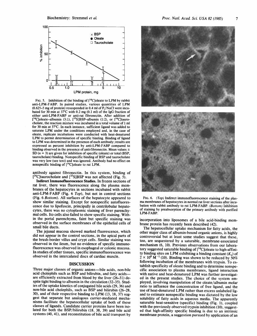
FIG. 5. Inhibition of the binding of [14C~oleate to LPM by rabbitanti-LPM-FABP. In paired studies, various quantities of LPM(0.625-5 mg of protein) resuspended in 0.4 ml of Pi/NaCl were incu-bated for 30 min at 37°C with 0.2 mg (0.1 ml) of the IgG fraction ofeither anti-LPM-FABP or anti-rat fibronectin. After addition of[14C]oleate-albumin (3:1), [35S]BSP-albumin (1:2), or [14C]tauro-cholate, the reaction mixture was incubated in a total volume of 1 mlfor 30 min at 37°C. In each instance, sufficient ligand was added tosaturate LPM under the conditions employed and, in the case ofoleate, replicate incubations were conducted with heat-denaturedLPM to permit determination of specific binding. Binding of ligandto LPM was determined in the presence of each antibody; results areexpressed as percent inhibition by anti-LPM-FABP compared tobinding observed in the presence of anti-fibronectin. Mean values ±SD (n = 3) are given for inhibition of specific (oleate) or total (BSP,taurocholate) binding. Nonspecific binding of BSP and taurocholatewas very low (see text) and was ignored. Antibody had no effect onnonspecific binding of [14C]oleate to rat LPM.
antibody against fibronectin. In this system, binding of[14C]taurocholate and [35S]BSP was not affected (Fig. 5).
Indirect Immunofluorescence Studies. In frozen sections ofrat liver, there was fluorescence along the plasma mem-branes of the hepatocytes in sections incubated with rabbitanti-LPM-FABP (Fig. 6 Top), but not in control sections(Fig. 6 Bottom). All surfaces of the hepatocyte appeared toshow similar staining. Except for nonspecific autofluores-cence due to lipofuscin, principally in centrilobular hepato-cytes, there was no cytoplasmic staining of liver parenchy-mal cells. Ito cells also failed to show specific staining. With-in the portal parenchyma, faint but specific staining wasobserved in the surface membranes of the epithelial cells ofsmall bile ducts.The jejunal mucosa showed marked fluorescence, which
did not appear in the control sections, in the apical parts ofthe brush-border villus and crypt cells. Similar staining wasobserved in the ileum, but no evidence of specific immuno-fluorescence was observed in esophageal or colonic mucosa.In studies of other tissues, specific immunofluorescence wasobserved in the intercalated discs of cardiac muscle.
DISCUSSIONThree major classes of organic anions-bile acids, non-bileacid cholephils such as BSP and bilirubin, and fatty acidsare efficiently extracted from blood by the liver (26-31) de-spite tight binding to albumin in the circulation (32-35). Stud-ies of the uptake kinetics of conjugated bile acids (29, 36) andnon-bile acid cholephils, such as BSP and bilirubin (26-28,30), and of their respective binding to LPM (13, 18, 37) sug-gest that separate but analogous carrier-mediated mecha-nisms facilitate the hepatocellular uptake of both of theseclasses of ligands. Candidate carrier proteins have been iso-lated for both the BSP/bilirubin (18, 38, 39) and bile acidsystems (40, 41), and reconstitution of bile acid transport by
FIG. 6. (Top) Indirect immunofluorescence staining of the plas-ma membranes of hepatocytes in normal rat liver sections after incu-bation with rabbit antibody to rat LPM-FABP. (Bottom) Inhibitionof staining by preabsorption of the primary antibody with purifiedLPM-FABP.
incorporation into liposomes of a bile acid-binding mem-brane protein has recently been described (42).The hepatocellular uptake mechanism for fatty acids, the
other major class of albumin-bound organic anions, is highlycontroversial but at least some studies suggest that these,too, are sequestered by a saturable, membrane-associatedmechanism (6, 10). Previous observations from our labora-tory suggested saturable binding of [14Cjoleate to high-affini-ty binding sites on LPM exhibiting a binding constant (Ka) of2 x 108 M-1 (10). Binding was shown to be reduced by 50%following incubation of the membranes with trypsin. To es-tablish specificity of oleate binding and to determine nonspe-cific association to plasma membranes, ligand interactionwith native and heat-denatured LPM was further investigat-ed in the present studies. The choice of the system em-ployed, involving manipulation of the oleate/albumin molarratio to influence the concentration of free ligand, and theuse of heat-denatured LPM rather than excess unlabeled lig-and to estimate nonspecific binding was dictated by the lowsolubility of fatty acids in aqueous media. The apparentlysaturable heat-sensitive (specific) binding (Fig. 1), coupledwith the previously observed trypsin inhibition (10), suggest-ed that high-affinity specific binding is due to an intrinsicmembrane protein, a suggestion pursued by application of an
Biochemistry: Stremmel et aL

8 Biochemistry: Stremmel et al.
affinity chromatography technique previously successfullyemployed to demonstrate a BSP/bilirubin-binding protein insimilar membrane preparations (18).The affinity chromatography data have established that
LPM do, in fact, contain a single protein with a high bindingaffinity for fatty acids but not for phospholipids, cholesterolesters, bile acids, BSP, or bilirubin. This protein (LPM-FABP) consists of a 40-kDa polypeptide chain with a pI of9.0, lacking either lipid or carbohydrate components. Basedon its molecular weight and the maximal specific binding ca-pacity of LPM for oleate, this protein would appear to con-stitute no more than 12% by weight of the intrinsic mem-brane protein of the hepatocyte. The protein does not shareimmunologic determinants with either ligandin or the cyto-solic fatty acid-binding protein (Z protein) previously de-scribed in liver (43) and small intestine (44). It is also distin-guished from the latter by physicochemical properties, sincethe Z protein has a molecular mass of 12 kDa and apI of 5.7.
Thus, we have demonstrated the presence in LPM of asingle, basic protein with a high affinity for fatty acids butnot for other hydrophobic membrane constitutents or forother classes of albumin-bound organic anions. Its precisephysiologic function remains to be established but its pI of9.0 is compatible with the hypothesis that, at physiologicpH, electrostatic interactions between a positively chargedmembrane protein and a negatively charged fatty acid anionmay contribute to the binding process. Moreover, immuno-fluorescence detection of its presence in liver, small intes-tine, and myocardium, which are major sites of fatty acidtransport, support the likelihood of a significant biologicfunction. Further support for this postulate derives from thespecific inhibition of fatty acid binding to liver-derived plas-ma membranes by antibodies to this protein, as reportedabove.The concept that insoluble fatty acids may be passed se-
quentially from albumin to the membrane binding protein de-scribed herein and thence to the cytosolic fatty acid-bindingprotein previously described is appealing, but remains only aworking hypothesis. The existence and characteristics ofthis protein are compatible with the general concept that sep-arate but analogous class-specific membrane transport sys-tems mediate the hepatocellular uptake of the three majorclasses of albumin-bound organic anions.
The authors are grateful to Dr. Michael Gerber for the electronmicroscopic examinations of liver plasma membrane fractions andto Dr. Robert Glickman for valuable suggestions. Ms. ReinhildHodtke and Mr. Mark Shepard provided expert technical assist-ance, and Ms. Annette Seaborough prepared the manuscript. Thiswork was supported by Grant STR 216/1 from the Deutsche Fors-chungsgemeinschaft, Federal Republic of Germany, by GrantAM26438 from the National Institute of Arthritis, Diabetes, Diges-tive and Kidney Diseases, and by gifts generously provided by theJack Martin Fund and the Polly Annenberg Levee Charitable Trust.
1. Heimberg, M., Goh, E. H., Klausner, H. A., Soler-Argilaga,C., Weinstein, I. & Wilcox, H. G. (1978) in Disturbances inLipid and Lipoprotein Metabolism, eds. Dietschy, J. M.,Gotto, A. M., Jr., & Onthro, J. A. (Am. Physiol. Soc., Bethes-da, MD), pp. 251-267.
2. Samuel, D., Paris, S. & Ailhaud, G. (1976) Eur. J. Biochem.64, 583-595.
3. Paris, S., Samuel, D., Romey, G. & Ailhaud, G. (1979) Biochi-mie 61, 361-367.
4. Abumrad, N. A., Perkins, R. C., Park, J. H. & Park, C. R.(1981) J. Biol. Chem. 256, 9183-9191.
5. Odom, R. E. (1975) Dissertation (Vanderbilt Univ., Nashville,TN).
6. Mahadevan, S. & Saver, F. (1979) Arch. Biochem. Biophys.164, 185-193.
7. DeGrella, R. F. & Light, R. J. (1980) J. Biol. Chem. 255, 9731-9738.
8. DeGrella, R. F. & Light, R. J. (1980) J. Biol. Chem. 255, 9739-9745.
9. Stremmel, W., Kochwa, S. & Berk, P. D. (1982) Clin. Res. 30,290A (abstr.).
10. Stremmel, W., Kochwa, S. & Berk, P. D. (1983) Biochem.Biophys. Res. Commun. 112, 88-95.
11. Barrett, P. V. D., Mullins, F. X. & Berlin, N. I. (1966) J. Lab.Clin. Med. 68, 905-912.
12. Fisher, M. M., Bloxam, D. L., Oda, M., Phillips, M. J. &Yousef, Y. M. (1975) Proc. Soc. Exp. Biol. Med. 150, 177-184.
13. Reichen, J., Blitzer, B. L. & Berk, P. D. (1981) Biochim.Biophys. Acta 640, 298-312.
14. Baudhuin, P., Evrard, P. & Berthet, J. (1967) J. Cell Biol. 32,181-191.
15. Lowry, 0. H., Rosebrough, N. J., Lewis Farr, A. & Randall,R. J. (1951) J. Biol. Chem. 193, 265-279.
16. Chen, R. F. (1967) J. Biol. Chem. 242, 173-181.17. Stremmel, W., Kochwa, S. & Berk, P. D. (1983) Biochim.
Biophys. Acta 756, 20-27.18. Stremmel, W., Gerber, M. A., Glezerov, V., Thung, S. N.,
Kochwa, S. & Berk, P. D. (1983) J. Clin. Invest. 71, 1796-1805.
19. Peters, T., Jr., Taniuchi, H. & Anfinsen, C. B., Jr. (1973) J.Biol. Chem. 248, 2447-2451.
20. Porath, J. & Sundberg, L. (1972) Nature (London) New Biol.238, 261-262.
21. Reichen, J. & Berk, P. D. (1979) Biochem. Biophys. Res. Com-mun. 91, 484-489.
22. Neville, D. M., Jr. (1971) J. Biol. Chem. 246, 6328-6334.23. Dewald, B., Dulaney, J. T. & Touster, 0. (1974) Methods En-
zymol. 32, 82-91.24. Ouchterlony, 0. (1958) Prog. Allergy 5, 1-78.25. Kochwa, S. (1961) J. Clin. Invest. 40, 874-883.26. Scharschmidt, B. F., Waggoner, J. G. & Berk, P. D. (1975) J.
Clin. Invest. 56, 1280-1292.27. Goresky, C. A. (1977) in Chemistry and Physiology ofBile Pig-
ments, eds. Berk, P. D. & Berlin, N. I. (U.S. GovernmentPrinting Office, Washington, DC), pp. 265-281.
28. Paumgartner, G. & Reichen, J. (1976) Clin. Sci. Mol. Med. 51,169-176.
29. Reichen, J. & Paumgartner, G. (1975) Gastroenterology 68,132-136.
30. Glasinovic, J. C., Duval, M. & Erlinger, S. (1975) J. Clin. In-vest. 55, 419-426.
31. Kushlan, M. C., Gollan, J., Ma, W.-L. & Ockner, R. (1981) J.Lipid Res. 22, 431-436.
32. Goodman, D. S. (1958) J. Am. Chem. Soc. 80, 3892-3898.33. Baker, K. J. & Bradley, S. E. (1966) J. Clin. Invest. 45, 281-
287.34. Ostrow, J. D. & Schmid, R. (1963) J. Clin. Invest. 42, 1286-
1298.35. Kramer, W., Buscher, H.-P., Gerok, K. & Kurz, G. (1979) J.
Biochem. 102, 1-9.36. Paumgartner, G. & Reichen, J. (1975) Experientia 31, 306-307.37. Accatino, L. & Simon, F. A. (1976) J. Clin. Invest. 57, 496-
508.38. Wolkoff, A. W. & Chung, C. T. (1980) J. Clin. Invest. 65,
1152-1161.39. Lunazzi, G., Tiribelli, C., Gazzen, B. & Sottocasa, G. (1982)
Biochim. Biophys. Acta 685, 117-122.40. Abberger, H., Bickel, U., Buscher, H.-P., Fuchte, K., Gerok,
W., Kramer, W. & Kutz, G. (1980) in Bile Acids and Lipids,eds. Paumgartner, G., Steihl, A. & Gerok, W. (MTP, Lancas-ter, England), pp. 233-246.
41. von Dippe, P. & Levy, D. (1983) J. Biol. Chem. 258, 8896-8901.
42. Levy, D. & von Dippe, P. (1983) Hepatology 3, 387 (abstr.).43. Levi, A. J., Gatmaitan, Z. & Arias, I. M. (1969) J. Clin. In-
vest. 48, 2156-2167.44. Ockner, R. K. & Manning, J. A. (1974) J. Clin. Invest. 54,
326-338.
Proc. NatL Acad Sci. USA 82 (1985)