Embed Size (px)
Citation preview

General rights Copyright and moral rights for the publications made accessible in the public portal are retained by the authors and/or other copyright owners and it is a condition of accessing publications that users recognise and abide by the legal requirements associated with these rights.
Users may download and print one copy of any publication from the public portal for the purpose of private study or research.
You may not further distribute the material or use it for any profit-making activity or commercial gain
You may freely distribute the URL identifying the publication in the public portal If you believe that this document breaches copyright please contact us providing details, and we will remove access to the work immediately and investigate your claim.
Downloaded from orbit.dtu.dk on: Mar 15, 2019
Isolation, Characterization, and Selection of Molds Associated to Fermented BlackTable Olives
Bavaro, Simona L.; Susca, Antonia; Frisvad, Jens Christian; Tufariello, Maria; Chytiri, Agathi; Perrone,Giancarlo; Mita, Giovanni; Logrieco, Antonio F.; Bleve, GianlucaPublished in:Frontiers in Microbiology
Link to article, DOI:10.3389/fmicb.2017.01356
Publication date:2017
Document VersionPublisher's PDF, also known as Version of record
Link back to DTU Orbit
Citation (APA):Bavaro, S. L., Susca, A., Frisvad, J. C., Tufariello, M., Chytiri, A., Perrone, G., ... Bleve, G. (2017). Isolation,Characterization, and Selection of Molds Associated to Fermented Black Table Olives. Frontiers in Microbiology,8, [1356]. DOI: 10.3389/fmicb.2017.01356

ORIGINAL RESEARCHpublished: 18 July 2017
doi: 10.3389/fmicb.2017.01356
Frontiers in Microbiology | www.frontiersin.org 1 July 2017 | Volume 8 | Article 1356
Edited by:
Joaquin Bautista-Gallego,
Instituto de la Grasa (CSIC), Spain
Reviewed by:
Maria Gougouli,
Perrotis College, American Farm
School, Greece
Anca Ioana Nicolau,
Dunarea de Jos University, Romania
*Correspondence:
Gianluca Bleve
Specialty section:
This article was submitted to
Food Microbiology,
a section of the journal
Frontiers in Microbiology
Received: 21 April 2017
Accepted: 04 July 2017
Published: 18 July 2017
Citation:
Bavaro SL, Susca A, Frisvad JC,
Tufariello M, Chytiri A, Perrone G,
Mita G, Logrieco AF and Bleve G
(2017) Isolation, Characterization, and
Selection of Molds Associated to
Fermented Black Table Olives.
Front. Microbiol. 8:1356.
doi: 10.3389/fmicb.2017.01356
Isolation, Characterization, andSelection of Molds Associated toFermented Black Table OlivesSimona L. Bavaro 1, Antonia Susca 1, Jens C. Frisvad 2, Maria Tufariello 3, Agathi Chytiri 4,
Giancarlo Perrone 1, Giovanni Mita 3, Antonio F. Logrieco 1 and Gianluca Bleve 3*
1Consiglio Nazionale delle Ricerche—Istituto di Scienze delle Produzioni Alimentari, Bari, Italy, 2Department of Biotechnology
and Biomedicine, Technical University of Denmark, Kongens Lyngby, Denmark, 3Consiglio Nazionale delle Ricerche—Istituto
di Scienze delle Produzioni Alimentari, Lecce, Italy, 4 Section of Food Chemistry, Department of Chemistry, University of
Ioannina, Ioannina, Greece
Table olives are one of the most important fermented food in the Mediterranean countries.
Apart from lactic acid bacteria and yeasts that mainly conduct the olive fermentation,
molds can develop on the brine surface, and can have either deleterious or useful
effects on this process. From the food safety point of view, occurring molds could
also produce mycotoxins, so, it is important to monitor and control them. In this
respect, identification of molds associated to two Italian and two Greek fermented
black table olives cultivars, was carried out. Sixty strains were isolated and molecularly
identified as Penicillium crustosum (21), P. roqueforti (29), P. paneum (1), P. expansum
(6), P. polonicum (2), P. commune (1). A group of 20 selected isolates was subjected
to technological (beta-glucosidase, cellulolytic, ligninolytic, pectolytic, and xylanolytic
activities; proteolytic enzymes) and safety (biogenic amines and secondary metabolites,
including mycotoxins) characterization. Combining both technological (presence of
desired and absence of undesired enzymatic activities) and safety aspects (no or low
production of biogenic amines and regulated mycotoxins), it was possible to select six
strains with biotechnological interest. These are putative candidates for future studies
as autochthonous co-starters with yeasts and lactic acid bacteria for black table olive
production.
Keywords: table olives, fermentation, molds, starter, mycotoxins
INTRODUCTION
During fermentation of table olives, molds can develop on the brine surface and produce athick layer on the top. Mold growth during storage in the market can result in appearance ofvisible mycelia. They are generally considered spoilage microorganisms responsible for productalterations, such as flesh softening and development of moldy taste, flavor, and appearance. In tableolives, the most representative identified mold genera are Aspergillus and Penicillium (Fernandezet al., 1997). Their presence reduces product acceptance by the consumers, and it is also of relevantinterest for the safety of table olives, since they can be responsible for mycotoxin production. Theoccurrence of Penicillium citrinum and P. verrucosum during fermentation, in particular in blackolives, was linked to the production of ochratoxin A (OTA) and citrinin, while the contaminationby aflatoxin B1 (AFB) is mainly related to Aspergillus flavus on damaged olives during drying andstorage (El Adlouni et al., 2006; Ghitakou et al., 2006; Heperkan et al., 2006, 2009).

Bavaro et al. Molds in Fermented Table Olives
A survey, performed in Turkey in 2000–2001, for the presenceof molds in several commercial products revealed that 77% oftable olives examined samples contained high amount of citrinin(Heperkan et al., 2006), whereas AFB was detected in all 30samples of table olives and olive pasta from Athens marketand OTA in two out of 30 samples. In black olives from theMarmara Region (Turkey), in addition to citrinin, aflatoxin,patulin, and penicillic acid were also detected (Korukluoglu et al.,2000). Although, their concentrations are very low, in Greek styleblack table olives produced in Morocco, OTA was detected inseven of ten samples, five samples contained OTA and citrinin,whereas four out of ten samples contained AFB (El Adlouni et al.,2006). In another study, although their levels were very low, AFBwas found in 4 loose and in 6 packed olive samples out of 40commercial samples, whereas OTA was found in 12 loose and in11 packed olive samples out of 40 samples (Franzetti et al., 2011).
Even if mycotoxin levels detected in table olives were toolow to cause diseases (Medina-Pradas and Arroyo-López, 2015),green and black table olives could be a possible source ofmycotoxins.
Thus, some measures have been suggested to reduce thepresence of molds and mycotoxin production along the entiretable olive chain by applying good handling procedure duringharvest, controlling storage conditions (temperature, packaging,salinity).
On the other hand, molds can be also useful microorganismsin food production. On sausage surfaces they can lead to desirableeffects mainly related to successful production or consumerappeal. Selection and production of industrialized mold startercultures allow olive producers to reduce the risks for consumersafety and to improve sausage organoleptic traits and taste. Infact, molds can have a role in sausage maturation, in aroma andtexture improvement, in shortening ripening time and/or shelflife expansion. They can produce enzymes responsible of lipidsand proteins modification and degradation and they can help inreduction of lipid oxidation (Sunesen and Stahnke, 2003).
In this paper, the identification of molds associated to twoItalian and two Greek fermented black table olives cultivars, wascarried out. For the first time, a multi-step selection protocol,consisting in both analytical and biochemical tests, has beenproposed in order to identify mold candidates to be tested asautochthonous co-starter (together with already selected yeastsand lactic acid bacteria) for black table olive production.
MATERIALS AND METHODS
Brine SamplesBrine samples were collected during season 2012–2013 and2013–2014 from Italian (Apulia) and Greek (Epirus) lab-scale(Bleve et al., 2014, 2015) and industrial fermentations of Cellinadi Nardò, Leccino, Kalamàta, and Conservolea table olives.
Isolation of FungiThe isolation of fungi from brines was carried out by serialdilution of samples in sterile water solution with 0.01% Tween80 (Sigma-Aldrich, Darmstadt, Germany) added to assist thedispersal of conidia and transferring them to agar. One
hundred microliter were spread on Dichloran Rose BengalChlorotetracycline (DRBC; Oxoid Ltd., Hampshire, UK) agarmedium (King et al., 1979), in 90-mm petri dishes in triplicateand incubated at 25◦C for 5–7 days in the dark. Afterincubation, single-spore isolations were made according toCrous et al. (2009) for representative colonies on the basisof their morphological traits (shape, size, and color of thecolony; shape of cells). All strains isolated in this study weredeposited in the ITEM collection (ITEM collection: http://www.ispa.cnr.it/Collection/). In addition, a subset of representativestrains of Penicillium subgenus Penicillium used by Frisvadand Samson (2004) and available in the ITEM collectionwere included in the analysis as reference strains for speciesidentification.
Molecular Identification of FungiDNA was extracted from mycelium of isolates grown inWickerham’s medium (glucose, 40 g; peptone, 5 g; yeast extract,3 g; malt extract, 3 g; and distilled water to 1 L) and incubatedin an orbital shaker (150 rpm) for 48 h at 25◦C. All componentsof Wickerham’s medium were purchased by LabM Limited(Lancashire, UK). Following incubation, themycelia were filteredand lyophilized for total DNA extraction. DNA was extractedstarting from 10mg of lyophilized mycelium, grinded with 5mmiron bead in Mixer Mill MM 400 (Retsch, Germany), andprocessed with “Wizard R© Magnetic DNA Purification System forFood” kit (Promega, Madison, WI, USA). The quality of genomicDNA was determined by electrophoresis and the quantificationusing a Spectrophotometer ND-1000 (Thermo Fisher, Waltham,MA, USA). Isolates recovered from DRBC agar medium weresequenced firstly in ITS region and only fungal spp. isolateswere further sequenced in beta-tubulin gene. Amplification of theITS and β-tubulin DNA regions was performed using primersITS4/ITS5 and Bt2a/Bt2b, respectively, specific to filamentousascomycetes (White et al., 1990; Glass and Donaldson, 1995),according to published protocols. PCR reactions were performedin 20µL reaction mixtures containing 1µL DNA template(20 ng/µL), 2µL PCR buffer, 15.3µL ultra pure sterile water,0.4µL dNTP (10mM), 0.6µL of each primer (10 pmol/µL),and 0.1µL Hot Master Taq DNA Polymerase (2.5U/µL, 5PRIME GmbH, Germany). Amplifications were performed in aGeneAmp PCR system 9700 (AB Applied Biosystems, CA).
PCR amplicons were purified using the enzymatic mixtureEXO/SAP (Exonuclease I, Escherichia coli/Shrimp AlkalinePhosphatase; Thermo Fisher Scientific, Waltham, MA, USA)and sequenced in both strands using standard conditionswith BigDyeTM Terminator v3.0 Ready reaction Kit (AppliedBiosystems, Foster City, CA, USA). Sequence reactions wereanalyzed using an ABI- Prism model 3730 Genetic Analyzer(Applied Biosystems, Foster City, CA, USA) after purification bygel filtration through Sephadex G-50 (Little Chalfont, UK).
Evolutionary analyses were conducted in MEGA5 (Tamuraet al., 2011) and inferred using the UPGMA method (Sneathand Sokal, 1973). The percentage of replicate trees in which theassociated taxa clustered together in the bootstrap test (1,000replicates) are shown next to the branches (Felsenstein, 1985).The evolutionary distances were computed using the number of
Frontiers in Microbiology | www.frontiersin.org 2 July 2017 | Volume 8 | Article 1356

Bavaro et al. Molds in Fermented Table Olives
differences method (Nei and Kumar, 2000) and are in the unitsof the number of base differences per sequence.
Biochemical AnalysesAll chemicals for biochemical analyses were purchased fromSigma-Aldrich (Darmstadt, Germany). To perform the screeningtests on the biochemical activity, Penicillium isolates werecultivated on basal medium (LBM) containing per liter:KH2PO41 g, ammonium tartrate 0.5 g, MgSO4 7H2O 0.01 g,CaCl2 2H2O 0.01 g, yeast extract 0.001 g; CuSO4 5H2O 0.001g; Fe2(SO4)3 0.001 g; and MnSO4 0.001 g, and incubated in thedark at 25◦C for 5 days. After this period, agar disks (6mm indiameter) of activemycelia were plated on solidmedia containingthe different substrates for the detection of beta-glucosidase,lypolitic activities, cellulase, laccase, tyrosinase, xylanase, ligninmodifying, pectolytic, protease (gelatin and milk agar) activities.For all enzyme activities a score 3 (intense brown), 2 (lightbrown), 1 (yellow-milky), 0 (white) was assigned.
Beta-Glucosidase ActivityThe activity of β-glucosidase was detected by growing the testfungus on agar containing esculin (6,7- dihydroxycomarin-6-glucosidase) as the sole carbon source. Cellulolysis BasalMedium (CBM; C4H12N2O6 5 g, yeast extract 0.1 g, KH3PO4 1 g,MgSO4·7H2O 0.5 g, CaCl2·2H2O 0.001 g, in 1 l distilled water)was supplemented with 0.5% esculin (w/v), 1.8% (w/v) agar andautoclaved. One ml of a sterile 2% (w/v) aqueous ferric sulfatesolution was aseptically added for each 100ml of CBM. Themedium was dispensed into Petri dishes, allowed to solidify,inoculated and incubated at 25◦C for 7 days in darkness. Ablack color developed in the medium by the colonies producingβ-glucosidase (Pointing et al., 1999).
Hemicellulolytic (Xylanolytic) Enzyme AssaysThis enzymatic activity was detected through use of the XBMmedium (C4H12O6 0.5 g, KH2PO4 1 g, MgSO4·7H2O 0.5 g, yeastextract 0.1 g, CaCl2·2H2O 0.001 g in 1 L distilled water) with 4%(w/v) xylan and 1.6% (w/v) agar and autoclaved. The mediumwas inoculated with the test fungus and incubated at 22◦Cfor 7 days in darkness. The plates were stained with iodine(0.25% w/v aqueous I2 and KI), xylan degradation around thecolonies appeared as a yellow-opaque area against a blue/reddish purple color for under grads xylan indicated endoxylanaseactivity (Pointing et al., 1999).
Proteolytic Plate AssayThe extracellular proteases were detected on agar plates, usingdifferent substrates Milk Agar (Tryptone 5 g, yeast extract 2.5 g,Dextrose 1.0 g, Skim Milk powder 1 g, 1.5% agar) supplementedwith 0.0015% Bromocresol green (BCG) reagent and MEAsupplemented with 1% of gelatin (MEAG) at 25◦C and inpresence or absence of 2.5% of NaCl. On Milk Agar, the enzymeactivity was detected as clearer areas surrounding the colony,indicating that hydrolysis of the substrate had occurred. Also, itwas developed a method to detect proteolytic activity using MEAas basal medium supplemented with 1% of gelatin. In this case,the detection of extracellular proteases was done after staining
with Coomassie Blue (0.25% w/v) in methanol–acetic acid–water(5:1:4 v/v/v) for 1 h at room temperature and destining withmethanol–acetic acid (Vermelho et al., 1996). Enzyme activitywas detected as clear regions surrounding the colony, indicatingthat hydrolysis of the substrate had occurred (Ludemann et al.,2004).
Pectinolytic ActivityThe extracellular pectinolytic activity was assessed by mediumcontained 500 mL of mineral salt solution, 1 g yeast extract, 15 gof agar, 5 g of pectin, and 500 mL of distilled water. The mineralsalts solution contained per liter: (NH4)2SO4 2 g, KH2 PO4 4 g,Na2 HPO4 6 g, FeSO4. 7 H2 O 0.2 g, CaCl2 1 mg, H3BO3 l g,MnSO4 l g, ZnSO4 l g, CuSO4 l g, MoO3 l g, pH 7, or pH 5 asneeded. This medium at pH 7 was used to detect pectate lyaseproduction. For all tests, plates were incubated for 5–10 days andthen flooded with 1% aqueous solution of hexadecyltrimethylammonium bromide. This reagent precipitates intact pectin inthe medium and thus a clear zone around a colony in anotherwise opaque medium indicates degradation of the pectin(Hankin et al., 1975).
Cellulase ActivityThe hydrolysis of cellulose into sugars was investigatedusing carboxymethylcellulose (CMC) plates. CBM medium wassupplemented with 2% low viscosity CMC and 1.6% agar. Theplates were incubated for 5–10 days in darkness at 25◦C. Whenthe colony diameters were ∼30mm were flooded with 2%aqueous solution of Congo Red and leaved for 15min. Removedthe stain and washed the agar surface with distilled water, theplates were flood with 1 M NaCl and discolored for 15 min.The CMC degradation around the colonies appeared as yellow-opaque area against a red color for under graded CMC (Pointinget al., 1999).
Laccase AssayLaccase activity was determined with 2,2′-azino-di-(3-ethylbenzothialozin-6-sulfonic acid) (ABTS) as the substrate.LBM medium was supplemented with 0.1% (w/v) glucose,1.6% (w/v) agar and sterilized. Aseptically was added 1 mL ofa sterilized solution of aqueous glucose 20% to each 100mL ofgrowth medium prepared. The medium was inoculated with thetest fungus. The plates were incubated for 10 days in darkness at25◦C. The production of laccase was detected as the formation ofgreen color in the growth medium (Pointing et al., 1999).
Tyrosinase AssayThe tyrosinase enzyme is implicated in the detoxificationof lignin breakdown products (Eaton and Hale, 1993). Theproduction of tyrosinase can be assayed by the well testprocedure using p-cresol (4-methoxyphenol). LBM medium wassupplemented with 1.6% (w/v) agar and sterilized. Asepticallywas added 1mL of a separately sterilized 20% (w/v) aqueousglucose solution to each 100 mL of growth medium prepared.Test microorganism were inoculated and incubated at 25◦C indarkness for 5–10 days. Spot tests were carried out as follows.Wells of approximately 5mm in diameter were done in the agarmedium and few drops of 0.1% (w/v) p-cresol in 0.05% (w/v)
Frontiers in Microbiology | www.frontiersin.org 3 July 2017 | Volume 8 | Article 1356

Bavaro et al. Molds in Fermented Table Olives
aqueous glycine solution were added inside them. Presence ofa red-brown color around the well indicated a positive result(Pointing et al., 1999).
Lignin Modifying EnzymesDecolorization of the Remazol Brilliant Blue R (RBBR)by fungi has been positively correlated with production ofthe polyphenol oxidases lignin peroxidase, Mn-dependentperoxidase (Boominathan and Reddy, 1992) and laccase(Pointing et al., 1999). The test foresees the use of LBM mediumsupplemented with 0.05% (w/v) RBBR and 1.6% (w/v) agar andsterilized. Aseptically was added 1ml of a separately sterilized20% (w/v) aqueous glucose solution to each 100 mL of growthmedium prepared. The fungi were inoculated and incubated at25◦C in darkness and examined plates daily for 10 days.
Production of Biogenic AminesTo assess the ability of the colonies to decarboxylate aminoacidsproducing biogenic amines a specific media has been designed.About 0.1 g of glucose, 0.06 g of bromocresol purple, 1.5% (w/v)agar, and 10 g of each amino acid to be tested were dissolvedin 900mL of demineralized water. After sterilization, 100 mLof yeast nitrogen base (Difco Laboratories, Franklin Lakes NJ,USA) solution (6.7% w/v), previously sterilized by filtration, wereaseptically added. Final pH was adjusted to 5.3± 0.02 using HCl.The amino acids tested were histidine, phenylalanine, tyrosine,ornithine, and lysine (Sigma-Aldrich, Darmstadt, Germany). Thecolonies were streaked on the surface of the agar plates and thenincubated at 25◦C for 4 d. At the pH of the plates the dye wasyellow. Slight increase of pH turned this color to purple. Thereaction was considered positive if a violet halo surrounded thecolonies (Gardini et al., 2006).
Lipolytic ActivityFor assaying total lipolytic activity olive oil and rhodamine Bwere used. Rhodamine B (1 mg/mL) was dissolved in distilledwater and sterilized by filtration. Growth medium contained perliter: nutrient broth, 8 g; sodium chloride, 4 g; and agar, 10 g.The medium was adjusted to pH 7.0, autoclaved, and cooledto about 60◦C. Then 2.5% (w/v) olive oil and 0.001% (w/v) ofrhodamine B solution were added with vigorous stirring andemulsified by mixing for 1 min. After the medium was allowed tostand for 10 min at 60◦C to reduce foaming, 20mL of mediumwas poured into each plastic petri dish. Plugs of Penicillium,previously cultivated on LBM for 5 days, were transferred on thesurface of lipolytic agar medium. Subsequently, the plates wereincubated at 25◦C for 48 h. Lipase activity was identified on theplate as an orange fluorescent halo around the colonies.
Secondary Metabolite ProfileDeterminationGrowth Media and ConditionsPenicillium isolates were grown on two different media: Czapekyeast autolysate (CYA) agar (BD Biosciences, San Jose, CA, USA),and CYA agar modified with 5% NaCl and by having pH 5.5. Allisolates were incubated in triplicates in both media for 12 days indarkness at 25◦C. Five agar plugs (diameter 6 mm) were cut out
of the colony from the center and in a radius toward the edge ofthe colony for the extraction of secondary metabolites was basedon a standard method for cultures grown on solid medium.
ChemicalsLC—MS and analytical grade chemicals (Sigma-Aldrich,Darmstadt, Germany) were used. ESI—TOF tune mix wasfrom (Agilent Technologies CA, USA). Approximately 1,500mycotoxins and microbial metabolites used as referencestandards derived from other studies (Frisvad and Thrane, 1987;Nielsen and Smedsgaard, 2003), commercial sources and fromother research groups. Other standards were obtained by Sigma-Aldrich, Cayman (Ann Arbor, MI), Calbiochem, (San Diego,CA), and ICN (Irvine, CA). TebuBio (Le-Perray-en-Yvelines,France), Axxora (Bingham, UK), Biopure (Tulln, Austria). Allstandards were tested for original UV—VIS data, accurate mass,and relative RT from previous studies (Frisvad and Thrane,1987). Agar plugs containing Penicillium colonies were extractedusing a (3:2:1) (ethyl acetate:dichloromethane:methanol) mixture(Smedsgaard, 1997).
Fungal Metabolites Analysis
(UHPLC—DAD—QTOFMS)A UHPLC—DAD—QTOF method was set up for screening,with typical injection volumes of 0.1–2 µL extract. All chemicalswere purchased from Sigma-Aldrich (Darmstadt, Germany).Separation was performed on a Dionex Ultimate 3,000 UHPLCsystem (Thermo Scientific, Dionex, CA, USA) equipped witha 100 × 2.1 mm, 2.6 µm, Kinetex C 18 column, held ata temperature of 40◦C, and using a linear gradient systemcomposed of A: 20mmol L−1 formic acid in water, and B:20mmol L−1 formic acid in acetonitrile. The flow was 0.4mLmin−1, 90% A graduating to 100% B in 10 min, 100% B10–13 min, and 90% A 13.1–15 min. Time-of-flight detectionwas performed using a maXis 3G QTOF orthogonal massspectrometer (Bruker Daltonics, Bremen, Germany) operatedat a resolving power of ∼50,000 full width at half maximum(FWHM). The instrument was equipped with an orthogonalelectrospray ionization source, and mass spectra were recordedin the range m/z 100–1,000 as centroid spectra, with five scansper second. For calibration, 1µL 10 mmol L−1 sodium formiatewas injected at the beginning of each chromatographic run, usingthe divert valve (0.3–0.4min). Data files were calibrated post-run on the average spectrum from this time segment, using theBruker HPC (high-precision calibration) algorithm. For ESI+the capillary voltage was maintained at 4,200 V, the gas flowto the nebulizer was set to 2.4 bar, the drying temperature was220◦C, and the drying gas flow was 12.0 L min−1. Transferoptics (ion-funnel energies, quadrupole energy) were tuned onHT-2 toxin to minimize fragmentation. For ESI—the settingswere the same, except that the capillary voltage was maintainedat −2,500 V. Unless otherwise stated, ion-cooler settings were:transfer time 50µs, radio frequency (RF) 55 V peak-to-peak(Vpp), and pre-pulse storage time 5µs. After changing thepolarity, the mass spectrometer needed to equilibrate thepower supply temperature for 1 h to provide stable massaccuracy.
Frontiers in Microbiology | www.frontiersin.org 4 July 2017 | Volume 8 | Article 1356

Bavaro et al. Molds in Fermented Table Olives
Automated Screening of Fungal SamplesTarget Analysis 1.2 (Bruker Daltonics, Bremen, Germany), wasused to process data-files, with the following typical settings:(A) retention time (if known) as ±1.2 min as broad, 0.8 minas medium, and 0.3 min as narrow range; (B) SigmaFit; 1,000(broad) (isotope fit not used), 40 (medium), and 20 (narrow);and (C) mass accuracy of the peak assessed at 4 ppm (broad),2.5 ppm (medium), and 1.5 ppm (narrow). Area cut-off wasset to 3,000 counts as default, but was often adjusted for veryconcentrated or dilute samples. The software DataAnalysis (DA)from Bruker Daltonics was used for manual comparison of allextracted-ion chromatograms (EIC) generated by Target Analysisto the base peak chromatograms (BPC), to identify non-detectedmajor peaks.
RESULTS
Fungal IsolatesIn the present study, molds were collected during fermentationfrom different lab- and industrial-scale table olive fermentationsperformed in Italy and Greece and from different Italian andGreek commercial products belonging to the four cultivarsLeccino, Cellina di Nardò, Kalamàta, and Conservolea. A totalof 60 fungal strains were isolated from DRBC medium plates.Molecular identification of all of those isolates was performedat species level by sequencing ITS and beta-tubulin gene.Species were identified using BLAST on the NCBI website(www.ncbi.nlm.nih.gov/BLAST/) and through comparison withthe sequence database of Penicillium type strains sequencedat ISPA-CNR (Figure 1). The analysis revealed 21 Penicilliumcrustosum isolates from all the cultivars (>99% similarity toAcc. n. KJ410745.1), one P. commune isolate from Kalamàta(100% similarity to EF198566), six P. expansum from Cellinadi Nardò and Kalamàta (>99% similarity to AY674400.1), oneP. paneum (>99% similarity to AY674389.1), two P. polonicumisolates from Cellina di Nardò (>99% similarity to EU128563.1),29 P. roqueforti isolates from all the cultivars (>99% similarity toAY674382.1).
Characterization of FungiA subgroup of 20 isolates (Table 1) was selected among the 60fungal isolates, in order to perform biochemical characterizationfor their technological and safety traits (Figure 2). The subsetincluded representative isolates for the different table olivecultivars, geographic localizations, morphotypes inside the samespecies. This subgroup consisted of 20 mold isolates in parallelsubjected to:
1. Technological tests: (i) the presence of beta-glucosidaseactivity, required to degrade oleuropein and of lypolyticactivity, involved in formation of several volatile compoundsable to improve the flavor of olives; (ii) the absenceof enzymatic activities (protease enzymes, cellulolytic,ligninolytic, pectolytic, and xylanolytic activities) with apossible negative effect on olive texture and quality.
2. Safety assessments for the production of biogenic amines andmycotoxins.
FIGURE 1 | Evolutionary relationships of 60 isolates from olives and 6 species
reference strains.
Concerning technological characterization, specific qualitativeplate tests were used to determine the presence of extracellularenzymatic activities (beta-glucosidase, lypolitic activities,cellulase, laccase, tyrosinase, xylanase, lignin modifying,pectolytic, and protease) in the 20 selected mold isolates.
All the isolates showed beta-glucosidase activity (Table 2). Theisolate ITEM 16756 revealed the highest lipolytic activity (score3), whereas ITEM 16728, 16733, 16740, 16741, 16750, 16753,16754, 16762, 16763, 16775, 17138, 17139, and 17134 showedslight activity (score 1). No lipolytic activity was revealed in theremaining isolates. Although, no isolates produced degradationactivities on milk agar, some isolate showed ability to degradegelatine (ITEM 16720 with score 3; ITEM 16727, 16743, 16750,16753, 16762, 16763, and 17139 with score 2; ITEM 16728, 16744,17138, and 17135 with score 1; Table 2). Moreover, all testedisolates revealed the presence of different levels of pectolyticactivity, whereas the ability to degrade xylan is absent in manyof them (ITEM 16720, 16728, 16744, 16754, 16756, 16762,16763, 16775, 17138, 17139, 17133; Table 2). Concerning ligninmodifying enzymes, they were detected at low andmedium levelsonly for the isolates ITEM 16721, 16741, 16756. Many isolates didnot produce detectable cellulolytic activities (ITEM 16720, 16727,16728, 16733, 16740, 16741, 16743, 16744, 16750, 16756, 16762,17134, and 17133;Table 2). Laccase and tyrosinase activities wereundetectable in all the tested molds (Table 2).
Frontiers in Microbiology | www.frontiersin.org 5 July 2017 | Volume 8 | Article 1356
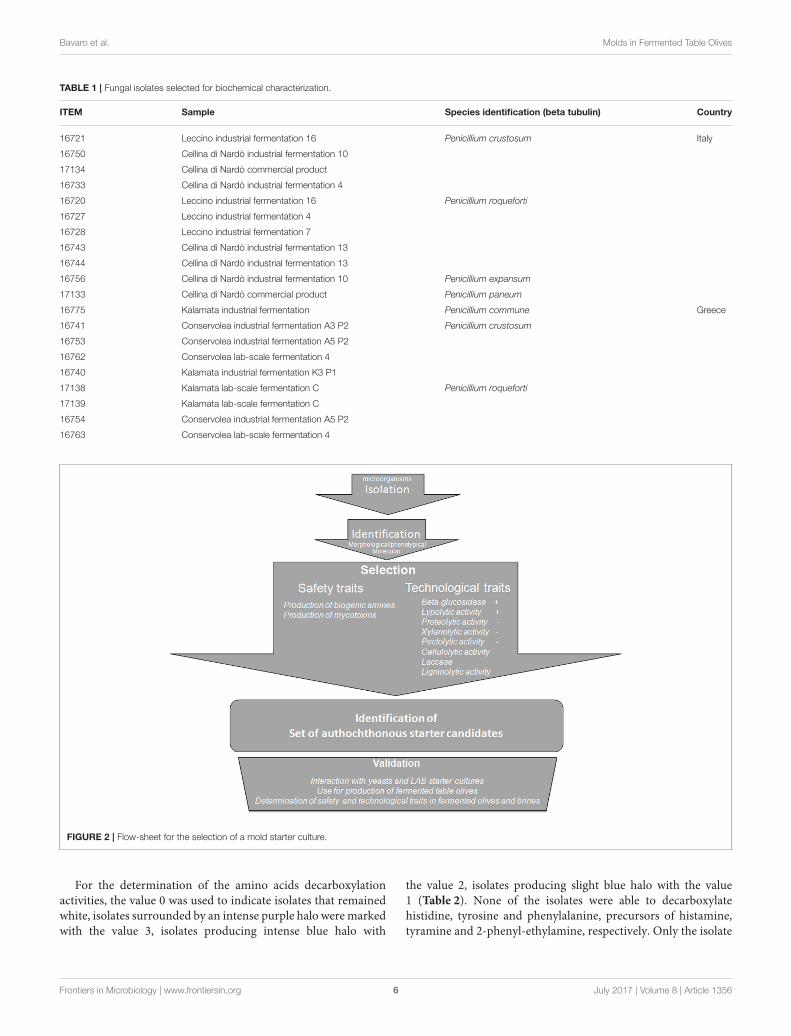
Bavaro et al. Molds in Fermented Table Olives
TABLE 1 | Fungal isolates selected for biochemical characterization.
ITEM Sample Species identification (beta tubulin) Country
16721 Leccino industrial fermentation 16 Penicillium crustosum Italy
16750 Cellina di Nardò industrial fermentation 10
17134 Cellina di Nardò commercial product
16733 Cellina di Nardò industrial fermentation 4
16720 Leccino industrial fermentation 16 Penicillium roqueforti
16727 Leccino industrial fermentation 4
16728 Leccino industrial fermentation 7
16743 Cellina di Nardò industrial fermentation 13
16744 Cellina di Nardò industrial fermentation 13
16756 Cellina di Nardò industrial fermentation 10 Penicillium expansum
17133 Cellina di Nardò commercial product Penicillium paneum
16775 Kalamata industrial fermentation Penicillium commune Greece
16741 Conservolea industrial fermentation A3 P2 Penicillium crustosum
16753 Conservolea industrial fermentation A5 P2
16762 Conservolea lab-scale fermentation 4
16740 Kalamata industrial fermentation K3 P1
17138 Kalamata lab-scale fermentation C Penicillium roqueforti
17139 Kalamata lab-scale fermentation C
16754 Conservolea industrial fermentation A5 P2
16763 Conservolea lab-scale fermentation 4
FIGURE 2 | Flow-sheet for the selection of a mold starter culture.
For the determination of the amino acids decarboxylationactivities, the value 0 was used to indicate isolates that remainedwhite, isolates surrounded by an intense purple halo weremarkedwith the value 3, isolates producing intense blue halo with
the value 2, isolates producing slight blue halo with the value1 (Table 2). None of the isolates were able to decarboxylatehistidine, tyrosine and phenylalanine, precursors of histamine,tyramine and 2-phenyl-ethylamine, respectively. Only the isolate
Frontiers in Microbiology | www.frontiersin.org 6 July 2017 | Volume 8 | Article 1356

Bavaro et al. Molds in Fermented Table Olives
TABLE2|Enzymatic
activitiesofthese
lectedmoldsisolatedfrom
tableolives.
ITEM
Beta
glucosidase
Lypolytic
activity
Cellulolytic
activity
Laccase
Tyrosinase
Lignin
modifying
enzymes
Protease
activity
(Milkagar)
Protease
activity(G
elatin)
Pectolytic
activity
Xilanolytic
activity
Aminoacid
decarboxylationactivities
Arginine
Phenylalanine
Tyrosine
Histidine
16720
++
−−
−−
−−
++
+++
−−
−−
−
16721
++
+−
++
−−
+−
−++
++
++
−−
−
16727
++
−−
−−
−−
++
++
-−
−−
16728
++
+−
−−
−−
++
++
−−
−−
−
16733
++
++
−−
−−
−−
++
++
++
−−
−
16740
++
++
−−
−−
−−
++
++
+++
−−
−
16741
++
++
−−
−+
−−
++
++
++
−−
−
16743
+−
−−
−−
−++
++
-−
−−
16744
++
−−
−−
−−
+++
−-
−−
−
16750
++
+−
−−
−−
++
++
+++
++
−−
−
16753
++
++
−−
−−
++
++
++
+++
−−
−
16754
++
+−
−−
−−
++
−-
−−
−
16756
++
++
++
−−
−++
−−
++
+−
-−
−−
16762
++
+−
−−
−−
++
++
−++
−+
++
−
16763
++
+−
−−
−++
++
−-
−−
−
16775
++
++
−−
−−
−+
++
−+
−−
−
17133
++
−−
−−
−−
++
++
−-
−−
−
17134
++
++
−−
−−
−−
++
+++
−−
−
17138
++
++
−−
−−
+++
−−
−−
−
17139
++
++
−−
−−
++
++
−−
−−
−
Frontiers in Microbiology | www.frontiersin.org 7 July 2017 | Volume 8 | Article 1356

Bavaro et al. Molds in Fermented Table Olives
ITEM 16762 showed good decarboxylation activity (value 3) oftyrosine. Various levels of arginine decarboxylation activity weredetected for the isolates ITEM 16721, 16733, 16740, 16741, 16750,16753, 16762, 16775, 17134.
Fungal ability to produce secondary metabolites, includedmycotoxins, was investigated by growing isolates in two differentconditions: namely in CYA medium and CYA medium withthe addition of the two constrains represented by salinity andacidity (5% NaCl and pH 5.5) (CYAS) in order to evaluate therole of salinity and acidity in affecting secondary metabolitesproduction. The secondary metabolites were analyzed by theTarget Analysis system (Klitgaard et al., 2014), able to screeneach extract for 3,000 compounds, considering mass accuracy,isotope fit, and retention time (RT), producing a qualitativerepresentation of all mycotoxins present in the sample (Table 3).
Aflatoxins, Ochratoxin, and Patulin were not detected in anymold isolates. Secondary metabolites produced by the isolateITEM 16728 (P. roqueforti) are not affected by the salt andacidity in the medium. At the two conditions (CYA and CYAS)it produced mycophenolic acid and roquefortine C.
The isolate ITEM 16775 produced cyclopiazonic acid andsclerotigenin in the two tested conditions, but FKL-3389 wasreleased only in the medium without salt and acidic conditions(CYA). The isolate ITEM 16756 (P. expansum), in additionto chaetoglobosin A, communesin A, and roquefortine C, inpresence of salt and acidity produced also chaetoglobosin Cand communesin B. Among isolates belonging to the speciesP. roqueforti, all of them produced mycophenolic acid in absenceof salt and acidity stress, whereas the isolates ITEM 16727 and16754 produced roquefortine C in the two tested conditions. Thislast metabolite was revealed in ITEM 16743 and 17138 grownon salt and acidic medium. The PR-toxin producers ITEM 16727and 17138 did not release this toxin in salt and acidic conditions.The isolates ITEM 16743, 16744, 16754, and 17138 also producedagroclavine in presence of salt and acidic pH (CYAS). Finally,the ITEM 17138 released roquefortine C and andrastatin A insalt and acidic conditions, whereas it produced roquefortine Xin absence of these stresses.
Considering P. crustosum isolates, all of them produceddehydrocyclopeptin in stress (salt and acid) conditions inaddition to andrastin A, cyclopenol, penitrem A, roquefortine C,viridicatol. In CYAS medium, in presence of the isolates ITEM16721 and 17134 there anacin and viridicatin were also detected,whereas viridicatin was produced by ITEM 16740 and 17134 andterrestric acid was produced only by ITEM 16740.
Summarizing, the presence of salt and low pH affectsin different way all metabolites: in particular they stimulateproduction of dehydrocyclopeptin and in some case alsoviridicatin in all P. crustosum (ITEM 16740 and 17134) strainstested and stimulated terrestric acid production in ITEM 16740;in P. roqueforti isolates ITEM 16743 and 17138 producedroquefortine C and only ITEM 17138 produced andrastin A,whereas a roquefortine derivative (present in ITEM 17138 grownin no salt and no acidic conditions) and PR-toxin (revealedin ITEM 17138 and ITEM 16727 in absence of salt andlow pH conditions) were not detected; P. expansum ITEM16756 produced chaetoglobosin C and communesin B but not
roquefortine C; marcfortine was not detected in P. paneumITEM 17133. P. roqueforti ITEM 16743, 16744, 16754, and 17138produced agroclavine.
Selection of FungiCombining qualitative tests for both technological (the presenceof desired enzymatic activities, i.e., beta-glucosidase and/orlipase activities, the reduced or absence of undesired traits,i.e., proteases, pectolytic and/or xylanolytic activities) andsafety assessments of tested fungal isolates, it was possibleto select some interesting isolates that can be consideredas putative candidates for future studies as co-starterswith yeasts and lactic acid bacteria. At the end of thisprocedure, the set of candidate strains for standardizedfermentation of table olives includes: P. roqueforti ITEM16728 selected from Leccino cultivar, P. paneum ITEM17133 and P. roqueforti ITEM 16744 selected from Cellinadi Nardò cultivar, P. roqueforti ITEM 17138 selected fromKalàmata cultivar and P. roqueforti ITEM 16754 selectedfrom Conservolea cultivar. It is to be considered, however,that table olives fermentations need to be performed in realconditions using the selected candidate strains in order toevaluate both safety and technological traits in olive and brinesamples.
DISCUSSION
Molds belonging to Penicillium and other molds genera(Aureobasidium, Aspergillus, Geotrichum) are often associatedto Black “Greek style” table olives. Their role in table olivesproduction is not completely understood yet. At present, theyare considered as contaminatingmicroorganisms, responsible forspoilage (texture softening, production of moldy odor and taste)and potentially toxicity (Heperkan, 2013).
The interest on molds associated to table olives startsfrom the consideration that they are used as secondarystarter cultures in Europe to process meat and cheeseproducts, affecting positively their flavor, taste, texture, offeringprotection against spontaneous undesired microorganisms,delay of rancidity, stabilization of color, oxygen, and lightprotection, etc. On the other end, they can also releasehighly toxic secondary metabolites such as mycotoxins. Non-toxigenic and technological mold starters can be exploited forstandardized fermentation, hindering the growth of undesiredmicroorganisms, similarly to what have been done for dry-cured meat using P. nalgiovense, P. chrysogenum, and recentlyof P. salamii (Sunesen and Stahnke, 2003; Delgado et al., 2016;Magistà et al., 2016); for the production of white cheeses usingP. camemberti (Brie and Camembert), and for the production ofblue cheeses (Roquefort, Gorgonzola, Stilton, Gammelost, etc.)using P. roqueforti (Geisen, 1993).
In the present paper, for the first time we (i) identified andcharacterized mold isolates associated to four different blacktable olives cultivars and (ii) selected some of them for theirpotential use as co-starter for table olives production. It canbe recommended to use species from series Roquefortorum(P. roqueforti and P. paneum) in olive fermentation,
Frontiers in Microbiology | www.frontiersin.org 8 July 2017 | Volume 8 | Article 1356

Bavaro et al. Molds in Fermented Table Olives
TABLE3|Secondary
metabolitesproducedin
cultu
rebymoldsisolatesfrom
tableolives.
ITEM
16775
ITEM
16721
ITEM
16740
ITEM
16741
ITEM
17134
ITEM
16756
ITEM
17133
ITEM
16727
ITEM
16728
ITEM
16743
ITEM
16744
ITEM
16754
ITEM
17138
P.commune
P.crustosum
P.crustosum
P.crustosum
P.crustosum
P.expansum
P.paneum
P.roqueforti
P.roqueforti
P.roqueforti
P.roqueforti
P.roqueforti
P.roqueforti
Chaetoglobosin
A
CYA
+
CYAS
+
Chaetoglobosin
C
CYA
CYAS
+
CommunesinA
CYA
+
CYAS
+
CommunesinB
CYA
CYAS
+
Marcfortine
CYA
+
CYAS
Cyclopiazo
nic
acid
CYA
+
CYAS
+
FKI-3389
CYA
+
CYAS
Sclerotig
enin
CYA
+
CYAS
+
Anacineor
similar
compound
CYA
++
CYAS
++
AndrastinA
CYA
++
++
++
+
CYAS
++
++
++
++
Cyclopenol
CYA
++
++
CYAS
++
++
Dehyd
ro
cyclopeptin
CYA
CYAS
++
++
Penitrem
ACYA
++
++
CYAS
++
++
(Continued)
Frontiers in Microbiology | www.frontiersin.org 9 July 2017 | Volume 8 | Article 1356

Bavaro et al. Molds in Fermented Table Olives
TABLE3|Contin
ued
ITEM
16775
ITEM
16721
ITEM
16740
ITEM
16741
ITEM
17134
ITEM
16756
ITEM
17133
ITEM
16727
ITEM
16728
ITEM
16743
ITEM
16744
ITEM
16754
ITEM
17138
P.commune
P.crustosum
P.crustosum
P.crustosum
P.crustosum
P.expansum
P.paneum
P.roqueforti
P.roqueforti
P.roqueforti
P.roqueforti
P.roqueforti
P.roqueforti
RoquefortineC
CYA
++
++
++
++
CYAS
++
++
++
++
+
RoquefortineX
CYA
+
CYAS
Viridicatol
CYA
++
++
CYAS
++
++
Viridicatin
CYA
CYAS
++
Mycophenolic
acid
CYA
++
++
++
CYAS
++
++
++
PR-toxin
CYA
++
CYAS
Terrestric
acid
CYA
CYAS
+
Agroclavine
CYA
CYAS
++
++
+denotesthepresenceofametabolite.
Frontiers in Microbiology | www.frontiersin.org 10 July 2017 | Volume 8 | Article 1356

Bavaro et al. Molds in Fermented Table Olives
because these are the only Penicillia that can tolerate higherconcentrations of acetic acid and lactic acid produced by thelactic acid bacteria (Frisvad et al., 2004; Houbraken et al., 2010).In other lactic acid and acetic acid containing foods and feedsRoquefortorum species are also the dominating Penicillia forexample cocoa, silage, rye bread, sauerkraut (O’Brien et al.,2006; Copetti et al., 2011; Gallo et al., 2015) etc., as long asthe concentration of acetic acid is sufficiently high. If theconcentration of these acids is low, toxigenic species suchas P. crustosum, P. commune, and P. expansum may thrive.These toxigenic fungi were found in olives in this study, so it isrecommended to secure that sufficiently high concentration ofacetic acid and lactic acid is present in the table olives.
For the first time, a flow sheet has been proposed forselection of molds associated to table olives. After identificationusing morphology/phenotypic characteristics and molecularapproaches, the mold isolates can be selected using two maincriteria (Figure 2): presence/absence of enzymatic activities andof toxic compounds. This selection step can help to individuatecandidates to be used in further validation step consisting in thestudy of their positive/negative interaction with already existingstarter cultures for table olives and in the use of them alone or incombination with yeasts and LAB for table olive production.
Molds associated to Leccino, Cellina di Nardó, Kalamàta, andConservolea fermented table olives were for the first time isolatedand identified. Classical morphological identification combinedwith molecular approach was used to improve resolution anddiscriminating power inside Penicillium species (Baffi et al.,2012). The P. commune, P. crustosum, P. expansum, P. roquefortiwere already reported in previous works as associated to tableolives (Ghitakou et al., 2006; Heperkan et al., 2006; Baffi et al.,2012), whereas the species P. paneum and P. polonicum werenot reported earlier in this product and they could representan occasional contamination, considering the low number ofcolonies observed. Following the selection strategy proposed foryeasts and lactic acid bacteria, both technological and safety testshave been chosen in order to perform a selection of mold isolates.
A subgroup (20) of them, selected in order to representthe overall observed molecular biodiversity, was assayed fortheir positive trait to produce beta-glucosidase, responsibleof polyphenols degradation (in particular oleuropein) anddebittering of table olives together with impact on flavor, dueto the production of secondary metabolites (Bevilacqua et al.,2013). Seventeen isolates showed good beta-glucosidase activity,representing promising candidates able to help other startersto degrade oleuropein, reducing time for debittering and NaClcontent in table olive processing. Several isolates (14) revealedthe presence of lipase activity, desired activity that could improvethe aromatic profile of fermented olives (Savitha et al., 2007;Rodríguez-Gómez et al., 2012).
In this preliminary study, following the approach previouslyused for yeasts and LAB (Arroyo-López et al., 2012; Bevilacquaet al., 2013; Bleve et al., 2014, 2015), the presence of proteolyticactivity was considered as a negative characteristic, since itis responsible for the release of aminoacids and ammoniacalnitrogen causing in turn a pH increase and the risk for theproduct to be unsafe (Tosi et al., 2008; Ledenbach and Marshall,
2009). Moreover, proteolytic activity could be also responsibleof undesired impact on olive quality provoking olive softening.Although, no isolates produced detectable activity on milk agar,12 of them metabolized gelatin at different levels (score 1–3). However, considering microbiological research performed sofar and scientific data available, it is not possible to definitelyestablish the real impact of these enzymatic activities ontable olives quality. In fact, the relative influence of fungalenzymatic activities could contribute to the maturation of aromatogether with other parameters, such as raw materials andprocessing conditions (Meynier et al., 1999; Harkouss et al.,2012). Other enzymatic activities that have been describedas undesired traits in yeasts, such as cellulases, xylanases,pectinases, need to be verified for molds when they are inoculatedin table olives. Also in this case, scarce information is stillavailable about the compounds and enzymes produced by themold population during fermentation and their concentrationsin packed olives. This is an important aspect since thepresence of molds that are able to secrete these enzymes intable olives could modify in a not predictable manner thenutritional composition of table olives and of their organolepticcharacteristics.
It is nevertheless worth noting that beta-glucosidase, cellulase,xylanase, pectinase, lignin modifying, lypolytic, and proteolitycenzymatic activities detected in many mold isolates can beof interest also for major industrial applications. Potentialbiotechnological uses of Penicillia, isolated from fresh olive fruits,olive paste and pomace, were suggested by Baffi et al. (2012).Analogously to the approach followed for yeasts (Bleve et al.,2011), mold isolates (as whole cells in free or immobilized forms)or enzymes purified from different mold species can be useful fortreatment and/or bioconversion of agro-food by-products, suchas wastewaters deriving from olive oil and table olives industry.
Table olives are one of the most contaminated fermented foodby biogenic amines like putrescine, cadaverine, and tyramine,as reported in “Zapatera” spoiled olives (Hornero-Mendez andGarrido-Fernandez, 1994) and in naturally fermented (Greek-style) table olives (Tofalo et al., 2012).
Recently, in addition to LAB, also yeasts have been consideredas a source of biogenic amines and preliminary assays have beenincluded in selection programs to check their ability to producethese compounds “in vitro” conditions (Bevilacqua et al., 2013;Bleve et al., 2014, 2015). In order to reduce the risks for consumer,these tests have been introduced in this study also for molds, toselect microbial sources unable to produce biogenic amines infermented table olives.
In this paper a preliminary screening of mold isolates,on media with or without salt and acidic constraints, wasalso carried out for qualitative analysis of many secondarymetabolites (including mycotoxins) in an attempt to identifyisolates potentially low producer of mycotoxins.
The three regulatedmycotoxins Aflatoxins, Ochratoxin A, andPatulin were not detected in any mold strains isolated from thefermented black olives.
P. expansum ITEM 16756 produced chaetoglobosin A andcommunesin A independently from the presence of absence ofsalt and low pH, whereas communesin B was revealed only in
Frontiers in Microbiology | www.frontiersin.org 11 July 2017 | Volume 8 | Article 1356

Bavaro et al. Molds in Fermented Table Olives
presence of these conditions. The isolation of chaetoglobosinA, characterized by embryolethal but not teratogenic effects onchickens was reported in the genus Penicillium (Veselý andJelínek, 1995). Besides patulin and roquefortine C, Andersenet al. (2004) described the presence of chaetoglobosin A andcommunesin B in P. expansum contaminated fruit juices andpotato pulp. Oral toxicity in cockerels, embryonic chickens,rats, and mice, cytotoxic activity in HeLa cells, teratogenicactivity in mice were demonstrated for chaetoglobosin A (Sekitaet al., 1982; Cherton et al., 1994), whereas communesin B iscytotoxic to lymphocytic leukemia cells (Numata et al., 1993).However, despite their negative effects reported in literature,these mycotoxins are not regulated as food contaminants.
The presence of cyclopiazonic acid (CPA), an indol–tetramicacid mycotoxin produced by the nearly ubiquitous moldsAspergillus and Penicillium, was revealed in P. commune ITEM16775.
Although, CPA is not considered to be a potent acutetoxin, it has different target organs (hepatic tissue, andspleen). Its toxicity and symptoms are linked to its abilityto alter normal intracellular calcium flux and potentialimmunomodulatory effect in “in vitro” studies. SeveralPenicillium species (P. camamberti, P. cyclopium, P. viridictum,P. griseofulvum, P. crustosum, and others) associated tocheese (Camembert, brie, cheddar, cream, cheese rind,etc.), acorn, barley, corn, peanuts, ham, and chicken meatwere reported as producers of CPA (Burdock and Flamm,2000).
The potent neurotoxin penitrem A has been detected inimportant agricultural commodities (maize, rice, wheat, oat, rye,barley, and sorghum) and in moldy cream cheese and it isproduced by several species of fungi. According to the resultsreported by El-Banna and Leistner (1988), also all isolates ofP. crustosum tested in this study were able to produce detectablelevels of penitrem A.
The P. expansum ITEM 16756 and all isolates of P. crustosumand of P. roqueforti, with the exception of the isolate ITEM16744, produced roquefortine C. This mycotoxin has neurotoxicproperties (Wagener et al., 1980), but the data on its neurotoxicityare equivocal (Fog Nielsen et al., 2006). Several studies haverevealed the presence of roquefortine C in blue-veined cheeses(Ohmomo et al., 1975; Scott and Kennedy, 1976; López-Díazet al., 1996). It is always found in cheeses, since fungal strains(P. camamberti and P. roqueforti) used as starters in the dairyindustry are able to produce this toxin in vitro. However, therelatively low toxicity and low concentrations of this toxin inblue cheese make this product safe to be consumed (Finoli et al.,2001).
According to data reported in this paper and by other authors(Frisvad et al., 2004), P. roqueforti is also known to producemycophenolic acid (MPA) and PR-toxin (detected only in ITEM16727 and 17138).
Although, MPA possess immunosuppressive effects (Bentley,2000), compared to other mycotoxins (T-2 toxin, gliotoxin,DON, and patulin), roquefortine C and MPA showed low acutecytotoxicity on the human intestinal cell line Caco-2 (Rasmussenet al., 2010).
PR-toxin is instead the most toxic metabolite produced by P.roqueforti because of a high toxic or lethal effects in rats, mice,and cats and is mutagenic in the Ames test (Arnold et al., 1978;Fog Nielsen et al., 2006). However, PR-toxin is unstable because itquickly reacts with different salts, amino acids (especially Sulfurcontaining amino acids), amines, casein, and the decompositionproducts resulting in the less toxic PR imine (Erdogan et al.,2003).
Despite the detection of variable contents roquefortine C andMPA in several blue-veined cheeses, no cases of intoxicationslinked to the consumption of these products have ever beenreported (Fontaine et al., 2015). Roquefortine C and MPAcombined exposure was studied on human intestinal cells (Caco-2 cells) and on monocytes (cancer lineage model cell THP-1) by Fontaine et al. (2015). These authors demonstratedthat roquefortine C and MPA may not create problems ofacute exposure in food, since human are exposed at relativelylow levels and these toxins show low acute cytotoxic effectsin comparison to other regulated mycotoxins. Agroclavineproduced by P. roqueforti is the precursor of isofumigaclavineA, and its hydrolysis product, isofumigaclavine B, are identicalto Roquefortine A and B, respectively. Low levels or traces ofIsofumigaclavine A and B were found in marbled and bluecheese. The toxicological effects of these substances are not well-known, but some neurotoxic data are described by several studies(Ohmomo et al., 1975; Scott and Kennedy, 1976; Scott, 1984).However, Agroclavine itself is probably not very toxic, althoughthere are few available data on its toxicity.
Although, several isolates of P. expansum were shown to beable to produce patulin and citrinin (Andersen et al., 2004), anddifferent isolates of P. paneum were responsible of production ofpatulin (O’Brien et al., 2006), none of these toxins were detectablefor all the tested isolates.
Considering technological and safety data collected for alltested isolates, at the end of this procedure, it is possible toselect some isolate, autochthonous for each table olive cultivar,suitable for future validation tests as co-starters with yeasts andlactic acid bacteria and for production of fermented table olives.Tests on an ad hoc optimized “olive agar” and then directly onfermented table olives and brine samples inoculated with selectedmolds might indicate the quantity and which biogenic aminesand mycotoxins will be eventually produced on the final productduring a fermentation process in real conditions.
AUTHOR CONTRIBUTIONS
Fundamental contributions to the conception and design of thework, acquisition, analysis and interpretation of data: GB, SB,AS, AL; Acquisition, analysis, elaboration, and interpretation ofdata: SB, AS, GB, JF, MT, AC; Drafting the work and revising itcritically for intellectual content: GB, AS, GP, GM,AL. All authorsapproved the final version of the manuscript to be submittedfor publication and agreed to be accountable for all aspects ofthe work in ensuring that questions related to the accuracy andintegrity of any part of the work are appropriately investigatedand resolved.
Frontiers in Microbiology | www.frontiersin.org 12 July 2017 | Volume 8 | Article 1356

Bavaro et al. Molds in Fermented Table Olives
ACKNOWLEDGMENTS
The authors wish to thank Dr. G. Cozzi for his valuableassistance in molds isolation and characterization andFilomena Epifani for fungal strains preservation. This paperis dedicated to the memory of our wonderful colleagueProf. Maria Tasioula-Margary. This study was supportedby the BIO-OLEA “Utilization of biophenols from Olea
Europea products—Olives, virgin olive oil and olive millwastewaters,” Interreg project co-funded by the EuropeanUnion (ERDF) and by the National funds of Greece andItaly under the European Territorial Cooperation ProgrammeGreece-Italy 2007–2013. The work was also supported inpart by “BIOTECA” project [Regione Puglia FSC 2007–2013 “Cluster Tecnologici Regionali 2014” (BURP n. 138/2014)].
REFERENCES
Andersen, B., Smedsgaard, J., and Frisvad, J. C. (2004). Penicillium expansum:
consistent production of patulin, chaetoglobosins, and other secondary
metabolites in culture and their natural occurrence in fruit products. J. Agric.Food Chem. 52, 2421–2428. doi: 10.1021/jf035406k
Arnold, D. L., Scott, P. M., McGure, P. F., Harwig, J., and Nera, E. A.
(1978). Acute toxicity studies of roquefortine and PR toxin, metabolites of
Penicillium roqueforti, in the mouse. Food Cosmet. Toxicol. 16, 369–371.
doi: 10.1016/S0015-6264(78)80009-1
Arroyo-López, F. N., Romero-Gil, V., Bautista-Gallego, J., Rodriguez-Gómez, F.,
Jiménez-Díaz, and R., García-García, P. (2012). Yeasts in table olive processing:
desirable or spoilage microorganisms. Int. J. Food Microbiol. 160, 42–49.
doi: 10.1016/j.ijfoodmicro.2012.08.003
Baffi, M. A., Romo-Sanchez, S., Ubeda-Iranzo, J., and Briones-Perez,
A. I. (2012). Fungi isolated from olive ecosystems and screening of
their potential biotechnological use. New Biotechnol. 29, 451–456.
doi: 10.1016/j.nbt.2011.05.004
Bentley, R. (2000). Mycophenolic acid: a one hundred year odyssey from antibiotic
immunosuppressant. Chem. Rev. 100, 3801–3825. doi: 10.1021/cr990097bBevilacqua, A., Beneduce, L., Sinigaglia, M., and Corbo, M. R. (2013). Selection
of yeasts as starter cultures for table olives. J. Food Sci. 78, M742–M751.
doi: 10.1111/1750-3841.12117
Bleve, G., Lezzi, C., Chiriatti, M. A., D’Ostuni, I., Tristezza, M., Di Venere, D., et al.
(2011). Selection of non-conventional yeasts and their use in immobilized form
for the bioremediation of olive oil mill wastewaters. Bioresour. Technol. 102,982–989. doi: 10.1016/j.biortech.2010.09.059.
Bleve, G., Tufariello, M., Durante, M., Perbellini, E., Ramires, F. A., Grieco, F., et al.
(2014). Physico-chemical and microbiological characterization of spontaneous
fermentation of Cellina di Nardò and Leccino table olives. Front. Microbiol.5:570. doi: 10.3389/fmicb.2014.00570
Bleve, G., Tufariello, M., Durante, M., Ramires, F. A., Grieco, F., Mita, G.,
et al. (2015). Physico-chemical characterization of natural fermentation process
of Conservolea and Kalamàta table olives and developement of a protocol
for the pre-selection of fermentation starters. Food Microbiol. 46, 368–382.doi: 10.1016/j.fm.2014.08.021
Boominathan, K., and Reddy, C. A. (1992). Fungal degradation of lignin:
biotechnological applications. Handbook Appl. Mycol. 4, 763–822.Burdock, W. G. A., and Flamm, G. (2000). Review article: safety assessment
of the mycotoxin cyclopiazonic. Acid. Int. J. Toxicol. 19, 195–218.
doi: 10.1080/10915810050074964
Cherton, J. C., Jallal, A., Lhommet, G., Loutelier, C., Dardoize, F., Lacoste, L., et al.
(1994). Unexpected production of chaeto- globosins from maize incubated by
Phomopsis lepstromiformis. I. Isolation and optimization of the production in
liquid media by LC monitoring. Analysis 22, 210–216.Copetti, M. V., Iamanaka, B. T., Frisvad, J. C., Pereira, J. L., Fungaro, M. H.,
and Taniwaki, M. (2011). Mycobiota of cacao. From farm to chocolate. FoodMicrobiol. 28, 1499–1504. doi: 10.1016/j.fm.2011.08.005
Crous, P. W., Verkley, G. J. M., Groenewald, J. Z., and Samson, R. A. (eds.).
(2009). Fungal Biodiversity. [CBS Laboratory Manual Series no.1.]. Utrecht:CBS-KNAW Fungal Biodiversity Centre.
Delgado, J., Peromingo, B., Núñez, F., and Asensio, M. A. (2016). Use of
moulds and their antifungal proteins for biocontrol of toxigenic moulds
on dry-ripened cheese and meats. Curr. Opin. Food Sci. 11, 40–45.
doi: 10.1016/j.cofs.2016.09.003
Eaton, R. A., and Hale, M. D. C. (1993). Wood: Decay, Pests and Protection.London: Chapman and Hall Ltd.
El Adlouni, C., Tozlovanu, M., Naman, F., Faid, M., and Pfohl-Leszkowicz, A.
(2006). Preliminary data on the presence of mycotoxins (ochratoxin A, citrinin
and aflatoxin B1) in black table olives “Greek style” of Moroccan origin. Mol.Nutr. Food Res. 50, 507–512. doi: 10.1002/mnfr.200600055
El-Banna, A. A., and Leistner, L. (1988). Production of penitrem A by
Penicillium crustosum isolated from foodstuffs. Int. J. Food Microbiol. 7, 9–17.doi: 10.1016/0168-1605(88)90067-0
Erdogan, A., Gurses, M., and Sert, S. (2003). Isolation of moulds capable of
producing mycotoxin from blue mouldy Tulum cheeses produced in Turkey.
Food Microbiol. 85, 83–85. doi: 10.1016/S0168-1605(02)00485-3Felsenstein, J. (1985). Confidence limits on phylogenies: an approach using the
bootstrap. Evolution 39, 783–791. doi: 10.1111/j.1558-5646.1985.tb00420.x
Fernandez, G. A., Adams, M. R., and Diez, M. J. F. (1997). Table Olives: Productionand Processing. London: Chapman et Hall. doi: 10.1007/978-1-4899-4683-6
Finoli, C., Vecchio, A., Galli, A., and Dragoni, I. (2001). Roquefortine C occurrence
in blue cheese. J. Food Protect. 64, 246–251. doi: 10.4315/0362-028X-64.2.246Fog Nielsen, K., Nielsen, O. G., Sumarah, M. W., Frisvad, J. C., and Miller, J. D.
(2006). Production of metabolites from the Penicillium roqueforti complex. J.Agric. Food Chem. 54, 3756–3763. doi: 10.1021/jf060114f
Fontaine, K., Passeró, E., Vallone, L., Hymery, N., Coton, M., Jany, J.-
L. et al. (2015). Occurrence of roquefortine C, mycophenolic acid and
aflatoxin M1 mycotoxins in blue-veined cheeses. Food Control 47, 634–640.doi: 10.1016/j.foodcont.2014.07.046
Franzetti, L., Scarpellini, M., Vecchio, A., and Planeta, D. (2011). Microbiological
and safety evaluation of green table olives marketed in Italy. Ann. Microbiol. 61,843–851. doi: 10.1007/s13213-011-0205-x
Frisvad, J. C., and Samson, R. A. (2004). Emericella venezuelensis, a new species
with stellate ascospores producing sterigmatocystin and aflatoxin B1. Syst. Appl.Microbiol. 27, 672–680. doi: 10.1078/0723202042369910
Frisvad, J. C., Smedsgaard, J., Larsen, T. O., Samson, R. A., and Robert, A. (2004).
Mycotoxins, drugs and other extrolites produced by species in Penicilliumsubgenus. Penicillium. Stud. Mycol. 49, 201–241.
Frisvad, J. C., and Thrane, U. (1987). Standardised High-Performance Liquid
Chromatography of 182 mycotoxins and other fungal metabolites based
on alkylphenone retention indices and UV-VIS spectra (Diode Array
Detection). J. Chromatogr. 404, 195–214. doi: 10.1016/S0021-9673(01)
86850-3
Gallo, A., Giuberti, G., Frisvad, J. C., Bertuzzi, T., and Nielsen, K. F. (2015).
Review on mycotoxin issues in ruminants: occurrence in forages, effects
of mycotoxin ingestion on health status and animal performance and
practical strategies to counteract their negative effect. Toxins 7, 3057–3111.doi: 10.3390/toxins7083057
Gardini, F., Tofalo, R., Belletti, N., Iucci, L., Suzzi, G., Torriani, S., et al. (2006).
Characterization of yeasts involved in the ripening of Pecorino Crotonese
cheese. Food Microbiol. 23, 641–648. doi: 10.1016/j.fm.2005.12.005
Geisen, R. (1993). Fungal starter cultures for fermented foods: molecular aspects.
Trends Food Sci. Technol. 4, 251–255. doi: 10.1016/0924-2244(93)90140-6Ghitakou, S., Koutras, K., Kanellou, E., and Markaki, P. (2006). Study of
aflatoxin B1 and ochratoxin A production by natural microflora andAspergillusparasiticus in black and green olives of Greek origin. Food Microbiol. 23,612–621. doi: 10.1016/j.fm.2005.12.008
Glass, N. L., and Donaldson, G. C. (1995). Development of primer sets designed for
use with the PCR to amplify conserved genes from filamentous Ascomycetes.
Appl. Environ. Microb. 61, 1323–1330.Hankin, L., Poincelot, R. P., and Anagnostakis, S. L. (1975). Microorganisms
from composting leaves: ability to produce extracellular degradative enzymes.
Microb. Ecol. 2, 296–308. doi: 10.1007/BF02011649
Frontiers in Microbiology | www.frontiersin.org 13 July 2017 | Volume 8 | Article 1356

Bavaro et al. Molds in Fermented Table Olives
Harkouss, R., Mirade, P. S., and Gatellier, P. (2012). Development of a rapid,
specific and efficient procedure for the determination of proteolytic activity
in dry-cured ham: definition of a new proteolysis index. Meat Sci. 92, 84–88.doi: 10.1016/j.meatsci.2012.04.017
Heperkan, D. (2013). Microbiota of table olive fermentations and
criteria of selection for their use as starters. Front Microbiol. 4:143.
doi: 10.3389/fmicb.2013.00143Heperkan, D., Dazkir, G. S., Kansu, D. Z., and Guler, F. K. (2009). Influence
of temperature on citrinin accumulation by Penicillium citrinum and
Penicillium verrucosum in black table olives. Toxin Rev. 28, 180–186.
doi: 10.1080/15569540903084982Heperkan, D., Meric, B. E., Sismanoglu, G., Dalkili,ç, G., and Güler, F. K. (2006).
Mycobiota, mycotoxigenic fungi, and citrinin production in black olives. Adv.Exp. Med. Biol. 571, 203–210. doi: 10.1007/0-387-28391-9_13
Hornero-Mendez, D., and Garrido-Fernandez, A. (1994). Biogenic- amines in
table olives analysis by high-performance liquid- chromatography.Analyst 119,2037–2041. doi: 10.1039/AN9941902037
Houbraken, J., Frisvad, J. C., and Samson, R. A. (2010). Sex in Penicillium series
Roqueforti. IMA Fungus 1, 171–180. doi: 10.5598/imafungus.2010.01.02.10King, A. D. Jr., Hocking, A. D., and Pitt, J. I. (1979). Dichloran-rose bengal medium
for enumeration and isolation of molds from foods. Appl. Environ. Microbiol.37, 959–964.
Klitgaard, A., Iversen, A., Andersen,M. R., Larsen, T. O., Frisvad, J. C., andNielsen,
K. F. (2014). Aggressive dereplication using UHPLC-DAD-QTOF - screening
extracts for up to 3000 fungal secondary metabolites. Anal. Bioanal. Chem. 406,1933–1943. doi: 10.1007/s00216-013-7582-x
Korukluoglu, M., Gürbüz, O., uylaser, V., Yildirim, A., and Sahin, I. (2000).
“Gemlik tipi zeytinlerde mikotoksin kirliliginin arastirilmasi, Türkiye 1,” in
Zeytincilik SempozyumuUludag Üniversitesi 6–9Haziran, Bildiri Kitabi (Bursa),218.
Ledenbach, L. H., and Marshall, R. T. (2009). “Microbiological spoilage of
dairy products,” in Compendium of the Microbiological Spoilage of Foodsand Beverages, Food Microbiology and Food Safety, eds W. H. Sperber and
M. P. Doyle (New York, NY: Springer Science+Business Media), 41–67.
doi: 10.1007/978-1-4419-0826-1_2López-Díaz, T. M., Román-Blanco, C., García-Arias, M. T., García-Fernández, M.
C., and García-López, M. L. (1996). Mycotoxins in two Spanish cheese varieties.
Int. J. Food Microbiol. 30, 391–395. doi: 10.1016/0168-1605(96)00957-9Ludemann, V., Pose, G., Pollio, M. L., and Segura, J. (2004). Determination of
growth characteristics and lipolytic and proteolytic activities of Penicilliumstrains isolated from Argentinean salami. Int. J. Food Microbiol. 96, 13–18.doi: 10.1016/j.ijfoodmicro.2004.03.003
Magistà, D., Ferrara, M., Del Nobile, M. A., Gammariello, D., Conte, A., and
Perrone, G. (2016). Penicillium salamii strain ITEM 15302: a new promising
fungal starter for salami production. Int. J. Food Microbiol. 231, 33–41.
doi: 10.1016/j.ijfoodmicro.2016.04.029
Medina-Pradas, E., and Arroyo-López, F. N. (2015). Presence of
toxic microbial metabolites in table olives. Front. Microbiol. 6:873.
doi: 10.3389/fmicb.2015.00873
Meynier, A., Novelli, E., Chizzolini, R., Zanardi, E., and Gandemer, G. (1999).
Volatile compounds of commercial Milano salami. Meat Sci. 51, 175–183.doi: 10.1016/S0309-1740(98)00122-3
Nei, M., and Kumar, S. (2000). Molecular Evolution and Phylogenetics. New York,
NY: Oxford University Press.
Nielsen, K. F., and Smedsgaard, J. (2003). Fungal metabolite screening: database
of 474 mycotoxins and fungal metabolites for de-replication by standardized
liquid chromatography-UV-mass spectrometry methodology. J. Chromatogr. A1002, 111–136. doi: 10.1016/s0021-9673(03)00490-4
Numata, A., Takahashi, C., Ito, Y., Takada, T., Kawai, K., Usami, Y., et al. (1993).
Communesins, cytotoxic metabolites of a fungus isolated from a marine alga.
Tetrahedron Lett. 34, 2355–2358. doi: 10.1016/s0040-4039(00)77612-xO’Brien, M., Nielsen, K. F., O’Kiely, P., Forristal, P. D., Fuller, H. T., and
Frisvad, J. C. (2006). Mycotoxins and other secondary metabolites produced
in vitro by Penicillium paneum Frisvad and Penicillium roqueforti Thom
isolated from baled grass silage in Ireland. J. Agric. Food Chem. 54, 9268–9276.doi: 10.1021/jf0621018
Ohmomo, S., Sato, T., Utagawa, T., and Abe, M. (1975). Isolation of
Festuclavine and three new indole alkaloids, roquefortine A, B and C from
cultures of Penicillium roqueforti. J. Agr. Chem. Soc. Jpn. 49, 615–623.
doi: 10.1271/bbb1961.39.1333
Pointing, S. B., Vrijmoed, L. L. P., and Jones, E. B. G. (1999). Laccase is produced as
the sole lignin modifying enzyme in submerged liquid culture by the white-rot
fungus Pycnoporus sanguineus.Mycologia 91, 345–349.Rasmussen, R. R., Binderup, M.-L., Larsen, T. O., and Rasmussen, P. H. (2010).
Mycotoxins in Maize Silage e Detection of Toxins and Toxicological Aspects. Kgs,Lyngby:: Technical University of Denmark (DTU).
Rodríguez-Gómez, F., Romero-Gil, V., Bautista-Gallego, J., Garrido-Fernández,
and A., Arroyo-López, F. N. (2012). Multivariate analysis to discriminate
yeast strains with technological applications in table olive processing. WorldJ. Microbiol. Biotechnol. 28, 1761–1770. doi: 10.1007/s11274-011-0990-1
Savitha, J., Srividya, S., Jagat, R., Payal, P., Priyanki, S., Rashmi, G.W.,., et al. (2007).
Identification of potential fungal strain (s) for the production of inducible,
extracellular and alkalophilic lipase. Afr. J. Biotechnol. 6, 564–568.Scott, P. M. (1984). “Mycotoxins production, isolation, separation and
purification,” in PR Toxin, ed V. Betina (Amsterdam: Elsevier), 469–474.
Scott, P. M., and Kennedy, B. P. C. (1976). Analysis of blue cheese for roquefortine
and others alkaloids from Penicillium roqueforti. J. Agr. Food Chem. 24,
865–868. doi: 10.1021/jf60206a028
Sekita, S., Yoshihira, K., Natori, S., Udagawa, S., Sakabe, F., Kurata, H., et al.
(1982). Chaetoglobosins, cytotoxic 10-(indol- 3-yl)-[13]cytochalasans
from Chaetomium spp. I. Production, isolation and some cytological
effects of chaetoglobosin A-J. Chem. Pharm. Bull. 30, 1609–1617.
doi: 10.1248/cpb.30.1609
Smedsgaard, J. (1997). Micro-scale extraction procedure for standardized
screening of fungal metabolite production in cultures. J. Chromatogr. A 760,
264–270. doi: 10.1016/S0021-9673(96)00803-5
Sneath, P. H. A., and Sokal, R. R. (1973).Numerical Taxonomy. San Francisco, CA:
Freeman.
Sunesen, L. O., and Stahnke, L. H. (2003). Mould starter cultures for
dry sausages-selection, application and effects. Meat Sci. 65, 935–948.
doi: 10.1016/S0309-1740(02)00281-4
Tamura, K., Peterson, D., Peterson, N., Stecher, G., Nei, M., and Kumar, S. (2011).
MEGA5: molecular evolutionary genetics analysis using maximum likelihood,
evolutionary distance, and maximum parsimony methods. Mol. Biol. Evol. 28,2731–2739. doi: 10.1093/molbev/msr121
Tofalo, R., Schirone, M., Perpetuini, G., Angelozzi, G., Suzzi, G., and Corsetti,
A. (2012). Microbiological and chemical profiles of naturally fermented table
olives and brines from different Italian cultivars. Anton. Leeuw. 102, 121–131.doi: 10.1007/s10482-012-9719-x
Tosi, F., Sandri, S., Tedeschi, G., Malacarne, M., and Fossa, E. (2008). Variazioni di
composizione e prorpietà fisico-chimiche del parmigiano-reggiano durante la
maturazione e in differenti zone della forma. Scienza e Tecnica Lattiero Casearia59, 507–528.
Vermelho, A. B., Meirelles, M. N., Lopes, A., Petinate, S. D., Chaia, A. A.,
and Branquinha, M. H. (1996). Detection of extracellular proteases from
microorganism on agar plates. Mem. Inst. Oswaldo Cruz. 91, 755–760.
doi: 10.1590/S0074-02761996000600020
Veselý, D., and Jelínek, R. (1995). Penicillium aurantiogriseum Dierckx produces
chaetoglobosin a toxic to embryonic chickens. Mycopathologia 132, 31–33.
doi: 10.1007/BF01138601
Wagener, R. E., Davis, N. D., and Diener, U. L. (1980). Penitrem A and
roquefortine production by Penicillium commune.Appl. Environ. Microbiol. 39,882–887.
White, T. J., Bruns, T., Lee, S., and Taylor, J. W. (1990). “Amplification and
direct sequencing of fungal ribosomal RNA genes for phylogenetics” in PCRProtocols: A Guide to Methods and Applications, eds M. A. Innis, D. H. Gelfand,
J. J.Sninsky, and T. J. White (New York, NY: Academic Press, Inc.), 315–322.
Conflict of Interest Statement: The authors declare that the research was
conducted in the absence of any commercial or financial relationships that could
be construed as a potential conflict of interest.
Copyright © 2017 Bavaro, Susca, Frisvad, Tufariello, Chytiri, Perrone, Mita,Logrieco and Bleve. This is an open-access article distributed under the termsof the Creative Commons Attribution License (CC BY). The use, distribution orreproduction in other forums is permitted, provided the original author(s) or licensorare credited and that the original publication in this journal is cited, in accordancewith accepted academic practice. No use, distribution or reproduction is permittedwhich does not comply with these terms.
Frontiers in Microbiology | www.frontiersin.org 14 July 2017 | Volume 8 | Article 1356