Embed Size (px)
Citation preview
Journal of Agricultural Science; Vol. 5, No. 7; 2013 ISSN 1916-9752 E-ISSN 1916-9760
Published by Canadian Center of Science and Education
262
Isolation and Identification of the Causal Pathogens for Kiwifruit Bacterial Canker and the Isolation of the Antagonistic Endophytic
Fungi From Kiwifruit in Sichuan, China Jinyi Yan1*, Yongliang Cui1*, Jian Ding2, Liangqiang Zhou2, Zuqiang Cheng2 & Min Zhang1
1 Department of Agriculture, Sichuan Agriculture University, Chengdu, China 2 Sichuan Provincial Institute of Natural Resource Sciences, Chengdu, China
Correspondence: Min Zhang, Department of Agriculture, Sichuan Agriculture University, Chengdu, China. E-mail: [email protected] *These two authors contribute equal work to this paper. Received: April 16, 2013 Accepted: May 18, 2013 Online Published: June 15, 2013 doi:10.5539/jas.v5n7p262 URL: http://dx.doi.org/10.5539/jas.v5n7p262 Abstract The kiwifruit bacterial canker has been recognized as the main kiwifruit pathogen in Sichuan and acquired strong resistance to chemicals during its long evolution under chemical evolutionary pressure. Base on biochemical testing, pathogenic testing and phylogenetic analyses, the results shown that Pseudomonas syringae pv. actinidiae is the causal agents of the “Hongyang” kiwifruit bacterial canker disease. Besides this bacterial pathogen, 14 endophytic fungi of kiwifruit leaves were also isolated and identified, including Penicillium sp. Colletotrichum sp. Phomopsis sp. Alternaria sp. and Nigrospora sp. 8 endophytic fungi were obtained from branches, such as Alternaria sp. Nigrospora sp. Phomopsis sp.and Sordaria sp. and one of the endophytic fungi Nigrospora sp. (No.J2) from branch showed good antagonistic activities to Pseudomonas syringae pv. actinidiae in vitro with the inhibition zone of 14 mm, therefore it has the potential of being used as a biological control agent against kiwifruit bacterial canker. Keywords: kiwifruit bacterial canker, endophytic fungi, “HongYang”, China, bio-control 1. Introduction China is one of the most important production areas for kiwifruit around the world and known as the country of origin of kiwifruit. Sichuan province, located in southwest of China, has a wealth of natural resources with about 700,000,000 m2 of kiwifruit growing areas and an annual production of 84,000,000 kg where “HongYang” kiwifruit is especially characterized with red color, good taste, and rich nutrient, renowned as the “national variety protection resource” and has become the main kiwifruit specie in Sichuan with good economical benefits and cultural values. However, the kiwifruit bacteria canker constitute causing serious yield loss in this area. The main disease symptoms were brown spots surrounded by yellow haloes on leaves and reddish exudates on twigs and trunks (Figure 1) which were similar to those caused by the bacterium Pseudomonas syringae pv. actinidiae (Psa.) in previous reports (Serizawa et al., 1989). Several studies have reported the kiwifruit bacterial canker in other countries, but the comprehensive research on the pathogens of kiwifruit bacterial canker in “HongYang” is still seldom. Endophytes are organisms able to live in plant tissues for a consistent part of their life cycle without inducing substantial damages to the host (Downing et al., 2000; Ryu et al., 2005). It is also known that endophytic fungi statement in abstract and introduction are an important source of bioactive compounds, some showing a real antifungal activity (Shimizu et al., 2000). As matter of facts, many have been already used as fungicide in a commercial way (Li et al., 2005). The outburst condition of kiwifruit bacterial canker in “HongYang” is especially serious in 2012, however, there is a particular kiwifruit plant still in healthy state while all of the kiwifruit plants surrounded were seriously affected. Therefore we have reason to suspect that maybe is the endophytic fungi in that particular plant that works. There are many researches on endophytic fungi in different plants, but no reports concern kiwifruit. In this study, we isolated and identified the kiwifruit bacterial canker pathogen on “HongYang” from reddish
www.ccsen
exudates. endophytebeen testedplant prelim
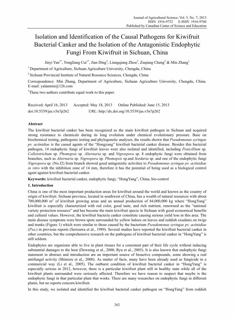
Figure 1. Tb
2. Materia2.1 IsolatioSince the “HongYan103°0′40.7chosen in wBacteriumApril, 201mixture. Tthe primeF3 (ACCT(Rees-GeoMastercycThe ampliThe gels wmixtures wwere spreadistilled wchosen andunder 28°(Genorise were detec
net.org/jas
Furthermore, es were identifd. Thus the obminary, in ord
The symptom obrown spots su
als and Methoon and Identifpathogens of
ng” producing 72″E), Dujianwhich the bact
m colonies were12. To isolate The mixture wers F1 (TTTGGTGAAGTorge et al., 20cler gradient 53fied products w
were visualizedwhich yielded ad onto the pla
water 1000 ml)d streaked on NC. Total DNAUniversal DN
cted by prime
endophytic ffied and the anbjective of the
der to find pote
of the kiwifruiurrounded by y
ods fication of the P
kiwifruit bacareas in Sich
ngyan (31°0′2terial canker ate isolated from
the pathogenwere directly teTTTGCTTTGCTTGGTCAGA010). PCR w331,German)were then anad under UV liamplicons a t
ates of nutrient). Plates were iNA plates for
A was extracteNA Mini Kit 1ers F1 (TTTTG
Journal of A
fungi from kintimicrobial ace study was toential biologica
it bacterial canyellow haloes o
Pathogen terial canker i
huan with diff2.99″N, 103°3ttack was seve
m the reddish en, the reddish ested for detecCACACCCGAAGC) / R4 (Cwas performed
). lyzed on a 2%ight in a transienfold serial dt agar (NA medincubated at 272 h to confirm
ed from bacter101076,AmeGCTTTGCAC
Agricultural Sci
263
iwifruit leavesctivity against o find the effecal control agen
nker in “HongYon leaves, and
in different plferent altitudes37′2.47″E), Sere. exudates produ
exudates werctable amplico
ATTT) / R2CGCACCCTTCd using an Ep
% agarose gel ailluminator. Thdilution was pedium) (beef ex
28°C for up to m their purity rium liquid m
erica) followinCACCCGATTT
ience
s and branchethe pathogen
ctive antagonints against kiw
Yang”, China:reddish exuda
laces may havs and soil conhifang (31°7′
uced on the trure collected a
ons by polyme2 (CACGCACCAATCAGGAppendorf Mas
and stained withe none-band-erformed and xtract 3 g,pep72 h. From thand inoculated
medium by Genng the manufacT) / R2 (CAC
es were obtaiof kiwifruit bstic strains froifruit bacterial
the main diseaates on twigs a
ve slight diffenditions:Ya/a′44.38″N, 104
unks of infectedand shaken interase chain reaCCCTTCAATATA) as it advster Cycler G
th ethidium br-mixture was raliquots (0.1 mptone 10 g,Nhe plates, Psa.-d into the liquinorise Univercturer”s instru
CGCACCCTTC
Vol. 5, No. 7;
ined; these fubacterial cankeom health kiwl canker.
ase symptoms and trunks
erences, three an (29°58′59.44°9′54.02″E)
d kiwifruit vinto a homogenaction (PCR) u
TCAGGATG) vised in the p
Gradient (END
romide (0.5 µgremoved. Fromml) of the dilu
NaCl 5 g, agar -like colonies id medium forrsal DNA Minuctions. The stCAATCAGGA
2013
ungal r has ifruit
were
main 45″N, were
nes in neous using
and paper
DORF
g/ml). m the utions 20 g, were 48 h
ni Kit trains ATG)

www.ccsenet.org/jas Journal of Agricultural Science Vol. 5, No. 7; 2013
264
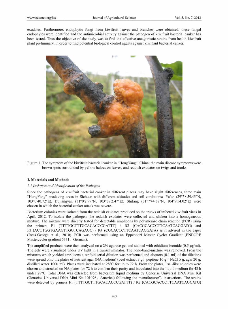
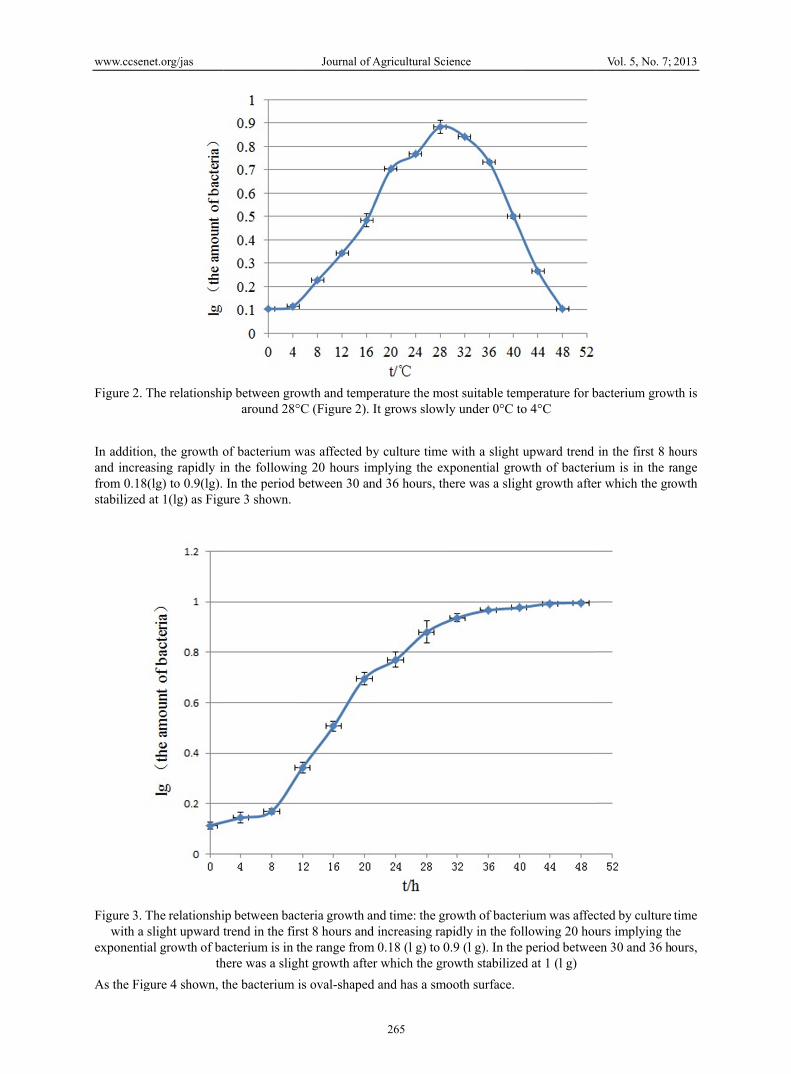
and F3 (ACCTGGTGAAGTTGGTCAGAGC) / R4 (CGCACCCTTCAATCAGGATA). The sequences were then compared to the database available at the National Center for Biotechnology Information (NCBI) using their Blast Search Software on the NCBI website (http://www.ncbi.nlm.nih.gov/genbank). The biochemical test was performed according to Lelliott & Stead (Lelliott & Stead, 1998), levan production, presence of oxidase, Gram staining, arginine dihydrolase, metabolism of glucose, presence of fluorescent pigment on King”s medium B, tobacco-hypersensitivity reaction. The effect of temperature on growth was measured according to the amount of the bacteria produced after being incubated for 24 h at different temperatures (Shanghai Yiheng light incubator MGC-250BP-2, Shanghai). The relationship between the growth and the time was monitored according to the amount of bacteria incubated at 28°C for different amount of times. Shape of the pathogen was observed by a scanning electronic microscope (Hitachi cold field emission scanning electronic microscope S-4800, Japan). Three 2-year-old healthy plants were inoculated with about 1×10⁷ CFU ml-1 of bacterium suspensions as described by Takikawa (Takikawa et al., 1989) as compared to water control. Only isolates bacterium reddish exudates from canes produced symptoms similar to those naturally observed on the buds and branches and identify the bacterium by molecular tests. The control plants were similarly incubated with sterile water. 2.2 Isolation and Identification of the Endophytic Fungi In July 2012, branch and leaf samples of the particular healthy kiwifruit plant were taken and transported in plastic bags in a cooler from the field sites to the laboratory. The isolation was carried out as following: branches and leaves were washed under running tap water for 3 min and than air-dried. Samples were pieced into the size of about 0.5 cm using sterile surgical blades, soaked in 75% ethanol for 2 min, washed by sterile distilled water for 3 min, immersed in 0.1% HgCl2 for 20 s, and then washed by sterile distilled water for 1 min. The treated pieces were cultured on the potato dextrose agar (PDA) medium (potato 200 g, glucose 17 g, agar 17 g, water 1000 ml) supplemented with streptomycin (100 μg/ml) sequentially. Total DNA was extracted by CTAB method (Wu et al., 2009) and PCR was performed using an Eppendorf Master Cycler Gradient. The strains were detected by the primers ITS1 (TCCGTAGGTGAACCTGCGG) and ITS4 (TCCTCCGCTTATTGATATGC). The amplified DNA fragments were purified and the sequences were then compared to the database available at the NCBI using their Blast Search Software on the NCBI website (http://www.ncbi.nlm.nih.gov/genbank). 2.3 The Antibacterial Activity Test of the Endophytic Fungi The antibacterial activity was determined using the oxford-cup-plate method (cited literatures). 1×10⁷ CFU suspension of PSA was dispersed on NA plates. Endophytic fungi were grown in 250 ml Erlenmeyer flasks containing 150 ml malt PDA liquid medium. The culture was grown with continuous shaking on a rotary shaker (180 rpm) at 26°C for 3 days. After incubation,endophytic fungi were harvested by a centrifugation at 5000 rpm for 10 min and 20 μl of endophytic fungi extracts were pipetted into a sterile oxford-cup (6 mm×10 min). These were placed on the surface of the plate and incubated at 30°C for 48 h. Sterile water was used as a control. Antibacterial activity was measured by the diameter of the inhibition zone. For the stains that showed good bacteriostasis, 7 generation was subcultured and the subculture was tested again to make sure the stability. 3. Results Three strains from three sites were obtained. For colony Dujiangyan, there was no obvious symptom on cane No.1 during the first 8 days. It produced the reddish exudates in Day 9 with water soaked lesions appeared around the wound. For colony Shifang it was until the 11 days that the cane appeared obvious symptom: reddish exudates produced on twigs. For colony Ya/an, it took 8 days to appear the symptom on cane No.3. Bacterium with morphological, biochemical and molecular characteristics identical to the original isolate were re-isolated from tissue showing symptoms. According to the techniques reported by Lelliott and Stead (1988), these strains were positive for levan production, tobacco-hypersensitivity reaction and metabolism of glucose. Negative for presence of oxidase, potato soft rot, arginine dihydrolase, Gram staining and the presence of fluorescent pigment on King”s medium B. The relationship between growth and temperature was analyzed. It was demonstrated that the most suitable temperature for bacterium growth is around 28°C (Figure 2). It grows slowly under 0°C to 4°C.
www.ccsen
Figure 2. T
In additionand increafrom 0.18(stabilized
Figure 3. Twith a s
exponentia
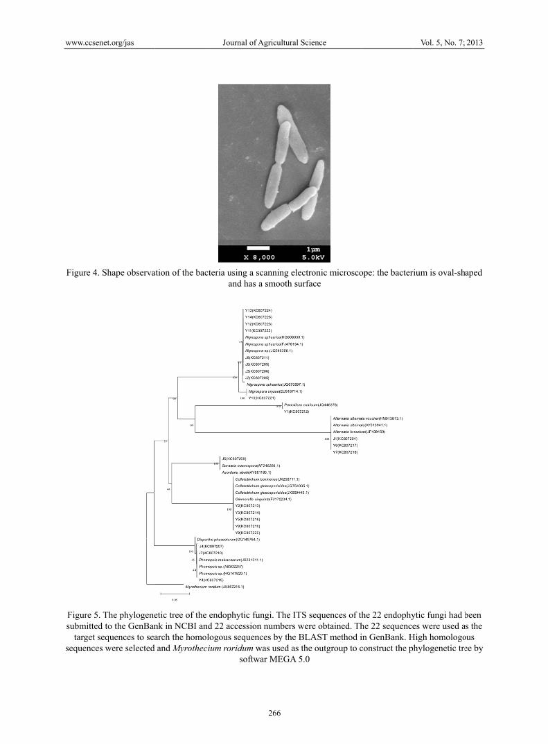
As the Fig
net.org/jas
The relationshi
n, the growth oasing rapidly in(lg) to 0.9(lg). at 1(lg) as Figu
The relationshislight upward tal growth of ba
thgure 4 shown, t
ip between groaround 28°
of bacterium wn the followinIn the period ure 3 shown.
ip between bactrend in the firacterium is in there was a sligthe bacterium i
Journal of A
owth and temp°C (Figure 2).
was affected byng 20 hours imbetween 30 an
cteria growth arst 8 hours andthe range fromht growth afteis oval-shaped
Agricultural Sci
265
erature the moIt grows slowl
y culture timemplying the exnd 36 hours, th
and time: the grd increasing rapm 0.18 (l g) to 0
r which the grd and has a smo
ience
ost suitable temly under 0°C to
e with a slight xponential growhere was a slig
rowth of bacterpidly in the fol0.9 (l g). In therowth stabilizeooth surface.
mperature for bo 4°C
upward trend wth of bacteriht growth afte
rium was affecllowing 20 hou
e period betweed at 1 (l g)
Vol. 5, No. 7;
bacterium grow
in the first 8 hium is in the rer which the gr
cted by cultureurs implying then 30 and 36 h
2013
wth is
hours range rowth
time he
hours,
www.ccsen
Figure 4. S
Figure 5. Tsubmitted
target sesequences
net.org/jas
Shape observa
The phylogeneto the GenBan
equences to seawere selected
ation of the bac
etic tree of thenk in NCBI anarch the homoand Myrothec
Journal of A
cteria using a sand has
endophytic fund 22 accessionlogous sequen
cium roridum wsoftw
Agricultural Sci
266
scanning electra smooth surfa
ungi. The ITS sn numbers wernces by the BLwas used as the
war MEGA 5.0
ience
ronic microscoace
sequences of thre obtained. Th
LAST method ie outgroup to c
ope: the bacter
he 22 endophyhe 22 sequencein GenBank. Hconstruct the p
Vol. 5, No. 7;
rium is oval-sh
ytic fungi had bes were used a
High homologohylogenetic tre
2013
haped
been as the ous ee by
www.ccsen
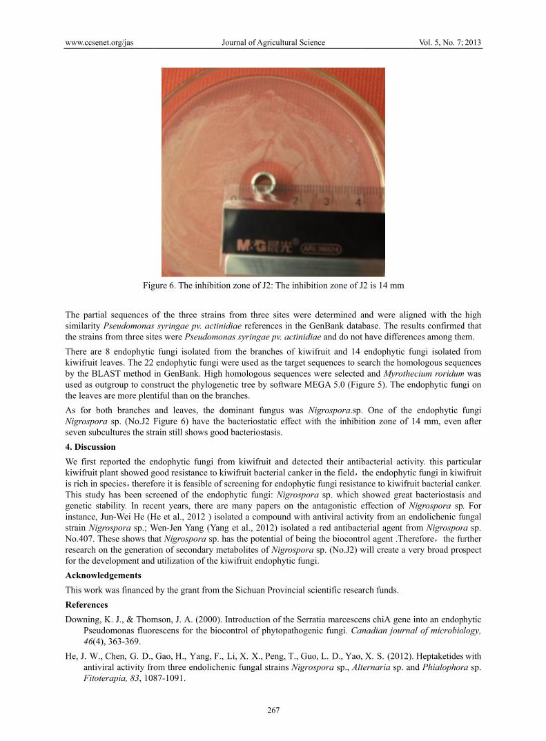
The partiasimilarity the strains There are kiwifruit leby the BLused as outhe leaves As for boNigrosporaseven subc4. DiscussWe first rkiwifruit pis rich in sThis studygenetic stainstance, Jstrain NigrNo.407. Tresearch ofor the devAcknowleThis work ReferenceDowning,
Pseud46(4)
He, J. W., antiviFitote
net.org/jas
Fig
al sequences oPseudomonasfrom three sit8 endophytic
eaves. The 22 LAST method iutgroup to conare more plen
oth branches a sp. (No.J2 Fcultures the strsion reported the enplant showed gspecies,therefoy has been scrability. In receJun-Wei He (Hrospora sp.; Whese shows thn the generatio
velopment andedgements
was financed es K. J., & Thom
domonas fluor), 363-369.
Chen, G. D., iral activity froerapia, 83, 108
gure 6. The inh
of the three st syringae pv. a
tes were Pseud fungi isolatedendophytic fuin GenBank. Hstruct the phyl
ntiful than on thand leaves, tFigure 6) haverain still shows
ndophytic fungood resistanceore it is feasiblreened of the ent years, ther
He et al., 2012 Wen-Jen Yang hat Nigrosporaon of secondar
d utilization of
by the grant fr
mson, J. A. (20rescens for the
Gao, H., Yangom three endo87-1091.
Journal of A
hibition zone o
trains from thactinidiae refe
domonas syringd from the br
ungi were usedHigh homologlogenetic tree he branches.the dominant e the bacterios good bacterio
ngi from kiwife to kiwifruit ble of screeningendophytic fure are many p) isolated a co(Yang et al., 2
a sp. has the pory metabolitesthe kiwifruit e
rom the Sichua
000). Introducte biocontrol of
g, F., Li, X. Xolichenic funga
Agricultural Sci
267
f J2: The inhib
hree sites wereerences in the gae pv. actinidranches of kiwd as the target sgous sequencesby software M
fungus was Nstatic effect wostasis.
fruit and detecbacterial cankeg for endophytungi: Nigrospopapers on the ompound with2012) isolatedotential of beins of Nigrosporendophytic fun
an Provincial s
tion of the Serf phytopathoge
X., Peng, T., Gual strains Nigro
ience
bition zone of J
e determined GenBank data
diae and do notwifruit and 14sequences to ss were selected
MEGA 5.0 (Fig
Nigrospora.spwith the inhibi
cted their antier in the field,tic fungi resistaora sp. which
antagonistic h antiviral activd a red antibacng the biocontra sp. (No.J2) wngi.
scientific resea
rratia marcesceenic fungi. Ca
uo, L. D., Yaoospora sp., Al
J2 is 14 mm
and were aligabase. The resut have differen endophytic f
search the homd and Myrothegure 5). The en
p. One of theition zone of
ibacterial activthe endophyti
ance to kiwifrushowed greateffection of Nvity from an e
cterial agent frtrol agent .Thewill create a v
arch funds.
ens chiA gene anadian journa
o, X. S. (2012)lternaria sp. an
Vol. 5, No. 7;
gned with the ults confirmed
nces among thefungi isolated
mologous sequeecium roridumndophytic fun
e endophytic f14 mm, even
vity. this partiic fungi in kiwuit bacterial cat bacteriostasisNigrospora spendolichenic furom Nigrosporerefore,the fuvery broad pro
into an endopal of microbio
. Heptaketidesnd Phialophor
2013
high d that em. from
ences m was
gi on
fungi after
cular ifruit
anker. s and . For ungal ra sp. urther spect
hytic logy,
with ra sp.
www.ccsenet.org/jas Journal of Agricultural Science Vol. 5, No. 7; 2013
268
Lelliott, R. A, Stead, D. E. (1988). Methods for the Diagnasis of Bacterial Diseases of Plants. Oxford, UK: Blackwell Scientific.
Li, Y., Sung, Y. C., Liu, J. Y., Ma, Y. M., Tan, R. X. (2005). Anti-Helicobacter pyori substances from endophytic fungal cultures. World J. Microbiol Biotechnol, 21, 553-558. http://dx.doi.org/10.1007/s11274-004-3273-2
Recs-George, J., Vanneste, J. L., Cornish, D. A., Pushparajah, I. P. S., Yu, J., Templeton, M. D., & Eerett, K. R. (2010). Detection of Pseudomonas syringae pv. actinidiae using polymerase chain reaction(PCR) primers based on the 16S-23S rDNA intertranscribed spacer region and comparison with PCR primers based on other gene regions. Plant Pathology, 59, 453-464.
Ryu, C. M., Kim, J. W., Cho, O. H., Park, S. Y., Park, S. H., & Park, C. S. (2005). Nature of a root associated Paenibacillus poymyxa from field-grown winter barley in Korea. J. Microbiol Biolechnol, 15, 984-991.
Serizawa, S., Ichikawa, T., Takikawa, Y., Tsuyumu, S., & Goto, M. (1989). Occurrence of bacterial canker of kiwifruit in Japan: description of symptoms, isolation of the pathogen and screening of bactericides. Annals of the Phytopathological Society of Japan, 55(4), 427-436.
Shimizu, M., Nakagawa, Y., Sato, Y., Furumai, T., Igarashi, Y., Onaka, H., … Kunch, H. (2000). Studies on endophytic actinomycetes(I) Streptomyces sp. Isolated from Rhodooendron and its antifungal activity. J. Gen Plant Pathol, 66, 360-366. http://dx.doi.org/10.1007/PL00012978
Takikawa, Y., Serizawa, S., Ichikawa, T., Tsuyumu, S., & Goto, M. (1989). Pseudomonas syringae pv. Actinidiae pv. nov.: the causa bacterium of canker of kiwifruit in Japan. Annals of Phytopathological, Society of Japan, 55, 437-44. http://dx.doi.org/10.3186/jjphytopath.55.437
Yang, W. J., Yang, C. S., Huang, C. J., Chen, K. S., & Lin, S. F. (2012). Bostrycin, a novel coupling agent for protein immobilization and prevention of biomaterial-centered infection produced by Nigrospora sp. No. 407. Enzyme and Microbial Technology, 50(6), 287-292.
Wu, F., Huang, D., Huang, X. L., Zhou, X., & Cheng, W. J. (2009). Comparing Study on Several Methods for DNA Extraction from Endophytic fungi. Chinese Agriculture Science Bulletin, 25(08), 62-64.
Copyrights Copyright for this article is retained by the author(s), with first publication rights granted to the journal. This is an open-access article distributed under the terms and conditions of the Creative Commons Attribution license (http://creativecommons.org/licenses/by/3.0/).