Embed Size (px)
Citation preview

1560
Brazilian Journal of Microbiology (2011) 42: 1560-1568 ISSN 1517-8382
ISOLATION AND CULTIVATION OF FUNGAL STRAINS FROM IN VITRO CELL CULTURES OF TWO MARINE
SPONGES (PORIFERA: HALICHONDRIDA AND HAPLOSCLERIDA)
Enrique E. Rozas
1, 4, Rodolpho M. Albano
2, Gisele Lôbo-Hajdu
2, Werner E.G. Müller
3, Heinz-C. Schröder
3, Márcio R.
Custódio 4 *
1Centro de Energia, Ambiente e Biodiversidade, Escola Superior de Ciências da Saúde, Universidade do Estado do Amazonas,
Manaus, AM, Brasil; 2Instituto de Biologia Roberto Alcântara Gomes, Universidade do Estado do Rio de Janeiro, Rio de Janeiro,
RJ, Brasil; 3Institut für Physiologische Chemie, Abt. Angewandte Molekularbiologie, Johannes Gutenberg-Universität, Mainz,
Germany; 4Departamento de Fisiologia Geral, Instituto de Biociências, Universidade de São Paulo, São Paulo, SP, Brasil.
Submitted: March 01, 2011; Returned to authors for corrections: March 30, 2011; Approved: May 16, 2011.
ABSTRACT
Despite the large number of reports describing sponge-microbe associations, limited knowledge is available
about associated fungi and their relationships with the hosts. In this work, specific fungal strains were
obtained directly from in vitro sponge cell cultures (primmorphs) and single sponge cells (cytospins) and
compared with those obtained from whole tissue preparations. A total of 27 fungal strains were isolated from
the marine sponges Hymeniacidon heliophila and Haliclona melana. Fifteen strains, nine from H. heliophila
and six from H. melana, were obtained from whole tissue and were considered as possible mesohyl
associated or transient fungi. Twelve strains were isolated from in vitro sponge cell cultures (primmorphs)
and were, therefore, considered as cell associated. From these, five different strains were obtained from H.
heliophila isolated cells, while five were identified from cytospins and two from primmorphs of H. melana.
The fungal strains obtained from cell cultures from both sponge species were different, and none of them
were detected in the whole tissue preparations of the same species. Nine H. heliophila and seven H. melana
strains shows low similarity with the sequences available in public databases and belong to potentially new
species. This is the first report of fungi isolated directly from sponge cells, which allowed the observation and
selection of specific strains that probably would not be obtained by usual culture dependent techniques.
Key words: Marine sponges, primmorphs, cytospins, fungal detection
INTRODUCTION
Marine sponges (Porifera) represent one of the oldest
extant multicellular animals (20). The more than 8,000
described species inhabit a variety of marine and freshwater
systems, from the tropics to Polar Regions (10). They are
sessile filter-feeding organisms that influence benthic and
pelagic processes by transferring large quantities of material
from the water column to the sea floor (25). The sponge body
is simple, constituted by an outer epithelial layer (pinacocytes)
*Corresponding Author. Mailing address: Rua do Matão. Trav. 14, n. 101. 05508-900. São Paulo, SP. Brazil.; Tel: 55 11 3091-7611 Fax: 55 11 3091-8095.; E-mail: [email protected]

1561
Rozas, E.E. et al. Fungal strains from in vitro cell cultures of two marine sponges
enclosing the mesohyl, formed by specialized cells,
extracellular matrix and a network of canals and chambers. In
these chambers, water is pumped continuously by flagellated
cells (choanocytes) and microorganisms and organic particles
are retained and phagocytized (29).
In the last 20 years, sponges have been considered one of
the most prolific sources of natural products (see 1, and
previous reviews of this series). Several compounds have been
isolated and tested, showing a wide range of pharmacological
activities such as antimicrobial, cytotoxic, antimalarial,
antitumour, antiviral and anti-inflammatory. The chemical
diversity is notable, including unusual nucleosides, terpenes,
cyclic peptides and alkaloids (31). However, many of these
substances are produced by associated microorganisms, and not
by the sponge itself. Recently, the isolation, identification and
cultivation of this microbiota have been the trend in the search
for new compounds (1).
Most sponges harbor large numbers of symbionts, which
include bacteria, cyanobacteria, unicellular algae, archaea and
fungi, sometimes comprising 40 to 60% of the total biomass (9,
16). In many cases, these communities are species-specific and
independent of the environmentally available strains (17, 42).
Of the different organisms, bacteria are the most thoroughly
investigated and usually reported as participating in the
chemical defenses or physiological processes of their hosts (32,
35, 37).
On the other hand, the knowledge about the diversity and
function of potentially sponge-associated fungi is still limited.
Despite the growing number of strains identified from sponge
samples (17, 18, 22, 40), little evidence has been published to
support the idea of true symbioses. The best example of such
relationship is represented by unidentified yeast vertically
transmitted in three Chondrilla species (19). In addition,
several fungi isolated from sponges and other marine substrates
were identified as belonging to terrestrial genera.
Consequently, even the debate about the real marine origin of
these strains is still open (26, 40). In general, the fungal strains
isolated from marine environments show different secondary
metabolite profiles when compared with those from terrestrial
habitats. For instance, some marine species of Penicillium
produce secondary metabolites unknown to closely related
strains of terrestrial origin (15).
In addition to the associated microbiota, as any other
marine substratum, sponges are also exposed to large numbers
of different microorganisms from the environment. These
animals can filter vast quantities of seawater, retaining over
80% of the suspended particles (23), and therefore a transient
microbiota is always present in its canals, tissues and surfaces
(24). The presence of these communities, frequently in huge
numbers, makes the studies of specific strains especially
complex. It can be difficult to distinguish between true sponge-
associated microorganisms and environment-derived
contaminants, even with the assistance of molecular
techniques. Moreover, it is possible that some strains would not
be obtained by usual culture-dependent approaches due to
overgrowth by other microorganisms. In order to avoid these
problems, in this work we use in vitro sponge cell cultures and
single cells as initial material to obtain fungal cultures. This
approach eliminated most of the mesohyl associated and
transient microorganisms, and allowed the focus on the
isolation and culture of strains specifically associated to sponge
cells.
METHODS
Sponges
A total of 12 samplings were performed at Praia Grande
area (23o49’23.76”S, 45
o25’01.79”W, São Sebastião, Brazil).
On each occasion, four individuals of Hymeniacidon heliophila
Parker, 1910 (Halichondrida: Halichondriidae) and four of
Haliclona melana Muricy and Ribeiro, 1999 (Haplosclerida:
Chalinidae) were collected. Individual specimens were placed
separately into plastic bags and brought to the laboratory in ice-
cooled boxes (18-20oC). These species have already been used

1562
Rozas, E.E. et al. Fungal strains from in vitro cell cultures of two marine sponges
in previous works on sponge cell biology (5, 6), and showed
good responses to culture conditions. Taxonomic identification
was performed by M.R. Custódio, using standard methods (21).
Sponge cell culture (Primmorphs)
Cell dissociation and primmorph formation followed
protocols described elsewhere (6). Sponge tissues were cleaned
of larger epibionts using forceps, cut in 3 to 4 mm pieces,
washed in seawater and incubated with CMFSW+E (calcium
and magnesium-free seawater with EDTA: 460 mM NaCl, 7
mM Na2SO4, 10 mM KCl, 10 mM HEPES, 2.5 mM EDTA, pH
8.2. Sigma) for 30 min in a low speed rotary shaker. The
supernatant was then discarded, and the fragments re-
suspended in fresh CMFSW+E solution. After continuous
shaking for 45 min, the supernatant was collected and filtered
through a 40 µm mesh. The cells were pelleted by
centrifugation at 250 x g / 10 min and re-suspended in sterile-
filtered (0.22 µm) natural seawater from the same collection
site, supplemented with antibiotics (kanamycin 100 mg/L,
gentamicin 50 mg/L and tetracycline 10 mg/L, Calbiochem)
and phenol red (16 mg/L, Sigma). This media was used for
maintenance of the primmorphs. In the first week, it was
renewed daily and in the following weeks, each three days.
Isolation and culture of the fungal strains
The primmorphs were observed daily on inverted
microscope, and the fungal hyphae that appeared on the surface
were picked and incubated in tubes with DY medium
(Dextrose 1% and Yeast Nitrogen base 1% (Fluka) in natural
seawater). After 24h, 100 µl of each suspension was
disseminated in DY agar (DY medium with 2% agar) to verify
the purity of the culture. The fungi were carefully selected
according to morphologic characteristics (7, 13), transferred to
125 ml Erlenmeyer flasks with 60 ml of DY medium and
incubated at 20oC. After 15 days, the cultures were sampled for
DNA extraction and identification.
To obtain cell-associated fungi, primmorphs were
cultivated for 21 days and then dissociated with CMFSW+E.
The cell suspension was counted using a Neubauer chamber,
and attached in low density to sterile glass coverslips using a
cytocentrifuge (80 x g / 5 min, 1.5 x 10⁴ cells per spot. Citospin
248, FANEM). The coverslips were allowed to dry in a sterile
laminar flow hood for 3h and then transferred to six-well plates
filled with DY medium, with the cell surface facing the
overlaying medium. For each plate, one well with a coverslip
prepared with CMFSW+E was used as control. Culture
observation was carried out using phase contrast inverted
microscopy (Eclipse TE300, NIKON), until the mycelia
became visible with the naked eye. Morphologically different
strains were collected and transferred to the DY agar to verify
the purity of the culture, and then cultivated as described
above. To differentiate between strains associated with the
cells and those incidentally present in the sponge surfaces and
canals, fungal cultures were established from sponge whole
tissues. Sponge fragments were washed with sterile
CMFSW+E (5 min) in order to release peripheral cells and
eventual transient fungi, gently compressed and the resulting
material was collected in a flask with sterile seawater. Then,
100 µl aliquots of this suspension were disseminated in DY
and isolates were purified by successive selection and
cultivated following the procedure described above.
DNA extraction and polymerase chain reaction (PCR)
amplification
The DNA was isolated from fresh samples (25 mg) of
mycelia from the fungal cultures using the PureLinkTM
Genomic DNA Kit (Invitrogen). The extracted DNAs were
analyzed in 1% agarose-Tris-borate-EDTA gels to determine
the quality and the amount of DNA obtained. Amplification of
the 18S rRNA gene was performed using the specific primers
EF3 (5’-TCCTCTAAATGACCAAGTTTG-3’), EF4 (5'-
GGAAGGG [G/A] TGTATTTATTAG-3') and Fung5 (5'-
GTAAAAGTCCTGGTTCCCC-3') (32). One microliter of
each DNA sample (100 ng) was used for PCR amplification.
1563
Rozas, E.E. et al. Fungal strains from in vitro cell cultures of two marine sponges
Primer sets EF4/EF3 and EF4/Fung5 were used for direct
amplification of 18S rDNA sequences from extracted fungal
DNA. PCR mixtures (50 µl) for both sets consisted of 1x PCR
buffer (Invitrogen), 2 mM MgCl2, 0.2 mM mix of dNTPs, 50 ng of
each primer and 0.3 µl Platinum Taq Polymerase (5 u/µl,
Invitrogen). The following thermocycling pattern was used: 94oC
for 3 min (1 cycle); 94oC for 1 min; 48
oC for 1 min; 72
oC for 3
min (35 cycles); and 72oC for 10 min (1 cycle). The PCR products
were separated on a 1% Agarose-Tris-borate-EDTA gel.
Partial sequencing of the fungal 18S rRNA gene
The PCR products were purified with the Illustra GFX PCR
DNA and the Gel Band Purification Kits (GE Healthcare) and
used in cycle sequencing reactions with the kit DYENAMIC ET
terminators (GE Healthcare). The 0.5 Kb fragments obtained from
each fungal strain, corresponding to the region amplified by the
EF4/EF3 primer set were sequenced with the EF4 primer. The
sequencing reactions were analyzed by capillary electrophoresis
on a MEGABACE 1000 sequencing platform (GE Healthcare).
Consensus sequences were obtained using the CAP3 Sequence
Assembly Program (10). Sequences were then manually edited
and analyzed by BLAST searches at the National Center for
Biotechnology Information (NCBI, accessed in July, 2010).
Sequence alignment of the 0.5 Kb fragments and phylogenetic
trees were prepared with POY v4.1.2 (39), using 60 random
sequence-addition repetitions and TBR branch swapping using
parsimony criterion. The sequence of the anamorphic fungi
Acremonium cellulolyticus (accession number BA474750) was
used as outgroup. All sequences were deposited in the GenBank,
and accession numbers are listed in Table 1.
Table 1. Fungal strains isolated and cultivated from the whole tissue, single cells and primmorphs of Hymeniacidon heliophila (Hy) and
Haliclona melana (Ha). Accession numbers (A.N.) and scores (%) are given. The closest sequence match corresponds to the top BLAST
score record (accessed in July 2010).
Hymeniacidon heliophila
Whole tissue Closest Sequence Match A.N. % Hy 1 (FJ477281) Penicillium waksmanii MMA12 (HM231102) 100
Hy 2 (FJ477282) Penicillium sp. HB-92 (AB521044) 99
Hy 3 (FJ477279) Penicillium waksmanii MMA12 (HM231102) 99
Hy 4 (FJ477283) Penicillium sp. HB-92 (AB521044) 97
Hy 5 (FJ477284) Penicillium sp. HB-92 (AB521044) 99
Hy 6 (FJ477293) Penicillium sp. HB-92 (AB521044) 99
Hy 7 (FJ477286) Penicillium sp. HB-92 (AB521044) 98
Hy 8 (FJ477278) Penicillium sp. ASR-151 (GU973852) 98
Hy 9 (FJ477287) Penicillium sp. HB-92 (AB521044) 96
Single cells
Hy I (FJ477273) Uncultured fungal contaminant (EF053580) 94
Hy II (FJ477272) Cryptosphaeria sp. MYA-4414 (FJ430577) 98
Hy III (FJ477274) Paraphaeosphaeria sp. B19 (GQ253350) 98
Hy IV (FJ477276) Ascomycota sp. Mo19 (EU887770) 98
Hy V (FJ477277) Cryptosphaeria sp. MYA-4414 (FJ430577) 97
Haliclona melana
Whole tissue Ha 1 (FJ477280) Penicillium sp. ASR-302 (GU973859) 97
Ha 2 (FJ477292) Penicillium sp. HB-92 (AB521044) 99
Ha 3 (FJ477291) Penicillium sp. HB-92 (AB521044) 96
Ha 4 (FJ477288) Penicillium sp. HB-92 (AB521044) 99
Ha 5 (FJ477289) Emericella nidulans (AB008403) 97
Ha 6 (FJ477290) Penicillium sp. HB-92 (AB521044) 99
Single cells
Ha I (FJ477269) Cladosporium cladosporioides (AB521051) 99
Ha II (FJ477267) Cladosporium cladosporioides (AB521051) 98
Ha III (FJ477268) Cladosporium cladosporioides (AB521051) 99
Ha IV (FJ477270) Xylaria polymorpha (AB274816) 97
Ha V (FJ477266) Uncultured fungus FAS 62 (GU072561) 98
Primmorphs
Ha VI (FJ477271) Escovopsis aspergilloides (AY172598) 97
Ha VII (FJ477275) Penicillium sp. YW01 (GU944770) 99
1564
Rozas, E.E. et al. Fungal strains from in vitro cell cultures of two marine sponges
RESULTS
Fungal isolation and cultivation
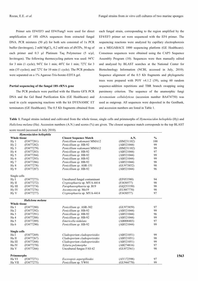
Five strains were obtained from single cells (cytospins) of
each sponge (Table 1). The hyphae of two of these fungi, Ha
IV and Ha V (Fig. 1a-f), were detected at early stages, always
associated or near the sponge cell nuclei. Both appear as small
clusters of round, bright structures from which the adult
mycelia originates (Fig. 1a, e). In Ha IV, large amounts of
insoluble hyaline material of unknown nature could be
observed deposited around the hyphae even at early stages
(Fig. 1b, c). In this strain, the hyphae were always embedded in
this substance, in which some crystals were formed in later
stages (Fig. 1d). In Ha V, the hyphae initially grew following
the host cell margins (Fig. 1e) and latter forming a branched,
three-dimensional mycelium (Fig. 1f). Such very early stages
of other strains were not detected, and they were observed
already as small mycelia overlapping or close to the sponge
cells. When incubated in liquid medium, these fungal strains
were detected 15 days after the preparation of the cytospins (36
days after the beginning of the sponge cell culture), reaching
the maximum size in five additional days. However, the strains
markedly reduced the growth speed after three successive
subcultures, and the maximum size was reached only after 28
days.
Using primmorphs, two strains were obtained from H.
melana, but none from H. heliophila (Table 1). The fungi
associated with the primmorphs appeared always as small
hyphae attached to the surface of the aggregates after 45 days
in culture. Using whole tissue preparations, nine
morphologically different strains were isolated from H.
heliophila, although two (Hy 1 and Hy 7) could not be
cultivated up to confluence. From H. melana whole tissue
preparations, six strains were isolated and cultivated.
Figure 1. Growth of strains Ha IV and
Ha V from H. melana cells in culture.
(a, b, c) Hyphae of Ha IV growing from
a single sponge cell immersed in DY
liquid media (b and c: same field). The
deposition of insoluble hyaline material
of unknown nature is already visible
since the early growth stages (n:
nuclei). (d) Ha IV mycelium immersed
in DY liquid media, 24h after the strain
started to grow from the sponge cell.
Large deposits of hyaline material
surround the hyphae and include
crystals (arrow). (e) Hyphae of Ha IV
(arrow) growing from a single sponge
cell. (f) Ha IV mycelium, 24h after the
strain started to grow from the sponge
cell.

1565
Rozas, E.E. et al. Fungal strains from in vitro cell cultures of two marine sponges
Fungal identification
The tentative fungal identifications based on the NCBI
database best matches and accession numbers for the strains
obtained in this work are summarized in Table 1. All fungi
isolated from whole tissue preparations from both sponges
belong to the genus Penicillium, with the only exception of one
strain related to Emericella nidulans (AB008403) in H.
melana. In contrast, this genus was poorly represented in the
material obtained from cell cultures, with only one strain (Ha
VII) found in H. melana (Penicillium sp. YW01. GU944770).
In both sponges, all strains obtained from cell-derived material
were distinct from those isolated from whole tissue
preparations of the same individual. In addition, different cell-
derived strains were obtained from each sponge species.
Phylogenetic trees inferred with the partial 18S rRNA
sequences showed that all cell-derived strains are related (Fig.
2), with the exception of Ha VII (Penicillium sp. YW01).
Figure 2. Phylogenetic tree depicting the relationship among the strains isolated from whole tissue and cell culture preparations of
H. heliophila (Hy) and H. melana (Ha). The GenBank best matches sequences are given (asterisks: strains obtained from marine
sources).

1566
Rozas, E.E. et al. Fungal strains from in vitro cell cultures of two marine sponges
DISCUSSION
Among all marine Phyla, Porifera is regarded as one of the
most important sources of metabolite-producing fungi (26, 36),
and in the last decade have increasingly become the target in
the search for new strains with biotechnological potential (14,
27). Different approaches have been used to isolate and
cultivate these microorganisms, mostly using whole
homogenates or dissected tissue samples (e.g. 12, 22, 26, 41).
Such studies have obtained a large number of different strains
and increased the knowledge about the diversity of sponge-
associated fungi. However, details about the mechanisms and
interactions with their hosts are scarce (17), and it is probable
that several strains are missed by overgrowth or competitive
interference by other species. By using in vitro sponge cell
cultures and single cells (cytospins), the focus in the present
work was to select only those strains specifically associated
with sponges.
From all fungal strains isolated from H. heliophila and H.
melana, only two have best match sequences similar to fungi
already obtained from marine sources (see Table 1). Thus Hy
IV was related to a Ascomycota strain collected in deep sea
termal vents (2), and Ha V was similar to an uncultured marine
fungus isolated from the sediment of the Arabian Sea (Jebaraj
et al. unpublished). For all other strains, the best match
sequences points to well known terrestrial species, most
belonging to the genus Penicillium and Cladosporium.
However, in recent years evidence has been accumulating that
indicates the presence of these (and others) ubiquitous
terrestrial fungi in the marine environment (17, 26, 30).
Interestingly, none of the strains isolated from the in vitro
cell cultures was found in the whole tissue preparations of the
same animal. Moreover, the fungal communities of cellular
origin of each sponge species were different. In contrast, the
strains obtained from the whole tissue preparations of H.
heliophila and H. melana were not species-specific, exhibiting
similar microbial profiles. In this case, the dominant fungal
strain was related to Penicillium, which was represented in the
cell-derived cultures by only one strain (Ha VII: Penicillium
sp. YW01). This dominance is reflected in the phylogenetic
analysis (Fig. 2), which groups all whole tissue strains in
opposition to the cell-derived lineages.
Of particular interest is the fact that two strains (Ha IV
and Ha V) were observed directly connected to single sponge
cells, appearing 15 days after the cytospins were placed in DY
media and 36 days after the sponge culture. It is possible that
these fungi were acquired from the original marine
environment or accidentally attached to the sponge cells
surface during manipulation. However, when isolated from the
cytospins and inoculated in fresh DY media, they reached the
maximum size in only five days. This suggests that the fungi
were indeed enclosed in the sponge cells, similarly to what has
been observed in Chondrilla (18), and the lapse in the initial
growth time was due to a latency of the enclosed fungi. This
condition would be interrupted after the sponge cell death,
forcing the fungus to grow towards the nutrient medium.
Moreover, after three successive subcultures the strains
reduced their growth speed noticeably. This indicates lack of a
putative component supplied by the sponge cells - growth
factor or nutrients - and suggests a facultative dependence. A
similar process could be responsible for the late appearance (45
days) of the two strains obtained from H. melana primmorphs.
Primmorphs are three-dimensional aggregates surrounded by a
continuous pinacoderm, in which the cells are fully capable of
physiological processes such as allogeneic recognition (5, 43)
and spicule secretion (3). Depending on the species, they can
be kept in plain seawater without additional nutrients for
several days or even months, after which they degenerate and
die (33, 38). After 45 days in culture, the primmorphs of H.
melana already started this declining process, and no longer
maintained the initial aspect of a rounded, dense cell mass with
a smooth surface. It is possible that at this stage, enclosed fungi
were released from the dormancy, in the same way as observed
in the isolated cells.

1567
Rozas, E.E. et al. Fungal strains from in vitro cell cultures of two marine sponges
The possibility that these fungi were admitted to
intracellular life by means of endocytosis depends on the
pathogen adaptations (28). The loss of virulence and the
production of anti-phagocytosis substances would allow the
initially intracellular parasite to become an endosymbiont (4,
8). Alternatively, many sponge cell types are phagocytes (29),
and fungal spores could enter the cells by this process without
disrupting the cell membranes. In this sense, a casual
infestation followed by phagocytosis would explain why the
fungi were present inside the cells and not in the whole tissue.
However, it would not explain why these strains were not
phagocysed and eliminated during the in vitro sponge cell
culture (21 days). This would mean a partial loss in the ability
to recognize non-self components, which seems unlikely. Thus,
the results suggest the presence of at least two intracellular
species: Ha VI and Ha VII, from H. melana, and the specificity
of the other strains isolated from the single cells and
primmorphs.
The identifications obtained by the best match sequences
in GenBank are provisional, and can only indicated affinities of
the obtained fungal strains. From all strains obtained in this
work, nine from H. heliophila and seven from H. melana show
sequence identity equal to or less than 98% compared to those
available in GenBank, indicating some probable new species.
In addition, five of these low identity strains from H. heliophila
and four from the H. melana were obtained directly from
sponge cells in culture. At this point, additional data, such as
linking morphological observations with a more
comprehensive molecular analysis is necessary to ensure the
correct identification. Nonetheless, this is the first report of
fungal strains isolated using in vitro sponge cell cultures, and
one can imagine that similar approach can be used for other
microorganisms such as bacteria. Our data show that
primmorphs and even single cells can be used as the initial
material for cultures, allowing detailed observation of the
emergence and growth of specific strains and the selection of
those that are not obtainable by usual techniques.
ACKNOWLEDGEMENTS
This work was funded by the Conselho Nacional de
Desenvolvimento Científico e Tecnológico and the
Bundesministerium für Bildung und Forschung (German-
Brazilian Cooperation in Marine Sciences - CNPq
590004/2005-0; BMBF 03F0451). The authors thank Dr.
Fernando P.L. Marques (Departamento de Zoologia. IB/USP)
for the assistance with the phylogenetic analysis.
REFERENCES
1. Blunt, J.W.; Copp, B.R.; Hu, W.P.; Munro, M.H.G.; Northcote, P.T.;
Prinsep, M.R. (2009). Marine natural products. Nat. Prod. Rep. 26, 170-
244.
2. Burgaud, G.; Le Calvez, Th.; Arzur, D.; Vandenkoornhuyse, Ph.;
Barbier, G. (2009). Diversity of culturable marine filamentous fungi
from deep-sea hydrothermal vents. Environ. Microbiol. 11, 1588-1600.
3. Cao, X.; Fu, W.; Yu, X.; Zhang, W. (2007). Dynamics of spicule
production in the marine sponge Hymeniacidon perlevis during in vitro
cell culture and seasonal development in the field. Cell. Tiss. Res. 329,
595-608.
4. Corsaro, D.; Venditti, D.; Padula, M.; Valassina, M. (1999). Intracellular
Life. Crit. Rev. Microbiol. 25, 39-79.
5. Custódio, M.R.; Hajdu, E.; Muricy, G. (2002). In vivo study of
microsclere formation in sponges of the genus Mycale (Demospongiae,
Poecilosclerida). Zoomorphology 121, 203-211.
6. Custódio, M.R.; Hajdu, E.; Muricy, G. (2004). Cellular dynamics of in
vitro allogeneic reactions of Hymeniacidon heliophila (Demospongiae:
Halichondrida). Mar. Biol. 144, 999-1010.
7. Dugan, F.M. (2006). The identification of fungi. American
Phytopathological Society Press, Saint Paul.
8. Goebel, W.; Gross, R. (2001). Intracellular survival strategies of
mutualistic and parasitic prokaryotes. TRENDS Microbiol. 9, 267-274.
9. Hentschel, U.; Usher, K.M.; Taylor, M.W. (2006). Marine sponges as
microbial fermenters. FEMS Microbiol. Ecol. 55, 167-177.
10. Hooper, J.N.A.; van Soest, R.W.M. (2002). Systema Porifera: a guide to
the classification of sponges. Kluwer Academic/Plenum Publishers, New
York.
11. Huang, X.; Madan, A. (1999). CAP3, A DNA sequence assembly
program. Genome Res. 9, 868-877.
12. Kennedy, J.; Codling, C.; Jones, B.; Dobson, A.; Marchesi, J. (2008).
Diversity of microbes associated with the marine sponge, Haliclona
simulans, isolated from Irish waters and identification of polyketide
1568
Rozas, E.E. et al. Fungal strains from in vitro cell cultures of two marine sponges
synthase genes from the sponge metagenome. Environ. Microbiol. 10,
1888-1902.
13. Kohlmeyer, J. (1984). Tropical marine fungi. Mar. Ecol. 5, 329-378.
14. König, G.; Kehraus, S.; Seibert, S.; Abdel-Lateff, A.; Müller, D. (2006).
Natural products from marine organisms and their associated microbes.
ChemBioChem 7, 229-238.
15. Lang, G.; Wiese, J.; Schmaljohann. R.; Imhoff, J. (2007). New pentaenes
from the sponge-derived marine fungus Penicillium rugulosum: structure
determination and biosynthetic studies. Tetrahedron 63, 11844-11849.
16. Lee, Y.; Lee, J.; Lee, H. (2001). Microbial symbiosis in marine sponges.
J. Microbiol. 39, 254-264.
17. Li, Q.; Wang, G. (2009). Diversity of fungal isolates from three
Hawaiian marine sponges. Microbiol. Res. 164, 233-241.
18. Liu, W.C.; Li, C.Q.; Zhu, P.; Yang, J.L.; Cheng, K.D. (2009).
Phylogenetic diversity of culturable fungi associated with two marine
sponges: Haliclona simulans and Gelliodes carnosa, collected from the
Hainan Island coastal waters of the South China Sea. Fungal Divers. 42
(1), 1-15.
19. Maldonado, M.; Cortadella, N.; Trilla, M.I.; Rützler, K. (2005).
Endosymbiotic yeast maternally transmitted in a marine sponge. Biol.
Bull. 209, 94-106.
20. Müller, W.E.G. (1998). Origin of Metazoa: sponges as living fossils.
Naturwissenschaften 85, 11-25.
21. Muricy, G.; Hajdu, E. (2006) Porifera Brasilis: Guia de identificação das
esponjas marinhas mais comuns do Sudeste do Brasil. Museu Nacional.
Série Livros 17. Rio de Janeiro.
22. Paz, Z.; Komon-Zelazowska, M.; Druzhinina, I.S.; Aveskamp, M.M.;
Shnaiderman, A.; Aluma, Y.; Carmeli, S.; Ilan, M.; Yarden, O. (2010).
Diversity and potential antifungal properties of fungi associated with a
Mediterranean sponge. Fungal Divers. 42, 17-26.
23. Pfannkuchen, M.; Fritz, G.B.; Schlesinger, S.; Bayer, K.; Brümmer, F.
(2009). In situ pumping activity of the sponge Aplysina aerophoba,
Nardo 1886. J. Exp. Mar. Biol. Ecol. 369, 65-71.
24. Pile, A.J.; Patterson, M.; Witman, J. (1996). In situ grazing on plankton
<10 microns by the boreal sponge Mycale lingua. Mar. Ecol. Prog. Ser.
141, 95-102.
25. Pile, A.J.; Young, C.M. (2006). The natural diet of a hexactinellid
sponge: benthic-pelagic coupling in a deep-sea microbial food web.
Deep-Sea Res. 53, 1148-1156.
26. Proksch, P.; Ebel, R.; Edrada, R.; Riebe, F.; Liu, H.; Diesel, A.; Bayer,
M.; Li, X.; Lin, W.H.; Grebenyuk, V.; Müller, W.E.G.; Draeger, S.;
Zuccaro, A.; Schulz, B. (2008). Sponge-associated fungi and their
bioactive compounds: the Suberites case. Bot. Mar. 51, 209-218.
27. Raghukumar, C. (2008). Marine fungal biotechnology: an ecological
perspective. Fungal Divers. 31, 19-35.
28. Selvin, J.; Ninawe, A.S, Kiran, G.S, Lipton, A. P. (2010). Sponge-
microbial interactions: Ecological implications and bioprospecting
avenues. Crit. Rev. Microbiol. 36, 82-90.
29. Simpson, T.L. (1984). The cell biology of sponges. Springer-Verlag,
New York.
30. Singh, P.; Raghukumar, C.; Verma, V.; Souche, Y. (2010). Phylogenetic
diversity of culturable fungi from the deep-sea sediments of the Central
Indian Basin and their growth characteristics. Fungal Divers. 40, 89-102.
31. Sipkema, D.; Franssen, M.C.R.; Osinga, R.; Tramper, J.; Wijffels, R.H.
(2005). Marine sponges as pharmacy. Mar. Biotechnol. 7, 142-162.
32. Sipkema, D.; Holmes, B.; Nichols, S.A.; Blanch, H.W. (2009).
Biological characterization of Haliclona (?gellius) sp.: sponge and
associated microorganisms. Microb. Ecol. 58, 903-920.
33. Sipkema, D.; Van Wielink, R.; Van Lammeren, A.A.M.; Tramper, J.;
Osinga, R.; Wijffels, R.H. (2003). Primmorphs from seven marine
sponges: formation and structure. J. Biotech. 100, 127-139.
34. Smit, E.; Leeflang, P.; Glandorf, B.; Van Elsas, J.; Wernars, K. (1999).
Analysis of fungal diversity in the wheat rhizosphere by sequencing of
cloned PCR-amplified genes encoding 18s rRNA and temperature
gradient gel electrophoresis. Appl. Environ. Microbiol. 65, 2614-2621.
35. Thakur, N.L.; Anil, A.C.; Müller, W.E.G. (2004). Culturable epibacteria
of the marine sponge Ircinia fusca: temporal variations and their possible
role in the epibacterial defense of the host. Aquat. Microb. Ecol. 37, 295-
304.
36. Thomas, T.R.A.; Kavlekar, D.P.; LokaBharathi, P.A. (2010). Marine
drugs from sponge-microbe association: a review. Mar. Drugs 8, 1417-
1468.
37. Vacelet, J.; Duport, E. (2004). Prey capture and digestion in the
carnivorous sponge Asbestopluma hypogea (Porifera: Demospongiae).
Zoomorphology 123, 179-190.
38. Valisano, L.; Bavestrello, G.; Giovine, M.; Cerrano, C. (2006).
Primmorphs formation dynamics: a screening among Mediterranean
sponges. Mar. Biol. 149, 1037-1046.
39. Varon, A.; Le Sy, V.; Wheeler, W.C. (2010). POY version 4,
phylogenetic analysis using dynamic homologies. Cladistics 26 (1), 72-
85.
40. Wang, G. (2006). Diversity and biotechnological potential of the sponge-
associated microbial consortia. J. Ind. Microbiol. Biotechnol. 33, 545-
551.
41. Wang, G.; Li, Q.; Zhu, P. (2008). Phylogenetic diversity of culturable
fungi associated with the Hawaiian sponges Suberites zeteki and
Gelliodes fibrosa. Antonie Leeuwenhoek 93, 163-174.
42. Webster, N.; Negri, A.; Munro, M.; Battershill, C.N. (2004). Diverse
microbial communities inhabit Antarctic sponges. Environ. Microbiol. 6,
288-300.
43. Wiens, M.; Perovic-Ottstadt, S.; Müller, I.M.; Müller, W.E.G. (2004).
Allograft rejection in the mixed cell reaction system of the demosponge
Suberites domuncula is controlled by differential expression of apoptotic
genes. Immunogenetics 56, 597-610.
All the content of the journal, except where otherwise noted, is licensed under a Creative Commons


![Enzymatic Cellulose Hydrolysis: Enzyme Reusability and ... · novel engineered fungal strains to produce more efficient lignocellulolytic enzyme systems [13]. However, recycling or](https://img.pdfslide.us/doc/110x75/5f1b5c0af788e165636ba193/enzymatic-cellulose-hydrolysis-enzyme-reusability-and-novel-engineered-fungal.jpg)


![Cultivation andPartial Characterization ofSpiroplasmas in ... · Spiroplasma citri and unidentified strains (corn stunt organism, 277F [tick isolate], powderpuff, BNR-1, honeybee,](https://img.pdfslide.us/doc/110x75/5e52f91966b58e76ac372278/cultivation-andpartial-characterization-ofspiroplasmas-in-spiroplasma-citri.jpg)