Embed Size (px)
Citation preview
Vol. 170, No. 6
Isolation and Characterization of Pathogenicity Genes ofPseudomonas syringae pv. tabaci
YANGKYO P. SALCH AND PAUL D. SHAW*
Department of Plant Pathology, University of Illinois at Urbana-Champaign, 1102 South Goodwin Avenue,Urbana, Illinois 61801
Received 14 September 1987/Accepted 29 February 1988
Pseudomonas syringae pv. tabaci BR2 produces tabtoxin and causes wildfire disease on tobacco and beanplants. Approximately 2,700 TnS insertion mutants of a plasmid-free strain, PTBR 2.024, were generated byusing suicide plasmid pGS9. Of these Tn5 mutants, 8 were no longer pathogenic on tobacco plants and 10showed reduced symptoms. All of the eight nonpathogenic mutants caused typical wildfire disease symptoms onbean plants. Two of the nonpathogenic mutants failed to produce tabtoxin. The eight nonpathogenic mutantshave TnS insertions into different EcoRI and SailI restriction fragments. The EcoRI fragments containing Tn5from the eight nonpathogenic mutants were cloned into vector pTZ18R or pLAFR3. A genomic library of theparent strain was constructed in the broad-host-range cosmid pLAFR3. Three different cosmid clones thathybridized to the cloned Tn5-containing fragment from one of the nonpathogenic mutants, PTBR 4.000, wereisolated from the genomic library. These clones contained six contiguous EcoRI fragments (a total of 57kilobases [kb]). A 7.2-kb EcoRI fragment common to all three restored pathogenicity to mutant PTBR 4.000.None of the six EcoRI fragments hybridized to TnS-containing fragments from the other seven mutants. The7.2-kb fragment was conserved in P. syringae pv. tabaci and P. syringae pv. angulata, but not in otherpathovars or strains. Because the mutants retained pathogenicity on bean plants and because of theconservation of the 7.2-kb EcoRI fragment only in pathovars of tobacco, we suggest that genes on the fragmentmight be related to host specificity.
Pseudomonas syringae pv. tabaci BR2 causes wildfiredisease on green bean and tobacco plants (29, 33). It pro-duces tabtoxin, a dipeptide phytotoxin containing tabtoxi-nine-,-lactam linked to either threonine or serine (40, 41).The toxin causes chlorotic halos on infected plants (43).Strain BR2 harbors one detectable plasmid, pBPW1 (31;B. J. Staskawicz, M. Sato, and N. J. Panopoulos, Phytopa-thology 71:257, 1981), which is not directly involved either intabtoxin production or in other aspects of pathogenicity in P.syringae pv. tabaci (29, 31).The development of Tn5 mutagenesis and recombinant
DNA procedures has provided tools for the isolation andcharacterization of large numbers of mutant derivatives ofphytopathogenic bacteria, defective in various aspects of thepathogenic process (6, 28). A number of genes involved inthe plant-pathogen interactions have been cloned; examplesare pathogenicity genes from P. syringae pv. phaseolicola(25), avirulence genes of P. syringae pv. glycinea (38), andtoxin-production genes of P. syringae pv. phaseolicola (30).
Little is known about the molecular genetics of pathoge-nicity mechanisms of P. syringae pv. tabaci, even thoughthe structure of tabtoxin is well established and its inhibitionmechanism is well understood (41). Turner and Taha (44)suggested that tabtoxin production is significant in pathoge-nicity, since their spontaneous nontoxigenic mutants of P.syringae pv. tabaci 11528 were nonpathogenic on tobaccoleaves of Nicotiana tabacum cv. White Burley. However,other P. syringae pv. tabaci strains incapable of producingtabtoxin were still pathogenic on tobacco plants (11). Also,nontoxigenic strains of P. syringae pv. phaseolicola werestill pathogenic on host plants (30).
In this study, a plasmid-free BR2 strain was selected for
* Corresponding author.
the investigation of pathogenicity traits by using Tn5 muta-genesis and recombinant DNA techniques.(A preliminary report of this study was presented at the
Third International Symposium on the Molecular Geneticsof Plant-Microbe Interactions, Montreal, Quebec, Canada,27 to 31 July 1986.)
MATERIALS AND METHODS
Bacterial strains, plasmids, and culture conditions. Thebacterial strains and plasmids used are listed in Table 1.Bacteria were grown in Luria-Bertani broth (LB) (24), KingB medium (KB) (23), Vogel-Bonner minimal medium (VB)(49), or M9 medium (5) at 26 or 37°C. Filter-sterilizedantibiotics (Sigma Chemical Co.) were added to media at thefollowing concentrations (in micrograms per milliliter) unlessotherwise stated: ampicillin, 300; kanamycin, 100; strepto-mycin, 100; nalidixic acid, 200; and tetracycline, 15.TnS mutagenesis. Bacterial conjugations with Escherichia
coli WA803 (containing the suicide plasmid pGS9::TnS) asthe donor and P. syringae pv. tabaci BR2 (strain PTBR2.024) as the recipient strain were carried out by liquidmating. Both strains were grown overnight in 5 ml of LBsupplemented with appropriate antibiotics. A 1-ml portion ofeach culture was transferred into 50 ml of fresh LB withoutantibiotics and grown to mid-log phase (A660 = 0.5). Thedonor and recipient cells (1 ml each) were mixed andincubated at 30°C with constant shaking overnight (about 16h). The cells were plated on minimal medium (VB) contain-ing kanamycin to select for TnS-containing transconjugants.For further characterization, the transconjugants were rep-lica plated on KB containing nalidixic acid and streptomycin(50 ,ug/ml). They were also replica plated on media contain-ing chloramphenicol (300 ,ug/ml) to test for the presence ofplasmid vector pGS9.
Pathogenicity, tabtoxin production, and resistance assays.
2584
JOURNAL OF BACTERIOLOGY, June 1988, p. 2584-25910021-9193/88/062584-08$02.00/0Copyright C 1988, American Society for Microbiology
on January 1, 2020 by guesthttp://jb.asm
.org/D
ownloaded from
PATHOGENICITY GENES OF P. SYRINGAE PV. TABACI
TABLE 1. Bacterial strains and plasmids used in this study
Strain or plasmid Genotype or phenotype' reference
P. syringae pv. tabaciPTBR 2.024 Nalr Tox+ Pat' Flu' Toxr 29PTBR 3.000 PTBR 2.024::Tn5 Pat- Tox- This studyPTBR 4.000 PTBR 2.024::TnS Pat- Tox+ This studyPTBR 5.000 PTBR 2.024::Tn5 Pat- Tox+ This studyPTBR 6.000 PTBR 2.024::TnS Pat- Tox+ This studyPTBR 7.000 PTBR 2.024::Tn5 Pat- Tox- This studyPTBR 8.000 PTBR 2.024::Tn5 Pat- Tox+ This studyPTBR 9.000 PTBR 2.024::Tn5 Pat- Tox+ This studyPTBR 10.000 PTBR 2.024::TnS Pat- Tox+ This studyPTBR 4.005 PTBR 4.000::pQS409 Pat' Tox+ This study
S. typhimuriumTA 1975 his Rif J. B. Johnston
E. coliHB101 hsd-20 (rB- mB-) recAl3 Strr arg pro ara gal F- 4WA803 met thi 51TB-1 rKj mK- ara thi lacZ Strr 48, 50NM522 thi pro lacI lacZ 14
PlasmidspGS9 30.5 kb, Tra+ (N-type), plSA replicon, Cmr Kanr (Tn) 35pTZ18R 2.86 kb, Ampr 27pWS6 12.0 kb, Ampr Kanr Strr 50pLAFR3 23.0 kb, Tetr cos rlx B. J. StaskawiczpTZ184.1 pTZ18R::12.9-kb EcoRI fragment::TnS from PTBR 4.000 This studypQS405 pTZ18R::7.2-kb EcoRI fragment from PTBR 2.024 This studypQS409 pWS6::7.2-kb EcoRI fragment from PTBR 2.024 This study
a Abbreviations: Nair, Strr, Kanr, Rif, Ampr, Tetr, and Cmr, resistant to nalidixic acid, streptomycin, kanamycin, rifampin, ampicillin, tetracycline, andchloramphenicol, respectively; Tox-, no toxin production by in vitro assay; Toxr, resistant to tabtoxin; Pat-, nonpathogenic for tobacco plants; Flu', fluorescentpigment on KB.
Pathogenicity tests were performed on detached tobacco (N.tabacum cv. Havana 38) or bean (Phaseolus vulgaris cv.Top Crop) leaves. Fully expanded leaves were removedfrom plants and washed with distilled water, and excesswater was removed by air drying at room temperature. Theleaves were placed in petri dishes containing Whatman no. 1filter paper moistened with sterile distilled water. Bacterialinocula were prepared by growing cells in 5 ml of VB at 26°Covernight and washing them with 0.9% sterile saline solu-tion. Inoculations were performed by gently pin-prickingleaves with a sterilized needle through droplets of inoculum(20 Ru, containing approximately 4 x 10⁷ viable cells).Inoculated leaves were incubated at 26°C and illuminatedcontinuously with fluorescent lights. Populations of bacteriain leaves at times after inoculation were estimated by themethod of Mills et al. (D. Mills, G. Bertoni, and P. Muko-padhyay, Abstr. 3rd. Int. Symp. Mol. Genet. Plant-MicrobeInteractions, abstr. no. 34, 1986). Tabtoxin production andtabtoxin resistance were assayed as described previously(31).DNA isolations. Minipreparations of plasmid DNAs were
carried out by the sodium dodecyl sulfate-alkaline lysismethod (34). Large-scale isolation of plasmid DNA wasperformed by the procedure of Johnston and Gunsalus (21).Total DNA was isolated by the procedure of Chesney et al.(7).Recombinant DNA techniques. Restriction digestion of
DNA, agarose gel electrophoresis, and DNA-DNA hybrid-ization were performed as described by Maniatis et al. (26).Restriction endonucleases obtained from Bethesda Research
Laboratories, Inc., or Anglian Biotechnology Ltd. wereused as recommended by the suppliers. Bacterial alkalinephosphatase (BAP) was obtained from Bethesda ResearchLaboratories and used as recommended by the manufac-turer. T4 DNA ligase was purchased from Promega Biotec,and ligation was performed under conditions specified by themanufacturer. DNAs used as probes were nick translatedwith [a-32P]dCTP (800puCi/pumol) by using a nick translationkit (from Bethesda Research Laboratories) to a specificactivity of more than 106 cpm/,ug of DNA by following theprotocol outlined in the kit. The DNA probe was separatedfrom unincorporated nucleotides by Sephadex G-50 (Sigma)column chromatography and denatured by boiling for 10 minprior to use. DNA was transferred onto GeneScreen mem-branes (New England Nuclear Corp.) under conditions de-scribed by the manufacturer. Multiple hybridizations wereperformed as described by Anderson and Young (1). Colonyhybridization was performed as described previously (16,17).
Transformation. Transformation was carried out by amodification of the procedure of Gross and Vidaver (15).Cells grown in 50 ml of LB were harvested at mid-log phasefor E. coli (A660 = 0.5) and at late log phase for P. syringaepv. tabaci BR2 (A660 = 1.0) by centrifugation at 6,000 rpm ina Sorvall SS34 rotor for 10 min. Cells were suspended in 20ml of transformation buffer containing 150 mM CaC12 and15% glycerol. The suspension was incubated on ice for 20min and then centrifuged as described above. The cells wereresuspended in 2.5 ml of the transformation buffer. Smallportions (0.2 ml) of the suspensions were dispensed into
VOL. 170, 1988 2585
on January 1, 2020 by guesthttp://jb.asm
.org/D
ownloaded from
2586 SALCH AND SHAW
a b C1 2 3 4 5 6 7 8 9 10 11 12 13 6 7 8 9 10 1 12 13 2 3 4 5 6 7 8 9 10 11 12 13
23.15-X*
9.6 _6.6 _
4.3
L~~~~~~~~~~~~I
b~~~~~~f'b .0
b v~~~~~~~~~~~~t
FIG. 1. Southern blot analysis ofTn5 insertions present in nonpathogenic mutants. DNA samples digested with EcoRI (a), EcoRI and Sall(b), or Sall (c) were separated by agarose gel electrophoresis, transferred onto a GeneScreen membrane, and probed with pGS9. Lanes: 1,lambda DNA Hindlll digest (fragment sizes are given in kilobases); 2, pGS9; 3, total DNA from E. coli WA803::pGS9; 4, total DNA fromPTBR 2.024 plus one equivalent of pGS9; 5, total DNA from PTBR 2.024; 6, PTBR 3.000; 7, PTBR 4.000; 8, PTBR 5.000; 9, PTBR 6.000;10, PTBR 7.000; 11, PTBR 8.000; 12, PTBR 9.000; 13, PTBR 10.000.
plastic tubes and stored at -80°C. For transformation, 0.2 mlof the cell suspensions thawed on ice prior to use was mixedwith 0.1 ml of DNA (1 ,ug) and incubated on ice for 30 min(for E. coli) or for 60 min (for BR2 strains). The cellsuspensions were heat shocked either at 42°C (for E. coli) orat 45°C (for BR2) for 2 min and then incubated at roomtemperature for 10 min. After 1 to 2 ml of fresh LB wasadded to the suspensions, the E. coli mixtures were incu-
bated at 37°C for 30 to 60 min and the BR2 mixtures were
incubated at 26°C for 2 h with shaking to allow time forexpression of resistance to the antibiotics used for selection.The cells were plated on media containing appropriateantibiotics.
Construction of genomic library. Total DNA from PTBR2.024 was partially digested with EcoRI (25 U/ml for 100 ,ugof DNA), and DNA fragments in the range of 20 to 30kilobase pairs (kb) were isolated by fractionation of thedigests on 1.25 to 5 M sodium chloride gradients (8). Thesefragments were ligated into cosmid vector pLAFR3 whichhad been digested to completion with EcoRI and dephos-phorylated with BAP. The ligated DNA was packaged invitro by using packaging extracts (Promega Biotec) as rec-ommended by the supplier. The packaged DNA was trans-duced into E. coli HB1O1, and the recombinant cosmidswere selected on a medium containing tetracycline. Thepackaging efficiency was 7.5 x 10⁴ CFU/,ug of DNA. Thecosmid clones were transferred onto sterilized nitrocellulosefilters (HATF; Millipore Corp.) which were sandwichedbetween two sheets of sterile Whatman 3MM filter paper.These were placed on tetracycline medium containing 0.8 gof K2HPO4 3H20, 0.2 g of KH2PO4, 0.25 g of MgSO4, 0.6g of trisodium citrate, and 44 ml of glycerol per 100 ml andstored at -80°C.
RESULTS
TnS insertion mutants. The TnS insertion mutants of P.syringae pv. tabaci BR2.024 (strain PTBR 2.024), isolated onminimal medium, produced fluorescent pigment on KB,were resistant to kanamycin, streptomycin, and nalidixicacid, and were sensitive to chloramphenicol. The pheno-types of the TnS insertion mutants indicate that these are
derived from the parent strain PTBR 2.024. A streptomycinresistance gene is encoded by TnS and is expressed inPseudomonas strains, but not in E. coli (36). The sensitivityof transconjugants to chloramphenicol indicates the absenceofpGS9 in the cells and indicates that TnS transpositions had
occurred. DNA was isolated from 10 randomly chosen Tn5mutants and digested to completion with EcoRI. Agarose gelelectrophoresis and Southern hybridization with pGS9 as theprobe showed that Tn5 had inserted into a range of differentEcoRI restriction fragments (data not shown).TnS mutants were tested for pathogenicity on detached
tobacco leaves. Of the 2,686 mutants generated, 8 (0.3%)showed no symptoms and 10 exhibited reduced symptoms(limited chlorosis without necrotic lesions) on tobaccoleaves. However, all 18 still produced wildfire symptoms ondetached bean leaves. When the eight nonpathogenic mu-tants (designated as PTBR 3.000, PTBR 4.000, PTBR 5.000,PTBR 6.000, PTBR 7.000, PTBR 8.000, PTBR 9.000, andPTBR 10.000) were assayed for tabtoxin production andresistance in vitro, two mutants (PTBR 3.000 and PTBR7.000) produced no tabtoxin and all eight were resistant totabtoxin.
Physical analysis of TnS insertions of the nonpathogenicmutants. Total genomic DNA was isolated from the eightnonpathogenic mutants and digested to completion withEcoRI or SalI, or both. TnS does not contain EcoRI recog-nition sites, but does contain one Sall site in the middle ofthe transposon (22). TnS insertions were detected by agarosegel electrophoresis followed by Southern hybridization anal-ysis with pGS9 as a probe. Similar results (data not shown)were obtained by using the internal Hindlll fragment of TnSas a probe. Mutants PTBR 3.000, PTBR 4.000, PTBR 6.000,PTBR 7.000, PTBR 8.000, and PTBR 10.000 (lanes 6, 7, 9,10, 11, and 13) contain one EcoRI fragment (Fig. la) and twoSall fragments (Fig. lb and c) that hybridize to TnS (the filmin Fig. 1 was deliberately overexposed to reveal the faintbands; e.g., panel a, lane 9). This indicates that only onecopy of Tn5 was present per mutant genome in these strains.Mutant PTBR 5.000, however, has additional EcoRI andSall fragments that hybridize to TnS (Fig. la and c, lanes 8).The EcoRI-SaiI double digest ofPTBR 5.000 also shows oneadditional band hybridizing to pGS9 (Fig. lb, lane .8). P'TBR9.000 shows two EcoRI bands (Fig. la, lane 12) and four Sallbands (Fig. lc, lane 12) or EcoRI-SaII bands (Fig. lb, lane12) hybridizing to TnS. Tn5 has a single internal Sail site butno EcoRI sites. These results therefore suggested that thePTBR 5.000 chromosome contained ISS0 as well as TnS andthat the PTBR 9.000 chromosome contained two copies ofTnS.
Bacterial growth of the mutant PTBR 4.000 in planta.Strain PTBR 4.000 was investigated further to determine the
J. BACTERIOL.
on January 1, 2020 by guesthttp://jb.asm
.org/D
ownloaded from
PATHOGENICITY GENES OF P. SYRINGAE PV. TABACI
3
43
0~
0 1 2 3 4 5
DYS
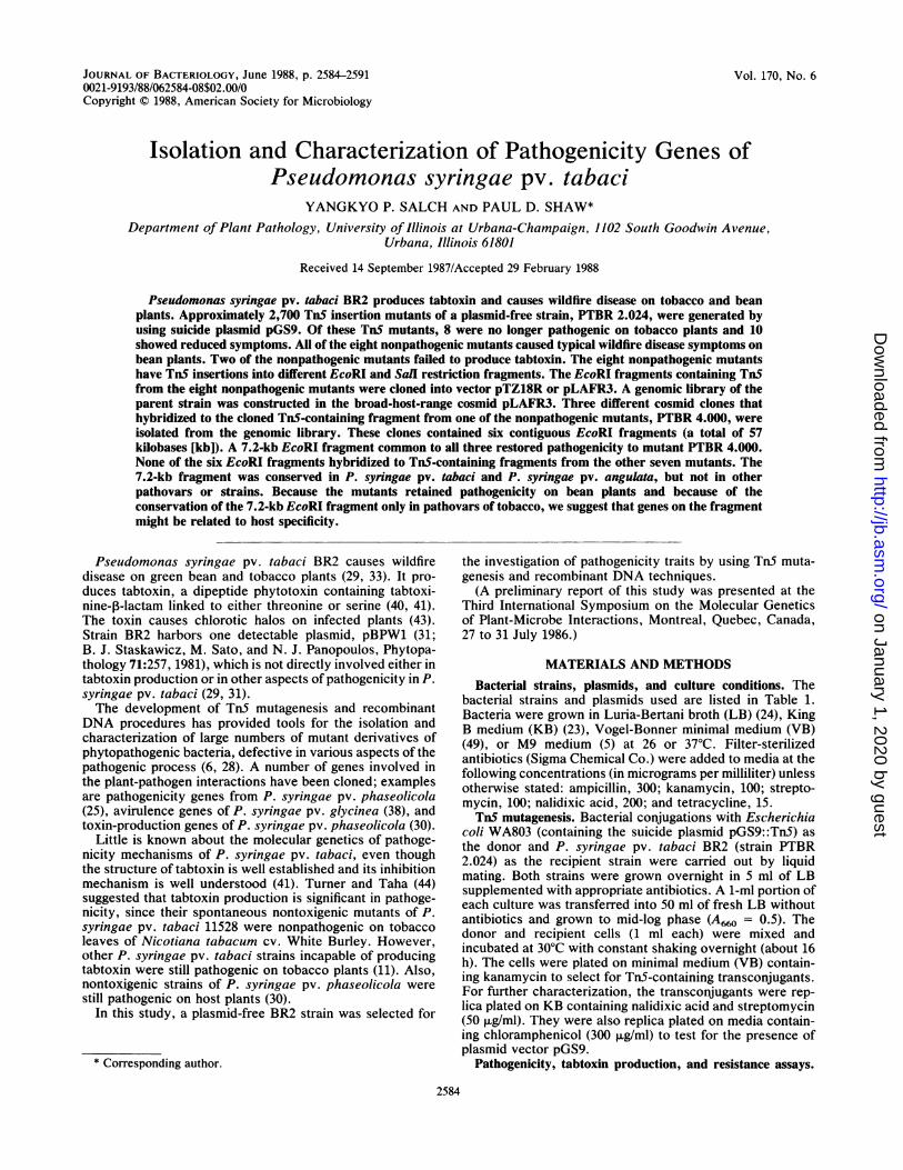
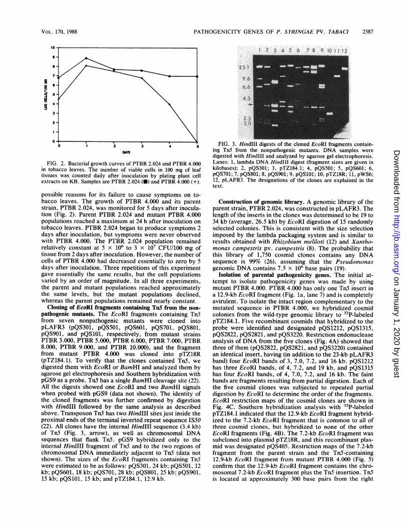
FIG. 2. Bacterial growth curves of PTBR 2.024 and PTBR 4.000in tobacco leaves. The number of viable cells in 100 mg of leaftissues was counted daily after inoculation by plating plant cellextracts on KB. Samples are PTBR 2.024 (U) and PTBR 4.000 (+).
possible reasons for its failure to cause symptoms on to-bacco leaves. The growth of PTBR 4.000 and its parentstrain, PTBR 2.024, was monitored for 5 days after inocula-tion (Fig. 2). Parent PTBR 2.024 and mutant PTBR 4.000populations reached a maximum at 24 h after inoculation ontobacco leaves. PTBR 2.024 began to produce symptoms 2days after inoculation, but symptoms were never observedwith PTBR 4.000. The PTBR 2.024 population remainedrelatively constant at 5 x 10⁶ to 3 x 10⁷ CFU/100 mg oftissue from 2 days after inoculation. However, the number ofcells of PTBR 4.000 had decreased essentially to zero by 5days after inoculation. Three repetitions of this experimentgave essentially the same results, but the cell populationsvaried by an order of magnitude. In all three experiments,the parent and mutant populations reached approximatelythe same levels, but the mutant populations declined,whereas the parent populations remained nearly constant.
Cloning of EcoRI fragments containing Tn5 from the non-pathogenic mutants. The EcoRI fragments containing Tn5from seven nonpathogenic mutants were cloned intopLAFR3 (pQS301, pQS501, pQS601, pQS701, pQS801,pQS901, and pQS101, respectively, from mutant strainsPTBR 3.000, PTBR 5.000, PTBR 6.000, PTBR 7.000, PTBR8.000, PTBR 9.000, and PTBR 10.000), and the fragmentfrom mutant PTBR 4.000 was cloned into pTZ18R(pTZ184.1). To verify that the clones contained Tn5, wedigested them with EcoRI or BamHI and analyzed them byagarose gel electrophoresis and Southern hybridization withpGS9 as a probe. Tn5 has a single BamHI cleavage site (22).All the digests showed one EcoRI and two BamHI signalswhen probed with pGS9 (data not shown). The identity ofthe cloned fragments was further confirmed by digestionwith HindIlI followed by the same analysis as describedabove. Transposon TnS has two HindlIl sites just inside theproximal ends of the terminal inverted repeat sequence IS50(22). All clones have the internal HindIll sequence (3.4 kb)of TnS (Fig. 3, arrow), as well as chromosomal DNAsequences that flank TnS. pGS9 hybridized only to theinternal HindlIl fragment of Tn5 and to the two regions ofchromosomal DNA immediately adjacent to Tn5 (data notshown). The sizes of the EcoRI fragments containing TnSwere estimated to be as follows: pQS301, 24 kb; pQS501, 12kb; pQS601, 18 kb; pQS701, 28 kb; pQS801, 25 kb; pQS901,15 kb; pQS101, 15 kb; and pTZ184.1, 12.9 kb.
1 2 3 4 5 6 78 9 101112
2 3.1
9.6
6.6
4.3
2.32.0
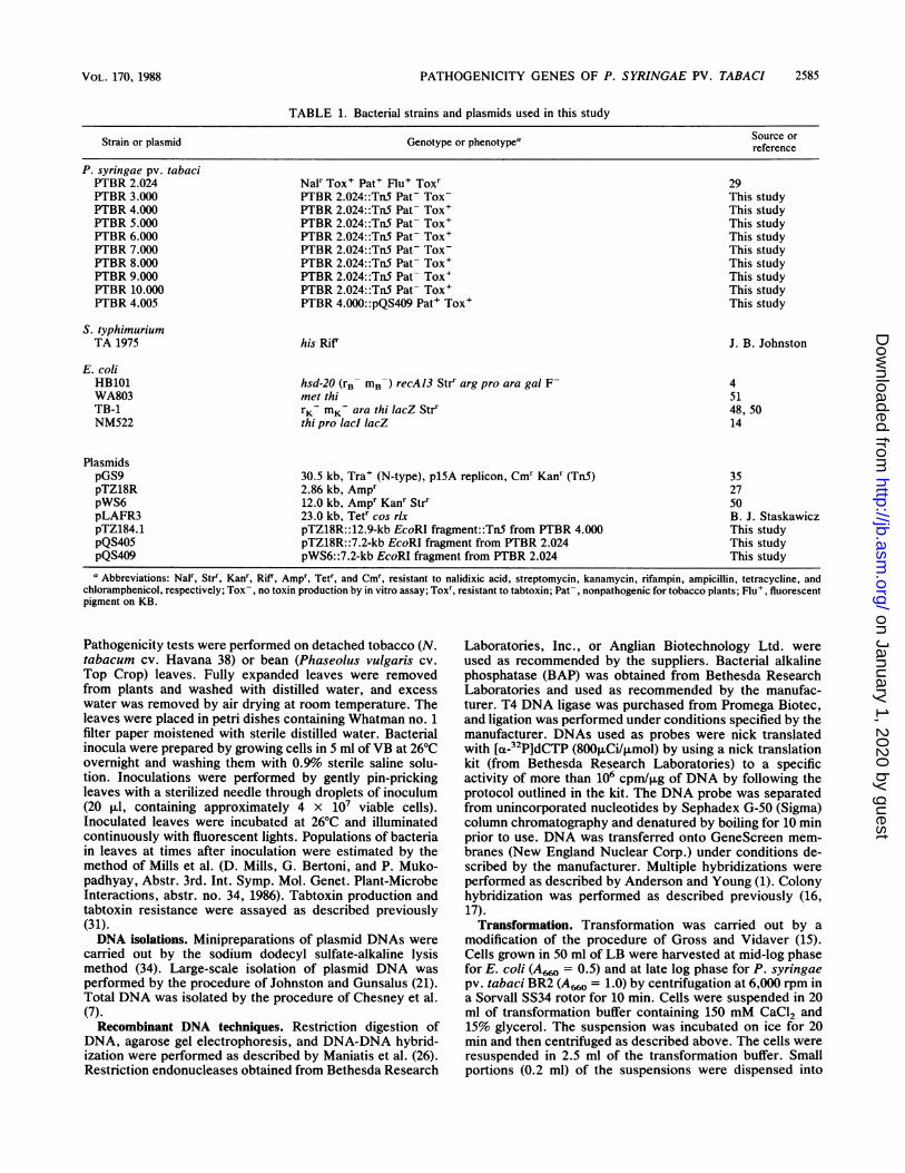
FIG. 3. HindIII digests of the cloned EcoRI fragments contain-ing Tn5 from the nonpathogenic mutants. DNA samples weredigested with Hindlll and analyzed by agarose gel electrophoresis.Lanes: 1, lambda DNA HindlIl digest (fragment sizes are given inkilobases); 2, pQS301; 3, pTZ184.1; 4, pQS501; 5, pQS601; 6,pQS701; 7, pQS801; 8, pQS901; 9, pQS101; 10, pTZ18R; 11, pWS6;12, pLAFR3. The designations of the clones are explained in thetext.
Construction of genomic library. A genomic library of theparent strain, PTBR 2.024, was constructed in pLAFR3. Thelength of the inserts in the clones was determined to be 19 to34 kb (average, 26.5 kb) by EcoRI digestion of 15 randomlyselected colonies. This is consistent with the size selectionimposed by the lambda packaging system and is similar toresults obtained with Rhizobium meliloti (12) and Xantho-monas campestris pv. campestris (8). The probability thatthis library of 1,750 cosmid clones contains any DNAsequence is 99% (26), assuming that the Pseudomonasgenomic DNA contains 7.5 x 10⁶ base pairs (19).
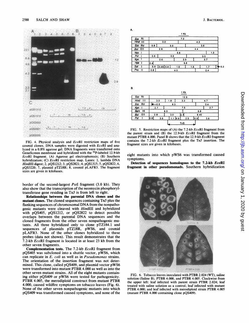
Isolation of parental pathogenicity genes. The initial at-tempt to isolate pathogenicity genes was made by usingmutant PTBR 4.000. PTBR 4.000 has only one TnS insert ina 12.9-kb EcoRI fragment (Fig. la, lane 7) and is completelyavirulent. To isolate the intact region complementary to themutated sequence in PTBR 4.000, we hybridized cosmidcolonies from the wild-type genomic library to 32P-labeledpTZ184.1. Five recombinant cosmids that hybridized to theprobe were identified and designated pQS1212, pQS1315,pQS2822, pQS2821, and pQS3220. Restriction endonucleaseanalysis of DNA from the five clones (Fig. 4A) showed thatthree of them (pQS2822, pQS2821, and pQS3220) containedan identical insert, having (in addition to the 23-kb pLAFR3band) four EcoRI bands of 3, 7.0, 7.2, and 16 kb. pQS1212has three EcoRI bands, of 4, 7.2, and 19 kb, and pQS1315has four EcoRI bands, of 4, 7.0, 7.2, and 16 kb. The faintbands are fragments resulting from partial digestion. Each ofthe five cosmid clones was subjected to repeated partialdigestion by EcoRI to determine the order of the fragments.EcoRI restriction maps of the cosmid clones are shown inFig. 4C. Southern hybridization analysis with 32P-labeledpTZ184.1 indicated that the 12.9-kb EcoRI fragment hybrid-ized to the 7.2-kb EcoRl fragment that is common to all ofthree cosmid clones, but hybridized to none of the otherEcoRI fragments (Fig. 4B). The 7.2-kb EcoRI fragment wassubcloned into plasmid pTZ18R, and this recombinant plas-mid was designated pQS405. Restriction maps of the 7.2-kbfragment from the parent strain and the TnS-containing12.9-kb EcoRI fragment from mutant PTBR 4.000 (Fig. 5)confirm that the 12.9-kb EcoRI fragment contains the chro-mosomal 7.2-kb EcoRI fragment plus the Tn5 insertion. Tn5is located at approximately 300 base pairs from the right
VOL. 170, 1988 2587
on January 1, 2020 by guesthttp://jb.asm
.org/D
ownloaded from
2588 SALCH AND SHAW
A.1 Kb
Ew RI 7.2Hind III 3.0 4.2Em RV 0.8 3.0 | 3.6Bst Ell 2.8 l 4.4 lIT 5.9 13
Pst 0.9 3.0 T 3.3__p_ r 2.6 2.0 2.7
I 0.41 6.8Sal I 0.9 10.6510.6 1.6 1 1.5 1 1.7PVu II 0.8 4.0 2.4
B.1 Kb
Ew RI 12.9Hind III 3.0 1.8 3.3 4.7Ew RV -0.5 8.3 3.4Kpn I 2.7 7.5 2.7Bam HI 6.3 6.8Bst Ell j 2.8 | 3.0 Io.71 6.45Pst I 0.81 3.0 1.1 l0.9 2.5 10.9 3.6
woS3220
FIG. 4. Physical analysis and EcoRI restriction maps of fivecosmid clones. DNA samples were digested with EcoRI and ana-
lyzed in a 0.8% agarose gel. DNA fragments were transferred ontoGeneScreen membrane and hybridized with the 32P-labeled 12.9-kbEcoRI fragment. (A) Agarose gel electrophoresis; (B) Southernhybridization; (C) EcoRI restriction map. Lanes: 1, lambda DNAHindIII digest; 2, pQS1212; 3, pQS2821; 4, pQS1315; 5, pQS2822; 6,pQS3220; 7, plasmid pTZ18R; 8, cosmid pLAFR3. The fragmentsizes are given in kilobases.
border of the second-largest PstI fragment (3.0 kb). Theyalso show that the transcription of the neomycin phosphoryl-transferase gene residing in TnS is from left to right.
Relationships between the parental DNA clones and themutant clones. The cloned sequences containing TnS plus theflanking sequences ofchromosomal DNA from the nonpatho-genic mutants were cleaved with HindlIl and hybridizedwith pQS405, pQS1212, or pQS2822 to detect possibleoverlaps between the parental DNA sequences and thecloned fragments from the other seven nonpathogenic mu-
tants. All three hybridized only to clone pTZ184.1 andsequences of plasmids pTZ18R, pWS6, and cosmidpLAFR3. None of the other clones hybridized to theseprobes (data not shown). This result demonstrates that the7.2-kb EcoRI fragment is located in at least 23 kb from theother seven fragments.Complementation tests. The 7.2-kb EcoRI fragment from
pQS405 was subcloned into a shuttle vector, pWS6, whichcan replicate in E. coli as well as in Pseudomonas strains.The orientation of the insertion fragment was not deter-mined. This clone, called pQS409, and plasmid vector pWS6were transformed into mutant PTBR 4.000 as well as into theother seven mutant strains. All of the eight mutants contain-ing either pQS409 or pWS6 were tested for pathogenicity.PTBR 4.005, the merodiploid construct from mutant PTBR4.000, caused wildfire symptoms on tobacco leaves (Fig. 6).None of the other seven nonpathogenic mutants into whichpQS409 was transformed caused symptoms, and none of the
FIG. 5. Restriction maps of (A) the 7.2-kb EcoRI fragment fromthe parent strain and (B) the 12.9-kb EcoRI fragment from themutant PTBR 4.000. Panel B shows that the 12.9-kb EcoRI fragmentcontains the 7.2-kb EcoRI fragment plus the TnS insertion. Thefragment sizes are given in kilobases.
eight mutants into which pWS6 was transformed causedsymptoms.
Detection of sequences homologous to the 7.2-kb EcoRIfragment in other pseudomonads. Southern hybridization
VI i 4 43* I:E
FIG. 6. Tobacco leaves inoculated with PTBR 2.024 (WT), salinesolution (Saline B), PTBR 4.000, and PTBR 4.005. Clockwise fromthe upper left: leaf infected with parent strain PTBR 2.024; leaftreated with saline solution as a control; leaf infected with mutantPTBR 4.000; and leaf infected with merodiploid strain PTBR 4.005(mutant PTBR 4.000 containing clone pQS409).
0.3
J. BACTERIOL.
4
ll.
rA
d0ft.as&
on January 1, 2020 by guesthttp://jb.asm
.org/D
ownloaded from
PATHOGENICITY GENES OF P. SYRINGAE PV. TABACI
TABLE 2. Pseudomonas strains used in homology test
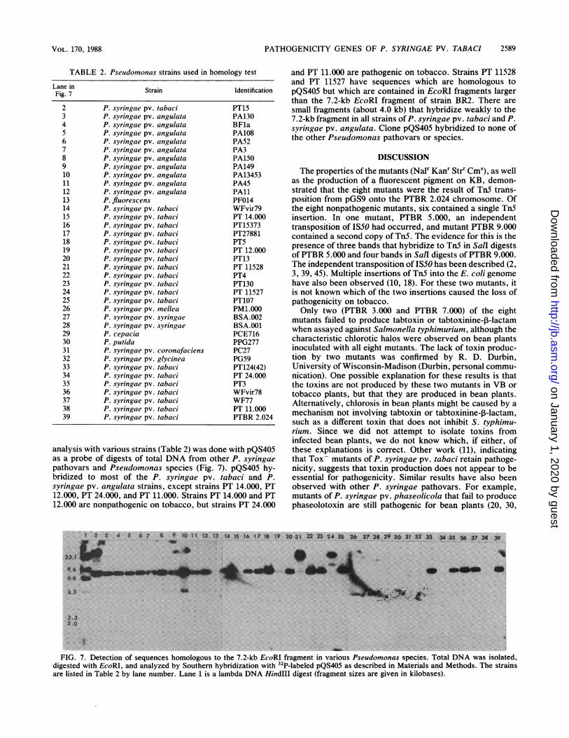
Lane in Strain IdentificationFig. 7
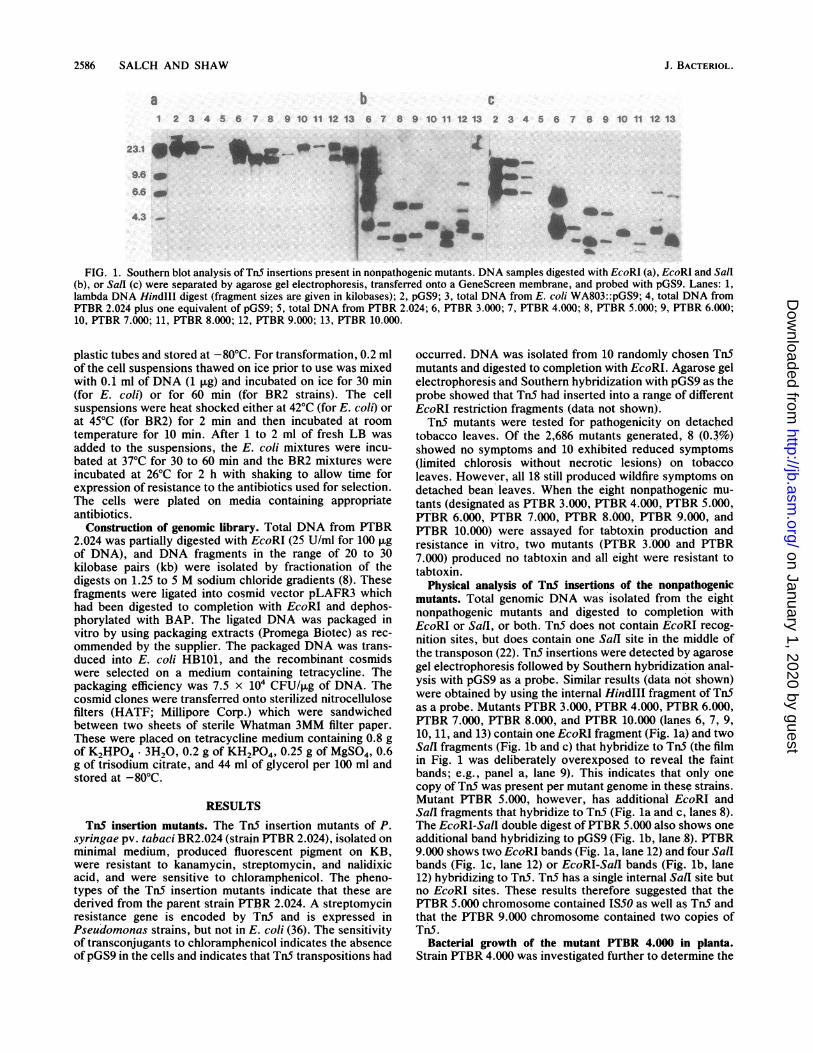
2 P. syringae pv. tabaci PT153 P. syringae pv. angulata PA1304 P. syringae pv. angulata BF1a5 P. syringae pv. angulata PA1086 P. syringae pv. angulata PA527 P. syringae pv. angulata PA38 P. syringae pv. angulata PA1509 P. syringae pv. angulata PA14910 P. syringae pv. angulata PA1345311 P. syringae pv. angulata PA4512 P. syringae pv. angulata PAll13 P. fluorescens PF01414 P. syringae pv. tabaci WFvir7915 P. syringae pv. tabaci PT 14.00016 P. syringae pv. tabaci PT1537317 P. syringae pv. tabaci PT2788118 P. syringae pv. tabaci PT519 P. syringae pv. tabaci PT 12.00020 P. syringae pv. tabaci PT1321 P. syringae pv. tabaci PT 1152822 P. syringae pv. tabaci PT423 P. syringae pv. tabaci PT13024 P. syringae pv. tabaci PT 1152725 P. syringae pv. tabaci PT10726 P. syringae pv. mellea PM1.00027 P. syringae pv. syringae BSA.00228 P. syringae pv. syringae BSA.00129 P. cepacia PCE71630 P. putida PPG27731 P. syringae pv. coronafaciens PC2732 P. syringae pv. glycinea PG5933 P. syringae pv. tabaci PT124(42)34 P. syringae pv. tabaci PT 24.00035 P. syringae pv. tabaci PT336 P. syringae pv. tabaci WFvir7837 P. syringae pv. tabaci WF7738 P. syringae pv. tabaci PT 11.00039 P. syringae pv. tabaci PTBR 2.024
analysis with various strains (Table 2) was done with pQS405as a probe of digests of total DNA from other P. syringaepathovars and Pseudomonas species (Fig. 7). pQS405 hy-bridized to most of the P. syringae pv. tabaci and P.syringae pv. angulata strains, except strains PT 14.000, PT12.000, PT 24.000, and PT 11.000. Strains PT 14.000 and PT12.000 are nonpathogenic on tobacco, but strains PT 24.000
and PT 11.000 are pathogenic on tobacco. Strains PT 11528and PT 11527 have sequences which are homologous topQS405 but which are contained in EcoRI fragments largerthan the 7.2-kb EcoRI fragment of strain BR2. There aresmall fragments (about 4.0 kb) that hybridize weakly to the7.2-kb fragment in all strains of P. syringae pv. tabaci and P.syringae pv. angulata. Clone pQS405 hybridized to none ofthe other Pseudomonas pathovars or species.
DISCUSSION
The properties of the mutants (Nalr Kanr Strr Cms), as wellas the production of a fluorescent pigment on KB, demon-strated that the eight mutants were the result of TnS trans-position from pGS9 onto the PTBR 2.024 chromosome. Ofthe eight nonpathogenic mutants, six contained a single Tn5insertion. In one mutant, PTBR 5.000, an independenttransposition of IS50 had occurred, and mutant PTBR 9.000contained a second copy of TnS. The evidence for this is thepresence of three bands that hybridize to TnS in Sall digestsofPTBR 5.000 and four bands in SalI digests of PTBR 9.000.The independent transposition of ISS0 has been described (2,3, 39, 45). Multiple insertions of TnS into the E. coli genomehave also been observed (10, 18). For these two mutants, itis not known which of the two insertions caused the loss ofpathogenicity on tobacco.Only two (PTBR 3.000 and PTBR 7.000) of the eight
mutants failed to produce tabtoxin or tabtoxinine-p-lactamwhen assayed against Salmonella typhimurium, although thecharacteristic chlorotic halos were observed on bean plantsinoculated with all eight mutants. The lack of toxin produc-tion by two mutants was confirmed by R. D. Durbin,University of Wisconsin-Madison (Durbin, personal commu-nication). One possible explanation for these results is thatthe toxins are not produced by these two mutants in VB ortobacco plants, but that they are produced in bean plants.Alternatively, chlorosis in bean plants might be caused by amechanism not involving tabtoxin or tabtoxinine-p-lactam,such as a different toxin that does not inhibit S. typhimu-rium. Since we did not attempt to isolate toxins frominfected bean plants, we do not know which, if either, ofthese explanations is correct. Other work (11), indicatingthat Tox- mutants of P. syringae pv. tabaci retain pathoge-nicity, suggests that toxin production does not appear to beessential for pathogenicity. Similar results have also beenobserved with other P. syringae pathovars. For example,mutants of P. syringae pv. phaseolicola that fail to producephaseolotoxin are still pathogenic for bean plants (20, 30,
1 2 3 4 5 6 7 8 9 10 11 12 13 14 S 16 17 IS 19 20 21 22 24 2s5 25 27 29X3 31 32 33 X35 36 37 38 3.........
g
A.3 -'
2.32.0
d
FIG. 7. Detection of sequences homologous to the 7.2-kb EcoRI fragment in various Pseudomonas species. Total DNA was isolated,digested with EcoRI, and analyzed by Southern hybridization with 32P-labeled pQS405 as described in Materials and Methods. The strainsare listed in Table 2 by lane number. Lane 1 is a lambda DNA Hindlll digest (fragment sizes are given in kilobases).
2589VOL. 170, 1988
on January 1, 2020 by guesthttp://jb.asm
.org/D
ownloaded from
2590 SALCH AND SHAW
32). There are conflicting results (44), however, which sug-gest that Tox- mutants of P. syringae pv. tabaci are stillpathogenic. Our results indicate that toxin production bythat organism does not necessarily lead to the production ofsymptoms. An alternative to the direct role of toxins indisease is the suggestion that toxins are produced by phyto-pathogenic bacteria to prevent coinfection of a plant byrelated bacteria (37, 47).PTBR 4.000 penetrates and multiplies in intercellular
spaces of tobacco leaf tissue to approximately the same levelas PTBR 2.024 does, but in contrast to the parent, it does notsurvive (Fig. 5). One possible explanation is that an initialsource of nutrients is available to the mutant, but that this iseventually exhausted, and other nutrient sources that can beaccessed by the parent are not available to the mutant,whose population therefore declines. An alternative expla-nation is that the mutants are unable to protect themselvesfrom plant defense mechanisms. Our results suggest that theproduction of disease symptoms is not dependent solely onthe size of the bacterial population in the leaves, but mightrequire that the population be maintained for some period. Adecline in the number of viable cells of the nonpathogenicmutants of P. syringae pv. syringae (Mills et al., Abstr. 3rdInt. Symp. Mol. Genet. Plant-Microbe Interactions) in hosttissues has also been observed. However, there are otherexamples (25, 42, 45) of nonpathogenic mutants of P. sy-ringae pathovars that appear to be unable to reach thepopulation levels of their pathogenic parents in plant tissues,and so in these instances, the population level might be afactor in symptom production.Lack of hybridization of the six EcoRI fragments in clones
pQS405, pQS1212, and pQS2822 to the TnS-containing frag-ments from the other seven mutants demonstrates that the7.2-kb EcoRI fragment is located at least 23 kb in onedirection and 26 kb in the other direction from the otherseven mutants. Since sequences complementary to the otherseven mutant sequences have not been isolated from theparental genomic library, we cannot comment on the orga-nization of the other Tn5-containing sequences. Thus, unlikethe pathogenicity genes from P. syringae pv. phaseolicola(25) and from X. campestris pv. campestris (46), which areclustered, at least one of the genes in PTBR 2.024 is removedfrom the others. pQS409, which contains the parental 7.2-kbfragment, restored the pathogenicity of mutant PITBR 4.000on tobacco by complementation tests, but did not comple-ment the other seven mutants. This supports the structuralevidence indicating that none of the other mutations map inclose proximity to the 7.2-kb EcoRI fragment. The isolationof parental DNA clones complementary to the other sevenmutants and cross-hybridization among those clones willreveal the organization of those genes.
Hybridization indicated that only strains of P. syringaepv. tabaci and P. syringae pv. angulata contain sequenceshomologous to the 7.2-kb EcoRI fragment from PTBR 2.024.In most strains, hybridization occurred to fragments of thesame size, but in two strains, hybridization took place withlarger EcoRI fragments (about 17 kb). This indicates thatsequences on the 7.2-kb EcoRI fragment are conserved instrains of P. syringae pv. tabaci and P. syringae pv. angu-lata, but there are sequence polymorphisms among thestrains. The weak hybridization to the small fragment (4.0kb) suggests the possibility of repeated sequences in the7.2-kb EcoRI fragment. Two of the P. syringae pv. tabacistrains that do not contain sequences homologous to the7.2-kb EcoRI fragment were nonpathogenic on tobacco.However, two other strains lacking the homology retained
pathogenicity on tobacco. There are no obvious explana-tions for the lack of hybridization, but it might suggest thatthe gene(s) on the 7.2-kb EcoRI fragment are not essentialfor pathogenesis in certain strains or that those strains havealternative mechanisms for overcoming the absence of thegene(s). Since most of the isolates of P. syringae pv. tabaciand P. syringae pv. angulata we tested were isolated fromtobacco plants in the field, the 7.2-kb EcoRI fragment mayencode host specificity functions (13) on tobacco. In addi-tion, the observation that all of the eight mutants causedwildfire symptoms indistinguishable from those caused bythe parent on bean plants supports the suggestion that all ofthe eight mutated genes are involved in host specificity ontobacco plants. It is likely that the mechanism of hostspecificity is a complex process and involves many genesincluding host range and avirulence genes (13). For example,Daniels et al. (9) failed to isolate a pathovar specificitygene(s) from X. campestris pathovars by cross-transferringcosmid clones from genomic libraries.The results obtained from the present study suggest that
the genes inactivated by TnS insertion of eight mutants arespecifically involved only in the pathogenicity on tobacco.This hypothesis must be supported by the isolation of a set ofgenes involved in host specificity on bean plants that isdifferent from those involved in host specificity on tobaccoplants when TnS insertion mutants are initially screened onbean leaves.
ACKNOWLEDGMENTS
We thank V. N. Iyer, B. J. Staskawicz, B. W. Kemper, and M. A.Schuler for generous gifts of plasmid vectors and strains. We arealso indebted to R. D. Durbin for the tabtoxin assay of the toxinmutants. We acknowledge A. Hepburn and S. Farrand for theircomments and suggestions in preparing the manuscript.
This work was funded in part by Biomedical Research SupportGrant PHS RR-7030 and by U.S. Department of Agriculture Com-petitive Grant 86-CRCR-1-2237.
LITERATURE CITED1. Anderson, M. L. M., and B. D. Young. 1985. Quantitative filter
hybridization, p. 73-111. In B. D. Hames and S. J. Higgins(ed.), Nucleic acid hybridization: a practical approach. IRLPress, Oxford.
2. Berg, D. E., L. Johnsrud, L. McDivitt, R. Ramabhadran, andB. J. Hirschel. 1982. Inverted repeats of TnS are transposableelements. Proc. Natl. Acad. Sci. USA 79:2632-2635.
3. Boucher, C., P. Barberis, A. Trigalet, and D. Demery. 1985.Transposon mutagenesis of Pseudomonas solanacearum: isola-tion of TnS-induced avirulent mutants. J. Gen. Microbiol.131:2449-2457.
4. Boyer, H. W., and D. Rouland-Dussoix. 1969. A complementa-tion analysis for the restriction and modification of DNA inEscherichia coli. J. Mol. Biol. 41:459-472.
5. Chang, A. C. Y., and S. N. Cohen. 1978. Construction andcharacterization of amplifiable multicopy DNA cloning vehiclesderived from the plSA cryptic miniplasmid. J. Bacteriol.134:1141-1156.
6. Chatterjee, A. K., and A. K. Vidaver. 1986. Genetics of patho-genicity factors: application to phytopathogenic bacteria. Aca-demic Press, Inc., New York.
7. Chesney, R. H., J. R. Scott, and D. Vapnek. 1979. Integration ofthe plasmid prophages P1 and P7 into the chromosome ofEscherichia coli. J. Mol. Biol. 130:161-173.
8. Daniels, M. J., C. E. Barber, P. C. Turner, M. K. Sawczyc,R. J. W. Byrde, and A. H. Fielding. 1984. Cloning of genesinvolved in pathogenicity of Xanthomonas campestris pv. cam-pestris using the broad-host range cosmid pLAFR1. EMBO J.3:3323-3328.
9. Daniels, M. J., P. C. Turner, C. E. Barber, M. K. Sawczyc, and
J. BACTERIOL.
on January 1, 2020 by guesthttp://jb.asm
.org/D
ownloaded from
PATHOGENICITY GENES OF P. SYRINGAE PV. TABACI
F. Dums. 1985. Molecular pathology of Xanthomonas, p.125-133. In John Innes Institute 73rd report covering the 2 years
1983-1984. The John Innes Institute, Norfolk, England.10. De Bruijn, F. J., and J. R. Lupski. 1984. The use of transposon
TnS mutagenesis in the rapid generation of correlated physicaland genetic maps of DNA segments cloned into multicopyplasmids a review. Gene 27:131-149.
11. Durbin, R. D. 1971. Chlorosis-inducing pseudomonad toxins:their mechanisms of action and structure, p. 369-385. In S. Akaiand S. Ouchi (ed.), Morphological and biochemical events inplant-parasite interactions. Mochizuki Publishing Co., Tokyo,Japan.
12. Friedman, A. M., S. R. Long, S. E. Brown, W. J. Buikema, andF. M. Ausubel. 1982. Construction of a broad host range cosmidvector and its use in the genetic analysis of Rhizobium mutants.Gene 18:289-296.
13. Gabriel, D. W. 1986. Specificity and gene function in plant-pathogen interactions. ASM News 52:19-25.
14. Gough, J. A., and N. E. Murray. 1983. Sequence diversityamong related genes for recognition of targets in DNA mole-cules. J. Mol. Biol. 166:1-19.
15. Gross, D. C., and A. K. Vidavar. 1981. Transformation ofPseudomonas syringae with nonconjugative R plasmids. Can. J.Microbiol. 27:759-765.
16. Grunstein, M., and D. S. Hogness. 1975. Colony hybridization: a
method for the isolation of cloned DNAs that contain a specificgene. Proc. Natl. Acad. Sci. USA 72:3%1-3%5.
17. Hanahan, D., and M. Meselson. 1980. Plasmid screening at highcolony density. Gene 10:63-67.
18. Harayama, S., E. T. Palva, and G. L. Hazelbauer. 1979. Trans-poson insertion mutants of Escherichia coli K-12 defective in a
component common to galactose and ribose chemotaxis. Mol.Gen. Genet. 171:193-203.
19. Holloway, B. W. 1974. Genetic organization of Pseudomonas, p.
133-161. In P. H. Clarke and M. H. Richmond (ed.), Geneticsand biochemistry of Pseudomonas. John Wiley & Sons, Inc.,New York.
20. Jamieson, A. F., R. L. Bieleski, and R. E. Mitchell. 1981.Plasmids and phaseolotoxin production in Pseudomonas sy-
ringae pv. phaseolicola. J. Gen. Microbiol. 122:161-165.21. Johnston, J. B., and I. C. Gunsalus. 1977. Isolation of metabolic
plasmid DNA from Pseudomonas putida. Biochem. Biophys.Res. Commun. 75:13-17.
22. Jorgensen, R. A., S. J. Rothstein, and W. S. Reznikoff. 1979. Arestriction enzyme cleavage map of TnS and location of a regionencoding neomycin resistance. Mol. Gen. Genet. 177:65-72.
23. King, E. O., M. K. Ward, and D. E. Raney. 1954. Two simplemedia for the demonstration of pyrocyanin and fluorescein. J.Lab. Clin. Med. 44:301-313.
24. Lennox, E. S. 1955. Transduction of linked genetic characters ofthe host bacteriophage P1. Virology 1:190-206.
25. Lindgren, P. B., R. C. Peet, and N. J. Panopoulos. 1986. Genecluster of Pseudomonas syringae pv. "phaseolicola" controlspathogenicity of bean plants and hypersensitivity on nonhostplants. J. Bacteriol. 168:512-522.
26. Maniatis, T., E. F. Fritsch, and J. Sambrook. 1982. Molecularcloning: a laboratory manual. Cold Spring Harbor Laboratory,Cold Spring Harbor, N.Y.
27. Mead, D. A., E. Szczesna-Skorupa, and B. Kemper. 1986.Single-stranded DNA "blue" T7 promoter plasmids: a versatiletandem promoter system for cloning and protein engineering.Protein Eng. 1:67-74.
28. Mills, D. 1985. Transposon mutagenesis and its potential forstudying virulence genes in plant pathogens. Annu. Rev. Phy-topathol. 23:297-320.
29. Obukowicz, M., and P. D. Shaw. 1985. Construction of Tn3-containing plasmids from plant-pathogenic pseudomonads andan examination of their biological properties. Appl. Environ.Microbiol. 49:468-473.
30. Peet, R. C., P. B. Lindgren, D. K. Willis, and N. J. Panopoulos.1986. Identification and cloning of genes involved in phaseolo-toxin production by Pseudomonas syringae pv. "phaseo-licola." J. Bacteriol. 166:1096-1105.
31. Piwowarski, J. M., and P. D. Shaw. 1982. Characterization ofplasmids from plant pathogenic pseudomonads. Plasmid 7:85-94.
32. Quigley, N. B., D. Lane, and P. L. Bergquist. 1985. Genes forphaseolotoxin synthesis are located on the chromosome ofPseudomonas syringae pv. phaseolicola. Curr. Microbiol. 12:295-300.
33. Ribeiro, R. de L. D., D. J. Hagedorn, R. D. Durbin, and T. F.Uchytil. 1979. Characterization of the bacterium inciting beanwildfire in Brazil. Phytopathology 69:208-212.
34. Rodriguez, R. L., and R. C. Tait. 1983. Recombinant DNAtechniques: an introduction, p. 50-51. The Benjamin/CummingsPublishing Company, Inc., Reading, Mass.
35. Selvaraj, G., and V. N. Iyer. 1983. Suicide plasmid vehicles forinsertion mutagenesis in Rhizobium meliloti and related bacte-ria. J. Bacteriol. 156:1292-1300.
36. Selvaraj, G., and V. N. Iyer. 1984. Transposon TnS specifiesstreptomycin resistance in Rhizobium spp. J. Bacteriol. 158:580-589.
37. Smidt, M. L., and A. K. Vidaver. 1982. Bacteriocin productionby Pseudomonas syringae PsW-1 in plant tissue. Can. J. Micro-biol. 28:600-604.
38. Staskawicz, B. J., D. Dahlbeck, and N. T. Keen. 1984. Clonedavirulence gene of Pseudomonas syringae pv. glycinea deter-mines race-specific incompatibility on Glycine max (L.) Merr.Proc. Natl. Acad. Sci. USA 81:6024-6028.
39. Staskawicz, B. J., D. Dahlbeck, J. Miller, and D. Damm. 1983.Molecular analysis of virulence genes in Pseudomonas solana-cearum, p. 345-350. In A. Puhler (ed.), Molecular genetics ofthe bacteria-plant interaction. Springer-Verlag KG, Berlin.
40. Stewart, W. W. 1971. Isolation and proof of structure of wildfiretoxin. Nature (London) 229:174-178.
41. Taylor, P. A., H. K. Schnoes, and R. D. Durbin. 1972. Charac-terization of chlorosis-inducing toxins from Pseudomonas sp.Biochim. Biophys. Acta 286:107-117.
42. Trigalet, A., and D. Demery. 1986. Invasiveness in tomatoplants of TnS-induced avirulent mutants of Pseudomonas sola-nacearum. Physiol. Mol. Plant Pathol. 28:423-430.
43. Turner, J. G. 1981. Tabtoxin, produced by Pseudomonas ta-baci, decreases Nicotiana tabacum glutamine synthetase andcauses accumulation of ammonia. Physiol. Plant Pathol.19:57-67.
44. Turner, J. G., and R. R. Taha. 1984. Contribution of tabtoxin tothe pathogenicity of Pseudomonas syringae pv. tabaci. Physiol.Plant Pathol. 25:55-69.
45. Turner, P., C. Barber, and M. Daniels. 1984. Behavior of thetransposons TnS and Tn7 in Xanthomonas campestris pv.campestris. Mol. Gen. Genet. 195:101-107.
46. Turner, P., C. Barber, and M. Daniels. 1985. Evidence forclustered pathogenicity genes in Xanthomonas campestris pv.campestris. Mol. Gen. Genet. 199:338-343.
47. Vidaver, A. K. 1976. Prospects for control of phytopathogenicbacteria by bacteriophages and bacteriocins. Annu. Rev. Phy-topathol. 14:451-465.
48. Vieira, J., and J. Messing. 1982. The pUC plasmids, anM13mp7-derived system for insertion mutagenesis and sequenc-ing with synthetic universal primers. Gene 19:259-268.
49. Vogel, H. J., and D. M. Bonner. 1956. Acetyl ornithinase ofEscherichia coli: partial purification and some properties. J.Biol. Chem. 218:97-106.
50. Werneke, J. M., S. G. Sligar, and M. A. Schuler. 1985. Devel-opment of broad-host-range vectors for expression of clonedgenes in Pseudomonas. Gene 38:73-84.
51. Wood, W. B. 1966. Host specificity of DNA produced byEscherichia coli: bacterial mutations affecting the restrictionand modification of DNA. J. Mol. Biol. 16:118-133.
2591VOL. 170, 1988
on January 1, 2020 by guesthttp://jb.asm
.org/D
ownloaded from