Embed Size (px)
Citation preview
JOURNAL OF BACTERIOLOGY, July 1970, p. 159-165 Vol. 103, No. 1Copyright 0 1970 American Society for Microbiology Printed in U.S.A
Isolation and Analysis of the Nucleic Acids andPolysaccharides from Clostridium welchii
G. K. DARBY, A. S. JONES, J. F. KENNEDY, AND R. T. WALKERDepartmentt of Chiemistry, Birmingh1am University, Edgbastoni, Birminighlam 15, Eniglaild
Received for publication 24 February 1970
A method previously described for the use of bentonite in the isolation of thenucleic acids from two gram-positive organisms was applied to the isolation of thenucleic acids from two strains of Clostridium welchii. The nucleic acids were sepa-rated from polysaccharides by the fractional precipitation of their cetyltrimethyl-ammonium salts from sodium chloride solution, and the base composition of thenucleic acids was determined. One strain of C. welchii investigated (NCTC 10578)was shown to produce considerable quantities of an acidic and also a weakly acidicor neutral polysaccharide; the other strain (ATCC 10543) gave very small quantitiesof the latter but none of the former polysaccharide. The monosaccharide com-position of these polysaccharides was determined and the acidic polysaccharidewas shown to resemble dermatan sulfate.
The isolation of nucleic acids from gram-posi-tive microorganisms is more difficult than thecorresponding isolation from gram-negativemicroorganisms, because of the difficulty en-countered in rupturing the cell walls of the formerwithout causing enzymatic and mechanicaldegradation of the nucleic acids. A procedurewhich can be used for many gram-positive bac-teria is that described by Marmur (19) in whichthe cell wall is first ruptured by the use of lyso-zyme and the nucleic acids are extracted withsodium dodecyl sulfate. However, some gram-positive bacteria are resistant to the action oflysozyme. Clostridium welchii is susceptible to theaction of lysozyme and the nucleic acids wereobtained from this organism by the method ofMarmur, but preliminary attempts at the isolationof nucleic acids from our strain of this bacteriumby the use of lysozyme failed, partly because thedeoxyribonucleic acid (DNA) was degraded to anextent which interfered with the separation ofDNA from ribonucleic acid (RNA) by thefractionation of their cetyltrimethylammoniumsalts. We therefore applied to the isolation of thenucleic acids of this organism a procedure previ-ously described (13), which involves the use ofbentonite and mechanical breakage of the orga-nisms followed by the isolation of RNA, DNA,and polysaccharides.
MATERIALS AND METHODSBentonite. A suspension of bentonite in 0.01 M
acetate buffer, pH 6 was prepared as previouslydescribed (5).
Strains and conditions of growth. The following twostrains of C. welchii were used: (i) a strain which has
been in our department for over 10 years and has nowbeen deposited in the National Collection of TypeCultures, NCTC 10578 type A (Central Public HealthLaboratory, London); (ii) C. welchii type A, NCIB8875 (also ATCC 10543). Both organisms were grownin Robertson cooked meat medium which had beenheated to 100 C and subsequently cooled to 37 Cbefore inoculation. The inoculum was incubated at37 C for 18 hr and then used to inoculate 500 ml of amedium containing 0.1% Lab-Lemco beef extract,0.2% Oxoid yeast extract, 0.5% Oxoid peptone, and0.5% NaCl. The air was expelled from this mediumby heating it in loosely closed screw-capped bottles inan autoclave for 15 min at 115 C. The medium wasrapidly cooled; 0.5% sterile glucose solution wasadded, followed by 2 ml of inoculum; the bottle wasfilled to the top with more medium from which the airhad been expelled, and the whole was incubated at 37C for 18 hr.
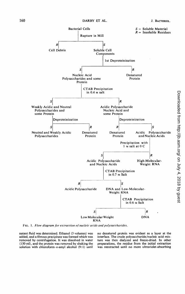
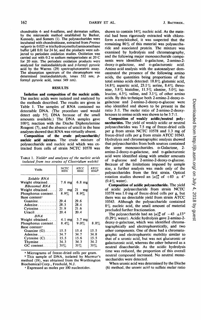
Harvesting, cell breakage, and extraction of solublecell components. A flow diagram of the extraction andfractionation procedures is shown in Fig. 1. The cellsfrom 20 liters of medium were obtained by centrifuga-tion of the medium at 0 C at 23,000 X g. The cellswere washed with 0.14 M NaCl solution, and added tothe vessel of a rotary ball mill (capacity 1 liter) with200 ml of 0.01 M acetate buffer (pH 6). Ballotinibeads (no. 5, 150 ml) and bentonite suspension (100ml) were added, and the mixture was shaken at 4 Cfor periods of 1 hr. The mixture was then centrifugedat 23,000 X g, the supernatant fluid was removed, andthe residue was shaken with fresh buffer. The amountof nucleic acid in the supematant fluid could be deter-mined directly from the absorbancy of the solution at260 nm. In one case the initial extract of cells from 20liters of culture of strain NCTC 10578 was examinedseparately. After shaking the cells for 1 hr at 4 C in arotary ball mill as described above, the cell debris wasremoved and the quantity of nucleic acid in the super-
159
on July 4, 2018 by guesthttp://jb.asm
.org/D
ownloaded from
DARBY ET AL.
R
Cell Debris
Bacterial Cells
Rupture in Mill
S
Soluble CellComponents
S
Nucleic AcidPolysaccharides and some
Protein
CTAB Precilin 0.4 M sa
S = Soluble MaterialR = Insoluble Residues
1st Deproteinization
R
DenaturedProtein
'pitationlIt
S
Weakly Acidic and NeutralPolysaccharides andsome Protein
Deproteinization
S
Neutral and Weakly AcidicPolysaccharides
R
Acidic PolysaccharideNucleic Acid andsome Protein
Deproteinization
R
DenaturedProtein
R
DenaturedProtein
S
Acidic Polysaccharideand Nucleic Acids
Precipitation withI M salt at 0 C
sAcidic Polysaccharideand Nucleic Acids
R
Acidic Polysaccharide
R
High-Molecular-Weight RNA
CTAB Precipitationin 0.7 M Salt
S
DNA and Low-Molecular-Weight RNA
CTAB Precipitationin 0.6 M Salt
s
Low Molecular WeightRNA
FIG. 1. Flow diagram for extractiont of nuicleic acids aid polysaccharides.
natant fluid was determined. Ethanol (3 volumes) wasadded, and a fibrous precipitate was formed which wasremoved by centrifugation. It was dissolved in water(150 ml), and the protein was removed by shaking thesolution with chloroform-n-amyl alcohol (9:1) until
no denatured protein was evident as a layer at theinterface. The crude polysaccharide/nucleic acid mix-ture was then dialyzed and freeze-dried. In otherpreparations, the residue from the initial extractionwas reextracted until no more ultraviolet-absorbing
R
DNA
160 J. BACTERIOL.
on July 4, 2018 by guesthttp://jb.asm
.org/D
ownloaded from
VOL. 103, 1970 NUCLEIC ACIDS F
material could be obtained (usually five or six extrac-tions were required); the material was precipitatedfrom the combined extracts with ethanol and depro-teinized as described above.
Fractionation of the macromolecular constituents.The deproteinized solution obtained above was made0.4 M with respect to sodium chloride, and cetyltri-methylammonium bromide (CTAB) was added untilprecipitation was complete. The precipitate of thecetyltrimethylammonium salt of the nucleic acids andany highly acidic polysaccharide were removed bycentrifugation, and the neutral and weakly acidicmaterial was precipitated from the supernatant fluidby the addition of ethanol (3 volumes). This latterprecipitate was dissolved in water to give a viscoussolution; it was dialyzed against water and freeze-dried.The cetyltrimethylammonium salt of the acidic
material was dissolved in 1 M NaCl, and the sodiumsalt was precipitated by the addition of ethanol (3volumes). The ribosomal RNA was precipitated asthe sodium salt from 1 M NaCl solution as previouslydescribed (13). The salt concentration was then low-ered to 0.7 M, and CTAB was added so that the finalCTAB concentration was 1%. The precipitate of thecetyltrimethylammonium salt of the acidic polysac-charides was removed by centrifugation, convertedinto the sodium salt in the normal way, dialyzedagainst water, and freeze-dried. The sodium chlorideconcentration of the solution was then reduced to 0.6M, and the cetyltrimethylammonium salt of the DNAwas removed by centrifugation and recovered in theusual way (13). Ethanol was added to the supernatantfraction from the 0.6 M NaCI-CTAB precipitation,and the sodium salts of the RNA soluble in 1 M sodiumchloride was recovered.
Analysis of nucleic acids. The total phosphoruscontent was estimated by the method of Jones et al.(11).The purine and pyrimidine contents of the nucleic
acids were determined as follows. DNA samples werehydrolyzed as described by Wyatt and Cohen (31)and the bases were determined as described by Wyatt(30). RNA samples were hydrolyzed and the baseswere determined as described by Markham and Smith(18) after separating the bases in the solvent systemdescribed by Kirby (15).
Analysis of polysaccharides. Polysaccharide prepa-rations were hydrolyzed for component identificationby dissolving them in 2 N HCl (3 mg/ml) and heatingat 100 C for 3 hr. The hydrolysates were evaporatedto dryness under reduced pressure and stored in avacuum desiccator over solid sodium hydroxide toremove final traces of HCl. Residues were separatedby ascending cellulose thin-layer chromatography inpyridine-ethyl acetate-acetic acid-water (5:5:3:1).Paper electrophoretograms of the residues were devel-oped on Whatman no. 2 paper, 0.05 M formate buffer(pH 3.5), with a potential of 20 v/cm. Componentswere revealed by the following methods: ultraviolet-absorbing materials by photography or visual exam-
ination in ultraviolet light; hexoses and hexuronicacids by silver nitrate or aniline hydrogen phthalate
'ROM C. WELCHII 161
sprays; and 2-amino-2-deoxyhexoses by the ninhydrinspray.
The crude mixture containing polysaccharide andnucleic acid from cells of strain NCTC 10578 wasexamined to determine its content of amino acid and2-amino-2-deoxyhexose. Amino acids and 2-amino-2-deoxyhexoses were liberated from the polysaccharide/nucleic acid mixture by the method of Spackman et al.(26)-hydrolysis in 6 N HCl at 110 C for 22 hr, withnorleucine used as an internal control for the purposeof correcting for destruction during hydrolysis andtransference loss. The composition of the hydrolysateswas determined by using a Technicon AutomaticAmino Acid Analyzer calibrated with a standardmixture containing 20 naturally occurring aminoacids, norleucine, 2-amino-2-deoxy-D-galactose, and2-amino-2-deoxy-D-glucose (0.05 ,mole of each).The polysaccharide fractions were hydrolyzed in
3.9 N HCl at 100 C for 9 hr, and the liberated 2-amino-2-deoxy-hexoses were determined by using the Sven-nerholm (27) modification of the Elson-Morganreaction (8). 2-Amino-2-deoxy-D-glucose hydrochlo-ride was used as a standard.
Further studies were made on the highly acidicpolysaccharide. With D-glucurono-6, 3-lactone usedas a standard, its uronic acid content was determinedby the carbazole method of Dische (6) and the modifi-cation introduced by Bitter and Muir (3). Inorganicsulfate was determined by the method of Jones andLetham (12) after hydrolysis in 2 N HCl at 100 C for3 hr. A standard solution of sodium sulfate was usedas a reference. N-acetyl was estimated after de-N-acetylation in 2 N HCl in methanol: the methyl acetateproduced was determined spectrophotometrically asthe hydroxamic-ferric complex (17).
The nature of the uronic acid moiety in the highlyacidic polysaccharide was determined, after hydrolysisof the polysaccharide in N HCI (10 mg/ml) at 100 Cfor 3 hr, by gas-phase chromatography. The hydroly-sate was evaporated to dryness by rotary evaporationin vacuo (bath temperature 30 C), and the residue wasdissolved in redistilled analytical reagent grade drypyridine (200 ,uliters). Trimethylsilylation was accom-plished by the addition of hexamethyldisilazane (50,uliters) and trimethylchlorosilane (25 ,lAiters) andwarming to 37 C for 10 min. Samples (10 jsliters) ofthe solution were analyzed by gas-phase chromatog-raphy with a Pye 104 double-column gas chromato-graph. The stationary phase was 10% silicone ester30 on celite as the solid support packed into glasscolumns (152 by 0.6 cm diameter), and the carrier gaswas nitrogen at a flow rate of 40 ml/min. Analyseswere made isothermally at 180 C: peaks were detectedby a flame ionization detector and recorded on aHoneywell 0-1 mv recorder. Standards of 1, 2-isopro-pylidene-L-idofuranurono-6, 3-lactone and dermatansulfate were hydrolyzed, and derivatives were madeunder identical conditions and analyzed by gas-phasechromatography. Standards of D-glucurono-6, 3-lactone and D-galacturonic acid were dissolved in NHCI (5 mg/ml), rotary-evaporated to dryness, andchromatographed as their trimethylsilyl derivatives.The highly acidic polysaccharide was also analyzed
for the acidic mucopolysaccharides, hyaluronic acid,
on July 4, 2018 by guesthttp://jb.asm
.org/D
ownloaded from
DARBY ET AL.
chondroitin 4- and 6-sulfates, and dermatan sulfate,by the microscale method established by Barker,Kennedy, and Somers (1). The polysaccharides wereincubated with chondroitinase, extracted from Proteusvulgaris in 0.025 M tris(hydroxymethyl)aminomethanebuffer (pH 8.0) for 24 hr, and the products were sub-jected to periodate oxidation studies. Oxidation wascarried out with 0.2 M sodium metaperiodate at 20 Cfor 20 min. The periodate oxidation products wereanalyzed for malondialdehyde and j3-formyl pyruvicacid by the Warren (29) spectrophotometric assay.The absorption spectrum of the chromophore wasdetermined (malondialdehyde, Xmax 532 nm; ,B-formyl pyruvic acid, Xmax 549 nm).
RESULTS
Isolation and composition of the nucleic acids.The nucleic acids were isolated and analyzed bythe methods described. The results are given inTable 1. The samples of RNA contained nodetectable DNA. (The procedure used woulddetect only 5% DNA because of the smallamounts available.) The DNA samples gave100% reaction with the Dische DiphenylamineReagent (7), and the absence of uracil in the baseanalyses showed that RNA was virtually absent.
Composition of the crude polysaccharide/nucleic acid mixture. The crude mixture ofpolysaccharide and nucleic acid which was ex-tracted from cells of strain NCTC 10578 was
TABLE 1. Yieldsa and analyses of the nucleic acidsisolated from two strains of Clostridium welchii
Yields 8NCTC ATCC ATCCYields ~10578 10543 10543b
Soluble RNAWeight obtained. 7.6 mg 6.8 mg
Ribosomal RNAWeight obtained. 22 mg 21 mgPhosphorus content.. 8.9%0 8.9%Base contentcGuanine ........... 29.4 29.6Adenine ........... 28.3 28.4Cytosine ........... 21.9 21.6Uracil ............. 20.4 20.4
DNAWeight obtained. 4.1 mg 3.7 mgPhosphorus content. 8.4% 9.0% 8.8%Base contentcGuanine (G) ....... 15.5 15.4 15.5Adenine ........... 34.7 34.7 34.8Cytosine (C) ....... 15.5 15.6 15.5Thymine ........... 34.3 34.3 34.2GC content ........ 31% 31% 31%a Micrograms of freeze-dried cells per gram.bThis sample of DNA, isolated by Marmur's
method (19), was obtained from the WorthingtonBiochemical Corp., Freehold, N.J.
c Expressed as moles per 100 nucleotides.
shown to contain 14% nucleic acid. As the mate-rial had been rigorously extracted with chloro-form n-amylalcohol, it was suspected that theremaining 86% of this material was polysaccha-ride and associated protein. The mixture wasexamined by hydrolysis and chromatography,and the following major monosaccharide compo-nents were identified: D-galactose, 2-amino-2-deoxy-D-galactose, and D-galacturonic acid.Amino acid analysis with the autoanalyser dem-onstrated the presence of the following aminoacids, the quantities being proportions of thetotal amino acids detected: 18.8% glutamic acid,14.8% aspartic acid, 23.1% serine, 8.4% threo-nine, 5.8% histidine, 11.5% alanine, 5.0% iso-leucine, 4.5% valine, and 3.1% of other aminoacids. By this technique both 2-amino-2-deoxy-D-galactose and 2-amino-2-deoxy-D-glucose werealso identified and shown to be present in theratio 3:1. The molar ratio of 2-amino-2-deoxy-hexoses to amino acids was shown to be 3.7:1.
Composition of weakly acidic/neutral poly-saccharides. The yield of weakly acidic or neutralpolysaccharides was 7.5 mg of freeze-dried cellsper g from strain NCTC 10578 and 1.3 mg offreeze-dried cells per g from strain ATCC 10543.Hydrolysis and chromatographic analysis showedthat polysaccharides from both sources containedthe same monosaccharides. D-Galactose, 2-amino-2-deoxy-D-galactose, and D-galacturonicacid were identified along with snmaller amountsof D-glucose and 2-amino-2-deoxy-D-glucose.Because of the limitations imposed by samplesize, a further analysis was made only of thepolysaccharides from the first strain. Opticalrotation studies showed an [a]' of +83 i 4°(0.4% water).Composition of acidic polysaccharide. The yield
of acidic polysaccharide from strain NCTC10578 was 1.0 mg of freeze-dried cells per g, butthere was no detectable yield from strain ATCC10543. Although the polysaccharide contained8% nucleic acid, the small amount of materialprecluded further fractionation.The polysaccharide had an [a]' of -43 4 13°
(0.29% water). Acidic hydrolysis gave 2-amino-2-deoxy-D-galactose, which was identified chroma-tographically and electrophoretically, and twoother components. One of these had a chromato-graphic and electrophoretic mobility similar tothat of a uronic acid, but was not glucuronic orgalacturonic acid, whereas the other behaved as aneutral disaccharide. As the acidic hydrolysistime was reduced, the proportion of this overallneutral compound increased. No neutral mono-saccharides were detected.When uronic acid was determined by the Dische
(6) method, the uronic acid to sulfate molar ratio
162 J. BACTERIOL.
on July 4, 2018 by guesthttp://jb.asm
.org/D
ownloaded from
NUCLEIC ACIDS FROM C. WELCHII
was 0.5:1. A ratio of 1.6:1 was obtained byusing the Bitter and Muir (3) modification of theDische (6) method. These results are consistentwith the uronic acid having the L-ido-configura-tion rather than the D-gluco-configuration, sincethe relative color yields of D-glucuronic acid andL-iduronic acid in the Dische (6) and Bitter andMuir (3) methods are 0.50:1 and 0.28:1 (sample0.32:1). The molar ratio of 2-amino-2-deoxy-hexose to sulfate was 1.12:1, and the molarratio of N-acetyl to sulfate was 1.24:1.
Gas-phase chromatography of the trimethyl-silyl derivatives of the acidic hydrolysis productsgave a peak which corresponded exactly with thesingle peak obtained under identical conditionsfor the standard L-idurono-6, 3-lactone. Apartfrom the minor peaks attributable to 2-amino-2-deoxy-D-galactose, the hydrolysis products of der-matan sulfate gave a single peak which corre-sponded exactly with the peak obtained for theL-idurono-6, 3-lactone standard and the acidicpolysaccharide. Similar analysis of D-glucurono-6,3-lactone and of D-galacturonic acid gave nopeaks in the vicinity of the one attributable to L-idurono-6, 3-lactone.
Assay (29) of the periodate oxidation productsof the chondroitinase-treated polysaccharidegave a sharp chromophore absorption maximumof 549 nm. This liberation of f-formyl pyruvicacid indicated hydrolysis of the parent polysac-charide by chondroitinase to give 4-deoxy-a-L-threo-hex-4-enosyluronic acids.
DISCUSSION
The isolation of pure nucleic acids from C.welchii NCTC 10578 was complicated by thepresence of polysaccharides which precipitatedwith the DNA throughout the fractionationprocedure and which also caused the solutions tohave a high viscosity which interfered profoundlywith the separation of DNA from RNA by thefractionation of their cetyltrimethylammoniumsalts in the manner previously described (10, 13).When the RNA was destroyed by treatment withribonuclease, the DNA samples so obtainedcontained about 50% polysaccharide. The modifi-cation of the fractionation procedure describedhere gave, however, essentially pure samples ofnucleic acids and polysaccharides, although thephosphorus content of the DNA was slightly low.In this procedure the weakly acidic or neutralpolysaccharide was separated from the remainderof the acidic material by precipitation of the latterfrom a solution of 0.4 M NaCl by CTAB. Allhighly acidic material, such as the nucleic acidsand sulfated polysaccharides, is precipitated atthis concentration of salt. The preliminary re-moval of the neutral polysaccharide resulted in a
considerable decrease in the viscosity of the solu-tion and facilitated the subsequent fractionationof the acidic material. The microsomal RNA wasseparated by precipitation from 1 M NaCl solu-tion, and the sulfated polysaccharide was sepa-rated from the DNA and soluble RNA by precipi-tation at 0.7 M NaCl with CTAB. Scott (24) hasshown that sulfated polysaccharides can be pre-cipitated by CTAB from 0.7 M NaCl, whereaspolysaccharides containing only carboxylic acidfunctions are precipitated at a much lowerconcentration. The DNA and soluble RNA wereseparated in the usual way (10).The molecular weight of 3 X 10⁶ obtained for
the DNA showed that, although some degrada-tion had occurred, the DNA was sufficientlyundegraded to be of use in cross hybridizationexperiments with high-molecular-weight DNA.The base composition of the DNA found here[31 % guanine plus cytosine (GC) content] lieswithin the range (26.5 to 32.0% GC) previouslyreported (20, 23).There was a marked difference in the results
obtained from the isolations from the two strainsNCTC 10578 and ATCC 10543. The latter con-tained hardly any neutral polysaccharide fractionand no detectable acidic polysaccharide. Further-more, a sample of DNA obtained from the Wor-thington Biochemical Corp. and isolated fromC. welchii ATCC 10543 by the method of Marmur(19) was pure. Had the bacterium originallycontained an acidic polysaccharide, the DNAwould have contained this as an impurity sincethe method fails to separate the two. Observationof the two strains, after growth under identicalconditions by phase-contrast microscopy, showedthat NCTC 10578 had a clearly defined capsulewhereas ATCC 10543 possessed no visible cap-sule. It was shown by Smith (25) that continuoussubculturing of smooth varieties of C. welchiiin meat broth often gives rise to mucoid variantswhich are covered with a slime layer. This wasfurther investigated by Izumi (9), who showedthat a polysaccharide was excreted into themedium by these variants. He separated thepolysaccharide into two fractions consisting of aneutral polysaccharide and an acidic mucopoly-saccharide-like substance. An attempt to isolate apolysaccharide by the method of Izumi (9) fromthe culture fluids in which the two strains usedhere had been grown gave no comparable amountof material. (He obtained 470 mg from 2 liters ofculture after a preliminary purification pro-cedure.) Thus it would appear that strain ATCC10543 does not produce a polysaccharide in anyquantity, and that NCTC 10578 has a capsulewhich is more rigidly attached to the cell than isthe slime layer of the mucoid variants.
VOL. 103, 1970 163
on July 4, 2018 by guesthttp://jb.asm
.org/D
ownloaded from
DARBY ET AL.
A crude sample of the polysaccharide materialfrom the capsule of the strain NCTC 10578 wasshown to contain a number of amino acids and,since the material had been deproteinized, it isunlikely that they arose from nonassociatedprotein. Many polysaccharides exist as proteincomplexes, and it has been shown that the hy-droxyl group of serine can be involved in thecarbohydrate to protein link (16). The high valuefor serine obtained in the amino acid analysis ofthis crude material may therefore be significant.The results obtained for the base content of the
nucleic acids are similar for the two strains, andfor DNA, to the results obtained for the commer-cially available sample of C. welchii DNA.The weakly acidic or neutral polysaccharide
obtained from the NCTC 10578 contained thesame monosaccharides as the polysaccharidefrom ATCC 10543. The polysaccharide(s)contained 2-amino-2-deoxy-D-galactose, 2-amino-2-deoxy-D-glucose, D-galactose, D-glucose, andD-galacturonic acid. It is unlikely that all thesemonosaccharides were derived from one poly-saccharide, and presumably the carbohydratematerial contained a mixture of polysaccharideswhich had the same general characteristics.The acidic polysaccharide was detected only in
strain NCTC 10578. Per mole of sulfate, thepolysaccharide contained 1.12 moles of 2-amino-2-deoxyhexose, 1.24 moles of N-acetyl, and 1.6moles of uronic acid when determined by theBitter and Muir (3) method. The 2-amino-2-deoxyhexose was identified as 2-amino-2-deoxy-D-galactose, whereas thin-layer chromatographicevidence indicated the presence of L-iduronicacid. This configurational assignment to theuronic acid was substantiated by the uronic aciddeterminations. Any protein present would havetended to give a high value for uronic acid.
Proof that the uronic acid was in fact iduronicacid was obtained by gas-phase chromatographyas the trimethylsilyl derivative. Although severalpeaks might be expected from the lactones andfree acid forms of a uronic acid, it has been shown(submitted for publication) that a single peak isobtainable under certain conditions as in thepresent investigation. The mobility of the deriva-tive was identical to that obtained from anL-idurono-6, 3-lactone standard and from adermatan sulfate standard.
Periodate oxidation of the chondroitinase-hydrolyzed polysaccharide showed that ,B-formylpyruvic acid was formed by periodate oxidation.This demonstrated that the chondroitinase hadhydrolyzed the polysaccharide to give 4-deoxy-a-L-threo-hex-4-enosyluronic acids. No malondial-dehyde, which would have arisen from 2-deoxy-D-ribose, was detected. The enzymatic action
therefore demonstrates that the polysaccharidecontains a j3-(1 -* 4)-2-acetamido-2-deoxy-D-hex-ose to uronic acid linkage, and that the uronic acidhas the D-glUCO- or L-ido-configuration-the en-zyme is specific for this linkage and these uronicacids (see reference 1). Combining this result withthe results of gas-phase chromatographic identifi-cation of the uronic acid and electrophoretic andpaper chromatographic identification of the 2-amino-2-deoxyhexose, it would appear that thepolysaccharide contains a fl-(1 -. 4)-2-acetamido-2-deoxy-D-galactose to L-iduronic acid linkage.
Since the polysaccharide also contained,within experimental limits, equimolar proportionsof sulfate, uronic acid, 2-amino-2-deoxy-hexose,and N-acetyl groups, the results indicate that itpossessed the repeating structure of the acidicmucopolysaccharide, dermatan sulfate, i.e.,(1 -, 4)-O-a-L-idopyranosyluronic acid-(1 -. 3)-2-acetamido-2-deoxy-4-O-sulfo- B -D - galactopyra-nose. The existence of a repeating unit was furthersubstantiated by the increased yield of an overallneutral disaccharide with decreased hydrolysistime. The approximate value for the specificrotation was of the same order as that quoted fordermatan sulfate (-59°) by Meyer et al. (21).
Mucopolysaccharides have been detectedpreviously in bacterial capsules. Hyaluronic acidhas been found in Aerobacter aerogenes (28),Streptococcus pyogenes (22), Pseudomonas aeru-ginosa (4), and encapsulated strains of Group Astreptococci (14). Although dermatan sulfate isusually isolated from mammalian tissues (1, 21),it appears to exist in the capsule of some strains ofC. welchii.
ACKNOWLEDGMENTS
We thank E. T. J. Chelton, F.I.M.L.T., for growing the orga-nisms, and the Science Research Council for a grant to G. K. D.We are grateful to D. Horton for the gift of I ,2-isopropylidene-L-idofuranurono-6,3-lactone and to K. von Berlepsch for thegift of dermatan sulfate.
LITERATURE CITED
1. Barker, S. A., J. F. Kennedy, and P. J. Somers. 1968. Methodsfor the microscale identification of some acidic muco-polysaccharides. Carbohyd. Res. 8:482-490.
2. Barker, S. A., J. F. Kennedy, and P. J. Somers. 1969. Theacidic mucopolysaccharides of human skin. Carbohyd.Res. 10:57-63.
3. Bitter, T., and H. Muir. 1962. A modified uronic acid car-bazole reaction. Anal. Biochem. 4:330-334.
4. Bonde, G. J., F. E. Carisen, and C. E. Jensen. 1957. Productionof hyaluronic acid by Pseudomonas aeruginosa. ActaPharmacol. Toxicol. 13:205-212.
5. Brownhill, T. J., A. S. Jones, and M. Stacey. 1959. The in-activitation of ribonuclease during the isolation of ribo-nucleic acid and ribonucleoproteins from yeast. Biochem.J. 73:434-438.
6. Dische, Z. 1947. A new specific color reaction of hexuronicacids. J. Biol. Chem. 167:189-198.
7. Dische, Z. 1955. Color reactions of nucleic acid components,
164 J. BACTERIOL.
on July 4, 2018 by guesthttp://jb.asm
.org/D
ownloaded from
NUCLEIC ACIDS FROM C. WELCHII
p. 285-305. In E. Chargaff and J. N. Davidson (ed.),The nucleic acids. Academic Press Inc., New York.
8. Elson, L. A., and W. T. J. Morgan. 1933. A colorimetricmethod for the determination of glucosamine and chon-drosamine. Biochem. J. 27:1824-1828.
9. Izumi, K. 1962. Mucopolysaccharides produced by a strainof Clostridium perfingens. J. Bacteriol. 83:956-959.
10. Jones, A. S. 1963. The use qf alkyltrimethylammoniumbromides for the isolation of ribo- and deoxyribonucleicacids. Nature (London) 199:280-282.
11. Jones, A. S., W. A. Lee, and A. R. Peacocke. 1951. Thedetermination of phosphorus in deoxypentose nucleicacids. J. Chem. Soc. 623-624.
12. Jones, A. S., and D. S. Letham. 1956. A spectrophotometricmethod for the determination of sub-micro quantities ofsulphur with 4-amino-4-chlorodiphenyl. Analyst 81:15-18.
13. Jones, A. S., and R. T. Walker. 1968. The isolation of nucleicacids from gram-positive micro-organisms. Arch. Biochem.Biophys. 128:579-582.
14. Kendall, F. E., M. Heidelberger, and M. H. Dawson. 1937.A serologically inactive polysaccharide elaborated bymucoid strains of group A hemolytic streptococcus. J.Biol. Chem. 118:61-69.
15. Kirby, K. S. 1955. Some new solvent systems for the paper
chromatography of nucleic acid degradation products.Biochim. Biophys. Acta 18:575-576.
16. Lindahl, U., and L. Roden. 1966. The chondroitin 4-sulphate-protein linkage. J. Biol. Chem. 241:2113-2119.
17. Ludowieg, J., and A. Dorfmann. 1960. A micromethod forthe colorimetric determination of N-acetyl groups in acidmucopolysaccharides. Biochim. Biophys. Acta 38:212-218.
18. Markham, R., and J. D. Smith. 1951. Chromatographicstudies of nucleic acids. 4. The nucleic acids of the tobaccoyellow mosaic virus including a note on the nucleic acidsof the tomato bushy stunt virus. Biochem. J. 49:401-407.
19. Marmur, J. 1961. A procedure for the isoiation of deoxy-
ribonucleic acid from micro-organisms. J. Mol. Biol.3:208-218.
20. Marmur, J., and P. Doty. 1962. Determination of the basecomposition of deoxyribonucleic acid from its thermaldenaturation temperature. J. Mol. Biol. 5:109-118.
21. Meyer, K., D. Davidson, A. Linker, and P. Hoffman. 1956.The acidic mucopolysaccharides of connective tissue.Biochim. Biophys. Acta 21:506-518.
22. Pierce, A. W., and A. G. C. White. 1954. Hyaluronic acidformation by Streptococcus pyogenes. Proc. Soc. Exp.Biol. Med. 87:50-54.
23. Schildkraut, C. L., J. Marmur, and P. Doty. 1962. Deter-mination of the base composition of deoxyribonucleicacid from its buoyant density in CsCl. J. Mol. Biol.4:430-443.
24. Scott, J. E. 1962. The precipitation of polyanions by long-chain aliphatic ammonium salts. 6. The affinity of substi-tuted ammonium cations for the anionic groups of some
biological polymers. Biochem. J. 84:270-275.25. Smith, H. W. 1959. The bacteriophages of Clostridium
perfringens. J. Gen. Microbiol. 21:622-630.26. Spackman, D. H., W. H. Stein, and S. Moore. 1958. Auto-
matic recording apparatus for use in the chromatographyof amino acids. Anal. Chem. 30:1190-1206.
27. Svennerholm, L. 1957. The determination of hexosamineswith special reference to nervous tissue. Acta Soc. Med.Upsal. 61:287-306.
28. Warren, G. H. 1950. The isolation of a mucopolysaccharidefrom Aerobacter aerogenes. Science 111:473-474.
29. Warren, L. 1959. The thiobarbituric acid assay of sialicacids. J. Biol. Chem. 234:1971-1975.
30. Wyatt, G. R. 1951. The purine and pyrimidine compositionof deoxypentose nucleic acids. Biochem. J. 48:584-590.
31. Wyatt, G. R., and S. S. Cohen. 1953. The bases of the nucleicacids of some bacterial and animal viruses: the occurrence
of 5-hydroxymethyl cytosine. Biochem. J. 55:774-782.
VOL. 103, 1970 165
on July 4, 2018 by guesthttp://jb.asm
.org/D
ownloaded from


![Capsular Polysaccharides Produced by the Bacterial ... · formation of biofilms [13]. Cell-surface polysaccharides have been shown to mediate the attachment of bacterial cells to](https://img.pdfslide.us/doc/110x75/5f20093a25e108007167d54a/capsular-polysaccharides-produced-by-the-bacterial-formation-of-biofilms-13.jpg)