Embed Size (px)
Citation preview
UBCMJ | MARCH 2012 3(2) | www.ubcmj.com
INTRODUCTION
Amyloidosis is a heterogeneous group of diseases in that abnormally folded, insoluble proteins are deposited in extracellular spaces. In each type of amyloidosis, distinct
soluble fibril precursor proteins are mis-folded into an abnormal protein conformation of anti-parallel β-pleated sheets. This folding results in the insoluble and stable properties exhibited by amyloid protein deposits. Distribution of these deposits may be diffuse or localized throughout the body, depending on the pathophysiology of the underlying amyloid type. Due to the mass effect of amyloid deposition, the structure and function of the effected organs may be compromised, creating the sequelae of clinical features.1
Secondary amyloidosis (AA amyloidosis) presents secondary to a multitude of chronic inflammatory conditions1, including
ABSTRACTIsolated pulmonary amyloid is a rare form of amyloidosis. The hallmark of amyloid consists of abnormal insoluble proteins that deposit in various locations throughout the body. Within the lungs, amyloid proteins may be deposited in the hilum, trachea, or parenchyma of the lung, either distributed diffusely or in an isolated nodule. These uncommon diagnoses can be easly mistaken for less rare presentations. In the case of isolated pulmonary nodular amyloid, diagnosis of bronchogenic carcinoma, metastatic disease, and focal fungal infections such as tuberculomas and histoplasmomas are considered first. Amyloid is diagnosed only with a tissue sample reviewed by a pathologist using a Congo Red stain demonstrating apple-green birefringence under polarized light. Such tissue samples are made difficult to obtain due to the hard and nodular consistency of the amyloid protein layered in beta-pleated sheets. Confusion of this relatively commonly benign process with more sinister diagnosis of primary or secondary neoplasm can lead to great emotional turmoil for the patient and family. A late diagnosis will also prevent inefficient use of medical resources, money, and time. Increased awareness of the rare presentations of pulmonary amyloid may aid in preventing a lengthy and tumultuous arrival at a proper diagnosis.
KEYWORDS: focal nodular hyperplasia, ruptured tumor, liver resection and radiofrequency ablation
Correspondence: Stephen W. Chung, [email protected]
CASE AND ELECTIVE REPORTS
34
rheumatoid arthritis, spondyloarthropathy, and inflammatory bowel disease, as well as chronic infections such as tuberculosis, osteomyelitis, bronchiectasis and leprosy.2 The chronic inflammation leads to an increased production of an acute phase reactant serum amyloid A (AA), a protein that can be measured, reflecting the burden of disease.2 Other types of amyloid include hereditary and senile forms that are much rarer.
The most common type, primary amyloidosis (AL amyloidosis) is a monoclonal plasma cell dyscrasia leading to
Isolated Pulmonary Nodular Amyloidosis: A Case Report of a Rare Presentation of Amyloidosis in the Lung Confused with Bronchogenic CarcinomaCody Pollock, BSca, Robin Gray, MD, FRCPCb, Alex Medellin, MD, FRCPCb
aMD Class of 2012, Faculty of Medicine, University of Calgary, Calgary, ABbDepartment of Radiology, University of Calgary, Calgary, AB
conventional medicine: an outcomes study comparing effectiveness in a primary care setting. J Altern Complement Med. 2001; 7(2):149-59.
7. Witt C, Ludtke R, Baur R, Willich S. Homeopathic medical practice: Long-term results of a cohort study with 3891 patients. BMC Public Health. 2005; 5:115.
8. Witt C, Keil T, Selim D, Roll S, Vance W, Wegscheider K, et al. Outcome and
costs of homeopathic and conventional treatment strategies: A comparative cohort study in patients with chronic disorders. Complement Ther Med. 2005; 13 (2):79-86.
In an event when clinical findings, radiographic
appearances and pathological conclusions are incongruent with these
common diagnoses, amyloid of the lung should be considered.
“
UBCMJ | MARCH 2012 3(2) | www.ubcmj.com
the production of immunoglobulin light chains, that conform into the abnormal protein fibril deposits of amyloid.3 Common organs involved include the kidney, heart, GI tract, nervous system, and soft tissue. This systemic form of the disease is known to be associated with other β-cell dyscrasias such as multiple myeloma and macroglobulinemia.4 It has been shown that 88% of patients with systemic AL amyloid have pulmonary involvement.5 Thus systemic forms of the disease affect the lung, often distributed diffusely throughout the parenchyma.
There are two main anatomical presentations of localized pulmonary amyloid; large airway deposits and parenchymal types, that are further divided into nodular or diffuse subtypes. Amyloid isolated to the parenchyma is given the diagnosis of isolated pulmonary amyloid once investigations have ruled out systemic forms.4 This distinction is necessary considering the prognosis of systemic forms is much worse; only 10 % are expected to be living at 5 years. Isolated forms may not create symptoms at all.1,6,7
CASE REPORTA male 78-year-old retired welder, who is a non-insulin dependent diabetic and ex-smoker for approximately 25 years, developed flu-like symptoms, intermittent dull chest pain and cough with no hemoptysis. After the failure of three different antibiotic treatments over a three-month period the patient was sent for a chest x-ray (Figure 1) showing a 5.3 cm mass in the right upper lobe. The patient was informed the mass was likely of a cancerous etiology.
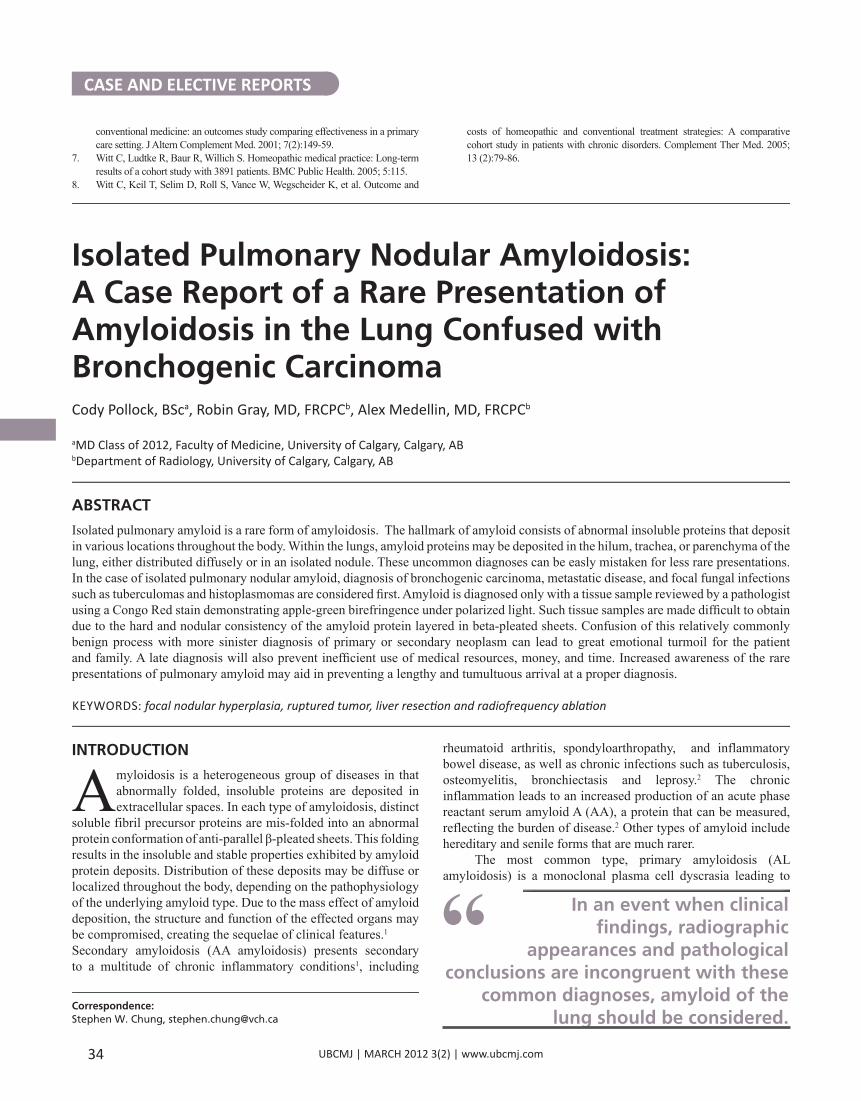
Three weeks later a CT of the chest (Figure 2) and abdomen were attained for staging of the assumed bronchogenic carcinoma. A speculated mass measuring 5.3 cm by 4.6 cm was found in the anterior segment of the right upper lobe. This mass abutted the superior vena cava, but no signs of compression or invasion were noted. There was no sign of destruction of the adjacent bone or distant isolated bony metastasis. Mediastinal lymph nodes were numerous, but the sizes were within normal limits. No metastases to other organs were identified.
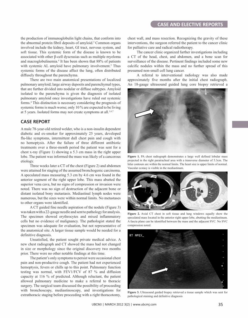
A CT guided fine needle aspiration of the nodule (Figure 3) was taken with a 22-gauge needle and sent to pathology for analysis. The specimen showed erythrocytes and mixed inflammatory cells but no evidence of malignancy. The pathologist stated the specimen was adequate for evaluation, but not representative of the anatomical site. A larger tissue sample would be needed for a definitive diagnosis.
Unsatisfied, the patient sought private medical advice. A new chest radiograph and CT showed the mass had not changed in size or morphology since the original discovery two months prior. There were no other notable findings at this time.
The patient’s only symptoms to persist were occasional chest pain and non-productive cough. The patient had not experienced hemoptysis, fevers or chills up to this point. Pulmonary function testing was normal, with FEV1/FCV of 87 % and diffusion capacity at 116 % of predicted. Although reluctant, the patient allowed pulmonary medicine to make a referral to thoracic surgery. The surgical team discussed the possibility of proceeding with bronchoscopy, mediastinoscopy, and investigations for extrathoracic staging before proceeding with a right thoracotomy,
CASE AND ELECTIVE REPORTS
35
chest wall, and mass resection. Recognizing the gravity of these interventions, the surgeon referred the patient to the cancer clinic for palliative care and radical radiotherapy.
The cancer clinic organized further investigations including a CT of the head, chest, and abdomen, and a bone scan for surveillance of the disease. Pertinent findings included some new calcific nodules within the mass and no further spread of this presumed non-small cell lung cancer.
A referral to interventional radiology was also made approximately five months after the initial chest radiograph. An 18-gauge ultrasound guided lung core biopsy retrieved a
Figure 1. PA chest radiograph demonstrates a large well defined lobular mass projected in the right paratracheal area with a transverse diameter of 5.3cm. The hilar contours are within the normal limits. The heart size is upper limits of normal. Vascular ecstasy is visible in the mediastinum.
Figure 2. Axial CT chest in soft tissue and lung windows equally show the speculated mass located in the anterior right upper lobe, abutting the mediastinum. A fascia plane can be identified between the mass and the adjacent SVC. No SVC compression noted.
Figure 3. Ultrasound guided biopsy retrieved a tissue sample which was sent for pathological staining and definitive diagnosis
UBCMJ | MARCH 2012 3(2) | www.ubcmj.com
CASE AND ELECTIVE REPORTS
36
including plaques, nodular, cavitated, and calcific forms. The variety of morphological classes creates a difficult entity to diagnose, especially when superimposed infections or unrelated pulmonary pathologies are present.
The rarity of such a diagnosis is exemplified by a study at the Mayo Clinic.1 Over a thirteen-year period, only 7 cases of isolated amyloidomas where found in the lung.1 Patients most commonly presented in the 6th decade. Variation in the presentations included sizes ranging from 0.55cm with an average of 3cm.1 Thirty to fifty percent of cases showed calcifications. Also, cavitations and spiculations have been noted in other cases.8
Solitary pulmonary nodular amyloidosis is an uncommon diagnosis. More common conditions that present similarly should be excluded first, such as neoplastic, infectious, or inflammatory conditions. In an event when clinical findings, radiographic appearances, and pathological conclusions are incongruent with these common diagnoses, amyloid of the lung should be considered.
Amyloid proteins are deposited in a protein conformation of anti-parallel β-pleated sheets and have unique properties. When stained with Congo Red, a pathognomonic pattern of apple-green birefringence may be visualized under polarized light.1,10 Immunohistochemical staining can further determine the protein type allowing for targeted treatments.10
The final diagnosis of amyloidoma of the lung was undoubtedly a surprise to the many clinicians involved in the case discussed. To further classify the diagnosis of an amyloidoma, systemic amyloidosis must be ruled out, since management differs between each subtype.
A regimented diagnostic workup suggested by Shah et al. includes first immunohistochemical staining to determine the protein type.9 This often involves ruling out secondary AA amyloidosis with a negative test and assigning the presumptive diagnosis of the much more common systemic AL type. In lung tissue, protein typing of this nature is ideal but not always achieved due to its lack of practicality.6,9 A diagnosis of
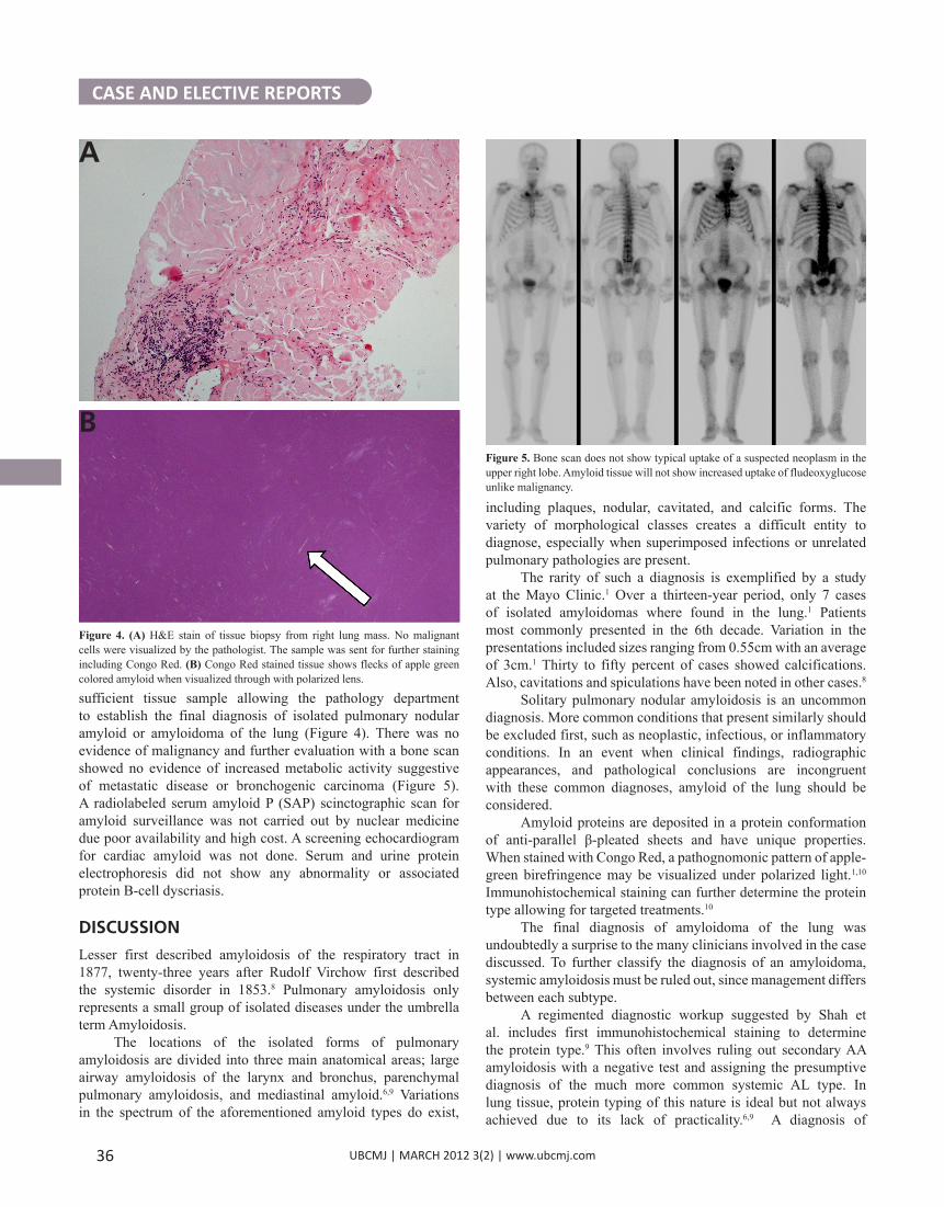
sufficient tissue sample allowing the pathology department to establish the final diagnosis of isolated pulmonary nodular amyloid or amyloidoma of the lung (Figure 4). There was no evidence of malignancy and further evaluation with a bone scan showed no evidence of increased metabolic activity suggestive of metastatic disease or bronchogenic carcinoma (Figure 5). A radiolabeled serum amyloid P (SAP) scinctographic scan for amyloid surveillance was not carried out by nuclear medicine due poor availability and high cost. A screening echocardiogram for cardiac amyloid was not done. Serum and urine protein electrophoresis did not show any abnormality or associated protein B-cell dyscriasis.
DISCUSSIONLesser first described amyloidosis of the respiratory tract in 1877, twenty-three years after Rudolf Virchow first described the systemic disorder in 1853.8 Pulmonary amyloidosis only represents a small group of isolated diseases under the umbrella term Amyloidosis.
The locations of the isolated forms of pulmonary amyloidosis are divided into three main anatomical areas; large airway amyloidosis of the larynx and bronchus, parenchymal pulmonary amyloidosis, and mediastinal amyloid.6,9 Variations in the spectrum of the aforementioned amyloid types do exist,
Figure 5. Bone scan does not show typical uptake of a suspected neoplasm in the upper right lobe. Amyloid tissue will not show increased uptake of fludeoxyglucose unlike malignancy.
A
Figure 4. (A) H&E stain of tissue biopsy from right lung mass. No malignant cells were visualized by the pathologist. The sample was sent for further staining including Congo Red. (B) Congo Red stained tissue shows flecks of apple green colored amyloid when visualized through with polarized lens.
B
UBCMJ | MARCH 2012 3(2) | www.ubcmj.com
secondary (AA) amyloid is associated with chronic inflammatory diseases, that should be treated first. Also ruling out commonly associated plasma cell dyscrasia, such as multiple myeloma and macroglobulinemia, is accomplished with bone marrow aspirate as well as urine and serum electrophoresis analysis.9
Alongside the abnormal amyloid fibrils, normal plasma glycoprotein and serum amyloid P (SAP) are also laid down.4,9 The uptake of this protein is not dependent on the deposition rate of new amyloid. By radiolabeling SAP, amyloid deposits can be detected by scintigraphic imaging2, thus providing a complete body evaluation of amyloid deposits. Solid organs such as liver and spleen allow for more sensitive localization than with organs such as the lungs. Thus scintigraphic imaging is a sensitive method for the evaluation of extra-pulmonary amyloid that may be associated with established amyloid of the lung.2,8
CONCLUSIONIn this case of an isolated pulmonary amyloidosis, the patient lived with a false presumed diagnosis of a bronchogenic carcinoma for almost half a year. From the initial discovery of the mass to final diagnosis, the patient was exposed to multiple CT and bone scans, chest x-rays and tissue biopsies. Undoubtedly he was forced to endure much emotional turmoil and unnecessarily address end of life issues. This emphasizes the importance of obtaining an adequate biopsy
CASE AND ELECTIVE REPORTS
37
SOAP Note
Subjective• 78 year-old retired welder• Past medical history of non-insulin dependent diabetes, carpel tunnel syn-drome and sciatic nerve pain• Quit smoking >25 years prior, pack years unknown• Currently presents with fever, intermittent chest pains and cough with no hematemesis• Estimates his weight loss at 3-4 pounds
Objective• Incidental finding of a 5.3cm mass in right upper lobe on PA chest radiograph• No lymphadenopathy on head and neck exam, no peripheral edema, no club-bing• 146/78 mmHg, 95% on room air, 65 regular beats/min• CT guided fine needle aspirate found no signs of malignancy• Treatment and palliative care for suspected non-small cell lung cancer discussed at length with pulmonary medicine, thoracic surgery and radiation oncology• Ultrasound guided 18 gauge core biopsy retrieved sufficient tissue for patho-logical assessment
Assessment• Final diagnosis made by pathologist >5 months after mass was initially found on plain radiographs• Biopsy showed pulmonary amyloidoma with the use of special Congo Red staining
Plan• Further investigations, bone scan and protein electrophoresis, were negative ruling out systemic disease• Isolated pulmonary amyloidoma was confirmed• Pulmonary function tests were normal• Patient had become asymptomatic• Apology for patient’s emotional turmoil, delay in diagnosis and money lost attaining unnecessary investigations all leading to the uncommon diagnosis of isolated pulmonary nodular amyloidosis.
early, allowing the correct diagnosis to be established and communicated to the patient. In order to ensure quality care, an attitude of vigilance and dedication will aid us in maintaining quality care as we investigate each clinical question to its complete end.
REFERENCES1. Utz JP, Swensen SJ, Gertz MA. Pulmonary amyloidosis. The Mayo Clinic
experience from 1980 to 1993. Ann Intern Med 1996 Feb 15;124(4):407-413.
2. Lachmann HJ, Goodman HJ, Gilbertson JA, et al. Natural history and outcome in systemic AA amyloidosis. N Engl J Med 2007;356:2361.
3. Husby G. Nomenclature and classification of amyloid and amyloidoses. J Intern Med 1992 Dec;232(6):511-512.
4. Aylwin AC, Gishen P, Copley SJ. Imaging appearance of thoracic amyloidosis. J Thorac Imaging 2005 Feb;20(1):41-46.
5. Smith RR, Hutchins GM, Moore GW, Humphrey RL. Type and distribution of pulmonary parenchymal and vascular amyloid. Correlation with cardiac amyloidosis. Am J Med 1979;66:96–104.
6. Lachmann HJ, Hawkins PN. Amyloidosis and the lung. Chron Respir Dis 2006;3(4):203-214.
7. Guidelines Working Group of UK Myeloma Forum; British Committee for Standards in Haematology, British Society for Haematology. Guidelines on the diagnosis and management of AL amyloidosis. Br J Haematol 2004;125: 681–700.
8. Gillmore JD, Hawkins PN. Amyloidosis and the respiratory tract. Thorax 1999 May;54(5):444-451.
9. Shah PL, Gillmore JD, Copley SJ, Collins JV, Wells AU, du Bois RM, et al. The importance of complete screening for amyloid fibril type and systemic disease in patients with amyloidosis in the respiratory tract. Sarcoidosis Vasc Diffuse Lung Dis 2002 Jun;19(2):134-142.
10. Picken MM. New insights into systemic amyloidosis: the importance of diagnosis of specific type. Curr Opin Nephrol Hypertens 2007;16:196.