Embed Size (px)
Citation preview
Islet Antigen-reactive B Cells as Participants and Therapeutic Targets in T1D
John C. CambierDepartment of Immunology and Microbiology
University of Colorado School of Medicine
No relevant COI
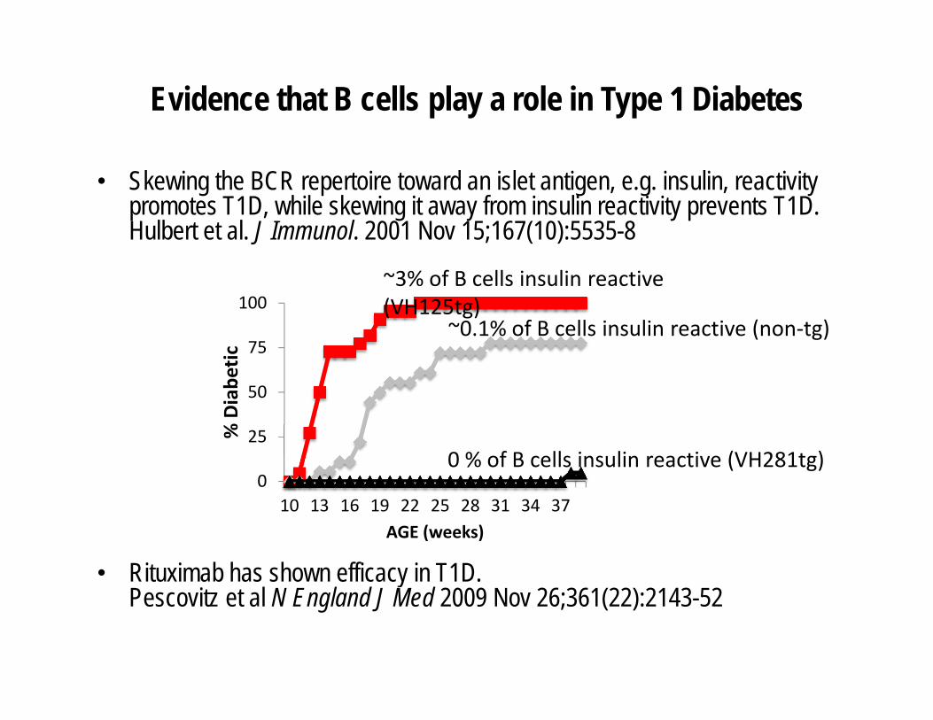
Evidence that B cells play a role in Type 1 Diabetes
• Skewing the BCR repertoire toward an islet antigen, e.g. insulin, reactivity promotes T1D, while skewing it away from insulin reactivity prevents T1D. Hulbert et al. J Immunol. 2001 Nov 15;167(10):5535-8
• Rituximab has shown efficacy in T1D. Pescovitz et al N England J Med 2009 Nov 26;361(22):2143-52
0
25
50
75
100
10 13 16 19 22 25 28 31 34 37
% Diabe
tic
AGE (weeks)
0 % of B cells insulin reactive (VH281tg)
~0.1% of B cells insulin reactive (non‐tg)
~3% of B cells insulin reactive (VH125tg)
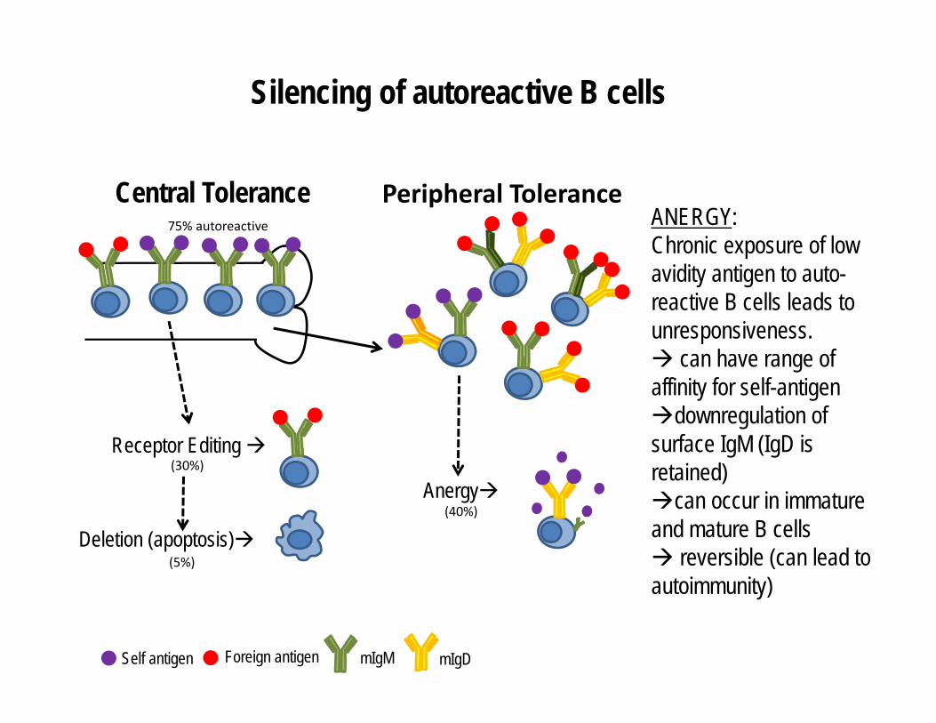
Silencing of autoreactive B cells
Central Tolerance
Receptor Editing
Deletion (apoptosis)
75% autoreactive
(30%)
(5%)
Peripheral Tolerance
Anergy(40%)
ANERGY:Chronic exposure of low avidity antigen to auto-reactive B cells leads to unresponsiveness. can have range of affinity for self-antigendownregulation of surface IgM (IgD is retained)can occur in immature and mature B cells reversible (can lead to autoimmunity)
Self antigen Foreign antigen mIgM mIgD

Hypothesis:
We posit that potentially pathogenic insulin-reactive B are normally silenced by anergy, but they become activated and
participate in development of T1D.
Questions:
Are potentially pathogenic insulin-reactive B cells found in anergic compartments in healthy subjects?
Do these B cells show signs of activation and move to the pancreas and PLN during disease development?

Anergic B cells in the healthy human
Andy Duty and Patrick Wilson (JEM, 2009): • Described two anergic B cell subpopulations in healthy humans. These
express autoreactive antigen receptors • Approximately 3% of peripheral blood B cells are IgD+IgM-
– Approximately 2.5% are naïve (BND cells) and 0.5% are memory(Cδ-CS cells)
Tam Quach and Inaki Sanz (JI, 2011): • Expanded definition to include cells with low surface levels of mIgM (IgMlo)
but relatively normal levels of IgD
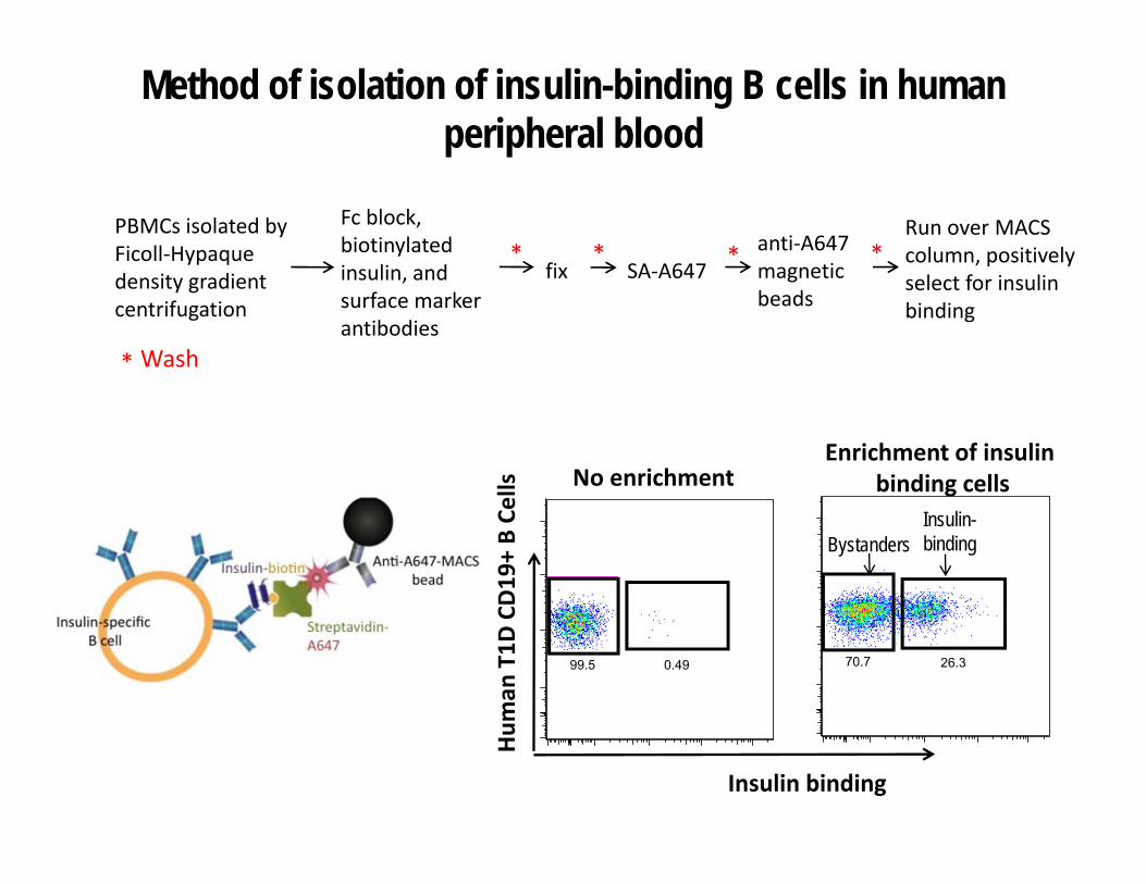
Method of isolation of insulin-binding B cells in human peripheral blood
PBMCs isolated by Ficoll‐Hypaque density gradient centrifugation
Fc block, biotinylated insulin, and surface marker antibodies
anti‐A647 magnetic beads
Run over MACS column, positively select for insulin binding
fix SA‐A647* * *
* Wash
*
26.370.70.4999.5
No enrichmentEnrichment of insulin
binding cellsHum
an T1D
CD19
+ B Ce
lls
Insulin binding
Insulin-bindingBystanders

0 50K 100K 150K 200K 250K
FSC-A
0
50K
100K
150K
200K
250K
SS
C-A
31.9
0 50K 100K 150K 200K 250K
FSC-H
0
50K
100K
150K
200K
250K
FSC
-A
99.8
0 102 103 104 105
CD27
0
102
103
104
105
CD
19
0 102 103 104 105
Insulin binding
0
102
103
104
105
CD
190 102 103 104 105
Insulin binding
0
102
103
104
105
CD
19
0 102 103 104 105
IgD
0
102
103
104
105
IgM
0 102 103 104 105
IgD
0
102
103
104
105
IgM
MemoryNaive
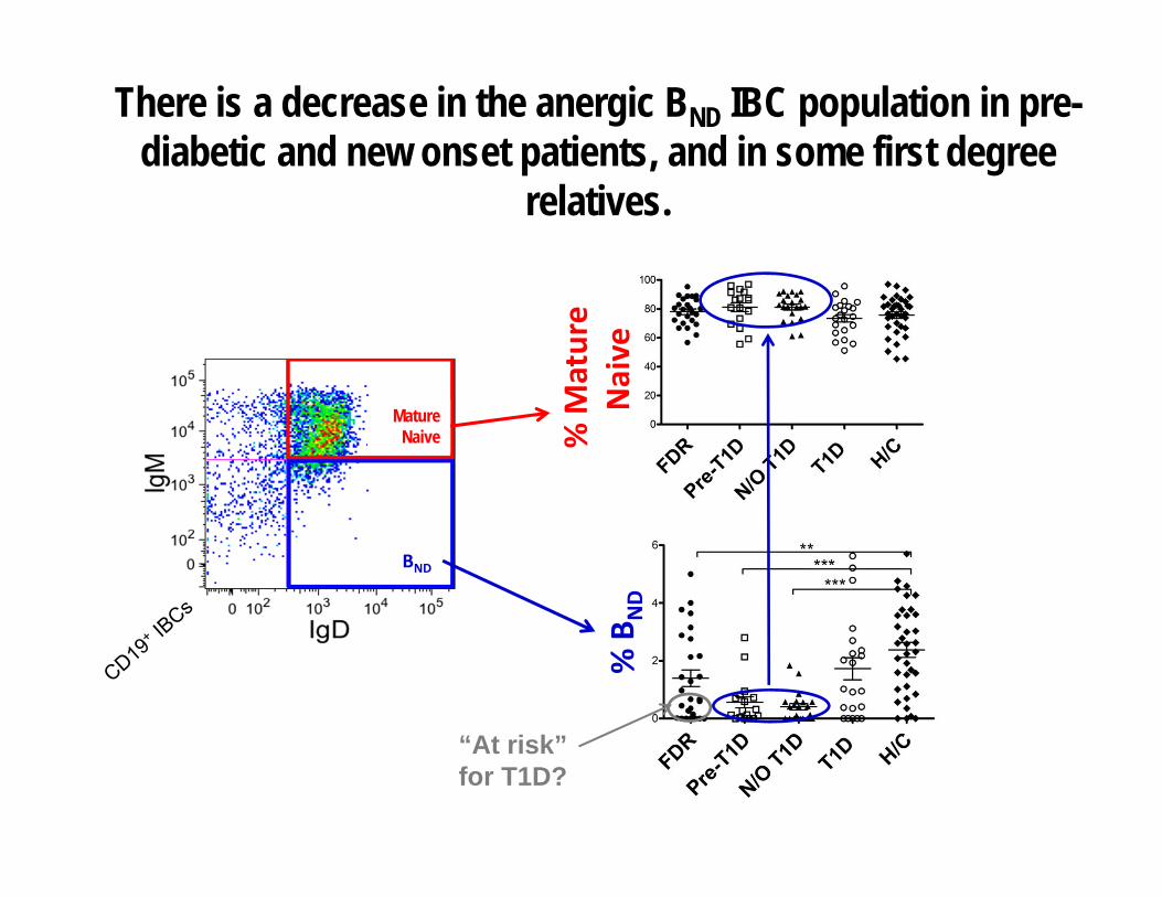
Gating strategy for identification of insulin-binding B cell subpopulations in PBL
MatureNaive
BND
IBCs
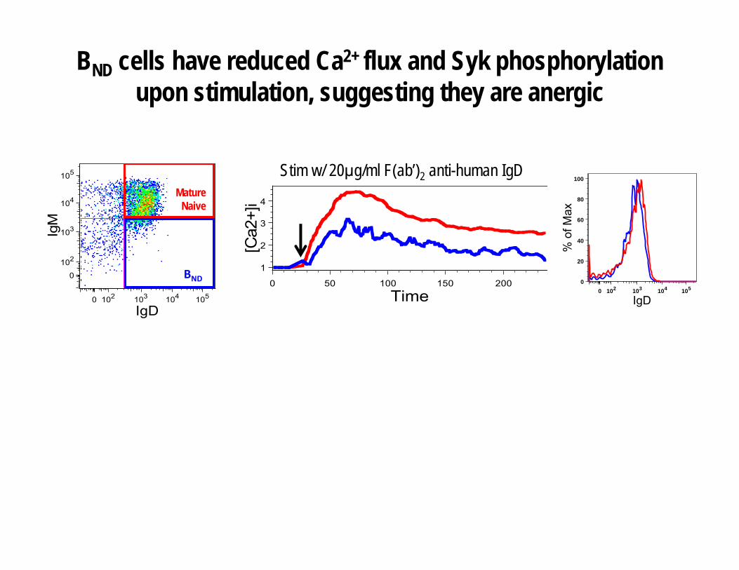
BND cells have reduced Ca2+ flux and Syk phosphorylation upon stimulation, suggesting they are anergic
0 102 103 104 105
pSyk
0
20
40
60
80
100
% o
f Max
pSyk
0 102 103 104 105
IgD
0
20
40
60
80
100
% o
f Max
0 50 100 150 200
Time
1
2
3
4
[Ca2
+]i
0 102 103 104 105
IgD
0
102
103
104
105
IgM
Naive
Bnd
Stim w/ 20μg/ml F(ab’)2 anti-human IgD
BND
Mature Naive

IBCs in the anergic BND fraction bear high affinity auto/polyreactivity antigen receptors
ChromatinInsulin LPS
Single cell sort MN and BND IBCscloned Ig variable regionsre-expressed as mAb
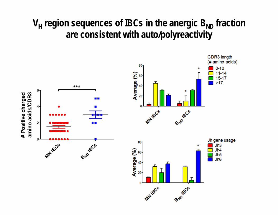
VH region sequences of IBCs in the anergic BND fraction are consistent with auto/polyreactivity

Serum antibodies from IAA+ new onsets show increased reactivity to both insulin and chromatin
Conclusions Part I
• The BND cells appear functionally anergic based on decreased BCR-mediated Ca2+
mobilization and Syk phosphorylation.• B cells bearing BCR with high affinity for insulin are found in the BND fraction
indicating normal silencing by anergy.• BND IBCs have a higher affinity for insulin than their mature naïve IBC counterparts,
suggesting the latter are ignorant of their autoantigen. • The anergic BND IBCs are polyreactive.
– These BND cells could be initially activated by antigens other than insulin, such as host or pathogen-derived DNA
• “New onsets” who make insulin autoantibodies also make chromatin autoantibodies.
– Suggests activation of the anergic, high affinity, polyreactive B cell population found in healthy controls
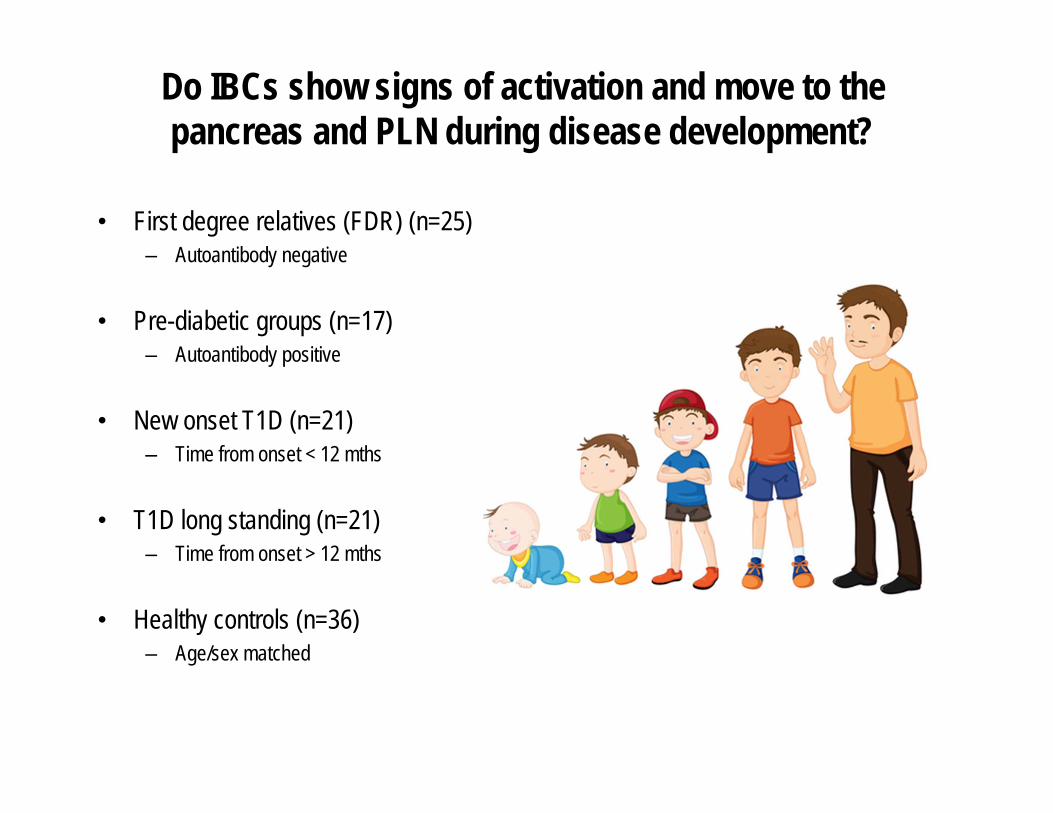
Do IBCs show signs of activation and move to the pancreas and PLN during disease development?
• First degree relatives (FDR) (n=25)– Autoantibody negative
• Pre-diabetic groups (n=17)– Autoantibody positive
• New onset T1D (n=21)– Time from onset < 12 mths
• T1D long standing (n=21)– Time from onset > 12 mths
• Healthy controls (n=36)– Age/sex matched

There is a decrease in the anergic BND IBC population in pre-diabetic and new onset patients, and in some first degree
relatives.
“At risk” for T1D?
% B
ND
% M
ature
Naive
BND
Mature Naive

Loss of BND in FDRs correlates with high risk HLA genotype

“Transient” loss of the total BND population in FDRs, pre-diabetics and new onsets
FDRPre-
T1DN/O
T1D T1D
H/C
0
2
4
6 *****
***
IBC
BN
D[%
of I
BC
s]
FDRPre-
T1DN/O
T1D T1D
H/C
0
2
4
6**
******
Tota
l BN
D[%
of t
otal
B c
ells
]
IBC
Total
0
2
4
6
p = 0.003
% B
ND
Insulin-binding Total
Conclusions Part II
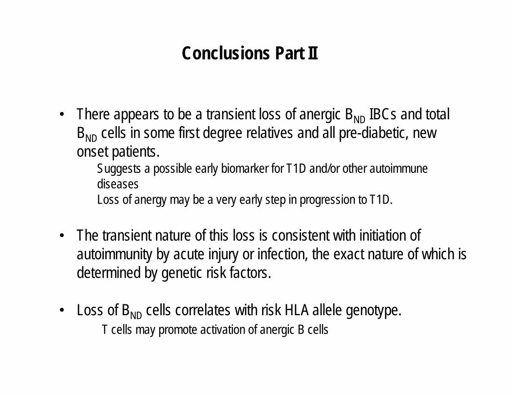
• There appears to be a transient loss of anergic BND IBCs and total BND cells in some first degree relatives and all pre-diabetic, new onset patients.
Suggests a possible early biomarker for T1D and/or other autoimmune diseasesLoss of anergy may be a very early step in progression to T1D.
• The transient nature of this loss is consistent with initiation of autoimmunity by acute injury or infection, the exact nature of which is determined by genetic risk factors.
• Loss of BND cells correlates with risk HLA allele genotype.T cells may promote activation of anergic B cells
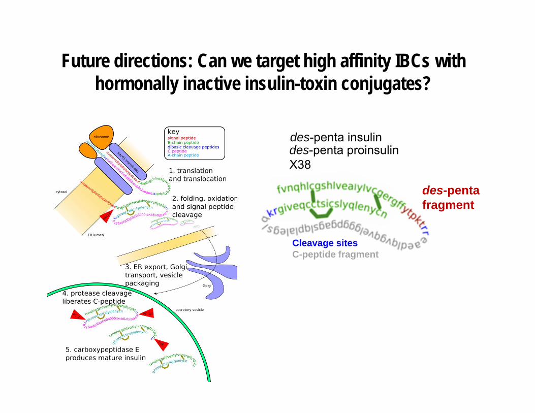
Future directions: Can we target high affinity IBCs with hormonally inactive insulin-toxin conjugates?
Cleavage sitesC-peptide fragment
des-pentafragment
des-penta insulindes-penta proinsulinX38

Acknowledgements
Cambier LabMia Smith
Tom PackardShannon O’NeillRochelle HinmanAndrew GetahunMelissa WalkerJanie Akerlund
Elizabeth FranksSoojin Kim
Peter Gottlieb LabLisa Fitzgerald-MillerMarynette Rihanek
Aimon Alkanani
Patrick Wilson LabCarole Dunand
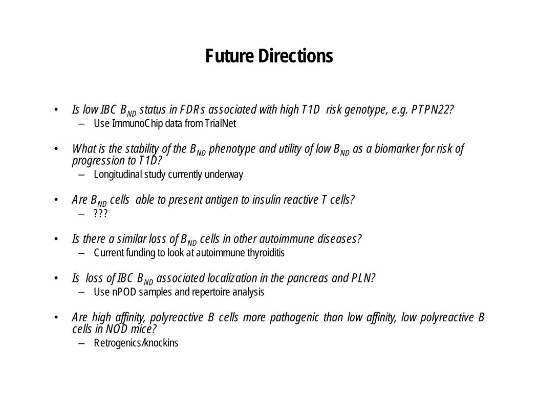
Future Directions
• Is low IBC BND status in FDRs associated with high T1D risk genotype, e.g. PTPN22? – Use ImmunoChip data from TrialNet
• What is the stability of the BND phenotype and utility of low BND as a biomarker for risk of progression to T1D?
– Longitudinal study currently underway
• Are BND cells able to present antigen to insulin reactive T cells?– ???
• Is there a similar loss of BND cells in other autoimmune diseases?– Current funding to look at autoimmune thyroiditis
• Is loss of IBC BND associated localization in the pancreas and PLN?– Use nPOD samples and repertoire analysis
• Are high affinity, polyreactive B cells more pathogenic than low affinity, low polyreactive Bcells in NOD mice?
– Retrogenics/knockins

Human Type 1 Diabetes (T1D)
• Autoimmune disorder characterized by destruction of beta cells in pancreas decreased production of insulin hyperglycemia
• Predicted by presence of genes and autoantibodies– e.g. HLA genes (esp DR3/4-DQ2/8 haplotype)– e.g. anti-insulin, GAD65, ICA, IA-2A, ZnT8
• Occurs more often in individuals < 30 years old; clinical symptoms include polyuria, polydipsia
• Both genetics and environment thought to play a role
• Incidence is rising globally and as much as 4% annually in U.S.
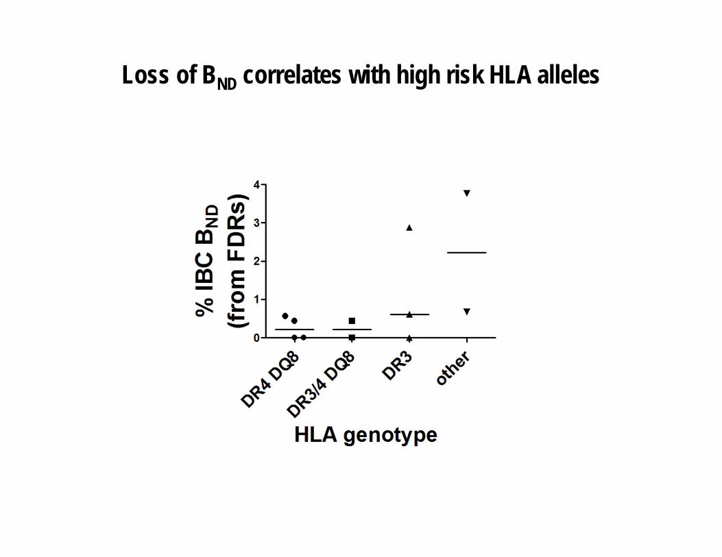
Loss of BND correlates with high risk HLA alleles
There is a decrease in the anergic BND IBC population in pre-diabetic and new onset patients, and in some first degree
relatives.
“At risk” for T1D?
% B
ND
% M
ature
Naive
BND
Mature Naive
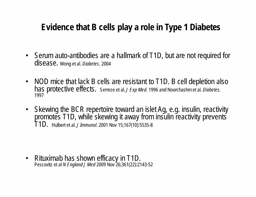
Evidence that B cells play a role in Type 1 Diabetes
• Serum auto-antibodies are a hallmark of T1D, but are not required for disease. Wong et al. Diabetes. 2004
• NOD mice that lack B cells are resistant to T1D. B cell depletion also has protective effects. Serreze et al. J Exp Med. 1996 and Noorchashm et al. Diabetes. 1997
• Skewing the BCR repertoire toward an islet Ag, e.g. insulin, reactivity promotes T1D, while skewing it away from insulin reactivity prevents T1D. Hulbert et al. J Immunol. 2001 Nov 15;167(10):5535-8
• Rituximab has shown efficacy in T1D. Pescovitz et al N England J Med 2009 Nov 26;361(22):2143-52