Embed Size (px)
Citation preview
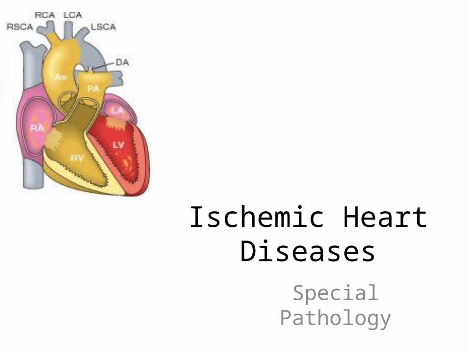
Ischemic Heart Diseases
Special Pathology
• Ischemic heart disease (IHD);– generic designation for a group of closely related
syndromes resulting from myocardial ischemia• an imbalance between the supply (perfusion) and demand of the
heart for oxygenated blood
– Ischemia comprises;• insufficiency of oxygen• reduced availability of nutrient substrates • inadequate removal of metabolites
– Isolated hypoxemia • diminished transport of oxygen by the blood• induced by cyanotic congenital heart disease, severe anemia, or
advanced lung disease
– is less deleterious than ischemia • because perfusion is maintained
– (including metabolic substrate delivery and waste removal)
• IHD is often termed coronary artery disease (CAD) or coronary heart disease
• In more than 90% of cases, the cause of myocardial ischemia is reduction in coronary blood flow
• due to atherosclerotic coronary arterial obstruction
– long period (decades) of silent, slowly progressive, coronary atherosclerosis
• before these disorders become manifest
– syndromes of IHD are only the late manifestations of coronary atherosclerosis
• probably began during childhood or adolescence
• Clinical manifestations of IHD;– divided into four syndromes:
• Myocardial infarction (MI), – most important form of IHD, in which the – duration and severity of ischemia is sufficient to cause death
of heart muscle
• Angina pectoris, – ischemia is less severe – does not cause death of cardiac muscle– three variants-stable angina, Prinzmetal angina, and unstable
angina-» the latter is the most threatening as a frequent harbinger
of MI.
• Chronic IHD with heart failure• Sudden cardiac death
• Certain conditions aggravate ischemia through either; – an increase in cardiac energy demand
• (e.g., hypertrophy)
– by diminished availability of blood or oxygen due to lowered systemic blood pressure
• shock or hypoxemia
• Tachycardia, increased heart rate not only – increases demand through
• more contractions per unit time
– decreases supply • by decreasing the relative time spent in diastole-when
coronary perfusion occurs
• The risk of an individual developing detectable IHD depends ;– in part on the number, distribution, and structure of atheromatous plaques, – degree of narrowing they cause.
• Clinical manifestations of IHD are not entirely predicted by these anatomic observations of disease burden
• Extraordinarily broad spectrum of the expression of disease;– elderly individuals with extensive coronary atherosclerosis who have never
had a symptom,– previously asymptomatic young adult in whom modestly obstructive disease
comes unexpectedly to medical attention as a result of acute MI or sudden cardiac death
• Reasons for clinical heterogeneity of the disease are complex• often precipitous and variable onset and natural history largely
depend on the pathologic basis of the so-called acute coronary syndromes of IHD – comprising unstable angina, acute MI, and sudden death)

• The acute coronary syndromes are frequently initiated by – plaque disruption or acute plaque change
• an unpredictable and abrupt conversion of a stable atherosclerotic plaque to an unstable and potentially life-threatening atherothrombotic lesion through
– superficial erosion, ulceration, fissuring, rupture, or – deep hemorrhage, usually with superimposed
thrombosis.
• Pathogenesis;– dominant influence in the causation of the IHD
syndromes – diminished coronary perfusion relative to
myocardial demand, owing largely to a complex and dynamic interaction among;
• fixed atherosclerotic narrowing of the epicardial coronary arteries
• intraluminal thrombosis overlying a disrupted atherosclerotic plaque
• platelet aggregation• vasospasm
• More than 90% of patients with IHD have atherosclerosis of one or more of the coronary arteries– Clinical manifestations of coronary atherosclerosis are generally due to;
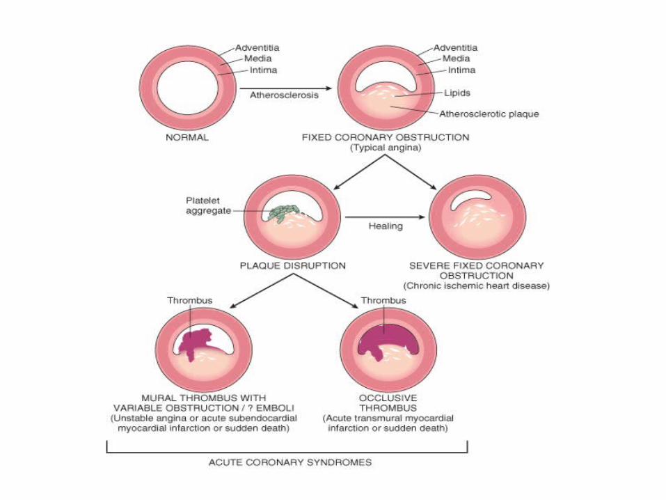
• progressive encroachment of the lumen leading to stenosis (chronic, "fixed" obstructions)
• acute plaque disruption with thrombosis (generally both sudden and dynamic), which compromises blood flow
– A fixed obstructive lesion of 75% or greater (i.e., only 25% or less lumen remaining) generally causes symptomatic ischemia induced by exercise;
• with this degree of obstruction, the augmented coronary flow provided by compensatory vasodilation is no longer sufficient to meet even moderate increases in myocardial demand
– A 90% stenosis can lead to inadequate coronary blood flow even at rest– Slowly developing occlusions may stimulate collateral vessels over time,
• protect against distal myocardial ischemia and infarction even with an eventual high-grade stenosis
• Although only a single major coronary epicardial trunk may be affected, – two or all three are often involved
• lateral anterior descending (LAD), left circumflex (LCX), and right coronary artery (RCA)
• Clinically significant stenosing plaques may be located anywhere within these vessels but tend to predominate – within the first several centimeters of the LAD and LCX – along the entire length of the RCA. – Sometimes the major secondary epicardial branches are also
involved • (i.e., diagonal branches of the LAD, obtuse marginal branches of the LCX, or
posterior descending branch of the RCA), • but atherosclerosis of the intramural branches is rare
– onset of symptoms and prognosis of IHD depend;• not only on the extent and severity of fixed, chronic anatomic disease, • but also critically on dynamic changes in coronary plaque morphology
• Role of Acute Plaque Change. – In most patients the myocardial ischemia underlying unstable angina,
acute MI, and (in many cases) sudden cardiac death is precipitated by • abrupt plaque change followed by thrombosis (Fig.)
– Most often, the initiating event is disruption of previously only partially stenosing plaques with any of the following:
• Rupture/fissuring, exposing the highly thrombogenic plaque constituents • Erosion/ulceration, exposing the thrombogenic subendothelial basement
membrane to blood • Hemorrhage into the atheroma, expanding its volume
– The events that trigger abrupt changes in plaque configuration and superimposed thrombosis are complex and poorly understood.
– Acute alterations in plaque imply the inability of a plaque to withstand mechanical stresses
– Important influences are both • intrinsic (e.g., plaque structure and composition)• extrinsic (e.g., blood pressure, platelet reactivity)
• Structure and composition of a plaque are dynamic and contribute to a propensity to disruption. – vulnerable plaques
• Plaques that contain large areas of foam cells and extracellular lipid, and those in which the fibrous caps are thin or contain few smooth muscle cells or have clusters of inflammatory cells, are more likely to rupture
– Fissures • frequently occur at the junction of the fibrous cap and the adjacent normal plaque-free
arterial segment, • a location at which the blood flow-inducing mechanical stresses within the plaque are
highest and the fibrous cap is thinnest.
– now recognized that the fibrous cap can undergo continuous remodeling• balance of synthetic and degradative activity of collagen• major structural component of the fibrous cap, accounts for its mechanical strength and
determines plaque stability and prognosis
– Collagen• produced by smooth muscle cells and • degraded by the action of metalloproteinases, enzymes elaborated by macrophages in
atheroma • considerable evidence that inflammation destabilizes the mechanical integrity of plaques
– Moreover, drugs such as statins;• inhibitors of HMG Co-A reductase, a key enzyme in the synthesis of cholesterol• reduce clinical events associated with IHD, are thought to stabilize plaques by;
– their lipid-lowering effect– reducing plaque inflammation
• Influences extrinsic to plaque are also important– Adrenergic stimulation can elevate physical stresses on
the plaque through • systemic hypertension or local vasospasm• adrenergic stimulation associated with awakening and arising
induces a pronounced circadian periodicity for the time of onset of acute MI, with a peak incidence between 6 a.m. and 12 noon,
• concurrent with a surge in blood pressure and immediately following heightened platelet reactivity
– Intense emotional stress can also contribute to plaque disruption;
• most dramatically illustrated by the marked increase in the incidence of sudden death that is associated with natural or other disasters
• earthquakes and the September 11, 2001 attacks in New York and Washington, DC
• Preexisting culprit lesion in patients who develop myocardial infarction and other acute coronary syndromes is;– not necessarily a severely stenotic and hemodynamically significant lesion
prior to its acute change • Pathologic and clinical studies show that plaques that undergo abrupt
disruption leading to coronary occlusion are;– those that previously produced only mild to moderate luminal stenosis
• Approximately two thirds of plaques that rupture with subsequent occlusive thrombosis caused occlusion of only;– 50% or less before plaque rupture, – 85% had initial stenosis less than 70%
• Worrisome conclusion is;– large number of now asymptomatic adults in the industrial world have a
real but unpredictable risk of a catastrophic coronary event– Regrettably, it is presently impossible to reliably predict plaque disruption
or subsequent thrombosis in an individual patient
• Accumulating evidence indicates;– plaque disruption and the ensuing platelet
aggregation and intraluminal thrombosis• common, repetitive, and often clinically silent
complications of atheroma
– Moreover, healing of subclinical plaque disruption and overlying thrombosis
• important mechanism of growth of atherosclerotic lesions
• Role of Inflammation;– play important roles at all stages of atherosclerosis,
• from its inception to the development of complications
– The establishment of the initial lesion requires the interaction between;
• endothelial cells and circulating leukocytes, – leading to the accumulation of T cells and macrophages in the arterial wall. – Entry of leukocytes into the wall is a consequence of the release of
chemokines by endothelial cells, and the increased expression of adhesion proteins (ICAM-1, VCAM-1, E-selectin and P-selectin) in these cells.
– T cells located in the arterial wall produce cytokines such as TNF, IL-6 and IFN-γ that stimulate endothelial cells and activate macrophages, which become loaded with oxidized LDL.
– At later stages of atherosclerosis, • destabilization and rupture of the plaque may involve the secretion
of metalloproteinases by macrophages.• These enzymes weaken the plaque by digesting collagen at the
fibrous cap or the shoulder of the lesion
• several proteins involved in inflammation may serve as potential markers of atherosclerosis– C-reactive protein (CRP);
• acute phase reactant made in the liver• suggested as a predictor of risk of coronary heart
disease• In some, but not all, studies CRP predicts risk
independently from risk estimates provided by serum lipid levels
• It could be used to estimate the risk of;– myocardium infarct in patients with angina– new infarcts in patients who are infarct survivors
• Role of Coronary Thrombus;– critical to the pathogenesis of the acute coronary syndromes
• Partial or total thrombosis associated with a disrupted plaque
– In the most serious form, acute transmural MI • thrombus superimposed on a disrupted but previously only partially
stenotic plaque converts it to a total occlusion
– In contrast, with unstable angina, acute subendocardial infarction, or sudden cardiac death, the extent of luminal obstruction by thrombosis is usually incomplete (mural thrombus)
• it may wax and wane with time.
– Mural thrombus in a coronary artery can also embolize. • Indeed, small fragments of thrombotic material in the distal
intramyocardial circulation or microinfarcts may be found at autopsy of patients who have had unstable angina or sudden death.
– Finally, thrombus is a potent activator of multiple growth-related signals in smooth muscle cells,
• can contribute to the growth of atherosclerotic lesions
• Role of Vasoconstriction – Vasoconstriction compromises lumen size, and, by
increasing the local mechanical forces, can potentiate plaque disruption.
– Vasoconstriction at sites of atheroma is stimulated by:
• (1) circulating adrenergic agonists, • (2) locally released platelet contents, • (3) impaired secretion of endothelial cell relaxing
factors relative to contracting factors (e.g., endothelin) due to atheroma - associated endothelial dysfunction
• (4) mediators released from perivascular inflammatory cells
• Summary (Fig. and Table)– Acute coronary syndromes-angina, acute MI, and sudden death-
share a common pathophysiologic basis in;• coronary atherosclerotic plaque disruption • associated intraluminal platelet-fibrin thrombus formation
– Critical consequence is downstream myocardial ischemia • Stable angina results from increases in myocardial oxygen demand that
outstrip the ability of markedly stenosed coronary arteries to increase oxygen delivery but is not usually associated with plaque disruption.
• Unstable angina derives from a sudden change in plaque morphology, which induces partially occlusive platelet aggregation or mural thrombus, and vasoconstriction leading to severe but transient reductions in coronary blood flow. In some cases, distal microinfarcts occur secondary to thromboemboli.
• In MI, acute plaque change induces total thrombotic occlusion• Finally, sudden cardiac death frequently involves a coronary lesion in
which disrupted plaque and often partial thrombus and possibly embolus have led to regional myocardial ischemia that induces a fatal ventricular arrhythmia


Coronary Artery Pathology in Ischemic Heart Disease
Syndrome Stenoses Plaque DisruptionPlaque-Associated
Thrombus
Stable angina >75% No No
Unstable angina Variable Frequent Nonocclusive, often with thromboemboli
Transmural myocardial infarction
Variable Frequent Occlusive
Subendocardial myocardial infarction
Variable Variable Widely variable, may be absent, partial/complete, or lysed
Sudden death Usually severe Frequent Often small platelet aggregates or thrombi and/or thromboemboli
• Angina pectoris;– symptom complex of IHD – characterized by paroxysmal and usually recurrent attacks of
substernal or precordial chest discomfort • (variously described as constricting, squeezing, choking, or knifelike)
– caused by transient (15 seconds to 15 minutes) myocardial ischemia – falls short of inducing the cellular necrosis that defines infarction– three overlapping patterns of angina pectoris:
• (1) stable or typical angina• (2) Prinzmetal or variant angina• (3) unstable or crescendo angina
– caused by varying combinations of increased myocardial demand and decreased myocardial perfusion,
• owing to fixed stenosing plaques, disrupted plaques, vasospasm, thrombosis, platelet aggregation, and embolization
– silent ischemia • being increasingly recognized that not all ischemic events are perceived by
patients, • even though such events may have adverse prognostic implications
• Stable angina;– most common form
• therefore called typical angina pectoris,
– appears to be caused by the reduction of coronary perfusion to a critical level by chronic stenosing coronary atherosclerosis;
• renders the heart vulnerable to further ischemia whenever there is increased demand
– produced by physical activity, emotional excitement, or any other cause of increased cardiac workload
– usually relieved by rest (thereby decreasing demand) or nitroglycerin, a strong vasodilator.
– Although the coronary arteries are usually maximally dilated by intrinsic regulatory influences,
• nitroglycerin also decreases cardiac work by dilating the peripheral vasculature.
• In particular instances, local vasospasm may contribute to the imbalance between supply and demand.
• Prinzmetal variant angina;– uncommon pattern of episodic angina that
• occurs at rest • due to coronary artery spasm
– Usually there is an elevated ST segment on the electrocardiogram (ECG),
• indicative of transmural ischemia.
– Although individuals with this form of angina may well have significant coronary atherosclerosis,
• anginal attacks are unrelated to physical activity, heart rate, or blood pressure
– Prinzmetal angina generally responds promptly to vasodilators,
• such as nitroglycerin and calcium channel blockers
• Unstable or crescendo angina– refers to a pattern of pain that occurs with progressively
increasing frequency• precipitated with progressively less effort, often occurs at rest• tends to be of more prolonged duration
– Mostly induced by disruption of an atherosclerotic plaque with superimposed partial (mural) thrombosis and possibly embolization or vasospasm (or both).
– unstable angina is often the prodrome of subsequent acute MI
• Although the ischemia that occurs in unstable angina falls precariously close to inducing clinically detectable infarction
– Preinfarction angina;• in the spectrum of IHD, unstable angina lies intermediate
between stable angina on the one hand and MI on the other
• Myocardial Infarction;– also known as "heart attack,"
• death of cardiac muscle resulting from ischemia
– most important form of IHD – alone is the leading cause of death in the United
States and industrialized nations. • About 1.5 million individuals in the United States suffer
an acute MI annually • approximately one third of them die
– At least 250,000 people a year die of a heart attack before they reach the hospital
• CHRONIC ISCHEMIC HEART DISEASE – cardiac findings in patients
• develop progressive heart failure as a consequence of ischemic myocardial damage
– ischemic cardiomyopathy• used by clinicians to describe CIHD
– prior MI and sometimes previous coronary arterial bypass graft surgery or other interventions
– CIHD usually constitutes postinfarction cardiac decompensation• exhaustion of the compensatory hypertrophy of noninfarcted viable
myocardium– itself in jeopardy of ischemic injury
– However, in other cases severe obstructive CAD may be present without acute or healed infarction but with diffuse myocardial dysfunction.
• Morphology;– Hearts from patients with CIHD are usually enlarged and
heavy, secondary to left ventricular hypertrophy and dilation
– Invariably there is moderate to severe stenosing atherosclerosis of the coronary arteries and sometimes total occlusion.
– Discrete, gray-white scars of healed infarcts are usually present.
– The mural endocardium is generally normal except for some superficial, patchy, fibrous thickenings, although mural thrombi may be present.
– The major microscopic findings include myocardial hypertrophy, diffuse subendocardial vacuolization, and scars of previously healed infarcts.
• Clinical diagnosis; – insidious onset of CHF in patients with past
episodes of MI or anginal attacks– progressive myocardial damage is entirely silent,
and heart failure is the first indication of CIHD. – diagnosis rests largely on the exclusion of other
forms of cardiac involvement• Such patients make up nearly half of cardiac transplant
recipients.
• SUDDEN CARDIAC DEATH – most commonly defined as unexpected death from cardiac causes
early after symptom onset (usually within 1 hour) or without the onset of symptoms.
– In many adults, SCD is a complication and often the first clinical manifestation of IHD
– With decreasing age of the victim, the following nonatherosclerotic causes of SCD become increasingly probable:
– Congenital structural or coronary arterial abnormalities • Aortic valve stenosis • Mitral valve prolapse • Myocarditis • Dilated or hypertrophic cardiomyopathy • Pulmonary hypertension • Hereditary or acquired abnormalities of the cardiac conduction system • Isolated hypertrophy, hypertensive or unknown cause.
– Increased cardiac mass is an independent risk factor for cardiac death; thus, some young patients who die suddenly, including athletes, have hypertensive hypertrophy or unexplained increased cardiac mass as the only finding.
• Ultimate mechanism of SCD is most often a lethal arrhythmia – asystole, ventricular fibrillation
• Mostly fatal arrhythmia is triggered by electrical irritability of myocardium
• distant from the conduction system, • induced by ischemia or other cellular abnormalities.
– Although ischemic injury can impinge on the conduction system and create electromechanical cardiac instability,
• Prognosis of patients vulnerable to SCD, especially those with chronic IHD,– markedly improved by implantation of an automatic
cardioverter defibrillator – senses and electrically counteracts an episode of ventricular
fibrillation
• Morphology;– Marked coronary atherosclerosis with critical (>75%)
stenosis involving one or more of the three major vessels is present in 80% to 90% of SCD victims;
• only 10% to 20% of cases are of nonatherosclerotic origin
– Usually there are high-grade stenoses (>90%), and acute plaque disruption is common
– A healed myocardial infarct is present in about 40%, • those who were successfully resuscitated from sudden
cardiac arrest, new MI is found in only 25% or less.
– Subendocardial myocyte vacuolization indicative of severe chronic ischemia is common
• Arrhythmias that occur in the absence of structural cardiac pathology can also precipitate sudden death– most important cause is the autosomal dominant
long QT syndrome (Romano-Ward syndrome),• causes heightened cardiac excitability and episodic
ventricular arrhythmias
• Mutations causing this disorder have been demonstrated in at least five different genes – encode components of cardiac ion channels
including potassium and sodium channels

Thank You