Embed Size (px)
Citation preview

Journal of Clinical and Analytical Medicine |
O
h
r
c
i
r
g
a
in
e
a
sl
e R
1
Hasan Ikbal Atilgan1, Murat Sadic2, Meliha Korkmaz2, Sinem Ozyurt3, Gökhan Koca2
1Ministry of Health Kahramanmaras Necip Fazıl City Hospital, Division of Nuclear Medicine, Kahramanmaras, 2Ministry of Health Ankara Training and Research Hospital, Department of Nuclear Medicine, Ankara,
3Ministry of Health Sami Ulus Children Hospital, Division of Nuclear Mecicine, Ankara, Turkey
Ureter Visualization on DMSA
Is Ureter Visualization Possible on Tc-99m DMSA Scintigraphy with Vesicoureteral Reflux Patients?
Vezikoüreteral Reflü’lü hastalarda Tc-99m DMSA Sintigrafisinde Üreter Vizüalizasyonu Mümkünmü?
DOI: 10.4328/JCAM.2652 Received: 03.07.2014 Accepted: 13.07.2014 Printed: 01.03.2016 J Clin Anal Med 2016;7(2): 158-62Corresponding Author: Murat Sadic, Department of Nuclear Medicine, Ministry of Health Ankara Training and Research Hospital, Ankara, Turkey. T.: +90 3125953608 F.: +90 3125953856 E-Mail: [email protected]
ÖzetAmaç: 99mTechnetium- dimercaptosuccinic acid (99mTc-DMSA) normalde renal korteks tarafından akümüle olan bir radyoaktif madde olup üreter veya pelvikaliseal sistemde vizüalize olmamaktadır. Bu çalışmada 99mTc-DMSA Sintigrafisinde üreterleri vizüalize olan vakalar derlenmiştir. Gereç ve Yön-tem: Çalışmaya ortalama yaşları 3.5 (min 2 ay-max 18 yaş) olan 18 hasta (5 kız, 13 erkek) dahil edildi. 99mTc-DMSA Sintigrafisinde18 hastanın 20 üreter ve/veya pelvisi görünür durumdaydı. İki hasta her iki üreterler vizüalize olmak-taydı. Tüm hastaların Vezikoüreteral Reflü (VUR) dereceleri, 99mTc-DMSA uptake’leri, Pelvikaliseal sistem durumları, Üre ve kreatinin seviyeleri ele alın-dı. Bulgular: Vizüalize olan üreterlerin 3 tanesinde pelvikaliektazi olması nede-niyle bu hastalar çalışmadan çıkarıldı. Geri kalan 17 üreterli hastanın değer-lendirilmesinde 3 hastada konjenital megaüreter mevcuttu. 3 hastada Gra-de 3 VUR, 3 hastada Grade 4 VUR vardı. Grade 5 VUR ise 7 hastada (8 üre-ter) var idi ve bu hastaların bir tanesinde bilateral vizüalize üreter mevcuttu. Tartışma: Konjenital megaüreterli ve VUR olan hastalarda 99mTc-DMSA Sin-tigrafisinde üreterler vizüalize olabilmektedir. Bu hastalarda ileri görüntüle-me modaliteleri önerilmektedir.
Anahtar KelimelerVezikoüreteral Reflü (VUR); Megaüreter; 99mTechnetium-Dimercaptosuccinic Acid (99mTc-DMSA)
AbstractAim: Ureter or pelvicalyceal system is not be vizualized with 99mTechne-tium- dimercaptosuccinic acid (99mTc-DMSA) which is accumulated by renal cortex normally. In this study the cases whose ureters are visible were re-viewed with 99mTc-DMSA scintigraphy. Material and Method: 18 patients (5 females, 13 males) with median age 3.5 years (min 2 months-max 18 years) were included in this study. Twenty ureters and/or pelvis of 18 patients were visible in 99mTc-DMSA scintigraphy. In two patients’s both ureters were visible. Vesicoureteral reflux (VUR) grade, 99mTc-DMSA uptake, renal size, status of pelvicalyceal system, urea, creatinine levels were evaluated in all patients. Results: Three of the visible ureters were actually due to pelvicali-ectasis. These pelvicaliectasic patients were excluded from the study. In the evaluation of the remaining 17 ureters of patients, congenital megaureter was present in three patients. Grade 3 VUR was detected in three patients, grade 4 was in three patients. VUR is seen as grade fıve in eight kidneys of seven patients because one of these patients has bilateral vizualized ureter. Discussion: In patients with congenital megaureter and VUR, ureters can be visible with 99mTc-DMSA scintigraphy and further imaging modalities are recommended for these patients.
KeywordsVesicoureteral Reflux (VUR); Megaureter; 99mTechnetium-Dimercaptosuc-cinic Acid (99mTc-DMSA)
| Journal of Clinical and Analytical Medicine158

| Journal of Clinical and Analytical Medicine
Ureter Visualization on DMSA
2
Introduction Vesicoureteral reflux (VUR) which is the reverse flow of the urine from bladder to the ureter or renal pelvis causes urinary system infections and renal scars in children [1]. It is usually mild or mod-erate and heals spontaneously, but in very rare cases it persists and causes pyelonephritis, renal scars and even renal failure [2]. Voiding cystourethrography (VCUG), radionuclide cystography and voiding urosonography are used for the diagnosis of VUR [3]. VCUG is the most commonly used method for the diagnosis [4]. 99mTechnetium-dimercaptosuccinic acid (99mTc-DMSA) is used in the diagnosis and follow up of pyelonephritis in VUR patients. Size, number, morphology, anatomic localization and functional capacity of the kidneys can be determined with 99mTc-DMSA scintigraphy [5]. 99mTc-DMSA scintigraphy is the most sensitive method with high specificity for the detection of re-nal paranchymal hypoactive/defective regions [6]. Diffusely decreased uptake of 99mTc-DMSA or hypo-active regions are seen in the presence of pyelone-phritis. 99mTc-DMSA is filtered in the glomeruli and then reabsorbed by proximal renal tubular epithelial cells via megalin- and cubilin-mediated endocytosis [7]. In normal urological system, 99mTc-DMSA is only accumulated by renal cortex and ureter or pelvicaly-ceal system can not be vizualized with this agent.Dilatation of collecting system is seen due to VUR, obstruction and urinary tract infections (UTI). Acute UTI may cause dilatation of the ureter beside the pel-vicalyceal system. Increase in ureteral dilatation is correlated to grade of VUR [8]. Congenital wide ure-ters can also be observed and named as megaureter. Megaureter was first described by Caulk in a patient with wide ureter and normal pelvicalyceal system [9]. In case of ureter dilatation due to VUR and megaure-ter, ureter can be visualized by 99mTc-DMSA scintig-raphy and up to date, only one case was presented with visibly dilated ureter [10]. In this study the cases of 99mTc-DMSA scintigraphy in which the ureters are visible were evaluated.
Material and Method3251 patients were referred to our nuclear medicine clinic for the evaluation of renal parenchymal scars and split function between July 2007 and April 2013. VUR history was present in 894 (27%) of 3251 pa-tients. Twenty ureters and/or pelvis of 18 patients were visible with 99mTc-DMSA scintigraphy. These 18 patients (5 females and 13 males) ranging in age from 2 months to 18 years (median age: 3.5) were included in this study. Patients were evaluated according to age, gender, urea and creatinine levels, kidney size, pelvic dilatation, 99mTc-DMSA uptake ratios and hypoactive/defective regions. After written informed consents were obtained from their parents, 99mTc-DMSA scintigraphies were taken. Dosage of 99mTc-DMSA was arranged in relation to the weights of pa-tients and was injected intravenously between 800 μCi and 5 mCi (27.2-170 MBq). The images were taken 3-4 hours after the injection of the radiopharmaceutical with low energy high reso-
lution parallel hole gamma camera (General Electric GE, Mille-nium MG, USA) in 140 keV±%20 peaks. Anterior, posterior and posterior oblique static images were obtained. Relative uptakes of the kidneys were calculated quantitatively and hypoactive/defective regions were recorded. A kidney uptake of 45–55 % of the total renal activity was considered as normal (symmetrical renal split function). VUR grades were noted according to their previous VCUG. Size of the kidneys and diameter of the pelvis were calculated by ultrasonography (USG) [Table 1].
ResultsTwenty ureters and/or pelvis of 18 patients were visible in 99mTc-DMSA scintigraphy. In two patients both ureters were visible. Proximal part of the three ureters of three patients were visible with 99mTc-DMSA scintigraphy, yet it was realized that these were due to the dilatation of renal pelvis resembling the ureter with 99mTechnetium mercaptoacetyltriglycine (99mTc MAG3) and/or USG imaging. These three patients were exclud-ed from the study. Remaining 17 ureters had congenital mega-ureter or VUR. Three of the ureters were dilated and visualized due to congenital megaureter [Figure 1] and the remaining 14
Table 1. Ultrasonographic findings of the patients
Patient no
Age Visible side Renal size (Visible side)
Renal size (Nonvisible side)
Pelvicalyceal system (Visible side)
Pelvicalyceal system (Nonvisible side)
1 11 months
Left distal ureter
Decreased Increased Normal Normal
2 10 Left whole ureter
Normal Normal Normal Normal
3 6 Right whole ureter
Decreased Normal Increased Normal
4 1 Left whole ureter
Normal Normal Normal Normal
5 2 Right 1/2 distal ureter
Decreased Normal Increased Normal
6 10 months
Right whole ureter
Normal Normal Normal Normal
7 3 Left whole ureter
Increased Agenetic Increased Agenetic
8 9 Bilateral whole ureter
Bılateral Increased
Increased Increased
9 18 Left whole ureter
Normal Normal Increased Increased
10 5 Left whole ureter
Increased Normal Normal Increased
11 4 Right distal ureter
Increased Decreased Normal Normal
12 7 Left whole ureter
Decreased Normal Increased Normal
13 13 Right whole ureter
Normal Decreased Normal Normal
14 2 months
Right whole ureter
Normal Agenetic Normal Agenetic
15 2 Bilateral whole ureter
Right decreased, left increased
- Increased Increased
Journal of Clinical and Analytical Medicine | 159
Ureter Visualization on DMSA
| Journal of Clinical and Analytical Medicine
Ureter Visualization on DMSA
3
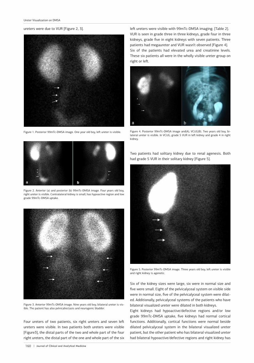
ureters were due to VUR [Figure 2, 3].
Four ureters of two patients, six right ureters and seven left ureters were visible. In two patients both ureters were visible [Figure3], the distal parts of the two and whole part of the four right ureters, the distal part of the one and whole part of the six
left ureters were visible with 99mTc-DMSA imaging. [Table 2].VUR is seen in grade three in three kidneys, grade four in three kidneys, grade five in eight kidneys with seven patients. Three patients had megaureter and VUR wasn’t observed [Figure 4].Six of the patients had elevated urea and creatinine levels. These six patients all were in the wholly visible ureter group on right or left.
Two patients had solitary kidney due to renal agenesis. Both had grade 5 VUR in their solitary kidney [Figure 5].
Six of the kidney sizes were large, six were in normal size and five were small. Eight of the pelvicalyceal system on visible side were in normal size, five of the pelvicalyceal system were dilat-ed. Additionally, pelvicalyceal systems of the patients who have bilateral visualized ureter were dilated in both kidneys.Eight kidneys had hypoactive/defective regions and/or low grade 99mTc-DMSA uptake, five kidneys had normal cortical functions. Additionally, cortical functions were normal beside dilated pelvicalyceal system in the bilateral visualized ureter patient, but the other patient who has bilateral visualized ureter had bilateral hypoactive/defective regions and right kidney has
Figure 1. Posterior 99mTc-DMSA image. One year old boy, left ureter is visible. Figure 4. Posterior 99mTc-DMSA image and(A), VCUG(B). Two years old boy, bi-lateral ureter is visible. In VCUG, grade 5 VUR in left kidney and grade 4 in right kidney.
Figure 5. Posterior 99mTc-DMSA image. Three years old boy, left ureter is visible and right kidney is agenetic.
Figure 2. Anterior (a) and posterior (b) 99mTc-DMSA image. Four years old boy, right ureter is visible. Contralateral kidney is small, has hypoactive region and low grade 99mTc-DMSA uptake.
Figure 3. Anterior 99mTc-DMSA image. Nine years old boy, bilateral ureter is vis-ible. The patient has also pelvicaliectasis and neurogenic bladder.
| Journal of Clinical and Analytical Medicine160
Ureter Visualization on DMSA

| Journal of Clinical and Analytical Medicine
Ureter Visualization on DMSA
4
low grade 99mTc-DMSA uptake [Table 3]. DiscussionVUR is graded into five grades (grade 1-5) according to the se-verity the dilation and tortuosity of ureter and pelvicalyceal sys-tem in case reflux reaches the kidney [11]. VUR is the most com-mon cause of antenatal hydronephrosis for 40% of intrauterin cases [12]. 30% of the children with attack of acute pyelone-phritis had also VUR [13]. In our study, VUR is seen in grade five in eight kidneys with seven patients, grade four in three kidneys, grade three in three kidneys. VUR influences the diameter of the ureter, but ureters may be widened without VUR in case of UTI. Bacterial infection may cause smooth muscle dysfunction in ureteral wall and so UTI may cause dilatation of the ureter [8]. In long standing ureters widen excessively and tortuoused in
high grade VUR cases. By USG examination, find-ings of pelvic and/or calyceal dilatation, ureteral dilatation, pelvic and/or ureteral wall thickening are to be considered as warning for VUR. Normal appearing urinary tract normally does not usually coexist with VUR [12]. VUR may effect the growth of kidneys. In unscarred kidneys as well as most of the kidneys with moderate scarring, normal growth is expected. Growth is impaired in case of severe scarring with little functioning paren-chyma and dilate ureters. In unilateral VUR cases, the abnormal kidney growth is impaired and the opposite normal kidney may get larger to com-pansate the excretion function [14]. First studies about VUR were based on intravenous urogra-phy. Later on, these were replaced with 99mTc-DMSA scintigraphy due to its high sensitivity for the detection on renal defects [15]. In our study, eight patients had abnormal 99mTc-DMSA imag-ing with hypoactive/defective regions and/or low grade activity accumulation. And also, one of the patient in bilateral visualized group had bilateral
hypoactive/defective regions and his right kidney has low grade 99mTc-DMSA uptake.Congenital dysplastic kidneys may be seen with dilated and tortuous ureter in case of high grade VUR [16]. VUR related congenital dysplasia is seen as dilated ureter and pelvicalyceal system with abnormally thin parenchyma and loss of cortico-medullary differentiation in USG examination. 99mTc-DMSA imaging varies in these cases, as asymmetrical uptake of activ-ity with small kidney around dilated pelvicalyceal system may be seen in severe cases [17]. In antenatal diagnosed dysplastic kidney, two normal neonate renal USG can exclude significant abnormalities and so VCUG is not needed in the evaluation of VUR [18]. VCUG is indicated for the first examination of VUR in boys, inadequate visualization of the bladder of kidney and spe-cific request for for urethral or bladder imaging [19]. When renal developmental abnormalities are recognized prenatally, VCUG is indicated 4-6 weeks after birth [20].99mTc-DMSA scintigraphy is used for the diagnosis and follow up of pyelonephritis with detecting the renal cortical defective/hypoactive regions and calculate the differential renal functions. 90% of 99mTc-DMSA is bound to plasma proteins and 0%-5% to red blood cells. 40-50% of injected activity is taken by the kidney within 3-4 hours of injection and 6%-9% of the dose is present in the blood at 14 hours after injection [21]. In normal conditions, 99mTc-DMSA is concentrated only by the cortex and pelvicalyceal system and ureters are invisible. Megaureter is the presence of enlarged ureter with or without dilatation of the upper collecting system and is possibly caused by congenital (primary) or abnormalities of bladder or urethra [22]. Diameter of normal ureter is almost always smaller than 5 mm [8]. Zelenko et al. defined the normal ureter diameter in ureterolithiasis patients by comparing the symptomatic and as-ymptomatic kidneys with unenhanced helical computed tomog-raphy. They described that in 96% of patients; normal ureter diameter was less than 3 mm with 6.6% of the patients’ ureter less than 3 mm and mentioned that 3 mm should be considered
Table 2. Demographic, biochemical and VCUG findings of the patients
Patient no
Age Gender Urea Level (mg/dL)
Creatinine level (mg/dL)
Visible side VUR grade
1 11 months Boy 25 0,5 Left distal ureter 3
2 10 Girl 42 1,42 Left whole ureter 3
3 6 Girl 34 1,35 Right whole ureter 3
4 1 Boy 27 0,6 Left whole ureter Megaureter
5 2 Boy 30 0,62 Right 1/2 distal ureter
4
6 10 months Girl 7 0,4 Right whole ureter 4
7 3 Boy 33 0,74 Left whole ureter Megaureter
8 9 Boy 27 0,9 Bilateral whole ureter Bilateral 5
9 18 Girl 69 1,32 Left whole ureter 5
10 5 Boy 24 0,77 Left whole ureter Megaureter
11 4 Boy 26 0,81 Right distal ureter 5
12 7 Girl 42 1,41 Left whole ureter 5
13 13 Boy 32 1,28 Right whole ureter 5
14 2 months Boy 18 0,59 Right whole ureter 5
15 2 Boy 40 0,67 Bilateral whole ureter Left VUR 5, Right VUR 4
Table 3. Side and part of the visible ureter with the 99mTc-DMSA, uptake ratios of the same side of the kidney are shown.
Patient no
Visible side DMSA uptake of kidney (%) (Side of visible ureter)
DMSA uptake of kidney (%) (Side of nonvisible ureter)
1 Left distal ureter 18 82
2 Left whole ureter 52 48
3 Right whole ureter 3 97
4 Left whole ureter 50 50
5 Right 1/2 distal ureter
11 89
6 Right whole ureter 31 69
7 Left whole ureter 100 0
8 Bilateral whole ureter Left 53, Right 47
9 Left whole ureter 57 43
10 Left whole ureter 92 8
11 Right distal ureter 90 10
12 Left whole ureter 20 80
13 Right whole ureter 76 24
14 Right whole ureter 100 0
15 Bilateral whole ureter Left 85, Right 15
Journal of Clinical and Analytical Medicine | 161
Ureter Visualization on DMSA

| Journal of Clinical and Analytical Medicine
Ureter Visualization on DMSA
5
as upper limit of normal size for nonobstructed ureters [23]. If megaureter is not due to VUR, diuresis renography is indicated [24]. In our cases, three of the patients had megaureter and don’t have VUR. In three cases, the proximal part of the ureter seemed widened, but in their USG and/or 99mTc MAG3 images, extremely dilated pelvicalyceal system were observed. Two of 15 patients had only one kidney and their left kidney was age-netic. Pelvicalyceal system on the visible side was dilated in eight kidneys of six patients in USG and other patients’ pelvi-calyceal system was not dilated. In conclusion, ureters may become visible with 99mTc-DMSA scintigraphy in patients with congenital megaureter and VUR, more even so in high grade VUR. Ureter visualization with 99mTc-DMSA scintigraphy should urge the clinician to perform further imaging methods like VCUG to define the grade and therapy of the patients even if 99mTc-DMSA scintigraphy is normal.
DisclosureThe authors stated that they had no interests which might be perceived as posing a conflict or bias.
Competing interestsThe authors declare that they have no competing interests. References 1. Garin EH1, Olavarria F, Garcia Nieto V, Valenciano B, Campos A, Young L. Clini-cal significance of primary vesicoureteral reflux and urinary antibiotic prophylaxis after acute pyelonephritis: a multicenter, randomized, controlled study. Pediatrics 2006;117(3):626-32.2. Coulthard MG. Vesicoureteric reflux is not a benign condition. Pediatr Nephrol 2009;24(2):227-32.3. Darge K, Riedmiller H. Current status of vesicoureteral reflux diagnosis. World J Urol 2004;22(2):88-95.4. Schneider K, Krüger-Stollfuss I, Ernst G, Kohn MM. Paediatric fluoroscopy—a survey of children’s hospitals in Europe. I. Staffing, frequency of fluoroscopic pro-cedures and investigation technique. Pediatr Radiol 2001;31(4):238-46.5. Goldraich NP, Goldraich IH. Update on dimercaptosuccinic acid renal scanning in children with urinary tract infection. Pediatr Nephrol 1995;9(2):221-6.6. Merrick MV, Uttley WS, Wild SR. The detection of pyelonephritic scarring in children by radioisotope imaging. Br J Radiol 1980;53(630):544-6.7. Lee BH, Lee SH, Choi HJ, Kang HG, Oh SW, Lee DS, et al. Decreased renal uptake of (99m)Tc-DMSA in patients with tubular proteinuria. Pediatr Nephrol 2009;24(11):2211-6.8. Hellström M, Jodal U, Mårild S, Wettergren B. Ureteral dilatation in chil-dren with febrile urinary tract infection or bacteriuria. AJR Am J Roentgenol 1987;148(3):483-6.9. Rabinowitz R, Barkin M, Schillinger JF, Jeffs RD. Surgical treatment of the mas-sively dilated primary megaureter in children. Br J Urol 1979;51(1):19-23.10. Turkolmez S, Ors D, Korkmaz M. Megaureter visualization on 99mTc-DMSA scintigraphy. Ann Nucl Med 2005;19(5):421-3.11. Jaswon MS, Dibble L, Puri S, Davis J, Young J, Dave R, et al. Prospective study of outcome in antenatally diagnosed renal pelvis dilatation. Arch Dis Child Fetal Neonatal Ed 1999;80(2):135-8.12. Avni EF, Ayadi K, Rypens F, Hall M, Schulman CC. Can careful ultrasound ex-amination of the urinary tract exclude vesicoureteric reflux in the neonate? Br J Radiol 1997;70(838):977-82.13. Rosenberg AR, Rossleigh MA, Brydon MP, Bass SJ, Leighton DM, Farnsworth RH. Evaluation of acut urinary tract infection in children by dimercaptosuccinic acid scintigraphy: A prospective study. J Urol 1992;148(5):1746-9.14. Smellie JM, Edwards D, Normand IC, Prescod N. Effect of vesicoureteric reflux on renal growth in children with urinary tract infection. Arch Dis Child 1981;56(8):593-600.15. Grattan-Smith JD, Little SB, Jones RA. Evaluation of reflux nephropathy, pyelo-nephritis and renal dysplasia. Pediatr Radiol 2008;38(1):83-105. 16. Risdon RA. The small scarred kidney in childhood. Pediatr Nephrol 1993;7(4):361-4.17. Coulthard MG. Vesicoureteric reflux is not a benign condition. Pediatr Nephrol 2009;24(2):227-32.18. Ismaili K, Avni FE, Alexander M, Schulman C, Collier F, Hall M. Routine voiding cystourethrography is of no value in neonates with unilateral multicystic dysplas-tic kidney. J Pediatr 2005;146(6):759-63.
19. Risdon RA. The small scarred kidney in childhood. Pediatr Nephrol 1993;7(4):361-4.20. John U, Rudnik-Schöneborn S, Zerres K, Misselwitz J.Kidney growth and re-nal function in unilateral multicystic dysplastic kidney disease. Pediatr Nephrol 1998;12(7):567-71.21. Treves ST, Baker A, Fahey FH, Cao X, Davis RT, Drubach LA, et al. Nuclear medicine in the first year of life. J Nucl Med 2011;52(6):905-25.22. Zerin JM. Hydronephrosis in the neonate and young infant: current concepts. Semin Ultrasound CT MR 1994;15(4):306-16.23. Zelenko N, Coll D, Rosenfeld AT, Smith RC. Normal ureter size on unenhanced helical CT. AJR Am J Roentgenol 2004;182(4):1039-41.24. Berrocal T, López-Pereira P, Arjonilla A, Gutiérrez J. Anomalies of the distal ureter, bladder, and urethra in children: embryologic, radiologic, and pathologic features. Radiographics 2002;22(5):1139-64.
How to cite this article:Atilgan HI, Sadic M, Korkmaz M, Ozyurt S, Koca G. Is Ureter Visualization Possible on Tc-99m DMSA Scintigraphy with Vesicoureteral Reflux Patients? J Clin Anal Med 2016;7(2): 158-62.
| Journal of Clinical and Analytical Medicine162
Ureter Visualization on DMSA