Embed Size (px)
Citation preview
EDITORIAL
Is nano too big?
Walter Mier & John Babich & Uwe Haberkorn
Published online: 3 October 2013# The Author(s) 2013. This article is published with open access at Springerlink.com
The possibility to utilize nanotechnology for drug develop-ment has led to widespread interest and funding opportunities.The public perception of the field of nanoscale science orig-inally referred to technologies dealing with nanomechanics,nanomaterials, nanooptics and nanoelectronics, but increas-ingly anticipates progress in nanomedicine. Today’s molecu-lar imaging possibilities are ideal for visualizing the pharma-cokinetics of the effectors used in new treatment strategiessuch as gene therapy [1] and therapeutic vaccination [2]. Withthe possibility to visualize and the salient, concomitant treat-ment, nuclear medicine has the greatest potential for use in thedevelopment of innovative treatment strategies. Consequent-ly, the eventual applications of nanosized tracers are of thegreatest interest for our discipline.
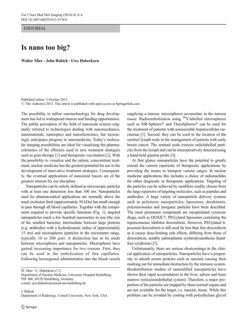
Nanoparticles can be strictly defined as microscopic particleswith at least one dimension less than 100 nm. Nanoparticlesused for pharmaceutical applications are normally above therenal exclusion limit (approximately 50 kDa) but small enoughto pass through all blood capillaries. Together with the compo-nents required to provide specific functions (Fig. 1), targetednanoparticles reach a few hundred nanometres in size (the sizeof the smallest bacteria), intermediate between large proteins(e.g. antibodies with a hydrodynamic radius of approximately15 nm) and microspheres (particles in the micrometre range,typically 10 to 200 µm). A distinction has to be madebetween microspheres and nanoparticles. Microspheres havegained increasing importance for two reasons. First, theycan be used in the embolization of fine capillaries.Following locoregional administration into the blood vessels
supplying a tumour, microspheres accumulate in the tumourtissue. Radioembolization using 90Y-labelled microspheressuch as SIR-Spheres® and TheraSpheres® can be used forthe treatment of patients with unresectable hepatocellular car-cinoma [3]. Second, they can be used in the location of thesentinel lymph node in the management of patients with earlybreast cancer. The sentinel node extracts radiolabelled parti-cles from the lymph and can be intraoperatively detected usinga hand-held gamma probe [4].
At first glance nanoparticles have the potential to greatlyextend the current repertoire of therapeutic applications byproviding the means to transport various cargos. In nuclearmedicine applications this includes a choice of radionuclidesfor either diagnostic or therapeutic applications. Targeting ofthe particles can be achieved by modifiers readily chosen fromthe large repertoire of targeting molecules, such as peptides andantibodies. A large variety of nanoparticle delivery systemssuch as polymeric nanoparticles, liposomes, dendrimers,polymerosomes and inorganic particles have been described.The most prominent compounds are encapsulated cytotoxicdrugs, such as DOXIL®, PEGylated liposomes containing thetopoisomerase inhibitor doxorubicin. However, PEGylated li-posomal doxorubicin is still used far less than free doxorubicinas it causes dose-limiting side effects, differing from those ofdoxorubicin, notably palmoplantar erythrodysaesthesia (hand-foot syndrome) [5].
Unfortunately, there are serious shortcomings in the clini-cal application of nanoparticles. Nanoparticles have a propen-sity to adsorb serum proteins such as opsonin causing theirmarking out for immediate destruction by the immune system.Biodistribution studies of unmodified nanoparticles haveshown their rapid accumulation in the liver, spleen and bonemarrow (reticuloendothelial system). Therefore, a major pro-portion of the particles are trapped by these normal organs andare not available for the target, i.e. tumour, tissue. While thisproblem can be avoided by coating with polyethylene glycol
W. Mier :U. Haberkorn (*)Department of Nuclear Medicine, University Hospital Heidelberg,INF 400, 69120 Heidelberg, Germanye-mail: [email protected]
J. BabichDepartment of Radiology, Cornell University, New York, USA
Eur J Nucl Med Mol Imaging (2014) 41:4–6DOI 10.1007/s00259-013-2574-9
[6], it should be born in mind that any coating chosen tosuppress recognition by the immune system is inevitablyassociated with interference with specific binding.
The lack of specificity of a naked nanoparticle has beenapproached by coupling the nanoparticles with targeting mol-ecules such as antibodies, receptor affine peptides, folatereceptor ligands or bisphosphonates on their surface [7].However, the number of potential targeting molecules is lim-ited to molecules that are not influenced by conjugation toparticles. The strategy to direct the accumulation to a specifictarget whilst limiting nonspecific accumulation faces twoproblems: (1) the affinity of the targeting molecule isinfluenced by the large particle to which it is attached, and(2) the biodistribution of the small molecule/peptide/antibodyis dominated by the large particle. All modifications requiredto facilitate targeting and biocompatibility, however, increasethe size of the particle, and such increase is detrimental to thequality of the pharmaceutical which generally decreases withincreasing size [8].
It is possible that nanoparticles can be transported throughthe interendothelial junctions which are enlarged in tumours.However, the likelihood that large constructs can penetratedeeply into tissue, and make their way back from sites whereno specific interaction takes place can be estimated from acomparison with oxygen. The permanent oxygen shortage inlarge solid tumours reveals the enormous diffusion barrier tobe overcome. The diffusion constants of O2 and macromole-cules differ by many orders of magnitude. Tumours show apoorly organized vascular architecture and compression ofblood and lymphatic vessels by cancer cells. In addition tohypoxia, this creates increased interstitial fluid pressure whichcan slow down the movement of molecules within the tumourand limits the delivery of drugs to cells located far from
functioning blood vessels [9]. Mobility is therefore a mainfactor defining the performance of a tracer in an organismexpressing a target at a defined specificity. Antibodies are aninstructive example of this. Their large size counteracts theirexcellent properties and binding affinity. However, antibodieshave the advantage of outstanding biocompatibility and there-fore attain exceptional circulation times. This increases theprobability of constructive interaction and thus compensatesfor their lower mobility.
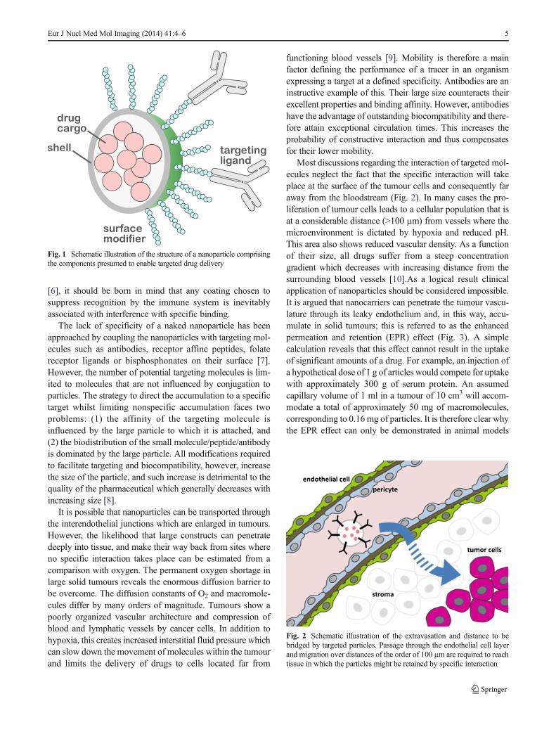
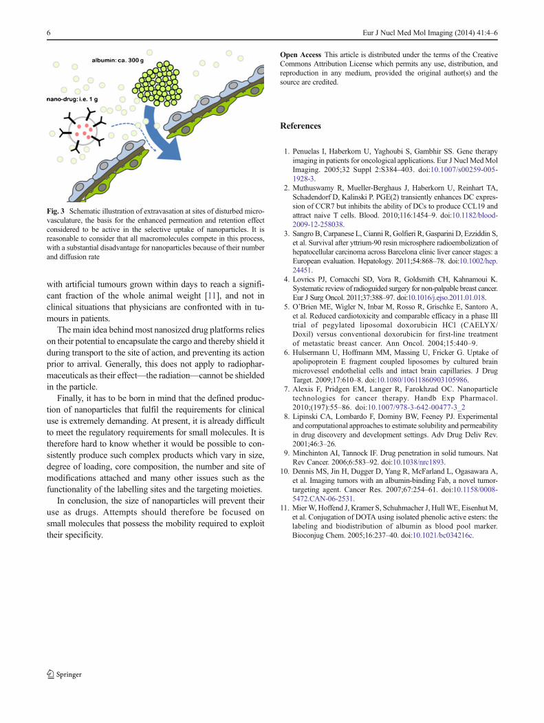
Most discussions regarding the interaction of targeted mol-ecules neglect the fact that the specific interaction will takeplace at the surface of the tumour cells and consequently faraway from the bloodstream (Fig. 2). In many cases the pro-liferation of tumour cells leads to a cellular population that isat a considerable distance (>100 µm) from vessels where themicroenvironment is dictated by hypoxia and reduced pH.This area also shows reduced vascular density. As a functionof their size, all drugs suffer from a steep concentrationgradient which decreases with increasing distance from thesurrounding blood vessels [10].As a logical result clinicalapplication of nanoparticles should be considered impossible.It is argued that nanocarriers can penetrate the tumour vascu-lature through its leaky endothelium and, in this way, accu-mulate in solid tumours; this is referred to as the enhancedpermeation and retention (EPR) effect (Fig. 3). A simplecalculation reveals that this effect cannot result in the uptakeof significant amounts of a drug. For example, an injection ofa hypothetical dose of 1 g of articles would compete for uptakewith approximately 300 g of serum protein. An assumedcapillary volume of 1 ml in a tumour of 10 cm3 will accom-modate a total of approximately 50 mg of macromolecules,corresponding to 0.16 mg of particles. It is therefore clear whythe EPR effect can only be demonstrated in animal models
Fig. 1 Schematic illustration of the structure of a nanoparticle comprisingthe components presumed to enable targeted drug delivery
Fig. 2 Schematic illustration of the extravasation and distance to bebridged by targeted particles. Passage through the endothelial cell layerand migration over distances of the order of 100 µm are required to reachtissue in which the particles might be retained by specific interaction
Eur J Nucl Med Mol Imaging (2014) 41:4–6 5
with artificial tumours grown within days to reach a signifi-cant fraction of the whole animal weight [11], and not inclinical situations that physicians are confronted with in tu-mours in patients.
The main idea behind most nanosized drug platforms relieson their potential to encapsulate the cargo and thereby shield itduring transport to the site of action, and preventing its actionprior to arrival. Generally, this does not apply to radiophar-maceuticals as their effect—the radiation—cannot be shieldedin the particle.
Finally, it has to be born in mind that the defined produc-tion of nanoparticles that fulfil the requirements for clinicaluse is extremely demanding. At present, it is already difficultto meet the regulatory requirements for small molecules. It istherefore hard to know whether it would be possible to con-sistently produce such complex products which vary in size,degree of loading, core composition, the number and site ofmodifications attached and many other issues such as thefunctionality of the labelling sites and the targeting moieties.
In conclusion, the size of nanoparticles will prevent theiruse as drugs. Attempts should therefore be focused onsmall molecules that possess the mobility required to exploittheir specificity.
Open Access This article is distributed under the terms of the CreativeCommons Attribution License which permits any use, distribution, andreproduction in any medium, provided the original author(s) and thesource are credited.
References
1. Penuelas I, Haberkorn U, Yaghoubi S, Gambhir SS. Gene therapyimaging in patients for oncological applications. Eur J Nucl MedMolImaging. 2005;32 Suppl 2:S384–403. doi:10.1007/s00259-005-1928-3.
2. Muthuswamy R, Mueller-Berghaus J, Haberkorn U, Reinhart TA,Schadendorf D, Kalinski P. PGE(2) transiently enhances DC expres-sion of CCR7 but inhibits the ability of DCs to produce CCL19 andattract naive T cells. Blood. 2010;116:1454–9. doi:10.1182/blood-2009-12-258038.
3. Sangro B, Carpanese L, Cianni R, Golfieri R, Gasparini D, Ezziddin S,et al. Survival after yttrium-90 resin microsphere radioembolization ofhepatocellular carcinoma across Barcelona clinic liver cancer stages: aEuropean evaluation. Hepatology. 2011;54:868–78. doi:10.1002/hep.24451.
4. Lovrics PJ, Cornacchi SD, Vora R, Goldsmith CH, Kahnamoui K.Systematic review of radioguided surgery for non-palpable breast cancer.Eur J Surg Oncol. 2011;37:388–97. doi:10.1016/j.ejso.2011.01.018.
5. O’Brien ME, Wigler N, Inbar M, Rosso R, Grischke E, Santoro A,et al. Reduced cardiotoxicity and comparable efficacy in a phase IIItrial of pegylated liposomal doxorubicin HCl (CAELYX/Doxil) versus conventional doxorubicin for first-line treatmentof metastatic breast cancer. Ann Oncol. 2004;15:440–9.
6. Hulsermann U, Hoffmann MM, Massing U, Fricker G. Uptake ofapolipoprotein E fragment coupled liposomes by cultured brainmicrovessel endothelial cells and intact brain capillaries. J DrugTarget. 2009;17:610–8. doi:10.1080/10611860903105986.
7. Alexis F, Pridgen EM, Langer R, Farokhzad OC. Nanoparticletechnologies for cancer therapy. Handb Exp Pharmacol.2010;(197):55–86. doi:10.1007/978-3-642-00477-3_2
8. Lipinski CA, Lombardo F, Dominy BW, Feeney PJ. Experimentaland computational approaches to estimate solubility and permeabilityin drug discovery and development settings. Adv Drug Deliv Rev.2001;46:3–26.
9. Minchinton AI, Tannock IF. Drug penetration in solid tumours. NatRev Cancer. 2006;6:583–92. doi:10.1038/nrc1893.
10. Dennis MS, Jin H, Dugger D, Yang R, McFarland L, Ogasawara A,et al. Imaging tumors with an albumin-binding Fab, a novel tumor-targeting agent. Cancer Res. 2007;67:254–61. doi:10.1158/0008-5472.CAN-06-2531.
11. MierW, Hoffend J, Kramer S, Schuhmacher J, Hull WE, EisenhutM,et al. Conjugation of DOTA using isolated phenolic active esters: thelabeling and biodistribution of albumin as blood pool marker.Bioconjug Chem. 2005;16:237–40. doi:10.1021/bc034216c.
Fig. 3 Schematic illustration of extravasation at sites of disturbed micro-vasculature, the basis for the enhanced permeation and retention effectconsidered to be active in the selective uptake of nanoparticles. It isreasonable to consider that all macromolecules compete in this process,with a substantial disadvantage for nanoparticles because of their numberand diffusion rate
6 Eur J Nucl Med Mol Imaging (2014) 41:4–6