Embed Size (px)
Citation preview
1
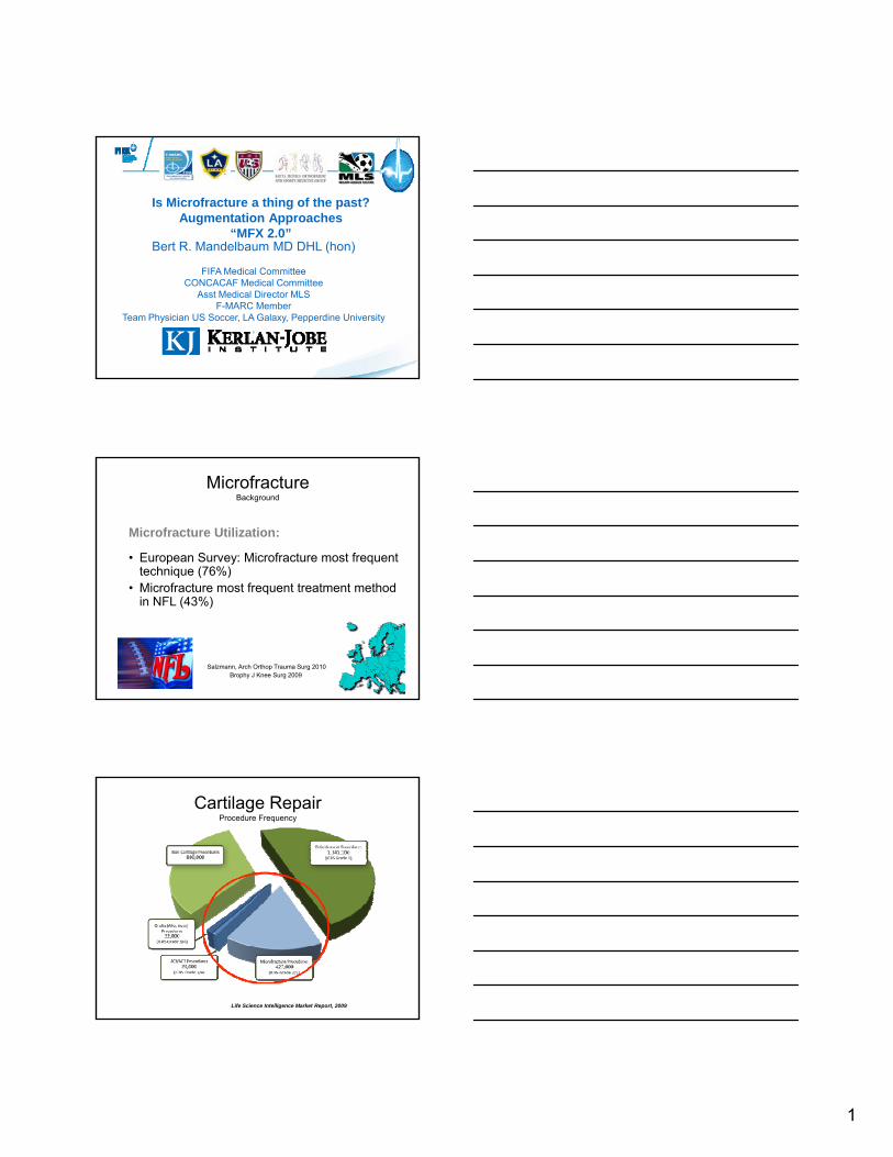
Is Microfracture a thing of the past?Augmentation Approaches
“MFX 2.0”Bert R. Mandelbaum MD DHL (hon)
FIFA Medical CommitteeCONCACAF Medical Committee
Asst Medical Director MLSF-MARC Member
Team Physician US Soccer, LA Galaxy, Pepperdine University,
MicrofractureBackground
Microfracture Utilization:
• European Survey: Microfracture most frequent technique (76%)
• Microfracture most frequent treatment method in NFL (43%)
Salzmann, Arch Orthop Trauma Surg 2010Brophy J Knee Surg 2009
Cartilage RepairProcedure Frequency
Life Science Intelligence Market Report, 2009
2
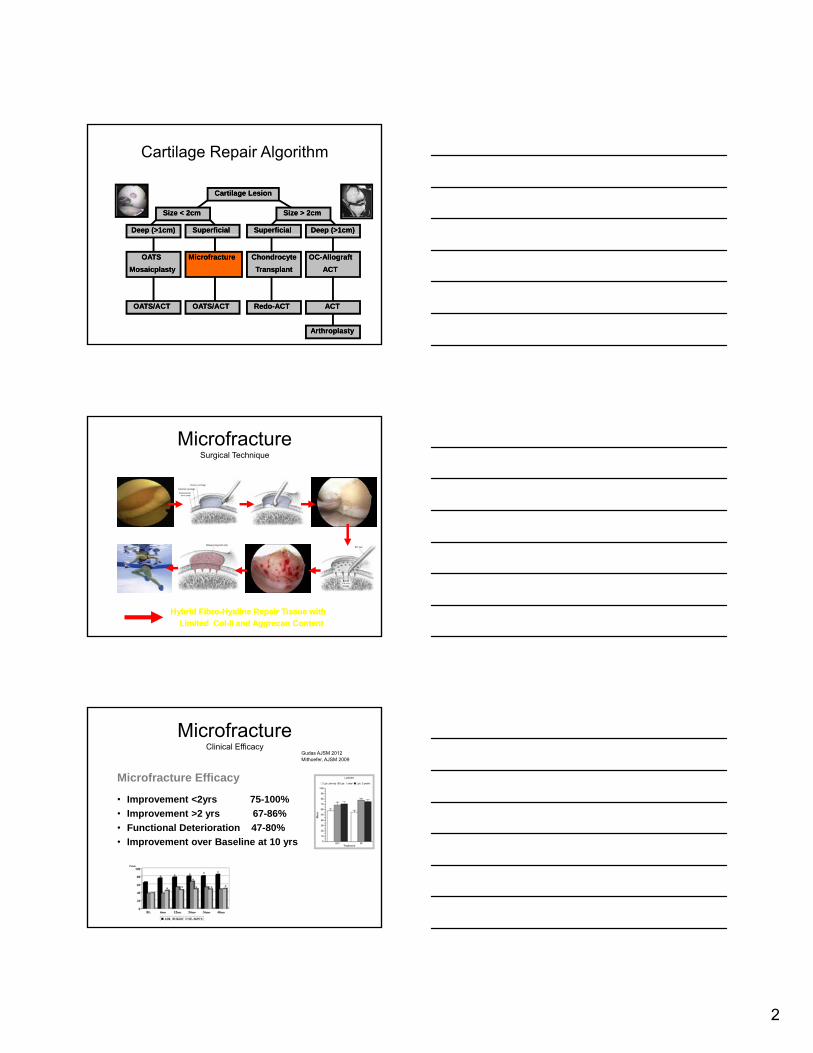
Cartilage Repair Algorithm
Cartilage LesionCartilage Lesion
SizeSize < 2cm< 2cm
Deep (>1cm)Deep (>1cm) SuperficialSuperficial SuperficialSuperficial
SizeSize > 2cm> 2cm
Deep (>1cm)Deep (>1cm)
MicrofractureMicrofracture
OATS/ACTOATS/ACT
OATSOATS
MosaicplastyMosaicplasty
OATS/ACTOATS/ACT
ChondrocyteChondrocyte
TransplantTransplant
OCOC--AllograftAllograft
ACTACT
RedoRedo--ACTACT ACTACT
ArthroplastyArthroplasty
MicrofractureSurgical Technique
Hybrid FibroHybrid Fibro--Hyaline Repair Tissue with Hyaline Repair Tissue with
Limited ColLimited Col--II and II and AggrecanAggrecan ContentContent
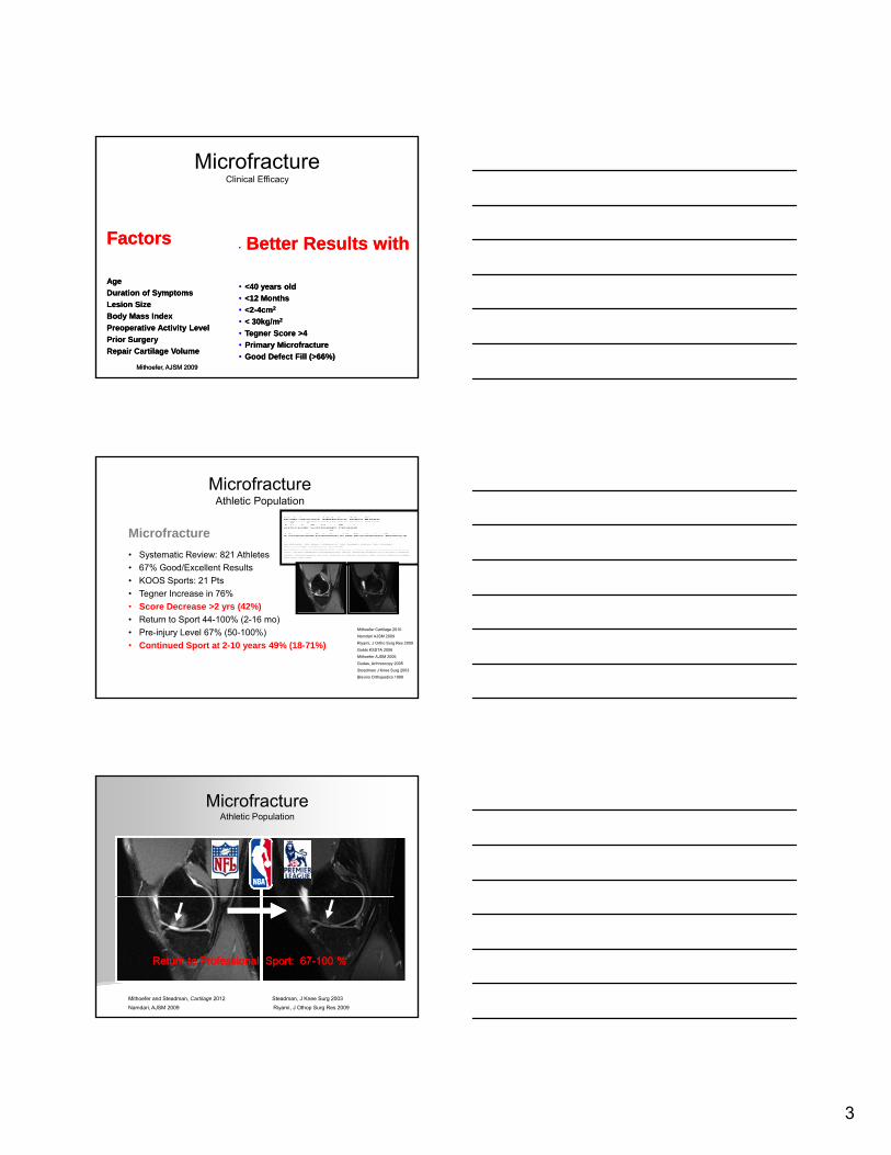
MicrofractureClinical Efficacy
Microfracture Efficacy
• Improvement <2yrs 75-100%
• Improvement >2 yrs 67-86%
Gudas AJSM 2012Mithoefer, AJSM 2009
p y
• Functional Deterioration 47-80%
• Improvement over Baseline at 10 yrs
3
MicrofractureClinical Efficacy
FactorsFactors•• Better Results with Better Results with
Mithoefer, AJSM 2009Mithoefer, AJSM 2009
AgeAge
Duration of SymptomsDuration of Symptoms
Lesion SizeLesion Size
Body Mass IndexBody Mass Index
Preoperative Activity LevelPreoperative Activity Level
Prior SurgeryPrior Surgery
Repair Cartilage VolumeRepair Cartilage Volume
•• <40 years old<40 years old
•• <12 Months<12 Months
•• <2<2--4cm4cm22
•• < 30kg/m< 30kg/m22
•• TegnerTegner Score >4Score >4
•• Primary Primary MicrofractureMicrofracture
•• Good Defect Fill (>66%)Good Defect Fill (>66%)
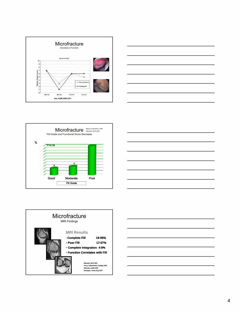
MicrofractureAthletic Population
Microfracture
• Systematic Review: 821 Athletes
• 67% Good/Excellent Results
KOOS S 21 P• KOOS Sports: 21 Pts
• Tegner Increase in 76%
• Score Decrease >2 yrs (42%)
• Return to Sport 44-100% (2-16 mo)
• Pre-injury Level 67% (50-100%)
• Continued Sport at 2-10 years 49% (18-71%)
Mithoefer Cartilage 2010
Namdari AJSM 2009
Riyami, J Ortho Surg Res 2009
Gobbi KSSTA 2006
Mithoefer AJSM 2006
Gudas, Arthroscopy 2005
Steadman J Knee Surg 2003
Blevins Orthopedics 1999
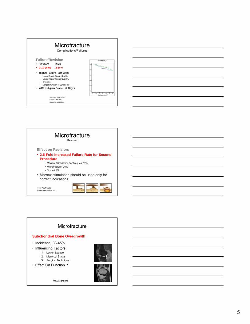
MicrofractureAthletic Population
RReturn to Professional Sporteturn to Professional Sport:: 6767--100 %100 %
Mithoefer and Steadman, Cartilage 2012 Steadman, J Knee Surg 2003
Namdari, AJSM 2009 Riyami, J Othop Surg Res 2009
4
MicrofractureDecrease of Function
KonKon, AJSM 2009+2011, AJSM 2009+2011
MicrofractureFill Grade and Functional Score Decrease
%%*P<0.05*P<0.05
Blevins Orthopedics 1999
Mithoefer JBJS 2005
Good Moderate PoorGood Moderate Poor
**
Fill GradeFill Grade
**
Microfracture MRI Findings
MRI ResultsComplete Fill 18Complete Fill 18--95%95%
Poor Fill 17Poor Fill 17 57%57%
Mithoefer Mithoefer JBJSJBJS 20052005
Kreuz, Osteoarthritis Cartilage 2006Kreuz, Osteoarthritis Cartilage 2006
Mithoefer, AJSM 2009Mithoefer, AJSM 2009
Ramappa, J Knee Surg 2007Ramappa, J Knee Surg 2007
Poor Fill 17Poor Fill 17--57%57%
Complete Integration 4Complete Integration 4--8%8%
Function Correlates with FillFunction Correlates with Fill
5
Microfracture Complications/Failures
Failure/Revision• <2 years 2.5%
• 2-10 years 2-38%
Hi h F il R t ith• Higher Failure Rate with:– Lower Repair Tissue Quality
– Lower Repair Tissue Quantity
– Smoking
– Longer Duration of Symptoms
• 48% Kellgren Grade I at 10 yrs
Salzmann KSSTA 2012
Gudas AJSM 2012
Mithoefer, AJSM 2009
MicrofractureRevision
Effect on Revision:
• 2.5-Fold Increased Failure Rate for Second Procedure
• Marrow Stimulation Techniques 26%
• Microfracture 20%
• Control 8%
• Marrow stimulation should be used only for correct indications
Minas AJSM 2009
Jungermann AJSM 2012
Microfracture
Subchondral Bone Overgrowth
• Incidence: 33-45%
• Influencing Factors:1 L i L ti1. Lesion Location
2. Meniscal Status
3. Surgical Technique
• Effect On Function ?
MithoeferMithoefer ICRS 2012ICRS 2012
6
MicrofractureBone Overgrowth
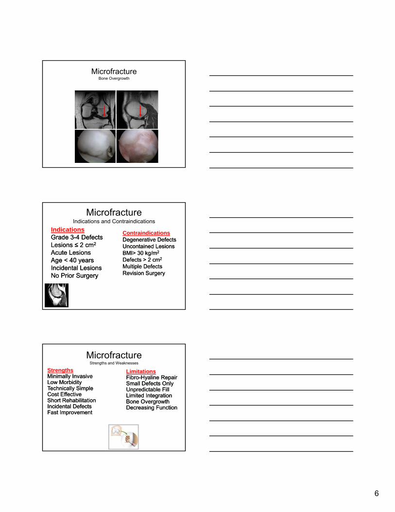
MicrofractureIndications and Contraindications
IndicationsGrade 3Grade 3--4 Defects4 DefectsLesions ≤ 2 cmLesions ≤ 2 cm22
Acute LesionsAcute Lesions
ContraindicationsDegenerative DefectsDegenerative DefectsUncontained LesionsUncontained LesionsBMI> 30 kg/mBMI> 30 kg/m22
Age < 40 yearsAge < 40 yearsIncidental LesionsIncidental LesionsNo Prior SurgeryNo Prior Surgery
ggDefects > 2 cmDefects > 2 cm22
Multiple DefectsMultiple DefectsRevision SurgeryRevision Surgery
MicrofractureStrengths and Weaknesses
StrengthsMinimally InvasiveMinimally InvasiveLow MorbidityLow MorbidityTechnically SimpleTechnically SimpleCost EffectiveCost Effective
LimitationsFibroFibro--Hyaline RepairHyaline RepairSmall Defects Only Small Defects Only Unpredictable FillUnpredictable FillLimited IntegrationLimited Integration
Short RehabilitationShort RehabilitationIncidental DefectsIncidental DefectsFast ImprovementFast Improvement
ggBone OvergrowthBone OvergrowthDecreasing FunctionDecreasing Function
7
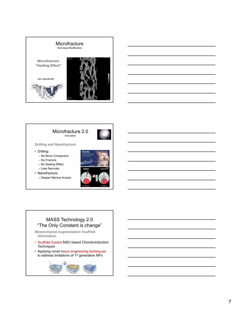
MicrofractureTechnique Modification
MicrofractureMicrofracture“Sealing Effect”“Sealing Effect”
Chen J Chen J OrthopOrthop Res 2009Res 2009
Microfracture 2.0Innovation
Drilling and Nanofracture
• Drilling:– No Bone CompactionNo Bone Compaction
– No Fracture
– No Sealing Effect
– Less Necrosis
• Nanofracture:– Deeper Marrow Access
MASS Technology 2.0“The Only Constant is change”
Mesenchymal Augmentation Scaffold Stimulation
• Scaffold-Guided MSC-based ChondroinductionScaffold Guided MSC based Chondroinduction Techniques
• Applying novel tissue engineering techniques to address limitations of 1st generation MFx
8
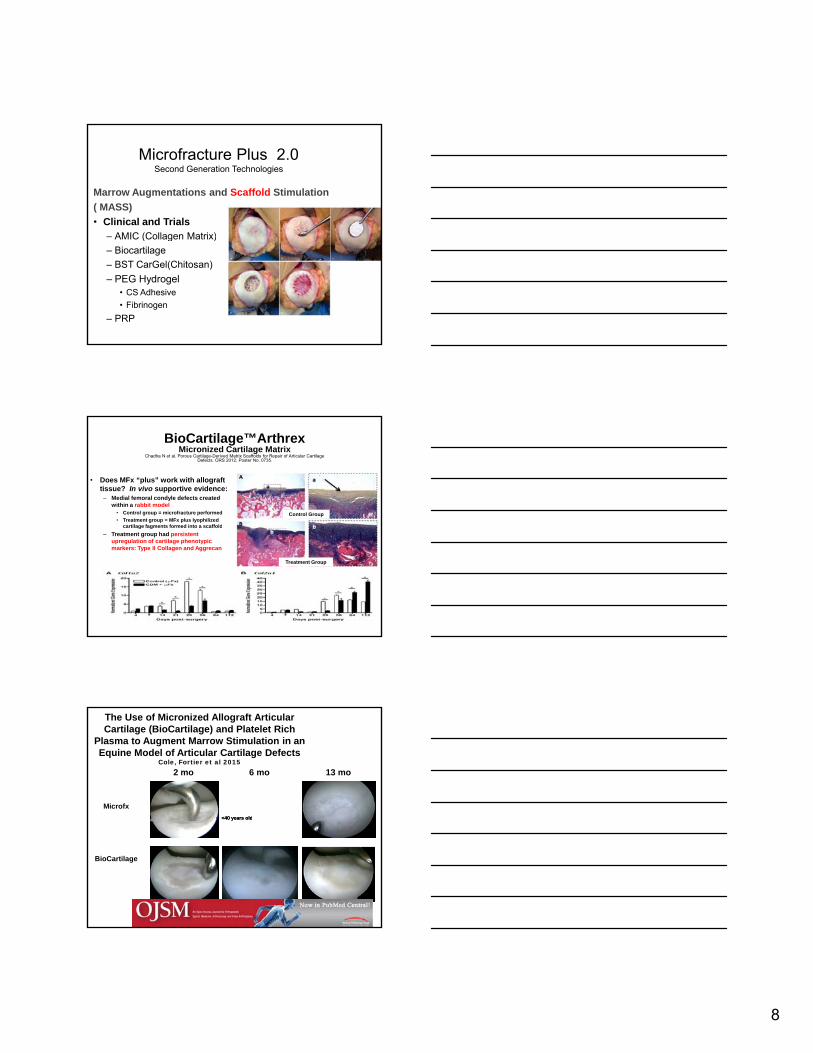
Microfracture Plus 2.0Second Generation Technologies
Marrow Augmentations and Scaffold Stimulation
( MASS)
• Clinical and TrialsAMIC (Collagen Matrix)– AMIC (Collagen Matrix)
– Biocartilage
– BST CarGel(Chitosan)
– PEG Hydrogel • CS Adhesive
• Fibrinogen
– PRP
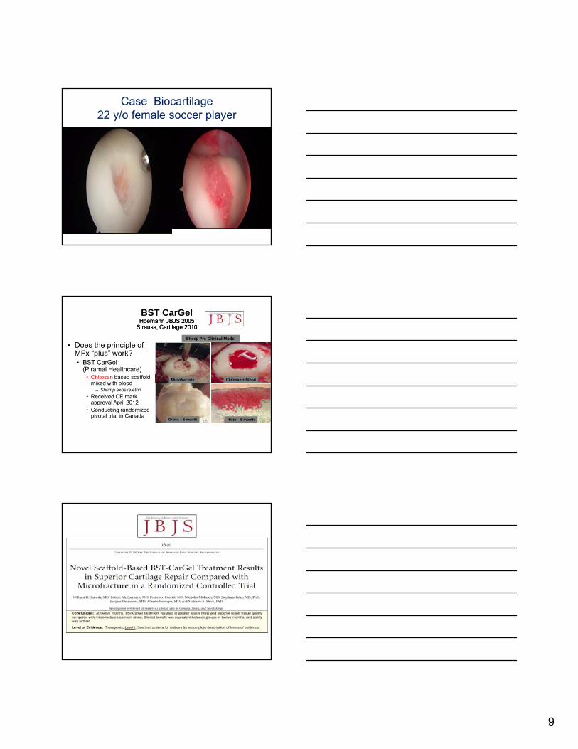
• Does MFx “plus” work with allograft tissue? In vivo supportive evidence:
– Medial femoral condyle defects created within a rabbit model
• Control group = microfracture performed
• Treatment group = MFx plus lyophilized cartilage fagments formed into a scaffold
Control Group
BioCartilage™ArthrexMicronized Cartilage Matrix
Chadha N et al. Porous Cartilage-Derived Matrix Scaffolds for Repair of Articular Cartilage Defects. ORS 2012; Poster No. 0735.
cartilage fagments formed into a scaffold
– Treatment group had persistent upregulation of cartilage phenotypic markers: Type II Collagen and Aggrecan
Treatment Group
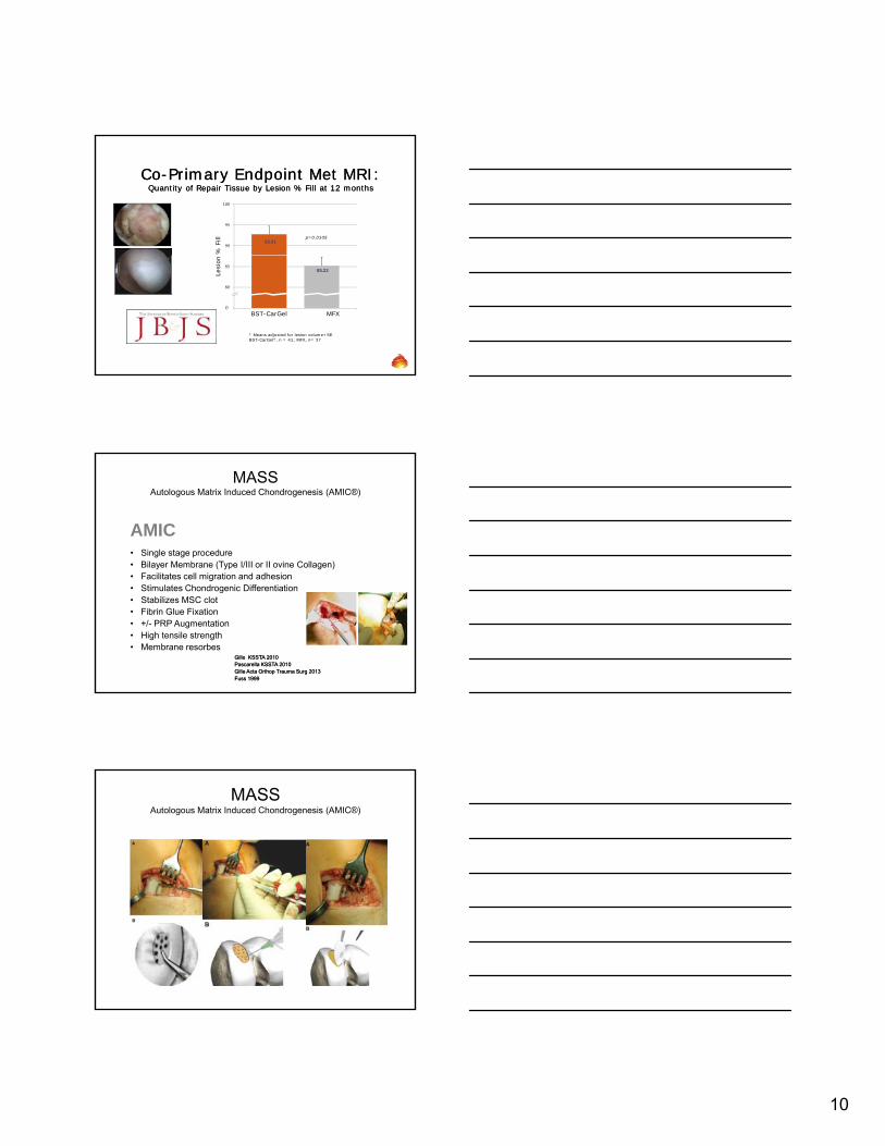
The Use of Micronized Allograft Articular Cartilage (BioCartilage) and Platelet Rich
Plasma to Augment Marrow Stimulation in an Equine Model of Articular Cartilage Defects
Cole, Fortier et al 2015
Microfx
2 mo 6 mo 13 mo
•• <40 years old<40 years old
BioCartilage
•• <40 years old<40 years old
9
Case Biocartilage 22 y/o female soccer player
• Does the principle of MFx “plus” work?• BST CarGel
(Piramal Healthcare)
Sheep Pre-Clinical Model
BST CarGelHoemannHoemann JBJS 2005JBJS 2005
Strauss, Cartilage 2010Strauss, Cartilage 2010
• Chitosan based scaffold mixed with blood
– Shrimp exoskeleton
• Received CE mark approval April 2012
• Conducting randomized pivotal trial in Canada
Microfracture Chitosan + Blood
Gross – 6 month Histo – 6 month
10
CoCo--Primary Endpoint Met MRI: Primary Endpoint Met MRI: Quantity of Repair Tissue by Lesion % Fill at 12 monthsQuantity of Repair Tissue by Lesion % Fill at 12 months
90
95
100
% F
ill 92.81p=0.0105
* Means adjusted for lesion volume+SEBST-CarGel®, n = 41; MFX, n= 37
75
80
85
Lesi
on
0
BST-CarGel MFX
85.22
MASSAutologous Matrix Induced Chondrogenesis (AMIC®)
AMIC• Single stage procedure• Bilayer Membrane (Type I/III or II ovine Collagen) • Facilitates cell migration and adhesion• Stimulates Chondrogenic Differentiation• Stabilizes MSC clot• Fibrin Glue Fixation• +/- PRP Augmentation• High tensile strength• Membrane resorbes
GilleGille KSSTA 2010KSSTA 2010PascarellaPascarella KSSTA 2010KSSTA 2010GilleGille ActaActa OrthopOrthop Trauma Trauma SurgSurg 20132013Fuss 1999Fuss 1999
MASSAutologous Matrix Induced Chondrogenesis (AMIC®)
11
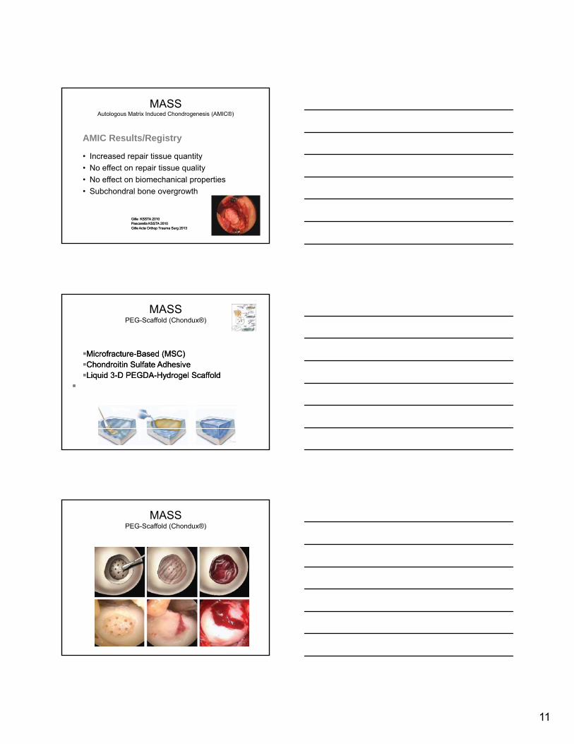
MASSAutologous Matrix Induced Chondrogenesis (AMIC®)
AMIC Results/Registry
• Increased repair tissue quantity
N ff t i ti lit• No effect on repair tissue quality
• No effect on biomechanical properties
• Subchondral bone overgrowth
GilleGille KSSTA 2010KSSTA 2010PascarellaPascarella KSSTA 2010KSSTA 2010GilleGille ActaActa OrthopOrthop Trauma Trauma SurgSurg 20132013
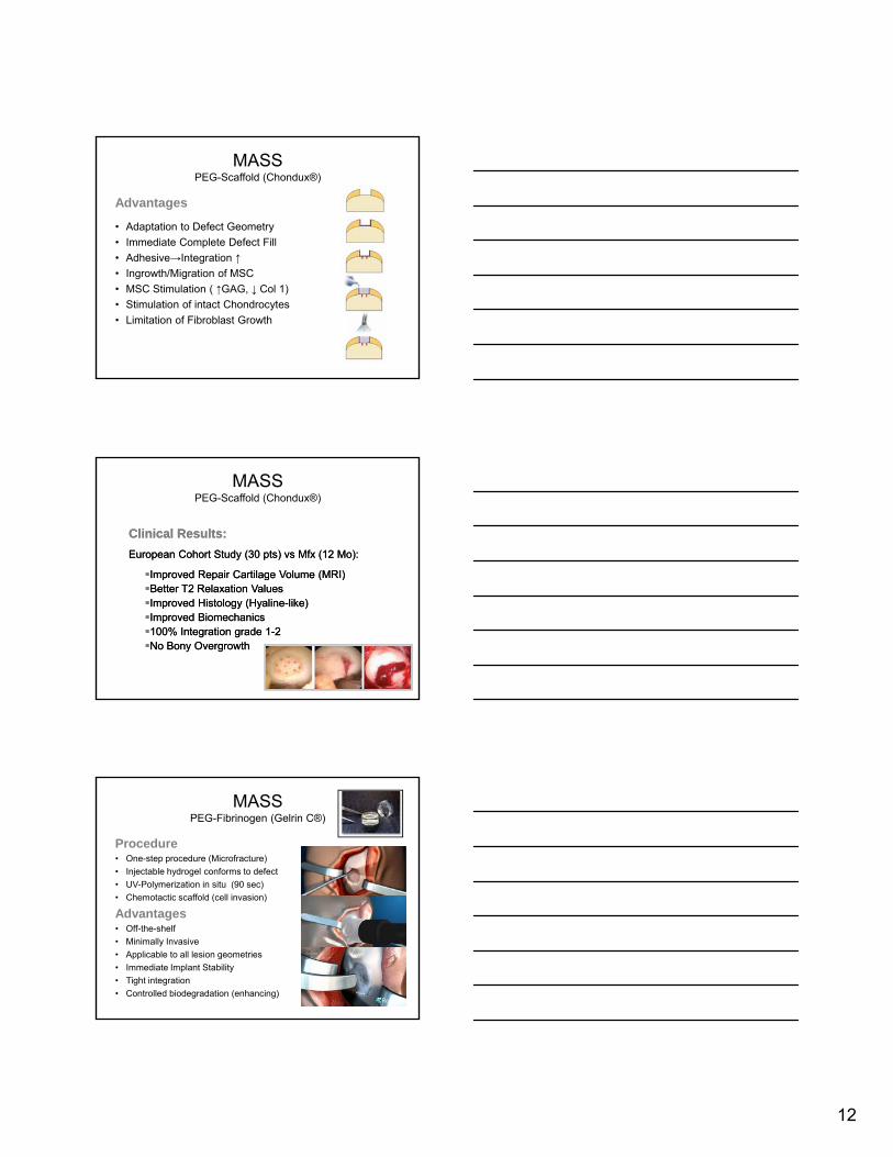
MicrofractureMicrofracture--Based (MSC)Based (MSC)ChondroitinChondroitin Sulfate AdhesiveSulfate Adhesive
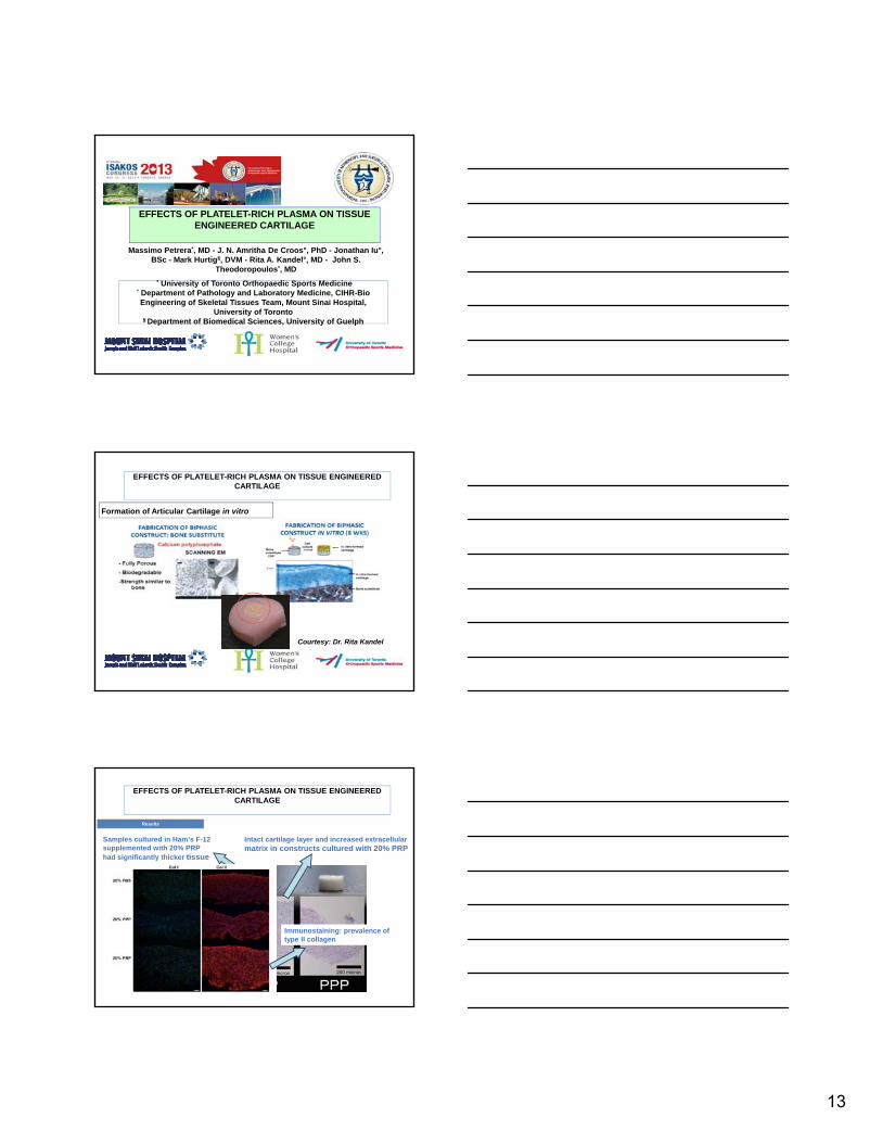
MASSPEG-Scaffold (Chondux®)
Liquid 3Liquid 3--D PEGDAD PEGDA--HydrogelHydrogel ScaffoldScaffold
MASSPEG-Scaffold (Chondux®)
12
MASSPEG-Scaffold (Chondux®)
Advantages
• Adaptation to Defect Geometry
• Immediate Complete Defect Fill
• Adhesive→Integration ↑• Adhesive→Integration ↑
• Ingrowth/Migration of MSC
• MSC Stimulation ( ↑GAG, ↓ Col 1)
• Stimulation of intact Chondrocytes
• Limitation of Fibroblast Growth
Clinical Results:Clinical Results:
European Cohort Study (30 pts) European Cohort Study (30 pts) vsvs MfxMfx (12 Mo):(12 Mo):
Improved Repair Cartilage Volume (MRI)Improved Repair Cartilage Volume (MRI)
MASSPEG-Scaffold (Chondux®)
p o ed epa Ca age o u e ( )p o ed epa Ca age o u e ( )Better T2 Relaxation ValuesBetter T2 Relaxation ValuesImproved Histology (HyalineImproved Histology (Hyaline--like)like)Improved BiomechanicsImproved Biomechanics100% Integration grade 1100% Integration grade 1--22No Bony OvergrowthNo Bony Overgrowth
MASSPEG-Fibrinogen (Gelrin C®)
Procedure• One-step procedure (Microfracture)
• Injectable hydrogel conforms to defect
• UV-Polymerization in situ (90 sec)
• Chemotactic scaffold (cell invasion)Chemotactic scaffold (cell invasion)
Advantages• Off-the-shelf
• Minimally Invasive
• Applicable to all lesion geometries
• Immediate Implant Stability
• Tight integration
• Controlled biodegradation (enhancing)
13
EFFECTS OF PLATELET-RICH PLASMA ON TISSUE ENGINEERED CARTILAGE
Massimo Petrera*, MD - J. N. Amritha De Croos°, PhD - Jonathan Iu°, BSc - Mark Hurtig§, DVM - Rita A. Kandel°, MD - John S.
Theodoropoulos*, MD
* University of Toronto Orthopaedic Sports Medicine° Department of Pathology and Laboratory Medicine, CIHR-Bio Engineering of Skeletal Tissues Team, Mount Sinai Hospital,
University of Toronto§ Department of Biomedical Sciences, University of Guelph
Formation of Articular Cartilage in vitro
EFFECTS OF PLATELET-RICH PLASMA ON TISSUE ENGINEERED CARTILAGE
Courtesy: Dr. Rita Kandel
Results
Intact cartilage layer and increased extracellular matrix in constructs cultured with 20% PRP
Samples cultured in Ham’s F-12 supplemented with 20% PRP had significantly thicker tissue
EFFECTS OF PLATELET-RICH PLASMA ON TISSUE ENGINEERED CARTILAGE
Immunostaining: prevalence of type II collagen
14
Results Mechanical testing
Tissue engineered cartilage cultured with 20% PRP showed superior compressive mechanical properties with an equilibrium modulus of 38.1±3.6
kPa versus 15.6±1.5kPa for 20% PPP (p=0.0002) and 20.4±3.5 kPa for 20% FBS (p=0.007)
Biochemistry Proteoglican
EFFECTS OF PLATELET-RICH PLASMA ON TISSUE ENGINEERED CARTILAGE
Biochemistry - Proteoglican
Samples supplemented with 20% PRP had significantly
higher GAG content (176.1±18.8µg GAG/mg dry
wt) compared to those supplemented with 20% FBS (112±10.6µg GAG/mg dry wt,
p=0.01 ) or 20% PPP (131.5±14.8µg GAG/mg dry wt,
p=0.11 )
Conclusions
• The presence of PRP in the culture media enhances the in vitro formation of cartilage with increased ECM and greater compressive
mechanical properties, while maintaining features of hyaline phenotype
EFFECTS OF PLATELET-RICH PLASMA ON TISSUE ENGINEERED CARTILAGE
phenotype
• This treatment may be a way to generate better tissue suitable to use for cartilage repair
• Further study to evaluate this tissue in vivo is required
•• Repeated platelet concentrate injections Repeated platelet concentrate injections 5 injections of ACP post 5 injections of ACP post microfxmicrofx: : macroscopically, histologically, and biomechanically superior to macroscopically, histologically, and biomechanically superior to microfxmicrofx alone after alone after 3, 6, and 12 months3, 6, and 12 months
ACP – 12 mo
Enhance reparative response of Enhance reparative response of microfracturesmicrofractures in the treatment of in the treatment of chondralchondraldefects of the knee: An experimental study in an animal modeldefects of the knee: An experimental study in an animal model
Milano et al. Milano et al. ArthroscopyArthroscopy. 2012.. 2012.
ACP – 12 mo
No ACP – 12 mo
ACP 12 mo
No ACP – 12 mo
15
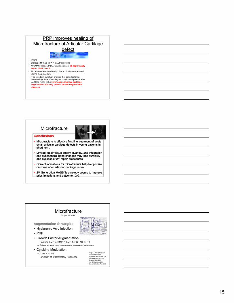
PRP improves healing of Microfracture of Articular Cartilage
defectSchneider, Klein, Dewitz
Koln, Germany ICRS 2013
• 36 pts
• 2 groups MFX vs MFX + 6 ACP injections
• WOMAC, Tegner, IKDC, Cincinnati score all significantly better of MFX+ACP
• No adverse events related to this application were noted during the procedure
• The results of our study showed that periodical intra-articular injections of autologous conditioned plasma after cartilage repair with microfracture improve cartilage regeneration and may prevent further degenerative changes
Microfracture
Conclusions
•• MicrofractureMicrofracture is effective firstis effective first--line treatment of acute line treatment of acute small articular cartilage defects in young patients in small articular cartilage defects in young patients in short term.short term.
•• Limited repair tissue quality, quantity, and integration Limited repair tissue quality, quantity, and integration and and subchondralsubchondral bone changes may limit durability bone changes may limit durability and success of 2and success of 2ndnd repair proceduresrepair procedures
•• Correct indications for Correct indications for microfracturemicrofracture help to optimize help to optimize outcome after outcome after articulararticular cartilage repair cartilage repair
•• 22ndnd Generation MASS Technology seems to improve Generation MASS Technology seems to improve prior limitations and outcome . 2.0 prior limitations and outcome . 2.0
MicrofractureImprovement
Augmentation Strategies
• Hyaluronic Acid Injection
• PRP
• Growth Factor Augmentation– Factors: BMP-2, BMP-7, BMP-4, FGF-18, IGF-1
– Stimulation of: MSC Differentiation, Proliferation, Metabolism
• Cytokine Modulation– IL-Ira + IGF-1
– Inhibition of Inflammatory Response
Kruger J Orthop Res 2011Fortier CORR 2011McIlwraith Arthroscopy 2011Yamaoka Cell Prol 2010Strauss AJSM 2009Kuo OA Cartilage 2006 Steinert J Orthop Res 2003


![Anisotropic crack propagation and deformation in dentin … · 2020. 8. 7. · microfracture behavior, crack-tip shielding and their anisotropy [46]. X-ray computed tomography (CT)](https://img.pdfslide.us/doc/110x75/60fae22e965f7e26f32792ad/anisotropic-crack-propagation-and-deformation-in-dentin-2020-8-7-microfracture.jpg)