Embed Size (px)
Citation preview
RESEARCH ARTICLE Open Access
Iroquois homeobox 2 suppresses cellularmotility and chemokine expression inbreast cancer cellsStefan Werner1*, Hauke Stamm1, Mutiha Pandjaitan1, Dirk Kemming2, Benedikt Brors3,4,5, Klaus Pantel1
and Harriet Wikman1
Abstract
Background: Disseminated tumor cells (DTCs) can be detected using ultrasensitive immunocytochemical assaysand their presence in the bone marrow can predict the subsequent occurrence of overt metastasis formation andmetastatic relapse. Using expression profiling on early stage primary breast tumors, low IRX2 expression waspreviously shown to be associated with the presence of DTCs in the bone marrow, suggesting a possible role ofIRX2 in the early steps of metastasis formation. The purpose of this study is to gain insights into the significance ofIRX2 protein function in the progression of breast cancer.
Methods: To assess the physiological relevance of IRX2 in breast cancer, we evaluated IRX2 expression in a largebreast cancer cohort (n = 1992). Additionally, constitutive IRX2 over expression was established in BT-549 andHs578T breast cancer cell lines. Subsequently we analyzed whether IRX2 overexpression effects chemokinesecretion and cellular motility of these cells.
Results: Low IRX2 mRNA expression was found to correlate with high tumor grade, positive lymph node status,negative hormone receptor status, and basal type of primary breast tumors. Also in cell lines low IRX2 expressionwas associated with mainly basal breast cancer cell lines. The functional studies show that overexpression of theIRX2 transcription factor in basal cell lines suppressed secretion of the pro-metastatic chemokines and inhibitedcellular motility but did not influence cell proliferation.
Conclusion: Our results imply that the IRX2 transcription factor might represent a novel metastasis associatedprotein that acts as a negative regulator of cellular motility and as a repressor of chemokine expression. Loss ofIRX2 expression could therefore contribute to early hematogenous dissemination of breast cancer by sustainingchemokine secretion and enabling mobilization of tumor cells.
Keywords: Breast cancer, Metastasis, Migration, IRX2, Chemokines, Disseminated tumor cells
BackgroundMetastasis - the main cause of cancer related deaths -is a complex multi-step process [1]. A key event isthe systemic spread of single tumor cells from theprimary lesion through the blood circulation into dis-tant organs. The presence of disseminated tumor cells(DTC) in the bone marrow (BM) of cancer patients isan independent predictor of metastatic relapse in
breast cancer and other solid tumors [2, 3]. Despiteprogress in the classification of tumors and the iden-tification and characterization of disseminated tumorcells in BM of cancer patients [4], the early events ofthe metastatic cascade, which leads to shedding ofsingle tumor cells from the primary tumor mass stillremains elusive.The transcription factor Iroquois Homeobox 2 (IRX2)
is a member of the Iroquois homeobox gene family.Members of this family appear to play multiple rolesduring pattern formation of vertebrate development[5–7]. In the breast tissue, IRX2 is expressed in ductal
* Correspondence: [email protected] of Tumor Biology, University Medical CenterHamburg-Eppendorf, Martinistrasse 52, 20246 Hamburg, GermanyFull list of author information is available at the end of the article
© 2015 Werner et al. Open Access This article is distributed under the terms of the Creative Commons Attribution 4.0International License (http://creativecommons.org/licenses/by/4.0/), which permits unrestricted use, distribution, andreproduction in any medium, provided you give appropriate credit to the original author(s) and the source, provide a link tothe Creative Commons license, and indicate if changes were made. The Creative Commons Public Domain Dedication waiver(http://creativecommons.org/publicdomain/zero/1.0/) applies to the data made available in this article, unless otherwise stated.
Werner et al. BMC Cancer (2015) 15:896 DOI 10.1186/s12885-015-1907-4

and lobular epithelium, but not in myoepithelium andit is suggested to function in linage-specific epithelialcell differentiation [8]. The relevance of the IRX2transcription factor throughout cancer progression issomewhat contradictory. Amplification of the IRX2chromosomal locus on 5p15.33 has been identified inbreast and soft tissue sarcomas [9, 10]. In breast can-cer this amplification may coexists with an activatingmutation of the PIK3CA gene [9]. In osteosarcomasknockdown of IRX2 inhibits cell proliferation and in-vasion [11], and elevated IRX2 expression is corre-lated with worse outcome and age in infant acutelymphoblastic leukemia [12]. Thus, these studies sug-gest a possible oncogenic function for the IRX2 pro-tein, especially in malignant cells of mesenchymalorigin. In contrast, other studies have shown thathypermethylation of IRX2 promoter region frequentlyoccurs in lung squamous cell and adenocarcinomas[13, 14]. Also one study showed that CpG islands inthe IRX2 gene were significantly more methylated inluminal A in comparison to basal tumors [15]. Mostof these studies have not performed functional valid-ation of the exact biological role of the IRX2 intumor progression. We have recently shown that lowIRX2 expression is associated with the presence ofDTCs in the bone marrow of breast cancer patients[16], suggesting a possible role of IRX2 as a metasta-sis suppressor protein in breast cancer.Although many of the early events of tumor cell dis-
semination and metastasis formation remain unclear, dif-ferent studies emphasize the importance of chemokinesin the microenvironment of the primary tumor and thesite of metastasis for cancer cell dissemination andmetastatic outgrowth [17]. For instance the expressionof the chemokine CCL5 (RANTES) can be correlatedwith progressive disease in breast cancer [18] and bonemetastasis of breast cancer cells is depending on signalingthrough the associated receptor CCR5 [19]. CoincidentlyCCR5 antagonists block metastasis formation of the breastcancer cell line MDA-MB-231 in mice, providing evidencefor a key role of CCL5/CCR5 in the invasiveness of basalbreast cancer cells [20]. Although accumulating evidenceemphasizes the central impact of chemokines on metasta-sis formation in breast cancer [21, 22], the mechanism forelevated levels of tumor cell derived chemokines secretionremains poorly understood.In this study, we aimed to validate the clinical import-
ance of IRX2 expression and to gain insights into thesignificance of IRX2 expression in the progression ofbreast cancer. The obtained data provide further evi-dence for IRX2 as a potential metastasis suppressor asectopic IRX2 expression diminished secretion of differ-ent chemokines and acts as negative regulator of cellularmotility of breast cancer cells.
ResultsExpression of IRX2 in primary breast tumorsWe previously found that low IRX2 gene expression inprimary breast tumors is associated with the presence ofDTCs in the bone marrow [16]. Low IRX2 was also asso-ciated with shortened survival of breast cancer patientsin one analyzed breast cancer data set [16]. To furtherinvestigate the patho-physiological relevance of IRX2gene expression in breast cancer, we evaluated IRX2gene expression in a large publically available patient co-hort comprised of 1992 patients (Table 1). We foundthat IRX2 is associated with several clinical prognosticfactors. Low IRX2 mRNA expression was found to becorrelated with high tumor stage (p = 0.004), hightumor grade (p < 0.001) and the presence of lymphnode metastasis (p = 0.044). On the other hand, lowIRX2 mRNA expression was found to be significantlycorrelated with low expression of both the estrogen(p = 0.001) and the progesterone (p < 0.001) steroid re-ceptors. Surprisingly, lower IRX2 mRNA expressionwas also significantly correlated with smaller tumorsize (p = 0.05). Finally we found that high IRX2 ex-pression is associated with the luminal A molecularsubtype and that low IRX2 expression is significantlymore frequent in tumors classified as basal and lu-minal B (p < 0.001). Taken together these analysesclearly show that low IRX2 expression is correlatedwith different parameters of poor prognosis, indicat-ing that loss of IRX2 expression is associated withless differentiated and more aggressive breast tumors.Nonetheless, no significant correlation between lowIRX2 expression and shortened survival was found inthis data set (data not shown).
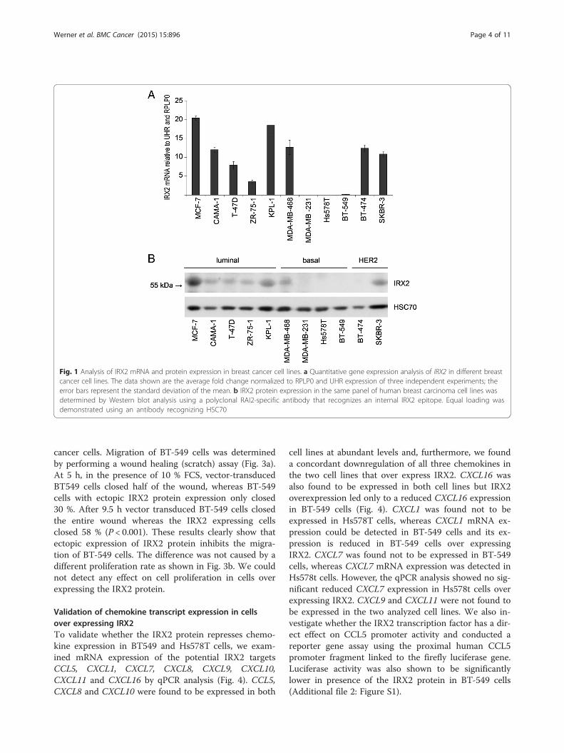
Expression of IRX2 in breast cancer cell linesTo further investigate the significance of IRX2 expres-sion in breast cancer, we evaluated IRX2 expression in apanel of breast cancer cell lines. IRX2 mRNA expressionwas detected in eight out of 11 breast cancer cell lines atvariable levels, as determined by quantitative real-timePCR analysis (Fig. 1a). The IRX2 protein expression wasdetermined by Western blot analysis (Fig. 1b) in thesame set of cell lines using a custom-made IRX2-specificpolyclonal antibody. The level of IRX2 protein expres-sion corresponds well with the level of IRX2 mRNA de-tected in these cells. In line with a potential metastasissuppressing function of IRX2, the poorly differentiated,highly metastatic basal breast cancer cell lines MDA-MB-231, Hs578T and BT-549 were completely negativefor IRX2 protein expression. IRX2 expression was foundin the luminal A cell lines (MCF7, CAMA-1, T-47D, ZR-75-1 and KPL1) as well as in the HER2 over expressingcell lines SKBR-3 and BT474. In BT-474 we found IRX2transcript but no protein expression.
Werner et al. BMC Cancer (2015) 15:896 Page 2 of 11
Analysis of IRX2 regulated networkExisting functional studies have so far described the bio-logical function of IRX2 mainly in the context of pattern
formation during embryonic development [5, 6]. To ob-tain evidence about the biological function of the IRX2transcription factor in breast epithelial cells and to iden-tify genes that might represent direct transcriptional tar-gets, we used SAM-analysis (Significance Analysis ofMicroarrays) on a large published data set of breast can-cer patients (GSE6532). Altogether 17 transcripts werefound significantly inversely correlated with IRX2 ex-pression and 43 transcripts were concurrently upregu-lated in patients with high IRX2 expression (Additionalfile 1: Table S1). Gene ontology analysis [23] was carriedout on these 60 most correlated and inversely correlatedgenes Interestingly, 6 out of 60 defined genes belongedto “chemokine signaling pathway” (p-value: <0.001;Benjamini: 0.001). Among the inversely correlatedgenes were four chemokines, CCL5, CXCL9, CXCL10and CXCL11 indicating that IRX2 might be activelyinvolved in the regulation of chemokine secretion bymediating the transcriptional repression of these che-mokines in breast cancer cells.
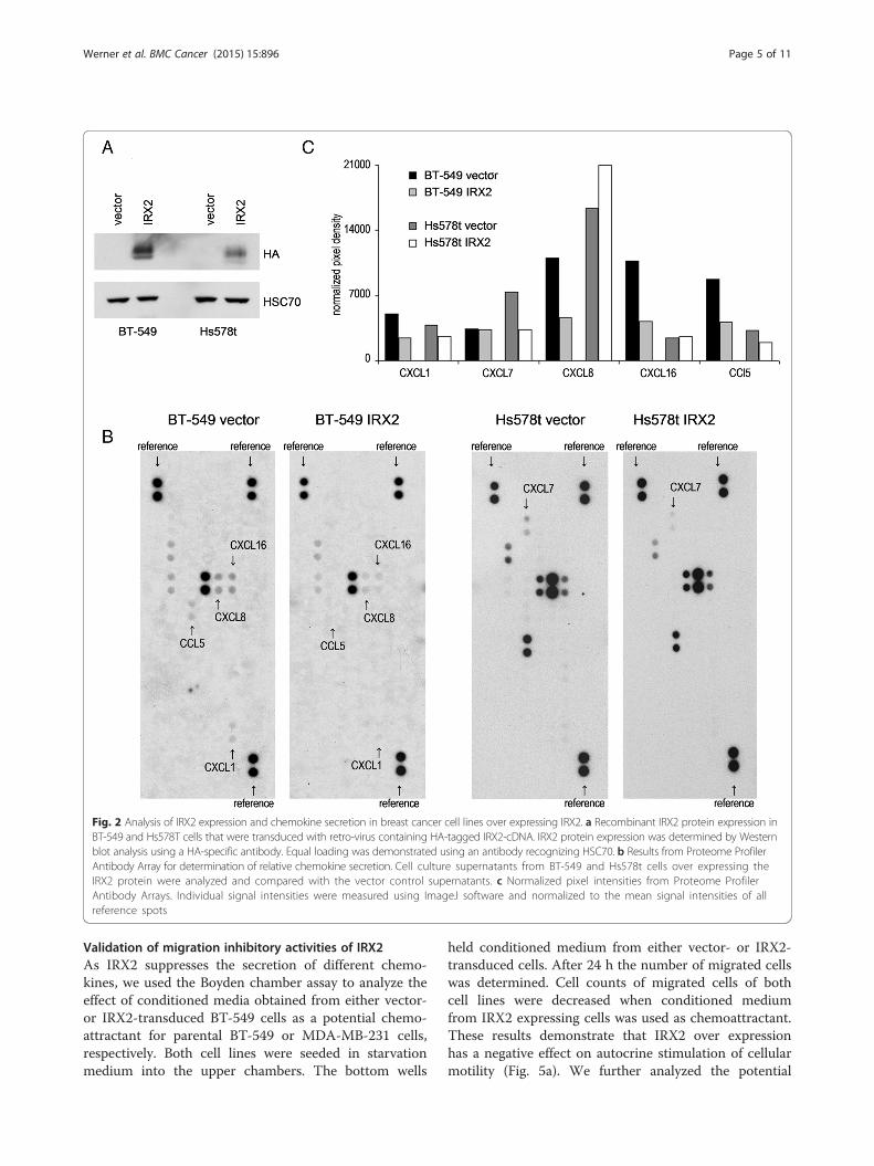
IRX2 protein expression represses chemokine secretion ofbreast cancer cellsTo experimentally validate the possible role of IRX2as a metastasis suppressing protein and repressor ofmigration and chemokine expression, we stably expresseda c-terminal HA-tagged IRX2 protein in IRX2-deficientBT-549 and Hs578t breast cancer cell lines. Successfulectopic IRX2 protein expression in both cell lines wasvalidated by Western Blot analysis using a HA-specificantibody (Fig. 2a).To investigate whether ectopic IRX2 expression has an
influence on chemokine secretion, we analyzed condi-tioned cell culture media from BT-549 and Hs578t cellswith a chemokine antibody array. In cell culture super-natants taken from BT-549 cells we found differences inthe presence of CCL5, CXCL1, CXCL8 and CXCL11.Remarkably, the expression of all four chemokines wasconsiderably lower in BT-549 cells overexpressing theIRX2 protein confirming the potential role of IRX2 inthe control of chemokine expression (Fig. 2b/c). In cellculture supernatants taken from Hs578t vector controlcells we found more CXCL7 expression in comparisonto supernatants taken form cells overexpressing IRX2(Fig. 2b/c). Interestingly, we also found reduced expres-sion of CCL5 in Hs578t cells overexpressing IRX2, toughthe overall expression of CCL5 was low abundant inHs578t cells.
Migration inhibitory activities of IRX2 in human breastcancer cellsTo further explore the possible role for IRX2 in tumorprogression, we investigated the ability of IRX2 expres-sion to modify cell migration and proliferation of breast
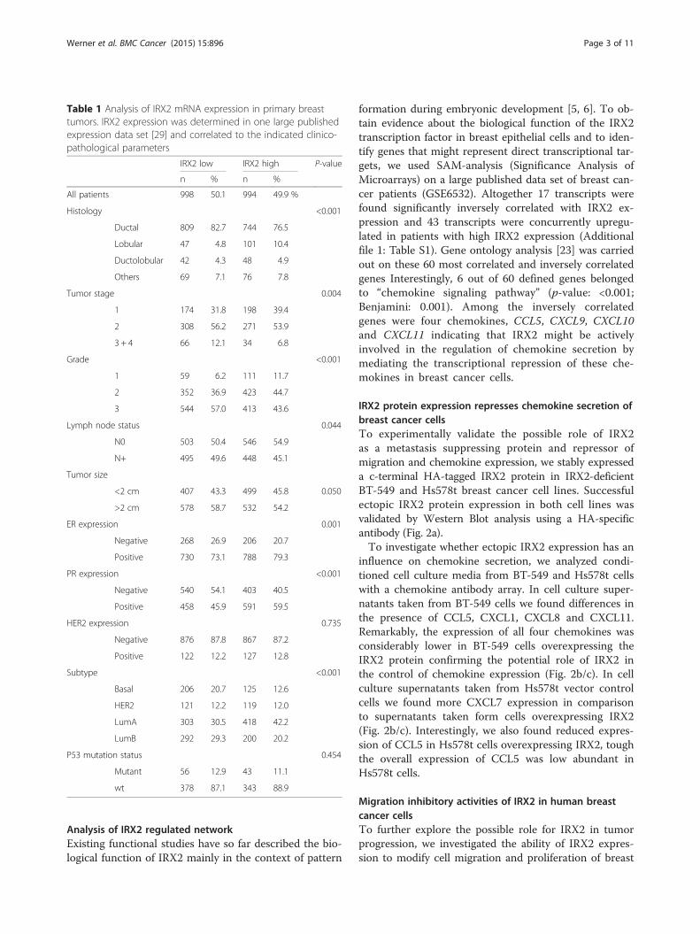
Table 1 Analysis of IRX2 mRNA expression in primary breasttumors. IRX2 expression was determined in one large publishedexpression data set [29] and correlated to the indicated clinico-pathological parameters
IRX2 low IRX2 high P-value
n % n %
All patients 998 50.1 994 49.9 %
Histology <0.001
Ductal 809 82.7 744 76.5
Lobular 47 4.8 101 10.4
Ductolobular 42 4.3 48 4.9
Others 69 7.1 76 7.8
Tumor stage 0.004
1 174 31.8 198 39.4
2 308 56.2 271 53.9
3 + 4 66 12.1 34 6.8
Grade <0.001
1 59 6.2 111 11.7
2 352 36.9 423 44.7
3 544 57.0 413 43.6
Lymph node status 0.044
N0 503 50.4 546 54.9
N+ 495 49.6 448 45.1
Tumor size
<2 cm 407 43.3 499 45.8 0.050
>2 cm 578 58.7 532 54.2
ER expression 0.001
Negative 268 26.9 206 20.7
Positive 730 73.1 788 79.3
PR expression <0.001
Negative 540 54.1 403 40.5
Positive 458 45.9 591 59.5
HER2 expression 0.735
Negative 876 87.8 867 87.2
Positive 122 12.2 127 12.8
Subtype <0.001
Basal 206 20.7 125 12.6
HER2 121 12.2 119 12.0
LumA 303 30.5 418 42.2
LumB 292 29.3 200 20.2
P53 mutation status 0.454
Mutant 56 12.9 43 11.1
wt 378 87.1 343 88.9
Werner et al. BMC Cancer (2015) 15:896 Page 3 of 11
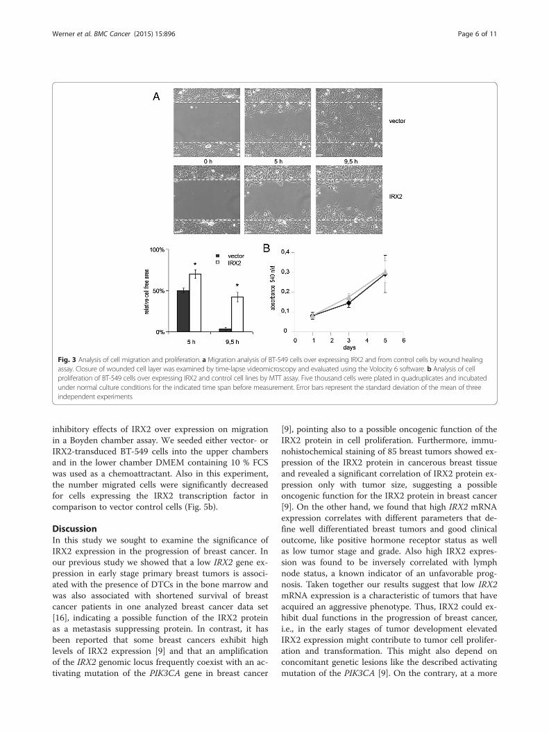
cancer cells. Migration of BT-549 cells was determinedby performing a wound healing (scratch) assay (Fig. 3a).At 5 h, in the presence of 10 % FCS, vector-transducedBT549 cells closed half of the wound, whereas BT-549cells with ectopic IRX2 protein expression only closed30 %. After 9.5 h vector transduced BT-549 cells closedthe entire wound whereas the IRX2 expressing cellsclosed 58 % (P < 0.001). These results clearly show thatectopic expression of IRX2 protein inhibits the migra-tion of BT-549 cells. The difference was not caused by adifferent proliferation rate as shown in Fig. 3b. We couldnot detect any effect on cell proliferation in cells overexpressing the IRX2 protein.
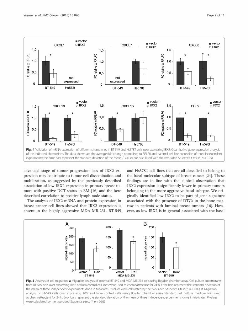
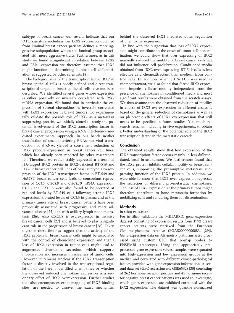
Validation of chemokine transcript expression in cellsover expressing IRX2To validate whether the IRX2 protein represses chemo-kine expression in BT549 and Hs578T cells, we exam-ined mRNA expression of the potential IRX2 targetsCCL5, CXCL1, CXCL7, CXCL8, CXCL9, CXCL10,CXCL11 and CXCL16 by qPCR analysis (Fig. 4). CCL5,CXCL8 and CXCL10 were found to be expressed in both
cell lines at abundant levels and, furthermore, we founda concordant downregulation of all three chemokines inthe two cell lines that over express IRX2. CXCL16 wasalso found to be expressed in both cell lines but IRX2overexpression led only to a reduced CXCL16 expressionin BT-549 cells (Fig. 4). CXCL1 was found not to beexpressed in Hs578T cells, whereas CXCL1 mRNA ex-pression could be detected in BT-549 cells and its ex-pression is reduced in BT-549 cells over expressingIRX2. CXCL7 was found not to be expressed in BT-549cells, whereas CXCL7 mRNA expression was detected inHs578t cells. However, the qPCR analysis showed no sig-nificant reduced CXCL7 expression in Hs578t cells overexpressing IRX2. CXCL9 and CXCL11 were not found tobe expressed in the two analyzed cell lines. We also in-vestigate whether the IRX2 transcription factor has a dir-ect effect on CCL5 promoter activity and conducted areporter gene assay using the proximal human CCL5promoter fragment linked to the firefly luciferase gene.Luciferase activity was also shown to be significantlylower in presence of the IRX2 protein in BT-549 cells(Additional file 2: Figure S1).
Fig. 1 Analysis of IRX2 mRNA and protein expression in breast cancer cell lines. a Quantitative gene expression analysis of IRX2 in different breastcancer cell lines. The data shown are the average fold change normalized to RPLP0 and UHR expression of three independent experiments; theerror bars represent the standard deviation of the mean. b IRX2 protein expression in the same panel of human breast carcinoma cell lines wasdetermined by Western blot analysis using a polyclonal RAI2-specific antibody that recognizes an internal IRX2 epitope. Equal loading wasdemonstrated using an antibody recognizing HSC70
Werner et al. BMC Cancer (2015) 15:896 Page 4 of 11
Validation of migration inhibitory activities of IRX2As IRX2 suppresses the secretion of different chemo-kines, we used the Boyden chamber assay to analyze theeffect of conditioned media obtained from either vector-or IRX2-transduced BT-549 cells as a potential chemo-attractant for parental BT-549 or MDA-MB-231 cells,respectively. Both cell lines were seeded in starvationmedium into the upper chambers. The bottom wells
held conditioned medium from either vector- or IRX2-transduced cells. After 24 h the number of migrated cellswas determined. Cell counts of migrated cells of bothcell lines were decreased when conditioned mediumfrom IRX2 expressing cells was used as chemoattractant.These results demonstrate that IRX2 over expressionhas a negative effect on autocrine stimulation of cellularmotility (Fig. 5a). We further analyzed the potential
Fig. 2 Analysis of IRX2 expression and chemokine secretion in breast cancer cell lines over expressing IRX2. a Recombinant IRX2 protein expression inBT-549 and Hs578T cells that were transduced with retro-virus containing HA-tagged IRX2-cDNA. IRX2 protein expression was determined by Westernblot analysis using a HA-specific antibody. Equal loading was demonstrated using an antibody recognizing HSC70. b Results from Proteome ProfilerAntibody Array for determination of relative chemokine secretion. Cell culture supernatants from BT-549 and Hs578t cells over expressing theIRX2 protein were analyzed and compared with the vector control supernatants. c Normalized pixel intensities from Proteome ProfilerAntibody Arrays. Individual signal intensities were measured using ImageJ software and normalized to the mean signal intensities of allreference spots
Werner et al. BMC Cancer (2015) 15:896 Page 5 of 11
inhibitory effects of IRX2 over expression on migrationin a Boyden chamber assay. We seeded either vector- orIRX2-transduced BT-549 cells into the upper chambersand in the lower chamber DMEM containing 10 % FCSwas used as a chemoattractant. Also in this experiment,the number migrated cells were significantly decreasedfor cells expressing the IRX2 transcription factor incomparison to vector control cells (Fig. 5b).
DiscussionIn this study we sought to examine the significance ofIRX2 expression in the progression of breast cancer. Inour previous study we showed that a low IRX2 gene ex-pression in early stage primary breast tumors is associ-ated with the presence of DTCs in the bone marrow andwas also associated with shortened survival of breastcancer patients in one analyzed breast cancer data set[16], indicating a possible function of the IRX2 proteinas a metastasis suppressing protein. In contrast, it hasbeen reported that some breast cancers exhibit highlevels of IRX2 expression [9] and that an amplificationof the IRX2 genomic locus frequently coexist with an ac-tivating mutation of the PIK3CA gene in breast cancer
[9], pointing also to a possible oncogenic function of theIRX2 protein in cell proliferation. Furthermore, immu-nohistochemical staining of 85 breast tumors showed ex-pression of the IRX2 protein in cancerous breast tissueand revealed a significant correlation of IRX2 protein ex-pression only with tumor size, suggesting a possibleoncogenic function for the IRX2 protein in breast cancer[9]. On the other hand, we found that high IRX2 mRNAexpression correlates with different parameters that de-fine well differentiated breast tumors and good clinicaloutcome, like positive hormone receptor status as wellas low tumor stage and grade. Also high IRX2 expres-sion was found to be inversely correlated with lymphnode status, a known indicator of an unfavorable prog-nosis. Taken together our results suggest that low IRX2mRNA expression is a characteristic of tumors that haveacquired an aggressive phenotype. Thus, IRX2 could ex-hibit dual functions in the progression of breast cancer,i.e., in the early stages of tumor development elevatedIRX2 expression might contribute to tumor cell prolifer-ation and transformation. This might also depend onconcomitant genetic lesions like the described activatingmutation of the PIK3CA [9]. On the contrary, at a more
Fig. 3 Analysis of cell migration and proliferation. a Migration analysis of BT-549 cells over expressing IRX2 and from control cells by wound healingassay. Closure of wounded cell layer was examined by time-lapse videomicroscopy and evaluated using the Volocity 6 software. b Analysis of cellproliferation of BT-549 cells over expressing IRX2 and control cell lines by MTT assay. Five thousand cells were plated in quadruplicates and incubatedunder normal culture conditions for the indicated time span before measurement. Error bars represent the standard deviation of the mean of threeindependent experiments
Werner et al. BMC Cancer (2015) 15:896 Page 6 of 11
advanced stage of tumor progression loss of IRX2 ex-pression may contribute to tumor cell dissemination andmobilization, as suggested by the previously describedassociation of low IRX2 expression in primary breast tu-mors with positive DCT status in BM [16] and the heredescribed correlation to positive lymph node status.The analysis of IRX2 mRNA and protein expression in
breast cancer cell lines showed that IRX2 expression isabsent in the highly aggressive MDA-MB-231, BT-549
and Hs578T cell lines that are all classified to belong tothe basal molecular subtype of breast cancer [24]. Thesefindings are in line with the clinical observation thatIRX2 expression is significantly lower in primary tumorsbelonging to the more aggressive basal subtype. We ori-ginally identified low IRX2 to be part of gene signatureassociated with the presence of DTCs in the bone mar-row in patients with luminal breast tumors [16]. How-ever, as low IRX2 is in general associated with the basal
Fig. 4 Validation of mRNA expression of different chemokines in BT-549 and Hs578T cells over expressing IRX2. Quantitative gene expression analysisof the indicated chemokines. The data shown are the average fold change normalized to RPLP0 and parental cell line expression of three independentexperiments; the error bars represent the standard deviation of the mean. P-values are calculated with the two-sided Student’s t-test (*, p < 0.05)
Fig. 5 Analysis of cell migration. a Migration analysis of parental BT-549 and MDA-MB-231 cells using Boyden chamber assay. Cell culture supernatantsfrom BT-549 cells over expressing IRX2 or from control cell lines were used as chemoattractant for 24 h. Error bars represent the standard deviation ofthe mean of three independent experiments done in triplicates. P-values were calculated by the two-sided Student’s t-test (*, p < 0.05). b Migrationanalysis of BT-549 cells over expressing IRX2 and from control cells using Boyden chamber assay Standard cell culture medium was usedas chemoattractant for 24 h. Error bars represent the standard deviation of the mean of three independent experiments done in triplicates. P-valueswere calculated by the two-sided Student’s t-test (*, p < 0.05)
Werner et al. BMC Cancer (2015) 15:896 Page 7 of 11
subtype of breast cancer, our results indicate that ourDTC signature including low IRX2 expression obtainedfrom luminal breast cancer patients defines a more ag-gressive subpopulation within the luminal group associ-ated with more aggressive traits. Furthermore, as in thisstudy we found a significant correlation between IRX2and ESR1 expression we therefore assume that IRX2might function in determining luminal cell differenti-ation as suggested by other scientists [8].The biological role of the transcription factor IRX2 in
breast epithelial cells is poorly defined and direct tran-scriptional targets in breast epithelial cells have not beendescribed. We identified several genes whose expressionis either positively or inversely correlated with IRX2mRNA expression. We found that in particular the ex-pression of several chemokines is inversely correlatedwith IRX2 expression in breast tumors. To experimen-tally validate the possible role of IRX2 as a metastasissuppressing protein, we initially aimed to study the po-tential involvement of the IRX2 transcription factor inbreast cancer progression using a RNA interference me-diated experimental approach. In our hands neithertransfection of small interfering RNAs nor viral trans-duction of shRNAs yielded a convenient reduction ofIRX2 protein expression in breast cancer cell lines,which has already been reported by other researchers[9]. Therefore, we rather stably expressed a c-terminalHA-tagged IRX2 protein in IRX2-deficient BT-549 andHs578t breast cancer cell lines of basal subtype. Overex-pression of the IRX2 transcription factor in BT-549 andHs578T breast cancer cells leads to concordant repres-sion of CCL5, CXCL8 and CXCL10 mRNA expression.CCL5 and CXCL8 were also found to be secreted atreduced levels by BT-549 cells following ectopic IRX2expression. Elevated levels of CCL5 in plasma and at theprimary tumor site of breast cancer patients have beenpreviously associated with progressive and more ad-vanced disease [25] and with axillary lymph node metas-tasis [26]. Also CXCL8 is overexpressed in invasivebreast cancer cells [27] and is believed to play a signifi-cant role in the progression of breast cancer [28]. Takentogether, these findings suggest that the activity of theIRX2 protein in breast cancer cells might be associatedwith the control of chemokine expression and that aloss of IRX2 expression in tumor cells might lead toaugmented chemokine secretion, which supportsmobilization and increases invasiveness of tumor cells.However, it remains unclear if the IRX2 transcriptionfactor is directly involved in the transcriptional regu-lation of the herein identified chemokines or whetherthe observed reduced chemokine expression is a sec-ondary effect of IRX2 overexpression. Further studiesthat also encompasses exact mapping of IRX2 bindingsites, are needed to unravel the exact mechanism
behind the observed IRX2 mediated down regulationof chemokine expression.In line with the suggestion that loss of IRX2 expres-
sion might contribute to the onset of tumor cell dissem-ination, we could show that over expressing of IRX2markedly reduced the motility of breast cancer cells butdid not influence cell proliferation. Conditioned mediaobtained from IRX2 over expressing BT-549 cells is lesseffective as a chemoattractant than medium from con-trol cells. In addition, when 10 % FCS was used aschemoattractant, we also found that forced IRX2 expres-sion impedes cellular motility independent from thepresence of chemokines in conditioned media and mostsignificant results were obtained from the scratch assays.We thus assume that the observed reduction of motilityin course of IRX2 overexpression in different assays isbased on the generic reduction of chemokines as well ason pleiotropic effects of IRX2 overexpression that stillneeds to be specified in future studies. Yet, much re-search remains, including in vivo experiments, to obtaina better understanding of the potential role of the IRX2transcription factor in the metastatic cascade.
ConclusionThe obtained results show that low expression of theIRX2 transcription factor occurs mainly in less differen-tiated, basal breast tumors. We furthermore found thatthe IRX2 protein inhibits cellular motility of breast can-cer cells, supporting the presumptive metastasis sup-pressing function of the IRX2 protein. In addition, wewere able to show that IRX2 over expression repressesthe secretion of different pro-metastatic chemokines.The loss of IRX2 expression at the primary tumor mighttherefore contribute to bone metastasis formation bymobilizing cells and rendering them for dissemination.
MethodsIn silico validationFor in-silico validation the METABRIC gene expressiondata set consisting of expression results from 1992 breastcancer patients were retrieved from the EuropeanGenome-phenome Archive (EGAS00000000083, [29]).Gene expression data on Affymetrix platforms were proc-essed using custom CDF that re-map probes toENSEMBL transcripts. Using the appropriately pre-processed gene expression values, samples were separatedinto high-expression and low expression groups at themedian and correlated with different clinico-pathologicalfactors provided with gene expression information. A sec-ond data set (GEO accession no. GSE6532) [30] consistingof 262 hormone receptor positive and 45 hormone recep-tor negative breast cancer patients was used to investigate,which genes expression are exhibited correltaed with theIRX2 expression. The dataset was quantile normalized
Werner et al. BMC Cancer (2015) 15:896 Page 8 of 11
and subsequently genes differentially expressed betweenthe extreme tertials of IRX2 expression were identifiedusing the significance analysis of microarrays (SAM) algo-rithm with a false discovery rate (FDR) of 5 % and with1000× repeated permutation [31]. To narrow down the re-sults, in a second, step only transcripts with a foldchange > 2 were taken into account.
Quantitative real-time PCR analysisqPCR analyses for the patients samples were performed on150 ng total RNA isolated by RNeasy Micro Kit (Qiagen).For cell line analyses 500 ng total RNA was transcribedusing First Strand cDNA Synthesis Kit (Fermentas St.Leon-Rot, Germany) together with 500 ng of universalhuman reference (UHR, Stratagene, Agilent technologies,Texas USA). Quantitative real-time PCR analyses wereperformed on Eppendorf Master Cycler using SYBRGreen (Fermentas, St. Leon-Rot, Germany) as fluores-cence detection method with the following primers;RPLPO-F: TGAGGTCCTCCTTGGTGAACA, RPLPO-R:CCCAGCTCTGGAGAAACTGC, IRX2-F: CCGAGAAACAAAAGCGAAGA, IRX2-R: AGCACGAGTGATCCGTGAG, CCL5-F: CTCGCTGTCATCCTCATTGC, CCL5-R:AAAGCAGCAGGGTGTGGTG, CXCL1-F: CTGAACAGTGACAAATCCAAC, CXCL1-R: CCTAAGCGATGCTCAAACAC, CXCL7-F: GAACTCCGCTGCATGTGTATAA, CXCL7-R: GCAATGGGTTCCTTTCCCGAT, CXCL8-F: GAATTCTCAGCCCTCTTCAAAAAC, CXCL8-R: GCCAAGGAGTGCTAAAGAACTTAG, CXCL10-F:GAAGGGTGAGAAGAGATGTC, CXCL10-R: TAGGGAAGTGATGGGAGAG, CXCL16-F: CCCGCCATCGGTTCAGTTC, CXCL16-R: CCCCGAGTAAGCATGTCCAC.The analyses were performed in triplicates and the meanvalues were used for each gene. The mRNA levels werenormalized to the mRNA level of the ribosomal RPLP0gene using ΔΔCT-method for quantification. The results,expressed as N-fold differences in target gene expressioncompared to universal human reference (UHR) or paren-tal cell line expression.
Cell cultureMDA-MD-231 and SK-BR-3 cells were obtained fromATCC. BT-549, BT-474, T-47D, ZR-75-1 and MDA-MD-468 cells were obtained from Cell Lines Service(Heidelberg). MCF-7 and Hs578T were a friendly giftfrom Dr. Steven Johnsen (University of Göttingen,Germany). Phoenix amphotropic retroviral packagingcells were a friendly gift from Dr. Volker Assmann(UKE, Hamburg, Germany). CAMA-1 and KPL-1 cellswere a friendly gift from Dr K. Iljin (VTT, Espoo, Finland).Cells were grown as monolayers according to standard con-ditions in either DMEM or RPMI supplemented with 10 %fetal calf serum and 2 mM L-glutamine (Invitrogen)at 37 °C in a humidified atmosphere containing 10 %
CO2 or 5 % CO2 respectively. Analysis of cellularviability was done by 3-(4,5-dimethylthiazol-2-yl)-2,5-diphenyltetrazolium bromide (MTT) assay. 5000 cellswere plated in quadruplicates and absorbance at 570 nmwas measured after 3 h treatment with MTT solution andlysis of cells in isopropanol containing 4 mM HCl and0.1 % (w/v) NP-40. Lysates were additionally treated withsonicator to achieve complete lysis of cells.
Expression plasmids and viral transductionIRX2 coding sequence was amplified (forward-primer:CCGCTGCTCGGCGTGACGCG, reverse-primer: TAGGTAGGGCTGGACGCCC) from cDNA obtained fromthe breast cancer cell line MCF-7 and cloned into theexpression plasmid phCMV3 (Genlantis) using EcoRIand KpNI restrictions sites. HA-tagged IRX2 cDNA se-quence was reamplified and cloned into the retroviralexpression plasmid pMXs-IRES-Puro (Cell Biolabs) usingEcoRI and NotI restrictions sites and afterwards the clonedcDNA sequence was verified by sequencing. For productionof retroviral particles ψNX-ampho cells were transfectedwith the retroviral expression plasmid using Lipofectamine2000 (Invitrogen) according to manufacturer’s suggestedprotocol. Viral transduction was done with 500 μl viralsupernatant added to 50 % confluent recipient cultures in6-well plates containing 1 ml cell culture medium. Positiveselection was achieved 24 h after transduction usingpuromycin-containing medium (2 μg/ml, Sigma-Aldrich).Cells were subsequently kept under puromycin for 4 days.
Western blot analysis and antibody productionWhole cell extracts from cultured cells were prepared bydirect lysis and sonication of cells in SDS sample buffercontaining proteinase and phosphatase inhibitors. Cellextracts were separated on denaturing 8 % polyacryl-amide gels and blotted onto PVDF membrane. Detectionof IRX2 protein was either done by incubation with anHA specific antibody (Sigma-Aldrich, H6908) or a cus-tom made IRX2 specific antibody. Detection of theHSC70 protein (Santa Cruz, clone B6) was used as load-ing control. To generate polyclonal antibodies againstIRX2, a peptide DDEDDDEEGERGLAPPKPVTSS corre-sponding to the central region of human IRX2 was syn-thesized, coupled to keyhole limpet hemocyanin and wasthen injected into rabbits. IRX2 specific antibodies wereisolated by immunoaffinity purification using the corre-sponding immunizing peptide coupled to a solid sup-port. Reactivity and specificity of the IRX2 specificantibody was verified by Western blot analysis.
Chemokine antibody arrayConditioned medium from confluent BT-549 andHs578t cells stably expressing either IRX2 or empty vec-tor were tested for chemokine secretion by chemokine
Werner et al. BMC Cancer (2015) 15:896 Page 9 of 11
antibody array following the manufacturer’s instructions(R&D Systems Proteome Profiler™ Human ChemokineArray Kit). Estimation of normalized signal intensity wasdone using ImageJ software.
In vitro scratch assayBT-549 cells which were either transduced with IRX2 orempty vector containing retrovirus were plated in a con-centration of of 7 × 10⁵ cells and grown to confluence in6 well plates under standard culture conditions. After-wards cells were incubated for 24 h in DMEM contain-ing 0.5 % FCS. In vitro scratch assay was performed asdescribed elsewhere [32]. Cell movement was recordedin real time using an Improvision Live Cell SpinningDisk Microscope and data analysis was done using theVolocity® software (Perkin Elmer).
Transwell migration assay5 × 104 BT-549 or MDA-MB-231 cells were plated inserum-free DMEM media into the upper chambers ofBD Cell Culture Inserts for 24-well plates with 8.0 μmpores (BD Falcon™). In the lower chamber conditionedmedium from confluent BT-549 cells stably expressingeither IRX2 or empty vector were used as chemoattract-ant. Plates were incubated at 37 °C, and migration wasallowed to proceed for 20 h. Afterwards non migratedcells in the upper chambers were removed with cottonswabs, and the remaining cells were fixed in 4 % parafor-maldehyde and stained with crystal violet. Cells werecounted by using a Zeiss light microscope. Four fieldswere counted on each of two filters. Results are expressedas average cells per field.
Ethical approvalSince all analyses were conducted on previously pub-lished expression data from breast cancer patients or oncell lines, no further ethics approval was required forthis study.
Additional files
Additional file 1: Table S1. Genes in primary breast tumors showingexhibited correlation with IRX2 expression and functional annotation ofthese genes. SAM-analysis (Significance Analysis of Microarrays) on andfunctional annotation was used on a large published data set of breastcancer patients (GSE6532). (XLS 79 kb)
Additional file 2: Figure S1. Gene reporter assay with reporter plasmidcontaining proximal CCL5 promoter fragment. The reporter plasmid wastransfected into BT-549 cells that over express IRX2 and into the controlcell line. For normalization, a co-transfection with the pGL4.74 plasmidcontaining the Renilla luciferase was performed. Each experiment wasperformed in triplicate. Error bars represent the standard deviation of themean. (PDF 18 kb)
AbbreviationsBM: Bone marrow; CCL5: Chemokine (C-C motif) ligand 5; CCR5: C-Cchemokine receptor type 5; CXCL: Chemokine (C-X-C motif) ligand;
CpG: CpG dinucleotides; DMEM: Dulbecco’s modified Eagle’s medium;DTC: Disseminated tumor cell; ESR1: Estrogen receptor 1 (alpha); FCS: Fetalcalf serum; FDR: False discovery rate; GEO: Gene expression omnibus;h: Hour; HA: Human influenza hemagglutinin; HER2: Human epidermalgrowth factor receptor 2; IRX2: Iroquois homeobox 2; mRNA: MessengerRNA; PCR: Polymerase chain reaction; PIK3CA: Phosphatidylinositol-4,5-bisphosphate 3-kinase; qPCR: Quantitative polymerase chain reaction;RPLPO: Ribosomal protein, large, P0; RPMI: Roswell park memorial institutemedium; SAM: Significance analysis of microarrays; shRNA: Small hairpin RNA.
Competing interestsThe authors declare that they have no competing interests.
Authors’ contributionsSW designed the study, developed methodology, acquired and interpreteddata and wrote the manuscript, HS and MP developed methodology,acquired and interpreted data and edited the manuscript, DK and BBperformed computational analyses and interpreted data, KP and HWdesigned and supervised the study and edited the manuscript. All authorshave read and approved the final manuscript.
AcknowledgementsWe are grateful for the skillful technical assistance from JolantheKropidlowski and Dr. Bernd Zobiak from the UKE Microscopy Imaging Facility(UMIF). This work was supported by Werner Otto Stiftung [grant number14/79] and European Commission [DISMAL].
Author details1Department of Tumor Biology, University Medical CenterHamburg-Eppendorf, Martinistrasse 52, 20246 Hamburg, Germany. 2EuropeanLaboratory Association, Ibbenbüren, Germany. 3Department of AppliedBioinformatics (G200), German Cancer Research Center (DKFZ), Heidelberg,Germany. 4National Center for Tumor Diseases (NCT), 69120 Heidelberg,Germany. 5German Consortium for Translational Cancer Research (DKTK),69120 Heidelberg, Germany.
Received: 6 July 2015 Accepted: 3 November 2015
References1. Pantel K, Brakenhoff RH, Brandt B. Detection, clinical relevance and specific
biological properties of disseminating tumour cells. Nat Rev Cancer.2008;8(5):329–40. doi:10.1038/nrc2375.
2. Braun S, Vogl FD, Naume B, Janni W, Osborne MP, Coombes RC, et al.A pooled analysis of bone marrow micrometastasis in breast cancer.N Engl J Med. 2005;353(8):793–802. doi:10.1056/NEJMoa050434.
3. Kang Y, Pantel K. Tumor cell dissemination: emerging biological insightsfrom animal models and cancer patients. Cancer Cell. 2013;23(5):573–81.doi:10.1016/j.ccr.2013.04.017.
4. Wan L, Pantel K, Kang Y. Tumor metastasis: moving new biological insightsinto the clinic. Nat Med. 2013;19(11):1450–64. doi:10.1038/nm.3391.
5. Gomez-Skarmeta JL, Modolell J. Iroquois genes: genomic organization andfunction in vertebrate neural development. Curr Opin Genet Dev.2002;12(4):403–8.
6. Cavodeassi F, Modolell J, Gomez-Skarmeta JL. The Iroquois family of genes:from body building to neural patterning. Development. 2001;128(15):2847–55.
7. Kim KH, Rosen A, Bruneau BG, Hui CC, Backx PH. Iroquois homeodomaintranscription factors in heart development and function. Circ Res.2012;110(11):1513–24. doi:10.1161/CIRCRESAHA.112.265041.
8. Lewis MT, Ross S, Strickland PA, Snyder CJ, Daniel CW. Regulated expressionpatterns of IRX-2, an Iroquois-class homeobox gene, in the human breast.Cell Tissue Res. 1999;296(3):549–54.
9. Kadota M, Sato M, Duncan B, Ooshima A, Yang HH, Diaz-Meyer N, et al.Identification of novel gene amplifications in breast cancer and coexistenceof gene amplification with an activating mutation of PIK3CA. Cancer Res.2009;69(18):7357–65. doi:10.1158/0008-5472.CAN-09-0064.
10. Adamowicz M, Radlwimmer B, Rieker RJ, Mertens D, Schwarzbach M,Schraml P, et al. Frequent amplifications and abundant expression of TRIO,NKD2, and IRX2 in soft tissue sarcomas. Genes Chromosomes Cancer.2006;45(9):829–38. doi:10.1002/gcc.20343.
Werner et al. BMC Cancer (2015) 15:896 Page 10 of 11
11. Liu T, Zhou W, Zhang F, Shi G, Teng H, Xiao J, et al. Knockdown of IRX2 inhibitsosteosarcoma cell proliferation and invasion by the AKT/MMP9 signalingpathway. Mol Med Rep. 2014;10(1):169–74. doi:10.3892/mmr.2014.2215.
12. Kang H, Wilson CS, Harvey RC, Chen IM, Murphy MH, Atlas SR, et al. Geneexpression profiles predictive of outcome and age in infant acutelymphoblastic leukemia: a Children’s Oncology Group study. Blood.2012;119(8):1872–81. doi:10.1182/blood-2011-10-382861.
13. Rauch TA, Wang Z, Wu X, Kernstine KH, Riggs AD, Pfeifer GP. DNAmethylation biomarkers for lung cancer. Tumour Biol. 2012;33(2):287–96.doi:10.1007/s13277-011-0282-2.
14. Sato T, Arai E, Kohno T, Takahashi Y, Miyata S, Tsuta K, et al. Epigeneticclustering of lung adenocarcinomas based on DNA methylation profiles inadjacent lung tissue: Its correlation with smoking history and chronicobstructive pulmonary disease. Int J Cancer. 2014;135(2):319–34.
15. Kamalakaran S, Varadan V, Giercksky Russnes HE, Levy D, Kendall J,Janevski A, et al. DNA methylation patterns in luminal breast cancersdiffer from non-luminal subtypes and can identify relapse riskindependent of other clinical variables. Mol Oncol. 2011;5(1):77–92.doi:10.1016/j.molonc.2010.11.002.
16. Werner S, Brors B, Eick J, Marques E, Pogenberg V, Parret A, et al.Suppression of early hematogenous dissemination of human breast cancercells to bone marrow by retinoic acid-induced 2. Cancer Discov.2015;5(5):506-19. doi:10.1158/2159-8290.CD-14-1042.
17. Roussos ET, Condeelis JS, Patsialou A. Chemotaxis in cancer. Nat Rev Cancer.2011;11(8):573–87. doi:10.1038/nrc3078.
18. Luboshits G, Shina S, Kaplan O, Engelberg S, Nass D, Lifshitz-Mercer B, et al.Elevated expression of the CC chemokine regulated on activation, normal Tcell expressed and secreted (RANTES) in advanced breast carcinoma. CancerRes. 1999;59(18):4681–7.
19. Karnoub AE, Dash AB, Vo AP, Sullivan A, Brooks MW, Bell GW, et al.Mesenchymal stem cells within tumour stroma promote breast cancermetastasis. Nature. 2007;449(7162):557–63. doi:10.1038/nature06188.
20. Velasco-Velazquez M, Jiao X, De La Fuente M, Pestell TG, Ertel A, Lisanti MP,et al. CCR5 antagonist blocks metastasis of basal breast cancer cells. CancerRes. 2012;72(15):3839–50. doi:10.1158/0008-5472.CAN-11-3917.
21. Ben-Baruch A. The multifaceted roles of chemokines in malignancy. CancerMetastasis Rev. 2006;25(3):357–71. doi:10.1007/s10555-006-9003-5.
22. Lv D, Zhang Y, Kim HJ, Zhang L, Ma X. CCL5 as a potentialimmunotherapeutic target in triple-negative breast cancer. Cell MolImmunol. 2013;10(4):303–10. doi:10.1038/cmi.2012.69.
23. da Huang W, Sherman BT, Lempicki RA. Systematic and integrative analysisof large gene lists using DAVID bioinformatics resources. Nat Protoc.2009;4(1):44–57. doi:10.1038/nprot.2008.211.
24. Neve RM, Chin K, Fridlyand J, Yeh J, Baehner FL, Fevr T, et al. A collection ofbreast cancer cell lines for the study of functionally distinct cancer subtypes.Cancer Cell. 2006;10(6):515–27. doi:10.1016/j.ccr.2006.10.008.
25. Niwa Y, Akamatsu H, Niwa H, Sumi H, Ozaki Y, Abe A. Correlation of tissueand plasma RANTES levels with disease course in patients with breast orcervical cancer. Clin Cancer Res. 2001;7(2):285–9.
26. Sauer G, Schneiderhan-Marra N, Kazmaier C, Hutzel K, Koretz K, MucheR, et al. Prediction of nodal involvement in breast cancer based onmultiparametric protein analyses from preoperative core needlebiopsies of the primary lesion. Clin Cancer Res. 2008;14(11):3345–53.doi:10.1158/1078-0432.CCR-07-4802.
27. Freund A, Chauveau C, Brouillet JP, Lucas A, Lacroix M, Licznar A, et al.IL-8 expression and its possible relationship with estrogen-receptor-negative status of breast cancer cells. Oncogene. 2003;22(2):256–65.doi:10.1038/sj.onc.1206113.
28. Freund A, Jolivel V, Durand S, Kersual N, Chalbos D, Chavey C, et al.Mechanisms underlying differential expression of interleukin-8 in breastcancer cells. Oncogene. 2004;23(36):6105–14. doi:10.1038/sj.onc.1207815.
29. Curtis C, Shah SP, Chin SF, Turashvili G, Rueda OM, Dunning MJ, et al. Thegenomic and transcriptomic architecture of 2,000 breast tumours revealsnovel subgroups. Nature. 2012;486(7403):346–52. doi:10.1038/nature10983.
30. Loi S, Haibe-Kains B, Desmedt C, Lallemand F, Tutt AM, Gillet C, et al.Definition of clinically distinct molecular subtypes in estrogen receptor-positive breast carcinomas through genomic grade. J Clinical Oncol.2007;25(10):1239–46. doi:10.1200/JCO.2006.07.1522.
31. Wikman H, Lamszus K, Detels N, Uslar L, Wrage M, Benner C, et al.Relevance of PTEN loss in brain metastasis formation of breast cancerpatients. Breast Cancer Res. 2012;14(2):R49. doi:10.1186/bcr3150.
32. Liang CC, Park AY, Guan JL. In vitro scratch assay: a convenient andinexpensive method for analysis of cell migration in vitro. Nat Protoc.2007;2(2):329–33. doi:10.1038/nprot.2007.30.
Submit your next manuscript to BioMed Centraland take full advantage of:
• Convenient online submission
• Thorough peer review
• No space constraints or color figure charges
• Immediate publication on acceptance
• Inclusion in PubMed, CAS, Scopus and Google Scholar
• Research which is freely available for redistribution
Submit your manuscript at www.biomedcentral.com/submit
Werner et al. BMC Cancer (2015) 15:896 Page 11 of 11