Embed Size (px)
Citation preview
Pertanika4(2), 141-147(1981)
Iron Tolerance by the Young Pig as Influencedby the Dietary Status of Vitamin E and Selenium1
K.K. KUAN and E.FL MILLER2
Faculty of Veterinary Medicine and Animal ScienceUniversiti Pertanian Malaysia
Key words: pigs; iron; vitamin E; Selenium; iron toxicity.
RINGKASAN
Tiga percubaan untuk menentukan pengaruh vitamin E and selenium terhadap kesan-kesan feramapabila ianya diberi dalam makanan atau disuntikkan dalam otot, telah dijalankan dengan menggunakan191 ekor babi. Anak-anak babi berumur tiga hari dari ibu babiyang telah diberi makanan kurang vitamin Edan selenium (Se) semasa bunting dan menyusu, tidak menunjukkan toleransi rendah apabila diberi suntikan100 mg feram dextran. Keracunan feram tidak berlaku pada babi yang dicerai susu pada lima minggu dariibu babi yang telah mendapat makanan kurang vitamin E-Se apabila babi tersebut diberi suntikan keduadengan 100 mg feram dextran. Babi yang membesar sehingga peringkat berat badan pasaran yang diberimakanan kurang vitamin E-Se tidak menunjukkan kesan-kesan keracunan feram apabila ransum itu mengan-dungi feram sehingga 850 'parts per million* (ppm). Babi yang berumur 9 minggu yang, diberi suntikan1000 mg feram dextran menunjukkan kenaikan paras feram serum yang lebih tinggi (P < 0.01 j jikalaudibandingkan dengan babi yang tidak disuntikan dengan galian itu. Tambahan pula, babi yangdiberi makanan dengan tambahan vitamin E dan disuntik dengan 1000 mg feram dextran menunjukkanparas feram serum yang lebih tinggi (P < 0.01 j apabila dibandingkan dengan babi yang diberi makanankurang vitamin E. Walaubagaimanapun, paras feram serum yang tinggi itu tidak membawa kesan-kesanburuk kepada babi itu.
SUMMARY
Three experiments using a total of 191 pigs, were conducted to determine the effects of iron, bothoral supplementation and intramuscular injection in young pigs as influenced by dietary vitamin E andSelenium (Se). Piglets from sows on low vitamin E-Se gestation and lactation diets did not show anyreduced tolerance to the standard dose of 100 mg iron from iron dextran when given at three days of age.Iron toxicosis was not produced when young pigs weaned at five weeks from sows on a low vitamin E-Sediet were given a second iron dextran injection of 100 mg iron. Pigs fed a low vitamin E-Se diet upto marketweight did not exhibit any conclusive evidence of iron toxicity when their feed contained up to 850 ppmiron. When nine week old pigs were given an intramuscular injection of 1000 mg iron from iron dextran,there was a highly significant increase in serum iron levels; pigs on the vitamin E supplemented diets hadsignificantly higher serum iron levels than those on the low vitamin E diet. However, the high serum-ironlevels did not produce any noticeable ill effect on the animals.
INTRODUCTION
One of the standard practices on the modernintensive pig farm is the administration of iron topiglets a few days after birth as a prophylaticmeasure against anemia. The supplementaryprovision of this element can be given either orallyor in the form of an intramuscular injectionalthough the latter is a superior method in termsof promoting maximum hemoglobin synthesis andin increasing weight gains (Wahlstrom et al, 1960).There have, however, been reports of iron poison-
ing following the routine administration of iron topiglets (Behrens, 1957; Brag, 1958; Henrikson,1962). Follow-up research (Arpi and Tollerz,1965; Patterson et al, 1969 and Patterson et al,1971) has indicated that vitamin E deficiencywill render piglets more susceptible to the toxiceffects of iron.
The present research was undertaken to furtherinvestigate the iron tolerance of the vitamin Edeficient young pig. Selenium deficiency may alsorender piglets more susceptible to the toxic
Part of M.S. Thesis submitted to Michigan State University.Dept. of Animal Husbandry, Michigan State University.
141
K. K. KUAN AND E. R. MILLER
effects of iron. Selenium functions as a biologicalantioxidant as the essential component ofglutathione peroxidase (Hoekstra et al.t 1973 andScott and Noguchi, 1973).
MATERIALS AND METHODS
Three experiments were conducted to examinethe pig's tolerance for iron as influenced byvitamin E and Se status.
General conduct of ExperimentsExperiment 1: Effect of a second iron injection
on vitamin E - Se deficient pigs.
A total of 127 piglets from 17 litters wereused in this experiment. All litters were from sowsfed a low vitamin E - Se ration throughoutgestation and lactation (Table 1). At three days ofage the piglets were given their first intramuscularinjection of 100 mg iron from iron dextran. Atfive weeks of age, these pigs were weaned to a lowvitamin E - Se starter ration (Table 1). Two daysafter weaning each litter was divided at randominto two groups. Pigs in one group received anintramuscular injection of 34 I.U. of vitamin Eplus 0.5 mg of Se, the other group served as acontrol. Two days after the vitamin E - Seinjection, each group of animals was furtherdivided into two sub-groups. One group of vitaminE - Se injected pigs and one group of non-injected
animals were given a second intramuscular injectionof 100 mg iron from iron dextran.
Rectal temperature was taken at 0, 1,4 and 6hours after the second iron injection, to determinewhether there was an anaphylactic reaction.
Experiment 2. Effect of dietary iron supple-mentation to low vitamin E and vitamin E supple-mented rations of young pigs.
Forty-eight pigs from sows fed a low vitaminE - Se diet were used. The pigs were weaned atfive weeks of age and allotted randomly by litterand weight into six groups of eight pigs per group.The feed provided following weaning and through-out the experimental period was a corn-soybeanmeal basal diet which contained about 0.05 ppmSe. (Table). The basal diet contained 345 ppm Fe.Additions of iron as ferrous sulphate (FeSO47H2O) and vitamin E as d-otocopheryl acetateto the basal diet were combined in a 3 X 2 factorialarrangement of treatments. The diets contained345, 550 and 850 ppm iron and contained eithera low level of vitamin E (none added) or weresupplemented with 22 I.U. of vitamin E/kg offeed. The pigs had ad libitum access to feed andwere weighed at weekly or fortnightly intervals.Blood for hematological studies and serum Feanalysis were collected at various intervals duringthe course of the experiment. Half of the animals
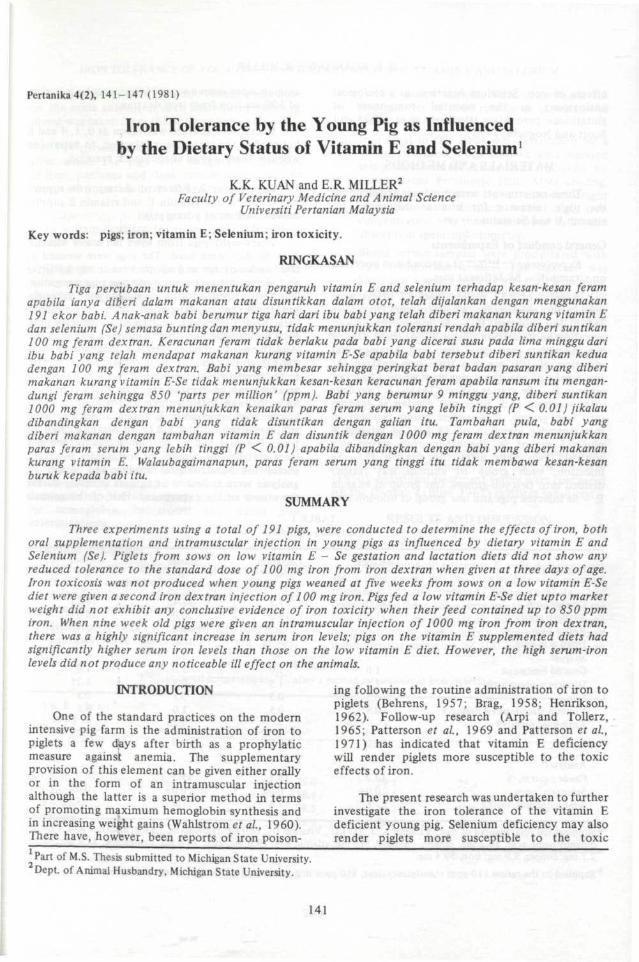
TABLE 1Composition of diets
Ingredients
Ground cornSoybean mealRolled oatsDried skin milkSucroseGround limestoneDicalcium phosphateSaltVitamin trace-mineral premixa
Antibiotic premixb
Analyses:Crude protein, %Selenium, ppmd-otocopherol, mg/kg
Gestation%
85.011.5---1.01.50.50.5
100.0
13.20.047.6
Lactation%
77.519.0
---
1.01.50.50.5
100.0
16.10.057.4
Starter%
51.4520.010.010.05.01.00.80.51.00.25
100.0
19.50.097.8
Grower%
78.7518.0
--_
1.01.250.50.5
100.0
16.00.057.5
aSupplied the following/kg of diet: Vitamin A, 3,300 I.U., Vitamin D2, 660 I.U.;riboflavin, 3.3 mg;nicotinic acid, 17.6 mg;d-pantothenic acid, 13.2 mg; choline chloride, 110 mg; vitamin B12,.19.8 mg; zinc, 74.8 mg; manganese 37.4 mg; iodine,2.7 mg; copper, 9.9 mg; iron, 59.4 mg.
bSupplied to the ration 110 ppm chlortetracycline, 110 ppm sulfamethazine and 55 ppm penicillin.
142
IRON TOLERANCE OF YOUNG PIGS AND DIETARY STATUS OF VITAMIN E AND SELENIUM
from each group were bled and bleeding was doneon the same animals at each subsequent bleeding.Blood was taken from the anterior vena cava.
The period of the experiment was 126 days,after which the pigs were slaughtered. Samplesof liver, pancreas and flank muscle were collectedfrom each animal for iron analysis.
Experiment 3: Effects of a large dose of irongiven intramuscularly to young pigs deficient invitamin E and Se.
Sixteen pigs were used in this study. Eightwere weaned from sows fed a low .vitamin E - Segestation and lactation ration, while the othereight were from sows whose diet had beensupplemented with 22 I.U. of vitamin E/kg of diet.All 16 pigs received their first iron dextran injection(100 mg of iron) at three days of age. At nineweeks of age, each group of animals was againdivided into two sub-groups. One sub-group fromeach of the two main groups received a second irondextran injection intramuscularly at a dose of1000 mg Fe. The animals were observed for signsof toxicity following the administration of themassive dose of iron.
Blood was collected prior to the administrationof the massive dose of iron and then at one day
and again at one week after the iron treatmentfor hemoglobin, hematocrit and serum irondeterminations.
Analytical procedures1. Hemoglobin was determined by the cyanmethe-
moglobin method of Crosby et aL, (1954). AColeman Junior II spectrophotometer wasused for optical density determinations.
2. Hematocrit was determined by the micro-capillary tube method (McGovern et aL,
1955). Blood samples were centrifuged forfive minutes at 1000 rpm in an InternationalHematocrit centrifuge.
3. Iron level in the feeds was determined by awet ash method. Feed samples were digestedusing concentrated nitric acid followed byconcentrated perchloric acid. After cooling,samples were diluted to a constant weightwith deionized distilled water and, ironconcentration was then determined by atomicabsorption spectrophotometry.
Blood serum samples were precipitated with20% trichloroacetic acid. The mixture washeated in a water bath at 90° for 15 minutes.Upon cooling, the mixture was centrifuged, thesupernatant decanted into acid washed test-tubes and iron concentration then determinedby atomic absorption spectrophotometry.
Liver, muscle and pancreas samples werehomogenized and a weighed amount of thehomogenate was digested and analyzed bythe procedure followed for the feedsamples.
Statistical analysis
The data for experiment 2 and 3 were analyzedfor statistical differences by the f-test of Snedecor(1950). Individual treatment values were comparedby the Duncan's (Bliss, 1967) multiple range test.
RESULTS AND DISCUSSION
Experiment 1The feeding of a vitamin E - Se deficient diet
alone to sows did not produce piglets with reducedtolerance to iron dextran. When three-day oldpiglets from these sows were injected with 100 mgiron dextran these animals did not exhibit any signsor symptoms of iron toxicity. Pigs given a secondintramuscular injection of 100 mg iron from iron
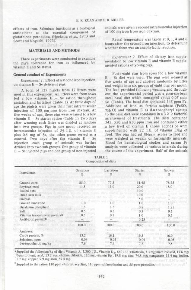
TABLE 2Average rectal temperatures, °C, after a second intramuscular iron injection
Vit. E - SeIron inj.
Time
Ohour1 hour4 hours6 hours
inj.a
intramuscular injection of 34 I.Uintramuscular injection of 100 m
-
39.039.439.539.3
. vitamin E + 0.5 mg See iron from iron dextran
+
39.11 39.5
39.739.4
+
39.139.639.539.3
++
39.139.539.439.4
143
K. K. KUAN AND E. R. MILLER
dextran, four days after weaning to a low vitaminE - Se starter diet, also did not develop anyreduced iron tolerance. One half of the experi-mental pigs had received a vitamin E - Se injection(34 I.U. vitamin E + 0.5 mg Se) two days prior tothe iron injection, irrespective of whether theanimals had been previously treated with vitaminE - Se injection or not, there was no evidence ofiron toxicity.
Rectal temperatures taken at 0, 1, 4 and 6hours following the second iron injection were allnormal with no evidence of anaphylaxis for any ofthe treatments (Table 2).
The results of this study appear to becontradictory to those of Lannek et ah, (1962)and Patterson et ai, (1969). Both groups ofworkers obtained piglets that were hypersensitiveto the toxic effects of iron by feeding gestatingsows a low vitamin E diet. It is important however,to point out that in both the above mentionedstudies, the oil and grain portions of theexperimental diets were subjected to oxidizingconditions by heating. This increased the peroxidevalue of the diet and further aggravated the lowvitamin E situation. In the present study nopolyunsaturated fatty acids were added to theexperimental diets and the peroxide level were notincreased by heat treatment.
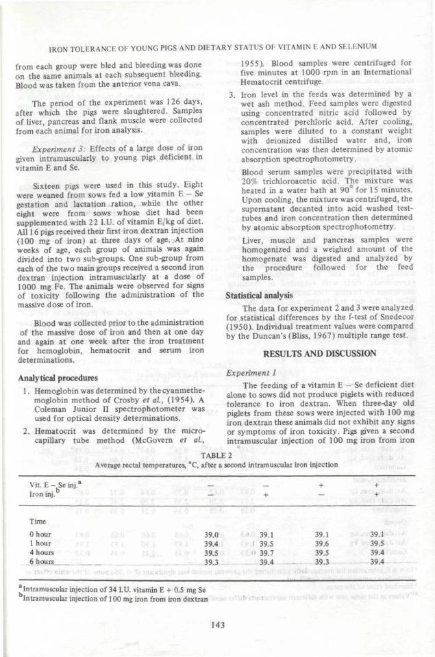
Experiment 2Pig performance data are presented in Table 3.
Average daily gains during the growing periodtended to increase with increasing levels of iron inpigs fed low vitamin E diets and decreased withincreasing levels of iron on high vitamin E diets.Treatment average daily gains did not differsignificantly during the finishing period or whenthe entire study was taken into consideration.Similarly there were only small differences amongtreatments in daily feed consumption or gain/feedratios.
The decrease in daily gains during the growingperiod with increasing levels of iron on the highvitamin E diet was rather unexpected. One possibleexplanation of the results is that a high level ofvitamin E is somehow incompatible with a highiron level. The exact mode of incompatibility (if itexists) cannot be explained without furtherinvestigation.
There was a significant (P<0.5)ironX vitamin Einteraction on gain during the growing period ofthe study. Here again, the indication suggests thathigh vita'min E is not compatible with high iron.
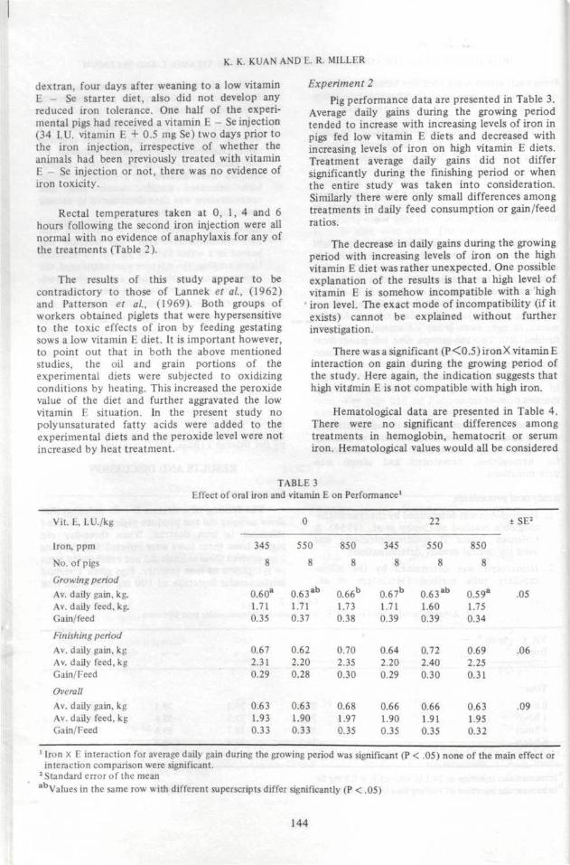
Hematological data are presented in Table 4.There were no significant differences amongtreatments in hemoglobin, hematocrit or serumiron. Hematological values would all be considered
TABLE 3Effect of oral iron and vitamin E on Performance1
Vit. E, l.U./kg
Iron, ppm
No. of pigs
Growing period
Av. daily gain, kg.
Av. daily feed, kg.
Gain/feed
Finishing periodAv. daily gain, kgAv. daily feed, kgGain/Feed
Overall
Av, daily gain, kgAv. daily feed, kgGain/ Feed
22 ± SE2
345
8
0.60a
1.710.35
0.672.310.29
0.631.930.33
550
8
0.6 3 a b
1.710.37
0.622.200.28
0.631.900.33
850
8
0.66b
1.730.38
0.702.350.30
0.681.970.35
345
8
0.67b
1.710.39
0.642.200.29
0.661.900.35
550
8
0.6 3 a b
1.600.39
0,722.400.30
0.661.910.35
850
8
0.59a
1.750.34
0.692.250.31
0.631.950.32
.05
.06
.09
1 Iron X E interaction for average daily gain during the growing period was significant (P < .05) none of the main effect orinteraction comparison were significant.
2 Standard error of the meanabValues in the same row with different superscripts differ significantly (P < .05)
144
IRON TOLERANCE OF YOUNG PIGS AND DIETARY STATUS 01 VITAMIN E AND SELENIUM
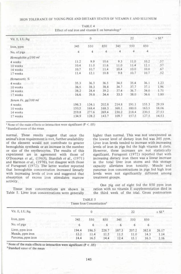
TABLE 4Effect of oral iron and vitamin E on hematology1
Vit. E, I.U./kg
Iron, ppm
No. of pigs
Hemoglobin g/100 ml4 weeks10 weeks14 weeks17 weeks
Hematocrit, %4 weeks10 weeks14 weeks17 weeks
Serum Fe, fjg/JOOml4 weeks10 weeks14 weeks17 weeks
0 22 1 SE
345
4
11.210,410.711.4
35.336.538.216.6
196.3150.5259.8134.9
550
4
9.911.010.712.1
36.338.328.439.8
134.1168.4277.6128.2
850
4
10.611.611.410.8
36.538.839.234.4
202.8168.3269.4143.7
345
4
9.311.010.49.8
34.534.737.433.3
214.8168.1230.2109.7
550
4
11.011.410.010.7
35.437.736.738.0
191.1180.0218.4157.5
850
4
10.212.110.010.7
36.137.134.036.6
155.3163.5230.5127.5
.57
.57
.67
.52
1.231.961.711.80
29.5918.0627.5314.53
1 None of the main effects or interaction were2 Standard error of the mean
normal. These results suggest that once theanimal's iron requirement is met, further availabilityof the element would not contribute to greaterhemoglobin synthesis or an increase in the numberor size of the erythrocytes. The results of thisexperiment are in agreement with those ofO'Donovan et al, (1963), Standish et al, (1971)and Harmon et al, (1970), but disagree with thoseof Furugouri (1972). The latter worker reportedthat hemoglobin concentration increased linearlywith increasing levels of iron and suggested thatabsorption of excess iron stimulates marrowactivity.
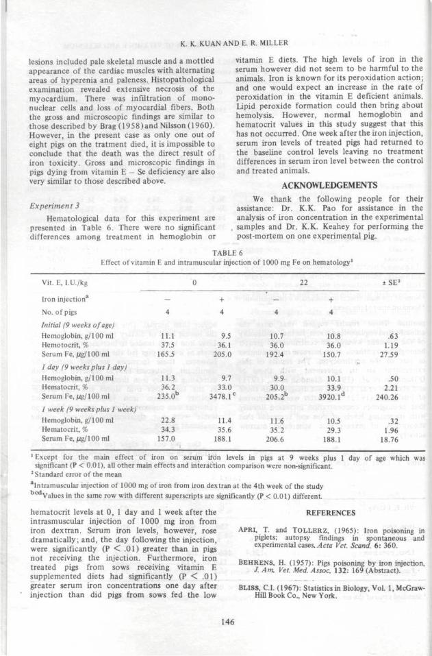
Tissue iron concentrations are shown inTable 5. Liver iron concentrations were generally
higher than normal. This was not unexpected asthe lowest level of dietary iron fed was 345 ppm.Liver iron levels tended to increase with increasinglevels of iron in pigs fed the high vitamin E diets.However, these increases are not statisticallysignificant. Furugouri (1972) reported that withincreasing dietary iron there was a linear increasein the total liver iron stores and this storagecapacity alleviates iron toxicity. Muscle andpancreas iron concentrations in pigs fed high ironlevels were not significantly different amongtreatment groups.
One pig out of eight fed the 850 ppm ironration with no vitamin E supplementation died inthe third week of the trial. Gross postmortem
TABLE 5Tissue Iron Concentration1
Vit. E, I.U./kg.
Iron, ppm
No. of pigs
Liver, ppm ironMuscle, ppm ironPancreas, ppm iron
345
6
194.412.214.4
0 22
550
6
186.311.416.5
850
6
228.711.514.4
345
6
187.311.512.4
550
6
207.211.015.1
850
6
382.814.316.3
± SE2
26.171.142.16
1 None of the main effects or interaction were significant (P < .05)2 Standard error of the mean
145
K. K. KUAN AND E. R. MILLER
lesions included pale skeletal muscle and a mottledappearance of the cardiac muscles with alternatingareas of hyperenia and paleness. Histopathologicalexamination revealed extensive necrosis of themyocardium. There was infiltration of mono-nuclear cells and loss of myocardial fibers. Boththe gross and microscopic findings are similar tothose described by Brag (1958)and Nilsson (1960).However, in the present case as only one out ofeight pigs on the tratment died, it is impossible toconclude that the death was the direct result ofiron toxicity. Gross and microscopic findings inpigs dying from vitamin E - Se deficiency are alsovery similar to those described above.
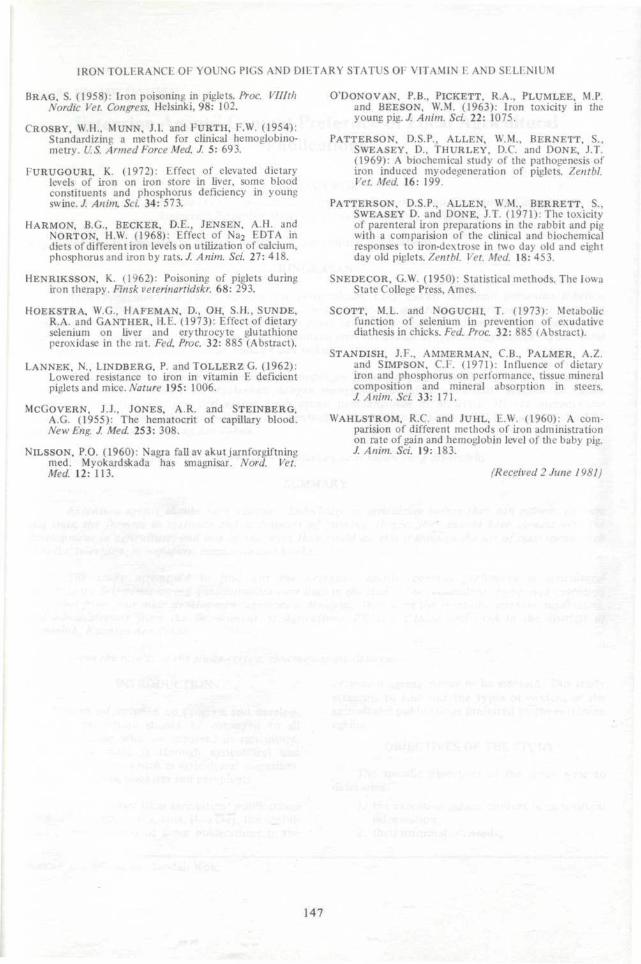
Experiment 3Hematological data for this experiment are
presented in Table 6. There were no significantdifferences among treatment in hemoglobin or
vitamin E diets. The high levels of iron in theserum however did not seem to be harmful to theanimals. Iron is known for its peroxidation action;and one would expect an increase in the rate ofperoxidation in the vitamin E deficient animals,lipid peroxide formation could then bring abouthemolysis. However, normal hemoglobin andhematocrit values in this study suggest that thishas not occurred. One week after the iron injection,serum iron levels of treated pigs had returned tothe baseline control levels leaving no treatmentdifferences in serum iron level between the controland treated animals.
ACKNOWLEDGEMENTS
We thank the following people for theirassistance: Dr. K.K. Pao for assistance in theanalysis of iron concentration in the experimentalsamples and Dr. K.K. Keahey for performing thepost-mortem on one experimental pig.
TABLE 6Effect of vitamin E and intramuscular injection of 1000 mg Fe on hematology1
Vit. E, I.U./kg
Iron injectiona
No. of pigs
Initial (9 weeks of age)Hemoglobin, g/100 mlHemotocrit, %Serum Fe, jug/100 ml
1 day (9 weeks plus 1 day)Hemoglobin, g/100 mlHematocrit, %Serum Fe, jug/100 ml
I week (9 weeks plus I week)Hemoglobin, g/100 mlHematocrit, %Serum Fe, //g/100 ml
-
4
11.137.5
165.5
11.336.2
235.0b
22.834.3
157.0
0
+
4
9.536.1
205.0
9.733.0
3478.1C
11.435.6
188.1
—
4
10.736.0
192.4
9.930.0
205.2b
11.635.2
206.6
22
+
4
10.836.0
150.7o
10.133.9
3920.1d
10.529.3
188.1
±SE2
.631.19
27.59
.502.21
240.26
.321.96
18.76
1 Except for the main effect of iron on serum iron levels in pigs at 9 weeks plus 1 day of age which wassignificant (P < 0.01), all other main effects and interaction comparison were non-significant.
2 Standard error of the meanaIntramusculai injection of 1000 mg of iron from iron dextran at the 4th week of the studybcdValues in the same row with different superscripts are significantly (P < 0.01) different
hematocrit levels at 0, 1 day and 1 week after theintrasmuscular injection of 1000 mg iron fromiron dextran. Serum iron levels, however, rosedramatically; and, the day following the injection,were significantly (P < .01) greater than in pigsnot receiving the injection. Furthermore, irontreated pigs from sows receiving vitamin Esupplemented diets had significantly (P < .01)greater serum iron concentrations one day afterinjection than did pigs from sows fed the low
REFERENCES
APRI, T. and TOLLERZ, (1965): Iron poisoning inpiglets; autopsy findings in spontaneous andexperimental cases. Acta Vet. Scand. 6: 360.
BEHRENS, H. (1957): Pigs poisoning by iron injection,J.Ant Vet. Med. Assoc. 132: 169 (Abstract).
BLISS, C.I. (1967): Statistics in Biology, Vol. 1, McGraw-Hill Book Co., New York.
146
IRON TOLHRANC I: OI YOUNG PIGS AND DIETARY STATUS 01 VITAMIN E AND SELENIUM
BRAG, S. (1958): Iron poisoning in piglets. Proc. VIHthNordic Vet. Congress, Helsinki, 98: 102.
CROSBY, W.H., MUNN, J.L and EURTH, F.W. (1954):Standardizing a method for clinical hemoglobino-metry. U.S. Armed Force Med. J. 5: 693.
EURUGOURI, K. (1972): Effect of elevated dietarylevels of iron on iron store in liver, some bloodconstituents and phosphorus deficiency in youngswine. J. Anim, Sci. 34: 573.
HARMON, B.G., BECKER, D.E., JENSEN, A.H. andNORTON, H.W. (1968): Effect of Na2 EDTA indiets of different iron levels on utilization of calcium,phosphorus and iron by rats. / Anim. Sci 27: 418.
HENRIKSSON, K. (1962): Poisoning of piglets duringiron therapy. Finsk veterinartidskr. 68: 293.
HOEKSTRA, W.G., HAFEMAN, D., OH, S.H., SUNDE,R.A. and GANTHER, H.E. (1973): Effect of dietaryselenium on liver and erythrocyte glutathioneperoxidase in the rat. Fed. Proc. 32: 885 (Abstract).
LANNEK, N., LlNDBERG, P. andTOLLERZG. (1962):Lowered resistance to iron in vitamin E deficientpiglets and mice. Nature 195: 1006.
MCGOVERN, J.J., JONES, A.R. and STEINBERG,A.G. (1955): The hematocrit of capillary blood.NewEng. J. Med 253: 308.
NlLSSON, P.O. (1960): Nagra fall av akut jarnforgiftningmed. Myokardskada has smagnisar. Nord. Vet.Med. 12: 113.
O'DONOVAN, P.B., PlCKETT, R.A., PLUMLEE, M.P.and BEESON, W.M. (1963): Iron toxicity in theyoung pig. J, Anim. Sci. 22: 1075.
PATTERSON, D.S.P., ALLEN, W.M., BERNETT, S.,SWEASEY, D., THURLEY, D.C and DONE, J.T.(1969): A biochemical study of the pathogenesis ofiron induced myodegeneration of piglets. Zentbl.Vet. Med. 16: 199.
PATTERSON, D.S.R, ALLEN, W.M., BERRETT, S.,SWEASEY D. and DONE, J.T. (1971): The toxicityof parenteral iron preparations in the rabbit and pigwith a comparision of the clinical and biochemicalresponses to iron-dextrose in two day old and eightday old piglets. Zentbl Vet. Med. 18: 453.
SNEDECOR, G.W. (1950): Statistical methods. The IowaState College Press, Ames.
SCOTT, M.L. and NOGUCHI, T. (1973): Metabolicfunction of selenium in prevention of exudativediathesis in chicks. Fed. Proc. 32: 885 (Abstract).
STANDISH, J.F., AMMERMAN, C.B., PALMER, A.Z.and SIMPSON, C.I . (1971): Influence of dietaryiron and phosphorus on performance, tissue mineralcomposition and mineral absorption in steers./ Anim. Sci. 33: 171.
WAHLSTROM, R.C. and JUHL, E.W. (1960): A com-parision of different methods of iron administrationon rate of gain and hemodobin level of the baby pig.J. Anim. Sci. 19: 183.
(Received 2 June 1981)
147