Embed Size (px)
Citation preview
RESEARCH Open Access
IPO5 promotes the proliferation andtumourigenicity of colorectal cancer cellsby mediating RASAL2 nucleartransportationWenjuan Zhang1, Yanxia Lu1, Xiaomin Li1, Jianming Zhang1,2, Weihao Lin1, Wei Zhang1, Lin Zheng1 andXuenong Li1*
Abstract
Background: Karyopherin nuclear transport receptors play important roles in tumour development and drugresistance and have been reported as potential biomarkers and therapeutic targets for tumour treatment. However,IPO5, one of the karyopherin nuclear transport receptor family members, remains largely uncharacterized in tumourprogression.
Methods: The TCGA data, quantitative reverse transcription-PCR (qRT-PCR), western blotting, and IHC analyses wereused to detect IPO5 expression in CRC tissues. A series of in vivo and in vitro experiments was utilized to demonstratethe function of IPO5 in CRC tissues. Mass spectrometry (MS), CO-IP technology, subcellular fractionation, andimmunofluorescence were utilized to investigate the possible mechanisms of CRC.
Results: IPO5 was highly expressed and positively correlated with the clinicopathological characteristics of colorectalcancer tissues. Functional experiments indicated that IPO5 could promote the development of CRC. Mechanistically, wescreened RASAL2, one cargo of IPO5, and further confirmed that IPO5 bound to the NLS sequence of RASAL2, mediatingRASAL2 nuclear translocation and inducing RAS signal activation, thereby promoting the progression of CRC.
Conclusions: Together, our results indicate that IPO5 is overexpressed in colorectal cancer cells. By transporting RASAL2,IPO5 may play a crucial role in CRC.
Keywords: Colorectal cancer, IPO5, RASAL2, NLS, Ras pathway, Karyopherins
BackgroundColorectal cancer has become a major threat to humanhealth. Although a large number of studies have been per-formed on CRC, the concrete molecular mechanisms ofthe formation and metastasis of CRC has not been com-pletely clear until recently. Therefore, an in-depth study ofthe molecular mechanisms of the occurrence and develop-ment of CRC and the identification of effective molecularmarkers among many factors is of great importance.Karyopherins are key regulatory molecules of nuclear
plasma transport and are the most classic cell transporter
proteins; they include both importins and exportins [1, 2].Transporter proteins that have molecular weights greaterthan 40 kDa transport a variety of molecules between thecytoplasm and nucleus through the nuclear pore complex;these include transcription factors, splicing factors andother proteins [3–5]. Nevertheless, the dysfunction of kar-yopherins may derail the transport activity and may causethe abnormal localization of oncogenic factors, thus lead-ing to tumourigenesis [6–8]. The currently reported kar-yopherins related to tumours include KPNA2, XPO5,XPO1, and KPNA7 [9–11]. IPO5, a member of the karyo-pherin beta subunit, locates in the 13q32 chromosomal re-gion and has been demonstrated to play a vital role in thetranslocation of various proteins. However, the role ofIPO5 in cancer progression has not been well defined. In
© The Author(s). 2019 Open Access This article is distributed under the terms of the Creative Commons Attribution 4.0International License (http://creativecommons.org/licenses/by/4.0/), which permits unrestricted use, distribution, andreproduction in any medium, provided you give appropriate credit to the original author(s) and the source, provide a link tothe Creative Commons license, and indicate if changes were made. The Creative Commons Public Domain Dedication waiver(http://creativecommons.org/publicdomain/zero/1.0/) applies to the data made available in this article, unless otherwise stated.
* Correspondence: [email protected] of Pathology, School of Basic Medical Sciences, SouthernMedical University, Guangzhou 510515, People’s Republic of ChinaFull list of author information is available at the end of the article
Zhang et al. Journal of Experimental & Clinical Cancer Research (2019) 38:296 https://doi.org/10.1186/s13046-019-1290-0
our previous research, we analysed the CRC gene expres-sion profile data of GSE41258 and sifted out a number ofdifferentially expressed genes in which the expression ofIPO5 was continuously increasing with the increasing se-verity from normal tissues to stage I, stage II, stage III,stage IV, and liver metastasis tumours (Additional file 1:Figure S1A) [12]. Meanwhile, the high expression of IPO5,especially in CRC cells, was also confirmed in the TCGAdatabase and Oncomine database (Additional file 1: FigureS1B). Therefore, we hypothesized that IPO5 may play akey role in the development of CRC.RASAL2, a member of the RAS GTPase-activating pro-
tein family, plays a role in negatively regulating RAS activ-ity by catalysing the hydrolysis of RAS-GTP to RAS-GDP.Therefore, it is a key regulator of the RAS signalling path-way and is involved in many cellular activities. RAS signal-ling is intimately associated with the proliferation andmetastasis of CRC cells. RASAL2 was reported to exhibitpro-tumourigenic or anti-tumourigenic effects in differenttypes of cancer. Its role in colorectal cancer remainscontroversial.In this study, we determined that the expression level
of IPO5 is significantly upregulated in CRC tissues.Functional assays revealed that IPO5 could promoteCRC growth in vitro and in vivo, and the mechanism in-volved was with the mediation of RASAL2 nucleartranslocation followed by the activation of the RAS sig-nalling pathway. Our study provides a potential onco-genic role for IPO5 in CRC development.
MethodsCell cultureCell lines SW620, SW480, HCT116, LoVo, HT-29, LS174 T, Caco-2, RKO were obtained from American TypeCulture Collection (ATCC, Manassas, VA, USA) andcultured in RPMI 1640 medium (Gibco, Grand Island,NY, USA) with 5% FBS at 37 °C with 5% CO2 (Gibco,USA). FHC cells were cultured in DMEM medium(Gibco, Grand Island, NY, USA) with 20% FBS (Gibco,Grand Island, NY, USA) at 37 °C in a humidified atmos-phere with 5% CO2.
Clinical specimens and animalsHuman colorectal cancer tissues specimens were collectedfrom patients with general surgery in Nanfang Hospital,Southern Medical University (Guangzhou, China), withinformed consent from all patients. The fresh surgicallyresected CRC tissues were immediately frozen in liquid ni-trogen and were stored at − 80 °C until further use. Theuse of clinical materials for research purposes has beenapproved by the Southern Medical University InstitutionalBoard (Guangzhou, China). Female, 4–5 weeks old BALB/C nude mice were purchased from the Animal Center of
Guangdong Province. All nude mice were raised underSPF conditions.
RNA extraction and real-time quantitative PCRThe total RNA was extracted according to the instructionsof Trizol reagent (Takara), and RNA reverse transcriptionwere performed using Takara Reversal Kit. Quantitativereal-time PCR (qRT-PCR) was done using the SybrGreenQpcr Mix (DBI Bioscience). Relative expression levelswere detected on ABI PRISM7500 Sequence DetectionSystem (Applied Biosystem) [13]. The primer sequencesare listed in Additional file 6: Table S4.
ImmunohistochemistryImmunohistochemistry staining and scoring were per-formed according to previous research [13]. IPO5 (Bioss,Beijing; #bs-17075R), Ki67 (zsgb-bio, Beijing; #ZA-0502)and RASAL2 (Bioss, Beijing; #bs-21160R) antibodieswere used for immunostaining.
Construction of plasmids and transfectionThe lentiviral constructs expressing or repressing IPO5were purchased from Genechem (Shanghai, China), andthe IPO5 siRNA was synthesized by Ribobio (Guangzhou,China). The RASAL2 wild type and RASAL2-NLS mutantplasmids were purchased from GeneCopoeia (USA). Celltransfection was performed using Lipofectamine 3000 asdescribed in the manufacturer’s protocol (Invitrogen, USA).
Western blot assayThe total protein was extracted using a lysis buffer (Key-Gen Biotech, Nanjing, China) and the protein concentra-tion was determined by bicinchoninic acid quantificationkit (KeyGen Biotech, Nanjing, China). The protein wasseparated with 10% SDS-PAGE, and transferred ontoPVDF membrane. Specific primary antibody was added:anti-GAPDH (proteintech, USA, #10494–1-AP), anti-p21(Abcam, Cambridge, USA, #ab109199), anti-p27 (Abcam,Cambridge, USA, #ab92741), anti-cyclinD1 (proteintech,USA, #60186–1-lg), anti-p53 (proteintech, USA, #60283–2-lg), anti-PARP (Cell Signaling, Beverly, MA, #9542),anti-p-PARP (Cell Signaling, Beverly, MA, #9548), anti-Caspase-3 (Cell Signaling, Beverly, MA, #9662), anti-p-Caspase-3 (Cell Signaling, Beverly, MA, #9664), anti-Akt(Cell Signaling, Beverly, MA, #9279), anti-p-Akt (Ser-473)(Cell Signaling, Beverly, MA, #4060), anti-Erk (Cell Signal-ing, Beverly, MA, #9102), anti-p-Erk1/2 (Cell Signaling,Beverly, MA, #4370), anti-Mek (Cell Signaling, Beverly,MA, #4694), anti-p-Mek (Cell Signaling, Beverly, MA,#3958), anti-CDK4 (proteintech, USA, #11026–1-AP),anti-CDK6 (proteintech, USA, #14052–1-AP), anti-myc(Abcam, Cambridge, USA, #ab32072), anti-bax (Abcam,Cambridge, USA, #ab32503), anti-bcl2 (Abcam, Cam-bridge, USA, #ab32124) and incubated at 4 °C overnight
Zhang et al. Journal of Experimental & Clinical Cancer Research (2019) 38:296 Page 2 of 14
followed by incubation with their respective second anti-bodies. The bands were visualized using Pierce ECL West-ern Blotting Substrate (Thermo Scientific, USA).
Nude mice tumorigenicity assayFemale BALB/c nude mice aged 4–5 weeks were usedand all animal experimental protocols were reviewedand approved by the Animal Care and Use Committeeof Southern Medical University. In briefly, 1 × 10 7 cellssuspended in PBS were injected subcutaneously in theback of nude mice (n = 7 per group). The diameter ofthe tumor was measured every 3–4 days, tumor volumewas calculated (V = 1/2*length*width*height). After 4weeks, the tumor was excised and fixed in 10% formalin,followed by haematoxylin–eosin (HE) staining.
CCK-8 cell proliferation, colony formation and transwellassayCell proliferation, colony formation assay, transwell mi-gration assays were performed as previous described [13].
Flow cytometryThe Cell Cycle Detection Kit (KeyGEN, Nanjing) andFluorescein isothiocyanate (FITC), Annexin V, propi-dium iodide Apoptosis Detection Kit (KeyGEN, Nanjing)were used according to the manufacturer’s instruction.
Subcellular fractionationThe nuclear plasma separation kit was purchased fromTransGen Biotech (Beijing) and was carried out accord-ing to the instructions. Western blotting was used toverify the extraction. GAPDH antibody (proteintech,USA, #10494–1-AP) was used as cytoplasmic internalreference and Lamin B antibody (proteintech, USA,#23498–1-AP) was used as nuclear internal reference.
Co-immunoprecipitationAppropriate volume of IPO5 and RASAL2 antibodies(Santa Cruz Biotechnology, USA; #sc-390,605) were addedto the extracted cell lysate and the mixture was shakenslowly overnight at 4 °C. Subsequent steps were performedas previous described and proteins was detected by west-ern blot [14].
ImmunofluorescenceCells were fixed and permeabilized with 0.5% TritonX-100, followed by blocked with 1% BSA for 30 min atroom temperature. Thereafter, the primary anti-IPO5(Santa Cruz Biotechnology; #sc-55,527) and RASAL2(Santa Cruz Biotechnology; #sc-390,605) were added andincubated at 4 °C overnight. The next steps are the sameas previous studies [14].
Statistical analysisSPSS 20.0 software (IBM) was used for data analysis. Alldata were presented as mean ± SD, comparison betweengroups using one-way ANOVA or independent t-test.The relationships between IPO5 expression and clinicalpathological parameters were determined by χ2 test. Re-lationships between IPO5 expression and RASAL2 nu-clear location were analysed by χ2 test. *p < 0.05, **p <0.01, ***p < 0.001 was considered statistically significant.
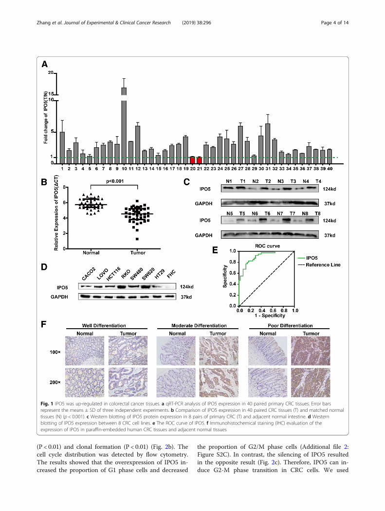
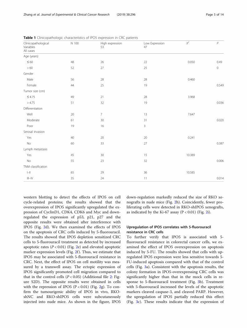
ResultsIPO5 is upregulated in colorectal cancer tissuesThe overexpression of IPO5 at the mRNA and proteinlevels was further verified using quantitative reversetranscription-PCR (qRT-PCR) and western blotting in 40and 8 pairs of primary CRC and normal tissues, respect-ively (Fig. 1a-c). By evaluating the expression levels ofIPO5 in eight colorectal cancer cell lines, LOVO, FHC,CACO2, RKO, SW620, SW480, HT29 and HCT116, wefound that compared with that in the normal epithelialcell line FHC, IPO5 is highly expressed in cancer cells,and the highest expression levels were found in SW620,a cell line with high metastatic potential (Fig. 1d). Tofurther investigate the potential clinical significance ofIPO5 in CRC, we constructed ROC curves, using therelative expression levels of IPO5 in CRC tissues andpaired non-cancerous tissues (AUC of 0.9050) (Fig. 1e).The data indicate IPO5 has potential as a biomarker forCRC diagnosis. Furthermore, the immunohistochemistryresults showed that IPO5 is located predominantly inthe cytoplasm and the positive staining of IPO5 was sig-nificantly higher in the cancer tissues than it was in thecorresponding normal tissues (Fig. 1f ). To further evalu-ate the relationship between the expression levels ofIPO5 and the clinicopathological parameters, the expres-sion of IPO5 was divided into a low-level group (n = 47)and a high-level group (n = 53). The results showed thatthe positive protein expression of IPO5 was positivelycorrelated with tumour size, differentiation, TNM stage,lymph node metastasis (Table 1).
Knockdown of IPO5 inhibits the growth and migration ofCRC cells in vitro and in vivoTo characterize the biological role of IPO5 in CRC cells,we overexpressed IPO5 in the SW480 and HCT116 celllines, simultaneously silenced IPO5 expression withIPO5-shRNA in SW620 and RKO cells (Fig. 2a), andthen selected IPO5-shRNA1 to construct lentiviral stableinterfering IPO5 cell lines for subsequent functionalstudies. The upregulation of IPO5 significantly increasedthe activity (P < 0.01) and clonogenicity (P < 0.01) ofHCT116 and SW480 cells compared to those of the nor-mal control cells (Additional file 2: Figure S2A and S2B).Conversely, interference with IPO5 reduces cell viability
Zhang et al. Journal of Experimental & Clinical Cancer Research (2019) 38:296 Page 3 of 14
(P < 0.01) and clonal formation (P < 0.01) (Fig. 2b). Thecell cycle distribution was detected by flow cytometry.The results showed that the overexpression of IPO5 in-creased the proportion of G1 phase cells and decreased
the proportion of G2/M phase cells (Additional file 2:Figure S2C). In contrast, the silencing of IPO5 resultedin the opposite result (Fig. 2c). Therefore, IPO5 can in-duce G2-M phase transition in CRC cells. We used
Fig. 1 IPO5 was up-regulated in colorectal cancer tissues. a qRT-PCR analysis of IPO5 expression in 40 paired primary CRC tissues. Error barsrepresent the means ± SD of three independent experiments. b Comparison of IPO5 expression in 40 paired CRC tissues (T) and matched normaltissues (N) (p < 0.001). c Western blotting of IPO5 protein expression in 8 pairs of primary CRC (T) and adjacent normal intestine. d Westernblotting of IPO5 expression between 8 CRC cell lines. e The ROC curve of IPO5. f Immunohistochemical staining (IHC) evaluation of theexpression of IPO5 in paraffin-embedded human CRC tissues and adjacent normal tissues
Zhang et al. Journal of Experimental & Clinical Cancer Research (2019) 38:296 Page 4 of 14
western blotting to detect the effects of IPO5 on cellcycle-related proteins; the results showed that theoverexpression of IPO5 significantly upregulated the ex-pression of CyclinD1, CDK4, CDK6 and Myc and down-regulated the expression of p53, p21, p27 and theopposite results were obtained after interference withIPO5 (Fig. 2d). We then examined the effects of IPO5on the apoptosis of CRC cells induced by 5-fluorouracil.The results showed that IPO5 depletion sensitized CRCcells to 5-fluorouracil treatment as detected by increasedapoptotic rates (P < 0.01) (Fig. 2e) and elevated apoptoticmarker expression levels (Fig. 2f ). Thus, we estimate thatIPO5 may be associated with 5-fluorouracil resistance inCRC. Next, the effect of IPO5 on cell motility was mea-sured by a transwell assay. The ectopic expression ofIPO5 significantly promoted cell migration compared tothat in the control cells (P < 0.05) (Additional file 2: Fig-ure S2D). The opposite results were obtained in cellswith the repression of IPO5 (P < 0.01) (Fig. 2g). To con-firm the tumourigenic ability of IPO5 in vivo, RKO-shNC and RKO-shIPO5 cells were subcutaneouslyinjected into nude mice. As shown in the figure, IPO5
down-regulation markedly reduced the size of RKO xe-nografts in nude mice (Fig. 2h). Coincidently, fewer pro-liferating cells were detected in RKO-shIPO5 xenografts,as indicated by the Ki-67 assay (P < 0.01) (Fig. 2i).
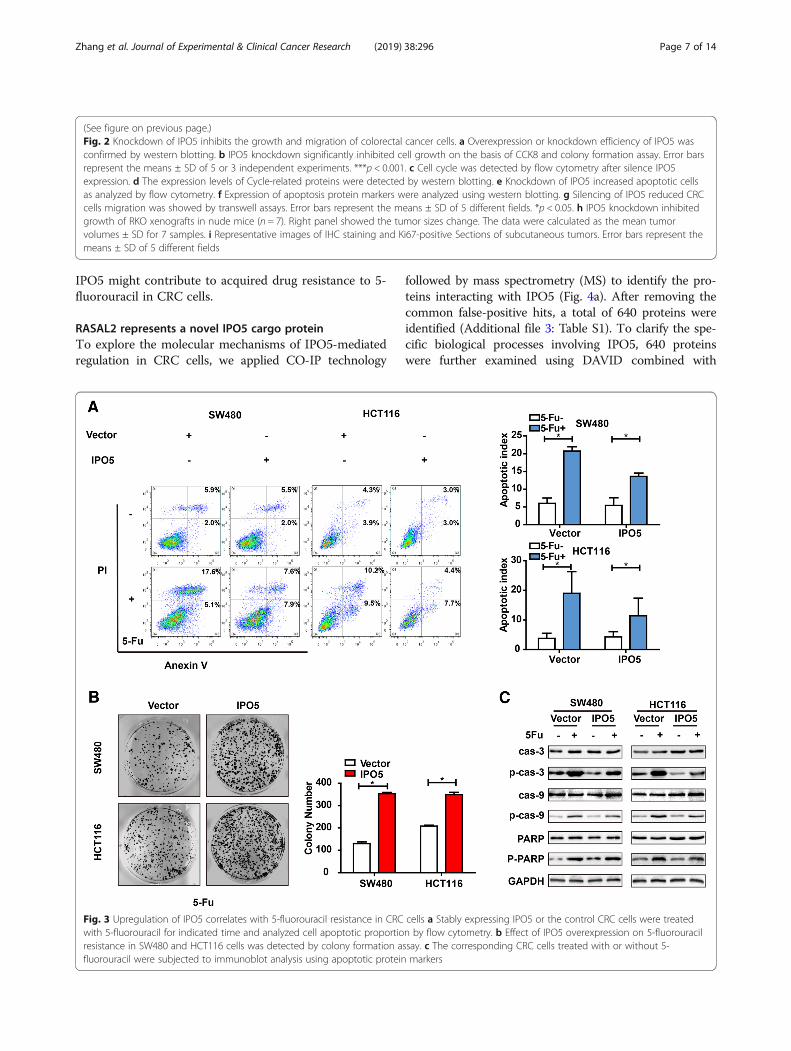
Upregulation of IPO5 correlates with 5-fluorouracilresistance in CRC cellsTo further verify that IPO5 is associated with 5-fluorouracil resistance in colorectal cancer cells, we ex-amined the effect of IPO5 overexpression on apoptosisinduced by 5-FU. The results showed that cells with up-regulated IPO5 expression were less sensitive towards 5-FU-induced apoptosis compared with that of the controlcells (Fig. 3a). Consistent with the apoptosis results, thecolony formation in IPO5-overexpressing CRC cells wassignificantly higher than that in the mock cells in re-sponse to 5-fluorouracil treatment (Fig. 3b). Treatmentwith 5-fluorouracil increased the levels of the apoptoticmarkers cleaved caspase-3, and cleaved PARP. However,the upregulation of IPO5 partially reduced this effect(Fig. 3c). These results indicate that the expression of
Table 1 Clinicopathologic characteristics of IPO5 expression in CRC patients
ClinicopathologicalVariablesAll cases
N 100 High expression53
Low Expression47
X2 P
Age (years)
≤ 60 48 26 22 0.050 0.49
> 60 52 27 25 0
Gender
Male 56 28 28 0.460
Female 44 25 19 0.549
Tumor size (cm)
≤ 4.75 49 21 28 3.968
> 4.75 51 32 19 0.036
Differentiation
Well 20 7 13 7.647
Moderate 61 30 31 0.020
Poor 19 16 3
Serosal invasion
Yes 40 20 20 0.241
No 60 33 27 0.387
Lymph metastasis
Yes 45 30 15 10.389
No 55 23 32 0.006
TNM classification
I–II 65 29 36 10.585
III–IV 35 24 11 0.014
Zhang et al. Journal of Experimental & Clinical Cancer Research (2019) 38:296 Page 5 of 14
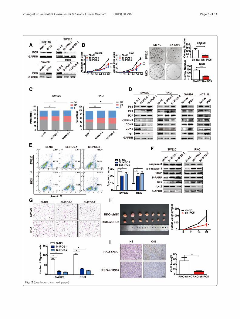
Fig. 2 (See legend on next page.)
Zhang et al. Journal of Experimental & Clinical Cancer Research (2019) 38:296 Page 6 of 14
IPO5 might contribute to acquired drug resistance to 5-fluorouracil in CRC cells.
RASAL2 represents a novel IPO5 cargo proteinTo explore the molecular mechanisms of IPO5-mediatedregulation in CRC cells, we applied CO-IP technology
followed by mass spectrometry (MS) to identify the pro-teins interacting with IPO5 (Fig. 4a). After removing thecommon false-positive hits, a total of 640 proteins wereidentified (Additional file 3: Table S1). To clarify the spe-cific biological processes involving IPO5, 640 proteinswere further examined using DAVID combined with
(See figure on previous page.)Fig. 2 Knockdown of IPO5 inhibits the growth and migration of colorectal cancer cells. a Overexpression or knockdown efficiency of IPO5 wasconfirmed by western blotting. b IPO5 knockdown significantly inhibited cell growth on the basis of CCK8 and colony formation assay. Error barsrepresent the means ± SD of 5 or 3 independent experiments. ***p < 0.001. c Cell cycle was detected by flow cytometry after silence IPO5expression. d The expression levels of Cycle-related proteins were detected by western blotting. e Knockdown of IPO5 increased apoptotic cellsas analyzed by flow cytometry. f Expression of apoptosis protein markers were analyzed using western blotting. g Silencing of IPO5 reduced CRCcells migration was showed by transwell assays. Error bars represent the means ± SD of 5 different fields. *p < 0.05. h IPO5 knockdown inhibitedgrowth of RKO xenografts in nude mice (n = 7). Right panel showed the tumor sizes change. The data were calculated as the mean tumorvolumes ± SD for 7 samples. i Representative images of IHC staining and Ki67-positive Sections of subcutaneous tumors. Error bars represent themeans ± SD of 5 different fields
Fig. 3 Upregulation of IPO5 correlates with 5-fluorouracil resistance in CRC cells a Stably expressing IPO5 or the control CRC cells were treatedwith 5-fluorouracil for indicated time and analyzed cell apoptotic proportion by flow cytometry. b Effect of IPO5 overexpression on 5-fluorouracilresistance in SW480 and HCT116 cells was detected by colony formation assay. c The corresponding CRC cells treated with or without 5-fluorouracil were subjected to immunoblot analysis using apoptotic protein markers
Zhang et al. Journal of Experimental & Clinical Cancer Research (2019) 38:296 Page 7 of 14
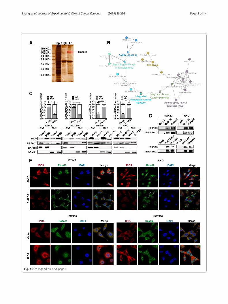
Fig. 4 (See legend on next page.)
Zhang et al. Journal of Experimental & Clinical Cancer Research (2019) 38:296 Page 8 of 14
KEGG pathway enrichment analysis. We found that inaddition to the known functionality in the database, thereare also notable features of interest, such as cell cycleregulation and cytoskeletal regulation (Additional file 4:Table S2). In addition, we built biological networks andanalysed the signal pathways involved in these 640 pro-teins by using Cytoscape software plug-in ClueGO in con-junction with the WikiPathway database, and we foundthat these proteins were involved in various cancer-relatedsignal pathways (Fig. 4b). To further pinpoint potentialproteins transported to the nucleus by IPO5, we used thecNLS Mapper Tool together with the COMPARTMENTSdatabase to determine the nuclear localization signal andsubcellular localization of the proteins. Finally, 15 proteinswith NLS sequences and shuttles through the nucleo-plasm were screened, which we believe are the most likelycargo candidates for IPO5 (Additional file 5: Table S3).From these 15 proteins, we focused on 8 cancer-relatedproteins (UBR5, ATRX, RASAL2, LIMK1, RAD51,RABL6, SIN3A, and DNAJB1) for further subcellular frac-tionation analysis. The results showed that RASAL2 wasthe most likely cargo protein for IPO5 in CRC cells (Add-itional file 2: Figure. S2E). To verify our results, we contin-ued to explore whether the subcellular localization ofRASAL2 was affected by IPO5. The results showed thatthe amounts of cytoplasmic RASAL2 increased togetherwith a concomitant decrease in the nuclear levels after si-lencing IPO5, whereas cells overexpressing IPO5 showedthe opposite results (Fig. 4c). Furthermore, we performedan endogenous reciprocal CO-IP assay to verify the inter-action of IPO5 and RASAL2 (Fig. 4d). Then, immuno-fluorescence and confocal microscopy were used todemonstrate that RASAL2 translocated into the nucleusin IPO5 upregulated cells but accumulated in the cyto-plasm and at the cell surface in si-IPO5-treated cells (Fig.4e). These results collectively suggest that IPO5 mediatesRASAL2 nuclear transport in CRC cells.
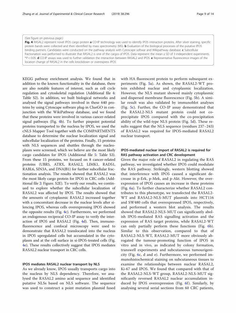
IPO5 mediates RASAL2 nuclear transport by NLSAs we already know, IPO5 usually transports cargo intothe nucleus by NLS dependency. Therefore, we ana-lysed the RASAL2 amino acid sequence and identifiedputative NLSs based on NLS software. The sequencewas used to construct a point mutation plasmid fused
with HA fluorescent protein to perform subsequent ex-periments (Fig. 5a). As shown, the RASAL2-WT pro-tein exhibited nuclear and cytoplasmic localization.However, the NLS mutant showed mainly cytoplasmicand dispersed membrane fluorescence (Fig. 5b). A simi-lar result was also validated by immunoblot analyses(Fig. 5c). Further, the CO-IP assay demonstrated thatthe RASAL2-NLS mutant protein could not co-precipitate IPO5 compared with the co-precipitationability of the wild-type NLS protein (Fig. 5d). These re-sults suggest that the NLS sequence (residues 237–243)of RASAL2 was required for IPO5-mediated RASAL2nuclear transport.
IPO5-mediated nuclear import of RASAL2 is required forRAS pathway activation and CRC developmentGiven the major role of RASAL2 in regulating the RASpathway, we investigated whether IPO5 could modulatethe RAS pathway. Strikingly, western blotting showedthat interference with IPO5 caused a significant de-crease in p-Erk, p-Mek, and p-Akt. However, the over-expression of IPO5 causes an increase in these proteins(Fig. 6a). To further characterize whether RASAL2 con-tributes to this phenotype, we transfected the RASAL2-WT and RASAL2-NLS-MUT plasmids into HCT116and SW480 cells that overexpressed IPO5, respectively,and performed a western blot analysis. The resultsshowed that RASAL2-NLS-MUT can significantly abol-ish IPO5-mediated RAS signalling activation and theexpression of RAS target proteins, while RASAL2-WTcan only partially perform these functions (Fig. 6b).Similar to this observation, compared to that ofRASAL2-NLS-WT, RASAL2-MUT more obviously ab-rogated the tumour-promoting function of IPO5 invitro and in vivo, as indicated by colony formation,transwell experiments and subcutaneous tumourigeni-city (Fig. 6c, d and e). Furthermore, we performed im-munohistochemical staining on subcutaneous tissues toexamine the relationships between nuclear RASAL2,Ki-67 and IPO5. We found that compared with that ofthe RASAL2-NLS-WT group, RASAL2-NLS-MUT sig-nificantly reversed RASAL2 nuclear accumulation in-duced by IPO5 overexpression (Fig. 6f ). Similarly, byanalysing several serial sections from 60 CRC patients,
(See figure on previous page.)Fig. 4 RASAL2 represent novel IPO5 cargo protein a CO-IP technology was used to identify IPO5 interaction proteins. After silver staining, specificprotein bands were collected and then identified by mass spectrometry (MS). b Evaluation of the biological processes of the putative IPO5binding partners. Candidates were conducted on the pathway analysis with Cytoscape softwar and Wikipathway database. c Subcellularfractionation was performed to illustrate that RASAL2 is one of the cargos of IPO5. Data represent the mean ± SD of 3 independent experiments.*P < 0.05. d CO-IP assays was used to further validation the interaction between RASAL2 and IPO5. e Representative fluorescence images of thelocation change of RASAL2 in the cells knockdown or overexpress IPO5
Zhang et al. Journal of Experimental & Clinical Cancer Research (2019) 38:296 Page 9 of 14
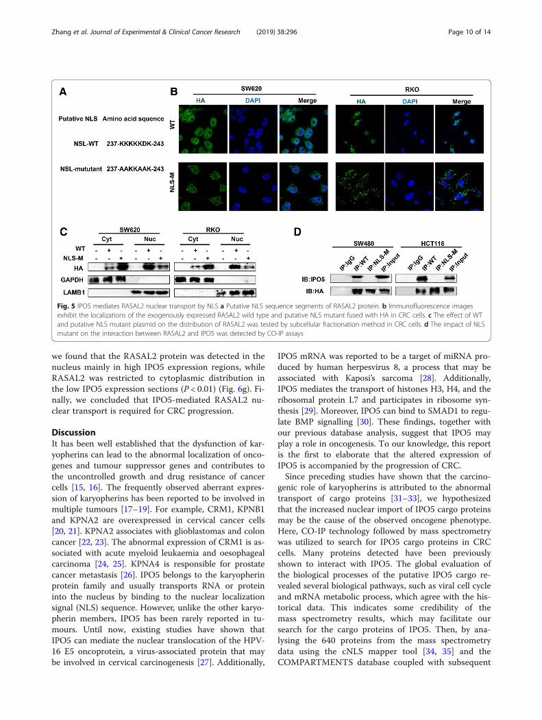
we found that the RASAL2 protein was detected in thenucleus mainly in high IPO5 expression regions, whileRASAL2 was restricted to cytoplasmic distribution inthe low IPO5 expression sections (P < 0.01) (Fig. 6g). Fi-nally, we concluded that IPO5-mediated RASAL2 nu-clear transport is required for CRC progression.
DiscussionIt has been well established that the dysfunction of kar-yopherins can lead to the abnormal localization of onco-genes and tumour suppressor genes and contributes tothe uncontrolled growth and drug resistance of cancercells [15, 16]. The frequently observed aberrant expres-sion of karyopherins has been reported to be involved inmultiple tumours [17–19]. For example, CRM1, KPNB1and KPNA2 are overexpressed in cervical cancer cells[20, 21]. KPNA2 associates with glioblastomas and coloncancer [22, 23]. The abnormal expression of CRM1 is as-sociated with acute myeloid leukaemia and oesophagealcarcinoma [24, 25]. KPNA4 is responsible for prostatecancer metastasis [26]. IPO5 belongs to the karyopherinprotein family and usually transports RNA or proteininto the nucleus by binding to the nuclear localizationsignal (NLS) sequence. However, unlike the other karyo-pherin members, IPO5 has been rarely reported in tu-mours. Until now, existing studies have shown thatIPO5 can mediate the nuclear translocation of the HPV-16 E5 oncoprotein, a virus-associated protein that maybe involved in cervical carcinogenesis [27]. Additionally,
IPO5 mRNA was reported to be a target of miRNA pro-duced by human herpesvirus 8, a process that may beassociated with Kaposi’s sarcoma [28]. Additionally,IPO5 mediates the transport of histones H3, H4, and theribosomal protein L7 and participates in ribosome syn-thesis [29]. Moreover, IPO5 can bind to SMAD1 to regu-late BMP signalling [30]. These findings, together withour previous database analysis, suggest that IPO5 mayplay a role in oncogenesis. To our knowledge, this reportis the first to elaborate that the altered expression ofIPO5 is accompanied by the progression of CRC.Since preceding studies have shown that the carcino-
genic role of karyopherins is attributed to the abnormaltransport of cargo proteins [31–33], we hypothesizedthat the increased nuclear import of IPO5 cargo proteinsmay be the cause of the observed oncogene phenotype.Here, CO-IP technology followed by mass spectrometrywas utilized to search for IPO5 cargo proteins in CRCcells. Many proteins detected have been previouslyshown to interact with IPO5. The global evaluation ofthe biological processes of the putative IPO5 cargo re-vealed several biological pathways, such as viral cell cycleand mRNA metabolic process, which agree with the his-torical data. This indicates some credibility of themass spectrometry results, which may facilitate oursearch for the cargo proteins of IPO5. Then, by ana-lysing the 640 proteins from the mass spectrometrydata using the cNLS mapper tool [34, 35] and theCOMPARTMENTS database coupled with subsequent
Fig. 5 IPO5 mediates RASAL2 nuclear transport by NLS a Putative NLS sequence segments of RASAL2 protein. b Immunofluorescence imagesexhibit the localizations of the exogenously expressed RASAL2 wild type and putative NLS mutant fused with HA in CRC cells. c The effect of WTand putative NLS mutant plasmid on the distribution of RASAL2 was tested by subcellular fractionation method in CRC cells. d The impact of NLSmutant on the interaction between RASAL2 and IPO5 was detected by CO-IP assays
Zhang et al. Journal of Experimental & Clinical Cancer Research (2019) 38:296 Page 10 of 14
Fig. 6 (See legend on next page.)
Zhang et al. Journal of Experimental & Clinical Cancer Research (2019) 38:296 Page 11 of 14
subcellular fractionation methods, we found thatRASAL2 may be one of the cargos transported byIPO5 in CRC cells.RASAL2 is a RAS-GTPase-activated protein and a
negative regulator of the RAS cascade. Its potentialoncosuppressor role was currently hypothesized in a var-iety of tumours by inhibiting the activation of the down-stream RAS pathway [36, 37]. Existing studies haveobserved that RASAL2 is mainly localized and plays arole in the cytoplasm [38, 39], but whether it could enterthe nucleus has not been reported. Here, via
immunofluorescence and immunohistochemical stainingcombined with protein localization database results, wefound that RASAL2 is distributed not only in the cyto-plasm but also in the nucleus and membranes of CRCcells. This led us to analyse its amino acid sequence byusing a related database, and we found that it containsone NLS sequence, which indicates that RASAL2 is verylikely to enter the nucleus with the NLS sequence.Therefore, we hypothesized that IPO5 may interact withthe NLS of RASAL2, resulting in RASAL2 nuclear trans-location and eliminating the inhibitory effect of RASAL2
(See figure on previous page.)Fig. 6 IPO5-mediated nuclear import of RASAL2 is required for RAS pathway activation and CRC development a Western blotting was used todetermine the expression of ERK, p-ERK, AKT, and p-AKT upon knockdown or overexpressing of IPO5 in CRC cells. b The NLS mutant of RASAL2resulted in reversing the IPO5-mediated RAS signaling activation showed by Western blotting. c and d The recovery effect of NLS mutant on theIPO5-mediated CRC cells migration and proliferation ability were detected by colony formation and transwell respectively. Scale bars, 200 μm. eSubcutaneous tumorigenesis in nude mice was applied to explore the impact of NLS mutant on tumorigenicity of IPO5 in vivo. Data representthe mean ± SD of 3 independent experiments. *P < 0.05. f Tumor xenograft tissues were embedded in paraffin and then assessed for theexpression of Ki-67 and RASAL2. Scale bars, 200 μm. g Correlation of nuclear RASAL2 expression and IPO5 expression in colorectal cancer tissuewas evaluated by Immunohistochemical. Scale bars, 50 μm
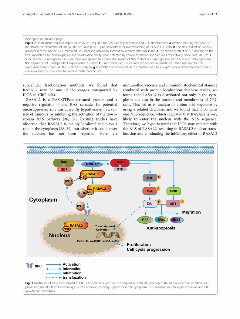
Fig. 7 Illustration of IPO5 involvement in CRC. IPO5 interacts with the NLS sequence of RASAL2 leading to RASAL2 nuclear transposition, Thispreventing RASAL2 from functioning as a RAS signaling pathway suppressor in the cytoplasm. Thus resulting in RAS signal activation and CRCgrowth and metastasis
Zhang et al. Journal of Experimental & Clinical Cancer Research (2019) 38:296 Page 12 of 14
in the cytoplasm; this would subsequently augment theRAS pathway and would ultimately drive CRC progres-sion. To validate this hypothesis, we constructed aRASAL2 NLS sequence plasmid with point mutationsand transfected cells for further functional experiments.Our results show that the NLS mutations of RASAL2disrupted the interaction between IPO5 and RASAL2and significantly impaired the IPO5-mediated malignantcharacteristics of CRC. There is evidence that some kar-yopherins share common cargo [40, 41], and this iscoupled with the fact that our exploration for the IPO5binding cargo still lacks specificity; therefore, we mustbear in mind that is a possibility that RASAL2 enters thenucleus, which is also mediated by other IPO5 familymembers. These results invite us to carry out furtherstudies. Because altered karyopherin protein functionsplay a vital role in drug resistance [42, 43], recent studiespoint to their potential usefulness as a novel strategy foranticancer therapy [44]. A small molecule inhibitor tar-geting CRM1 has been shown to be beneficial to the an-ticancer effects [45–47]. INI-43, an inhibitor of KPNB1,is a potent chemotropic agent of malignancies [48].However, because normal cells also share the nucleartransport machinery with cancer cells, drugs targetingthese proteins are currently limited. Therefore, prospect-ive basic research with regard to the tumourigenicmechanisms of karyopherin proteins is lacking and isdesperately required for effective cancer therapy.
ConclusionsIn summary, the current study demonstrated thatIPO5 is an oncogene involved in CRC cell prolifera-tion and migration. This highlights the significance ofIPO5 in 5-fluorouracil-resistant CRC cells. The onco-genic function of IPO5 was mediated by promotingRAS signalling by increasing the nuclear translocationof RASAL2 (Fig. 7). Our results provide new informa-tion on the carcinogenic role mediated by IPO5. It isexpected to be a promising therapeutic target.
Additional files
Additional file 1: Figure S1. The expression of IPO5, data from publicdatabase. (A) Analysis of IPO5 expression in different types ofmalignancies. (B) Analysis of IPO5 expression in TCGA CRC large samplegenomic database. (C) Analysis of IPO5 expression using the CRC geneexpression profile data GSE41258. (JPG 849 kb)
Additional file 2: Figure S2. Effects of IPO5 over-expression on CRC cellproliferation and migration in vitro (A and B) Up-regulation of IPO5 in-creased cell proliferation (P < 0.01) and clonogenicity (P < 0.01) as com-pared to controls . (C) The effect of IPO5 on the cell cycle distributionwas detected by flow cytometry in CRC cells. (D) The impact of ectopicexpression of IPO5 on cell migration was validated by transwell assay(P <0.01). (E) Screen out IPO5 transporting cargos using subcellular fraction-ation methods followed by immunoblotting. (JPG 5002 kb)
Additional file 3: Table S1. List of IPO5 binding candidates identifiedby mass spectrometry. (DOCX 55 kb)
Additional file 4: Table S2. Pathway analysis of IPO5 bindingcandidates using DAVID tool. (DOCX 16 kb)
Additional file 5: Table S3. List of 15 IPO5 binding proteins with NLSsequence. (DOCX 20 kb)
Additional file 6: Table S4. Primer sequences used in RT-qPCR analysis.Table S5: Nucleotide sequences used for knockdown. (DOC 40 kb)
Abbreviations5-FU: 5-fluorouracil; BSA: Albumin from bovine serum; CCK-8: Cell countingkit-8; CO-IP: Co-Immunoprecipitation; CRC: Colorectal cancer; CRM1: ExportinCRM1; FBS: Fetal bovine serum; IF: Immunofluorescence;IHC: Immunohistochemistry; IPO5: Importin 5; MS: Mass spectrometry;NC: Negative control; NLS: Nuclear localization sequence; NPC: Nuclear porecomplex; qPCR: Quantitative real-time polymerase chain reaction;RASAL2: RAS protein activator like 2; TCGA: The Cancer Genome Atlas;TNM: Tumor Node Metastasis
AcknowledgementsNot applicable.
Authors’ contributionsXNL contributed to study design, analysis, obtaining funding, studysupervision. WJZ carried out the experiments and writing up the manuscript.YXL and XML contributed in data acquisition, analysis, and interpretation ofthe data. JMZ and WZ contributed in statistical analysis. WHL contributed inthe collection of patient samples and clinical input. All authors read andapproved the final manuscript.
FundingThis study was supported by the National Natural Science Foundation ofChina (grant numbers 81874074, 81672429 and 81502479).
Availability of data and materialsThe datasets used and/or analysed during the current study are availablefrom the corresponding author on reasonable request.
Ethics approval and consent to participateThis study was approved by the Ethics Committees of Nanfang Hospital,Southern Medical University. Written informed consent was obtained from allpatients.
Consent for publicationNot applicable.
Competing interestsThe authors declare that they have no competing financial interests in ourstudy.
Author details1Department of Pathology, School of Basic Medical Sciences, SouthernMedical University, Guangzhou 510515, People’s Republic of China.2Department of General Surgery, Nanfang Hospital, Southern MedicalUniversity, Guangzhou, China.
Received: 22 January 2019 Accepted: 25 June 2019
References1. Chook YM, Blobel G. Karyopherins and nuclear import. Curr Opin Struct Biol.
2001;11:703–15.2. Çağatay T, Chook YM. Karyopherins in cancer. Curr Opin Cell Biol. 2018;
52:30–42.3. Lund E, Guttinger S, Calado A, Dahlberg JE, Kutay U. Nuclear export of
microRNA precursors. Science. 2004;303:95–8.4. Silver PA, Kau TR, Way JC. Nuclear transport and cancer: from mechanism to
intervention. Nat Rev Cancer. 2004;4:106–17.
Zhang et al. Journal of Experimental & Clinical Cancer Research (2019) 38:296 Page 13 of 14
5. Tran EJ, King MC, Corbett AH. Macromolecular transport between thenucleus and the cytoplasm: advances in mechanism and emerging links todisease. Biochim Biophys Acta. 2014;1843:2784–95.
6. Senapedis WT, Baloglu E, Landesman Y. Clinical translation of nuclear exportinhibitors in cancer. Semin Cancer Biol. 2014;27:74–86.
7. Kimura M, Imamoto N. Biological significance of the importin-beta family-dependent nucleocytoplasmic transport pathways. Traffic. 2014;15:727–48.
8. Kosyna FK, Depping R. Controlling the gatekeeper: therapeutic targeting ofnuclear transport. Cells. 2018;7:221.
9. Shigeyasu K, Okugawa Y, Toden S, Boland CR, Goel A. Exportin-5 functionsas an oncogene and a potential therapeutic target in colorectal cancer. ClinCancer Res. 2017;23:1312–22.
10. Kim J, McMillan E, Kim HS, Venkateswaran N, Makkar G, Rodriguez-Canales J,et al. XPO1-dependent nuclear export is a druggable vulnerability in KRAS-mutant lung cancer. Nature. 2016;538:114–7.
11. Vuorinen EM, Rajala N, Rauhala HE, Kallioniemi A. Abstract 76: KPNA7nuclear import protein - a critical regulator of cancer cell growth. CancerRes. 2016;76:76.
12. Qi L, Ding Y. Construction of key signal regulatory network in metastaticcolorectal cancer. Oncotarget. 2018;9:6086–94.
13. Lin C, Zhang J, Lu Y, Li X, Zhang W, Zhang W, et al. NIT1 suppresses tumourproliferation by activating the TGFβ1–Smad2/3 signalling pathway incolorectal cancer. Cell Death Dis. 2018;9:263.
14. Zeng Z, Li Y, Pan Y, Lan X, Song F, Sun J, et al. Cancer-derived exosomalmiR-25-3p promotes pre-metastatic niche formation by inducing vascularpermeability and angiogenesis. Nat Commun. 2018;9:5395.
15. Weis K. Regulating access to the genome: nucleocytoplasmic transportthroughout the cell cycle. Cell. 2003;112:441–51.
16. Beck M, Schirmacher P, Singer S. Alterations of the nuclear transport systemin hepatocellular carcinoma - new basis for therapeutic strategies. J Hepatol.2017;67:1051–61.
17. Zhao X, Chen Y, Tan X, Zhang L, Zhang H, Li Z, et al. Advanced glycationend-products suppress autophagic flux in podocytes by activatingmammalian target of rapamycin and inhibiting nuclear translocation oftranscription factor EB. J Pathol. 2018;245:235-48.
18. Yan D, Pomicter AD, Tantravahi S, Mason CC, Senina AV, Ahmann JM, et al.Nuclear-cytoplasmic transport is a therapeutic target in myelofibrosis. ClinCancer Res. 2018;25:2323-35.
19. Kau TR, Silver PA. Nuclear transport as a target for cell growth. Drug DiscovToday. 2003;8:78–85.
20. van der Watt PJ, Maske CP, Hendricks DT, Parker MI, Denny L, Govender D,et al. The Karyopherin proteins, Crm1 and Karyopherin β1, areoverexpressed in cervical cancer and are critical for cancer cell survival andproliferation. Int J Cancer. 2009;124:1829–40.
21. Angus L, van der Watt PJ, Leaner VD. Inhibition of the nuclear transporter,Kpnβ1, results in prolonged mitotic arrest and activation of the intrinsicapoptotic pathway in cervical cancer cells. Carcinogenesis. 2014;35:1121–31.
22. Zhang Y, Zhang M, Yu F, Lu S, Sun H, Tang H, et al. Karyopherin alpha 2 is anovel prognostic marker and a potential therapeutic target for coloncancer. J Exp Clin Cancer Res. 2015;34:145.
23. Li J, Liu Q, Liu Z, Xia Q, Zhang Z, Zhang R, et al. KPNA2 promotes metabolicreprogramming in glioblastomas by regulation of c-myc. J Exp Clin CancerRes. 2018;37:194.
24. Kojima K, Kornblau SM, Ruvolo V, Dilip A, Duvvuri S, Davis RE, et al.Prognostic impact and targeting of CRM1 in acute myeloid leukemia. Blood.2013;121:4166–74.
25. Yang X, Cheng L, Yao L, Ren H, Zhang S, Min X, et al. Involvement ofchromosome region maintenance 1 (CRM1) in the formation andprogression of esophageal squamous cell carcinoma. Med Oncol. 2014;31:1–13.
26. Yang J, Lu C, Wei J, Guo Y, Liu W, Luo L, et al. Inhibition of KPNA4attenuates prostate cancer metastasis. Oncogene. 2017;36:2868–78.
27. Krawczyk E, Hanover JA, Schlegel R, Suprynowicz FA. Karyopherin beta3: anew cellular target for the HPV-16 E5 oncoprotein. Biochem Biophys ResCommun. 2008;371:684–8.
28. Quan L, Qiu T, Liang J, Li M, Zhang Y, Tao K. Identification of target genesregulated by KSHV miRNAs in KSHV-infected lymphoma cells. Pathol OncolRes. 2015;21:875–80.
29. Soniat M, Cagatay T, Chook YM. Recognition elements in the histone H3and H4 tails for seven different importins. J Biol Chem. 2016;291:21171–83.
30. Baas R, Sijm A, van Teeffelen HA, van Es R, Vos HR, Marc TH. Quantitativeproteomics of the SMAD (suppressor of mothers against decapentaplegic)transcription factor family identifies importin 5 as a bone morphogenicprotein receptor SMAD-specific importin. J Biol Chem. 2016;291:24121–32.
31. Vuorinen EM, Rajala NK, Rauhala HE, Nurminen AT, Hytonen VP, KallioniemiA. Search for KPNA7 cargo proteins in human cells reveals MVP and ZNF414as novel regulators of cancer cell growth. Biochim Biophys Acta Mol basisDis. 2017;1863:211–9.
32. Wang CI, Chien KY, Wang CL, Liu HP, Cheng CC, Chang YS, et al.Quantitative proteomics reveals regulation of karyopherin subunit alpha-2(KPNA2) and its potential novel cargo proteins in nonsmall cell lung cancer.Mol Cell Proteomics. 2012;11:1105–22.
33. Alshareeda AT, Negm OH, Green AR, Nolan CC, Tighe P, Albarakati N, etal. KPNA2 is a nuclear export protein that contributes to aberrantlocalisation of key proteins and poor prognosis of breast cancer. Br JCancer. 2015;112:1929–37.
34. Lin JR, Hu J. SeqNLS: nuclear localization signal prediction based onfrequent pattern mining and linear motif scoring. PLoS One. 2013;8:e76864.
35. Li K, Mo C, Gong D, Chen Y, Huang Z, Li Y, et al. DDX17 nucleocytoplasmicshuttling promotes acquired gefitinib resistance in non-small cell lungcancer cells via activation of β-catenin. Cancer Lett. 2017;400:194–202.
36. McLaughlin SK, Olsen SN, Dake B, De Raedt T, Lim E, Bronson RT, et al. TheRasGAP gene, RASAL2, is a tumor and metastasis suppressor. Cancer Cell.2013;24:365–78.
37. Shen J, Wang Y, Hung MC. RASAL2: wrestling in the combat of Rasactivation. Cancer Cell. 2013;24:277–9.
38. Olsen SN, Wronski A, Castano Z, Dake B, Malone C, De Raedt T, et al. Loss ofRasGAP tumor suppressors underlies the aggressive nature of luminal Bbreast cancers. Cancer Discov. 2017;7:202–17.
39. Hui K, Gao Y, Huang J, Xu S, Wang B, Zeng J, et al. RASAL2, a RAS GTPase-activating protein, inhibits stemness and epithelial-mesenchymal transitionvia MAPK/SOX2 pathway in bladder cancer. Cell Death Dis. 2017;8:e2600.
40. Friedrich B, Quensel C, Sommer T, Hartmann E, Kohler M. Nuclearlocalization signal and protein context both mediate importin alphaspecificity of nuclear import substrates. Mol Cell Biol. 2006;26:8697–709.
41. Duan J, Tang Z, Mu H, Zhang G. [retracted] nuclear import of prototypefoamy virus transactivator bel1 is mediated by KPNA1, KPNA6 and KPNA7.Int J Mol Med. 2017;39:771.
42. Ishizawa J, Kojima K, Hail NJ, Tabe Y, Andreeff M. Expression, function, andtargeting of the nuclear exporter chromosome region maintenance 1(CRM1) protein. Pharmacol Ther. 2015;153:25–35.
43. Mahipal A, Malafa M. Importins and exportins as therapeutic targets incancer. Pharmacol Ther. 2016;164:135–43.
44. Mutka SC, Yang WQ, Dong SD, Ward SL, Craig DA, Timmermans PB, et al.Identification of nuclear export inhibitors with potent anticancer activity invivo. Cancer Res. 2009;69:510–7.
45. Tai YT, Landesman Y, Acharya C, Calle Y, Zhong MY, Cea M, et al. CRM1inhibition induces tumor cell cytotoxicity and impairs osteoclastogenesis inmultiple myeloma: molecular mechanisms and therapeutic implications.Leukemia. 2014;28:155–65.
46. Hing ZA, Fung HY, Ranganathan P, Mitchell S, El-Gamal D, Woyach JA, et al.Next-generation XPO1 inhibitor shows improved efficacy and in vivotolerability in hematological malignancies. Leukemia. 2016;30:2364–72.
47. Walker CJ, Oaks JJ, Santhanam R, Neviani P, Harb JG, Ferenchak G, et al.Preclinical and clinical efficacy of XPO1/CRM1 inhibition by the karyopherininhibitor KPT-330 in Ph+ leukemias. Blood. 2013;122:3034–44.
48. van der Watt PJ, Chi A, Stelma T, Stowell C, Strydom E, Carden S, et al.Targeting the nuclear import receptor Kpnbeta1 as an anticancertherapeutic. Mol Cancer Ther. 2016;15:560–73.
Publisher’s NoteSpringer Nature remains neutral with regard to jurisdictional claims inpublished maps and institutional affiliations.
Zhang et al. Journal of Experimental & Clinical Cancer Research (2019) 38:296 Page 14 of 14