Embed Size (px)
Citation preview

Chapter 2
IONIZING RADIATION UNITS AND STANDARDS © M. Ragheb
7/19/2018
2.1 INTRODUCTION
Radiation is a natural phenomenon that existed long before the advent of humans,
and permeates the known universe. Humans have used radiation in useful and useless
applications such as nuclear medicine, food preservation, power production and nuclear
weaponry, adding to the natural sources of radiation.
The term “radiation” is broad and encompasses photons in the electromagnetic
spectrum (Fig. 1), such as visible light, microwaves, radio waves and x and gamma rays,
as well as particles such as cosmic rays, electrons, and alpha particles. However the term
is often used to mean ionizing radiation.
Figure 1. The electromagnetic radiation spectrum. Left scale is wave length in cms, and
right scale is energy in eV.

Ionizing radiation is radiation that has enough energy to remove electrons from
atoms, consequently creating charged ions and radicals in the materials it interacts with.
Ionizing radiation includes beta and alpha particles, neutrons, protons, as well as x and
gamma radiation. Radio waves and microwaves used in communications do not possess
enough energy to ionize matter. Microwaves used in cellular phones, for instance, are
not considered as ionizing radiation even though they can cause damage by delivering
energy to the adjacent tissue, in this case, the brain.
There are two types of ionizing radiation:
1. Directly ionizing radiation: Forms of radiation that can eject orbital electrons directly
from atoms by interacting with the Coulomb force. These include charged particles such
as electrons, protons and alpha particles.
2. Indirectly ionizing radiation: These are forms of radiation that indirectly transfer
energy to charger particles of the absorber atom such as electrons or protons. These
include gamma rays and neutrons (Fig. 2).
Figure 2. Ionization created by a neutron as indirectly ionizing radiation.
Directly ionizing radiation tends to deposit its energy at a localized range in
materials, whereas indirectly ionizing radiation deposits it along its whole path, as shown
in Fig. 3.
Atoms or nuclei that emit ionizing radiation exhibits the process of radioactivity
and can be naturally occurring or human made.
2.2 ACTIVITY
Activity refers to the rate of radioactive transformations of a radioactive isotope.
The generally accepted is the number of transformations of decays per unit time.

The Système International (SI) unit is the Becquerel:
1 Becquerel = 1 Bq = 1 [Disintegration/sec] (1)
The conventional unit of activity is the Curie:
101 Curie = 1 Ci = 3.7 x 10 [Disintegrations/sec], (2)
which corresponds to the activity of 1 gram of the radium226 isotope. We can also write:
-121 Bq = 27 x 10 Ci. (3)
Table 1 shows the activities in Bq of some materials and common objects.
Figure 3. Different patterns of energy deposition in tissue of directly and indirectly
ionizing radiation.
2.3 RADIATION FIELD AND EXPOSURE
In the same way that one can define an electric, magnetic or gravitational field, a
radiation field can be defined as a domain where ionization caused by radiation can be
detected and its effects measured. Since electromagnetic radiation in the form of x rays
or gamma rays can cause ionization in air, primarily through the processes of pair
production, Compton scattering and the photoelectric effect, the presence of this
ionization in air defines the presence of a radiation field.

Table 1: Activity in Becquerels of some materials and objects.
Material or Object Activity,
[Bq]
Adult human (70 kgs with 100 Bq/kg) 7 x 10³
One kg of coffee 1 x 10³
One kg of super phosphate fertilizer (uranium and its
decay chain daughters such as radium)
5 x 10³
The air in a 100 m2 home from radon gas 3 x 10³ – 3 x 10⁴
Household smoke detector using the americium241
isotope
3 x 10⁴
Radioisotope for medical diagnosis 7 x 10⁷
Radioisotope source for medical therapy 1 x 10¹⁴
One kg 50 year old vitrified high-level nuclear waste 1 x 10¹³
One kg of freshly separated and manufactured pure
uranium
2.5 x 10⁷
One kg uranium 15 percent natural uranium Canadian
ore
2.5 x 10⁷
One kg uranium of 0.3 percent natural uranium
Australian ore
5 x 10⁵
One kg low level radioactive waste 1 x 10⁶
One kg of coal ash 2 x 10³
One kg of granite 1 x 10³
This radiation field is considered as incident on a body immersed in the field at
any point. The biological effects of radiation being a function of the ionization produced
in air, it becomes important to measure the degree of “exposure” to the electromagnetic
radiation field in terms of the number of ions or electrons produced in the adjacent
atmosphere. Thus one can define the unit of exposure to a radiation field in air as:
Δq Coulomb
Exposure: X=Δm kgof air
(4)

where: q is the sum of electrical charges on all the ions of one sign produced in air when
all the electrons, negative and positive, liberated by photons in a volume of air
after being completely stopped.
m is the mass of the volume of air under consideration. The electrons could be
stopped outside the volume of interest.
In honor of the discoverer of x rays, the unit of exposure is the Röntgen defined
as:
-4 CbR: 1 Röntgen=2.58 10
kg
, (5)
which is equivalent to the production of 1 electrostatic unit (esu) of charge of one sign
from the interaction of x rays or gamma rays in 0.001293 grams of air at standard
temperature and pressure (STP). This is equivalent to 1 cm3 of air at atmospheric
pressure and 0 degrees Celsius, where:
-101 esu =3.33 10 [Cb]
For a smaller magnitude, the milliRöntgen is defined as:
-31mR=10 R (6)
The exposure rate is defined as:
dX R mR
X= ordt sec hr
(7)
It must be noticed that the concepts of exposure and exposure rates apply only to
x rays and gamma rays not to other forms of radiation such as neutrons or charged
particles, and that they are defined only in air and not in inert or biological materials.
The radiation field can be associated with a considerable amount of ionization in
air.
EXAMPLE
For instance, by following a process of dimensional analysis, let us calculate the
number of ions created per second per cubic centimeter, or ions rate density, associated
with the exposure to a modest 1 [mR/hr] gamma ray field in air:

-3 -4
-19 3 3
-3 -4
-19 3 3
3
mR1
hr
mR R 1 hr Cb/kg 1 ions 1 kg gm=1 10 2.58 10 0.001293
hr mR 60x60 sec R 1.6x10 Cb 10 gm cm air
1 1 1 ions=1 10 2.58 10 0.001293
60 60 1.6 10 10 cm .sec
ions=578[ ]
cm .sec
This amounts to the creation of a considerable amount of about 580 ions per cubic
centimeter of air per second.
2.4 ABSORBED DOSE UNIT
If we want to measure the effects of radiation on materials rather than air, we
consider that the biological effects will depend on the amount of imparted energy that the
radiation imparts to an arbitrary volume or control volume of material. The imparted
energy must take into account the possibility of the occurrence of nuclear reactions
within the volume of material of interest. Carrying out an energy balance over this
control volume:
D in out vImparted energy: ΔE = E -E +Q (8)
where: Qv is the Q values of the nuclear reactions taking place in the control volume,
Ein is the input energy to the control volume,
Eout is the output energy from the control volume.
This leads to the definition of the concept of the absorbed dose of radiation as:
DΔEimparted energyAbsorbed dose: D= =
mass Δm (9)
The absorbed dose at a point can be also defined as a limiting condition:
Dp
Δm 0
ΔED = lim
Δm (10)
A unit for the absorbed dose in the conventional system of units is the Rad
standing for Radiation Absorbed Dose as:
Joule ergs
1Rad =0.01 =100kg gm
(11)

where 1 Joule = 107 ergs.
A smaller unit is:
-31 mRad =10 Rad. (12)
In the Système International (SI) system of units, the unit for the absorbed dose is:
Joule
1 Gray = 1 Gy = 1 = 100 radskg
(13)
with smaller units:
-3
-2
1mGy=10 Gy,
1cGy=10 Gy. (14)
Similarly to the exposure rate, the absorbed dose rate is defined as:
dD Rad Gy
D = ordt sec sec
(15)
Only in situations when indirectly ionizing radiation; primarily fast neutrons, are
under consideration, the Kerma (Kinetic Energy Released in Material) or Kerma Dose is
defined as:
K = [Sum of the initial energies of all the charged, ionizing particles
released by indirectly ionizing radiation; mostly neutrons, per unit mass of the
substance at hand]
The units of K are rads or Grays; the same as the absorbed dose units.
The human senses cannot detect radiation or discern whether a material is
radioactive. However, a variety of instruments can detect and measure radiation reliably
and accurately.
The amount of ionizing radiation, or dose, received by a person is measured in
terms of the energy absorbed in the body tissue.
Since the Gray is also equal to 100 radiation absorbed doses (rads) in the
conventional system of units. A commonly used unit is one hundredth of a Gray or
centiGray ,and is also equal to 1 rad of absorbed dose.
1 centiGray = 1 cGy = 1 rad (16)
2.5 THE RELATIVE BIOLOGICAL EFFECTIVENESS: RBE
To account for the biological effects of different types of radiation on biological
matter an experimental approach is adopted where a tissue culture or organ is irradiated

with standard 100 keV x rays or gamma rays, then irradiated with the other type of
radiation and the resulting effect such as cell survival is observed. The Relative
Biological Effectiveness (RBE) of the considered type of radiation is defined as:
Effect produced byother typeof radiation
RBE=Effect produced by100keV x or γ rays
(17)
The RBE of 100 keV x or gamma rays is taken as a reference with an arbitrary
assigned value of unity. Table 2 shows the values of the Quality Factor for different
types of radiation.
Table 2. Assigned values of the Quality Factor for different types of radiation.
Radiation Type Quality Factor
Qf
x rays or gamma rays 1
Alpha particles, protons, fast neutrons 10
Heavy recoil nuclei 20
Thermal neutrons 2
1 MeV neutrons 11
20 MeV neutrons 8
Beta particles above 30 keV 1
Beta particles below 30 keV 1.7
Heavy ions 20
Being an experimental value, the value of the RBE is normally a fraction of a
number. The round-off of the RBE value is an assigned value designated as the Quality
Factor:
fQuality Factor: Q = Round-off of RBE (18)
The Quality Factor is directly related to the degree of ionization that different
types of radiation can create along their paths in materials. The higher the degree of
ionization per unit length, then the higher the resulting biological damage. This is
expressed in terms of the Linear Energy Transfer (LET) for radiation:
dE keV
LET = -dx μm
(19)
Alpha particles have a higher quality factor than x or gamma rays since they can
cause a high degree of ionization in materials through collisions with the atomic nuclei.
The ionization is notoriously high near the end of their tracks when the alpha particle has
been slowed down, as shown in Fig. 4. This effect is known as the Bragg effect.
Beta particles tracks appear farther apart from those of alpha particles displaying a
process of straggling as shown in Fig. 5. As the beta particle slows down, the path

becomes more erratic and the ions are formed closer together, with the tracks at the very
end behaving like the alpha particles tracks as shown in Fig. 4.
Figure 4. Alpha particles tracks originating from a source on the right hand side in a
cloud chamber.

Figure 5. Beta particles tracks in a cloud chamber.
Table 3 shows the relationship between the LET and the Quality Factor.
Table 3. Relationship between the LET and the Quality factor of radiation.
Linear energy Transfer
LET [keV/micron]
Quality Factor
Qf
3.5 or less 1
7 2
23 5
53 10
175 and above 20
2.6 EFFECTIVE DOSE, DOSE EQUIVALENT OR BIOLOGICAL
DOSE
Equal exposure to different types of radiation expressed in Grays do not however
necessarily produce equal biological effects. One Gray of alpha radiation, for example,
will have a greater effect than one Gray of beta radiation.
Using the concept of the Radio Biological Effectiveness and the Quality Factor
the Biological Dose or Dose Equivalent or Effective Dose is defined as the product of the
absorbed dose and the quality factor as:

f
Effective Dose= Absorbed Dose x Qualityfactor
H = D.Q (20)
Similarly, an effective dose rate is defined as:
fH=D.Q (21)
The conventional unit of the effective dose is the radiation equivalent man or rem,
defined as:
f1 rem=1 Q . rad (22)
with a smaller unit of:
1 mrem = 10-3 rem (23)
In the Système International (SI) system of units it is defined as the Sievert:
1 Sievert = 1 Sv = 100 rem. (24)
For x rays and gamma rays where the quality factor is unity, 1 rad of absorbed
dose gives an effective dose of 1 rem.
Now, regardless of the type of radiation, one Sievert of radiation produces the
same biological effect.
Smaller dose equivalent quantities are expressed in centiSievert (cSv),
milliSievert (mSv) or microSievert (µSv). The Sievert is equal to 100 radiation
equivalent man (rem) in the conventional system of units:
1 centiSievert = 1 cSv = 1 rem (25)
2.7 NATURAL BACKGROUND RADIATION
The natural background radiation is low intensity radiation that is a part of our
natural environment. Part of it is from constituents in the soil such as the heavy metals of
thorium and uranium and their decay chains daughter nuclides such as radium and radon.
These are continuously transforming through the process of radioactive decay giving up
emissions of alpha, beta and gamma radiation. Although not very abundant, they are
widely spread and minerals containing them are practically everywhere.
Cosmic rays from outer space also bombard the Earth with streams of highly
energetic particles, and high energy particles from the solar wind continuously fall on the
Earth and increase in intensity during solar flares and at the peaks of the 11 years solar
cycle.
There are sources of radiation inevitably within the body itself such as carbon14
and potassium40. The sources of radiation exposure in the USA estimated as an average
exposure to the gonads is shown in Table 4.

Naturally occurring background radiation is the main source of radiation exposure
for most people. Levels typically range from about 0.150-0.350 centiSieverts (cSv) per
person per year or 150-350 mrem/(person.yr)] but can be reach more than 5
[cSv/(person.yr)] or 5 [rem/(person.yr)].
The highest known level of background radiation affecting a substantial
population is in Kerala and Madras States in India where some 140,000 people receive
doses which average over 1.5 centiSievert per year from gamma radiation in addition to a
similar dose from radon gas. Comparable levels occur in Brazil and Sudan, with average
exposures up to about 4 cSv/yr to many people. The source is environmental radiation
from deposits of thorium and uranium, with radon as one of its decay daughter nuclides.
Several places are known in Iran, India and Europe where natural background
radiation gives an annual per capita dose of more than 5 cSv and up to 26 cSv at Ramsar
in Iran. Lifetime doses from natural radiation range up to several thousand milliSieverts.
There is no evidence of increased cancers or other health problems arising from these
high natural levels.
Radiation is all around us. It is naturally present in our environment and has been
since the birth of Earth as a planet. Consequently, life has evolved in an environment
which has significant levels of ionizing radiation. It comes from outer space as cosmic
radiation, the ground as terrestrial radiation, and even from within our own bodies like
carbon14 and potassium40. It is present in the air we breathe, the food we eat, the water
we drink, and in the construction materials used to build our homes. Certain foods such
as bananas (K40) and Brazil nuts (Th232) naturally contain higher levels of radiation than
other foods. Brick and stone homes have higher natural radiation levels than homes
made of other building materials such as wood. The USA’s Capitol, and Central Station
in New York which are largely constructed of granite, contain higher levels of natural
radiation than most homes. Thunderstorms do generate measurable amounts of gamma
radiation.
Levels of natural or background radiation can vary greatly from one location to
the next. For example, people residing in Colorado are exposed to more natural radiation
than residents of the east or west coast of the USA because Colorado has more cosmic
radiation at a high altitude of one mile above sea level and more terrestrial radiation from
soils enriched in naturally occurring uranium and thorium. Furthermore, a lot of our
natural exposure is due to radon and its decay products; a gas from the Earth's crust from
the decay chain of thorium and uranium that is present in the air we breathe.
The average annual radiation dose equivalent from natural sources to an
individual in the USA is about 126 millirems or 0.126 centiSieverts. Radon gas accounts
for two thirds of this exposure, while cosmic, terrestrial, and internal radiation account
for the remainder. No adverse health effects have been discerned from doses arising
from these levels of natural radiation exposure.
2.8 MAN MADE RADIATION
In 1895 x rays were discovered by Röntgen in Germany and have become since
then a useful source of medical diagnosis and therapy. In 1896 radioactivity was
discovered by Henry Becquerel in France. In 1934 it was discovered that radioisotopes
can be made and their usage spread in hospitals laboratories and industries. In 1945

atomic devices were developed and tested originally in the atmosphere, underground, and
then their testing was banned. The fission fragments and resulting from the atomic
testing in 1950s still exist in the stratosphere in the upper hemisphere where atmospheric
nuclear tests were conducted, and are descending to the Earth’s surface as radioactive
fallout.
Man made sources of radiation from medical, commercial, and industrial
activities contribute another 672 mrem or 0.672 mSv to our annual radiation exposure.
One of the largest of these sources of exposure is medical x-rays. Diagnostic medical
procedures account for about 50 mrem or 0.5 mSv each year.
Other sources of technological radiation exposure include high flying airplanes,
space travel, particle accelerators, and television and computer screens. In addition, some
consumer products such as tobacco, fertilizer, welding rods, gas mantles, luminous watch
dials, and smoke detectors contribute another 6.2 mrem or 0.062 mSv to our annual
radiation exposure.
Considering the data from Table 4 it appears that man made radiation from all
sources is being absorbed at nearly half the rate (0.672 mSv) of natural radiation (1.26
mSv). The additional dosage is primarily as a result of the use of x rays in the search for
decayed teeth, broken bones, lung lesions, swallowed objects, instruments or towels
inadvertently left inside the body in surgical procedures, and other useful or useless,
unprofitable or profitable procedures; in that order.
Ionizing radiation is generated in a range of useful applications and medical,
commercial and industrial activities. The most familiar and, in national terms, the largest
of these sources of exposure is medical x rays. A typical breakdown between natural
background and artificial sources of radiation is shown in Table 4.
Natural radiation contributes about 65 percent of the annual dose to the population
and medical procedures most of the remaining 35 percent. Natural and artificial
radiations are not different in kind or effect.
Table 4. Sources and magnitude of radiation dose equivalent in the USA, estimated as an
average exposure to the gonads.
Source
Yearly per capita dose equivalent
[cSv/(person.year)],
[rem/(person.year)]
Natural sources
External to body
Cosmic radiation 0.050
From Earth minerals 0.047
Building materials 0.003
Internal sources
Air inhalation 0.005
Elements naturally occurring in tissue 0.021
Total, natural sources 0.126
Man-made sources

Medical procedures
Diagnostic x rays 0.050
Radiotherapy x rays, radioisotopes 0.010
Internal diagnosis, therapy 0.001
Subtotal 0.061
Atomic energy industry, laboratories 0.0002
Television tubes, computer screens,
industrial waste
0.002
Radioactive weapons testing fallout 0.004
Subtotal 0.0062
Total, man-made sources 0.0672
Total, natural and man-made 0.1932
The United Nations Scientific Committee on the Effects of Atomic Radiation
(UNSCEAR) 2000 Report suggests that the average annual radiation dose from
diagnostic x rays in the USA has increased slightly so that the percentage of exposure
from medical procedures may be higher than that represented in Table 4. Some
Computed Tomography (CT) body scans can be of great benefit for the early detection of
tumors, but delivers one thousand times the dose from a typical chest x ray. A large
number of dental x rays are also delivered to the head close to the vital organs of the
brain and eyes.
2.9 RADIATION PROTECTION PRINCIPLES
Because exposure to high levels of ionizing radiation carries a risk, criteria for the
safe handling of radiation have been established. Exposure to low level radiation is
unavoidable. Radiation has always been present in the environment and in our bodies.
The consensus is that we can and should minimize unnecessary exposure to significant
levels of man-made radiation.
Radiation is easily detected. There is a range of simple, sensitive instruments
capable of detecting minute amounts of radiation from natural and man-made sources.
There are four ways in which people are protected from identified radiation sources:
1. Limiting time: For people who are exposed to radiation in addition to natural
background radiation through their work, the dose is reduced and the risk of illness
essentially eliminated by limiting exposure time.
2. Distance: In the same way that heat from a fire is less the further away one is the
intensity of radiation decreases in an inverse square manner with distance from its source.

3. Shielding: Barriers of lead, concrete or water give good protection from penetrating
radiation such as gamma rays. Radioactive materials are often stored or handled under
water, or by remote control in rooms constructed of thick concrete or lined with lead.
4. Containment: Radioactive materials are confined and kept out of the environment.
Radioactive isotopes for medical use, for example, are dispensed in closed handling
facilities, while nuclear reactors operate within closed systems with multiple barriers
which keep the radioactive materials contained. Rooms have a reduced air pressure so
that any leaks occur into the room and not out from the room.
2.10 SOURCES OF RADIATION EXPOSURE
The ionizing radiations of primary concern are alpha and beta particles, gamma
rays, and x rays. Alpha and beta particles and gamma rays can come from natural
sources or can be technologically produced. Most of the x ray exposure people receive is
technologically produced. Natural radiation comes from cosmic rays, naturally occurring
radioactive elements found in the Earth's crust such as uranium, thorium, C14 and K40,
and radioactive decay products such as radon gas and its subsequent decay products. The
latter group represents the majority of the radiation exposure of the general public.
In addition to these natural sources, radiation can come from such wide ranging
sources as hospitals, research institutions, nuclear reactors and their support facilities,
certain manufacturing processes, and Federal facilities involved in nuclear weapons
production.
Any release of radioactive material is a potential source of radiation exposure to
the population. In addition to exposure from external sources, radiation exposure can
occur internally by ingesting, inhaling, injecting, or absorbing radioactive materials.
Both external and internal sources may irradiate the whole body or a portion of the body.
In the USA, the average person is exposed to an effective dose equivalent of
approximately 0.360 cSv or rem of whole body exposure per year from all sources. For
comparison, the per capita yearly gonads exposure from Table 4 is 0.1932 cSv or rem.
2.11 CONSEQUENCES OF EXPOSURE
Ionizing radiation affects people by depositing energy in body tissue, which can
cause cell damage or cell death. In some cases there may be no effect. In other cases, the
cell may survive but become abnormal, either temporarily or permanently, or an
abnormal cell may become malignant. Large doses of radiation can cause extensive
cellular damage and result in death. With smaller doses, the person or particular
irradiated organ may survive, but the cells are damaged, increasing the chance of cancer.
The extent of the damage depends upon the total amount of energy absorbed, the time
period and dose rate of exposure, and the particular organ exposed.
Evidence of injury from low or moderate doses of radiation may not show up for
months or even years. For leukemia, the minimum time period between the radiation
exposure and the appearance of disease or latency period is 2 years. For solid tumors, the
latency period is more than 5 years. The types of effects and their probability of
occurrence can depend on whether the exposure occurs chronically over a large part of a

person's lifespan or acutely during a very short portion of the lifespan. It should be noted
that all of the health effects of exposure to radiation can also occur in unexposed people
due to other causes. Also, there is no detectable difference in appearance between
radiation induced cancers and genetic effects and those due to other causes.
2.12 CHRONIC AND ACUTE RADIATION EXPOSURE
Chronic exposure to ionizing radiation is continuous or intermittent exposure to
low levels of radiation over a long period of time. Chronic exposure is considered to
produce only effects that can be observed some time following initial exposure. These
include genetic effects and other effects such as cancer, precancerous lesions, benign
tumors, cataracts, skin changes, and congenital defects.
Acute exposure is exposure to a large, single dose of radiation, or a series of
doses, for a short period of time. Large acute doses can result from accidental or
emergency exposures or from special medical procedures such as radiation therapy. In
most cases, a large acute exposure to radiation can cause both immediate and delayed
effects. For humans and other mammals, acute exposure, if large enough, can cause rapid
development of radiation sickness, evidenced by gastrointestinal disorders, bacterial
infections, hemorrhaging, anemia, loss of body fluids, and electrolyte imbalance.
Delayed biological effects can include cataracts, temporary sterility, cancer, and genetic
effects. Extremely high levels of acute radiation exposure can result in death within a
few hours, days or weeks.
2.13 HEALTH EFFECTS RISKS
People are chronically exposed to background levels of radiation present in the
environment. Many people also receive additional chronic exposures and/or relatively
small acute exposures. For populations receiving such exposures, the primary concern is
that radiation could increase the risk of cancers or harmful genetic effects.
The probability of a radiation caused cancer or genetic effect is related to the total
amount of radiation accumulated by an individual. Based on current scientific evidence,
any exposure to radiation can be harmful or can increase the risk of cancer. However, at
very low exposures, the estimated increases in risk are very small. For this reason, cancer
rates in populations receiving very low doses of radiation may not show increases over
the rates for unexposed populations.
For information on the effects at high levels of exposure, scientists largely depend
on epidemiological data on survivors of the Japanese atomic bomb explosions and on
people receiving large doses of radiation medically. These data demonstrate a higher
incidence of cancer among exposed individuals and a greater probability of cancer as the
level of exposure increases. In the absence of more direct information, that data is also
used to estimate what the effects could be at lower exposures. Where questions arise,
scientists try to extrapolate based on information obtained from laboratory experiments,
but these extrapolations are acknowledged to be only estimates.
For radon, scientists largely depend on data collected on underground miners.
Professionals in the radiation protection field prudently assume that the chance of a fatal
cancer from radiation exposure increases in proportion to the magnitude of the exposure

and that the risk is as high for chronic exposure as it is for acute exposure. In other
words, it is assumed that no radiation exposure is completely risk free.
Table 5 gives an indication of the likely effects of a range of whole body radiation
effective doses and dose rates to individuals. Table 6 lists the yearly allowable per capita
effective dose rates.
Table 5. Observed effects from different effective doses.
Effective
dose,Dose
Equivalent
cSv, rem
Observed Effect
1,000 10 Sieverts as a short-term and whole-body dose would cause immediate
sickness, such as nausea and decreased white blood cell count, and
subsequent death within a few weeks. Between 2 and 10 Sieverts in a
short-term dose would cause severe radiation sickness with increasing the
likelihood that this would be fatal.
100 1 Sievert in a short term dose is about the threshold for causing immediate
radiation sickness in a person of average physical attributes, but would be
unlikely to cause death. Above 100 cSv, severity of illness increases with
dose. If doses greater than 100 cSv occur over a long period they are less
likely to have early health effects but they create a definite risk that cancer
will develop years later.
10 Above about 10 cSv, the probability of cancer, rather than the severity of
illness, increases with dose. The estimated risk of fatal cancer is 5 of
every 100 persons exposed to a dose of 100 cSv. If the normal incidence
of fatal cancer were 25%, this dose would increase it to 30%.
5 5 cSv is, conservatively, the lowest dose at which there is any evidence of
cancer being caused in adults. It is also the highest dose which is allowed
by regulation in any one year of occupational exposure. Dose rates
greater than 5 cSv/yr arise from natural background levels in several parts
of the world but do not cause discernible harm to local populations.
Table 6. Allowable yearly per capita effective dose rates.
Effective
Dose rate
[cSv/yr],
[rem/yr]
Allowable limits

2 Averaged over 5 years is the limit for radiological personnel such as
employees in the nuclear industry, uranium or mineral sands miners
and hospital workers, who are all closely monitored.
1 Is the maximum actual dose rate received by a uranium miner.
0.3-0.5 The typical dose rate above background received by uranium
miners.
0.3 Is the typical background radiation from natural sources in North
America, including an average of almost 0.2 cSv/yr from radon in
air.
0.2 The typical background radiation from natural sources, including an
average of 0.07 cSv/yr from radon in air. This is close to the
minimum dose received by all humans anywhere on Earth.
0.03-0.06 Is a typical range of dose rates from artificial sources of radiation,
mostly medical.
0.005 A small fraction of natural background radiation, is the design
target for maximum radiation at the perimeter fence of a nuclear
electricity generating station. In practice, the actual dose is less.
2.14 RADIATION PROTECTION STANDARDS
Most nations have their own systems of radiological protection which are often
based on the recommendations of the International Commission on Radiological
Protection (ICRP). The authority of the ICRP comes from the scientific standing of its
members and the merit of its recommendations.
The three key co0nsiderations behind the ICRP's recommendations are:
1. Justification: No practice should be adopted unless its introduction produces a
positive net benefit.
2. Optimization: All exposures should be kept as low as reasonably achievable,
economic and social factors being taken into account.
3. Limitation: The exposure of individuals should not exceed the limits recommended
for the appropriate circumstances.
National radiation protection standards are based on ICRP recommendations for
both Occupational and Public exposure categories.
The ICRP recommends that the maximum permissible dose for occupational
exposure should be 2 cSv or rem per year averaged over five years. This implies 10 cSv
or rems in 5 years with a maximum of 5 cSv or rems in any one year.
For the public’s exposure, 0.1 cSv or rem per year averaged over five years is the
limit. In both categories, the figures are over and above background levels, and exclude
medical exposure.

An array of international and national agencies promulgates the standards
concerning the use of radiation:
ICRP: International Commission on Radiological protection,
NCRP: National Council on Radiation Protection and Measurement
FRC: Federal Radiation Council
NRC: Nuclear Regulatory Commission
EPA: Environmental Protection Agency
The standards for the protection against radiation are enunciated in the Code of
Federal Regulations (CFR), in Title 10, Part 20 or 10-CFR-20. To strictly abide by the
rules and regulations pertaining to the safe use of radiation is a matter of utmost
importance in professional ethics among scientists and engineers who are bound to follow
and apply them for the protection of their subordinates, the public at large, and of
themselves. Failure to follow these rules and regulations is not just unprofessional or
unethical, but is a de facto crime that could lead to prosecution.
Some of the standards for maximum allowable radiation dose are shown in Table
7. Members of the public are allowed only one tenth the amount allowed to occupational
workers.
Table 7. Standards for Limiting radiation effective doses.
Category
Maximum yearly per capita effective dose
[cSv/(person.year)]
[rem/(person.year)]
Occupational workers 5.0
Members of the public 0.5
Whole population average (all sources
other than medical)
0.17
Occupational Workers:
Gonads, total body, red bone marrow 0.005
Skin and bone 0.030
Other internal organs 0.015
The allowable effective dose is a cumulative figure that depends on age, thus over
a lifetime the cumulative radiation effective dose to an occupational worker is:
cumulative
cSv remEffective Dose =2 (N-18) or
person person
(26)
where N is the age of the exposed individual in years.
Notice that the value of 2 is used here for the averaging process, rather than the
maximum value of 5 incorrectly used in some publications.
This relationship suggests that occupational workers can only be exposed to
radiation above the age of 18 years, after the body has nearly completed its growth and
maturation stages. It also implies that should an individual be exposed to an amount
exceeding the limit of 2 cSv in a given year, for instance 5 cSv as a result of an
emergency, then the exposure in the following year should be reduced to restore the
average value.
Above background levels of radiation exposure, the NRC requires that its
licensees limit maximum radiation exposure to individual members of the public to 100
mrem or 1 mSv per year, and limit occupational radiation exposure to adults working
with radioactive material to 5,000 mrem (50 mSv) per year. NRC regulations and
radiation exposure limits contained in Title 10 of the Code of Federal Regulations under
Part 20, are consistent with recommendations of national and international scientific
organizations and with practices in other developed nations.
At the boundary of a nuclear power plant the NRC maximum per capita dose
equivalent is 5 [mrem/year], which amounts to 1 percent of the individual limit, 3 percent
of the whole population limit as regulated by the ICRP and the FRC, and 1/20 of the
natural background at about 102 [rem/year].
2.15 POPULATION EFFECTIVE DOSE
It can be noticed from Table 7 that the maximum allowable population effective
dose or dose equivalent is lower than the maximum dose equivalent for an individual
within the population. For a population of N individuals exposed to an effective dose H:
0
( )N N H dH
(27)
The population effective dose is defined as:
0
. ( )populationH H N H dH
(28)
and its units are in [person.rem], or [person.cSv].
EXAMPLE
To calculate the population effective dose for a population of 2 million people
half of them receiving 100 mrem and the other half receiving 200 mrem per year, the
population effective dose is:
6 -3 6 -3
population
5
H =(10 x100x10 )+(10 x200x10 )[person.rem]
=3x10 [person.rem]
=3,000[person.Sievert]
Individual radiation doses vary from one individual to another, and depend on
their different life styles, as shown in Table 8 which can be used to compute an
individual’s approximate yearly dose equivalent.

Table 8. Computation of an individual’s yearly effective dose.
2.16 COMMITTED DOSE
The committed dose or dose commitment is a modified concept that considers the
commitment to dose that occurs when a radionuclide is released to the environment or
when it is taken into the body. For instance, if radioactive cesium is taken into the body,
it is eliminated with an effective or biological half life of about 100 days, even though its
physical half life is about 30 years. A dose commitment is considered to have taken
place the moment radioactive cesium enters the body even though it will be one hundred
days before that dose is actually accumulated. The concept is of more use for

radionuclides such as plutonium that could become permanently fixed in human tissue.
The committed dose is commonly calculated for a 50 year period.
2.17 LARGE ACUTE DOSES
The effects of large acute doses of radiation can be classified as early and late
effects. The early effects are observed at less than 2 months after exposure and are
normally expressed in terms of the LD50/30 factor defined as:
50LD /30=Lethal Dosefor 50% of individuals within 30days
=500cSv or remsfor humans
=10,000cGy or rads for bacteria and adult insects
(29)
The larger lethal dose for insects and bacteria is the background for science
fiction works suggesting a world where insects, bacteria, and dolphins (shielded by
water), would become the dominant species in the case of a global nuclear conflict.
Table 9 shows the early effects of large acute doses of radiation.
Table 9. Early effects of large acute effective doses of radiation.
Large acute effective dose
[cSv, rem] Early clinical effect
0-50 No observable effect
50-100 Slight blood count changes
100-200 Vomiting, blood changes. Recovery within weeks.
200-600 Vomiting within 2 hours, severe blood changes,
hemorrhage, and infection. Recovery of 20-100 percent
of individuals within 1 month to a year.
600-1,000 Vomiting within 1 hour, blood changes, hemorrhage,
infection, loss of hair. Death of 80-100 percent of
exposed individuals within 2 months.
Table 10. Acute Radiation sickness statistics to workers and emergency response
personnel in the Chernobyl accident.
Patient Classification
Effective
Dose
[cSv, rem]
Skin
burns
Number of
exposed
individuals
Deaths Death
period
Fourth degree,
extremely severe
600-1,600 all 22 21 4-50 days
Third degree, severe 400-600 6 out
of 7
23 7 2-7 weeks
Second degree,
moderate
200-400 few 53 1 -
First degree, slight 80-200 None 105 None None

_________
Total 29
The most severe accident involving acute radiation exposure was at the Chernobyl
reactor number 4 on April 26, 1986. The exposure was mainly to fire fighters who acted
heroically in their line of duty in containing the fire that resulted from the accident. Their
exposure was associated with skin burns from the radioactive smoke particulates
containing fission products beta emitters. Table 10 shows the acute radiation sickness
statistics from the Chernobyl accident.
Information about the late effects of the large acute doses of radiation originate
from 82,000 survivors from the Hiroshima and Nagasaki bombings. Below 100 rems of
dose equivalent no difference is observed in the leukemia incidence. In excess of 100
rems, an excess incidence of 1 case of leukemia can be observed per million persons per
year per rem. The threshold for the occurrence of cataracts is 200 rems. Brief infertility
occurred below a dose of 1,500 rads, and permanent infertility occurred at doses above
800 rads. The mutation rate doubled at dose equivalents between 20 and 200 rems. Life
shortening and degenerative effects have also been observed among radiation
practitioners such as radiologists.
2.18 LINEAR NON THRESHOLD HYPOTHESIS (LNTH)
Radiation protection standards are based on the conservative assumption that the
risk is directly proportional to the dose, even at the lowest levels, though there is no
evidence of risk at low levels. This assumption, called the linear non threshold
hypothesis (LNTH), is recommended for radiation protection purposes only such as
setting allowable levels of radiation exposure of individuals. It cannot properly be used
for predicting the consequences of an actual exposure to low levels of radiation. For
example, it suggests that, if the dose is halved from a high level where effects have been
observed, there will be half the effect, and so on. This could be very misleading if
applied to a large group of people exposed to trivial levels of radiation and could lead to
inappropriate actions to avert the doses.
Much of the evidence which has led to today's standards derives from the atomic
bomb survivors in 1945, which were exposed to high doses incurred in a very short time.
In setting occupational risk estimates, some allowance has been made for the body's
ability to repair damage from small exposures, but for low level radiation exposure the
degree of protection may be unduly conservative.
2.19 CHRONIC LOW DOSES
Given a long period of time the body reconstructive processes are at work and are
capable of repairing the damage resulting from low doses of radiation. It appears that
damage from radiation could have a threshold below which this damage is repairable.
The suggestion comes from experiments that observed that mutations in mice increased
only above a dose of 300 cGy or rads, and not below it. Some other experiments even
reported a longer life span in animals exposed to low chronic doses of radiation, the
suggested explanation being that low doses of radiation may enhance the defense

mechanisms of the immune system which can then protect the body against other forms
of disease and injury.
In dealing with radiation a conservative assumption must be made which involves
the linear extrapolation to low chronic doses of the observed effects from large doses of
100 cSv or rems or more.
The conservative assumption here is that the effects of radiation are directly
proportional to the dose and independent of the rate at which the dose is received. This is
not true in all cases since cataract incidence has indeed a threshold of 200 cSv or rems.
The extrapolated slope of the fatal cancer risk per person against the whole body
dose from large doses to low doses is as shown in Fig. 6:
-4 -4 Fatal cancers1.8 10 2.0 10
person.cSv
To place this in perspective 1 person out of 617 people dies of cancer in the USA,
implying a random risk of death from cancer per year as:
31
0.00162 1.62 10617
.
Over a 30 years period this probability is:
1
30 30 0.00162 0.0486617
For an individual at random receiving 1cSv or rem of whole body radiation over a
30 year period, the chances of acquiring a fatal cancer are:
42.0 10 .
Thus the increase in the probability of acquiring a fatal cancer is just:
42.0 10
0.0041 0.41 percent.0.0486
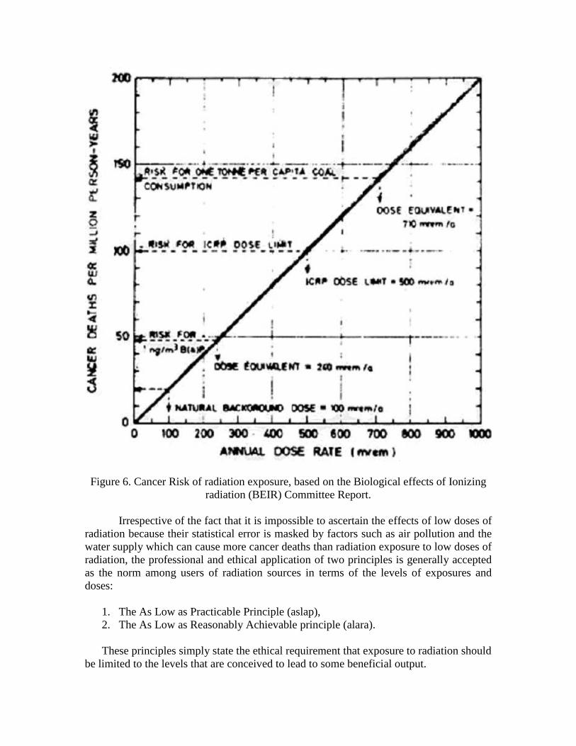
Figure 6. Cancer Risk of radiation exposure, based on the Biological effects of Ionizing
radiation (BEIR) Committee Report.
Irrespective of the fact that it is impossible to ascertain the effects of low doses of
radiation because their statistical error is masked by factors such as air pollution and the
water supply which can cause more cancer deaths than radiation exposure to low doses of
radiation, the professional and ethical application of two principles is generally accepted
as the norm among users of radiation sources in terms of the levels of exposures and
doses:
1. The As Low as Practicable Principle (aslap),
2. The As Low as Reasonably Achievable principle (alara).
These principles simply state the ethical requirement that exposure to radiation should
be limited to the levels that are conceived to lead to some beneficial output.

2.20 COMPARATIVE RISKS
Other sources of societal risks can cause cancer deaths, particularly chemical
pollution. For instance, benzo(a)pyrene is a carcinogenic substance occurring in cigarette
smoke, car exhaust and coal burning. The carcinogenicity of benzo(a)pyrene as well as
radiation has been fully established partly from human experience and partly from human
experiments out at high levels of exposure. Ionizing radiation is not likely to be
generating any new type of harm since humans have been exposed to a fairly large dose
of natural radiation background. A comparison of the risks from benzo(a)pyrene and
radiation exposure is shown in Fig. 6.
The natural background dose at 100 cSv or rem per year per capita leads to a risk
of about 20 cancer deaths per million persons per year. A concentration of 1 nanogram
per cubic meter results in a dose equivalent of about 240 mrem per year per capita, and
results in about 50 cancer deaths per million persons per year. However the risk from
burning one metric tonne (1,000 kgs) of coal per person per year has a dose equivalent of
710 mrem per person per year will be exceeding the maximum allowable ICRP dose limit
of 500 mrem per person per year.
Table 11. Fractional exposure to radiation sources.
Source of exposure Fraction
Natural background 67.60
Medical irradiation 30.70
Fallout from weapons testing 0.60
Miscellaneous sources 0.50
Occupational exposure 0.45
Nuclear Energy 0.15
Total 100.0
2.21 DISCUSSION
Humans receive radiation from a number of sources both natural and human
made. Depending on the style of living one can receive more or less radiation from some
of these sources. For instance, living in a brick house contributes a dose equivalent of
50-100 mrem per person per year, in a concrete house 70-100 mrem, and in a wooden
house 30-50 mrem.
Cosmic rays contribute 45 mrems to the annual per capita dose equivalent,
radioactive minerals in the soil 15 mrems, water food and air 25 mrems, air travel for a
round trip from London to New York 4 mrems, diagnostics x rays 20 mrems, and living
in the vicinity of a nuclear power plant 1 mrem.
Most of human exposure to radiation comes from the natural radiation
background and the medical applications of radiation as shown in Table 11.
Exposure to radiation has been, and will forever remain, a fact of life since the
beginning of life on Earth.

EXERCISES
1. The radiation dose received by someone handling a kilogram of a 15 percent uranium
ore will be about the same as the dose from a kilogram of fresh separated uranium.
Explain why this statement is true or false.
2. Consider the isotope Ra226. Using Avogadro’s law, calculate its activity and discuss its
relationship to the Curie unit of activity. You can obtain the half life of the radium226
isotope from the Table of the Nuclides.
3. Calculate the yearly dose accumulated by yourself according to your lifestyle, and
compare it to the average natural background dose.
4. Calculate the energy in eV of photons of electromagnetic radiation of:
a) X-rays,
b) Gamma rays,
c) Visible light,
d) Ultra violet light,
e) Infrared.
Start from an appropriate value of the wave length, and use:
,
, speed of light, h = Planck's constant, = radiation wave length
E h
cc
5. The naturally occurring isotope K40 is widely spread in the environment. In fact, the
average concentration of potassium in the crustal rocks is 27 [g/kg] and in the oceans is
380 [mg/liter]. K40 occurs in plants and animals, has a half-life of 1.3 billion years and an
abundance of 0.0119 atomic percent.
Potassium's concentration in humans is 1.7 [g/kg]. In urine, potassium's concentration is
1.5 [g/liter].
a) Calculate the specific activity of K40 in Becquerels per gram and in Curies/gm of K40.
b) Calculate the specific activity of K40 in Becquerels per gram and in Curies per gm of
overall potassium.
c) Calculate the specific activity of K40 in urine in [Bq/liter].
d. A beta activity above 200 transformations (disintegrations) per minute per liter of urine
following accidental exposure to fission products is indicative of an internal deposition in
the body, and requires intervention. How does this "body burden" criterion compare to
the activity caused by the one due to the naturally occurring potassium?
6. The production of Carbon14 with a half life of 5730 years is an ongoing nuclear
transformation from the neutrons originating from cosmic rays bombarding Nitrogen14 in
the Earth’s atmosphere: 1 14 1 14
0 7 1 6
14 0 14
6 1 7
1 0 1
0 1 1
n N H C
C e N
n e H
Where Nitrogen14 and Carbon14 appear as catalysts in the overall reaction, leading to the
disintegration of a neutron into a proton and an electron.
The atmospheric radiocarbon exists as C14O2 and is inhaled by all fauna and flora.
Because only living plants continue to incorporate C14, and stop incorporating it after
death, it is possible to determine the age of organic archaeological artifacts by measuring
the activity of the carbon present.
Two grams of carbon from a piece of wood found in an ancient temple are analyzed and
found to have an activity of 20 disintegrations per minute. Estimate the approximate age
of the wood, if it is assumed that the current equilibrium specific activity of C14 in carbon
has been constant at 13.56 disintegrations per minute per gram.
REFERENCES
1. W. E. Kisieleski and R. Baserga, "Radioisotopes and life Processes," USAEC,
Division of Technical Information, 1967.
2. I. Asimov and T. Dobzhansky, "The Genetic Effects of Radiation," USAEC, Division
of Technical Information, 1966.