Embed Size (px)
Citation preview
RESEARCH PAPER
Ion mobility spectrometry combined with multivariate statisticalanalysis: revealing the effects of a drug candidate for Alzheimer’sdisease on Aβ1-40 peptide early assembly
Serena Lazzaro1& Nina Ogrinc2 & Lieke Lamont2 & Graziella Vecchio3
& Giuseppe Pappalardo1& Ron M. A. Heeren2
Received: 6 April 2019 /Revised: 4 July 2019 /Accepted: 10 July 2019 /Published online: 12 August 2019# The Author(s) 2019
AbstractInhibition of the initial stages of amyloid-β peptide self-assembly is a key approach in drug development for Alzheimer’s disease,in which soluble and highly neurotoxic low molecular weight oligomers are produced and aggregate in the brain over time. Herewe report a high-throughput method based on ion mobility mass spectrometry and multivariate statistical analysis to rapidlyselect statistically significant early-stage species of amyloid-β1-40 whose formation is inhibited by a candidate theranostic agent.Using this method, we have confirmed the inhibition of a Zn-porphyrin-peptide conjugate in the early self-assembly of Aβ40peptide. The MS/MS fragmentation patterns of the species detected in the samples containing the Zn-porphyrin-peptide conju-gate suggested a porphyrin-catalyzed oxidation at Met-35(O) of Aβ40. We introduce ion mobility MS combined with multivar-iate statistics as a systematic approach to perform data analytics in drug discovery/amyloid research that aims at the evaluation ofthe inhibitory effect on the Aβ early assembly in vitro models at very low concentration levels of Aβ peptides.
Keywords Alzheimer’s disease (AD) . Amyloid β-peptide oligomers . Electrospray ionization-ion mobility-mass spectrometry(ESI-IM-MS) .Multivariate statistical analysis (MVA)
Introduction
Alzheimer’s disease (AD) is a neurodegenerative disorder,generally associated with the accumulation of misfoldedamyloid-β (Aβ) peptides. The World Health Organization(WHO) and Alzheimer’s Disease International estimate thenumber of people affected by AD alone will reach 81 million
worldwide by 2040, leading to a costly burden of disease [1].Although substantial progress has been made in understand-ing AD, the failure of symptomatic treatments for clinicallydiagnosed AD in phase III clinical trials indicates that ourunderstanding of this disease is still incomplete. The searchfor an effective and safe drug therefore continues. Despite thecontinuous debate about the amyloid hypothesis, experimen-tal and clinical evidence support the concept that proteolyticcleavage of APP [2, 3], leaving behind soluble peptides, pri-marily Aβ40 and Aβ42, is a very central factor to the devel-opment of AD. Interestingly, Aβ40 is ten times more preva-lent in the brain than Aβ42 but less fibrillogenic [4–9]. Inaddition, controversy still exists as to which of the two formsis toxic to neurons. While the Aβ monomer form remainsbenign [10], evidence indicates that pre-fibrillar soluble as-semblies of both Aβ40 and Aβ42 peptides are the actualinitiators of AD pathogenesis causing neuronal dysfunctionand memory impairment [11–13]. These peptides are low mo-lecular weight oligomers (LMWs) and not, as expected, ma-ture end-stage amyloid fibrils. Reducing the prevalence ofLMWs, intermediates with suitable inhibitors of the early-stage aggregation of Aβ peptides might decrease neuronaltoxicity [14–21]. A valid therapeutic strategy proposes the
Published in the topical collection Close-Up of Current Developments inIon Mobility Spectrometry with guest editor Gérard Hopfgartner.
Electronic supplementary material The online version of this article(https://doi.org/10.1007/s00216-019-02030-7) contains supplementarymaterial, which is available to authorized users.
* Ron M. A. [email protected]
1 Institute of Biostructures and Bioimaging (IBB), National ResearchCouncil, Via Paolo Gaifami N.18, 95126 Catania, Italy
2 The Maastricht Multimodal Molecular Imaging institute M4I-Division of Imaging Mass Spectrometry, Maastricht University,Minderbroedersberg 4-6, 6211 LK Maastricht, The Netherlands
3 Department of Chemical Sciences, Catania University, Viale AndreaDoria, 6, 95125 Catania, Italy
Analytical and Bioanalytical Chemistry (2019) 411:6353–6363https://doi.org/10.1007/s00216-019-02030-7
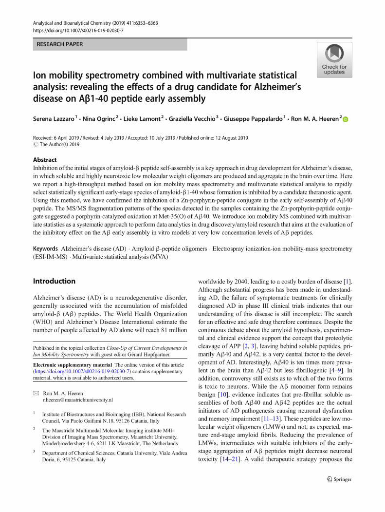
use of short peptides, which recognize Aβ’s aggregation-prone amino acid sequences, as the key disruptors of Aβ’sself-assembly. It is known that the penta-peptide KLVFF,which is homologous to the Aβ(16–20) region, strongly in-teracts with the full-length Aβ peptide to prevent fibril forma-tion [22, 23]. In our previous work [24], we reported the de-sign, synthesis, and anti-fibrillogenic activity of a novel pep-tide conjugate (Fig. 1). This conjugate consists of a fluorescentzinc-containing porphyrin macrocycle, which is linkedthrough a GPG peptide spacer to the KLVFF sequence ofAβ peptides (Zn-Porph).
Matrix-assisted laser desorption ionization-time of flight-mass spectrometry (MALDI-ToF-MS) experiments indicatedthat Zn-Porph interacts with monomeric Aβ42 in a 1:1 molarratio [24]. Yet, Thioflavin-T (ThT) kinetics and circular di-chroism (CD) data showed that Zn-Porph prevented the con-formational transition of Aβ42 to a β-sheet structure. Basedon these results, we hypothesize an interaction mechanisminvolving the zinc ion and the KLVFF peptide of theporphyrin-peptide conjugate as recognition sites of the histi-dine residues and hydrophobic region of Aβ42, respectively.In continuation of our studies, we would like to further eluci-date whether the Zn-Porph also inhibits the Aβ early assem-bly. Specifically, to survey the Zn-Porph’s in vitro binding andinhibitory effects on LMWs of Aβ40 peptide. In this regard,recent studies have shown that electrospray ionization ionmobility mass spectrometry (ESI-IM-MS) is a promising an-alytical tool to investigate the size and conformational distri-bution of the Aβ early-stage LMWs in vitro models [25–36].In respect to the screening of inhibitors [37], only a few IM-MS publications have attempted to identify small inhibitors ofthe initial assembly of Aβ40 at very low concentration levels[38–41]. In these studies, small molecules were added at dif-ferent ratios to solutions of synthetic or recombinant Aβ40 atconcentrations ranging from 10 to 32 μM. Herein, we inves-tigated the in vitro efficacy of the Zn-Porph as the inhibitor ofthe early-stage assembly of synthetic Aβ40 at 5 μM and20 μM. We combined IM-MS with multivariate statistical
analysis (MVA) [42] to compare the IM-MS profiles of mul-tiple samples [43] and to reveal a subset of statistically signif-icant early-stage species of Aβ40 whose formation wasinhibited in the presence of the Zn-Porph. From a chemomet-ric standpoint, statistical feature selection involves discrimi-nant techniques (supervised models). The main differencecompared with the unsupervised models (PCA) is that super-visedmodels use a priori knowledge about the class to which aspecific sample belongs. Geometrically, this is the same asidentifying regions in the hyperspace of the variables corre-sponding to the different classes [44]. Supervised models aredesigned to build an algorithm between a set of descriptivevariables (e.g., drift time_ m/z pairs with corresponding ionintensity value in the IM-MS spectra) and the membership to adefined class of samples [45]. As a modification of the PLSalgorithm, in the OPLS-DA model, the systematic variationsin X are separated into two parts: one linear and one orthog-onal to Y. Hence, the OPLS-DA model comprises two blocksof model variations: (1) the Y-predictive block, which repre-sents the inter-classes variation, and (2) the Y-orthogonalblock which constitutes the intra-class variation [46, 47].The latter augments classification performance in cases wherethe individual classes exhibit divergence in within-class vari-ation. This facilitates the interpretation of the model varia-tions. In another words, OPLS-DA is an excellent tool to find“What’s the difference” between sample classes, such as be-tween in vitro models not containing and containing a drugcandidate. In case of 2-class models, indeed, the OPLS-DA S-plot helps to quickly select reliable features (drift time_ m/zpairs), which capture the bulk of the ion intensity variationbetween the control group (e.g., samples not containing theinhibitor) and treated group (e.g., samples containing the in-hibitor). The S-plot combines the information from a tradition-al loading plot (PLS or OPLS) and the confidence limits col-umn plot (plot XVariance, XVar) resulting in an easier filteringout of low confidence limit features. The plot visualizes var-iable according to their contribution to the inter-classes sepa-ration, based on the covariance parameter magnitude, (p[1]),
Fig. 1 Zinc-porphyrin conjugatedwith the central hydrophobicmotif (KLVFF) of the Aβ peptide(abbr. Zn-Porph)
6354 Lazzaro S. et al.
and to their reliability, based on the correlation parameter val-ue, (p(corr)[1]). Both of these two parameters have a theoret-ical minimum of − 1 and maximum of + 1. The selection ofmeaningful discriminative features needs, therefore, a combi-nation of variable contribution (covariance, p[1]) and variableconfidence (correlation, p(corr)[1]) values, which is the pur-pose of the S-plot. The selected features can be further rankedby the variable of importance (VIP) plot [48]. The plot ranksthe overall contribution of each variable to the model takinginto account both p(corr)[1] and p[1] values. The variableswith VIP value greaten than 1.0 can be selected as top “reli-able ions with highest discriminatory capacity.” In this study, astringent threshold confidence interval was employed to se-lect, among all the Aβ40 early-stage species detected in sam-ple classes not containing the inhibitor, the meaningful ATDpeaks (with low intra-class ion intensity variability) whoseintensity was significantly affected by the inhibitor in the sam-ple classes containing ZnPorph.
Material and methods
Sample preparation
Samples were prepared from independent solutions of syn-thetic Aβ40 peptide (purity > 95%) purchased fromGenScript. Eight units of 1.0 mg of solid Aβ40 were dis-solved in pre-chilled 1,1,1,3,3,3-hexafluoro-2-propanol(HFIP) (Merck) to obtain a peptide concentration of0.5 mM. The Aβ40 solution was sonicated for 5 min at roomtemperature (RT), the tube was chilled on ice for 1 min. TheAβ40 solution was split into aliquots in siliconized tubes.From each aliquot, the HFIP was removed under the fumehood overnight and all traces evaporated using nitrogen. Theday before theMS analysis, HFIP-treated Aβ40 films were re-dissolved in dimethyl sulfoxide (DMSO, max 0.025% water,Merck). Each solution was sonicated for 5 min and subse-quently incubated for 24 h at 25 °C. Prior to MS analysis,the solutions were diluted into 10 mM ammonium acetatebuffer (CH3COONH4, Aldrich), pH 6.9 (in which DMSOconstitutes the 1% v/v of the final volume) to a final peptideconcentration of 5 and 20 μM. All samples were subsequentlycentrifuged at 13,000g for 10 min at 4 °C. The supernatantsolution was stored on ice for 5 min before injection. Anotherset of samples at 20 μM was also incubated at 37 °C for 2 hbefore storing them on ice prior to ESI-IM-MS analysis. Zn-Porph (previously dissolved in CH3COONH4, 10 mM,pH 6.9) was added to monomeric Aβ40 in DMSO (as pre-pared above) in a 1:1 Aβ40: Zn-Porph molar ratio to study theeffect on Aβ40 assembly. Summarizing, three sample setswere investigated: at 5 μM, 20 μM, and at 20 μM incubatedat 37 °C for 2 h prior to injection. The 16 samples of eachsample set were grouped into 2 sample classes identified as
“Aβ40” (eight samples) and “Aβ40 plus Porph” (eight sam-ples) to compare the ESI-IM-MS profiles of LMWs of Aβ40peptide in the presence and absence of equimolar amounts ofZn-Porph. Aβ40 peptide solutions at 100 μM were used tooptimize the IM-MS settings in both resolution and sensitivitymode.
MS method and instrumentation
Direct infusion experiments were performed on a Synapt G2-Si instrument (Waters Corp., Milford, MA). Measurementswere performed at a 7 μL/min injection flow rate for 5 min.Data were acquired in full scan mode using a mass range ofm/z 800–3000 at 1 scan/sec. ESI was operated in the positiveion mode with a capillary voltage of 2.8 kVand sample conevoltage of 38 V. The source and desolvation temperatureswere set at 80 and 40 °C, respectively. Nitrogen was used asa cone gas with the flow rate of 38 L/h and as desolvation gaswith a flow rate of 650 L/h. The mobility T-wave cell wasoperated at a pressure of 3.19 mbar of nitrogen, with awave velocity of 650 m/s and amplitude of 39 V. MS/MSspectra were acquired by CID fragmentation in the TRAPcell using collision energy of 70 V after precursor ionselection at LM resolution 6.5. Peak assignments wereperformed using their 13C isotope distributions of the spe-cies separated in the IM dimension with the MS operatingin resolution mode.
Data processing and multivariate statistical analysis
Data acquisition was carried out with MassLynx (V4.1) andDriftScope (V2.8) software. The total arrival time distribution(ATD) files classified as “Aβ40” and “Aβ40 plus Porph”were thus exported from DriftScope (V2.8) to Progenesis QI(64-bit, Nonlinear Dynamics). The Progenesis QI data analy-sis software is a small molecule discovery tool predominantlyused to identify the significantly changing compounds in yourdataset. In this particular case, the software was used for drifttime alignment, peak picking, and normalization using totalion intensity. We obtained three data matrices, one for each ofthe investigated data set. Multiple features with same drifttime and different m/z may belong to the same compounddue to the fragmentation, adduct formation, or clustering.The three data matrices were then exported from ProgenesisQI to the statistical package EZinfo (V3.0.1.0, Umetrics). Thiswas used to build 2-class orthogonal projection to latentstructure-discriminant (OPLS-DA) models and S-plots foreach sample set under investigation. Protein ProspectorV5.22.1 (UCSF Mass Spectrometry Facility) and FragmentIon Calculator (ISB Data Access Server) were used to analyzethe MS/MS fragmentation ions from peptides.
Ion mobility spectrometry combined with multivariate statistical analysis: revealing the effects of a drug... 6355
Results
ESI-IM-MS analysis revealed that Aβ40 predominantlyoligomerizes through dimers and trimers
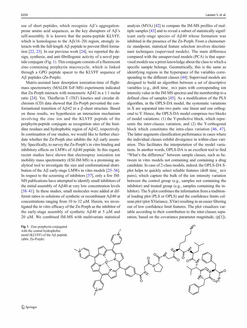
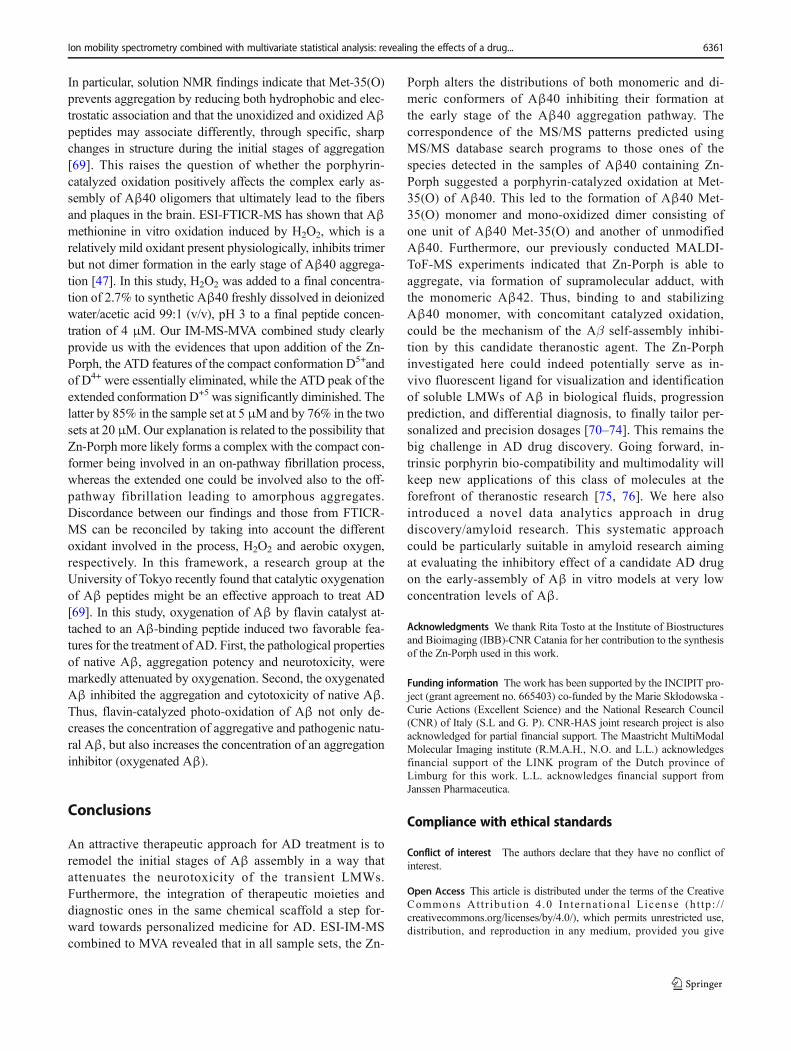
In agreement with previously reported data [25, 38, 39], theESI-IM-MS analysis (Fig. 2) reveals that in the range 5–20 μM, the initially monomeric Aβ40 (M) oligomerizes pre-dominantly through dimers (D) and trimers (TRI), the latterdetectable with a signal-to-noise ratio lower than three(S/N<3). Peak assignments were performed as described inthe Electronic Supplementary Material (ESM). As a result,the signal with a monoisotopic (mon) m/z at 1082.79 wasassigned to [M+4H]4+; the signal at 1443.39 (mon) to [M+3H]3+; the three signals with the same (mon) m/z at 2164.58were assigned to the [M+2H]2+, [D+4H]4+, and [TRI+6H]6+,respectively; the signal at average m/z 2598.92 was identifiedas [TRI+5H]5+; the signal at average m/z 2887.58 as [D+3H]3+. The two signals with the same (mon) m/z at 1731.87but different mobilities (dt) were attributed to the compact andextended forms of the [D+5H]5+.
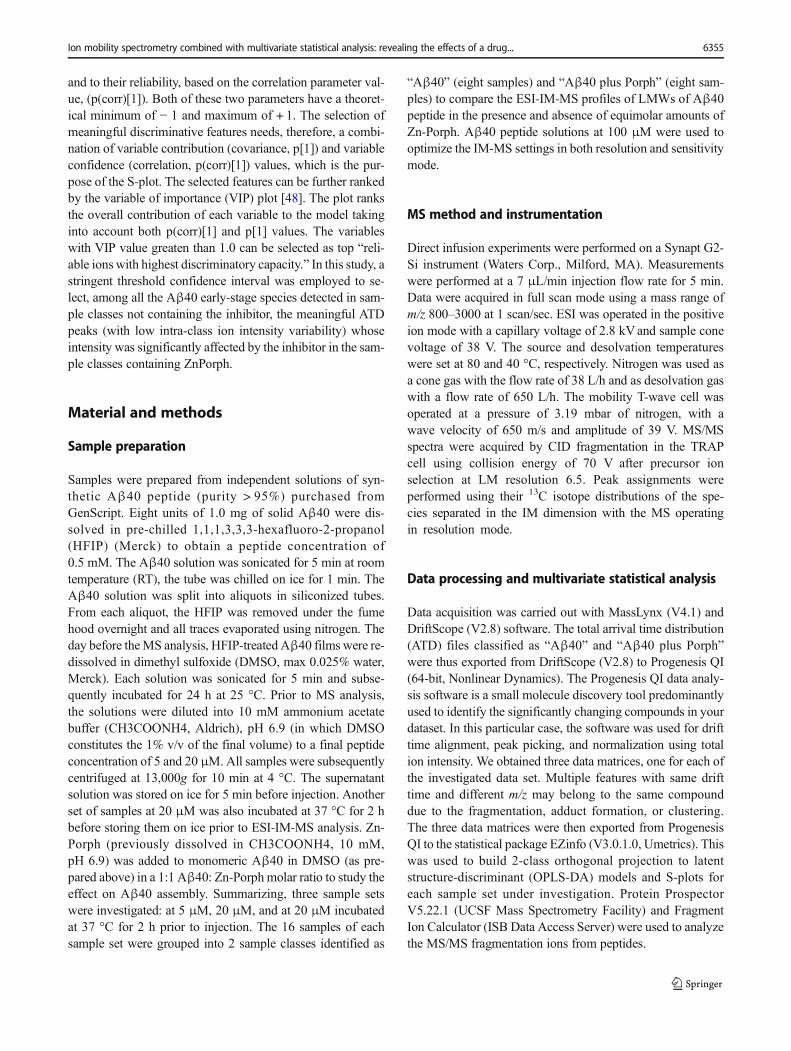
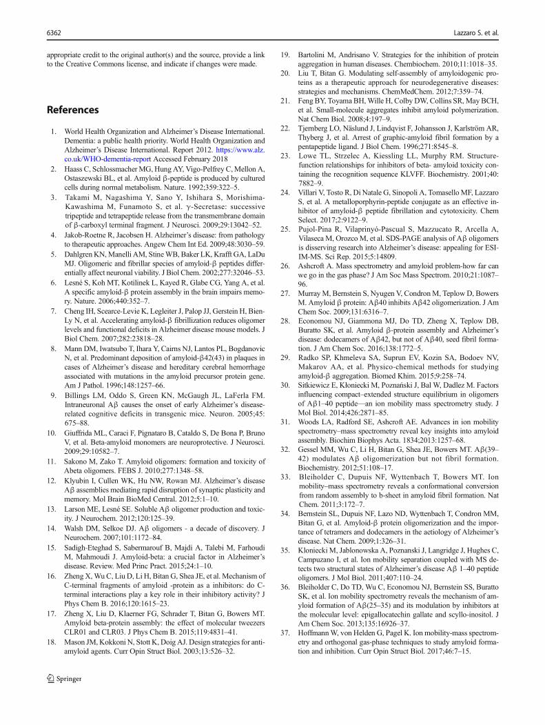
Upon the addition of the inhibitor (Aβ40: Zn-Porph, 1:1),the IM-MS of the “Aβ40 plus Porph” sample sets revealed thedetection of four new signals (Fig. 3) with monoisotopic(mon) m/z at 1086.79, 1448.72, 1735.08, and at 2172.63, re-spectively. All of these peaks were associated to the corre-sponding charge state (see ESM Fig. S3). Furthermore, thesignals of the Aβ40 species previously detected in the“Aβ40” sample sets (Fig. 2) were below the detection limitor reduced. Moreover, no other ATD peaks with S/N>3 andthe same isotopic envelope of the two major [D+5H]5+ con-formations were detected at drift times close to their drift timesof the two major conformations.
Identification of the new detected species
MS/MS experiments were performed to identify the four newspecies shown in Fig. 3. We first investigated the fragmenta-tion pattern (see ESM Fig. S4) of the predominant monomericM3+ species of Aβ40 with (mon) m/z at 1443.39 detected in“Aβ40” sample sets (Fig. 2). We mostly observed doubly andtriply charged b-type ions covering the residues 11–39. MS/
a
b
Fig. 2 Aβ monomers in rapidequilibrium with low-order Aβoligomers. At the top a 2DDriftScope IM-MS plot shows IMdrift time versus m/z versus in-tensity. The signal amplitude iscolor-coded, increasing from pur-ple (low intensity) to bright yel-low (high intensity). The plotshows the Aβ40 species detected5 min after diluting monomericAβ40 to the final peptide con-centration of 20 μM. The peakapexes have been accentuated bydots and annotated as monomers,dimers, and trimers of Aβ40marked with M, D, and TRI withtheir charge (protonation) states.The peaks assigned to the M4+,M3+, D5+, M2+, D4+, and TRI+6
are labeled with the correspond-ing experimental monoisotopicm/z (in parenthesis). The peaksassigned as TRI5+ and as D3+ arelabeled with the experimental av-erage m/z (in parenthesis). At thebottom b ESI-MS spectrum of20 μM Aβ40. In red, the speciesdetected with a S/N< 3. Satellitepeaks observed correspond to al-kali metal adduct commonly ob-served in ESI-MS
6356 Lazzaro S. et al.
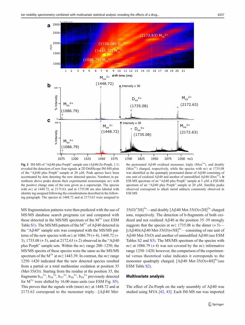
MS fragmentation patterns were then predicted with the use ofMS/MS database search programs (or and compared withthose detected in the MS/MS spectrum of the M3+ (see ESMTable S1). TheMS/MS pattern of theM3+ of Aβ40 detected inthe “Aβ40” sample sets was compared with the MS/MS pat-terns of the new species withm/z at 1086.79 (+ 4), 1448.72 (+3), 1735.08 (+ 5), and at 2172.63 (+ 2) observed in the “Aβ40plus Porph” sample sets. Within the m/z range 200–1250, theMS/MS spectra of these species were the same as the MS/MSspectrum of the M3+ atm/z 1443.39. In contrast, the m/z range1250–1420 indicated that the new detected species resultedfrom a partial or a total methionine oxidation at position 35(Met-35(O)). Starting from the residue at the position 35, thefragments b35
3+, b363+, b37
3+, b383+, b39
3+ previously detectedfor M3+ were shifted by 16.00 mass units (see ESM Fig. S5).This proves that the signals with (mon) m/z at 1448.72 and at2173.63 correspond to the monomer triply—[Aβ40 Met-
35(O)+3H]3+—and doubly [Aβ40 Met-35(O)+2H]2+ chargedions, respectively. The detection of b-fragments of both oxi-dized and not oxidized Aβ40 at the position 35–39 stronglysuggests that the species at m/z 1735.08 is the dimer (+ 5)—[(Aβ40)(Aβ40 Met-35(O))+5H]5+—consisting of one unit ofAβ40 Met-35(O) and another of unmodified Aβ40 (see ESMTables S2 and S3). The MS/MS spectrum of the species withm/z at 1086.79 (+ 4) was not covered by the m/z informativerange 1250–1420; however, the comparison of the experimen-tal versus theoretical value indicates it corresponds to themonomer quadruply charged, [Aβ40 Met-35(O)+4H]4+(seeESM Table S2).
Multivariate analysis
The effect of Zn-Porph on the early assembly of Aβ40 wasstudied using MVA [42, 43]. Each IM-MS run was imported
a
b
c
Fig. 3 IM-MS of “Aβ40 plus Porph” sample sets (Aβ40:Zn-Porph, 1:1)revealed the detection of new four signals. a 2D DriftScope IM-MS plotsof the “Aβ40 plus Porph” sample at 20 μM. Peak apexes have beenaccentuated by dots denoting the new detected species. Numbers in pa-renthesis above peaks denote their experimental monoisotopic m/z withthe positive charge state of the ions given as a superscript. The specieswith m/z at 1448.72, at 2173.63, and at 1735.08 are also labeled withidentity tag assigned following the considerations described in the follow-ing paragraph. The species at 1448.72 and at 2173.63 were assigned to
the protonated Aβ40 oxidized monomer, triply (Mox3+), and doubly(Mox2+) charged, respectively, while the species with m/z at 1735.08was identified as the quintuply protonated dimer of Aβ40 consisting ofone unit of oxidized Aβ40 and another of unmodified Aβ40 (Dox5+). bESI-MS spectrum of an “Aβ40 plus Porph” sample at 5 μM. c ESI-MSspectrum of an “Aβ40 plus Porph” sample at 20 μM. Satellite peaksobserved correspond to alkali metal adducts commonly observed inESI-MS
Ion mobility spectrometry combined with multivariate statistical analysis: revealing the effects of a drug... 6357
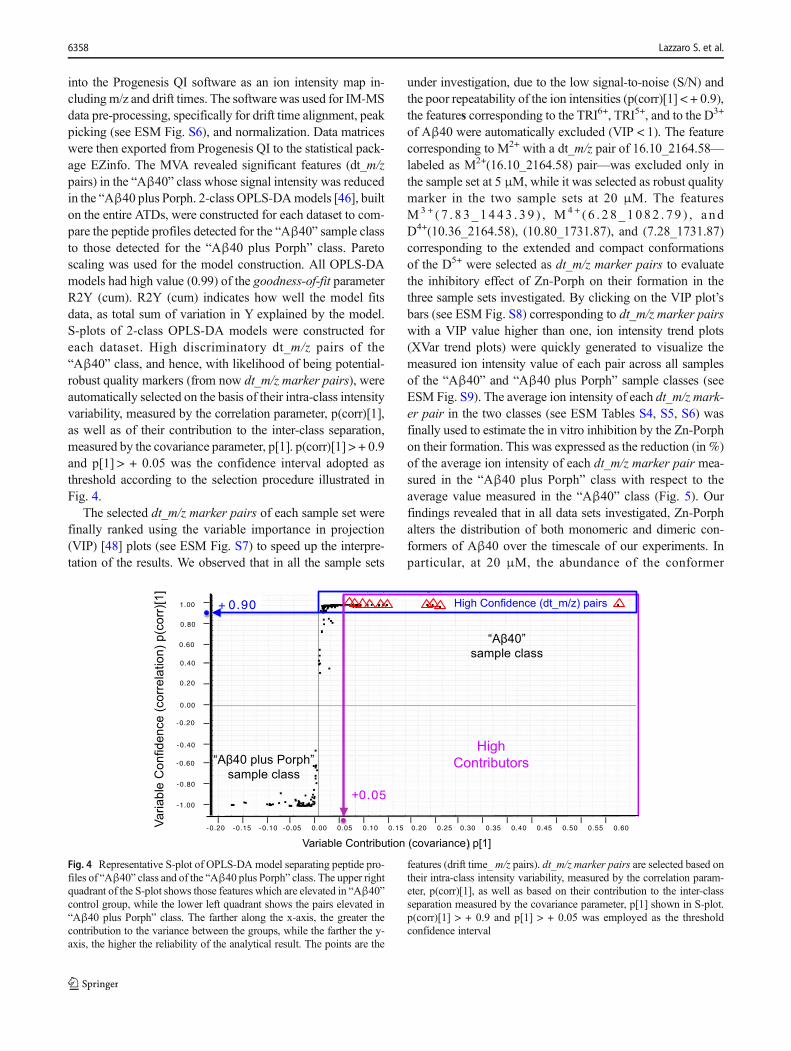
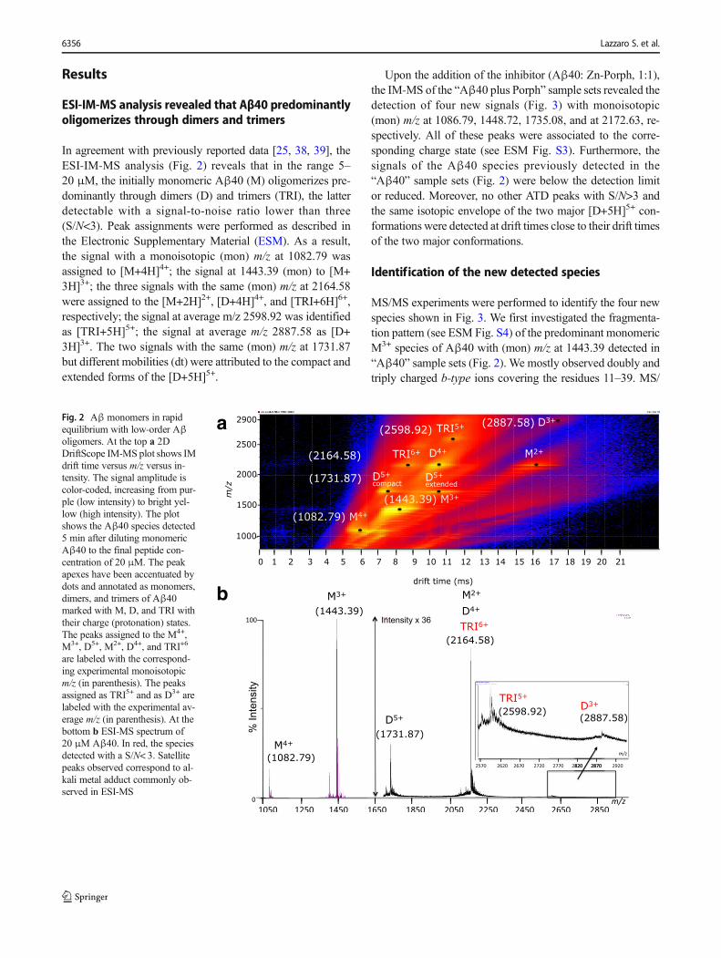
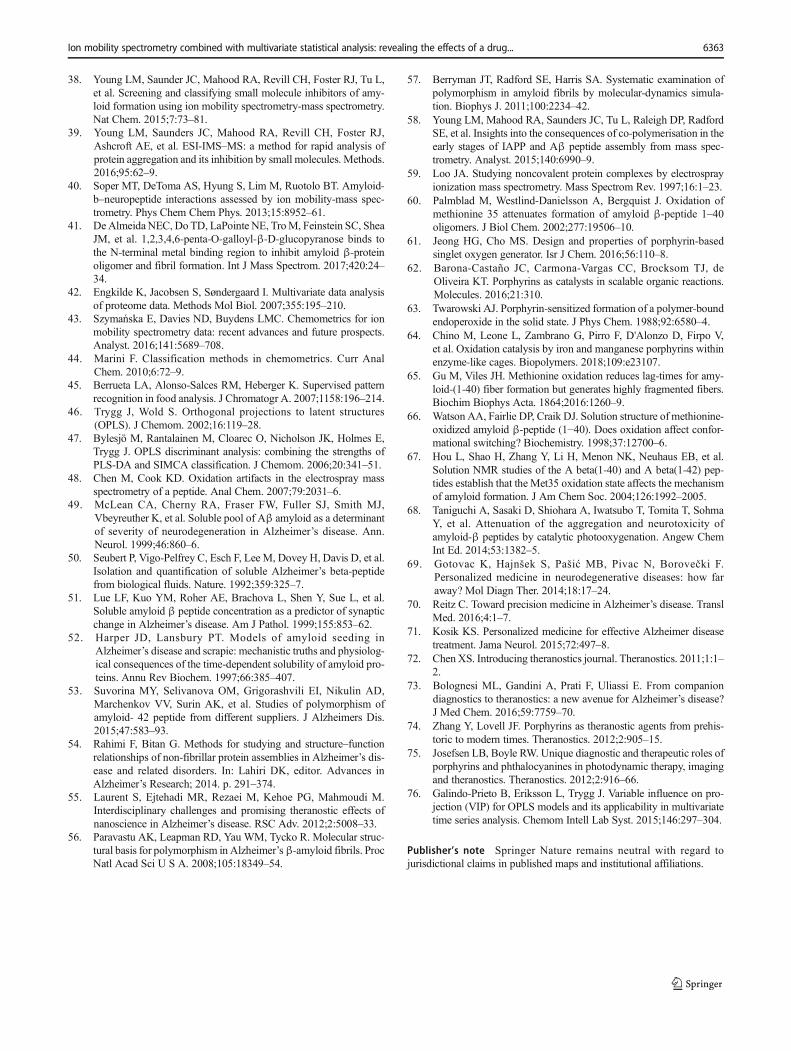
into the Progenesis QI software as an ion intensity map in-cluding m/z and drift times. The software was used for IM-MSdata pre-processing, specifically for drift time alignment, peakpicking (see ESM Fig. S6), and normalization. Data matriceswere then exported from Progenesis QI to the statistical pack-age EZinfo. The MVA revealed significant features (dt_m/zpairs) in the “Aβ40” class whose signal intensity was reducedin the “Aβ40 plus Porph. 2-class OPLS-DAmodels [46], builton the entire ATDs, were constructed for each dataset to com-pare the peptide profiles detected for the “Aβ40” sample classto those detected for the “Aβ40 plus Porph” class. Paretoscaling was used for the model construction. All OPLS-DAmodels had high value (0.99) of the goodness-of-fit parameterR2Y (cum). R2Y (cum) indicates how well the model fitsdata, as total sum of variation in Y explained by the model.S-plots of 2-class OPLS-DA models were constructed foreach dataset. High discriminatory dt_m/z pairs of the“Aβ40” class, and hence, with likelihood of being potential-robust quality markers (from now dt_m/z marker pairs), wereautomatically selected on the basis of their intra-class intensityvariability, measured by the correlation parameter, p(corr)[1],as well as of their contribution to the inter-class separation,measured by the covariance parameter, p[1]. p(corr)[1] > + 0.9and p[1] > + 0.05 was the confidence interval adopted asthreshold according to the selection procedure illustrated inFig. 4.
The selected dt_m/z marker pairs of each sample set werefinally ranked using the variable importance in projection(VIP) [48] plots (see ESM Fig. S7) to speed up the interpre-tation of the results. We observed that in all the sample sets
under investigation, due to the low signal-to-noise (S/N) andthe poor repeatability of the ion intensities (p(corr)[1] < + 0.9),the features corresponding to the TRI6+, TRI5+, and to the D3+
of Aβ40 were automatically excluded (VIP < 1). The featurecorresponding to M2+ with a dt_m/z pair of 16.10_2164.58—labeled as M2+(16.10_2164.58) pair—was excluded only inthe sample set at 5 μM, while it was selected as robust qualitymarker in the two sample sets at 20 μM. The featuresM3+ ( 7 . 8 3_1443 . 3 9 ) , M4 + ( 6 . 2 8_1082 . 7 9 ) , a ndD4+(10.36_2164.58), (10.80_1731.87), and (7.28_1731.87)corresponding to the extended and compact conformationsof the D5+ were selected as dt_m/z marker pairs to evaluatethe inhibitory effect of Zn-Porph on their formation in thethree sample sets investigated. By clicking on the VIP plot’sbars (see ESM Fig. S8) corresponding to dt_m/z marker pairswith a VIP value higher than one, ion intensity trend plots(XVar trend plots) were quickly generated to visualize themeasured ion intensity value of each pair across all samplesof the “Aβ40” and “Aβ40 plus Porph” sample classes (seeESM Fig. S9). The average ion intensity of each dt_m/z mark-er pair in the two classes (see ESM Tables S4, S5, S6) wasfinally used to estimate the in vitro inhibition by the Zn-Porphon their formation. This was expressed as the reduction (in %)of the average ion intensity of each dt_m/z marker pair mea-sured in the “Aβ40 plus Porph” class with respect to theaverage value measured in the “Aβ40” class (Fig. 5). Ourfindings revealed that in all data sets investigated, Zn-Porphalters the distribution of both monomeric and dimeric con-formers of Aβ40 over the timescale of our experiments. Inparticular, at 20 μM, the abundance of the conformer
Variable Contribution (covariance) p[1]
High
Contributors
“Aβ40”
sample class
+ 0.90
VariableConfidence(correlation)p(corr)[1]
+0.05
“Aβ40 plus Porph”
sample class
High Confidence (dt_m/z) pairs
I I I I I I I I I I I I I I I I I
-0.20 -0.15 -0.10 -0.05 0.00 0.05 0.10 0.15 0.20 0.25 0.30 0.35 0.40 0.45 0.50 0.55 0.60
II
II
II
II
II
I
-1.00
1.00
-0.80
-0.60
-0.40
-0.20
0.00
0.20
0.40
0.60
0.80
Fig. 4 Representative S-plot of OPLS-DA model separating peptide pro-files of “Aβ40” class and of the “Aβ40 plus Porph” class. The upper rightquadrant of the S-plot shows those features which are elevated in “Aβ40”control group, while the lower left quadrant shows the pairs elevated in“Aβ40 plus Porph” class. The farther along the x-axis, the greater thecontribution to the variance between the groups, while the farther the y-axis, the higher the reliability of the analytical result. The points are the
features (drift time_m/z pairs). dt_m/z marker pairs are selected based ontheir intra-class intensity variability, measured by the correlation param-eter, p(corr)[1], as well as based on their contribution to the inter-classseparation measured by the covariance parameter, p[1] shown in S-plot.p(corr)[1] > + 0.9 and p[1] > + 0.05 was employed as the thresholdconfidence interval
6358 Lazzaro S. et al.
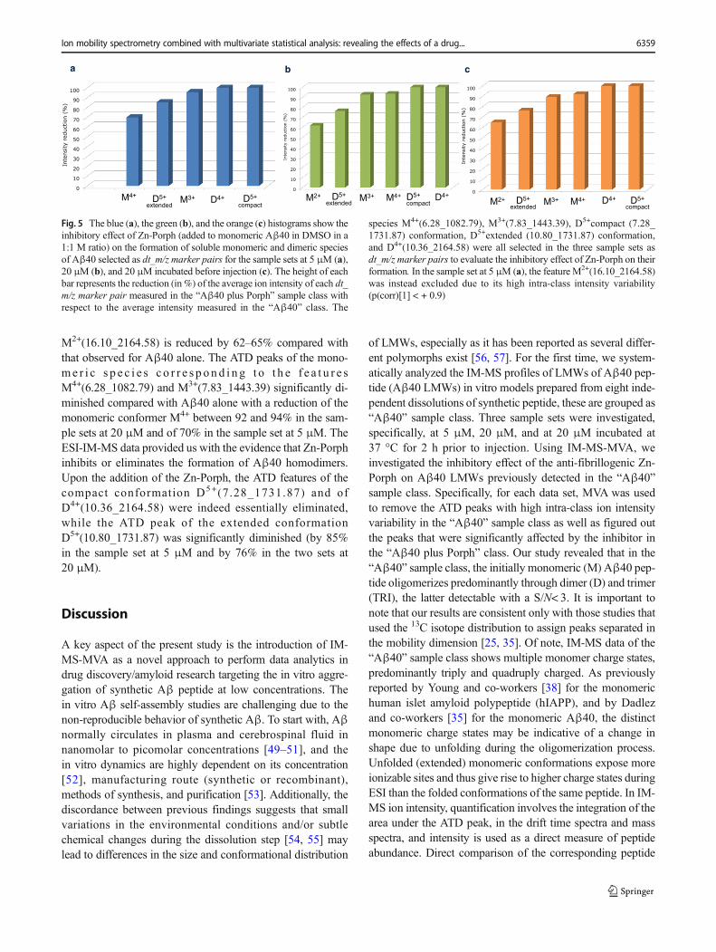
M2+(16.10_2164.58) is reduced by 62–65% compared withthat observed for Aβ40 alone. The ATD peaks of the mono-me r i c s p e c i e s c o r r e s p on d i n g t o t h e f e a t u r e sM4+(6.28_1082.79) and M3+(7.83_1443.39) significantly di-minished compared with Aβ40 alone with a reduction of themonomeric conformer M4+ between 92 and 94% in the sam-ple sets at 20 μM and of 70% in the sample set at 5 μM. TheESI-IM-MS data provided us with the evidence that Zn-Porphinhibits or eliminates the formation of Aβ40 homodimers.Upon the addition of the Zn-Porph, the ATD features of thecompact conformation D5+(7.28_1731.87) and ofD4+(10.36_2164.58) were indeed essentially eliminated,while the ATD peak of the extended conformationD5+(10.80_1731.87) was significantly diminished (by 85%in the sample set at 5 μM and by 76% in the two sets at20 μM).
Discussion
A key aspect of the present study is the introduction of IM-MS-MVA as a novel approach to perform data analytics indrug discovery/amyloid research targeting the in vitro aggre-gation of synthetic Aβ peptide at low concentrations. Thein vitro Aβ self-assembly studies are challenging due to thenon-reproducible behavior of synthetic Aβ. To start with, Aβnormally circulates in plasma and cerebrospinal fluid innanomolar to picomolar concentrations [49–51], and thein vitro dynamics are highly dependent on its concentration[52], manufacturing route (synthetic or recombinant),methods of synthesis, and purification [53]. Additionally, thediscordance between previous findings suggests that smallvariations in the environmental conditions and/or subtlechemical changes during the dissolution step [54, 55] maylead to differences in the size and conformational distribution
of LMWs, especially as it has been reported as several differ-ent polymorphs exist [56, 57]. For the first time, we system-atically analyzed the IM-MS profiles of LMWs of Aβ40 pep-tide (Aβ40 LMWs) in vitro models prepared from eight inde-pendent dissolutions of synthetic peptide, these are grouped as“Aβ40” sample class. Three sample sets were investigated,specifically, at 5 μM, 20 μM, and at 20 μM incubated at37 °C for 2 h prior to injection. Using IM-MS-MVA, weinvestigated the inhibitory effect of the anti-fibrillogenic Zn-Porph on Aβ40 LMWs previously detected in the “Aβ40”sample class. Specifically, for each data set, MVA was usedto remove the ATD peaks with high intra-class ion intensityvariability in the “Aβ40” sample class as well as figured outthe peaks that were significantly affected by the inhibitor inthe “Aβ40 plus Porph” class. Our study revealed that in the“Aβ40” sample class, the initially monomeric (M) Aβ40 pep-tide oligomerizes predominantly through dimer (D) and trimer(TRI), the latter detectable with a S/N< 3. It is important tonote that our results are consistent only with those studies thatused the 13C isotope distribution to assign peaks separated inthe mobility dimension [25, 35]. Of note, IM-MS data of the“Aβ40” sample class shows multiple monomer charge states,predominantly triply and quadruply charged. As previouslyreported by Young and co-workers [38] for the monomerichuman islet amyloid polypeptide (hIAPP), and by Dadlezand co-workers [35] for the monomeric Aβ40, the distinctmonomeric charge states may be indicative of a change inshape due to unfolding during the oligomerization process.Unfolded (extended) monomeric conformations expose moreionizable sites and thus give rise to higher charge states duringESI than the folded conformations of the same peptide. In IM-MS ion intensity, quantification involves the integration of thearea under the ATD peak, in the drift time spectra and massspectra, and intensity is used as a direct measure of peptideabundance. Direct comparison of the corresponding peptide
0
10
20
30
40
50
60
70
80
90
100
M2+ D5+ Extended
M3+ M4+ D5+ compact
D4+
Inte
nsity
red
uctio
n (%
)
a b c
0
10
20
30
40
50
60
70
80
90
100
M4+ D5+ Extended
M3+ D4+ D5+ compact
Inte
nsity
red
uctio
n (%
)
0
10
20
30
40
50
60
70
80
90
100
M2+ D5+ Extended
M3+ M4+ D4+ D5+ compact
Inte
nsity
red
uctio
n (%
)
M4+ D5+ extended
M3+ D4+ D5+ compact
M2+ D5+ extended M3+ M4+ D5+
compact D4+
M2+ D5+ extended M3+ M4+ D5+
compact D4+
Fig. 5 The blue (a), the green (b), and the orange (c) histograms show theinhibitory effect of Zn-Porph (added to monomeric Aβ40 in DMSO in a1:1 M ratio) on the formation of soluble monomeric and dimeric speciesof Aβ40 selected as dt_m/z marker pairs for the sample sets at 5 μM (a),20 μM (b), and 20 μM incubated before injection (c). The height of eachbar represents the reduction (in %) of the average ion intensity of each dt_m/z marker pair measured in the “Aβ40 plus Porph” sample class withrespect to the average intensity measured in the “Aβ40” class. The
species M4+(6.28_1082.79), M3+(7.83_1443.39), D5+compact (7.28_1731.87) conformation, D5+extended (10.80_1731.87) conformation,and D4+(10.36_2164.58) were all selected in the three sample sets asdt_m/z marker pairs to evaluate the inhibitory effect of Zn-Porph on theirformation. In the sample set at 5 μM (a), the feature M2+(16.10_2164.58)was instead excluded due to its high intra-class intensity variability(p(corr)[1] < + 0.9)
Ion mobility spectrometry combined with multivariate statistical analysis: revealing the effects of a drug... 6359
ATD peak area across different samples allows for the relativequantification of peptides. We initially observed that compar-ison of ATD peaks of the Aβ40 species within the “Aβ40”sample class was complicated by ATD drifts. These can bedue to a host of different factors, including sample stability,temperature and pressure fluctuations, deposit build-up, andheterogeneity and dynamic nature of Aβ40 peptide and of itsLMWs. The presence of interferences, e.g., other analyteswith a similar ion mobility, and changing of peak positionsdependent on environmental conditions, e.g., in the field op-erations, could strongly hamper proper analyte identificationand quantification. For the purpose of this study, we focusedon the major species detected in “Aβ40” sample class thatgive distinguishable peaks in the extracted ATDs (see ESMFig. S2). In this respect, as previously reported [58], the struc-tural heterogeneity and dynamic nature of the D+5 species isreflected in their ATD peak shape. ATD peaks that deviateslightly from Gaussian are consistent with multiple states, in-dicating the existence of multiple conformers rapidlyinterconverting on the ESI-IM-MS timescale [58]. Weadopted an automatic alignment procedure, which compen-sates for small variation between runs in the IM drift timesto combine and compare IM-MS profiles of Aβ40 early spe-cies from different dissolutions without ATD peak distortions(see text in ESM). Using the automatic alignment tool, framesdetected in all runs are automatically aligned with the baseframes detected in a sample of the “Aβ40” sample class, se-lected as the reference run. Alignment is an essential 1-D IM-MS data pre-processing step before MVA to achieve IM spec-tra that are reproducible between different samples and condi-tions [43]. It corrects for small variations in the temperatureand pressure of the drift tube, resulting in changes in analytedrift time. This results in an increased precision of the ion-abundance measurement of a peptide feature (dt_m/z pair)across multiple runs. Although the drift time alignment scoresare dependent on the degree of overlap between features, andmisalignment of conflicting features may still yield positivealignment scores, we used these scores as a qualitative mea-sure of the IM-MS alignment, along with visual interpretationto determine alignment success. All 16 samples from the twosample classes were determined to have good alignmentscores (> 80%). From these analyses, we conclude that drifttime alignment scores should be at least > 80%, and ideally >90%, to minimize variation and improve precision in ion-abundance measurements. This process is important becauseit facilitates consistent peak picking across multiple runs, en-ables appropriate normalization of data, reduces complica-tions in assigning peptide identity, and allows the direct com-parison of Aβ40 LMW features across the “Aβ40” and“Aβ40 plus Porph” sample classes (see ESM Fig. S6). Thisstrategy is not only relevant to Aβ40 assembly but may beuseful to the studies focusing on the inhibition on Aβ42 as-sembly and on other aggregation diseases such as Parkinson
(PD) or amyotrophic lateral sclerosis (ALS). Our findingsrevealed that in all data sets investigated, Zn-Porph alters thedistribution of both monomeric and dimeric conformers ofAβ40 over the timescale of our experiments, inhibiting theirformation at the early stage of the aggregation pathway ofAβ40. However, no complexes of Aβ40 and Zn-Porph wereobserved. In this regard, it is important to note that the ESIprocess does not maintain hydrophobic interactions in the gas-phase for very large molecular weight complexes [59], espe-cially, when the ligand has a molecular weight higher than800 Da, as in the case of Zn-Porph. The correspondence ofMS/MS patterns predicted using de novo peptide sequencingalgorithms to those ones of the species at m/z 1448.72 (+3),1735.08 (+5), and at 2172.63 (+2) detected in the “Aβ40 plusPorph” sample sets detected in the “Aβ40 plus Porph” sampleclasses (see ESM Table S3) suggested a porphyrin-catalyzedoxidation in position 35 (Met-35(O)). This led to the detectionof Aβ40 Met-35(O) monomer and mono-oxidized dimer; thelatter consisting of one unit of Aβ40 Met-35(O) and anotherof unmodified Aβ40—[(Aβ40)(Aβ40 Met-35(O)]. The de-tection of one major ATD peak for the mono-oxidized dimer(Dox5+) with m/z (mon) at 1735.08 and with discernable iso-topic distribution pattern (Fig. 3 and ESM Fig. S3) was anindication that the oxidation could possibly occur at Met-35on one of the two D+5 conformers. A previous study [60]conducted on synthetic Aβ40 by the sensitive electrosprayionization Fourier transform ion cyclotron resonance massspectrometry (ESI-FTICR-MS) has shown that spectra ac-quired between 20 and 30 min immediately after dissolvingthe peptide contained less than 1% of Aβ40 Met-35(O). We,thus, exclude that the species detected in the “Aβ40 plusPorph” sample classes could be a result of a spontaneousAβ40 oxidation over the timescale of our experiments. Wealso exclude that these could be oxidation artifacts of theESI process [61] having used gentle ESI conditions (the cap-illary voltage and cone voltage were 2.8 kVand 38 V, respec-tively). Our results therefore strongly suggest a porphyrin-catalyzed oxidation at the position 35 following the dilutionof monomeric Aβ40 peptide with acetate buffer containing anequimolar amount of Zn-Porph. Such event is not unexpectedsince, as previously reported, porphyrin and its analogueshave exhibited catalytic activity for highly selective mono-oxygenation reactions which proceed through singlet oxygen(1O2) generation [62–65]. The generated singlet oxygen read-ily reacted with the unoxidized Aβ40 leading to the detectionof the new detected species caused by the oxidation of Met-35. It has been suggested that oxidation of Met-35(O) in Aβpeptides significantly inhibits fiber formation. In vitro oxida-tion of Aβ, by the physiological oxidant hydrogen peroxide(H2O2), was monitored using Thioflavin-T (ThT), transmis-sion electron microscopy (TEM) [66], circular dichroism(CD) [67], and solution NMR [68]. All of these studies sug-gested a disrupting effect of Met-35(O) on β-sheet formation.
6360 Lazzaro S. et al.
In particular, solution NMR findings indicate that Met-35(O)prevents aggregation by reducing both hydrophobic and elec-trostatic association and that the unoxidized and oxidized Aβpeptides may associate differently, through specific, sharpchanges in structure during the initial stages of aggregation[69]. This raises the question of whether the porphyrin-catalyzed oxidation positively affects the complex early as-sembly of Aβ40 oligomers that ultimately lead to the fibersand plaques in the brain. ESI-FTICR-MS has shown that Aβmethionine in vitro oxidation induced by H2O2, which is arelatively mild oxidant present physiologically, inhibits trimerbut not dimer formation in the early stage of Aβ40 aggrega-tion [47]. In this study, H2O2 was added to a final concentra-tion of 2.7% to synthetic Aβ40 freshly dissolved in deionizedwater/acetic acid 99:1 (v/v), pH 3 to a final peptide concen-tration of 4 μM. Our IM-MS-MVA combined study clearlyprovide us with the evidences that upon addition of the Zn-Porph, the ATD features of the compact conformation D5+andof D4+ were essentially eliminated, while the ATD peak of theextended conformation D+5 was significantly diminished. Thelatter by 85% in the sample set at 5 μM and by 76% in the twosets at 20 μM. Our explanation is related to the possibility thatZn-Porph more likely forms a complex with the compact con-former being involved in an on-pathway fibrillation process,whereas the extended one could be involved also to the off-pathway fibrillation leading to amorphous aggregates.Discordance between our findings and those from FTICR-MS can be reconciled by taking into account the differentoxidant involved in the process, H2O2 and aerobic oxygen,respectively. In this framework, a research group at theUniversity of Tokyo recently found that catalytic oxygenationof Aβ peptides might be an effective approach to treat AD[69]. In this study, oxygenation of Aβ by flavin catalyst at-tached to an Aβ-binding peptide induced two favorable fea-tures for the treatment of AD. First, the pathological propertiesof native Aβ, aggregation potency and neurotoxicity, weremarkedly attenuated by oxygenation. Second, the oxygenatedAβ inhibited the aggregation and cytotoxicity of native Aβ.Thus, flavin-catalyzed photo-oxidation of Aβ not only de-creases the concentration of aggregative and pathogenic natu-ral Aβ, but also increases the concentration of an aggregationinhibitor (oxygenated Aβ).
Conclusions
An attractive therapeutic approach for AD treatment is toremodel the initial stages of Aβ assembly in a way thatattenuates the neurotoxicity of the transient LMWs.Furthermore, the integration of therapeutic moieties anddiagnostic ones in the same chemical scaffold a step for-ward towards personalized medicine for AD. ESI-IM-MScombined to MVA revealed that in all sample sets, the Zn-
Porph alters the distributions of both monomeric and di-meric conformers of Aβ40 inhibiting their formation atthe early stage of the Aβ40 aggregation pathway. Thecorrespondence of the MS/MS patterns predicted usingMS/MS database search programs to those ones of thespecies detected in the samples of Aβ40 containing Zn-Porph suggested a porphyrin-catalyzed oxidation at Met-35(O) of Aβ40. This led to the formation of Aβ40 Met-35(O) monomer and mono-oxidized dimer consisting ofone unit of Aβ40 Met-35(O) and another of unmodifiedAβ40. Furthermore, our previously conducted MALDI-ToF-MS experiments indicated that Zn-Porph is able toaggregate, via formation of supramolecular adduct, withthe monomeric Aβ42. Thus, binding to and stabilizingAβ40 monomer, with concomitant catalyzed oxidation,could be the mechanism of the Aβ self-assembly inhibi-tion by this candidate theranostic agent. The Zn-Porphinvestigated here could indeed potentially serve as in-vivo fluorescent ligand for visualization and identificationof soluble LMWs of Aβ in biological fluids, progressionprediction, and differential diagnosis, to finally tailor per-sonalized and precision dosages [70–74]. This remains thebig challenge in AD drug discovery. Going forward, in-trinsic porphyrin bio-compatibility and multimodality willkeep new applications of this class of molecules at theforefront of theranostic research [75, 76]. We here alsointroduced a novel data analytics approach in drugdiscovery/amyloid research. This systematic approachcould be particularly suitable in amyloid research aimingat evaluating the inhibitory effect of a candidate AD drugon the early-assembly of Aβ in vitro models at very lowconcentration levels of Aβ.
Acknowledgments We thank Rita Tosto at the Institute of Biostructuresand Bioimaging (IBB)-CNR Catania for her contribution to the synthesisof the Zn-Porph used in this work.
Funding information The work has been supported by the INCIPIT pro-ject (grant agreement no. 665403) co-funded by the Marie Skłodowska -Curie Actions (Excellent Science) and the National Research Council(CNR) of Italy (S.L and G. P). CNR-HAS joint research project is alsoacknowledged for partial financial support. The Maastricht MultiModalMolecular Imaging institute (R.M.A.H., N.O. and L.L.) acknowledgesfinancial support of the LINK program of the Dutch province ofLimburg for this work. L.L. acknowledges financial support fromJanssen Pharmaceutica.
Compliance with ethical standards
Conflict of interest The authors declare that they have no conflict ofinterest.
Open Access This article is distributed under the terms of the CreativeCommons At t r ibut ion 4 .0 In te rna t ional License (h t tp : / /creativecommons.org/licenses/by/4.0/), which permits unrestricted use,distribution, and reproduction in any medium, provided you give
Ion mobility spectrometry combined with multivariate statistical analysis: revealing the effects of a drug... 6361
appropriate credit to the original author(s) and the source, provide a linkto the Creative Commons license, and indicate if changes were made.
References
1. World Health Organization and Alzheimer’s Disease International.Dementia: a public health priority. World Health Organization andAlzheimer’s Disease International. Report 2012. https://www.alz.co.uk/WHO-dementia-report Accessed February 2018
2. Haass C, Schlossmacher MG,HungAY, Vigo-Pelfrey C,Mellon A,Ostaszewski BL, et al. Amyloid β-peptide is produced by culturedcells during normal metabolism. Nature. 1992;359:322–5.
3. Takami M, Nagashima Y, Sano Y, Ishihara S, Morishima-Kawashima M, Funamoto S, et al. γ-Secretase: successivetripeptide and tetrapeptide release from the transmembrane domainof β-carboxyl terminal fragment. J Neurosci. 2009;29:13042–52.
4. Jakob-Roetne R, Jacobsen H. Alzheimer’s disease: from pathologyto therapeutic approaches. Angew Chem Int Ed. 2009;48:3030–59.
5. DahlgrenKN,Manelli AM, StineWB, Baker LK, Krafft GA, LaDuMJ. Oligomeric and fibrillar species of amyloid-β peptides differ-entially affect neuronal viability. J Biol Chem. 2002;277:32046–53.
6. Lesné S, Koh MT, Kotilinek L, Kayed R, Glabe CG, Yang A, et al.A specific amyloid-β protein assembly in the brain impairs memo-ry. Nature. 2006;440:352–7.
7. Cheng IH, Scearce-Levie K, Legleiter J, Palop JJ, Gerstein H, Bien-Ly N, et al. Accelerating amyloid-β fibrillization reduces oligomerlevels and functional deficits in Alzheimer disease mouse models. JBiol Chem. 2007;282:23818–28.
8. Mann DM, Iwatsubo T, Ihara Y, Cairns NJ, Lantos PL, BogdanovicN, et al. Predominant deposition of amyloid-β42(43) in plaques incases of Alzheimer’s disease and hereditary cerebral hemorrhageassociated with mutations in the amyloid precursor protein gene.Am J Pathol. 1996;148:1257–66.
9. Billings LM, Oddo S, Green KN, McGaugh JL, LaFerla FM.Intraneuronal Aβ causes the onset of early Alzheimer’s disease-related cognitive deficits in transgenic mice. Neuron. 2005;45:675–88.
10. Giuffrida ML, Caraci F, Pignataro B, Cataldo S, De Bona P, BrunoV, et al. Beta-amyloid monomers are neuroprotective. J Neurosci.2009;29:10582–7.
11. Sakono M, Zako T. Amyloid oligomers: formation and toxicity ofAbeta oligomers. FEBS J. 2010;277:1348–58.
12. Klyubin I, Cullen WK, Hu NW, Rowan MJ. Alzheimer’s diseaseAβ assemblies mediating rapid disruption of synaptic plasticity andmemory. Mol Brain BioMed Central. 2012;5:1–10.
13. Larson ME, Lesné SE. Soluble Aβ oligomer production and toxic-ity. J Neurochem. 2012;120:125–39.
14. Walsh DM, Selkoe DJ. Aβ oligomers - a decade of discovery. JNeurochem. 2007;101:1172–84.
15. Sadigh-Eteghad S, Sabermarouf B, Majdi A, Talebi M, FarhoudiM, Mahmoudi J. Amyloid-beta: a crucial factor in Alzheimer’sdisease. Review. Med Princ Pract. 2015;24:1–10.
16. ZhengX,Wu C, Liu D, Li H, Bitan G, Shea JE, et al. Mechanism ofC-terminal fragments of amyloid -protein as a inhibitors: do C-terminal interactions play a key role in their inhibitory activity? JPhys Chem B. 2016;120:1615–23.
17. Zheng X, Liu D, Klaerner FG, Schrader T, Bitan G, Bowers MT.Amyloid beta-protein assembly: the effect of molecular tweezersCLR01 and CLR03. J Phys Chem B. 2015;119:4831–41.
18. Mason JM, Kokkoni N, Stott K, Doig AJ. Design strategies for anti-amyloid agents. Curr Opin Struct Biol. 2003;13:526–32.
19. Bartolini M, Andrisano V. Strategies for the inhibition of proteinaggregation in human diseases. Chembiochem. 2010;11:1018–35.
20. Liu T, Bitan G. Modulating self-assembly of amyloidogenic pro-teins as a therapeutic approach for neurodegenerative diseases:strategies and mechanisms. ChemMedChem. 2012;7:359–74.
21. Feng BY, Toyama BH,Wille H, Colby DW, Collins SR,May BCH,et al. Small-molecule aggregates inhibit amyloid polymerization.Nat Chem Biol. 2008;4:197–9.
22. Tjernberg LO, Näslund J, Lindqvist F, Johansson J, Karlström AR,Thyberg J, et al. Arrest of graphic-amyloid fibril formation by apentapeptide ligand. J Biol Chem. 1996;271:8545–8.
23. Lowe TL, Strzelec A, Kiessling LL, Murphy RM. Structure-function relationships for inhibitors of beta- amyloid toxicity con-taining the recognition sequence KLVFF. Biochemistry. 2001;40:7882–9.
24. Villari V, Tosto R, Di Natale G, Sinopoli A, TomaselloMF, LazzaroS, et al. A metalloporphyrin-peptide conjugate as an effective in-hibitor of amyloid-β peptide fibrillation and cytotoxicity. ChemSelect. 2017;2:9122–9.
25. Pujol-Pina R, Vilaprinyó-Pascual S, Mazzucato R, Arcella A,Vilaseca M, Orozco M, et al. SDS-PAGE analysis of Aβ oligomersis disserving research into Alzheimer’s disease: appealing for ESI-IM-MS. Sci Rep. 2015;5:14809.
26. Ashcroft A. Mass spectrometry and amyloid problem-how far canwe go in the gas phase? J Am Soc Mass Spectrom. 2010;21:1087–96.
27. Murray M, Bernstein S, Nyugen V, CondronM, Teplow D, BowersM. Amyloid β protein: Aβ40 inhibits Aβ42 oligomerization. J AmChem Soc. 2009;131:6316–7.
28. Economou NJ, Giammona MJ, Do TD, Zheng X, Teplow DB,Buratto SK, et al. Amyloid β-protein assembly and Alzheimer’sdisease: dodecamers of Aβ42, but not of Aβ40, seed fibril forma-tion. J Am Chem Soc. 2016;138:1772–5.
29. Radko SP, Khmeleva SA, Suprun EV, Kozin SA, Bodoev NV,Makarov AA, et al. Physico-chemical methods for studyingamyloid-β aggregation. Biomed Khim. 2015;9:258–74.
30. Sitkiewicz E, Kłoniecki M, Poznański J, Bal W, Dadlez M. Factorsinfluencing compact–extended structure equilibrium in oligomersof Aβ1–40 peptide—an ion mobility mass spectrometry study. JMol Biol. 2014;426:2871–85.
31. Woods LA, Radford SE, Ashcroft AE. Advances in ion mobilityspectrometry–mass spectrometry reveal key insights into amyloidassembly. Biochim Biophys Acta. 1834;2013:1257–68.
32. Gessel MM, Wu C, Li H, Bitan G, Shea JE, Bowers MT. Aβ(39–42) modulates Aβ oligomerization but not fibril formation.Biochemistry. 2012;51:108–17.
33. Bleiholder C, Dupuis NF, Wyttenbach T, Bowers MT. Ionmobility–mass spectrometry reveals a conformational conversionfrom random assembly to b-sheet in amyloid fibril formation. NatChem. 2011;3:172–7.
34. Bernstein SL, Dupuis NF, Lazo ND, Wyttenbach T, Condron MM,Bitan G, et al. Amyloid-β protein oligomerization and the impor-tance of tetramers and dodecamers in the aetiology of Alzheimer’sdisease. Nat Chem. 2009;1:326–31.
35. Kloniecki M, Jablonowska A, Poznanski J, Langridge J, Hughes C,Campuzano I, et al. Ion mobility separation coupled with MS de-tects two structural states of Alzheimer’s disease Aβ 1–40 peptideoligomers. J Mol Biol. 2011;407:110–24.
36. Bleiholder C, Do TD, Wu C, Economou NJ, Bernstein SS, BurattoSK, et al. Ion mobility spectrometry reveals the mechanism of am-yloid formation of Aβ(25–35) and its modulation by inhibitors atthe molecular level: epigallocatechin gallate and scyllo-inositol. JAm Chem Soc. 2013;135:16926–37.
37. HoffmannW, von Helden G, Pagel K. Ion mobility-mass spectrom-etry and orthogonal gas-phase techniques to study amyloid forma-tion and inhibition. Curr Opin Struct Biol. 2017;46:7–15.
6362 Lazzaro S. et al.
38. Young LM, Saunder JC, Mahood RA, Revill CH, Foster RJ, Tu L,et al. Screening and classifying small molecule inhibitors of amy-loid formation using ion mobility spectrometry-mass spectrometry.Nat Chem. 2015;7:73–81.
39. Young LM, Saunders JC, Mahood RA, Revill CH, Foster RJ,Ashcroft AE, et al. ESI-IMS–MS: a method for rapid analysis ofprotein aggregation and its inhibition by small molecules. Methods.2016;95:62–9.
40. Soper MT, DeToma AS, Hyung S, Lim M, Ruotolo BT. Amyloid-b–neuropeptide interactions assessed by ion mobility-mass spec-trometry. Phys Chem Chem Phys. 2013;15:8952–61.
41. DeAlmeidaNEC, Do TD, LaPointe NE, TroM, Feinstein SC, SheaJM, et al. 1,2,3,4,6-penta-O-galloyl-β-D-glucopyranose binds tothe N-terminal metal binding region to inhibit amyloid β-proteinoligomer and fibril formation. Int J Mass Spectrom. 2017;420:24–34.
42. Engkilde K, Jacobsen S, Søndergaard I. Multivariate data analysisof proteome data. Methods Mol Biol. 2007;355:195–210.
43. Szymańska E, Davies ND, Buydens LMC. Chemometrics for ionmobility spectrometry data: recent advances and future prospects.Analyst. 2016;141:5689–708.
44. Marini F. Classification methods in chemometrics. Curr AnalChem. 2010;6:72–9.
45. Berrueta LA, Alonso-Salces RM, Heberger K. Supervised patternrecognition in food analysis. J Chromatogr A. 2007;1158:196–214.
46. Trygg J, Wold S. Orthogonal projections to latent structures(OPLS). J Chemom. 2002;16:119–28.
47. Bylesjö M, Rantalainen M, Cloarec O, Nicholson JK, Holmes E,Trygg J. OPLS discriminant analysis: combining the strengths ofPLS-DA and SIMCA classification. J Chemom. 2006;20:341–51.
48. Chen M, Cook KD. Oxidation artifacts in the electrospray massspectrometry of a peptide. Anal Chem. 2007;79:2031–6.
49. McLean CA, Cherny RA, Fraser FW, Fuller SJ, Smith MJ,Vbeyreuther K, et al. Soluble pool of Aβ amyloid as a determinantof severity of neurodegeneration in Alzheimer’s disease. Ann.Neurol. 1999;46:860–6.
50. Seubert P, Vigo-Pelfrey C, Esch F, Lee M, Dovey H, Davis D, et al.Isolation and quantification of soluble Alzheimer’s beta-peptidefrom biological fluids. Nature. 1992;359:325–7.
51. Lue LF, Kuo YM, Roher AE, Brachova L, Shen Y, Sue L, et al.Soluble amyloid β peptide concentration as a predictor of synapticchange in Alzheimer’s disease. Am J Pathol. 1999;155:853–62.
52. Harper JD, Lansbury PT. Models of amyloid seeding inAlzheimer’s disease and scrapie: mechanistic truths and physiolog-ical consequences of the time-dependent solubility of amyloid pro-teins. Annu Rev Biochem. 1997;66:385–407.
53. Suvorina MY, Selivanova OM, Grigorashvili EI, Nikulin AD,Marchenkov VV, Surin AK, et al. Studies of polymorphism ofamyloid- 42 peptide from different suppliers. J Alzheimers Dis.2015;47:583–93.
54. Rahimi F, Bitan G. Methods for studying and structure–functionrelationships of non-fibrillar protein assemblies in Alzheimer’s dis-ease and related disorders. In: Lahiri DK, editor. Advances inAlzheimer’s Research; 2014. p. 291–374.
55. Laurent S, Ejtehadi MR, Rezaei M, Kehoe PG, Mahmoudi M.Interdisciplinary challenges and promising theranostic effects ofnanoscience in Alzheimer’s disease. RSC Adv. 2012;2:5008–33.
56. Paravastu AK, Leapman RD, Yau WM, Tycko R. Molecular struc-tural basis for polymorphism in Alzheimer’sβ-amyloid fibrils. ProcNatl Acad Sci U S A. 2008;105:18349–54.
57. Berryman JT, Radford SE, Harris SA. Systematic examination ofpolymorphism in amyloid fibrils by molecular-dynamics simula-tion. Biophys J. 2011;100:2234–42.
58. Young LM, Mahood RA, Saunders JC, Tu L, Raleigh DP, RadfordSE, et al. Insights into the consequences of co-polymerisation in theearly stages of IAPP and Aβ peptide assembly from mass spec-trometry. Analyst. 2015;140:6990–9.
59. Loo JA. Studying noncovalent protein complexes by electrosprayionization mass spectrometry. Mass Spectrom Rev. 1997;16:1–23.
60. Palmblad M, Westlind-Danielsson A, Bergquist J. Oxidation ofmethionine 35 attenuates formation of amyloid β-peptide 1–40oligomers. J Biol Chem. 2002;277:19506–10.
61. Jeong HG, Cho MS. Design and properties of porphyrin-basedsinglet oxygen generator. Isr J Chem. 2016;56:110–8.
62. Barona-Castaño JC, Carmona-Vargas CC, Brocksom TJ, deOliveira KT. Porphyrins as catalysts in scalable organic reactions.Molecules. 2016;21:310.
63. Twarowski AJ. Porphyrin-sensitized formation of a polymer-boundendoperoxide in the solid state. J Phys Chem. 1988;92:6580–4.
64. Chino M, Leone L, Zambrano G, Pirro F, D'Alonzo D, Firpo V,et al. Oxidation catalysis by iron and manganese porphyrins withinenzyme-like cages. Biopolymers. 2018;109:e23107.
65. Gu M, Viles JH. Methionine oxidation reduces lag-times for amy-loid-(1-40) fiber formation but generates highly fragmented fibers.Biochim Biophys Acta. 1864;2016:1260–9.
66. Watson AA, Fairlie DP, Craik DJ. Solution structure of methionine-oxidized amyloid β-peptide (1−40). Does oxidation affect confor-mational switching? Biochemistry. 1998;37:12700–6.
67. Hou L, Shao H, Zhang Y, Li H, Menon NK, Neuhaus EB, et al.Solution NMR studies of the A beta(1-40) and A beta(1-42) pep-tides establish that the Met35 oxidation state affects the mechanismof amyloid formation. J Am Chem Soc. 2004;126:1992–2005.
68. Taniguchi A, Sasaki D, Shiohara A, Iwatsubo T, Tomita T, SohmaY, et al. Attenuation of the aggregation and neurotoxicity ofamyloid-β peptides by catalytic photooxygenation. Angew ChemInt Ed. 2014;53:1382–5.
69. Gotovac K, Hajnšek S, Pašić MB, Pivac N, Borovečki F.Personalized medicine in neurodegenerative diseases: how faraway? Mol Diagn Ther. 2014;18:17–24.
70. Reitz C. Toward precision medicine in Alzheimer’s disease. TranslMed. 2016;4:1–7.
71. Kosik KS. Personalized medicine for effective Alzheimer diseasetreatment. Jama Neurol. 2015;72:497–8.
72. Chen XS. Introducing theranostics journal. Theranostics. 2011;1:1–2.
73. Bolognesi ML, Gandini A, Prati F, Uliassi E. From companiondiagnostics to theranostics: a new avenue for Alzheimer’s disease?J Med Chem. 2016;59:7759–70.
74. Zhang Y, Lovell JF. Porphyrins as theranostic agents from prehis-toric to modern times. Theranostics. 2012;2:905–15.
75. Josefsen LB, Boyle RW. Unique diagnostic and therapeutic roles ofporphyrins and phthalocyanines in photodynamic therapy, imagingand theranostics. Theranostics. 2012;2:916–66.
76. Galindo-Prieto B, Eriksson L, Trygg J. Variable influence on pro-jection (VIP) for OPLS models and its applicability in multivariatetime series analysis. Chemom Intell Lab Syst. 2015;146:297–304.
Publisher’s note Springer Nature remains neutral with regard tojurisdictional claims in published maps and institutional affiliations.
Ion mobility spectrometry combined with multivariate statistical analysis: revealing the effects of a drug... 6363