Embed Size (px)
Citation preview
Introduction
It is common knowledge that the ocular bioavailability of drugs applied topically as eye-drops is very poor. The absorption of drugs in the eye is severely limited by some protective mechanisms that ensure the proper functioning of the eye, and by other concomitant factors like, drainage of the instilled solutions, lachrymation and tear turnover, metabolism, tear evaporation, non-productive absorption/adsorption, limited corneal area and poor corneal permeability, binding by the lachrymal proteins. The drainage of the administered dose via the nasolachrymal system into the nasopharynx and the
gastrointestinal tract takes place when the volume of fluid in the eye exceeds the normal lachrymal volume of 7–10 μL. Thus, the portion of the instilled dose (1–2 drops, corresponding to 50–100 μL) that is not eliminated by spillage from the palpebral fissure is drained quickly. Therefore, the contact time of the dose with the absorbing surfaces (cornea and sclera) is reduced to a maximum of 2 min. Sometimes systemic absorption of the drug drained through the nasolachrymal duct may result in some undesirable side effects (Maurice, 1987; Middleton, Leung, & Robinson, 1990; Schoenwald, 1990). To over-come these problems various ophthalmic vehicles, such as suspensions, ointments, inserts and aqueous gels,
Journal of Drug TargetingJournal of Drug Targeting, 2010; 18(7): 499–505
2010
18
7
499
505
Address for Correspondence: Dr. Sanyog Jain, National Institue of Pharmaceutical Education and Research, Sector 67, SAS Nagar, Punjab-160062, India. E-mail: [email protected], [email protected]
16 August 2009
09 October 2009
24 November 2009
1061-186X
1029-2330
© 2010 Informa UK Ltd
10.3109/10611860903508788
R E S E A R C H A R T I C L E
Ion- and pH-activated novel in-situ gel system for sustained ocular drug delivery
Himanshu Gupta1, T. Velpandian2, and Sanyog Jain3
1Institute of Nuclear Medicine and Allied Sciences, Defence Research and Development Organization, Delhi, India, 2Ocular Pharmacology Department, Dr. R.P. Centre for Ophthalmic Sciences, All India Institute of Medical Sciences, Ansari Nagar, New Delhi, India, and 3National Institute of Pharmaceutical Education and Research, SAS Nagar, Punjab, India
AbstractPoor bioavailability (<1%) of drugs from conventional eye drops is mainly due to the various precorneal loss factors which include rapid tear turnover, systemic drug absorption through naso-lachrymal duct, transient residence time of the drug solution in the cul-de-sac and the relative impermeability of the drugs to corneal epithelial membrane. The present study describes the formulation and evaluation of chitosan and gellan gum based novel in-situ gel system activated by dual physiological mechanisms. Chitosan (a pH-sensitive polymer) in combination with gellan gum (an ion-activated polymer) were used as gelling agent. Timolol maleate, the drug which is frequently used for glaucoma therapy was used as model drug to check the efficacy of the formulation. The developed formulation was characterized for various in vitro parameters, for example, clarity, gelation pH, isotonicity, sterility, viscosity, transcorneal permeation profile, and ocular irritation. Ocular retention was studied by gamma scintigraphy and a significant increase in retention time was observed. The formulation was also found to be nonirritant and well tolerable. The developed system can be a viable alternative to conventional eye drops for the treatment of various ocular diseases and is suitable for clinical application.
Keywords: In-situ gel system; chitosan; gellan gum; ophthalmic drug delivery; Timolol maleate; gamma scintigraphy
DRT
451311
(Received 16 August 2009; revised 09 October 2009; accepted 24 November 2009)
ISSN 1061-186X print/ISSN 1029-2330 online © 2010 Informa UK LtdDOI: 10.3109/10611860903508788 http://www.informahealthcare.com/drt
Jour
nal o
f D
rug
Tar
getin
g D
ownl
oade
d fr
om in
form
ahea
lthca
re.c
om b
y N
orth
east
ern
Uni
vers
ity o
n 04
/16/
13Fo
r pe
rson
al u
se o
nly.
500 Himanshu Gupta, T. Velpandian, and Sanyog Jain
have been investigated in attempts to extend the ocular residence time of medications for topical application to the eye (Desai & Blanchard, 1994; Ranade & Hollinger, 1996). These ocular drug delivery systems offer some improvement over a conventional liquid dosage form but because of blurred vision (e.g., ointments) or lack of patient compliance (e.g., inserts); they have not been universally accepted. As a result, good ocular bioavail-ability by topical delivery of a drug remains a challenge yet to be satisfactorily resolved (Chiou & Watanabe, 1986; Felt et al., 1999a).
The use of preformed gels still has a number of lacu-nas, which has limited their use in ophthalmic drug delivery. These preformed gel formulations do not allow accurate and reproducible administration of drugs and after administration these often produce blurred vision, crusting of eyelids, and lachrymation. In-situ gel system is a new and recent approach, which combines the advantages of both gels and solution so that an accurate dose can be administered with ease of admin-istration. These formulations remain to a solution state before administration but however transforms to gel after administration in to cul-de-sac of the eye (Gurny, Ibrahim, & Buri, 1993). Srividya, Cardoza, & Amin (2001) prepared a pH-sensitive in-situ ophthalmic gel of ofloxacin hydrochloride using HPMC and carbopol 934 as polymers for treatment of bacterial conjuctivi-tis. Ion-activated in-situ gel forming system of various drugs using Gelrite (gellan gum) as polymer base has also been reported (Balasubramaniam & Pandit, 2003; Balasubramaniam, Kant, & Pandit, 2003; Sultana, Aqil, & Ali, 2006). Chitosan and Pluronic F-127 based pH and temperature triggered in-situ gel for sustained ocular drug delivery has been reported by our group (Gupta et al., 2007, 2009).
The present study describes the formulation and evaluation of an in-situ ocular drug delivery system based on the concept of pH- and ion-triggered in-situ gelation. Chitosan (pH-sensitive polymer which also act as per-meation enhancer) along with gellan gum (ion-activated gelling polymer) was used as gelling agent. Timolol maleate, the drug which is frequently used for glaucoma therapy was used as model drug to check the efficacy of the formulation.
Materials and methods
Materials
The drug timolol maleate was received as gift sample from M/s Ven petrochem & Pharma (India) Pvt. Ltd., Mumbai, India. Chitosan (practical grade, 75–85% deacetylated, molecular weight 150 kDa) was obtained as kind gift from M/s, India Sea Foods, Cochin, India.
Gellan gum (Gelrite® CP Kelco, USA) was obtained as a gift sample from Applied Biosciences, Mumbai, India. All other chemicals and solvents used were purchased from local suppliers and of analytical grade unless mentioned.
Drug polymer interaction studies
Aqueous solutions of chitosan, gellan gum, and timolol maleate were prepared individually and in combina-tions and were autoclaved. The ultraviolet (UV) spectra were taken before and after autoclaving using Shimadzu 1601 double beam UV-visible spectrophotometer. Both spectra were compared for any possible change in solution content due to interactions between different ingredients.
Preparation of in-situ gelling system
Placebo in-situ gelling systems were prepared using different polymer ratios and evaluated for their gelling capacity to identify a suitable composition (Table 1). Chitosan was dissolved in acetate buffer, pH 5.5–6.0. Gellan gum was dissolved in ultrapure water with heating at 70–80°C with stirring. The gelling capacity was deter-mined by placing a drop of the system in a vial containing 2 mL of freshly prepared simulated tear fluid (STF) using sodium chloride 0.670 g, sodium bicarbonate 0.200 g, cal-cium chloride dihydrate 0.008 g and purified water q.s. 100 g (Rozier et al., 1989) and visually assessing the gel formation, noting the time taken for gelation and time taken by the formed gel to dissolve.
Preparation of medicated formulation
For antiglaucoma activity, eye drops of timolol maleate are generally available as 0.25–0.5% w/v drug solution. Hence, a dose of 0.25% was used to prepare the formu-lation. The calculated amount of timolol maleate was placed in volumetric flask and dissolved in ultrapure water. A 0.1% of methyl paraben was added as a preserva-tive. Osmolarity of the formulation was determined by osmometer (Fiske Associate, USA) and required amount of glycerol was added and mixed thoroughly to make the solution isotonic. Complete formula of the developed formulation is given in Table 2. The solution was trans-ferred into amber-colored bottles, each with a cap that was fitted and that carried a dropper fitted with a teat. The resulting solutions were sterilized by autoclaving at 121°C for 20 min at 15 psi.
Physicochemical characterization
The clarity of the formulations after and before gel-ling was determined by visual examination of the
Jour
nal o
f D
rug
Tar
getin
g D
ownl
oade
d fr
om in
form
ahea
lthca
re.c
om b
y N
orth
east
ern
Uni
vers
ity o
n 04
/16/
13Fo
r pe
rson
al u
se o
nly.
Novel in-situ gel system for sustained ocular drug delivery 501
formulations under light alternatively against white and black backgrounds. For Gelation pH, formulation was taken in a vial and 1 M NaOH was added dropwise with continuous stirring. pH was checked using pH meter (Equiptronics digital pH meter) and viscosity was deter-mined by using Brookfield’s viscometer (RVT model) in small volume adapter at 20 rpm. The pH at which sudden change in viscosity was observed was noted as gelation pH. Osmolarity was determined by osmometer (Fiske Associate, USA).
In vitro transcorneal permeation study
Whole eyeballs of goat were procured from a local slaughter house and transported to laboratory in cold condition in normal saline. The corneas were carefully removed along with 5–6 mm of surround-ing scleral tissue and stored in freshly prepared STF, pH 7.4. Goat corneas were used to study the transorneal permeability of timolol maleate from developed for-mulation. The study was carried out in modified Franz diffusion chamber (designed at ocular pharmacology lab, Dr. R.P. Centre for Ophthalmic Sciences, All India Institute of Medical Sciences, New Delhi). It consisted of four cells and each cell consisted of upper and lower chamber. The upper chamber served as a donor com-partment in which 100 μL of drug solution/ formulation under study was placed. The upper and lower chambers were separated by goat cornea. The lower chamber served as a receiver compartment that was infused continuously with STF at the rate of 20 μL/min. The whole system was maintained at 37 ± 0.5°C. The per-fusate was collected at periodic time intervals for up to 4 h in preweighed microcentrifuge tubes and subjected
to the quantification of timolol maleate by using HPLC method as described earlier and validated in our laboratory (Higashiyama et al., 2004; Gupta et al., 2009). At the end of the study, amount of the drug remaining in the donor compartment was collected and esti-mated by HPLC method after appropriate dilution and from the amount of drug permeated and amount of drug in the donor compartment, the amount of the drug retained in the corneal membrane is calculated (Table 3).
Ocular irritation test (HET–CAM test)
For the present study, modified HET–CAM test (Hen’s Egg test or Huhner-Embroynen-test) as described by Velpandian et al. (2006) and Gupta et al. (2007) was car-ried out. Briefly, fertilized hen’s eggs were obtained from poultry farm. Three eggs for each formulation weighing between 50 and 60 g were selected and candled in order to discard the defective ones. These eggs were incubated in humidified incubator at a temperature of 37 ± 0.5°C for 3 days. The trays containing eggs were rotated manu-ally in a gentle manner after every 12 h. On the third day, egg albumin (~3 mL) was removed by using sterile tech-niques from the pointed end of the egg. The hole was sealed by sterilized parafilm (American Can Company, USA) with the help of heated spatula. The eggs were kept in the equatorial position for the development of chori-oallantoic membrane (CAM) away from the shell. The eggs were candled on the fifth day of incubation and everyday, thereafter nonviable embryos were removed. On the tenth day, a window (2 × 2 cm) was made on the equator of the eggs through which formulations (0.5 mL) were instilled.
0
20
40
60
80
100
120
0 1 2 3 4 5
Cum
ulat
ive
amou
nt o
f dru
gpe
rmea
ted
(µg)
Time (h)
Time (Hours)Drug in normal saline
Drug in chitosan and gellan gum based in-situ gel system
Drug in gellan gum based in-situ gel system
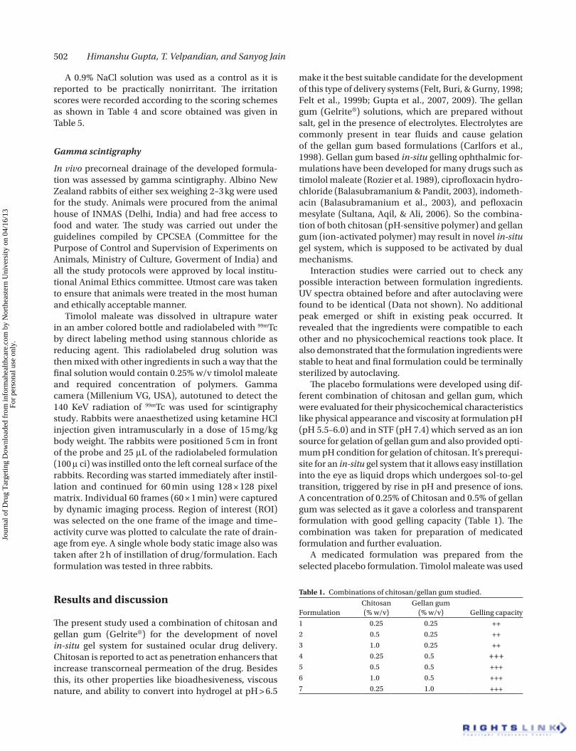
Figure 1. In vitro transcorneal permeation profile of various formulations. Values are expressed as mean ± SD (n=5).
Jour
nal o
f D
rug
Tar
getin
g D
ownl
oade
d fr
om in
form
ahea
lthca
re.c
om b
y N
orth
east
ern
Uni
vers
ity o
n 04
/16/
13Fo
r pe
rson
al u
se o
nly.
502 Himanshu Gupta, T. Velpandian, and Sanyog Jain
A 0.9% NaCl solution was used as a control as it is reported to be practically nonirritant. The irritation scores were recorded according to the scoring schemes as shown in Table 4 and score obtained was given in Table 5.
Gamma scintigraphy
In vivo precorneal drainage of the developed formula-tion was assessed by gamma scintigraphy. Albino New Zealand rabbits of either sex weighing 2–3 kg were used for the study. Animals were procured from the animal house of INMAS (Delhi, India) and had free access to food and water. The study was carried out under the guidelines compiled by CPCSEA (Committee for the Purpose of Control and Supervision of Experiments on Animals, Ministry of Culture, Goverment of India) and all the study protocols were approved by local institu-tional Animal Ethics committee. Utmost care was taken to ensure that animals were treated in the most human and ethically acceptable manner.
Timolol maleate was dissolved in ultrapure water in an amber colored bottle and radiolabeled with 99mTc by direct labeling method using stannous chloride as reducing agent. This radiolabeled drug solution was then mixed with other ingredients in such a way that the final solution would contain 0.25% w/v timolol maleate and required concentration of polymers. Gamma camera (Millenium VG, USA), autotuned to detect the 140 KeV radiation of 99mTc was used for scintigraphy study. Rabbits were anaesthetized using ketamine HCl injection given intramuscularly in a dose of 15 mg/kg body weight. The rabbits were positioned 5 cm in front of the probe and 25 μL of the radiolabeled formulation (100 μ ci) was instilled onto the left corneal surface of the rabbits. Recording was started immediately after instil-lation and continued for 60 min using 128 × 128 pixel matrix. Individual 60 frames (60 × 1 min) were captured by dynamic imaging process. Region of interest (ROI) was selected on the one frame of the image and time–activity curve was plotted to calculate the rate of drain-age from eye. A single whole body static image also was taken after 2 h of instillation of drug/formulation. Each formulation was tested in three rabbits.
Results and discussion
The present study used a combination of chitosan and gellan gum (Gelrite®) for the development of novel in-situ gel system for sustained ocular drug delivery. Chitosan is reported to act as penetration enhancers that increase transcorneal permeation of the drug. Besides this, its other properties like bioadhesiveness, viscous nature, and ability to convert into hydrogel at pH > 6.5
make it the best suitable candidate for the development of this type of delivery systems (Felt, Buri, & Gurny, 1998; Felt et al., 1999b; Gupta et al., 2007, 2009). The gellan gum (Gelrite®) solutions, which are prepared without salt, gel in the presence of electrolytes. Electrolytes are commonly present in tear fluids and cause gelation of the gellan gum based formulations (Carlfors et al., 1998). Gellan gum based in-situ gelling ophthalmic for-mulations have been developed for many drugs such as timolol maleate (Rozier et al. 1989), ciprofloxacin hydro-chloride (Balasubramanium & Pandit, 2003), indometh-acin (Balasubramanium et al., 2003), and pefloxacin mesylate (Sultana, Aqil, & Ali, 2006). So the combina-tion of both chitosan (pH-sensitive polymer) and gellan gum (ion-activated polymer) may result in novel in-situ gel system, which is supposed to be activated by dual mechanisms.
Interaction studies were carried out to check any possible interaction between formulation ingredients. UV spectra obtained before and after autoclaving were found to be identical (Data not shown). No additional peak emerged or shift in existing peak occurred. It revealed that the ingredients were compatible to each other and no physicochemical reactions took place. It also demonstrated that the formulation ingredients were stable to heat and final formulation could be terminally sterilized by autoclaving.
The placebo formulations were developed using dif-ferent combination of chitosan and gellan gum, which were evaluated for their physicochemical characteristics like physical appearance and viscosity at formulation pH (pH 5.5–6.0) and in STF (pH 7.4) which served as an ion source for gelation of gellan gum and also provided opti-mum pH condition for gelation of chitosan. It’s prerequi-site for an in-situ gel system that it allows easy instillation into the eye as liquid drops which undergoes sol-to-gel transition, triggered by rise in pH and presence of ions. A concentration of 0.25% of Chitosan and 0.5% of gellan gum was selected as it gave a colorless and transparent formulation with good gelling capacity (Table 1). The combination was taken for preparation of medicated formulation and further evaluation.
A medicated formulation was prepared from the selected placebo formulation. Timolol maleate was used
Table 1. Combinations of chitosan/gellan gum studied.
FormulationChitosan (% w/v)
Gellan gum (% w/v) Gelling capacity
1 0.25 0.25 ++
2 0.5 0.25 ++
3 1.0 0.25 ++
4 0.25 0.5 +++5 0.5 0.5 +++
6 1.0 0.5 +++
7 0.25 1.0 +++
Jour
nal o
f D
rug
Tar
getin
g D
ownl
oade
d fr
om in
form
ahea
lthca
re.c
om b
y N
orth
east
ern
Uni
vers
ity o
n 04
/16/
13Fo
r pe
rson
al u
se o
nly.
Novel in-situ gel system for sustained ocular drug delivery 503
in a concentration 0.25% w/v which is normally present in conventional eye drops formulations for glaucoma therapy. Methyl paraben (0.1%) was added as preserva-tive and glycerol was added in calculated amount to maintain isotonicity of the formulation and final formula is given in Table 2.
The developed formulations were further character-ized for various physiological parameters, like clarity, gelation pH, viscosity, and osmolarity. The developed formulation was iso-osmotic and gelation pH was found to be 6.5–7.0. Formulation also displayed good gelling property in STF (pH 7.4). The results were shown in Table 3.
Conducted in vitro transcorneal permeation studies on developed formulation showed a significantly higher (P < 0.05) permeation across goat cornea after 4 h as com-pared to plain drug solution and gellan gum alone based
in-situ gel system (Figure 1). The amount of the drug in the cornea was also calculated at the end of the study and a more than six times of the drug was found to be in the corneal membrane in case of chitosan and gellan gum based in-situ gel system as compared to free drug solu-tion and gellan gum alone based in-situ gel (Table 5). This might be due to the well-known permeation enhancer property of chitosan.
For packing the formulation the amber colored bottle closed with rubber closure and dropper with teat were used and found to be appropriate packaging system for current formulation. The packaging was tested for resist-ance for autoclaving, leakage, and pourability. Packaging passed all the tests and proved to be a good choice for packaging of present ocular formulation. Sterilization of the product was done by autoclaving at 121οC for 20 min at 15 psig and test for sterility was performed on autoclaved packaging according to IP 1996 standards. There has been no growth/microbial contamination was observed up to 14 days of incubation. Hence the formulation passed the sterility test.
Ocular irritation of the developed formulation was checked by Hen’s egg chorioallantoic membrane test which is a rapid, sensitive, and inexpensive test. Testing with incubated eggs is a borderline case between in vivo and in vitro systems and does not conflict with the ethical and legal obligations. The Chorioallantoic membrane of the chick embryo is a complete tissue including veins, arteries, and capillaries and is technically very easy to study. It responds to injury with a complete inflamma-tory process, a process similar to that induced in the conjuctival tissue of the rabbit eyes (Spielmann, 1997). Developed formulation was tested by this method and results were compared with those obtained using normal saline, which was used as control that is supposed to be practically nonirritant. A means score of 0 was obtained for normal saline. Chitosan/gellan gum based formula-tion was nonirritant up to 1 h (mean score 0) while the
Table 4. Scoring chart for HET–CAM test.
Effect Score Inference
No visible hemorrhage 0 Nonirritant
Just visible membrane discoloration 1 Mild irritant
Structures are covered partially due to membrane discoloration or hemorrhage 2 Moderately irritant
Structures are covered totally due to membrane discoloration or hemorrhages 3 Severe irritant
Table 5. In vitro transcorneal permeation of timolol maleate from free drug solution, gellan gum alone and chitosan and gellan gum based in-situ gel systems.
Formulation
Amount of the drug (µg) after 4 h
In the receptor compartment In the donor compartment In the corneal membrane
Free drug solution 48.25 ± 6.17 9776.00 ± 84.52 175.75 ± 12.31
Drug in gellan gum based in-situ gel system 55.82 ± 6.86 9750.73 ± 82.35 193.45 ± 15.26
Drug in chitosan and gellan gum based in-situ gel system
87.77 ± 7.43 8663.59 ± 54.65 1248.64 ± 23.85
Values are expressed as mean ± SD (n=5).
Table 2. Formula of the developed in-situ formulation.
Ingredients Concentration (w/v)
Timolol maleate 0.25%
Chitosan 0.25%
Gellan gum 0.50%
Glycerol q.s.
Methyl paraben 0.1%
Purified water (q.s.) 100%
Table 3. Physicochemical properties of the developed in-situ gel formulation.
Parameter Inference
Clarity Clear solution
pH 6.0
Osmolarity 298–302 mOsmol
Gelation pH 6.5-7.0
Viscosity (at pH 6.0) 40 ± 2.3 cps
Viscosity (at pH 7.4) 150 ± 9.5 cps
Values are expressed as mean ± SD (n=5).
Jour
nal o
f D
rug
Tar
getin
g D
ownl
oade
d fr
om in
form
ahea
lthca
re.c
om b
y N
orth
east
ern
Uni
vers
ity o
n 04
/16/
13Fo
r pe
rson
al u
se o
nly.
504 Himanshu Gupta, T. Velpandian, and Sanyog Jain
mean score was found to be 0.67 after 24 h (Table 6). The study demonstrated that the formulation is nonirritant to mild irritant and is well tolerable.
For scintigraphic studies, timolol maleate was radi-olabeled with radionuclide Tc-99m. It was chosen as it emits low energy gamma rays which do not lead to seri-ous health hazards. Timolol maleate was instantaneously labeled with Tc-99m. After prelabeling efficiency studies which includes labeling parameters like SnCl
2 concentra-
tion and pH were optimized and a 100 μg SnCl2 concen-
tration at pH 6.5 was found to give the maximum labeling efficiency (98.2%) (Gupta et al., 2009). At these conditions minimum colloids (1%) were produced. In vitro stability of the Tc-99m labeled complex was also tested and the
0
100
200
300
400
500
600
700
0 3 6 9 12 15 18 21 24 27 30 33 36 39 42 45 48 51 54 57 60Time (min)
Cou
nts/
sec
Free Timolol maleate Timolol maleate in-situ gel
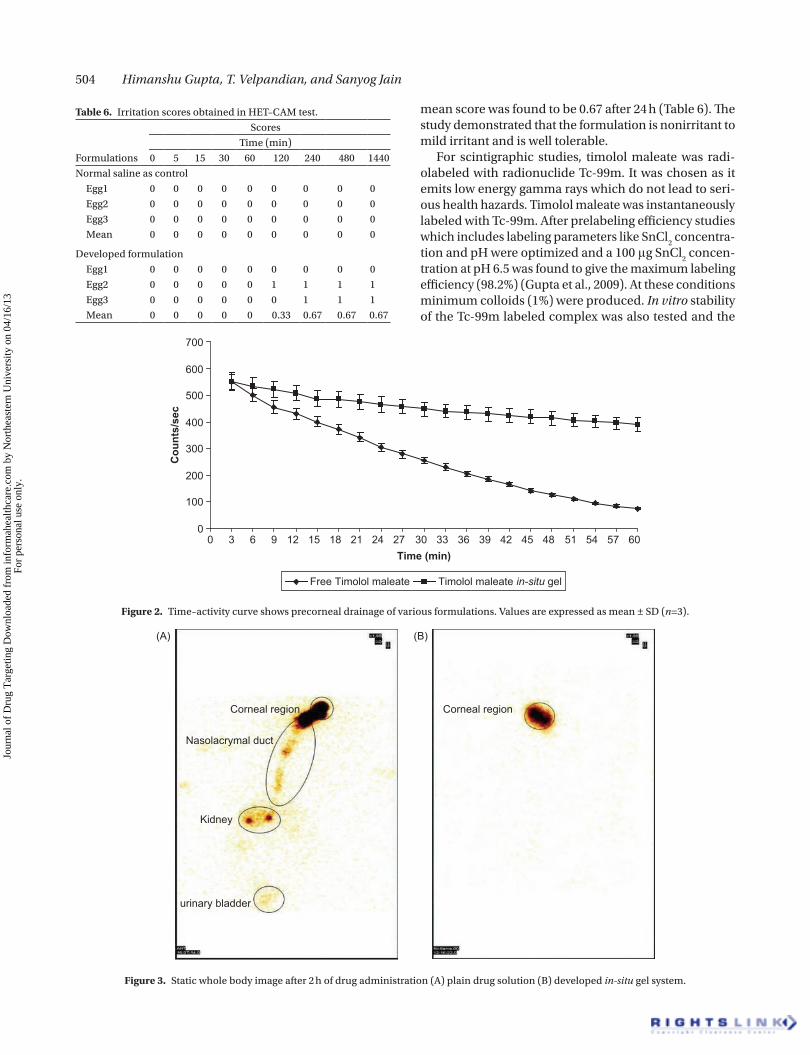
Figure 2. Time–activity curve shows precorneal drainage of various formulations. Values are expressed as mean ± SD (n=3).
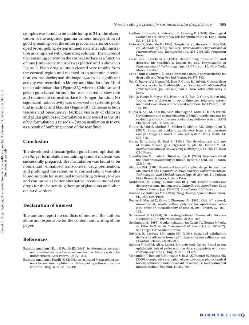
Corneal region Corneal region
Nasolacrymal duct
Kidney
urinary bladder
(A) (B)
Figure 3. Static whole body image after 2 h of drug administration (A) plain drug solution (B) developed in-situ gel system.
Table 6. Irritation scores obtained in HET–CAM test.
Formulations
Scores
Time (min)
0 5 15 30 60 120 240 480 1440
Normal saline as control
Egg1 0 0 0 0 0 0 0 0 0
Egg2 0 0 0 0 0 0 0 0 0
Egg3 0 0 0 0 0 0 0 0 0
Mean 0 0 0 0 0 0 0 0 0
Developed formulation
Egg1 0 0 0 0 0 0 0 0 0
Egg2 0 0 0 0 0 1 1 1 1
Egg3 0 0 0 0 0 0 1 1 1
Mean 0 0 0 0 0 0.33 0.67 0.67 0.67
Jour
nal o
f D
rug
Tar
getin
g D
ownl
oade
d fr
om in
form
ahea
lthca
re.c
om b
y N
orth
east
ern
Uni
vers
ity o
n 04
/16/
13Fo
r pe
rson
al u
se o
nly.
Novel in-situ gel system for sustained ocular drug delivery 505
complex was found to be stable for up to 24 h. The obser-vation of the acquired gamma camera images showed good spreading over the entire precorneal area for devel-oped in-situ gelling system immediately after administra-tion as compared with plain drug solution. The curves of the remaining activity on the corneal surface as a function of time (time–activity curve) was plotted and is shown in Figure 2. Plain drug solution cleared very rapidly from the corneal region and reached in to systemic circula-tion via nasolachrymal drainage system as significant activity was recorded in kidney and bladder after 2 h of ocular administration (Figure 3A), whereas Chitosan and gellan gum based formulation was cleared at slow rate and retained at corneal surface for longer duration. No significant radioactivity was observed in systemic pool, that is, kidney and bladder (Figure 3B). Chitosan is both viscous and bioadhesive. Further, viscosity of chitosan and gellan gum based formulation is increased as the pH of the formulation is raised (>7) upon instillation in to eye as a result of buffering action of the tear fluid.
Conclusion
The developed chitosan/gellan gum based ophthalmic in-situ gel formulation containing timolol maleate was successfully prepared. The formulation was found to be nonirritant, enhanced transcorneal drug permeation, and prolonged the retention at corneal site. It was also found suitable for sustained topical drug delivery to eyes and can prove as better alternative to conventional eye drops for the better drug therapy of glaucoma and other ocular disorders.
Declaration of interest
The authors report no conflicts of interest. The authors alone are responsible for the content and writing of the paper.
References
Balasubramaniam J, Kant S, Pandit JK. (2003). In vitro and in vivo eval-uation of the Gelrite gellan gum-based ocular delivery system for indomethacin. Acta Pharm, 53: 251–261.
Balasubramaniam J, Pandit JK. (2003). Ion-activated in situ gelling sys-tems for sustained ophthalmic delivery of ciprofloxacin hydro-chloride. Drug Deliv, 10: 185–191.
Carlfors J, Edsman K, Petersson R, Jörnving K. (1998). Rheological evaluation of Gelrite in situ gels for ophthalmic use. Eur J Pharm Sci, 6: 113–119.
Chiou GCY, Watanabe K. (1986). Drug delivery to the eyes. In: Ihler GM, ed. Methods of Drug Delivery: International Encyclopedia of Pharmacology and Therapeutics (pp. 203–210). UK: Pergamon Press.
Desai SD, Blanchard J. (1994). Ocular drug formulation and delivery. In: Swarbick J, Boylan JC, eds. Encyclopedia of Pharmaceutical Technology (pp. 43–75). vol. 11. New York: Marcel Dekker.
Felt O, Buri P, Gurny R. (1998). Chitosan: a unique polysaccharide for drug delivery. Drug Dev Ind Pharm, 24: 979–993.
Felt O, Baeyens V, Zignani M, Bury P, Gurny R. (1999a). Mucosal drug delivery: ocular. In: Mathiowith E, ed. Encyclopedia of Controlled Drug Delivery (pp. 605–626). vol. 1. New York: John Wiley & Sons.
Felt O, Furrer P, Mayer JM, Plazonnet B, Buri P, Gurny R. (1999b). Topical use of chitosan in ophthalmology: tolerance assess-ment and evaluation of precorneal retention. Int J Pharm, 180: 185–193.
Gupta H, Aqil M, Khar RK, Ali A, Bhatnagar A, Mittal G, Jain S. (2009). Development and characterization of 99mTc-timolol maleate for evaluating efficacy of in situ ocular drug delivery system. AAPS PharmSciTech, 10: 540–546.
Gupta H, Jain S, Mathur R, Mishra P, Mishra AK, Velpandian T. (2007). Sustained ocular drug delivery from a temperature and pH triggered novel in situ gel system. Drug Deliv, 14: 507–515.
Gurny R, Ibrahim H, Buri P. (1993). The development and use of in-situ formed gels triggered by pH. In: Edman Y, ed. Biopharmaceutics of Ocular Drug Delivery (pp. 81–90). FL, USA: CRC Press.
Higashiyama M, Inada K, Ohtori A, Tojo K. (2004). Improvement of the ocular bioavailability of timolol by sorbic acid. Int J Pharm, 272: 91–98.
Maurice DM. (1987). Kinetics of topically applied drugs. In: Saettone MS, Bucci P, eds. Ophthalmic Drug Delivery: Biopharmaceutical, Technological and Clinical Aspects (pp. 19–26). vol. 11. Padova: Fidia Research Series, Liviana Press.
Middleton DL, Leung SS, Robinson JR. (1990). Ocular bioadhesive delivery systems. In: Lenaerts V, Gurny R, eds. Bioadhesive Drug Delivery Systems (pp. 179–202). Boca Raton: CRC Press.
Ranade VV, Hollinger MA. (1996). Drug Delivery Systems. Boca Raton, FL, USA: CRC Press.
Rozier A, Mazuel C, Grove J, Plazonnet B. (1989). Gelrite®: a novel, ion-activated, in-situ gelling polymer for ophthalmic vehi-cles: effect on bioavailability of timolol. Int J Pharm, 57: 163– 168.
Schoenwald RD. (1990). Ocular drug delivery. Pharmacokinetic con-siderations. Clin Pharmacokinet, 18: 255–269.
Spielmann H. (1997). Ocular irritation. In: Castle JV, Gomez MJ, eds. In Vitro Methods in Pharmaceutical Research (pp. 265–287). San Diego, CA: Academic Press.
Srividya B, Cardoza RM, Amin PD. (2001). Sustained ophthalmic delivery of ofloxacin from a pH triggered in situ gelling system. J Control Release, 73: 205–211.
Sultana Y, Aqil M, Ali A. (2006). Ion-activated, Gelrite-based in situ ophthalmic gels of pefloxacin mesylate: comparison with con-ventional eye drops. Drug Deliv, 13: 215–219.
Velpandian T, Bankoti R, Humayun S, Ravi AK, Kumari SS, Biswas NR. (2006). Comparative evaluation of possible ocular photochemical toxicity of fluoroquinolones meant for ocular use in experimental models. Indian J Exp Biol, 44: 387–391.
Jour
nal o
f D
rug
Tar
getin
g D
ownl
oade
d fr
om in
form
ahea
lthca
re.c
om b
y N
orth
east
ern
Uni
vers
ity o
n 04
/16/
13Fo
r pe
rson
al u
se o
nly.