Embed Size (px)
Citation preview
RESEARCH ARTICLE Open Access
Involvement of Slit–Robo signaling in thedevelopment of the posterior commissureand concomitant swimming behavior inXenopus laevisYasuhiko Tosa1†, Kiyohito Tsukano1†, Tatsuya Itoyama1, Mai Fukagawa1, Yukako Nii1, Ryota Ishikawa1,Ken-ichi T. Suzuki3, Makiko Fukui1, Masahumi Kawaguchi2 and Yasunori Murakami1*
Abstract
Introduction: During vertebrate development, the central nervous system (CNS) has stereotyped neuronal tracts(scaffolds) that include longitudinal and commissural axonal bundles, such as the medial longitudinal fascicle or theposterior commissure (PC). As these early tracts appear to guide later-developing neurons, they are thought toprovide the basic framework of vertebrate neuronal circuitry. The proper construction of these neuronal circuits isthought to be a crucial step for eliciting coordinated behaviors, as these circuits transmit sensory information to theintegrative center, which produces motor commands for the effective apparatus. However, the developmental planunderlying some commissures and the evolutionary transitions they have undergone remain to be elucidated. Littleis known about the role of axon guidance molecules in the elicitation of early-hatched larval behavior as well.
Results: Here, we report the developmentally regulated expression pattern of axon-guidance molecules Slit2 ligandand Robo2 receptor in Xenopus laevis and show that treatment of X. laevis larvae with a slit2- or robo2-morpholinoresulted in abnormal swimming behavior. We also observed an abnormal morphology of the PC, which is part ofthe early axonal scaffold.
Conclusion: Our present findings suggest that expression patterns of Slit2 and Robo2 are conserved in tetrapods,and that their signaling contributes to the construction of the PC in Xenopus. Given that the PC also includes severaltypes of neurons stemming from various parts of the CNS, it may represent a candidate prerequisite neuronal tract inthe construction of subsequent complex neuronal circuits that trigger coordinated behavior.
IntroductionExternal stimuli received by several types of sensory re-ceptors located on the body’s surface are transferred to theperipheral nerves. Subsequently, the nerve afferents enterthe brain and send information to relay nuclei, which inturn project to higher centers in which the various sensoryinputs are integrated. The motor center then outputscommands to motoneurons located in the hindbrain or inthe spinal cord. Thus, the construction of a precise circuitis a crucial step in eliciting appropriate behavioral re-sponses. If such neuronal circuits are disorganized during
ontogenesis, early larvae may be unable to perform coor-dinated body movements. In the developing vertebratecentral nervous system (CNS), early-differentiating neu-rons extend axons toward their target regions, formingstereotyped tracts (scaffolds) consisting of longitudinaland commissural axonal bundles [1–8]. In later develop-ment, these early tracts are thought to serve as guidepostsfor later-developing axons [9]. The basic framework ofthese tracts is highly conserved in vertebrate evolution[2, 10]. The early tracts consist of longitudinal (extendingalong the anteroposterior axis) and commissural (connect-ing to the left and right side of the brain) tracts. Theformer include the lateral longitudinal fascicle, tracts ofthe postoptic commissure (TPOC), and the supraoptictract (SOT); the latter include the anterior, habenular(HC), and posterior (PC) commissures.
* Correspondence: [email protected]†Equal contributors1Graduate School of Science and Engineering, Ehime University, 2-5Bunkyo-cho, Matsuyama 790-8577, JapanFull list of author information is available at the end of the article
© 2015 Tosa et al. Open Access This article is distributed under the terms of the Creative Commons Attribution 4.0International License (http://creativecommons.org/licenses/by/4.0/), which permits unrestricted use, distribution, andreproduction in any medium, provided you give appropriate credit to the original author(s) and the source, provide a link tothe Creative Commons license, and indicate if changes were made. The Creative Commons Public Domain Dedication waiver(http://creativecommons.org/publicdomain/zero/1.0/) applies to the data made available in this article, unless otherwise stated.
Tosa et al. Zoological Letters (2015) 1:28 DOI 10.1186/s40851-015-0029-9
Developing vertebrate brains are typically subdividedinto series of segments called neuromeres, and those lo-cated in the diencephalon are called prosomeres [11–17].It is known that some commissural bundles are located ata specific region on the neural tube corresponding to pro-someric compartments. The HCs and PCs are formed inprosomere 2 (thalamus) and prosomere 1 (pretectum), re-spectively, in many vertebrate groups [14]. The highlyconserved framework of these commissures implies that astrictly maintained neurodevelopmental program involvedin their wiring has been inherited during vertebrate evolu-tion. In fact, some transcription factors and axon-guidance molecules have been shown play an importantrole in the formation of the network of these early tracts[18–20]. Previous studies have revealed that the inter-action between Slit (ligand) and Robo (receptor), whichacts as a repulsive guidance signal, plays an essential rolein the formation of early scaffolds, e.g., the inhibition ofSlit2 or Robo2 results in an abnormal morphology of theTPOC [21–23] and SOT [24]. These molecules are alsoinvolved in the formation of commissural tracts in insects[25, 26] and vertebrates (Slit:[27, 28]; Robo:[29, 30]). Inzebrafish robo3 mutant, the axons of the Mauthnerneuron fail to cross the midline [31]. In mammals, theSlit/Robo interaction is involved in the formation of thecorpus callosum, which is a type of commissural systemthat connects the cerebral hemispheres [32]. The simi-larity of the Slit-Robo interaction in teleosts and ro-dents leads to the possibility that the role of slit androbo is conserved in vertebrate evolution. To study suchevolutionary conservation in the vertebrate lineage, it isimportant to study the function of slit and robo in am-phibians, as these animals are thought to have divergedbetween the teleost and amniote lineages. In addition, thedevelopment of locomotion patterns in larval stages iswell-described in anuran species [33–35]. For these rea-sons, Xenopus laevis, an anuran species, may be a suitablemodel for use in phylogenetic studies and behavioral ana-lysis in early hatched larvae. The aim of the present studyis to identify the role of Slit-Robo signaling in the forma-tion of early tracts and/or the elicitation of swimmingbehavior in Xenopus. We also tried to identify the evolu-tionary transition of Slit-Robo signaling. To this end, westudied the expression pattern of Slit2 and Robo2 inXenopus embryos, then perturbed their signals using mor-pholino antisense oligonucleotides (MO) and analyzed theswimming behavior of early larvae. We found that expres-sion domains of Slit2 and Robo2 in Xenopus are similar tothose of amniotes, indicating that the axon guidancemechanism that depends on Slit-Robo signaling is evolu-tionarily conserved in the forebrain of tetrapods. We alsofound a disorganized swimming behavior and an abnor-mal morphology of the PC in both slit2- and robo2-MO-injected larvae. These results indicate that interaction
between Slit2 and Robo2 is involved in the construction ofthe PC and the formation of neuronal element(s) that con-trol coordinated body movement in Xenopus larvae.
Materials and methodsXenopus embryosAdult Xenopus laevis were purchased from a local farm(Hamamatsu Seibutsu Kyozai Co. Ltd; Shizuoka Prefecture,Japan), and fertilized eggs were obtained in the laboratoryvia artificial fertilization. Fertilized eggs were then placed infresh water and incubated at 20 °C. Embryonic stages weredetermined based on Nieuwkoop and Faber (1967) [36].The studies were performed according to the EthicalGuidelines for Animal Use of the Animal Care Committeeat Ehime University.
Isolation of axon-guidance genes in X. laevisXenopus homologues of slit2 and robo2 were isolated bypolymerase chain reaction (PCR) using X. laevis embry-onic cDNA as a template. The primers of Xlslit2 were de-signed based on a published sequence (NM_001087668.1).The following primers were used: Xlslit2-F, 5’–TGAATCAGCACCACCAATGG–3’, Xlslit2-R, 5’–CTAGTCTCGATACCTTCTCG–3’; Xlrobo2-F, 5’–TGGATTGTAGAGTGCTGAGG–3’, Xlrobo2-R, 5’–CACGGAGCAATGCTACTTCC–3’.The PCR products included in the agarose gel were
purified using the Wizard SV Gel and PCR Clean-UpSystem (Promega), and the DNA fragment was cloned intopGEM-T Easy (pGEM-T Easy Vector Systems; Promega).
Injection of morpholinosTo inhibit Slit2 or Robo2 signals specifically in neuronaltissues, a slit2 MO (GCCACCCAAGGAAAGAACCCAACCA; Gene Tools, LLC) and a robo2 MO (AGCCACCAGAAAGCCCATGTTTCCC) were injected at the8-cell stage into the small blastomere of the animal pole,which differentiates into the CNS [37]. To visualizeinjected cells, enhanced green fluorescent protein (eGFP)mRNA was co-injected into the blastomere. Morpholinoscontaining five mismatched nucleotides were used as acontrol. As controls of slit2 and robo2, GCgAgCCAAcGAAAcAAgCCAACCA and AGCCACgAcAAAcCCgATcTTTCCC were used, respectively (the mismatched nu-cleotide is shown in lower case). We injected 2.5 pmol ofslit2, robo2, and control MOs. Injected embryos were in-cubated in 3 % ficoll until the blastula stage; subsequently,embryos were replaced in 0.3 ×MMR (100 mM NaCl,5 mM MgCl2, 0.5 mM CaCl2, 5 mM EGTA, 20 mMHEPES-NaOH, pH 7.5) at 20 °C. At stage 28, we checkedeGFP illumination under a fluorescence microscope(Lumar V12; Carl Zeiss SMT GmbH, Oberkochen,Germany). Embryos showing neuron-specific localizationof eGFP were incubated and used for further analyses.
Tosa et al. Zoological Letters (2015) 1:28 Page 2 of 16
Morphological observationParaformaldehyde (PFA)-fixed specimens were observedunder a stereomicroscope (Lumar V12; Carl Zeiss). Thebody length, curvature of the body axis, and surface areaof the eyes were measured using the Axio Vision soft-ware (release 4.7.2; Carl Zeiss). Ten larvae were exam-ined in each measurement.
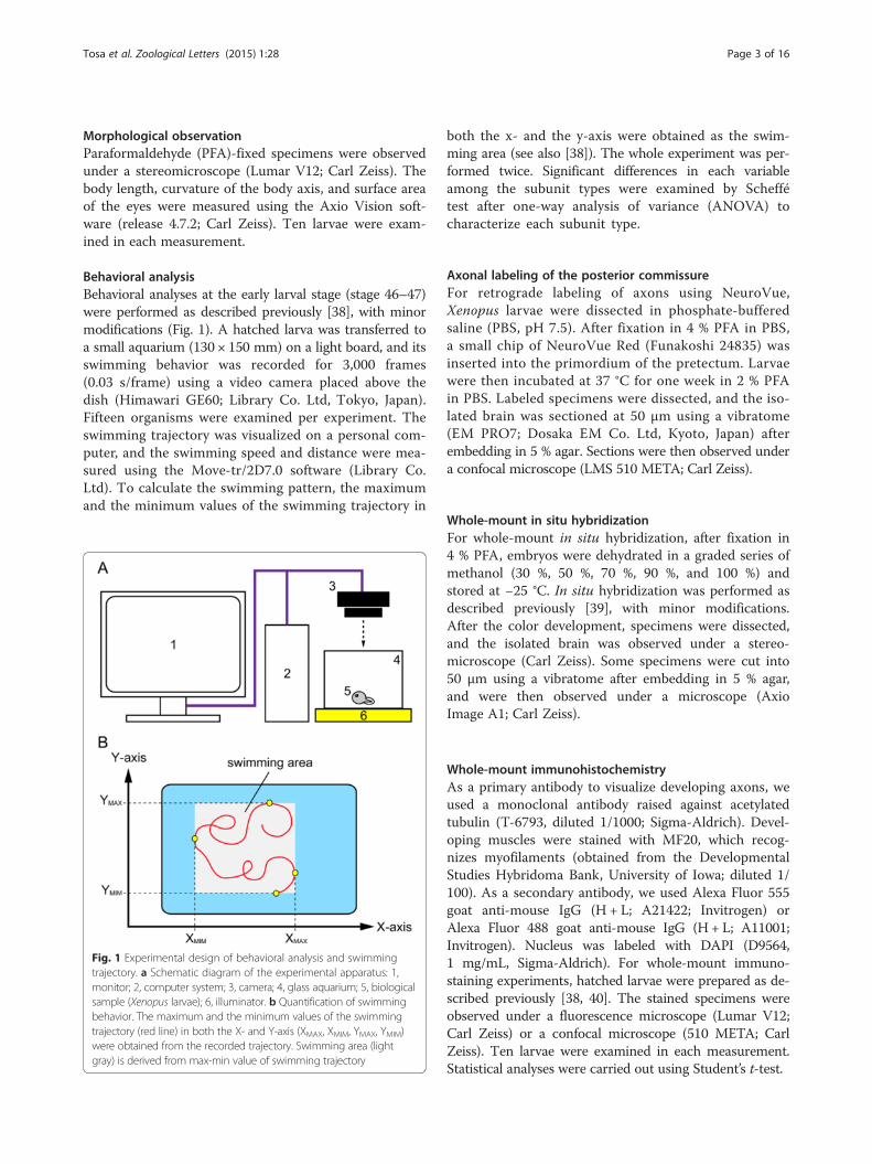
Behavioral analysisBehavioral analyses at the early larval stage (stage 46–47)were performed as described previously [38], with minormodifications (Fig. 1). A hatched larva was transferred toa small aquarium (130 × 150 mm) on a light board, and itsswimming behavior was recorded for 3,000 frames(0.03 s/frame) using a video camera placed above thedish (Himawari GE60; Library Co. Ltd, Tokyo, Japan).Fifteen organisms were examined per experiment. Theswimming trajectory was visualized on a personal com-puter, and the swimming speed and distance were mea-sured using the Move-tr/2D7.0 software (Library Co.Ltd). To calculate the swimming pattern, the maximumand the minimum values of the swimming trajectory in
both the x- and the y-axis were obtained as the swim-ming area (see also [38]). The whole experiment was per-formed twice. Significant differences in each variableamong the subunit types were examined by Scheffétest after one-way analysis of variance (ANOVA) tocharacterize each subunit type.
Axonal labeling of the posterior commissureFor retrograde labeling of axons using NeuroVue,Xenopus larvae were dissected in phosphate-bufferedsaline (PBS, pH 7.5). After fixation in 4 % PFA in PBS,a small chip of NeuroVue Red (Funakoshi 24835) wasinserted into the primordium of the pretectum. Larvaewere then incubated at 37 °C for one week in 2 % PFAin PBS. Labeled specimens were dissected, and the iso-lated brain was sectioned at 50 μm using a vibratome(EM PRO7; Dosaka EM Co. Ltd, Kyoto, Japan) afterembedding in 5 % agar. Sections were then observed undera confocal microscope (LMS 510 META; Carl Zeiss).
Whole-mount in situ hybridizationFor whole-mount in situ hybridization, after fixation in4 % PFA, embryos were dehydrated in a graded series ofmethanol (30 %, 50 %, 70 %, 90 %, and 100 %) andstored at −25 °C. In situ hybridization was performed asdescribed previously [39], with minor modifications.After the color development, specimens were dissected,and the isolated brain was observed under a stereo-microscope (Carl Zeiss). Some specimens were cut into50 μm using a vibratome after embedding in 5 % agar,and were then observed under a microscope (AxioImage A1; Carl Zeiss).
Whole-mount immunohistochemistryAs a primary antibody to visualize developing axons, weused a monoclonal antibody raised against acetylatedtubulin (T-6793, diluted 1/1000; Sigma-Aldrich). Devel-oping muscles were stained with MF20, which recog-nizes myofilaments (obtained from the DevelopmentalStudies Hybridoma Bank, University of Iowa; diluted 1/100). As a secondary antibody, we used Alexa Fluor 555goat anti-mouse IgG (H + L; A21422; Invitrogen) orAlexa Fluor 488 goat anti-mouse IgG (H + L; A11001;Invitrogen). Nucleus was labeled with DAPI (D9564,1 mg/mL, Sigma-Aldrich). For whole-mount immuno-staining experiments, hatched larvae were prepared as de-scribed previously [38, 40]. The stained specimens wereobserved under a fluorescence microscope (Lumar V12;Carl Zeiss) or a confocal microscope (510 META; CarlZeiss). Ten larvae were examined in each measurement.Statistical analyses were carried out using Student’s t-test.
Fig. 1 Experimental design of behavioral analysis and swimmingtrajectory. a Schematic diagram of the experimental apparatus: 1,monitor; 2, computer system; 3, camera; 4, glass aquarium; 5, biologicalsample (Xenopus larvae); 6, illuminator. b Quantification of swimmingbehavior. The maximum and the minimum values of the swimmingtrajectory (red line) in both the X- and Y-axis (XMAX, XMIM, YMAX, YMIM)were obtained from the recorded trajectory. Swimming area (lightgray) is derived from max-min value of swimming trajectory
Tosa et al. Zoological Letters (2015) 1:28 Page 3 of 16
In situ hybridization combined withimmunohistochemistryWhole-mount in situ hybridization was performed asdescribed above. Then, the samples were washed severaltimes with Tris-buffered saline with 0.1 % triton-x100(TBST). The nerve fibers were visualized by an immuno-histochemistry by described above. Briefly, samples wereincubated for 1 overnight in TBST containing 5 % skimmilk (TSTM). They were then treated with the anti-acetylated tubulin antibody. Samples were incubated inthe antibody for 2 days at RT in TSTM containing 0.02 %NaN3. The samples were then washed in TBST four timesand subsequently incubated in the secondary antibody(Alexa488 anti-mouse IgG, diluted 1:500) for 2 days at RTin TSTM. The samples were then washed four times inTBST. The specimens were observed under a fluorescencestereomicroscope (Lumar V12; Carl Zeiss).
ResultsSwimming behavior in MO-treated larvaeTo study the molecular functions of Xenopus cognates ofslit2 (Xlslit2) and robo2 (Xlrobo2) in the developing ner-vous system, MO were injected into the blastomere, whichdifferentiates into the CNS. Although many MO-injectedindividuals showed no abnormal morphology at stage 44,a small number of larvae exhibited head curvature or eyereduction in both the control- and slit2-MO-injectedgroups. Conversely, the robo2-MO-injected specimensand their control-MO-injected specimens showed a moreasymmetrical craniofacial morphology than did slit2-MO-and control-MO-injected specimens. Thus, in the subse-quent analysis, we used slit2-MO-injected larvae and theircontrol-MO-injected larvae (with normal body morph-ology), whereas we used robo2-MO-injected larvae andtheir control-MO-injected larvae, which have a slightlyasymmetrical shape in the head or eye, in addition tolarvae with normal body morphology.As Slit2 or Robo2 is involved in the formation of the
early neuronal circuit in many vertebrates [27–30], wehypothesized that MO-treated larvae possess abnormal-ities not only in their neuronal elements, but in behavioras well. In fact, it has been reported that early-bornneuronal frameworks play an essential role in the regula-tion of body movement [41]. Previous studies haveshown that the first alternating body movements in X.laevis occur on both sides of the body during the earlyswimming stage (stages 28–33); however, the embryodoes not move through the water. The myotomal mus-culature is fully developed during the free-swimmingstage (from stage 33), and the embryo is able to movethrough the water and swim [33–35]. Larvae beginswimming to search for food at the early larval stage(from stage 45 on), at which point their oral apparatusbecomes functional, enabling them to eat. As this free
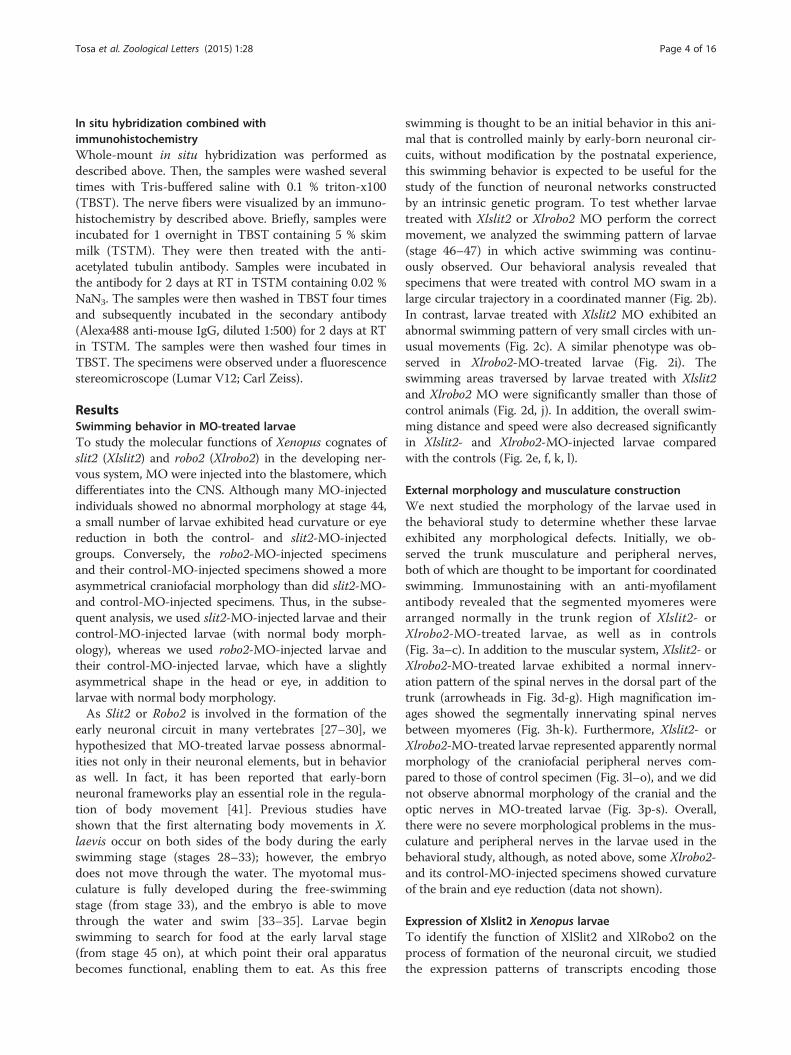
swimming is thought to be an initial behavior in this ani-mal that is controlled mainly by early-born neuronal cir-cuits, without modification by the postnatal experience,this swimming behavior is expected to be useful for thestudy of the function of neuronal networks constructedby an intrinsic genetic program. To test whether larvaetreated with Xlslit2 or Xlrobo2 MO perform the correctmovement, we analyzed the swimming pattern of larvae(stage 46–47) in which active swimming was continu-ously observed. Our behavioral analysis revealed thatspecimens that were treated with control MO swam in alarge circular trajectory in a coordinated manner (Fig. 2b).In contrast, larvae treated with Xlslit2 MO exhibited anabnormal swimming pattern of very small circles with un-usual movements (Fig. 2c). A similar phenotype was ob-served in Xlrobo2-MO-treated larvae (Fig. 2i). Theswimming areas traversed by larvae treated with Xlslit2and Xlrobo2 MO were significantly smaller than those ofcontrol animals (Fig. 2d, j). In addition, the overall swim-ming distance and speed were also decreased significantlyin Xlslit2- and Xlrobo2-MO-injected larvae comparedwith the controls (Fig. 2e, f, k, l).
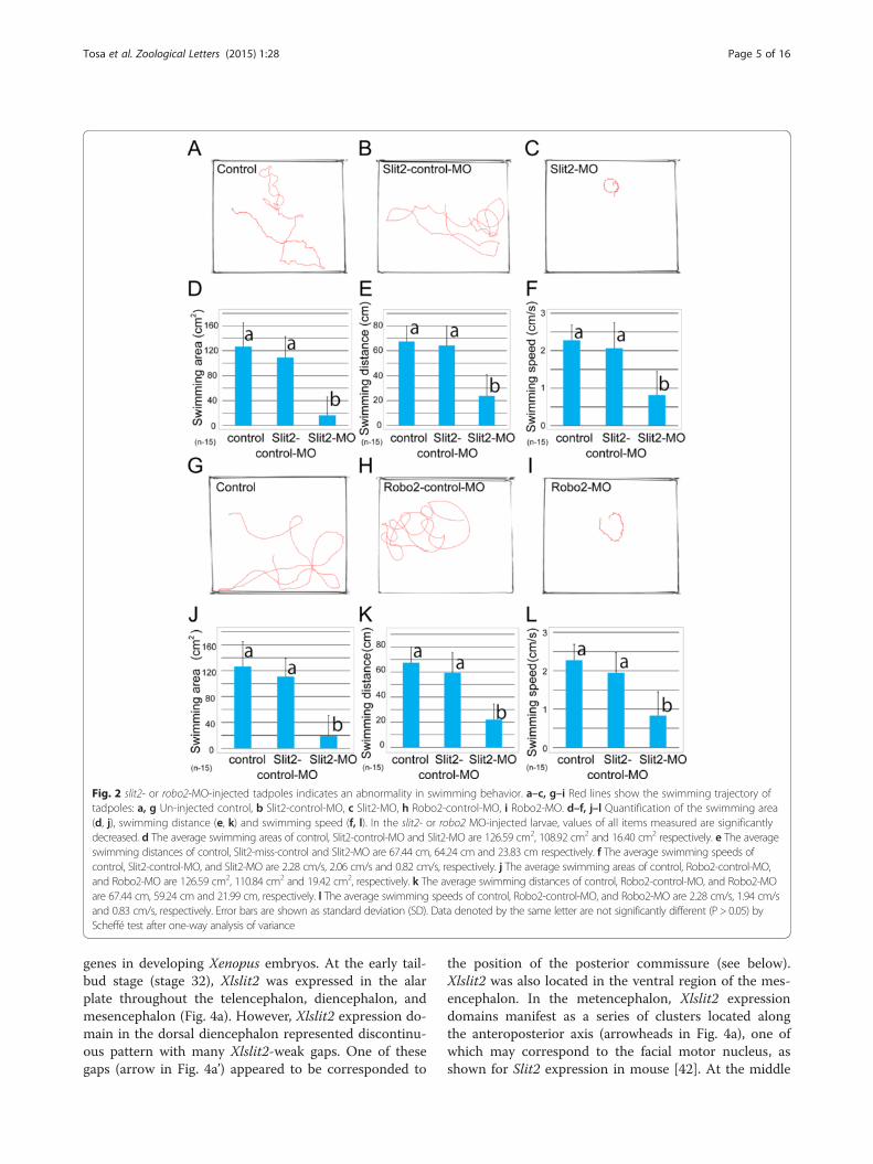
External morphology and musculature constructionWe next studied the morphology of the larvae used inthe behavioral study to determine whether these larvaeexhibited any morphological defects. Initially, we ob-served the trunk musculature and peripheral nerves,both of which are thought to be important for coordinatedswimming. Immunostaining with an anti-myofilamentantibody revealed that the segmented myomeres werearranged normally in the trunk region of Xlslit2- orXlrobo2-MO-treated larvae, as well as in controls(Fig. 3a–c). In addition to the muscular system, Xlslit2- orXlrobo2-MO-treated larvae exhibited a normal innerv-ation pattern of the spinal nerves in the dorsal part of thetrunk (arrowheads in Fig. 3d-g). High magnification im-ages showed the segmentally innervating spinal nervesbetween myomeres (Fig. 3h-k). Furthermore, Xlslit2- orXlrobo2-MO-treated larvae represented apparently normalmorphology of the craniofacial peripheral nerves com-pared to those of control specimen (Fig. 3l–o), and we didnot observe abnormal morphology of the cranial and theoptic nerves in MO-treated larvae (Fig. 3p-s). Overall,there were no severe morphological problems in the mus-culature and peripheral nerves in the larvae used in thebehavioral study, although, as noted above, some Xlrobo2-and its control-MO-injected specimens showed curvatureof the brain and eye reduction (data not shown).
Expression of Xlslit2 in Xenopus larvaeTo identify the function of XlSlit2 and XlRobo2 on theprocess of formation of the neuronal circuit, we studiedthe expression patterns of transcripts encoding those
Tosa et al. Zoological Letters (2015) 1:28 Page 4 of 16
genes in developing Xenopus embryos. At the early tail-bud stage (stage 32), Xlslit2 was expressed in the alarplate throughout the telencephalon, diencephalon, andmesencephalon (Fig. 4a). However, Xlslit2 expression do-main in the dorsal diencephalon represented discontinu-ous pattern with many Xlslit2-weak gaps. One of thesegaps (arrow in Fig. 4a’) appeared to be corresponded to
the position of the posterior commissure (see below).Xlslit2 was also located in the ventral region of the mes-encephalon. In the metencephalon, Xlslit2 expressiondomains manifest as a series of clusters located alongthe anteroposterior axis (arrowheads in Fig. 4a), one ofwhich may correspond to the facial motor nucleus, asshown for Slit2 expression in mouse [42]. At the middle
Fig. 2 slit2- or robo2-MO-injected tadpoles indicates an abnormality in swimming behavior. a–c, g–i Red lines show the swimming trajectory oftadpoles: a, g Un-injected control, b Slit2-control-MO, c Slit2-MO, h Robo2-control-MO, i Robo2-MO. d–f, j–l Quantification of the swimming area(d, j), swimming distance (e, k) and swimming speed (f, l). In the slit2- or robo2 MO-injected larvae, values of all items measured are significantlydecreased. d The average swimming areas of control, Slit2-control-MO and Slit2-MO are 126.59 cm2, 108.92 cm2 and 16.40 cm2 respectively. e The averageswimming distances of control, Slit2-miss-control and Slit2-MO are 67.44 cm, 64.24 cm and 23.83 cm respectively. f The average swimming speeds ofcontrol, Slit2-control-MO, and Slit2-MO are 2.28 cm/s, 2.06 cm/s and 0.82 cm/s, respectively. j The average swimming areas of control, Robo2-control-MO,and Robo2-MO are 126.59 cm2, 110.84 cm2 and 19.42 cm2, respectively. k The average swimming distances of control, Robo2-control-MO, and Robo2-MOare 67.44 cm, 59.24 cm and 21.99 cm, respectively. l The average swimming speeds of control, Robo2-control-MO, and Robo2-MO are 2.28 cm/s, 1.94 cm/sand 0.83 cm/s, respectively. Error bars are shown as standard deviation (SD). Data denoted by the same letter are not significantly different (P > 0.05) byScheffé test after one-way analysis of variance
Tosa et al. Zoological Letters (2015) 1:28 Page 5 of 16
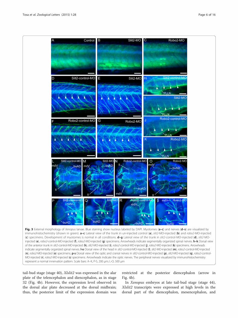
tail-bud stage (stage 40), Xlslit2 was expressed in the alarplate of the telencephalon and diencephalon, as in stage32 (Fig. 4b). However, the expression level observed inthe dorsal alar plate decreased at the dorsal midbrain;thus, the posterior limit of the expression domain was
restricted at the posterior diencephalon (arrow inFig. 4b).In Xenopus embryos at late tail-bud stage (stage 44),
Xlslit2 transcripts were expressed at high levels in thedorsal part of the diencephalon, mesencephalon, and
Fig. 3 External morphology of Xenopus larvae. Blue staining show nucleus labeled by DAPI. Myotomes (a–c) and nerves (d–s) are visualized byimmunohistochemistry (shown in green). a–c Lateral view of the trunk in un-injected control (a), slit2-MO-injected (b) and robo2-MO-injected(c) specimens. Development of myotomes is normal in all conditions. d–g Lateral view of the trunk in slit2-control-MO-injected (d), slit2-MO-injected (e), robo2-control-MO-injected (f), robo2-MO-injected (g) specimens. Arrowheads indicate segmentally organized spinal nerves. h–k Dorsal viewof the anterior trunk in slit2-control-MO-injected (h), slit2-MO-injected (i), robo2-control-MO-injected (j), robo2-MO-injected (k) specimens. Arrowheadsindicate segmentally organized spinal nerves. l-o Dorsal view of the head in slit2-control-MO-injected (l), slit2-MO-injected (m), robo2-control-MO-injected(n), robo2-MO-injected (o) specimens. p-s Dorsal view of the optic and cranial nerves in slit2-control-MO-injected (p), slit2-MO-injected (q), robo2-control-MO-injected (r), robo2-MO-injected (s) specimens. Arrowheads indicate the optic nerves. The peripheral nerves visualized by immunohistochemistryrepresent a normal innervation pattern. Scale bars: A–K, P-S, 200 μm; L-O, 500 μm
Tosa et al. Zoological Letters (2015) 1:28 Page 6 of 16
metencephalon (Fig. 4c). At this stage, Xlslit2 expressionregions became broader compared with those of stage40 embryos, although the Xlslit2 expression domain wasstill restricted to the specific part of the metencephalon(Fig. 4c). Conversely, Xlslit2 transcripts were weaklyexpressed in the transitional region between the di-encephalon and mesencephalon (arrow in Fig. 4c).
Expression of Xlrobo2 in Xenopus larvaeNext, we observed the expression pattern of the Xenopusortholog of robo2 (Xlrobo2), which is a putative receptorof Xlslit2. At stage 32, Xlrobo2 was expressed at highlevels in the metencephalon (Fig. 4d). Furthermore, theXlrobo2 transcript was observed in the diencephalonand ventral telencephalon (Fig. 4d). At stage 40, the
Fig. 4 Expression patterns of Xlslit2 and Xlrobo2 and morphology of axonal tracts in Xenopus larvae. a–c Transcripts of Xlslit2 are detected throughthe dorsal midline in the diencephalon at stage 32 (a), stage 40 (b) and stage 44 (c). Xlslit2-weak gap is found between diencephalon and mesencephalon(arrows in A and C, A’ is a dorsal view). In the metencephalon (met), expression domains of Xlslit2 are observed along the antero-posterior axis (arrowheads).d–f Expression pattern of Xlrobo2 at stage 32 (d), stage 40 (e) and stage 44 (f). (d) At early tail-bud stage, Xlrobo2 is expressed in the telencephalon (te),diencephalon (di), mesencephalon (mes), dorsal metencephalon (met) and notochord (nc). e, f At middle and late tail-bud stage, XlRobo2 expression isdetected at high levels in the dorsal CNS. g, h Dorsal (g) and lateral (h) view of the developing Xenopus larva at stage 46. Axons in the PNS and CNS arevisualized by anti-acetylated tubulin antibody. Habenular and posterior commissures are located the diencephalon (HC and PC). Scale bars: A-F, 200 μm; Gand H, 500 μm
Tosa et al. Zoological Letters (2015) 1:28 Page 7 of 16
Xlrobo2 mRNA was detected throughout the dorsal levelof the brain (Fig. 4e). In particular, it was expressed inthe dorsal sides of the mesencephalon and metencepha-lon, whereas it was weakly expressed in the ventralneural tube. We did not detect Xlrobo2 transcripts inthe spinal cord (data not shown). At stage 44, Xlrobo2was expressed at higher levels throughout the dorsal partof the brain, as in the previous stage (Fig. 4f ).
Morphology of the nervous system in Xenopus larvaeImmunostaining using an anti-acetylated tubulin anti-body was performed to investigate the developmentalprocess of the Xenopus nervous system. In the embry-onic stage 46, several cranial nerves, including the olfac-tory (the first cranial nerve, I), the optic (the secondcranial nerve, II), the trigeminal (mdV), the anteriorlateral line (buAD), and the posterior lateral line (PLLN)nerves, were observed (Fig. 4g, h), as in matured tadpolelarvae. We also identified spinal nerves arranged seg-mentally in the trunk region (data not shown). Thesenerves showed clear segregation and projected to theircorrect targets. Next, we studied the axonal organizationof the CNS, which receives input from the peripheralnerves. At stage 46, we observed several longitudinal orcommissural axonal bundles, in which three distinctcommissural tracts were observed in the dorsal side ofthe neural tube (Fig. 4g, h). Among those, the HC waslocated in the anterior part of the dorsal diencephalon.The PC was observed in the posterior part of the dorsaldiencephalon (apparently corresponding to the pretectum).The commissures in the cerebellum (CC), which mayinclude commissure cerebelli and commissure vestibulo-lateralis [43], were located on the cerebellum and acrossthe midline on the dorsal side.
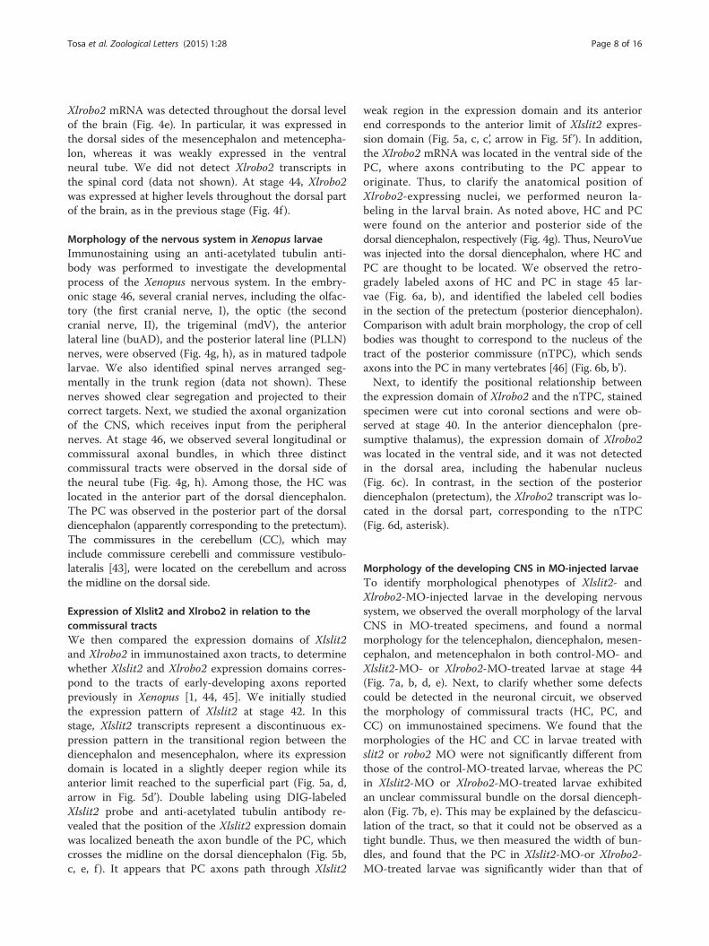
Expression of Xlslit2 and Xlrobo2 in relation to thecommissural tractsWe then compared the expression domains of Xlslit2and Xlrobo2 in immunostained axon tracts, to determinewhether Xlslit2 and Xlrobo2 expression domains corres-pond to the tracts of early-developing axons reportedpreviously in Xenopus [1, 44, 45]. We initially studiedthe expression pattern of Xlslit2 at stage 42. In thisstage, Xlslit2 transcripts represent a discontinuous ex-pression pattern in the transitional region between thediencephalon and mesencephalon, where its expressiondomain is located in a slightly deeper region while itsanterior limit reached to the superficial part (Fig. 5a, d,arrow in Fig. 5d’). Double labeling using DIG-labeledXlslit2 probe and anti-acetylated tubulin antibody re-vealed that the position of the Xlslit2 expression domainwas localized beneath the axon bundle of the PC, whichcrosses the midline on the dorsal diencephalon (Fig. 5b,c, e, f ). It appears that PC axons path through Xlslit2
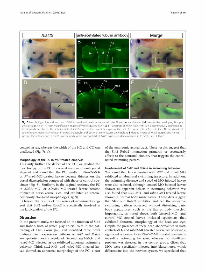
weak region in the expression domain and its anteriorend corresponds to the anterior limit of Xlslit2 expres-sion domain (Fig. 5a, c, c’, arrow in Fig. 5f ’). In addition,the Xlrobo2 mRNA was located in the ventral side of thePC, where axons contributing to the PC appear tooriginate. Thus, to clarify the anatomical position ofXlrobo2-expressing nuclei, we performed neuron la-beling in the larval brain. As noted above, HC and PCwere found on the anterior and posterior side of thedorsal diencephalon, respectively (Fig. 4g). Thus, NeuroVuewas injected into the dorsal diencephalon, where HC andPC are thought to be located. We observed the retro-gradely labeled axons of HC and PC in stage 45 lar-vae (Fig. 6a, b), and identified the labeled cell bodiesin the section of the pretectum (posterior diencephalon).Comparison with adult brain morphology, the crop of cellbodies was thought to correspond to the nucleus of thetract of the posterior commissure (nTPC), which sendsaxons into the PC in many vertebrates [46] (Fig. 6b, b’).Next, to identify the positional relationship between
the expression domain of Xlrobo2 and the nTPC, stainedspecimen were cut into coronal sections and were ob-served at stage 40. In the anterior diencephalon (pre-sumptive thalamus), the expression domain of Xlrobo2was located in the ventral side, and it was not detectedin the dorsal area, including the habenular nucleus(Fig. 6c). In contrast, in the section of the posteriordiencephalon (pretectum), the Xlrobo2 transcript was lo-cated in the dorsal part, corresponding to the nTPC(Fig. 6d, asterisk).
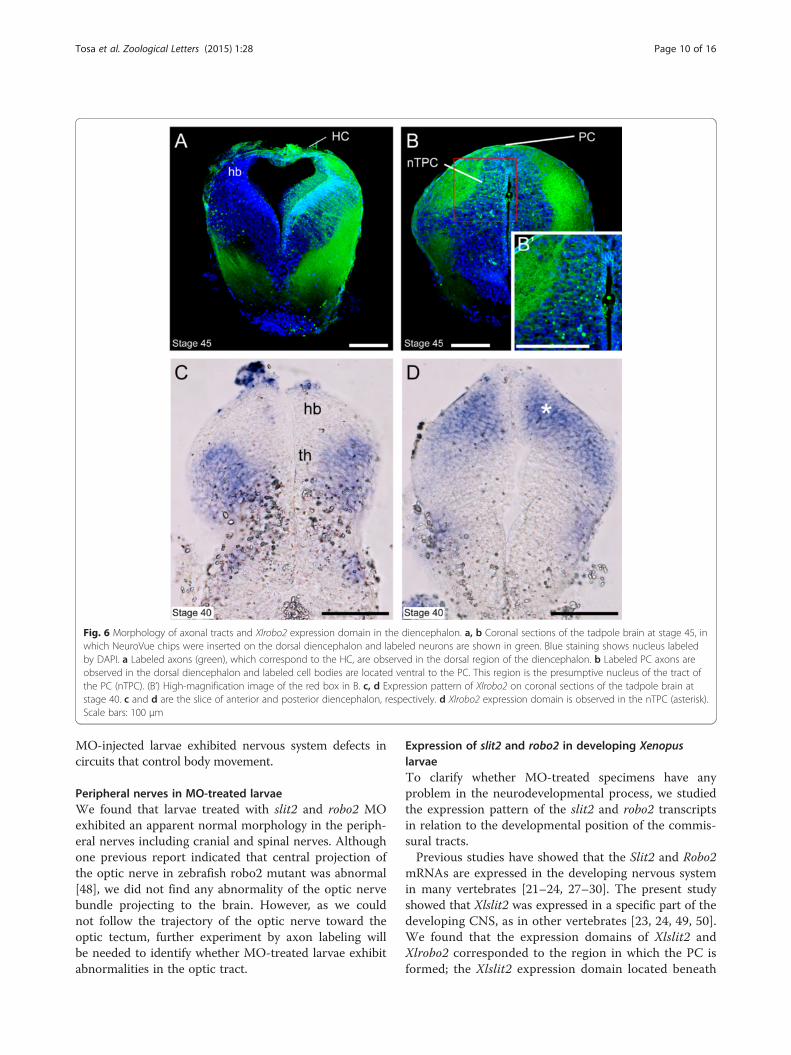
Morphology of the developing CNS in MO-injected larvaeTo identify morphological phenotypes of Xlslit2- andXlrobo2-MO-injected larvae in the developing nervoussystem, we observed the overall morphology of the larvalCNS in MO-treated specimens, and found a normalmorphology for the telencephalon, diencephalon, mesen-cephalon, and metencephalon in both control-MO- andXlslit2-MO- or Xlrobo2-MO-treated larvae at stage 44(Fig. 7a, b, d, e). Next, to clarify whether some defectscould be detected in the neuronal circuit, we observedthe morphology of commissural tracts (HC, PC, andCC) on immunostained specimens. We found that themorphologies of the HC and CC in larvae treated withslit2 or robo2 MO were not significantly different fromthose of the control-MO-treated larvae, whereas the PCin Xlslit2-MO or Xlrobo2-MO-treated larvae exhibitedan unclear commissural bundle on the dorsal dienceph-alon (Fig. 7b, e). This may be explained by the defascicu-lation of the tract, so that it could not be observed as atight bundle. Thus, we then measured the width of bun-dles, and found that the PC in Xlslit2-MO-or Xlrobo2-MO-treated larvae was significantly wider than that of
Tosa et al. Zoological Letters (2015) 1:28 Page 8 of 16
control larvae, whereas the width of the HC and CC wasunaffected (Fig. 7c, f ).
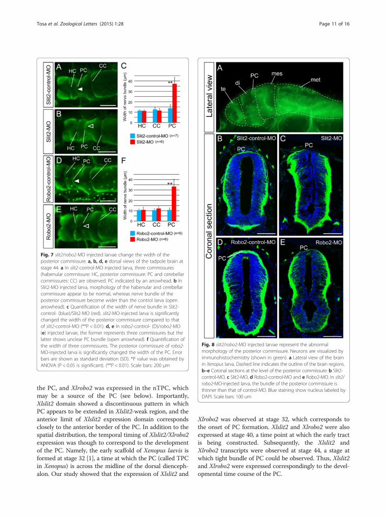
Morphology of the PC in MO-treated embryosTo clarify further the defect of the PC, we studied themorphology of the PC in coronal sections of embryos atstage 44 and found that the PC bundle in Xlslit2-MO-or Xlrobo2-MO-treated larvae became thinner on thedorsal diencephalon compared with those of control spe-cimen (Fig. 8). Similarly, in the sagittal sections, the PCin Xlslit2-MO- or Xlrobo2-MO-treated larvae becamethinner in dorso-ventral axis, and exhibited an antero-posteriorly elongated morphology (Fig. 9).Overall, the results of this series of experiments sug-
gest that Slit2 and/or Robo2 is specifically involved inthe fasciculation of the PC.
DiscussionIn the present study, we focused on the function of Slit2and Robo2, both of which play crucial roles in the pat-terning of CNS axons [47], and identified three novelfindings. First, expression patterns of Slit2 and Robo2are spatiotemporally regulated. Second, slit2-MO- androbo2-MO injected larvae exhibited abnormal swimmingbehavior. Third, slit2-MO- and robo2-MO-injected lar-vae showed an abnormal morphology of the PC, a part
of the embryonic axonal tract. These results suggest thatthe Slit2–Robo2 interaction primarily or secondarilyaffects to the neuronal circuitry that triggers the coordi-nated swimming pattern.
Involvement of Slit2 and Robo2 in swimming behaviorWe found that larvae treated with slit2 and robo2 MOexhibited an abnormal swimming trajectory. In addition,the swimming distance and speed of MO-injected larvaewere also reduced, although control-MO-injected larvaeshowed no apparent defects in swimming behavior. Wealso found that slit2-MO- and robo2-MO-treated larvaeshowed a normal body musculature. These data suggestthat Slit2 and Robo2 inhibition induced the abnormalswimming pattern observed, without disturbing basicbody apparatuses, such as the fins or body muscles.Importantly, as noted above, both Xlrobo2-MO- andcontrol-MO-treated larvae included specimens thatexhibited abnormal morphology of the head and eye.Despite the presence of these head abnormalities in bothcontrol-MO- and robo2-MO-treated larvae, we observed asignificant abnormality in Xlrobo2-MO-treated specimensregarding swimming behavior, whereas no significantproblem was detected in the control group. Given thatMOs were specifically injected into blastomeres, whichdifferentiate into the nervous system, we speculated that
Fig. 5 Morphology of axonal tracts and Xlslit2 expression domain in the neural tube. Dorsal (a-c) and lateral (d-f) view of the developing Xenopuslarva at stage 42. (A’-F’) High-magnification images of white squares in A-F. a, c Transcripts of Xlslit2. Xlslit2 mRNA is discontinuously expressed inthe dorsal diencephalon. The anterior limit of Xlslit2 attach to the superficial region of the brain (arrow in D). b, e Axons in the CNS are visualizedby immunohistochemistry (shown in green). Habenular and posterior commissures are visible. c, f Merged image of Xlslit2 (purple) and nerves(green). The anterior end of the PC corresponds to the anterior limit of Xlslit2 expression domain (arrow in F). Scale bars: 100 μm
Tosa et al. Zoological Letters (2015) 1:28 Page 9 of 16
MO-injected larvae exhibited nervous system defects incircuits that control body movement.
Peripheral nerves in MO-treated larvaeWe found that larvae treated with slit2 and robo2 MOexhibited an apparent normal morphology in the periph-eral nerves including cranial and spinal nerves. Althoughone previous report indicated that central projection ofthe optic nerve in zebrafish robo2 mutant was abnormal[48], we did not find any abnormality of the optic nervebundle projecting to the brain. However, as we couldnot follow the trajectory of the optic nerve toward theoptic tectum, further experiment by axon labeling willbe needed to identify whether MO-treated larvae exhibitabnormalities in the optic tract.
Expression of slit2 and robo2 in developing XenopuslarvaeTo clarify whether MO-treated specimens have anyproblem in the neurodevelopmental process, we studiedthe expression pattern of the slit2 and robo2 transcriptsin relation to the developmental position of the commis-sural tracts.Previous studies have showed that the Slit2 and Robo2
mRNAs are expressed in the developing nervous systemin many vertebrates [21–24, 27–30]. The present studyshowed that Xlslit2 was expressed in a specific part of thedeveloping CNS, as in other vertebrates [23, 24, 49, 50].We found that the expression domains of Xlslit2 andXlrobo2 corresponded to the region in which the PC isformed; the Xlslit2 expression domain located beneath
Fig. 6 Morphology of axonal tracts and Xlrobo2 expression domain in the diencephalon. a, b Coronal sections of the tadpole brain at stage 45, inwhich NeuroVue chips were inserted on the dorsal diencephalon and labeled neurons are shown in green. Blue staining shows nucleus labeledby DAPI. a Labeled axons (green), which correspond to the HC, are observed in the dorsal region of the diencephalon. b Labeled PC axons areobserved in the dorsal diencephalon and labeled cell bodies are located ventral to the PC. This region is the presumptive nucleus of the tract ofthe PC (nTPC). (B’) High-magnification image of the red box in B. c, d Expression pattern of Xlrobo2 on coronal sections of the tadpole brain atstage 40. c and d are the slice of anterior and posterior diencephalon, respectively. d Xlrobo2 expression domain is observed in the nTPC (asterisk).Scale bars: 100 μm
Tosa et al. Zoological Letters (2015) 1:28 Page 10 of 16
the PC, and Xlrobo2 was expressed in the nTPC, whichmay be a source of the PC (see below). Importantly,Xlslit2 domain showed a discontinuous pattern in whichPC appears to be extended in Xlslit2-weak region, and theanterior limit of Xlslit2 expression domain correspondsclosely to the anterior border of the PC. In addition to thespatial distribution, the temporal timing of Xlslit2/Xlrobo2expression was though to correspond to the developmentof the PC. Namely, the early scaffold of Xenopus laevis isformed at stage 32 [1], a time at which the PC (called TPCin Xenopus) is across the midline of the dorsal dienceph-alon. Our study showed that the expression of Xlslit2 and
Xlrobo2 was observed at stage 32, which corresponds tothe onset of PC formation. Xlslit2 and Xlrobo2 were alsoexpressed at stage 40, a time point at which the early tractis being constructed. Subsequently, the Xlslit2 andXlrobo2 transcripts were observed at stage 44, a stage atwhich tight bundle of PC could be observed. Thus, Xlslit2and Xlrobo2 were expressed correspondingly to the devel-opmental time course of the PC.
Fig. 7 slit2/robo2-MO injected larvae change the width of theposterior commissure. a, b, d, e dorsal views of the tadpole brain atstage 44. a In slit2-control-MO injected larva, three commissures(habenular commissure: HC, posterior commissure: PC and cerebellarcommissures: CC) are observed. PC indicated by an arrowhead. b InSlit2-MO injected larva, morphology of the habenular and cerebellarcommissure appear to be normal, whereas nerve bundle of theposterior commissure become wider than the control larva (openarrowhead). c Quantification of the width of nerve bundle in Slit2-control- (blue)/Slit2-MO (red). slit2-MO-injected larva is significantlychanged the width of the posterior commissure compared to thatof slit2-control-MO (**P < 0.01). d, e In robo2-control- (D)/robo2-MO(e) injected larvae, the former represents three commissures but thelatter shows unclear PC bundle (open arrowhead). f Quantification ofthe width of three commissures. The posterior commissure of robo2-MO-injected larva is significantly changed the width of the PC. Errorbars are shown as standard deviation (SD). *P value was obtained byANOVA (P < 0.05 is significant). (**P < 0.01). Scale bars: 200 μm
Fig. 8 slit2/robo2-MO injected larvae represent the abnormalmorphology of the posterior commissure. Neurons are visualized byimmunohistochemistry (shown in green). a Lateral view of the brainin Xenopus larva. Dashed line indicates the outline of the brain regions.b–e Coronal sections at the level of the posterior commissure: b Slit2-control-MO, c Slit2-MO, d Robo2-control-MO and e Robo2-MO. In slit2/robo2-MO-injected larva, the bundle of the posterior commissure isthinner than that of control-MO. Blue staining show nucleus labeled byDAPI. Scale bars: 100 um
Tosa et al. Zoological Letters (2015) 1:28 Page 11 of 16
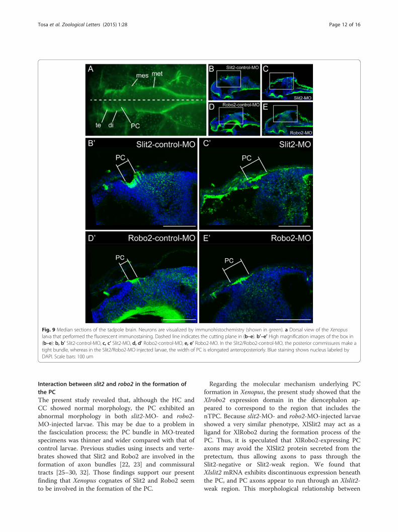
Interaction between slit2 and robo2 in the formation ofthe PCThe present study revealed that, although the HC andCC showed normal morphology, the PC exhibited anabnormal morphology in both slit2-MO- and robo2-MO-injected larvae. This may be due to a problem inthe fasciculation process; the PC bundle in MO-treatedspecimens was thinner and wider compared with that ofcontrol larvae. Previous studies using insects and verte-brates showed that Slit2 and Robo2 are involved in theformation of axon bundles [22, 23] and commissuraltracts [25–30, 32]. Those findings support our presentfinding that Xenopus cognates of Slit2 and Robo2 seemto be involved in the formation of the PC.
Regarding the molecular mechanism underlying PCformation in Xenopus, the present study showed that theXlrobo2 expression domain in the diencephalon ap-peared to correspond to the region that includes thenTPC. Because slit2-MO- and robo2-MO-injected larvaeshowed a very similar phenotype, XlSlit2 may act as aligand for XlRobo2 during the formation process of thePC. Thus, it is speculated that XlRobo2-expressing PCaxons may avoid the XlSlit2 protein secreted from thepretectum, thus allowing axons to pass through theSlit2-negative or Slit2-weak region. We found thatXlslit2 mRNA exhibits discontinuous expression beneaththe PC, and PC axons appear to run through an Xlslit2-weak region. This morphological relationship between
Fig. 9 Median sections of the tadpole brain. Neurons are visualized by immunohistochemistry (shown in green). a Dorsal view of the Xenopuslarva that performed the fluorescent immunostaining. Dashed line indicates the cutting plane in (b–e). b’–e’ High magnification images of the box in(b–e): b, b’ Slit2-control-MO, c, c’ Slit2-MO, d, d’ Robo2-control-MO, e, e’ Robo2-MO. In the Slit2/Robo2-control-MO, the posterior commissures make atight bundle, whereas in the Slit2/Robo2-MO injected larvae, the width of PC is elongated anteroposteriorly. Blue staining shows nucleus labeled byDAPI. Scale bars: 100 um
Tosa et al. Zoological Letters (2015) 1:28 Page 12 of 16
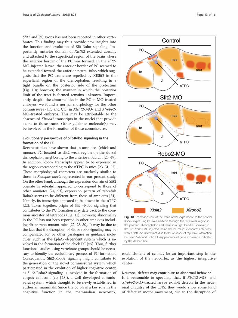
Slit2 and PC axons has not been reported in other verte-brates. This finding may thus provide new insights intothe function and evolution of Slit-Robo signaling. Im-portantly, anterior domain of Xlslit2 extended dorsallyand attached to the superficial region of the brain wherethe anterior border of the PC was formed. In the slit2-MO-injected larvae, the anterior border of PC seemed tobe extended toward the anterior neural tube, which sug-gests that the PC axons are repelled by XlSlit2 in thesuperficial region of the diencephalon, resulting in atight bundle on the posterior side of the pretectum(Fig. 10); however, the manner in which the posteriorlimit of the tract is formed remains unknown. Import-antly, despite the abnormalities in the PC in MO-treatedembryos, we found a normal morphology for the othercommissures (HC and CC) in Xlslit2-MO- and Xlrobo2-MO-treated embryos. This may be attributable to theabsence of Xlrobo2 transcripts in the nuclei that provideaxons to those tracts. Other guidance molecule(s) maybe involved in the formation of those commissures.
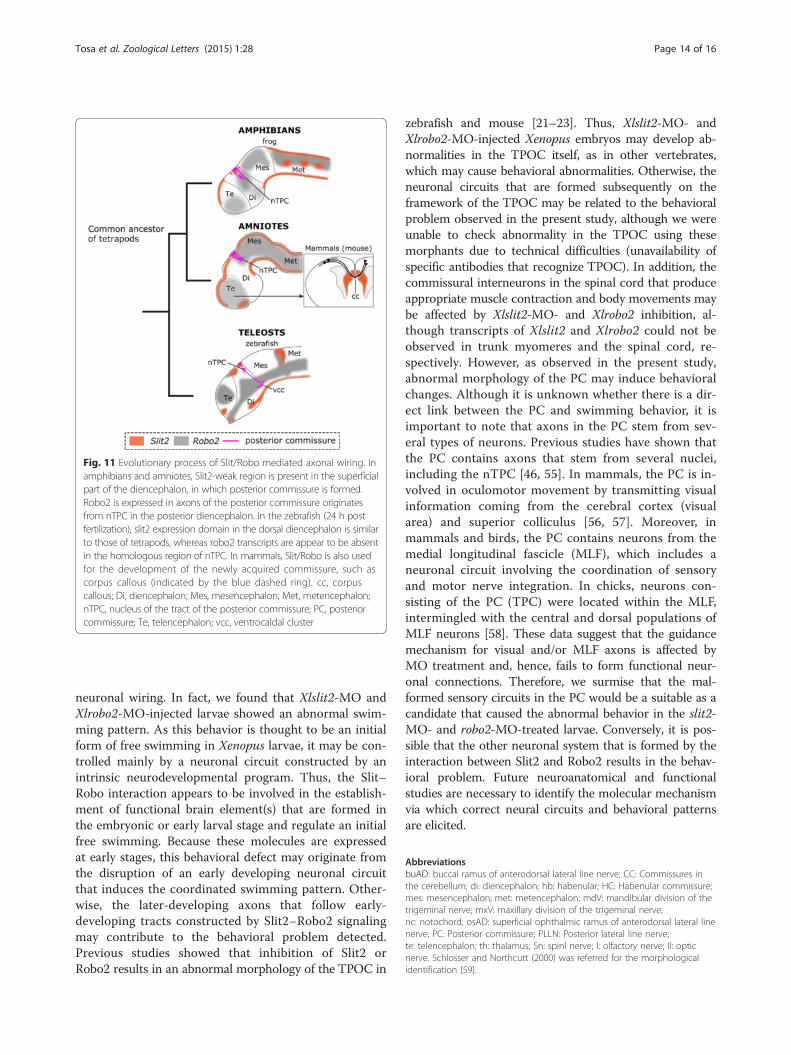
Evolutionary perspective of Slit-Robo signaling in theformation of the PCRecent studies have shown that in amniotes (chick andmouse), PC located to slit2 weak region on the dorsaldiencephalon neighboring to the anterior midbrain [23, 49].In addition, Robo2 transcripts appear to be expressed inthe region corresponding to the nTPC in mice [23, 51, 52].These morphological characters are markedly similar tothose in Xenopus laevis represented in our present study.On the other hand, although the expression domain of Slit2cognate in zebrafish appeared to correspond to those ofother amniotes [24, 53], expression pattern of zebrafishRobo2 seems to be different from those of amniotes [54].Namely, its transcripts appeared to be absent in the nTPC[22]. Taken together, origin of Slit –Robo signaling thatcontributes to the PC formation may date back to the com-mon ancestor of tetrapods (Fig. 11). However, abnormalityin the PC has not been reported in other amniotes includ-ing slit or robo mutant mice [27, 28, 30]. It may be due tothe fact that the disruption of slit or robo signaling may becompensated for by other paralogues or guidance mole-cules, such as the EphA7-dependent system which is in-volved in the formation of the chick PC [55]. Thus, furtherfunctional studies using vertebrate groups should be neces-sary to identify the evolutionary process of PC formation.Consequently, Slit2-Robo2 signaling might contribute tothe generation of the novel commissural system whichparticipated in the evolution of higher cognitive center,as Slit2-Robo2 signaling is involved in the formation ofcorpus callosum (cc; [28]), a well developed commis-sural system, which thought to be newly established ineutherian mammals. Since the cc plays a key role in thecognitive function in the mammalian neocortex,
establishment of cc may be an important step in theevolution of the neocortex as the highest integrativecenter.
Neuronal defects may contribute to abnormal behaviorIt is reasonable to speculate that, if Xlslit2-MO- andXlrobo2-MO-treated larvae exhibit defects in the neur-onal circuitry of the CNS, they would show some kindof defect in motor movement, due to the disruption of
Fig. 10 Schematic view of the result of the experiment. In the control,Robo2-expressing PC axons extend through the Slit2-weak region inthe posterior diencephalon and result in a tight bundle. However, inthe slit2-/robo2-MO-injected larvae, the PC makes elongates anteriorlywith a defasciculated tract, due to the absence of repulsive interactionbetween Slit2 and Robo2. Disappearance of gene expression indicatedby the dashed line
Tosa et al. Zoological Letters (2015) 1:28 Page 13 of 16
neuronal wiring. In fact, we found that Xlslit2-MO andXlrobo2-MO-injected larvae showed an abnormal swim-ming pattern. As this behavior is thought to be an initialform of free swimming in Xenopus larvae, it may be con-trolled mainly by a neuronal circuit constructed by anintrinsic neurodevelopmental program. Thus, the Slit–Robo interaction appears to be involved in the establish-ment of functional brain element(s) that are formed inthe embryonic or early larval stage and regulate an initialfree swimming. Because these molecules are expressedat early stages, this behavioral defect may originate fromthe disruption of an early developing neuronal circuitthat induces the coordinated swimming pattern. Other-wise, the later-developing axons that follow early-developing tracts constructed by Slit2–Robo2 signalingmay contribute to the behavioral problem detected.Previous studies showed that inhibition of Slit2 orRobo2 results in an abnormal morphology of the TPOC in
zebrafish and mouse [21–23]. Thus, Xlslit2-MO- andXlrobo2-MO-injected Xenopus embryos may develop ab-normalities in the TPOC itself, as in other vertebrates,which may cause behavioral abnormalities. Otherwise, theneuronal circuits that are formed subsequently on theframework of the TPOC may be related to the behavioralproblem observed in the present study, although we wereunable to check abnormality in the TPOC using thesemorphants due to technical difficulties (unavailability ofspecific antibodies that recognize TPOC). In addition, thecommissural interneurons in the spinal cord that produceappropriate muscle contraction and body movements maybe affected by Xlslit2-MO- and Xlrobo2 inhibition, al-though transcripts of Xlslit2 and Xlrobo2 could not beobserved in trunk myomeres and the spinal cord, re-spectively. However, as observed in the present study,abnormal morphology of the PC may induce behavioralchanges. Although it is unknown whether there is a dir-ect link between the PC and swimming behavior, it isimportant to note that axons in the PC stem from sev-eral types of neurons. Previous studies have shown thatthe PC contains axons that stem from several nuclei,including the nTPC [46, 55]. In mammals, the PC is in-volved in oculomotor movement by transmitting visualinformation coming from the cerebral cortex (visualarea) and superior colliculus [56, 57]. Moreover, inmammals and birds, the PC contains neurons from themedial longitudinal fascicle (MLF), which includes aneuronal circuit involving the coordination of sensoryand motor nerve integration. In chicks, neurons con-sisting of the PC (TPC) were located within the MLF,intermingled with the central and dorsal populations ofMLF neurons [58]. These data suggest that the guidancemechanism for visual and/or MLF axons is affected byMO treatment and, hence, fails to form functional neur-onal connections. Therefore, we surmise that the mal-formed sensory circuits in the PC would be a suitable as acandidate that caused the abnormal behavior in the slit2-MO- and robo2-MO-treated larvae. Conversely, it is pos-sible that the other neuronal system that is formed by theinteraction between Slit2 and Robo2 results in the behav-ioral problem. Future neuroanatomical and functionalstudies are necessary to identify the molecular mechanismvia which correct neural circuits and behavioral patternsare elicited.
AbbreviationsbuAD: buccal ramus of anterodorsal lateral line nerve; CC: Commissures inthe cerebellum; di: diencephalon; hb: habenular; HC: Habenular commissure;mes: mesencephalon; met: metencephalon; mdV: mandibular division of thetrigeminal nerve; mxV: maxillary division of the trigeminal nerve;nc: notochord; osAD: superficial ophthalmic ramus of anterodorsal lateral linenerve; PC: Posterior commissure; PLLN: Posterior lateral line nerve;te: telencephalon; th: thalamus; Sn: spinl nerve; I: olfactory nerve; II: opticnerve. Schlosser and Northcutt (2000) was referred for the morphologicalidentification [59].
Fig. 11 Evolutionary process of Slit/Robo mediated axonal wiring. Inamphibians and amniotes, Slit2-weak region is present in the superficialpart of the diencephalon, in which posterior commissure is formed.Robo2 is expressed in axons of the posterior commissure originatesfrom nTPC in the posterior diencephalon. In the zebrafish (24 h postfertilization), slit2 expression domain in the dorsal diencephalon is similarto those of tetrapods, whereas robo2 transcripts are appear to be absentin the homologous region of nTPC. In mammals, Slit/Robo is also usedfor the development of the newly acquired commissure, such ascorpus callous (indicated by the blue dashed ring). cc, corpuscallous; Di, diencephalon; Mes, mesencephalon; Met, metencephalon;nTPC, nucleus of the tract of the posterior commissure; PC, posteriorcommissure; Te, telencephalon; vcc, ventrocaldal cluster
Tosa et al. Zoological Letters (2015) 1:28 Page 14 of 16
Competing interestWe declare that no actual or potential competing interests in relation to thisarticle exist.
Authors’ contributionsYT and KT carried out the gene expression analyses, immunohistochemicalobservations and the neuronal labeling, and participated in the experimentsof the functional inhibition using MO and drafted the manuscript. MKparticipated in the gene isolation and expression analyses. TI carried on theimmunohistochemical and behavioral analyses, and participated in thestatistical assay. M. Fukagawa and YN carried on the gene expression andthe immunohistochemical analyses. KTS participated in the experimentsof the functional inhibition using MO. M. Fukui carried on theimmunohistochemical analysis. YM conceived of the study, andparticipated in its design and coordination and helped to draft the manuscript.All authors read and approved the final manuscript.
AcknowledgementsWe thank Dr. Mikio Inoue, Tetsuya Kominami, Kei Nakayama and HiromiTakata for technical support and valuable discussions. We thank all past andpresent members of the YM laboratory for support and constructive discussions.Work in YM’s laboratory was supported by RIKEN, Kobe, Japan and the JapanSociety for the Promotion of Science (JSPS; grant number 24650178 and26430018 to YM, and grant number 21770238 and 23657149 to MK).
Author details1Graduate School of Science and Engineering, Ehime University, 2-5Bunkyo-cho, Matsuyama 790-8577, Japan. 2Graduate School of Medicine andPharmaceutical Sciences, University of Toyama, 2630 Sugitani, Toyama930-0194, Japan. 3Graduate School of Science, Hiroshima University, 1-3-1Kagamiyama, Higashi-Hiroshima, Hiroshima 739-8526, Japan.
Received: 23 January 2015 Accepted: 31 August 2015
References1. Anderson RB, Key B. Novel guidance cues during neuronal pathfinding in
the early scaffold of axon tracts in the rostral brain. Development.1999;126:1859–68.
2. Barreiro-Iglesias A, Villar-Cheda B, Abalo XM, Anadon R, Rodicio MC. Theearly scaffold of axon tracts in the brain of a primitive vertebrate, the sealamprey. Brain Res Bull. 2008;75:42–52.
3. Chitnis AB, Kuwada JY. Axonogenesis in the brain of zebrafish embryos.J Neurosci. 1990;10:1892–905.
4. Doldan MJ, Prego B, Holmqvist B, Helvik JV, de Miguel E. Emergence ofaxonal tracts in the developing brain of the turbot (Psetta maxima).Brain Behav Evol. 2000;56:300–9.
5. Easter Jr SS, Ross LS, Frankfurter A. Initial tract formation in the mouse brain.J Neurosci. 1993;13:285–99.
6. Figdor MC, Stern CD. Segmental organization of embryonic diencephalon.Nature. 1993;363:630–4.
7. Ishikawa Y, Kage T, Yamamoto N, Yoshimoto M, Yasuda T, Matsumoto A,et al. Axonogenesis in the medaka embryonic brain. J Comp Neurol.2004;476:240–53.
8. Ross LS, Parrett T, Easter Jr SS. Axonogenesis and morphogenesis in theembryonic zebrafish brain. J Neurosci. 1992;12:467–82.
9. Pike SH, Melancon EF, Eisen JS. Pathfinding by zebrafish motoneurons intheabsence of normal pioneer axons. Developmen. 1992;114:825–31.
10. Nieuwenhuys R, TenDonkelaar HJ, Nicholson C, editors. The central nervoussystem of vertebrates. Heidelberg: Springer-Verlag; 1998.
11. Bergquist H, Källén B. On the development of neuromeres to migrationareas in the vertebrate cerebral tube. Act Anat. 1953;18:65–73.
12. Lumsden A, Keynes R. Segmental patterns of neuronal development in thechick hindbrain. Nature. 1989;337:424–8.
13. Orr H. Contribution to the embryology of the lizard. J Morphol. 1887;1:311–72.14. Puelles L, Rubenstein JL. Forebrain gene expression domains and the
evolving prosomeric model. Trends Neurosci. 2003;26:469–76.15. Shimamura K, Hartigan DJ, Martinez S, Puelles L, Rubenstein JL. Longitudinal
organization of the anterior neural plate and neural tube. Development.1995;21:3923–33.
16. Vaage S. The segmentation of the primitive neural tube in chick embryos(Gallus domesticus). Ergeb Anat Entw Gesch. 1969;4:1–88.
17. von Baer K. Über die Entwickelungsgeschichte der Thiere. Königsberg; 1828.18. Cariboni A, Andrews WD, Memi F, Ypsilanti AR, Zelina P, Chedotal A, et al.
Slit2 and Robo3 modulate the migration of GnRH-secreting neurons.Development. 2012;139:3326–31.
19. Kennedy TE, Serafini T, de la Torre JR, Tessier-Lavigne M. Netrins arediffusible chemotropic factors for commissural axons in the embryonicspinal cord. Cell. 1994;78:425–35.
20. Rubenstein JL, Shimamura K, Martinez S, Puelles L. Regionalization of theprosencephalic neural plate. Annu Rev Neurosci. 1998;21:445–77.
21. Barresi MJ, Hutson LD, Chien CB, Karlstrom RO. Hedgehog regulated Slitexpression determines commissure and glial cell position in the zebrafishforebrain. Development. 2005;132:3643–56.
22. Devine CA, Key B. Robo-Slit interactions regulate longitudinal axonpathfinding in the embryonic vertebrate brain. Dev Biol. 2008;313:371–83.
23. Ricano-Cornejo I, Altick AL, Garcia-Pena CM, Nural HF, Echevarria D,Miquelajauregui A, et al. Slit-Robo signals regulate pioneer axon pathfindingof the tract of the postoptic commissure in the mammalian forebrain.J Neurosci Res. 2011;89:1531–41.
24. Zhang C, Gao J, Zhang H, Sun L, Peng G. Robo2–slit and Dcc–netrin1coordinate neuron axonal pathfinding within the embryonic axon tracts.J Neurosci. 2012;32:12589–602.
25. Kidd T, Bland KS, Goodman CS. Slit is the midline repellent for the roboreceptor in Drosophila. Cell. 1999;96:785–94.
26. Rothberg JM, Jacobs JR, Goodman CS, Artavanis-Tsakonas S. Slit: an extracellularprotein necessary for development of midline glia and commissural axonpathways contains both EGF and LRR domains. Genes Dev. 1990;4:2169–87.
27. Plump AS, Erskine L, Sabatier C, Brose K, Epstein CJ, Goodman CS, et al. Slit1and Slit2 cooperate to prevent premature midline crossing of retinal axonsin the mouse visual system. Neuron. 2002;33:219–32.
28. Shu T, Sundaresan V, McCarthy MM, Richards LJ. Slit2 guides bothprecrossing and postcrossing callosal axons at the midline in vivo.J Neurosci. 2003;23:8176–84.
29. Hocking JC, Hehr CL, Bertolesi GE, Wu JY, McFarlane S. Distinct roles forRobo2 in the regulation of axon and dendrite growth by retinal ganglioncells. Mech Dev. 2010;127:36–48.
30. Lopez-Bendito G, Flames N, Ma L, Fouquet C, Di Meglio T, Chedotal A, et al.Robo1 and Robo2 cooperate to control the guidance of major axonal tractsin the mammalian forebrain. J Neurosci. 2007;27:3395–407.
31. Burgess HA, Johnson SL, Granato M. Unidirectional startle responses anddisrupted left-right co-ordination of motor behaviors in robo3 mutantzebrafish. Genes Brain Behav. 2009;8:500–11.
32. Shu T, Richards LJ. Cortical axon guidance by the glial wedge during thedevelopment of the corpus callosum. J Neurosci. 2001;21:2749–58.
33. Ten D. Anurans. In: Nieuwenhuys R, TenDonkelaar HJ, Nicholson C, editors.The central nervous system of vertebrates. 2nd ed. Heidelberg: Springer-Verlag;1998. p. 1151–314.
34. van Mier P, ten Donkelaar HJ. Structural and functional properties ofreticulospinal neurons in the early-swimming stage Xenopus embryo.J Neurosci. 1989;9:25–37.
35. van Mier P, Armstrong J, Roberts A. Development of early swimming inXenopus laevis embryos: myotomal musculature, its innervation andactivation. Neuroscience. 1989;32:113–26.
36. Nieuwkoop PD, Faber J. Normal Table of Xenopus laevis (Daudin).Amsterdam: North-Holland; 1967.
37. Moody SA, Kline MJ. Segregation of fate during cleavage of frog (Xenopuslaevis) blastomeres. Anat Embryol (Berl). 1990;182:347–62.
38. Kawaguchi M, Sugahara Y, Watanabe T, Irie K, Ishida M, Kurokawa D, et al.Nervous system disruption and concomitant behavioral abnormality in earlyhatched pufferfish larvae exposed to heavy oil. Environ Sci Pollut Res Int.2011;19:2488–97.
39. Takio Y, Kuraku S, Murakami Y, Pasqualetti M, Rijli FM, Narita Y, et al. Hoxgene expression patterns in Lethenteron japonicum embryos–insights intothe evolution of the vertebrate Hox code. Dev Biol. 2007;308:606–20.
40. Kuratani SC, Eichele G. Rhombomere transplantation repatterns thesegmental organization of cranial nerves and reveals cell-autonomousexpression of a homeodomain protein. Development. 1993;117:105–17.
41. Roberts A, Conte D, Hull M, Merrison-Hort R, al Azad AK, Buhl E, et al. Cansimple rules control development of a pioneer vertebrate neuronal networkgenerating behavior? J Neurosci. 2014;34:608–21.
Tosa et al. Zoological Letters (2015) 1:28 Page 15 of 16
42. Geisen MJ, Di Meglio T, Pasqualetti M, Ducret S, Brunet JF, Chedotal A, et al.Hox paralog group 2 genes control the migration of mouse pontineneurons through slit-robo signaling. PLoS Biol. 2008;6:e142.
43. Nieuwenhuys R. Comparative anatomy of the cerebellum. Prog Brain Res.1967;25:1–93.
44. Easter Jr SS, Taylor JS. The development of the Xenopus retinofugal pathway:optic fibers join a pre-existing tract. Development. 1989;107:553–73.
45. Key B, Anderson RB. Neuronal pathfinding during development of therostral brain in Xenopus. Clin Exp Pharmacol Physiol. 1999;26:752–4.
46. Lázár G, Pál E. Neuronal connections through the posterior commissure inthe frog Rana esculenta. J Hirnforsch. 1999;39:369–74.
47. Dickson BJ. Molecular mechanisms of axon guidance. Science.2002;298:1959–64.
48. Hutson LD, Chien CB. Pathfinding and error correction by retinal axons: therole of astray/robo2. Neuron. 2002;33:205–17.
49. Holmes G, Niswander L. Expression of slit-2 and slit-3 during chick development.Dev Dyn. 2001;222:301–7.
50. De Bellard ME, Rao Y, Bronner-Fraser M. Dual function of Slit2 in repulsionand enhanced migration of trunk, but not vagal, neural crest cells. J CellBiol. 2003;162:269-79.
51. Farmer WT, Altick AL, Nural HF, Dugan JP, Kidd T, Charron F, et al. Pioneerlongitudinal axons navigate using floor plate and Slit/Robo signals.Development. 2008;135:3643–53.
52. Mastick GS, Easter Jr SS. Initial organization of neurons and tracts in theembryonic mouse fore- and midbrain. Dev Biol. 1996;173:79–94.
53. Miyasaka N, Sato Y, Yeo SY, Hutson LD, Chien CB, Okamoto H, et al. Robo2is required for establishment of a precise glomerular map in the zebrafisholfactory system. Development. 2005;132:1283–93.
54. Lee JS, Ray R, Chien CB. Cloning and expression of three zebrafishroundabout homologs suggest roles in axon guidance and cell migration.Dev Dyn. 2001;221:216–30.
55. Stanic K, Vera A, González M, Recabal A, Astuya A, Torrejón M, et al.Complementary expression of EphA7 and SCO-spondin duringposteriorcommissure development. Front Neuroanat. 2014;8:49.
56. Bhidayasiri R, Plant GT, Leigh RJ. A hypothetical scheme for the brainstemcontrol of vertical gaze. Neurology. 2000;54:1985–93.
57. Leichnetz GR, Gonzalo-Ruiz A, DeSalles AA, Hayes RL. The origin ofbrainstem afferents of the paramedian pontine reticular formation in thecat. Brain Res. 1987;422:389–97.
58. Ware M, Schubert FR. Development of the early axon scaffold in the rostralbrain of the chick embryo. J Anat. 2011;219:203–16.
59. Schlosser G, Northcutt RG. Development of neurogenic placodes inXenopus laevis. J Comp Neurol. 2000;418:121–46.
Submit your next manuscript to BioMed Centraland take full advantage of:
• Convenient online submission
• Thorough peer review
• No space constraints or color figure charges
• Immediate publication on acceptance
• Inclusion in PubMed, CAS, Scopus and Google Scholar
• Research which is freely available for redistribution
Submit your manuscript at www.biomedcentral.com/submit
Tosa et al. Zoological Letters (2015) 1:28 Page 16 of 16