Embed Size (px)
Citation preview

Journal of Cellular Biochemistry 98:1615–1628 (2006)
Involvement of Phosphatidylcholine-SelectivePhospholipase C in Activation of Mitogen-ActivatedProtein Kinase Pathways in Imidazoline ReceptorAntisera-Selected Protein
Fei Li,1 Ning Wu,1 Rui-Bin Su,1 Jian-Quan Zheng,1 Bo Xu,1 Xin-Qiang Lu,1 Bin Cong,2 and Jin Li1*1Beijing Institute of Pharmacology and Toxicology, Beijing 100850, China2Department of Forensic Medicine, Hebei Medical University, Shijiazhuang 050017, China
Abstract Imidazoline receptor antisera-selected protein (IRAS) is considered as a candidate for the I1-imidazolinereceptor (I1R), but the signaling pathway mediated by IRAS remains unknown. In our study, the signal transductionpathways of IRAS were investigated in CHO cells stably expressing IRAS (CHO-IRAS), and compared to the native I1Rsignaling pathways. Rilmenidine or moxonidine (10 nM–100 mM), I1R agonists, failed to stimulate [35S]-GTPgS binding inCHO-IRAS cell membrane preparations, suggesting that G protein may not be involved in IRAS signaling pathway.However, incubation of CHO-IRAS with rilmenidine or moxonidine for 5 min could induce an upregulation ofphosphatidylcholine-selective phospholipase C (PC-PLC) activity, and an increase in the accumulation of diacylglycerol(DAG), the hydrolysate of PC-PLC, in a concentration-dependent manner. The elevated activation of PC-PLC byrilmenidine or moxonidine (100 nM) could be blocked by efaroxan, a selective I1R antagonist. Cells treated withrilmenidine or moxonidine showed an increased level of extracellular signal-regulated kinase (ERK) phosphorylation in aconcentration-dependent manner, which could be reversed by efaroxan or D609, a selective PC-PLC inhibitor. Theseresults suggest that the signaling pathway of IRAS in response to I1R agonists coupled with the activation of PC-PLC and itsdownstream signal transduction molecule, ERK. These findings are similar to those in the signaling pathways of native I1R,providing some new evidence for the relationship between I1R and IRAS. J. Cell. Biochem. 98: 1615–1628, 2006.� 2006 Wiley-Liss, Inc.
Key words: imidazoline receptor antisera-selected protein; I1-imidazoline receptor; Nischarin; G protein; phospha-tidylcholine-selective phospholipase C; diacylglycerol; extracellular signal-regulated kinase
The concept of imidazoline receptors was firstset up by Bousquet et al. [1984] when theystudied antihypertension effect of clonidine. Itis now accepted that there are at least twosubtypes of imidazoline receptors, I1-imidazo-line receptor (I1R) and I2-imidazoline receptor(I2R). I1R is characterized by a high affinity to agroup of agents including clonidine, rilmeni-dine, and moxonidine, which act on the brain
stem to reduce blood pressure [Ernsbergeret al., 1995a; Eglen et al., 1998]. I2R shows highaffinity to other imidazolines or guanidine,which presents a novel recognition site onmonoamine oxidase [Ernsberger et al., 1995a;Eglen et al., 1998]. In 2000, a gene encoding anI1R candidate protein, named imidazolinereceptor antisera-selected protein (IRAS), wascloned from human hippocampus. It was iden-tified that IRAS mRNA encodes a proteincontaining 1,504-amino acid residues, yield a167 kDa protein, which could be broken downinto 85 kDa and smaller protein molecules[Piletz et al., 2000].
Evidence supported that IRAS is a candidatefor I1R. First, IRAS mRNA was shown to beappropriately localized in brain neurons asexpected for I1R-binding sites [Ivanov et al.,1998], and a positive correlation (r¼ 0.7) wasestablished between the mRNA for IRAS and
� 2006 Wiley-Liss, Inc.
Fei Li and Ning Wu made equal contribution to the work.
Grant sponsor: National Basic Research Program of China;Grant number: 2003CB515400.
*Correspondence to: Dr. Jin Li, Beijing Institute ofPharmacology and Toxicology, Beijing 100850, China.E-mail: [email protected], [email protected]
Received 28 October 2005; Accepted 19 December 2005
DOI 10.1002/jcb.20806

membranous I1-binding sites (Bmax) over arange of native rat tissues [Piletz et al., 1999].Second, transfection of IRAS cDNA into theChinese hamster ovary (CHO) cells resulted inhigh affinity I1-like binding sites without theappearance of a2-AR or the other major subtypeof imidazoline binding sites [Piletz et al., 2000,2003]. Third, it has been revealed that IRAScould function on promoting cell survival [Don-tenwill et al., 2003a], anti-apoptosis [Donten-will et al., 2003b], and proliferation [Sano et al.,2002], which is similar to the intracellularfunctions of I1R [Dupuy et al., 2004]. Recentstudies have also found that the transfectionactivation of human IRAS is able to inhibit acellular model of opioid dependence (cAMPovershoot) in morphine-dependent CHO cellsstably co-expressing both IRAS and m opioidreceptor [Wu et al., 2005], which parallels issimilar to thefindings in vivo that the activationof I1R inhibits tolerance and dependence onmorphine [Georges and Aston-Jones, 2003; Suet al., 2003]. These findings collectively suggestthat IRAS is a strong candidate for I1R.
However, the above similarities betweenIRAS and I1R are not enough to prove thatIRAS expressed from the cloned gene is a nativeI1R. It is well known that besides such char-acters as distribution and functions, the fore-most is the identity in the signal transductionpathways between a receptor and its clonedprotein. Little evidence has been shown to provethat IRAS is coupled with the same signalingpathways as I1R. Although the signal pathwaysof I1R have been studied systemically, those ofIRAS remain unclear.
Previous studies have shown that the activa-tion of I1R increases the accumulation ofdiacylglycerol (DAG) [Liedtke and Ernsberger,1995; Separovic et al., 1996; Separovic et al.,1997], and the release of arachidonic acid (AA)[Ernsberger, 1998], and eicosanoids [Ernsber-ger et al., 1995; Separovic et al., 1997]. DAG orAA may activate protein kinase C (PKC)[Edward et al., 2001] which phosphorylatesand activates mitogen-activated proteinkinases (MAPK) [Edward et al., 2001; Zhanget al., 2001]. The phosphorylation of MAPK canbe blocked by phosphatidylcholine-selectivephospholipase C (PC-PLC) inhibitor D609,suggesting the involvement of PC-PLC in I1Rsignaling pathway. The phospholipid metabo-lism signaling pathway of I1R has been identi-fied in rat pheochromocytoma (PC12) cells and
some other tissues expressing native I1R. How-ever, it is not clear if I1R acts via the mostcommon G-protein linked systems. ClassicalGTP shift analyses in bovine brainstem, humanplatelets, and rat PC12 cells showed thatthe binding of agnists to I1R was sensitive tothe hydrolysis-resistant guanine nucleotideGpp(NH)p or GTPgS, suggesting I1R is a G-protein coupled receptor (GPCR) [Molderinget al., 1993; Ernsberger andShen, 1997; Takadaet al., 1997]. In contrast, similar GTP shiftstudies in other labs failed to prove that I1R is aGPCR [Piletz and Sletten, 1993; Bricca et al.,1994]. The paradox is probably due to theinfluence of other receptors that cannot beexcluded in the experimental model, especiallya2-AR (a2-adrenoceptors).
Although IRAS gene has been cloned andexpressed in some cell lines, the signalingpathways through IRAS are not well defined.The mouse homologue of IRAS, previouslyidentified as Nischarin, has been shown in theabsence of imidazolines to interact with the a5subunit of integrin and inhibit cell migration[Alahari et al., 2000], but the relevance of thishas been disputed [Lim and Hong, 2004].Therefore, we chose to use human IRAS trans-fected into CHO, a host cell line that completelylacks native I1R [Piletz et al., 2000]. Previousstudieshave shown that I1Ragonists canbind tothe I1-like sites encoded by transfected humanIRAS in CHO cells. Considering the similaritiesbetween IRAS and I1R, we hypothesized thatI1R agonistsmay trigger signaling transductionthrough IRAS in a way similar to I1R. Herein,we have investigated IRAS-mediated signalingpathways in CHO cells stably expressing IRAS(CHO-IRAS) and compared the IRAS signalingpathways with the native I1R pathways.
MATERIALS AND METHODS
Materials
IRAS-pcDNA3.1 (þ) plasmidwasakindly giftof Dr. J.E. Piletz (Jackson State University,Jackson, MS). CHO and CHO cell stablyexpressing m opioid receptor (CHO-m) cells wereprovided by Dr. L.Y. Liu-Chen (Temple Uni-versity School of Medicine, Philadelphia, PA).[3H]clonidine (55.5 Ci/mmol) and [35S]-GTPgS(1250 Ci/mmol) were purchased from NEN LifeSciences (Boston, MA). Geneticin, lipofecta-mine, and RPMI 1640 medium were purchased
1616 Li et al.

from Invitrogen Corporation (GibcoTM, GrandIsland, NY). Fetal bovine serum was purchasedfrom HyClone-Pierce (HyClone 1, SouthLogan, UT). Guanosine 50-O-(3-thiotriphos-phosate) (GTPgS), guanosine 50-diphosphate(GDP), moxonidine, rilmenidine, clonidine,efaroxan, D609, 1,2-dioleoyl-sn-glycerol, leu-peptin, pepstatin, aprotinin, 1.10-phenanthro-line monohydrate, were purchased from SigmaChemical Co. (St. Louis, MO). Anti-extracellu-lar signal-regulated kinase (ERK) antibody,anti-phospho-ERK antibody, anti-rabbit andanti-mouse horseradish peroxidase antibodies,and enhanced chemiluminesence detection(ECL). The western blotting detection reagentwas purchased from Santa Cruz Biotechnology(Santa Cruz, CA). [D-Ala2, N-Me-Phe4, Gly-ol]-enkephalin (DAMGO), GF/C filters werepurchased from Whatman (Whatman, UK).High-performance thin-layer chromatography(HPTLC) plates (10� 10 cm, glass plate of silicagel 60)were purchased fromMerck (Darmstadt,Germany).
Generation of Cell Lines Expressing HumanIRAS and Cell Culture
IRAS stably expressing CHO cell lines weregenerated by transfectingHuman IRAS expres-sion vector (hIRAS-pcDNA3.1(þ)) into CHOcells with Lipofectamine reagent following themanufacturer’s instructions. Transfected cellswere selected using 1 mg/ml geneticin for 4–6weeks. Clones were obtained and one of theclones was used as the representative in thisstudy.CHO cells were cultured in RPMI 1640
supplemented with 10% heat-inactivated fetalbovine serum, 100 U/ml penicillin, and 100 mg/ml streptomycin at 378C with humidified atmo-sphere consisting of 95% air and 5% CO2.Medium for CHO-IRAS cells was the same asthat for CHO cells except for the 200 mg/mlgeneticin contained.
Membrane Protein Preparation
Membrane proteins were isolated followingthe method of Zhu et al. [1997]. Isolatedmembrane protein was diluted with ice-coldassay buffer (5.0 mM HEPES, 0.5 mMMgCl2, 0.5 mM EGTA, 0.5 mM EDTA, pH7.5) and the protein concentration was deter-mined using Brad-ford method. All membrane
protein samples used in the experiments werefreshly prepared.
Radioligand-Binding Assay
HME assay buffer (5.0 mM HEPES, 0.5 mMMgCl2, 0.5 mM EGTA, 0.5 mM EDTA, freshlyadded with 100 mM ascorbic acid and 100 mMPMSF, pH 7.5) was used to optimize the bindingcondition for I1R [Ernsberger et al., 1995b]. Forsaturation analyses, [3H]clonidine rangingfrom 1.5 to 48 nMwas added to 20 mgmembraneproteins in HME assay buffer with a finalvolume of 500 ml and incubated in a 218Cwaterbath for 1 h. Nonspecific binding wasdefined with idazoxan (100 mM). For the compe-tition analyses, 20 mg membrane protein sam-ples were incubated with 30 nM [3H]clonidine(2�Kd value) and the competitor ligands atvarious concentrations in a 218C waterbath for1h.Reactionswere terminatedbyadding 5ml ofice-cold 50 mM Tris-HCl buffer (pH 7.5), andrapid vacuum filtration through GF/C glassfiber filters using a cell harvester, followed bywashing with cold Tris-HCl buffer (pH 7.5) forthree times. GF/C glass fiber filters werepresoaked in 0.2% BSA at room temperaturefor 30 min to lower the nonspecific binding.Radioactivity infilterswasdeterminedby liquidscintillation counting.
[35S]-GTPgS-Binding Assay
[35S]-GTPgS-binding assaywas carried out asdescribed previously [Zhu et al., 1997]. Briefly,membrane proteins of CHO-IRAS and CHO-mcells were prepared as described above. Bindingof [35S]-GTPgS to the CHO-m cell membraneprotein was in the assay buffer containing15 mM GDP, 50 mM HEPES, 5 mM MgCl2,1mMEDTA,100mMNaCl, pH7.4 (plus 0.1mMascorbic acid, 0.1mMDTT, 0.1mMPMSF, 1 mg/ml leupeptin, pepstatin and aprotinin justbefore the experiments), incubated at 258C for1 h. Stimulated binding of [35S]-GTPgS to CHO-m cell membrane preparation was determinedusing DAMGO (10 mM). Different concentra-tions of GDP (0.1–50 mM), MgCl2 (0–20 mM),and NaCl (0–100 mM), different temperatures(21, 25, 308C ), and time needed of reaction(30 min, 1, 1.5, 2 h) were tested to optimize the[35S]-GTPgS-binding assay condition of IRAS.Stimulated binding of [35S]-GTPgS to IRAS wasdetermined using moxonidine or rilmenidine(10 nM–100 mM). Nonspecific binding wasdefined by GTPgS (40 mM). Each reaction was
Activation of Mitogen Activation Protein Kinase Pathways in IRAS 1617

set up in the following order: [35S]-GTPgS(0.2 nM); agonists at different concentrationsor GTPgS and membrane proteins (20 mgprotein/tube) in a total volume of 500 ml assaybuffer per tube. The increased percentage overbasal binding level of [35S]-GTPgS was calcu-lated as follows: 100� [(mean total samplecpm�mean basal sample cpm)/mean basalsample cpm]. Basal binding was defined as[35S]-GTPgS binding in the absence of agonists.Because m opioid receptor is a classical GPCR[Burford et al., 2000], we used CHO-m cells asexperimental control in this assay in order tomake sure our experiment is correct.
PC-PLC Experiment
The PC-PLC experiment was preparedaccording to the method of Greney [Greneyet al., 2000] with some modifications. CHO-IRAS cells were seeded at 1� 106 cells/well in a6-well plate in RPMI1640 containing 10% FBS.After 24 h of culture, cells were rinsed twicewith serum-free RPMI1640 followed by theaddition of the agonists. Cells were washedthree times with serum- and drug-freeRPMI1640, and were lysed by adding 1.0 ml ofice-cold buffer [3 mM 1,4-piperazinediethane-sulfonic acid (PIPES), 0.6 mM EDTA, 0.03% 3-[(3-cholamidopropyl)dimethylammonio]-1-pro-panesulfonic acid (CHAPS) (pH 7.4)]. Sampleswere frozen at�208C followed by thawing at theroom temperature. Lysed cells were scraped offthe plate and the PC-PLC activity of cell lysateswas measured according to the protocol of theAmplex Red PC-PLC kit (Molecular Probes,Interchim, France). The free choline generatedby PC-PLD was also determined using theAmplex Red PC-PLC assay kit.
Measurement of DAG Accumulation
Extraction of DAG was carried out asdescribed previously [Lee et al., 1991]. CHO-IRAS cells were detached from culture flasks byadding 2 mM EDTA in 0.01 M PBS buffer (pH7.4) when they reached 90% confluence. Cellswere collectedby centrifugationat 1,500 rpm for10 min, and washed with the buffer (142 mMNaCl, 5.6 mM KCl, 2.2 mM CaCl2, 3.6 mMNaHCO3, 1 mM MgCl2, 5.6 mM D-glucose, and30 mM HEPES, pH 7.4). Cells were resus-pended in the buffer and incubated with 1 mlI1R agonists at various concentrations forindicated time. Reactions were terminated byadding 3ml of chloroform plusmethanol (1:2) to
reaction tubes. The cells were sonicated for 20 sin anultrasonic bath followed by the centrifuga-tion at 1,500g for 10 min. The supernatant wastransferred to another tube and the pellet wasresuspended in 2 ml chloroform: methanol (1:1)followed by centrifugation at 1,500g for 10 min.The supernatant was combined to the previoussupernatant, and 1 ml of chloroform and 1.4 mlof 0.9% NaCl solution were added, mixed well,and centrifuged (1,000g, 5min). Theupper layerwas removed and the lower layer was separatedagain by adding 0.5 ml CHC13, mixing well andcentrifuging (1,000g, 5min) once again. Finally,the lower layer was dried under N2 and theresidue was dissolved in a small volume ofchloroform.
DAG was purified and identified usingHPTLC assay. 1,2-dioleoyl-sn-glycerol (RF
0.67) was used as the standard. 1,2-dioleoyl-sn-glycerol (2 mg) was spotted on each plate andused to determine the material of identical RF
present in the cell extracts. HPTLC plates (fullheight) were initially pretreated with chloro-form: methanol (1:1) to remove impurities, andactivated by incubating at 1108C for 1 h. Thelipids were separated using the methoddescribed by Yao and Rastetter [1985] withsome modifications. The plate was first devel-oped in solvent system I containing benzene:-diethyl ether:ethanol:acetic acid (65:40:1:0.5).The solvent front was allowed to migrate 5.5 cmabove the origin (<10 min). The plate was thendried by directing hot air towards the glass side(�5 min) to remove acetic acid completely. Theplate was cooled to the room temperature anddeveloped in solvent system II (hexane: diethylether 94:6) to a 7 cm above the preadsorbentlayer. The plate was thoroughly dried under hotair, and cooled to the room temperature. Thecharring reagent was a mixture of 100 g/LCuSO4 and 80 g/L H3PO4. The density of thespots was analyzed by TLC scanner (CS-930)under UV light (365 nM).
Western Blotting
CHO-IRAS cells were grown on F 100 mmsterile dishes and treated with drugs (0.01 nM–10 mM) for indicated time. Afterward the cellswere washed twice with cold 0.01 M PBS andlysed at 48C with 200 ml/well of ice-cold lysisbuffer (50 mM Tris-HCl, 150 mM NaCl, 1 mMEGTA, 1 mM EDTA, 1% (v/v) NP40, 1 mMsodium orthovanadate, 1 mM sodium fluoride,1 mM DTT, 1 mM PMSF, 1 mg/ml leupeptin,
1618 Li et al.

pepstatin, and aprotinin, pH 7.5) for 30 min.Cell lysates were centrifuged at 12,000g for20 min at 48C. Equal amount of proteins(20 mg) were subjected to 12% SDS–PAGE gels,transferred onto nitrocellulose membranes.The membranes were blocked with TBST/5%dried milk solution for 2 h at the room tem-perature before being incubated with rabbitanti-ERK (1:1,000 diluted) and mouse anti-phosphor-ERK (1:1,000 diluted) antibodies at48C overnight. Immunoreactive bands werevisualized by incubating membranes withHRP-conjugated anti-rabbit (1:5,000 diluted)and anti-mouse secondary antibodies (1:2,000diluted) for 1 h at the room temperature anddetected by ECL reagents. Film images werequantified using a scanning densitometer.Results were expressed as a ratio betweenanti-phospho-ERK and anti-ERK blots.
Data Analysis
Data were presented as mean�SEM. Allexperiments were performed at least threetimes, each on a different culture. Statisticalanalyses were performed by ANOVA followedbyStudent–Newman–Keuls test to analyze thevariance. Densitometric quantification of theWestern blotting signals was performed by theBeta 4.0.2 of Scion Image software.
RESULTS
Ligand Binding Studies in CHO-IRASCells Membrane
Saturation binding experiments were per-formed with [3H]clonidine to determine thedensity (Bmax) and affinity (Kd) of IRAS inCHO-IRAS cells (Fig. 1A). No [3H]clonidine-specific binding was detected in parental CHOcells (data not shown). The Bmax and Kd valuesin the membrane fraction of CHO-IRAScells were 713.3� 102 fmol/mg protein and13.84� 1.75 nM, respectively (n¼ 3). The affi-nity of the expressed IRAS inCHO-IRAS cells issimilar to that of wild-type I1R in the bovineadrenal membrane (Kd¼ 16� 3 nM in thebovine adrenal membranes), but the level ofIRAS expression (Bmax¼ 713.3� 102 fmol/mgprotein) is higher than that of wild-type I1R inthe bovine adrenal membrane (Bmax¼ 44�8 fmol/mg protein) [Moldering et al., 1993].Competition binding experiments were per-
formed using two I1R agonists, moxonidine andclonidine, and a2-AR agonist norepinephrine.
TheI1Ragonists,moxonidineandclonidine,wereable to inhibit [3H]clonidine binding in CHO-IRAS cells membrane preparation, while the a2-AR agonist norepinephrine failed (Fig. 1B). Thisresult is consistent with a previous report thatparental CHO cells lack a2-AR [Fraser et al.,1989].
[35S]-GTPgS-Binding Assay
Initially,CHO-mwasusedasa classicalGPCRexperimental control [Burford et al., 2000]. Them opioid receptor agonist DAMGO (10 mM)increased [35S]-GTPgS binding in CHO-mcells membrane preparation to about 200%of the basal binding value (Fig. 2A). However,under the same assay condition with experi-mental control, moxonidine or rilmenidine
0 10 20 30 40 50 600
100
200
300
400
500
600
700
[3H]Clonidine (nmol/l)
Spe
cifi
c [3 H
]clo
nidi
ne b
indi
ng (
fmol
/mg
prot
ein)
A
0
20
40
60
80
100
120
-10 -9 -8 -7 -6 -5 -4 -3
Competitor concentration (Log M)
Spec
ific
bin
ding
(% o
f co
mpe
titor
abs
ence
)
clonidine moxonidine norepinephorineB
Fig. 1. [3H]clonidine binding to IRAS. A: Saturation curve forspecific [3H]clonidine binding to IRAS. Specific binding of[3H]clonidine was determined by subtraction of nonspecificbinding in the presence of idazoxan (100 mM) from total binding.B, competition experiments were performed with 30 nM[3H]clonidine for IRAS. The ligands used as competitors weremoxonidine, clonidine, and norepinephrine. The values shownare at least four separate experiments.
Activation of Mitogen Activation Protein Kinase Pathways in IRAS 1619
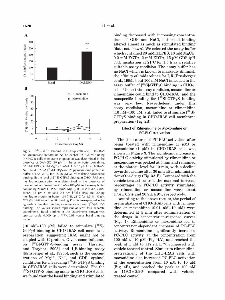
(10 nM–100 mM) failed to stimulate [35S]-GTPgS binding in CHO-IRAS cell membranepreparation, suggesting IRAS might not becoupled with G-protein. Given some influenceon [35S]-GTPgS-binding assay [Harrisonand Traynor, 2003] and I1R-binding assay[Ernsberger et al., 1995b], such as the concen-trations of Mg2þ, Naþ, and GDP, optimalconditions for measuring [35S]-GTPgS bindingin CHO-IRAS cells were determined. For the[35S]-GTPgS-binding assay in CHO-IRAS cells,we found that the basal binding and stimulated
binding decreased with increasing concentra-tions of GDP and NaCl, but basal bindingaltered almost as much as stimulated binding(data not shown). We selected the assay bufferwhich contained 20mMHEPES, 10mMMgCl2,0.2 mM EGTA, 2 mM EDTA, 15 mM GDP (pH7.4), incubation at 218C for 1.5 h as a relativesuitable assay condition. The assay buffer hasno NaCl which is known to markedly diminishthe affinity of imidazolines for I1R [Ernsbergeret al., 1995b], but 100mMNaCl is needed in theassay buffer of [35S]-GTPgS binding in CHO-mcells. Under this assay condition,moxonidine orrilmenidine could bind to CHO-IRAS, and thenonspecific binding for [35S]-GTPgS bindingwas very low. Nevertheless, under thisassay condition, moxonidine or rilmenidine(10 nM–100 mM) still failed to stimulate [35S]-GTPgS binding in CHO-IRAS cell membranepreparation (Fig. 2B).
Effect of Rilmenidine or Moxonidine onPC-PLC Activation
The time course of PC-PLC activation afterbeing treated with rilmenidine (1 mM) ormoxonidine (1 mM) in CHO-IRAS cells wasshown in Figure 3. The significant increase inPC-PLC activity stimulated by rilmenidine ormoxonidine was peaked at 5 min and remainedat the plateau level for 10 min, with a declinetowards baseline after 30min after administra-tion of the drugs (Fig. 3A,B). Comparedwith thevehicle-treated control, the maximal increasepercentages in PC-PLC activity stimulatedby rilmenidine or moxonidine were about17.4� 6.2% and 20.2� 4.9%, respectively.
According to the above results, the period ofpreincubation of CHO-IRAS cells with rilmeni-dine or moxonidine (0.01 nM–10 mM) weredetermined at 5 min after administration ofthe drugs in concentration-response curves(Fig. 4). Rilmenidine or moxonidine led toconcentration-dependent increase of PC-PLCactivity. Rilmenidine significantly increasedPC-PLC activity at the concentration from100 nM to 10 mM (Fig. 4A), and reached thepeak at 1 mM to 117.2� 1.7% compared withvehicle-treated control. Similar to rilmenidine,pretreatment of the CHO-IRAS cells withmoxonidine also increased PC-PLC activationat the concentration from 10 nM to 10 mM(Fig. 4B), and reached the peak at 100 nMto 119.3� 2.9% compared with vehicle-treated control.
Basal DAMGO0
50
100
150
200
250
300**
[35S]
-GT
P¦Ã
S bi
ndin
g (%
of
basa
l)
A
B
-20
0
20
40
60
80
100
-9 -8 -7 -6 -5 -4 -3
Concentration (log M)
[35S]
-GT
PS
bind
ing
(% o
f bas
al)
Rilmenidine
Moxonidine
Fig. 2. [35S]-GTPgS binding in CHO-m cells and CHO-IRAScells membrane preparation.A: The level of [35S]-GTPgS bindingin CHO-m cells membrane preparation was determined in thepresence of DAMGO (10 mM) in the assay buffer containing50 mM HEPES, 5 mM MgCl2, 1 mM EDTA, 15 mM GDP, 100 mMNaCl (add 0.2 nM [35S]-GTPgS and 20 mg membrane protein inbuffer, pH 7.4, 258C) for 1 h, 40 mM GTPgS to define nonspecificbinding. B: the level of [35S]-GTPgS binding in CHO-IRAS cellsmembrane preparation was determined in the presence ofmoxonidine or rilmenidine (10 nM–100 mM) in the assay buffercontaining 20 mM HEPES, 10 mM MgCl2, 0.2 mM EGTA, 2 mMEDTA, 15 mM GDP (add 0.2 nM [35S]-GTPgS and 20 mgmembrane protein in buffer, pH 7.4), 218C for 1.5 h, 40 mMGTPgS to define nonspecific binding. Rusults are expressed as theagonists stimulated binding increase over basal [35S]-GTPgSbinding. The values shown represent at least four separateexperiments. Basal binding in the experiments shown wasapproximately 4,000 cpm. **P<0.01 versus basal bindingvalue.
1620 Li et al.

In order to test whether the upregulation ofrilmenidine or moxonidine on PC-PLC activitywas mediated by IRAS, the cells were pre-treated with efaroxan for 15 min, an I1Rantagonist. Efaroxan (10 mM) abolished theincrease in PC-PLC activity induced by mox-onidine (1 mM) or rilmenidine (1 mM), but had nosignificant effect on PC-PLC activity whengiven alone (Fig. 5A). Furthermore, no changein PC-PLC activity was detected in parentalCHO cells treated by rilmenidine (1 mM) ormoxonidine (1 mM) for 5 min (data not shown).These results indicated that IRASmediates thePC-PLC activation. In addition to PC-PLC
activation, the PLD activation could also causepositive results measured by this assay kit. Toconfirm that IRAS activates PC-PLC ratherthan PC-PLD, we added D609, a putative PC-PLC inhibitor, to the cells to see if it could blockthe lipase activity in response to moxonidine orrilmenidine (1 mM). Figure 5B indicated thatpretreatment with D609 (1 mM) for 30 mincompletely inhibited the lipase activity, sug-gesting that the binding of moxonidineor rilmenidine to IRAS causes the activationof PC-PLC but not PC-PLD. These resultsare consistent with previous studies of nativeI1R in PC12 cells [Separovic et al., 1996;Ernsberger, 1999].
A
B
0 10 20 30 40 50 600
90
100
110
120
130Rilmenidine
**
Phos
phoc
holin
e fl
uore
scen
ce
(% o
f co
ntro
l)
Time (min)
0 10 20 30 40 50 600
90
100
110
120
130Moxonidine
**
Phos
phoc
holin
e fl
uore
scen
ce
(% o
f co
ntro
l)
Time (min)
Fig. 3. Time course of PC-PLC activation in CHO-IRAS cellstreated by rilmenidine or moxonidine. The cells (1�106 cells/well) were incubated with rilmenidine (1 mM) for indicated time.Cells were collected and lysed. The PC-PLC activity wasdetermined by measuring the value of fluorescence (excitationat 535 nm and emission detection at 590 nm), which was red after45 min development of the enzymatic reaction as describedunder Amplex Red phosphatidylcholine-specific PLC assay kitExperimental Procedures. The results represent at least sixexperiments, each performed with separate cell cultures, andshown as increase percentage of fluorescence compared withvehicle-treated control. *P<0.05 versus vehicle-treated control(by Student–Newman–Keuls test). A: Rilmenidine-treatedgroup. B: Moxonidine-treated group.
-11 -10 -9 -8 -7 -6 -50
100
110
120
130
*
*
*
Phos
phoc
holin
e fl
uore
scen
ce
(% o
fcon
trol
)
Rilmenidine Concentration (Log M)
-11 -10 -9 -8 -7 -6 -50
100
110
120
130
*
**
*
Phos
phoc
holin
e fl
uore
scen
ce
(% o
f co
ntro
l)
Moxonidine Concentration (Log M)
A
B
Fig. 4. Concentration-dependent curves of PC-PLC activationstimulated by rilmenidine or moxonidine in CHO-IRAS cells. Thecells (1� 106 cells/well) were incubated with different concen-tration of rilmenidine or moxonidine (0.01 nM–10mM) for 5 min.Cells were collected and lysed. The PC-PLC activity wasdetermined by measuring the value of fluorescence (excitationat 535 nm and emission detection at 590 nm), which was red after45 min development of the enzymatic reaction as describedunder Amplex Red phosphatidylcholine-specific PLC assay kitExperimental Procedures. The results represent at least sixexperiments, each performed with separate cell cultures, andshown as increase percentage of fluorescence compared withvehicle-treated control. *P<0.05 versus vehicle-treated control(by Student–Newman–Keuls test). A: Rilmenidine-treatedgroup. B: Moxonidine-treated group.
Activation of Mitogen Activation Protein Kinase Pathways in IRAS 1621

Effect of Rilmenidine or Moxonidine onDAG Accumulation
The DAG was isolated and analyzed byHPTLC. As DAG is the hydrolysate of PC, weassumed that DAG accumulation may be chan-ged at 5 min. In order to obtain the optimalreaction time, the cells were treated withrilmenidine (1 mM) or moxonidine (1 mM) for0.5, 1, 2, and5min, respectively. Pretreatment ofCHO-IRAS with rilmenidine or moxonidine for0.5–2 min had no significant influence on DAGaccumulation, but when the incubation periodwas prolonged to 5 min, the DAG accumulationwas significantly raised (Fig. 6A,B). Treatmentwithmoxonidine (1mM)or rilmenidine (1 mM) for5 min in CHO-IRAS cells led to an obvious
increase in DAG accumulation by 57.1� 15.0%and 60.1� 15.6%, respectively.
Effect of Rilmenidine or Moxonidine onERK Activation
Exposure of CHO-IRAS cells to rilmenidineor moxonidine (0.01 nM–10 mM) for15 min resulted in a concentration-dependentelevation of phosphorylated ERK (Fig. 7).Rilmenidine significantly increased ERKphosphorylation at the concentration from10 nM to 1 mM and reached the peak at100 nM in CHO-IRAS cells. Compared withvehicle-treated control, ERK phosphorylationwas increased by 74.5� 18.6% at 100 nM(Fig. 7A). Like rilmenidine, moxonidine signifi-cantly increased ERK phosphorylation at theconcentration from10nMto10mM,and reachedthe peak at 100 nM by 66.8� 11.1% comparedwith vehicle-treated control (Fig. 7B). High
Con Efa Ril Ril+Efa MoxMox+Efa0
90
100
110
120
130
+
#
+
**
Phos
phoc
holin
e fl
uore
scen
ce
(% o
f co
ntro
l)
Con D609 Ril Ril+D609 Mox Mox+D6090
90
100
110
120
130
#+
*
*
Phos
phoc
holin
e fl
uore
scen
ce
(% o
f co
ntro
l)
A
B
Fig. 5. Effect of efaroxan or D609 on PC-PLC activationstimulated by rilmenidine or moxonidine. A: CHO-IRAS cellswere pretreated with efaroxan (10 mM) or vehicle alone for 15min, followed by the treatment of rilmenidine or moxonidine (1mM) for 5 min.B: CHO-IRAS cells were pretreated with D609 (10mM) or vehicle alone for 30 min, followed by the treatment ofrilmenidine or moxonidine (1 mM) for 5 min. The enzymaticreaction was performed as described under Amplex Redphosphatidylcholine-specific PLC assay kit Experimental Proce-dures. *P< 0.05 versus vehicle-treated control, þP< 0.05 versusrilmenidine-treated group, #P<0.05 versus moxonidine-treatedgroup (by Student–Newman–Keuls test).
Con 0.5 min 1 min 2 min 5 min0
50
100
150
200
*
DA
G a
ccul
umat
ion
(%
of c
ontr
ol)
Time
Con Ril Mox0
50
100
150
200 **
DA
G a
ccul
umat
ion
(% o
f co
ntro
l)
A
B
Fig. 6. The level of DAG accumulation stimulated by mox-onidine or rilmenidine in CHO-IRAS cells. A: CHO-IRAS cellswere incubated with rilmenidine (1 mM) for 30 s, 1, 2, and 5 min.B: CHO-IRAS cells were incubated with moxonidine orrilmenidine (1 mM) for 5 min. Then DAG was extracted andquantified. The density of the spots was analyzed by dualwavelength TLC scanner (CS-930) under UV light (365 nM). Theresults were determined from at least five experiments, eachperformed with separate cell cultures. *P< 0.05 versus vehicle-treated control.
1622 Li et al.

concentrations of rilmenidine or moxonidine(10 mM) may elicit an attenuated response,suggesting this concentration may be supra-maximal. By the high concentration of rilmeni-dine or moxonidine, other signaling pathwaysmay be induced which would interfere withIRAS signaling response or induce cytotoxicity.The similar biphasic concentration-responsecurves had been observed inDAG accumulationand ERK activation in native I1R of PC12 cells[Separovic et al., 1996; Edward et al., 2001].To further investigate whether the effect of
rilmenidine or moxonidine on ERK stimulationwas mediated by IRAS, we pretreated the cellswith efaroxan, an I1R inhibitor, for 15 min.Efaroxan (10 mM) abolished ERK phosphoryla-tion stimulated by moxonidine (100 nM) orrilmenidine (100 nM), but had no significant
effect on ERK phosphorylation when givenalone (Fig. 8A). Furthermore, we also foundthat no change in phosphorylated ERK wasdetected in parental CHO cells treated withrilmenidine ormoxonidine (0.01 nM–10 mM) for15 min (data not shown), indicating that thephosporylation of ERK in response to rilmeni-dine or moxonidine was mediated by IRAS. ThePC-PLC inhibitor, D609, was also used in thisstudy to determine the relationship betweenPC-PLC and ERK activation. The cells werepretreated with D609 (10 mM) for 30 min andthen incubated with moxonidine (100 nM) orrilmenidine (100 nM) for 15 min. Figure 8Bshowed that D609 blocked the increase of ERKphosphorylation induced by moxonidine orrilmenidine, but had no significant effect onERK phosphorylation when given alone.
Fig. 7. Concentration-dependent curve of ERK phosphoryla-tion stimulated by rilmenidine or moxonidine in CHO-IRAScells. The cells were treated with rilmenidine or moxonidine(0.01 nM–10 mM) for 15 min. Then the cells were lysed,separated by SDS–PAGE and the protein levels of phosho-ERKand ERK were detected with anti-phospho-ERK antibody or anti-ERK antibody. Immunoreactive bands were visualized byincubation of membranes with an HRP-conjugated anti-mouse(1:2,000 dilution) and anti-rabbit secondary antibody (1:5,000
dilution) for 1 h at room temperature and detected bychemiluminescence. a: protein levels of pERK and total ERK. b:Data from a analyse of determining the radio of optical densitybetween the pERK42 and Pan-ERK42 blot. The results representat least five experiments, each performed with separate cellcultures. *P<0.05 versus vehicle-treated control (by Student–Newman–Keuls test). A: Rilmenidine-treated group. B: Moxoni-dine-treated group.
Activation of Mitogen Activation Protein Kinase Pathways in IRAS 1623

DISCUSSION
Our study demonstrated that PC-PLC isinvolved in IRAS mediating ERK activationinduced by the I1R agonists, rilmenidine ormoxonidine. These results provide direct evi-dence that the signaling pathways mediated byIRAS are similar to those mediated by I1R.
In our study, we found that IRAS activationby rilmenidine or moxonidine significantlyincreased the PC-PLC activity at 5 min andlasted for at least 10min. DAG, the hydrolysateof PC-PLC, could be detected after 5 min ofrilmenidine or moxonidine treatment. It isgenerally accepted that DAG may be formedby two phases. The initial phase is transient(�1 min) and primarily derived from thehydrolysis of phosphatidylinositol phospholi-pase C (PI-PLC). The sustained phase is moreprolonged, which ismediated by PC-PLC or PC-PLD [Billah and Anthes, 1990; Exton, 1990,1994; Lee and Severson, 1994]. Either PC-PLCor PC-PLD can hydrolyze phosphatidylcholine,
yielding DAG and phosphocholine, or phospha-tidic acid and choline, respectively. Phosphati-dic acid is subsequently converted into DAG byphosphatide phosphohydrolase [Billah et al.,1989; Murthy and Makhlouf, 1995]. In ourstudy, DAGaccumulation could not be observedwithin initial phase, which suggests PI-PLCmay not contribute to DAG accumulation, butpossibly PC-PLC or PC-PLCD mediates DAGaccumulation. Our results indicate that PC-PLC, but not PC-PLD, is involved in IRASsignaling. First, the PC-PLC activity increasedby rilmenidine or moxonidine could be reversedbyD609, a PC-PLC inhibitor. Second, accordingto manufacturer’s instructions of Amplex RedPC-PLC assay kit, phosphocholine produced byPC-PLC action on PC is hydrolyzed by alkalinephosphatase to generate choline. In the non-alkaline phosphated state, the enzyme-depen-dent fluorescent product would therefore resultfrom PC-PLD. In order to determine whetherPLD is involved, we used the assay kit in theabsence of alkaline phosphatase, and found that
Fig. 7. (Continued )
1624 Li et al.

Fig. 8. Efaroxan or D609 inhibits rilmenidine or moxonidineinduced ERK phosphorylation in CHO-IRAS cells. A: Cells werepretreated with efaroxan (10 mM) or vehicle alone for 15 min, andthen the cells were treated with moxonidine (100 nM) orrilmenidine (100 nM) for 15 min. B: Cells were pretreated withD609 (10 mM) or vehicle alone for 30 min, followed bymoxonidine (100 nM) or rilmenidine (100 nM) treatment for
15 min. a: The protein levels of pERK and total ERK b:Data from aanalyse of determining the radio of optical density between thepERK42 and Pan-ERK42 blot. The results represent at least sixexperiments, each performed with separate cell cultures.*P<0.05 versus vehicle-treated control, #P< 0.05 versus mox-onidine-treated group, þP<0.05 versus rilmenidine-treatedgroup (by Student–Newman–Keuls test).
Activation of Mitogen Activation Protein Kinase Pathways in IRAS 1625

the final fluorescence sharply decreased to thevalue which is equal to the negative control(data not shown), and the fluorescence inagonists-treated groups had no significantdifference compared with that in vehicle-treated control, suggesting that PLD was notstimulated in CHO-IRAS cells treated by rilme-nidine or moxonidine. Third, PC-PLC has beenreported to be involved in cell growth [Johansenet al., 1994] and death [Yonghong et al., 1998],which is consistent with known IRAS-mediatedintracellular functions [Sano et al., 2002; Don-tenwill et al., 2003a]. Our findings thus illumi-nate PC-PLC and its hydrolysate DAG involvedin the signal pathway of IRAS.
However, what stimulates PC-PLC remainsunknown. Our study demonstrated that theIRAS signaling pathway might not be coupledwith G proteins, suggesting that PC-PLC maynot be stimulated by G proteins. According tothe structure of IRAS, which has integrin-binding motif plus a proline-rich region and aPX domain [Piletz et al., 2000], it is likely thatIRASmay bind to a tyrosine kinase like Src andstimulate a signaling cascade leading to theactivation of PC-PLC. However, further studiesneed to be done to identify this.
ERK typically controls cellular processes,such as proliferation, differentiation, develop-ment, stress response, and apoptosis [Gutkind,1998; Aplin and Juliano, 1999; Davis, 2000].Previous studieshave shown that amodest levelof IRAS by co-transfection into the HEK293cells with insulin receptor substrate proteins(IRSs) led to a twofold rise in the activated stateof ERKby the stimulation of insulin [Sano et al.,2002], and that NGF treatment resulted in afivefold increase in phospho-ERK level in PC12cells stably transfected with IRAS compared tothat in the nontransfected cells [Piletz et al.,2003]. These results indicate that IRAS mightwork on ERK in the absence of I1R agonists. Inour study, we proved that ERKphosphorylationwas activated in the presence of I1R agonists.Rilmenidine or moxonidine induced a concen-tration-dependent increase in the phosphoryla-tion of ERK, and this increase was reversedby efaroxan, the selective I1R antagonist,suggesting the involvement of ERK in thesignaling pathway of IRAS. In order to identifythe relationship between PC-PLC and ERKactivation, we pretreated CHO-IRAS cellswith D609, the selective PC-PLC antagonist.Results showed that D609 attenuated the ERK
phosphorylation induced by rilmenidine ormoxonidine. The findings indicate that PC-PLCmediates the activation of ERK in responseto IRAS activation.
Although the sequence analysis of the IRAScDNA indicated that the product might not be aGPCR [Piletz et al., 2000], there is no directfunctional evidence to show this. Because of theinterference of other receptors, especially a2-AR, whether native I1R couples to G protein iscontroversial. In order to investigate if IRAS iscoupled with G protein, we used [35S]-GTPgS-binding assay in CHO-IRAS cells. In this cellline, the interference of a2-AR is ruled out. Theadvantage of [35S]-GTPgS-binding assay is thatit measures a functional consequence of thereceptor occupancy at one of the earliest eventsmediated by the receptor [Harrison and Tray-nor, 2003]. Moreover, the assay is the mostdirectmethod to test the relationship between areceptor and the G protein [Lazareno, 1997;Sovago et al., 2001]. Considering [35S]-GTPgS-binding assay and I1R-binding assay areinfluenced by many factors, including the con-centration of Mg2þ, Naþ, GDP, temperature,and time of reaction [Ernsberger et al., 1995b;Lazareno, 1997; Harrison and Traynor, 2003],we optimized the assay condition based on theinfluencing factors. However, under any condi-tion, moxonidine or rilmenidine failed to stimu-late [35S]-GTPgS binding in CHO-IRAS cellmembrane preparation. Our data did not sup-port IRAS coupling with G protein, which isconsistent with the sequence analysis of theIRAS cDNA.
Compared to previous studies of I1R in ratPC12 cells, we found that clonidine had slightlyhigher affinity than moxonidine for the trans-fected IRAS (Fig. 1B),which is the opposite rankorder reported [Greney et al., 2000]. In additionto this, our findings that IRAS activation byrilmenidine increased the PC-PLC activity,DAG acculumation and ERK phosphorylationin CHO-IRAS cells are consistent with previousreported findings on I1R activated by rilmeni-dine in NGF-induced differentiated PC12 cells[Zhang et al., 2001]. However, our results aredifferent from Edward et al. [2001] and Separo-vic et al. [1997] findings in the time course ofDAG accumulation and ERK activation. Theyreported that DAG accumulation was signifi-cantly increased in 15 s and ERK phosphoryla-tion peaked at 90 min by moxonidine in NGF-induced differentiated PC12 cells. Additionally,
1626 Li et al.

our result of PC-PLC activity elevation inducedby moxonidine in CHO-IRAS cells (19%) waslower than Greney’s result in PC12 cells (37%)[Greney et al., 2000]. We think that thesedifferences maybe due to species differences orunknown factors.In summary, we found that IRAS activation
by imidazolines causes PC-PLC hydrolysis andDAG accumulation, apparently leading to ERKphosphorylation. These results are similarto those reported for native I1R [Liedtke andErnsberger, 1995; Separovic et al., 1996;Edward et al., 2001; Zhang et al., 2001],including some reports that I1R do not coupleto G proteins [Piletz and Sletten, 1993; Briccaet al., 1994]. Our findings therefore support thehypothesis that IRAS is an I1R protein.
ACKNOWLEDGMENTS
We thank Dr. J.E. Piletz for kindly providingIRAS-pcDNA 3.1 (þ) plasmid and Dr. L.Y. Liu-Chen for kindly providing CHO and CHO-mcell lines.
REFERENCES
Alahari SK, Lee JW, Juliano RL. 2000. Nischarin, anovel protein that interacts with the integrin alpha5subunits and inhibits cells migration. J Cell Biol151:1141–1154.
Aplin AE, Juliano RL. 1999. Integrin and cytoskeletalregulation of growth factor signal to the MAP kinasepathway. J Cell Sci 112:695–706.
Billah MM, Anthes C. 1990. The regulation and cellularfunctions of phosphaidylcholine hydrolysis. Biochem J269:281–291.
Billah MM, Eckel S, Mullmann TJ, Egan RW, Siegel MI.1989. Phosphatidylcholine hydrolysis by phospholipaseD determines phosphatidate and diglyceride levels inchemotactic peptide-stimulated human neutrophils.J Biol Chem 264:17069–17077.
Bousquet P, Feldman J, Schwartz J. 1984. Centralcardiovascular effects of the alpha adrenergic drugs:Differences between catecholamines and imidazolines.J Pharmacol Exp Ther 230:232–236.
Bricca G, Greney H, Zhang J, Dontenwill M, Stutzmann J,Belcourt A, Bousquet P. 1994. Human brain imidazolinereceptors: Further characterazation with [3H]clonidine.Eur J Pharmacol 266:25–33.
Burford N, Wang D, Saddee W. 2000. G-protein coupling ofmu-opioid (OP3): Elevated basal signalling activity.Biochem J 348:531–537.
Davis RJ. 2000. Signal transduction by the JNK group ofMAP kinases. Cell 103:239–252.
Dontenwill M, Pascal G, Piletz JE, Chen M, Baldwin J,Ronde P, Dupuy L, Urosevic D, Greney H, Takeda K,Bousquet P. 2003a. IRAS, the human homologue ofnischarin, prolongs survival of transfected PC12 cell. CellDeath Differ 10:933–935.
Dontenwill M, Piletz JE, Chen M, Baldwin J, Pascal G,Ronde P, Dupuy L, Greney H, Takeda K, Bousquetd P.2003b. IRAS is an anti-apoptotic protein. Ann N Y AcadSci 1009:400–412.
Dupuy L, Urosevic D, Greney H, Quaglia W, Pigini M,Brasili L, Dontenwill M, Bousquet P. 2004. I1 imidazo-line receptor-mediated effects on apoptotic processes inPC12 cells. Cell Death Differ 11:1049–1052.
Edward L, Fishman D, Horowitz P, Nicole B, KesterM, Ernsberge P. 2001. The imidazoline-1 receptorin PC12 pheochromocytoma cells activatives proteinkinase C, extracelluar signal-regulated kinase (ERK)and c-jun N-terminal kinase (JUK). J Neurochem79:931–940.
Eglen RM, Hudson AL, Kendall DA, Nutt DJ, Morgan NG,Wilson VG, Dillon MP. 1998. Seeing through a glassdarkly: Casting light on imidazoline ‘I’sites. TrendsPharmacol Sci 19:381–390.
Ernsberger P. 1998. Arachidonic acid release from PC12pheochromocytoma cells is regulated by I1-imidazolinereceptors. J Auton Nerv Syst 72:147–154.
Ernsberger P. 1999. The I1-imidazoline receptor and itscellular signaling pathways. Ann N Y Acad Sci 881:35–53.
Ernsberger P, Shen IH. 1997. Membrane localization andguanine nucleotide sensitivity of medullary I1-imidazo-line binding sites. Neurochem Int 30:17–28.
Ernsberger P, Graves ME, Graff LM, Zakieh N, Nguyen P,Collins LA, Westbrooks KL, Johnson GG. 1995a. Imida-zoline receptors. Definition, characterization, distribu-tion, and transmembrane signaling. Ann N Y Acad Sci763:22–42.
Ernsberger P, Piletz JE, Graff LM, Graves ME. 1995b.Optimization of radioligand binding assays for I1-imida-zoline sites. Ann N Y Acad Sci 763:163–168.
Exton JH. 1990. Signaling through phosphatidylcholinebreakdown. J Biol Chem 265:1–4.
Exton JH. 1994. Phosphatidylcholine breakdown andsignal transduction. Biochim Biophys Acta 212:26–42.
Fraser CM, Arakawa S, McCombie WR, Venter JC. 1989.Cloning, sequence analysis, and permanent expression ofa human a2-adrenergic receptor in Chinese hamsterovery cells. J Biol Chem 264:11754–11761.
Georges F, Aston-Jones G. 2003. Prolonged activation ofmesolimbic dopaminergic neurons by morphine with-drawal following clonidine: Participation of imidazolineand norepinephrine receptors. Neuropsychopharmacol28:1140–1149.
Greney H, Ronde P, Magnier C, Maranca F, Rascente C,Quaglia W, Giannella M, Pigini M, Brasili L, Lugnier C,Bousquet P, Dontenwill M. 2000. Coupling of I1 imidazo-line receptors to the cAMP pathway: Studies with ahighly selective ligand benazoline. Mol Pharmacol57:1142–1151.
Gutkind JS. 1998. Cell growth control by G protein-coupledreceptors: From signal transduction to signal integration.Oncogene 17:1331–1342.
Harrison C, Traynor JR. 2003. The [35S]GTPgS bindingassay: Approaches and applications in pharmacology.Life Sci 74; 489–508.
Ivanov TR, Jones JC, Dontenwill M, Bousquet P, Piletz JE.1998. Charactetization of a partial cDNA clone detectedby imidazoline receptor-selective antisera. J Auton NervSyst 72:98–110.
Activation of Mitogen Activation Protein Kinase Pathways in IRAS 1627

Johansen T, Bjorkoy G, Overvatn A, Diaz-Meco MT,Traavik T, Moscat J. 1994. NIH 3T3 cells stablytransfected with the gene encoding phosphatidylcho-line-hydrolyzing phospholipase C from Bacillus cereusacquire a transformed phenotype. Mol Cell Biol 14:646–654.
Lazareno S. 1997. Measurement of agonists-stimulated[35S]GTPgS to cell membranes. Methods Mol Biol 83:107–116.
Lee MW, Severson DL. 1994. Signal transductionin vascular smooth muscle: Diacylglycerol second mes-sengers and PKC action. Am J Physiol 267:C659–C678.
Lee C, Fishers SK. Agranoff BW, HajraA K. 1991.Quantitative analysis of molecular species of diacylgly-cerol and phosphatidate formed upon muscarinic recep-tor activation of human SK-N-SH neuroblastoma cells.J Biol Chem 266:22837–22846.
Liedtke CM, Ernsberger P. 1995. Regulation of electrolytetransport in rabbit tracheal epithelial cells by the I1-imidazoline agonist moxonidine. Ann N Y Acad Sci763:401–404.
Lim Koh-Pang, Hong Wanjin. 2004. Human Nischarin/IRAS is targeted to the endosomes by a combined actionof a PX domain and a coiled-coil region. J Biol Chem279:54770–54782.
Moldering GJ, Moura D, Fink K, Boisch H, GothertM. 1993. Binding of [3H]clonidine to I1-imidazoline sitesin bovine adrenal medullary membranes. NaunynSchmiedebergs Arch Pharmacol 348:70–76.
Murthy KS, Makhlouf GM. 1995. Agonist mediated activa-tion of phosphatidylcholine specific phospholipase C andD in intestinal smooth muscle. Mol pharmacol 48:293–304.
Piletz JE, Sletten K. 1993. Nonadrenergic imidazolinebinding sites on human platelets. J Pharmacol Exp Ther267:1493–1502.
Piletz JE, Jones JC, Zhu H, Bishara O, ErnsbergerP. 1999. Imidazoline receptor antisera-selected cDNAand mRNA distribution. Ann NY Acad Sci 881:1–7.
Piletz JE, Ivanov TR, Sharp JD, Ernsberger P, Chang CH,Pickard RT, Gold G, Roth B, Zhu H, Jones JC, Baldwin J,Reis DJ. 2000. Imidazoline receptor antisera-selected(IRAS) cDNA: Cloning and characterization. DNA CellBoil 19:319–329.
Piletz JE, Wang G, Zhu H. 2003. Cell signaling byimidazoline-1 receptor candidate, IRAS, and thenischarin homologue. Ann N Y Acad Sci 1009:392–399.
Sano H, Liu SCH, Lane WS, Pileta JE, Lienhard G. 2002.Insulin receptor substrate 4 associates with the proteinIRAS. J Biol Chem 277:19439–19447.
Separovic D, Kester M, Ernsberger P. 1996. Coupling of I1-imidazoline receptors to diacylglyceride accumulation inPC12 rat pheochromocytoma cells. Mol Pharmacol49:668–675.
Separovic D, Kester M, Haxhiu MA, Piletz JE. 1997.Activation of phosphatidylcholine selective phospholi-pase C by I1-imidazoline receptors in PC12 cells androstral ventrolateral medulla. Brain Res 749:335–339.
Sovago J, Dupuis DS, Gulyas B, Hall H. 2001. An overviewon functional receptors autoradiography using[35S]GTPgS. Brain ResRev 38:149–164.
Su RB, Li J, Qin BY. 2003. A biphasic opioid functionmodulator: Agmatine. Acta Pharmacol Sin 24:631–636.
Takada K, Hayashi Y, Kamibayashi T, Mammoto T,Yamatodani A, Kitamura S, Yoshiya I. 1997. Theinvolvement of pertussis toxin-sensitive G proteins inthe post receptor mechanism of central imidazoline-1receptors. Br J Pharmacol 120:1575–1581
Wu N, Su RB, Xu B, Lu XQ, Liu Y, Zheng JQ, Piletz JE, LiJ, Qin BY. 2005. IRAS, a candidate for I1-imidazolinereceptor, mediates inhibitory effect of agmatine oncellular morphine dependence. Biochem Pharmacol70:1079–1087.
Yao JK, Rastetter GM. 1985. Microanalysis of complextissure lipid by high-performance thin-layer chromato-graghy. Anal Biochem 150:111–116.
Yonghong L, Pamela M, David S. 1998. Phosphatidylcho-line-specific phospholipase C regulates glutamate-induced nerve cell death. Proc Natl Acad Sci USA95:7748–7753.
Zhang J, El-Ms MM, Abdel-Rahman AA. 2001. ImidazolineI1 receptor-induced activation of phosphatidylcholine-specific phospholipase C elicits mitogen-activated proteinkinase phosphorylation in PC12 cells. Eur J Pharmacol415:117–125.
Zhu J, Luo LY, Li JG, Chen C, Liu-Chen LY. 1997.Activation of the cloned human kappa opioid receptorby agonists enhances [35S]GTPgS binding to membranes:Determination of potencies and efficacies of ligands. JPharmacol Exp Ther 282:676–684.
1628 Li et al.