Embed Size (px)
Citation preview
Article
The Rockefeller University Press $30.00J. Exp. Med. Vol. 208 No. 11 2279-2290www.jem.org/cgi/doi/10.1084/jem.20111106
2279
Chronic inflammation is a major driving force for the initiation and progression of tumors in many tissues (Mantovani et al., 2008). For ex-ample, in patients with ulcerative colitis (UC), one of the major forms of chronic inflamma-tory bowel diseases in humans, there is an in-creased risk of colon cancer, which is related to the duration, extent, and severity of inflamma-tory disease (Rutter et al., 2004; Gupta et al., 2007). The molecular mechanisms underlying this neoplastic transformation are poorly un-derstood, but immune cells that infiltrate the tumor and produce tumor-promoting cyto-kines are thought to make a major contribution to colon cancer growth (Mantovani et al., 2008). The mouse model of colitis-associated colon cancer (CAC), which is induced by administra-tion of azoxymethane (AOM) followed by re-peated oral administration of dextran sulfate sodium (DSS), has been highly informative. In this
model, neutralization of TNF (Popivanova et al., 2008) and/or inactivation of the NF-B path-way reduces the number and size of tumors (Greten et al., 2004). An additional contribu-tion to the colon cancer cell growth is provided, either directly or indirectly, by cytokines involved in the differentiation and function of T helper (Th)-17 lymphocytes, such as IL-6, IL-23, and IL-17A (Grivennikov et al., 2009; Wu et al., 2009). These cytokines activate signal trans-ducer and activator of transcription 3 (STAT3) in tumor cells and tumor-associated inflam-matory cells, enhancing tumor survival and promoting angiogenesis (Terzic et al., 2010). Collectively, these observations support the
CORRESPONDENCE Giovanni Monteleone: [email protected]
Abbreviations used: AOM, azoxymethane; CAC, colitis-associated colon cancer; DSS, dextran sulfate sodium; IEC, intestinal epithelial cell; LPMC, lamina propria mononuclear cell; STAT3, signal transducer and activator of transcription 3; TIC, tumor-infiltrating immune cell; UC, ulcerative colitis.
Involvement of interleukin-21 in the regulation of colitis-associated colon cancer
Carmine Stolfi,1 Angelamaria Rizzo,1 Eleonora Franzè,1 Angela Rotondi,1 Massimo Claudio Fantini,1 Massimiliano Sarra,1 Roberta Caruso,1 Ivan Monteleone,1 Pierpaolo Sileri,2 Luana Franceschilli,2 Flavio Caprioli,3 Stefano Ferrero,4 Thomas T. MacDonald,5 Francesco Pallone,1 and Giovanni Monteleone1
1Gastroenterology Unit, Department of Internal Medicine, 2Department of Surgery, University of Rome Tor Vergata, 00133 Rome, Italy3Unit of Gastroenterology 2 and 4Pathology Unit, Fondazione IRCCS Cà Granda Ospedale Maggiore Policlinico, 20122 Milan, Italy
5Centre for Immunology and Infectious Disease, Blizard Institute of Cell and Molecular Science, Barts and the London School of Medicine and Dentistry, London, UK
Chronic inflammation is a major driving force in the development of cancer in many tissues, but the array of factors involved in this neoplastic transformation are not well understood. We have investigated the role of interleukin (IL)-21 in colitis-associated colon cancer (CAC), as this cytokine is overexpressed in the gut mucosa of patients with ulcerative colitis (UC), a chronic inflammatory disease associated with colon cancer. IL-21 was increased in the gut of patients with UC-associated colon cancer, and in mice with CAC induced by azoxymethane (AOM) and dextran sulfate sodium (DSS). After AOM+DSS treatment, IL-21 KO mice showed reduced mucosal damage, reduced infiltration of T cells, and diminished production of IL-6 and IL-17A. IL-21–deficient mice also developed fewer and smaller tumors compared with wild-type (WT) mice. Absence of IL-21 reduced signal transducer and activator of transcrip-tion 3 activation in tumor and stromal cells. Administration of a neutralizing IL-21 antibody to WT mice after the last DSS cycle decreased the colonic T cell infiltrate and the production of IL-6 and IL-17A and reduced the number of tumors. These observations indicate that IL-21 amplifies an inflammatory milieu that promotes CAC, and suggest that IL-21 blockade may be useful in reducing the risk of UC-associated colon cancer.
© 2011 Stolfi et al. This article is distributed under the terms of an Attribution– Noncommercial–Share Alike–No Mirror Sites license for the first six months after the publication date (see http://www.rupress.org/terms). After six months it is available under a Creative Commons License (Attribution–Noncommercial–Share Alike 3.0 Unported license, as described at http://creativecommons.org/licenses/by-nc-sa/3.0/).
The
Journ
al o
f Exp
erim
enta
l M
edic
ine
Dow
nloaded from http://rupress.org/jem
/article-pdf/208/11/2279/1204673/jem_20111106.pdf by guest on 26 M
arch 2022
2280 IL-21 promotes colitis-associated colon cancer | Stolfi et al.
We next analyzed the expression of IL-21 in DSS- induced colitis and AOM+DSS-induced CAC. In initial ex-periments, WT mice were given 2 cycles of DSS-supplemented water preceded by AOM treatment, and then sacrificed at the end of the second DSS cycle (day 29). Analysis of IL-21 in the colon showed that DSS-colitis in WT mice was accompanied
notion that distinct pathways, perhaps at different stages of the disease, promote CAC, and that neutralization of such path-ways may help inhibit colitis-driven colon cancer progression.
IL-21 is a cytokine synthesized by a range of CD4+ Th cells, including Th1 and Th17 cells, activated NKT cells, and T fol-licular helper cells (Monteleone et al., 2009b). IL-21 is a key regulator of the proliferation and/or effector function of B cells, T cells, and natural killer cells, and also affects regulatory T cells (Monteleone et al., 2009b). IL-21 can also regulate the activity of nonimmune cells, and it plays a crucial role in many pathological responses, such as allergy and autoimmunity (Spolski and Leonard, 2008; Monteleone et al., 2009b). We have recently shown that IL-21 is overexpressed in the colonic mucosa of UC patients (Monteleone et al., 2005), where it positively regulates Th17 cell responses (Fina et al., 2008). These observations raise the intriguing possibility that blockade of IL-21 can dampen the ongoing mucosal inflammation in UC, thus reducing the risk of CAC. However, studies conducted in classical murine cancer models have shown that IL-21 inhibits, rather than promotes, the growth of tumors. Indeed, using a range of methods of IL-21 delivery, such as IL-21–transfected tumor cell lines, IL-21–expressing plasmids, and recombinant mouse IL-21, different groups have shown that IL-21 exerts antitumor effects both on established xenografts and dissemi-nated tumors when used as monotherapy or in combination with other therapies (Skak et al., 2008). The antitumor activity of IL-21 is mainly mediated by NK cells and CD8+ T cells (Wang et al., 2003; Furukawa et al., 2006), with a requirement for IFN- and perforin (Ma et al., 2003; Di Carlo et al., 2004). IL-21 therapy is also undergoing clinical trials for metastatic melanoma and renal cell carcinoma (Thompson et al., 2008; Schmidt et al., 2010). An unanswered question, however, is whether the antitumor effects of IL-21 are also involved in inflammation-associated malignancy. If this was the case, IL-21 inhibitors proposed for the therapy of inflammation in UC may compromise immune surveillance and enhance the risk of UC-associated colon cancer. In this study, we have tried to clarify the role of IL-21 in the initiation and growth of CAC.
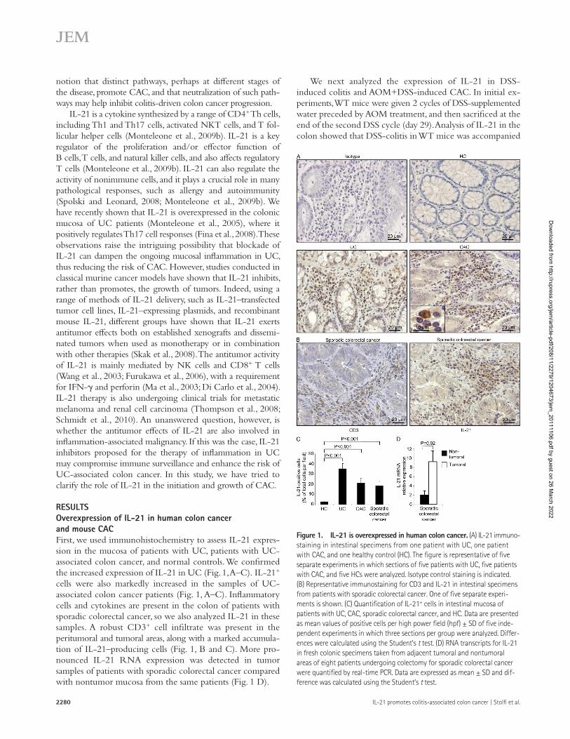
RESULTSOverexpression of IL-21 in human colon cancer and mouse CACFirst, we used immunohistochemistry to assess IL-21 expres-sion in the mucosa of patients with UC, patients with UC- associated colon cancer, and normal controls. We confirmed the increased expression of IL-21 in UC (Fig. 1, A–C). IL-21+ cells were also markedly increased in the samples of UC- associated colon cancer patients (Fig. 1, A–C). Inflammatory cells and cytokines are present in the colon of patients with sporadic colorectal cancer, so we also analyzed IL-21 in these samples. A robust CD3+ cell infiltrate was present in the peritumoral and tumoral areas, along with a marked accumula-tion of IL-21–producing cells (Fig. 1, B and C). More pro-nounced IL-21 RNA expression was detected in tumor samples of patients with sporadic colorectal cancer compared with nontumor mucosa from the same patients (Fig. 1 D).
Figure 1. IL-21 is overexpressed in human colon cancer. (A) IL-21 immuno-staining in intestinal specimens from one patient with UC, one patient with CAC, and one healthy control (HC). The figure is representative of five separate experiments in which sections of five patients with UC, five patients with CAC, and five HCs were analyzed. Isotype control staining is indicated. (B) Representative immunostaining for CD3 and IL-21 in intestinal specimens from patients with sporadic colorectal cancer. One of five separate experi-ments is shown. (C) Quantification of IL-21+ cells in intestinal mucosa of patients with UC, CAC, sporadic colorectal cancer, and HC. Data are presented as mean values of positive cells per high power field (hpf) ± SD of five inde-pendent experiments in which three sections per group were analyzed. Differ-ences were calculated using the Student’s t test. (D) RNA transcripts for IL-21 in fresh colonic specimens taken from adjacent tumoral and nontumoral areas of eight patients undergoing colectomy for sporadic colorectal cancer were quantified by real-time PCR. Data are expressed as mean ± SD and dif-ference was calculated using the Student’s t test.
Dow
nloaded from http://rupress.org/jem
/article-pdf/208/11/2279/1204673/jem_20111106.pdf by guest on 26 M
arch 2022

JEM Vol. 208, No. 11
Article
2281
CD4/CD8, and double-positive CD4/NK1.1 cells, whereas no production was seen in CD4/CD8+ and Thy1.2high/SCA-1+ cells (Fig. 2 C). IL-21–producing CD4+ T cells coex-pressed mostly ROR-t and, to a lesser extent, T-bet (Fig. 2 C). Collectively, these data indicate that IL-21 is overexpressed in the colon during CAC.
IL-21 KO mice are resistant to CACWT and IL-21 KO mice were treated with AOM+DSS and monitored for tumor formation. Endoscopy on day 82 showed that WT mice developed multiple and large tumors, whereas the number and size of tumors were reduced in
by increased IL-21 (Fig. 2 A). The treatment of mice with AOM+DSS did not induce a significant increase in IL-21 ex-pression compared with mice treated with DSS alone (Fig. 2 A). In parallel experiments, mice were treated with AOM and DSS and monitored for the development of tumors. At day 82, colonoscopy showed multiple tumors in all mice. Animals were sacrificed on day 84, and IL-21 expression was assessed in proteins extracted from tumor and nontumor samples by ELISA. IL-21 was significantly increased in tumor samples (Fig. 2 B). Flow cytometry analysis of mononuclear cells iso-lated from colonic tumors of WT mice sacrificed at day 84 showed that IL-21 was produced by CD4+, double-positive
Figure 2. Involvement of IL-21 in the experimental model of CAC. (A) Untreated mice (naive) and mice treated with AOM, DSS, or both were sacrificed at day 29. IL-21 pro-tein expression was evaluated by ELISA, and data are expressed as pg/mg total proteins. Values are mean ± SEM of 4 independently performed experiments containing at least 5 mice per group. (B) AOM+DSS-treated WT and IL-21 KO mice were sacrificed at day 84. IL-21 protein expression was evaluated by ELISA, and data are expressed as pg/mg total pro-teins. Values are mean ± SEM of four inde-pendently performed experiments in which at least five mice per group were considered. (C) WT mice treated with AOM+DSS were sacrificed at day 84. Representative histo-grams showing the percentage of IL-21+ TICs. IL-21+ cells were gated and then analyzed for the indicated markers. The example is repre-sentative of two independent experiments in
which cells isolated from 10 mice were analyzed. (D) Representative endoscopic pictures showing colon tumors in AOM+DSS-treated WT and IL-21 KO mice at day 82. Graphs show the endoscopic scoring of tumors, calculated as indicated in the Materials and methods section, and the number of lesions. Data indicate mean ± SEM of four experiments in which at least five mice per group were considered.
Figure 3. IL-21 deficiency is associated with a reduction in the numbers of CD8- and DX5-expressing LPMC. Representative dot-plots show-ing CD8+ and DX5+ cells in LPMC samples isolated from the colon of both WT and IL-21 KO mice sacrificed at day 0, at the end of the second DSS cycle (day 29), and at the end of the experiment (day 84). Numbers indicate the percentages of cells in the designated gates. One of three representative experi-ments in which at least five mice per group were considered is shown.
Dow
nloaded from http://rupress.org/jem
/article-pdf/208/11/2279/1204673/jem_20111106.pdf by guest on 26 M
arch 2022
2282 IL-21 promotes colitis-associated colon cancer | Stolfi et al.
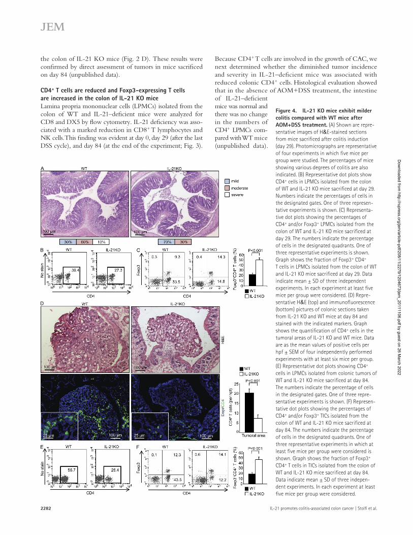
Because CD4+ T cells are involved in the growth of CAC, we next determined whether the diminished tumor incidence and severity in IL-21–deficient mice was associated with reduced colonic CD4+ cells. Histological evaluation showed that in the absence of AOM+DSS treatment, the intestine of IL-21–deficient mice was normal and there was no change in the numbers of CD4+ LPMCs com-pared with WT mice (unpublished data).
the colon of IL-21 KO mice (Fig. 2 D). These results were confirmed by direct assessment of tumors in mice sacrificed on day 84 (unpublished data).
CD4+ T cells are reduced and Foxp3-expressing T cells are increased in the colon of IL-21 KO miceLamina propria mononuclear cells (LPMCs) isolated from the colon of WT and IL-21–deficient mice were analyzed for CD8 and DX5 by flow cytometry. IL-21 deficiency was asso-ciated with a marked reduction in CD8+ T lymphocytes and NK cells. This finding was evident at day 0, day 29 (after the last DSS cycle), and day 84 (at the end of the experiment; Fig. 3).
Figure 4. IL-21 KO mice exhibit milder colitis compared with WT mice after AOM+DSS treatment. (A) Shown are repre-sentative images of H&E-stained sections from mice sacrificed after colitis induction (day 29). Photomicrographs are representative of four experiments in which five mice per group were studied. The percentages of mice showing various degrees of colitis are also indicated. (B) Representative dot plots show CD4+ cells in LPMCs isolated from the colon of WT and IL-21 KO mice sacrificed at day 29. Numbers indicate the percentages of cells in the designated gates. One of three represen-tative experiments is shown. (C) Representa-tive dot plots showing the percentages of CD4+ and/or Foxp3+ LPMCs isolated from the colon of WT and IL-21 KO mice sacrificed at day 29. The numbers indicate the percentage of cells in the designated quadrants. One of three representative experiments is shown. Graph shows the fraction of Foxp3+ CD4+ T cells in LPMCs isolated from the colon of WT and IL-21 KO mice sacrificed at day 29. Data indicate mean ± SD of three independent experiments. In each experiment at least five mice per group were considered. (D) Repre-sentative H&E (top) and immunofluorescence (bottom) pictures of colonic sections taken from IL-21 KO and WT mice at day 84 and stained with the indicated markers. Graph shows the quantification of CD4+ cells in the tumoral areas of IL-21 KO and WT mice. Data are as the mean values of positive cells per hpf ± SEM of four independently performed experiments with at least six mice per group. (E) Representative dot plots showing CD4+ cells in LPMCs isolated from colonic tumors of WT and IL-21 KO mice sacrificed at day 84. The numbers indicate the percentage of cells in the designated gates. One of three repre-sentative experiments is shown. (F) Represen-tative dot plots showing the percentages of CD4+ and/or Foxp3+ TICs isolated from the colon of WT and IL-21 KO mice sacrificed at day 84. The numbers indicate the percentage of cells in the designated quadrants. One of three representative experiments in which at least five mice per group were considered is shown. Graph shows the fraction of Foxp3+ CD4+ T cells in TICs isolated from the colon of WT and IL-21 KO mice sacrificed at day 84. Data indicate mean ± SD of three indepen-dent experiments. In each experiment at least five mice per group were considered.
Dow
nloaded from http://rupress.org/jem
/article-pdf/208/11/2279/1204673/jem_20111106.pdf by guest on 26 M
arch 2022

JEM Vol. 208, No. 11
Article
2283
However, at the end of the last DSS cycle (day 29), IL-21–deficient mice had less severe inflammation than WT mice (Fig. 4 A). At this time point, IL-21–deficient mice also had a reduced percentage of CD4+ LPMCs compared with WT mice (Fig. 4 B). Because IL-21 inhibits the differentiation of Foxp3-expressing T cells (Fantini et al., 2007), we also examined the expression of Foxp3 in the colon of WT and IL-21 KO mice. IL-21–deficient mice had an increased fraction of Foxp3- expressing CD4 T cells at day 29 (Fig. 4 C).
To examine whether there were reduced CD4+ cells in tumors of IL-21–deficient mice, some animals were sacrificed at day 84, and tumors were excised for histology. Micros-copic examination of hematoxylin and eosin (H&E)–stained colonic sections and immunofluorescence revealed mucosal infiltration by CD4+ cells in WT mice, but little infiltration in IL-21–deficient mice (Fig. 4 D). These differences were asso-ciated with higher numbers of dysplastic glands in WT mice. Flow cytometry analysis of mononuclear cells isolated from tumoral areas confirmed that IL-21–deficient mice had a di-minished number of CD4+ cells (Fig. 4 E), and an increased fraction of CD4+Foxp3+ cells (Fig. 4 F).
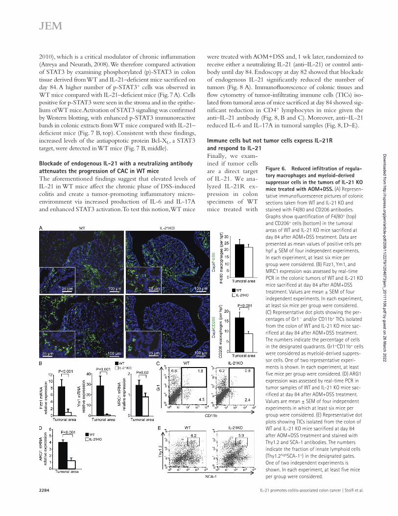
IL-21 KO mice produce less IL-6 and IL-17AThe contribution of CD4+ T cells, and in general of immune cells, in the growth of CAC is via cytokine production (Terzic et al., 2010), so we analyzed the cytokine profile in the colon of WT and IL-21 KO mice. In both types of mice, there was an increase in IL-6 and IL-17A transcripts after AOM+DSS treatment at day 29 (Fig. 5 A). However, transcripts for IL-6 and IL-17A were significantly lower in IL-21–deficient mice (Fig. 5 A). Analysis of cytokine protein by ELISA confirmed the reduced IL-6 and IL-17A in IL-21–deficient mice (Fig. 5 B). RNA transcripts and protein for IFN- were not different between IL-21–deficient mice and WT mice (unpublished data). Using real-time PCR and ELISA of samples taken from tumoral areas, we confirmed that lack of IL-21 was associated with a reduction in the expression of IL-6 and IL-17A (Fig. 5, C and D), whereas IFN- remained unchanged (not de-picted). TNF expression was not different between WT and IL-21–deficient mice at the different time points analyzed (unpublished data). Flow cytometry analysis of immune cells isolated from colonic tumors of WT mice sacrificed at day 84 showed that IL-6 was produced by T cells, F4/80+ cells, F4/80+CD206+ regulatory macrophages, and CD11b+GR1+ cells, but not Thy1.2high/SCA-1+ cells, whereas IL-17A was mostly produced by T cells and, to a lesser extent, Thy1.2high/SCA-1+ cells (Fig. 5 E). Immunofluorescence (Fig. 6 A) and flow cytometry (not depicted) indicated that IL-21 KO mice have a reduced infiltration of alternatively activated macrophages into the tumors as compared with WT mice. This finding was con-firmed by the reduced expression of alternatively activated macrophages-related markers in tumor samples of IL-21 KO mice as compared with WT mice (Fig. 6 B). IL-21–deficient mice also showed a reduced number of tumor infiltrating myeloid-derived suppressor cells as compared with WT mice (Fig. 6 C), paralleled by a reduction in the content of the
immunosuppressive factor ARG1 (Fig. 6 D). In contrast, the per-centage of Thy1.2high/SCA-1+ cells in the tumoral areas did not differ between WT and IL-21 KO mice (Fig. 6 E).
Overall, these findings indicate that the reduced formation of colonic tumors seen in IL-21–deficient mice is associated with a diminished infiltration of both myeloid and T cells into the tumor and decreased production of IL-6 and IL-17A.
STAT3 activation is reduced in IL-21 KO mice treated with AOM+DSSIL-21, like IL-6 and IL-17A, is a powerful activator of the tran-scription factor STAT3 (Caprioli et al., 2008; Hirahara et al.,
Figure 5. IL-21 KO mice produce less IL-6 and IL-17A. (A and B) IL-6 and IL-17A expression was assessed by real-time PCR (A) and ELISA (B) in colon of WT and IL-21 KO mice sacrificed at either day 0 or at day 29 after AOM+DSS treatment. Values are mean ± SEM of four independent experi-ments containing at least six mice per group. (C and D) IL-6 and IL-17A expression were assessed by real-time PCR (C) and ELISA (D) in the colon of WT and IL-21 KO mice sacrificed at day 84 after AOM+DSS treatment. Values are mean ± SEM of four independent experiments containing at least six mice per group. (E) IL-6– and IL-17A–producing cells in the tumoral areas of WT mice treated with AOM+DSS. Representative histo-grams showing the percentage of IL-6– and IL-17A–producing TICs. IL-6+ and IL-17A+ cells were gated and analyzed for the indicated markers. The example is representative of two independent experiments in which cells isolated from 10 mice were analyzed.
Dow
nloaded from http://rupress.org/jem
/article-pdf/208/11/2279/1204673/jem_20111106.pdf by guest on 26 M
arch 2022
2284 IL-21 promotes colitis-associated colon cancer | Stolfi et al.
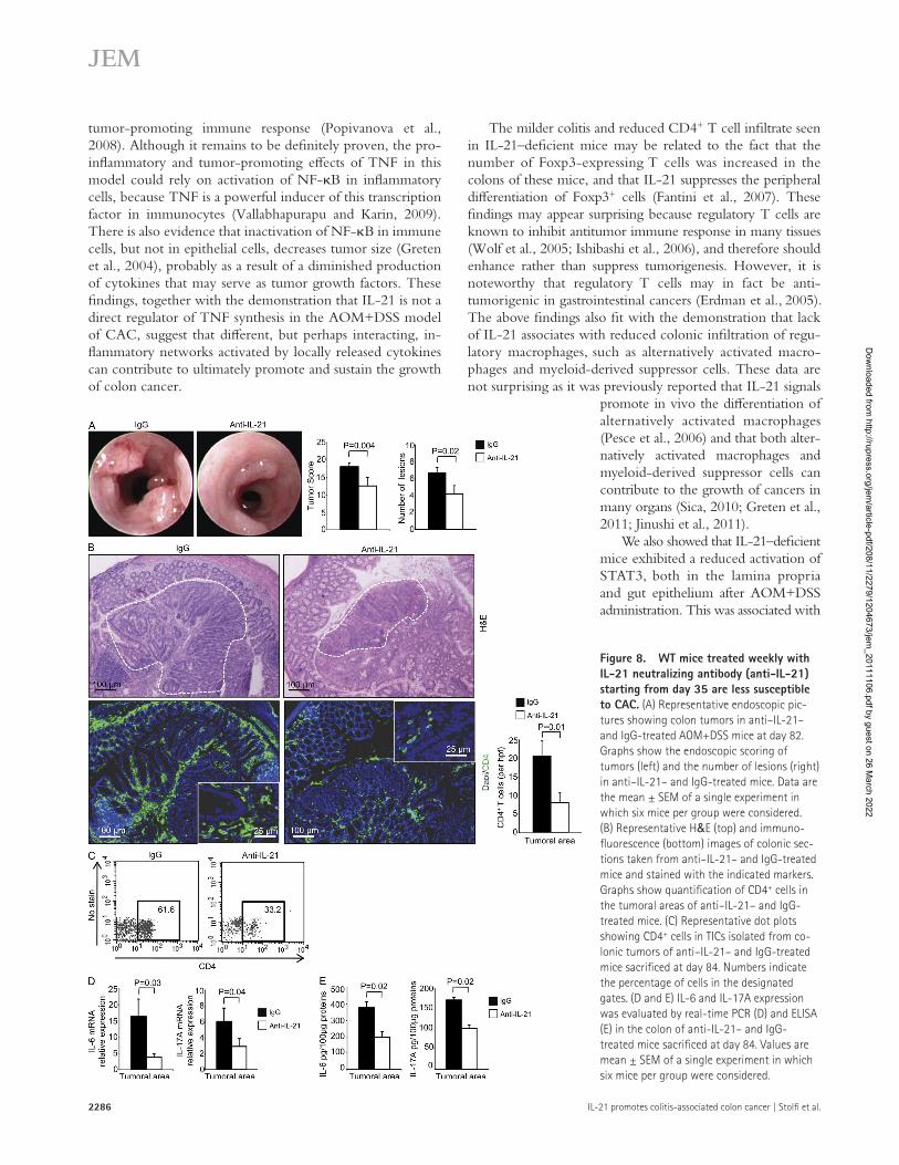
were treated with AOM+DSS and, 1 wk later, randomized to receive either a neutralizing IL-21 (anti–IL-21) or control anti-body until day 84. Endoscopy at day 82 showed that blockade of endogenous IL-21 significantly reduced the number of tumors (Fig. 8 A). Immunofluorescence of colonic tissues and flow cytometry of tumor-infiltrating immune cells (TICs) iso-lated from tumoral areas of mice sacrificed at day 84 showed sig-nificant reduction in CD4+ lymphocytes in mice given the anti–IL-21 antibody (Fig. 8, B and C). Moreover, anti–IL-21 reduced IL-6 and IL-17A in tumoral samples (Fig. 8, D–E).
Immune cells but not tumor cells express IL-21R and respond to IL-21Finally, we exam-ined if tumor cells are a direct target of IL-21. We ana-lyzed IL-21R ex-pression in colon specimens of WT mice treated with
2010), which is a critical modulator of chronic inflammation (Atreya and Neurath, 2008). We therefore compared activation of STAT3 by examining phosphorylated (p)-STAT3 in colon tissue derived from WT and IL-21–deficient mice sacrificed on day 84. A higher number of p-STAT3+ cells was observed in WT mice compared with IL-21–deficient mice (Fig. 7 A). Cells positive for p-STAT3 were seen in the stroma and in the epithe-lium of WT mice. Activation of STAT3 signaling was confirmed by Western blotting, with enhanced p-STAT3 immunoreactive bands in colonic extracts from WT mice compared with IL-21–deficient mice (Fig. 7 B, top). Consistent with these findings, increased levels of the antiapoptotic protein Bcl-XL, a STAT3 target, were detected in WT mice (Fig. 7 B, middle).
Blockade of endogenous IL-21 with a neutralizing antibody attenuates the progression of CAC in WT miceThe aforementioned findings suggest that elevated levels of IL-21 in WT mice affect the chronic phase of DSS-induced colitis and create a tumor-promoting inflammatory micro-environment via increased production of IL-6 and IL-17A and enhanced STAT3 activation. To test this notion, WT mice
Figure 6. Reduced infiltration of regula-tory macrophages and myeloid-derived suppressor cells in the tumors of IL-21 KO mice treated with AOM+DSS. (A) Represen-tative immunofluorescence pictures of colonic sections taken from WT and IL-21 KO and stained with F4/80 and CD206 antibodies. Graphs show quantification of F4/80+ (top) and CD206+ cells (bottom) in the tumoral areas of WT and IL-21 KO mice sacrificed at day 84 after AOM+DSS treatment. Data are presented as mean values of positive cells per hpf ± SEM of four independent experiments. In each experiment, at least six mice per group were considered. (B) Fizz1, Ym1, and MRC1 expression was assessed by real-time PCR in the colonic tumors of WT and IL-21 KO mice sacrificed at day 84 after AOM+DSS treatment. Values are mean ± SEM of four independent experiments. In each experiment, at least six mice per group were considered. (C) Representative dot plots showing the per-centages of Gr1 and/or CD11b+ TICs isolated from the colon of WT and IL-21 KO mice sac-rificed at day 84 after AOM+DSS treatment. The numbers indicate the percentage of cells in the designated quadrants. Gr1+CD11b+ cells were considered as myeloid-derived suppres-sor cells. One of two representative experi-ments is shown. In each experiment, at least five mice per group were considered. (D) ARG1 expression was assessed by real-time PCR in tumor samples of WT and IL-21 KO mice sac-rificed at day 84 after AOM+DSS treatment. Values are mean ± SEM of four independent experiments in which at least six mice per group were considered. (E) Representative dot plots showing TICs isolated from the colon of WT and IL-21 KO mice sacrificed at day 84 after AOM+DSS treatment and stained with Thy1.2 and SCA-1 antibodies. The numbers indicate the fraction of innate lymphoid cells (Thy1.2highSCA-1+) in the designated gates. One of two independent experiments is shown. In each experiment, at least five mice per group were considered.
Dow
nloaded from http://rupress.org/jem
/article-pdf/208/11/2279/1204673/jem_20111106.pdf by guest on 26 M
arch 2022

JEM Vol. 208, No. 11
Article
2285
Analysis of mechanisms by which IL-21 mediates this effect revealed that the lack of IL-21 was paralleled by a marked reduction in the number of cytotoxic lymphocytes and NK cells, both before and after induction of colitis. These data are consistent with previous studies showing that IL-21 is impor-tant for the growth and activity of CD8+ and NK cells (Spolski and Leonard, 2008), and indicate that the resistance of IL-21–deficient mice against AOM+DSS-induced colon cancer is not caused by enhanced immune surveillance. In contrast, our data suggest that IL-21 may sustain an inflam-matory circuit that promotes the growth of colon cancer during inflammation. Indeed, WT mice given a neutralizing IL-21 antibody exhibited markedly reduced colonic infiltra-tion with CD4+ cells and produced less inflammatory cyto-kines, such as IL-6 and IL-17A, thought to be master regulators of tumor-associated inflammation and tumorigen-esis in the colon (Atreya and Neurath, 2008; Maniati et al., 2010). By flow cytometry, we also showed that the majority of IL-6–producing cells were myeloid cells, whereas IL-17A was mostly produced by T cells. Because these cell types express IL-21R and IL-21 is produced by CD4+ cells (Monteleone et al., 2009b), it is conceivable that IL-21 produced in excess during colitis activates intracellular pathways in in-flammatory cells in an autocrine/paracrine manner, thereby triggering the production of IL-6 and IL-17A with the down-stream effect of amplifying colonic inflammation and favor-ing carcinogenesis. Our data confirm a recent study showing that innate lymphoid cells (i.e., Thy1.2high/SCA-1+ cells) produce IL-17A (Buonocore et al., 2010), whereas IL-6 and IL-21 were not expressed by these cells types. Moreover, we were not able to detect IL-21R on these cells, arguing against the possibility that innate lymphoid cells are a target of IL-21 in the colon. No change in the expression of IFN- and TNF was seen in IL-21–deficient mice, or WT mice given anti–IL-21. These data are somewhat different from recent studies where the incidence of AOM+DSS-induced colon cancer was markedly reduced in mice lacking TNF receptor p55, or in mice receiving a TNF blocker (Popivanova et al., 2008). Interestingly, TNF did not appear to function as a trophic factor for epithelial cells, but was needed to activate a
AOM+DSS and sacrificed at day 84. LPMCs and TICs expressed IL-21R, whereas no staining was seen in epithe-lial cells (Fig. 9 A). Flow cytometry analysis confirmed that TICs, but not tumor cells, expressed IL-21R (Fig. 9 B). Moreover, we could not detect IL-21R in two different mouse colon cancer cell lines, i.e., CT26 and MC38, whereas IL-21R was detectable in whole colonic extracts (Fig. 9 C). Additional characterization of IL-21R–expressing TICs showed that IL-21R is broadly expressed by T and myeloid cells, but not Thy1.2high/SCA-1+ cells (Fig. 9 D). No STAT3 activation was seen in MC38 cells stimulated with IL-21 (Fig. 10 A). IL-21 activated STAT3 in TICs of both WT and IL-21 KO mice (Fig. 10 B), but not in epi-thelial cells isolated from the same mice (not depicted). IL-21 increased the production of IL-6 and IL-17A via LPMCs isolated from the colon of both WT and IL-21 KO naive mice, and cultured in the presence or absence of anti-CD3 (Fig. 10, C and D).
DISCUSSIONThis study was undertaken to clarify the role of IL-21 in the process of colitis–induced colon carcinogenesis. Initially we showed up-regulation of IL-21 in the colonic mucosa of pa-tients with UC, but also in the neoplastic areas of patients with UC-associated colon cancer and sporadic colorectal cancer, raising the possibility that IL-21 can play a major role in colon carcinogenesis during chronic inflammation. We then extended our studies using a well-accepted animal model of colon cancer (Tanaka et al., 2003). Oral administration of DSS-supplemented water to mice is a reliable model of UC, recapitulating some histological changes (i.e., mucosal neutro-phil infiltration, epithelial cell loss, and ulcerations) observed in the colon of UC patients. Moreover, AOM treatment fol-lowed by repeated cycles of DSS results in chronic inflamma-tion and the development of colon tumors in nearly 100% of the treated mice. In the absence of IL-21, colonic inflamma-tion was reduced, as was the tumor incidence and size. These results were confirmed by functional studies in WT mice that developed fewer and smaller tumors when given a neutralizing IL-21 antibody than mice treated with a control antibody.
Figure 7. Reduced STAT3 activation in the tumors of IL-21 KO mice treated with AOM+DSS. (A) Representative immunofluores-cence pictures of colonic sections taken from WT and IL-21 KO mice sacrificed at day 84 and stained with the indicated markers. Graph shows quantification of p-STAT3+ cells in the tumoral areas of colonic sections from WT and IL-21 KO mice. Data are presented as mean values of posi-tive cells per hpf ± SD of 4 independent experi-ments analyzing at least 5 mice per group. (B) Colonic tumor extracts from two WT and two IL-21 KO AOM+DSS-treated mice sacrificed at day 84 were analyzed for phosphorylated STAT3 and BCL-XL by Western blotting. One of three repre-sentative experiments is shown.
Dow
nloaded from http://rupress.org/jem
/article-pdf/208/11/2279/1204673/jem_20111106.pdf by guest on 26 M
arch 2022
2286 IL-21 promotes colitis-associated colon cancer | Stolfi et al.
The milder colitis and reduced CD4+ T cell infiltrate seen in IL-21–deficient mice may be related to the fact that the number of Foxp3-expressing T cells was increased in the colons of these mice, and that IL-21 suppresses the peripheral differentiation of Foxp3+ cells (Fantini et al., 2007). These findings may appear surprising because regulatory T cells are known to inhibit antitumor immune response in many tissues (Wolf et al., 2005; Ishibashi et al., 2006), and therefore should enhance rather than suppress tumorigenesis. However, it is noteworthy that regulatory T cells may in fact be anti-tumorigenic in gastrointestinal cancers (Erdman et al., 2005). The above findings also fit with the demonstration that lack of IL-21 associates with reduced colonic infiltration of regu-latory macrophages, such as alternatively activated macro-phages and myeloid-derived suppressor cells. These data are not surprising as it was previously reported that IL-21 signals
promote in vivo the differentiation of alternatively activated macrophages (Pesce et al., 2006) and that both alter-natively activated macrophages and myeloid-derived suppressor cells can contribute to the growth of cancers in many organs (Sica, 2010; Greten et al., 2011; Jinushi et al., 2011).
We also showed that IL-21–deficient mice exhibited a reduced activation of STAT3, both in the lamina propria and gut epithelium after AOM+DSS administration. This was associated with
tumor-promoting immune response (Popivanova et al., 2008). Although it remains to be definitely proven, the pro-inflammatory and tumor-promoting effects of TNF in this model could rely on activation of NF-B in inflammatory cells, because TNF is a powerful inducer of this transcription factor in immunocytes (Vallabhapurapu and Karin, 2009). There is also evidence that inactivation of NF-B in immune cells, but not in epithelial cells, decreases tumor size (Greten et al., 2004), probably as a result of a diminished production of cytokines that may serve as tumor growth factors. These findings, together with the demonstration that IL-21 is not a direct regulator of TNF synthesis in the AOM+DSS model of CAC, suggest that different, but perhaps interacting, in-flammatory networks activated by locally released cytokines can contribute to ultimately promote and sustain the growth of colon cancer.
Figure 8. WT mice treated weekly with IL-21 neutralizing antibody (anti-IL-21) starting from day 35 are less susceptible to CAC. (A) Representative endoscopic pic-tures showing colon tumors in anti–IL-21– and IgG-treated AOM+DSS mice at day 82. Graphs show the endoscopic scoring of tumors (left) and the number of lesions (right) in anti–IL-21– and IgG-treated mice. Data are the mean ± SEM of a single experiment in which six mice per group were considered. (B) Representative H&E (top) and immuno-fluorescence (bottom) images of colonic sec-tions taken from anti–IL-21– and IgG-treated mice and stained with the indicated markers. Graphs show quantification of CD4+ cells in the tumoral areas of anti–IL-21– and IgG-treated mice. (C) Representative dot plots showing CD4+ cells in TICs isolated from co-lonic tumors of anti–IL-21– and IgG-treated mice sacrificed at day 84. Numbers indicate the percentage of cells in the designated gates. (D and E) IL-6 and IL-17A expression was evaluated by real-time PCR (D) and ELISA (E) in the colon of anti-IL-21– and IgG-treated mice sacrificed at day 84. Values are mean ± SEM of a single experiment in which six mice per group were considered.
Dow
nloaded from http://rupress.org/jem
/article-pdf/208/11/2279/1204673/jem_20111106.pdf by guest on 26 M
arch 2022

JEM Vol. 208, No. 11
Article
2287
We were however unable to detect IL-21R in tumor cells of mice administered AOM+DSS, in primary colonic epithelial cells isolated from WT mice and in CT26 and MC38, two murine CC cell lines. Stimulation of both pri-mary and tumoral epithelial cells with IL-21 did not result in changes in cell growth, activation of STAT3, and synthesis of IL-6 and IL-17A, thus suggesting that the reduced activation of STAT3 in cancer cells of IL-21–deficient mice is not caused by the lack of a direct effect of IL-21 on these cells,
but rather could be related to the diminished synthesis of IL-6 and IL-17A, given that these two cytokines directly activate STAT3
reduced expression of Bcl-XL, a STAT3-induced antiapop-totic protein. Because STAT3 activation in tumor cells and tumor-associated inflammatory cells plays a key role in tumor progression by augmenting tumor survival and angiogenesis and suppressing antitumor immunity (Yu et al., 2007), and because active STAT3 has been documented in CAC (Grivennikov et al., 2009), it is plausible that the reduced inci-dence and size of tumors seen in IL-21–deficient mice are in part dependent on the diminished activation of STAT3.
Figure 9. TICs, but not colonic epithelial cells, express IL-21R. (A) IL-21R immuno-staining in colon of WT mice treated with AOM+DSS and sacrificed at day 84. The figure is representative of four separate experiments. (B) Representative dot plots showing the ex-pression of IL-21R in TICs and IECs in WT mice treated with AOM+DSS and sacrificed at day 84. The numbers indicate the percentages of IL-21R+ cells in the designated gates. One of three representative experiments in which at least five mice per group were considered is shown. (C) RNA transcripts for IL-21R were evaluated by real-time PCR in MC38 and CT26 colon cancer cell lines. Colonic extracts from WT mice were used as positive control (Ve+). (D) Representative histograms showing the percentage of IL-21R+ TICs. IL-21R+ cells were gated and analyzed for the indicated markers by flow cytometry. The example is representa-tive of two independent experiments in which cells isolated from 10 mice were analyzed.
Figure 10. IL-21 activates STAT3 in TICs but not in epithelial cells. (A) MC38 cells were stimu-lated with IL-21 or IL-6 or left unstimulated for 30 min, and p-STAT3 (Y705) was assessed by flow cytometry. Isotype control stain is indicated. Numbers indicate the percentages of p-STAT3+ cells in the designated gates. One of three representative experi-ments is shown. (B) TICs of both WT and IL-21 KO mice were stimulated or not with 50 ng/ml IL-21 for the indicated time points, and p-STAT3 (Y705) was assessed by flow cytometry. The numbers indicate the percentages of p-STAT3+ cells in the designated gates. One of three representative experiments is shown. (C and D) LPMCs were isolated from the colon of WT and IL-21 KO naive mice. Cells were cultured in the presence or absence of anti-CD3 (2 µg/ml) and stimulated or not with 50 ng/ml IL-21 for 48 h. The presence of IL-6 (C) and IL-17A (D) in cell culture supernatants was quantified by ELISA. Data are ex-pressed as picograms/milliliter of supernatant. Values are mean ± SD of three independent experiments.
Dow
nloaded from http://rupress.org/jem
/article-pdf/208/11/2279/1204673/jem_20111106.pdf by guest on 26 M
arch 2022

2288 IL-21 promotes colitis-associated colon cancer | Stolfi et al.
injection, mice were given 2% DSS (mol wt, 9,000–20,000) in the drinking water for 4 d. 1 wk after the discontinuation of DSS administration, mice received a second i.p. injection of AOM (5 mg/kg). 7 d after the second AOM injection, mice were again given 2% DSS in the drinking water for 4 d, followed by regular water until the end of the experiment (day 84).
Analysis of cytokine expression in the CAC model. Colonic extracts of both WT and IL-21 KO mice, treated or not with AOM, DSS, or both, were homogenized in the following lysis buffer: 10 mmol/l Hepes, pH 7.9, 1 mmol/l EDTA, 60 mmol/l KCl, 0.2% Igepal CA-630, 1 mmol/l sodium fluoride, 10 µg/ml aprotinin, 10 µg/ml leupeptin, 1 mmol/l dithiothreitol, and 1 mmol/l PMSF. IL-21, IL-6, IL-17A, and IFN- protein expression was measured by ELISA according to the manufacturer’s protocol (R&D Systems).
Endoscopic procedures. For continuous monitoring of colitis and tumori-genesis was used the Coloview high-resolution mouse endoscopic system (Karl-Storz; Becker et al., 2005). Tumors observed during endoscopy were counted to obtain the overall number of lesions. Tumor sizes of all tumors in a given mouse were scored using the protocol described by Becker et al. (2005). Tumor sizes were graded as follows: grade 1 (very small but detectable tumor), grade 2 (tumor covering up to one eighth of the colonic circumfer-ence), grade 3 (tumor covering up to a quarter of the colonic circumference), grade 4 (tumor covering up to half of the colonic circumference), and grade 5 (tumor covering more than half of the colonic circumference).
Histochemistry. Histopathological analysis was performed on colonic cryosections after H&E staining. The degree of inflammation at the end of colitis induction (day 29) was scored as absent, mild, moderate, or severe based on the density and extent of both the acute and the chronic inflamma-tory infiltrate, loss of goblet cells, and bowel wall thickening. An inflamma-tory infiltrate of low cellularity confined to the mucosa was scored as mild inflammation, and transmural inflammation with extension into the peri-colonic adipose tissue with high cellularity was scored as severe. Intermediate changes were scored as moderate inflammation.
Immunofluorescence was performed using TSA Cy3 and fluorescein sys-tems (Perkin Elmer) and a fluorescence microscope (BX51; Olympus) using primary antibodies directed against p-STAT3 (Santa Cruz Biotechnology, Inc.), CD4 and F4/80 (both from BD), and CD206 (AbD Serotec). The nuclei were counterstained with Hoechst 3342 (Invitrogen). CD4-, F4/80-, CD206- and p-STAT3+ cells were subsequently counted in 6–10 high power fields from each slide. To assess IL-21R–expressing cells, paraffin- embedded colonic sections taken from WT mice undergoing AOM+DSS treatment and sacrificed at day 84 were stained either with anti–IL-21R anti-body (Santa Cruz Biotechnology, Inc.) or with a control antibody (Dako).
Isolation of LPMCs, TICs, and intestinal epithelial cells (IECs). LPMCs and TICs were isolated as follows: entire colons from each group were longitudinally cut and washed with Ca+- and Mg+-free Hank’s bal-anced salt solution (HBSS) to remove feces and debris. Tumors and tumor-free colon pieces were finely minced and incubated in HBSS containing 5 mM EDTA, 0.145 mg/ml dithiothreitol, 1 M Hepes (Lonza), 10% FBS, and 1% penicillin/streptomycin (P/S) at 37°C for 15 min for 2 cycles. EDTA was then removed by 3 washes in HBSS, and the tissue was digested in RPMI 1640 containing 0.4 mg/ml collagenase D (Roche) and 0.01 mg/ml DNase I (Roche) for 60 min (tumor-free colon pieces) or 45 min (tumor pieces) at 37°C on a shaking platform. After collagenase digestion, the me-dium containing the mononuclear cells was collected and centrifuged at 400 g for 10 min and the resulting cells were resuspended in RPMI 1640 supple-mented with 10% FBS and 1% P/S, and then used for flow cytometry analysis and RNA extraction. IECs were obtained by collecting the colon supernatants from HBSS/EDTA washings and then purified by negative se-lection using CD45 magnetic beads (Miltenyi Biotec).
Cell cultures. The murine colon cancer cell lines MC38 and CT26 were cultured in DMEM and RPMI 1640 medium, respectively, both supplemented
in tumor cells (Liu et al., 2010; Wang et al., 2009). We cannot, however, exclude the possibility that in patients with CAC, IL-21 may target directly tumor cells, as our previous study showed that human colonic epithelial cells, including human colon cancer cell lines, express IL-21R and respond to IL-21 by up-regulating chemokine synthesis (Caruso et al., 2007).
Although data in the present study support the crucial role of IL-21 in the control of CAC, there is evidence indi-cating that forced overexpression of IL-21 in tumor cells sup-presses their growth, through enhanced antitumor immunity (Skak et al., 2008). Like other cytokines, IL-21 could thus have opposing functions on the growth of tumors, depending on the tissue context and the local immune activation.
In conclusion, our studies reveal for the first time the cru-cial involvement of IL-21 in the mechanisms that control the incidence and ultimate growth of tumors driven by chronic inflammation. Given that inflammation has been implicated in the growth and progression of many tumors and IL-21 plays a pathogenic role in many chronic inflammatory processes asso-ciated with enhanced risk of cancers (e.g., Helicobacter pylori gastritis and celiac disease; Monteleone et al., 2009a), the use of IL-21 blockers could represent an attractive and novel ap-proach for preventing and/or treating inflammation-associated malignancies, as well as the underlying inflammation.
MATERIALS AND METHODSAssessment of IL-21–producing cells in human samples. Tissue sec-tions from 5 patients with UC, 5 patients with CAC, 5 patients with sporadic colon cancer, and 5 healthy controls were cut, deparaffinized, dehydrated through xylene and ethanol, and incubated with a rabbit anti–human IL-21 antibody (Millipore) for 1 h at room temperature. Immunoreactive cells were visualized using MACH4 Universal HRP-Polymer kit with DAB (Biocare Medical), according to the manufacturer’s instructions, and lightly counter-stained with hematoxylin. Isotype control sections were prepared under iden-tical immunohistochemical conditions, replacing the primary antibody with a purified, normal rabbit IgG control antibody (Dako). Tissue sections of spo-radic colorectal cancer were also stained with a monoclonal antibody against CD3 (Santa Cruz Biotechnology, Inc.). IL-21+ and CD3+ cells in 6–10 high-power fields were subsequently counted in all patients. The human studies were approved by the ethics committee of the University of Rome Tor Ver-gata, and each patient gave written informed consent.
Mice. WT and IL-21 KO (129S5-Il21tm1Lex) mice, both on the same genetic background (C57BL/6J), were purchased from Lexicon Genetics Inc. IL-21 KO mice are viable and do not exhibit any phenotype. Mice were main-tained in standard animal cages under specific pathogen–free conditions in the animal facility at the University of Rome Tor Vergata. Mice were main-tained under a strict 12-h light cycle (lights on at 7:00 am and off at 7:00 pm), and given a regular chow diet (Mucedola) ad libitum. Mice were routinely tested (every 6 mo) for heath status and infections according to the Federa-tion of European Laboratory Animal Science Associations (FELASA) guide-lines. Mice resulted negative for all pathogens included in this protocol. Mice were also negative for the presence of Helicobacter hepaticus and Helico-bacter bilis. WT and IL-21 KO mice were bred in heterozygous colonies and co-caged. Mice were genotyped and divided in separate cages at the begin-ning of each experiment. All animal experiments were approved by the local Institutional Animal Care and Use Committee.
Model of CAC. All the reagents were obtained from Sigma-Aldrich unless specified. To induce CAC, 6–8-wk-old female WT and IL-21 KO mice re-ceived a first i.p. injection of 10 mg/kg AOM on day 0. 7 d after the AOM
Dow
nloaded from http://rupress.org/jem
/article-pdf/208/11/2279/1204673/jem_20111106.pdf by guest on 26 M
arch 2022
JEM Vol. 208, No. 11
Article
2289
This work received support from the Fondazione Umberto di Mario, Rome; the Broad Medical Research Program Foundation; Associazione Italiana per la Ricerca Sul Cancro; and Giuliani SpA, Milan, Italy.
G. Monteleone has filed a patent entitled “A treatment for inflammatory diseases” (patent nr. 08154101.3), but the remaining authors have no conflict of interests to disclose.
Submitted: 1 June 2011Accepted: 2 September 2011
REFERENCESAtreya, R., and M.F. Neurath. 2008. Signaling molecules: the pathogenic
role of the IL-6/STAT-3 trans signaling pathway in intestinal inflam-mation and in colonic cancer. Curr. Drug Targets. 9:369–374. http://dx.doi.org/10.2174/138945008784221116
Becker, C., M.C. Fantini, S. Wirtz, A. Nikolaev, R. Kiesslich, H.A. Lehr, P.R. Galle, and M.F. Neurath. 2005. In vivo imaging of colitis and colon cancer development in mice using high resolution chromoendos-copy. Gut. 54:950–954. http://dx.doi.org/10.1136/gut.2004.061283
Buonocore, S., P.P. Ahern, H.H. Uhlig, I.I. Ivanov, D.R. Littman, K.J. Maloy, and F. Powrie. 2010. Innate lymphoid cells drive interleukin-23-dependent innate intestinal pathology. Nature. 464:1371–1375. http://dx.doi.org/10.1038/nature08949
Caprioli, F., M. Sarra, R. Caruso, C. Stolfi, D. Fina, G. Sica, T.T. MacDonald, F. Pallone, and G. Monteleone. 2008. Autocrine regula-tion of IL-21 production in human T lymphocytes. J. Immunol. 180: 1800–1807.
Caruso, R., D. Fina, I. Peluso, C. Stolfi, M.C. Fantini, V. Gioia, F. Caprioli, G. Del Vecchio Blanco, O.A. Paoluzi, T.T. Macdonald, et al. 2007. A functional role for interleukin-21 in promoting the synthesis of the T-cell chemoattractant, MIP-3alpha, by gut epithelial cells. Gastroenterology. 132:166–175. http://dx.doi.org/10.1053/j.gastro.2006.09.053
Di Carlo, E., A. Comes, A.M. Orengo, O. Rosso, R. Meazza, P. Musiani, M.P. Colombo, and S. Ferrini. 2004. IL-21 induces tumor rejection by specific CTL and IFN-gamma-dependent CXC chemokines in syn-geneic mice. J. Immunol. 172:1540–1547.
Erdman, S.E., J.J. Sohn, V.P. Rao, P.R. Nambiar, Z. Ge, J.G. Fox, and D.B. Schauer. 2005. CD4+CD25+ regulatory lymphocytes induce re-gression of intestinal tumors in ApcMin/+ mice. Cancer Res. 65:3998–4004. http://dx.doi.org/10.1158/0008-5472.CAN-04-3104
Fantini, M.C., A. Rizzo, D. Fina, R. Caruso, C. Becker, M.F. Neurath, T.T. Macdonald, F. Pallone, and G. Monteleone. 2007. IL-21 regu-lates experimental colitis by modulating the balance between Treg and Th17 cells. Eur. J. Immunol. 37:3155–3163. http://dx.doi.org/10.1002/ eji.200737766
Fina, D., M. Sarra, M.C. Fantini, A. Rizzo, R. Caruso, F. Caprioli, C. Stolfi, I. Cardolini, M. Dottori, M. Boirivant, et al. 2008. Regulation of gut inflammation and th17 cell response by interleukin-21. Gastroenterology. 134:1038–1048. http://dx.doi.org/10.1053/j.gastro .2008.01.041
Furukawa, J., I. Hara, H. Nagai, A. Yao, S. Oniki, and M. Fujisawa. 2006. Interleukin-21 gene transfection into mouse bladder cancer cells results in tumor rejection through the cytotoxic T lymphocyte response. J. Urol. 176:1198–1203. http://dx.doi.org/10.1016/j.juro.2006.04.037
Greten, F.R., L. Eckmann, T.F. Greten, J.M. Park, Z.W. Li, L.J. Egan, M.F. Kagnoff, and M. Karin. 2004. IKKbeta links inflammation and tumorigenesis in a mouse model of colitis-associated cancer. Cell. 118: 285–296. http://dx.doi.org/10.1016/j.cell.2004.07.013
Greten, T.F., M.P. Manns, and F. Korangy. 2011. Myeloid derived suppres-sor cells in human diseases. Int. Immunopharmacol. 11:802–807. http://dx.doi.org/10.1016/j.intimp.2011.01.003
Grivennikov, S., E. Karin, J. Terzic, D. Mucida, G.Y. Yu, S. Vallabhapurapu, J. Scheller, S. Rose-John, H. Cheroutre, L. Eckmann, and M. Karin. 2009. IL-6 and Stat3 are required for survival of intestinal epithelial cells and development of colitis-associated cancer. Cancer Cell. 15:103–113. http://dx.doi.org/10.1016/j.ccr.2009.01.001
Gupta, R.B., N. Harpaz, S. Itzkowitz, S. Hossain, S. Matula, A. Kornbluth, C. Bodian, and T. Ullman. 2007. Histologic inflammation is a risk fac-tor for progression to colorectal neoplasia in ulcerative colitis: a cohort
with 10% FBS and 1% P/S (all from Lonza). Cells were starved overnight and then stimulated with 50 ng/ml IL-21 or 50 ng/ml IL-6 for 15–60 min. To evaluate whether IL-21 induces the production of IL-6 and IL-17A in vitro, LPMCs isolated from the colon of both WT and IL-21 KO naive mice were cultured in the presence or absence of 2 µg/ml anti-CD3 (Invitrogen) and stimu-lated or not with 50 ng/ml IL-21 for 48 h. Cell-culture supernatants were assessed for the presence of IL-6 and IL-17A by ELISA (R&D Systems).
Flow cytometry analysis. LPMCs and TICs were stained with surface anti-bodies against CD4, CD8, CD49b (DX5), NK1.1 IL-21R, Ly6C/G (GR1), CD11b (all from BD), F4/80 (Invitrogen), and CD206 (BioLegend) for 30 min at 4°C. Innate lymphoid cells were characterized as previously described (Buonocore et al., 2010) using the following antibodies: Ly6A/E (SCA-1; eBioscience), CD90.2 (Thy1.2; Miltenyi Biotec), CD117 (c-Kit; eBiosci-ence), CD127 (IL-7R; BioLegend), and ROR-t (eBioscience). Cells were then analyzed by flow cytometry. Intracellular staining with antibodies for IL-21, Foxp3, and ROR-t (eBioscience) and IL-6, IL-17A, and T-bet (BD) was performed after 5-h stimulation with 40 ng/ml phorbol 12-myristate 13-acetate and 1 µg/ml ionomycin, in the presence of 2 mol/l monensin (eBiosci-ence) according to standard protocols. The percentage of IL-21–, IL-6–, and IL-17A–producing cells was evaluated by flow cytometry. To assess IL-21R expression, TICs and IECs were stained with an antibody against IL-21R (BD). STAT3 activation was evaluated in TICs and IECs, isolated from both WT and IL-21 KO mice, and MC38 cells stimulated with IL-21 and IL-6 for 15–60 min. Cells were then stained with a monoclonal antibody recognizing the phosphorylated-Y705 residue of STAT3 (BD) according to the manufac-turer’s instruction, and analyzed by flow cytometry. Cells were stained in parallel with the respective control isotype antibodies.
RNA extraction, cDNA preparation, and real-time PCR. Total RNA was extracted from cells by using TRIzol reagent, according to the manufac-turer’s instructions (Invitrogen). A constant amount of RNA (1 µg/sample) was reverse-transcribed into complementary DNA (cDNA), and 1 µl of cDNA/sample was then amplified by real-time PCR using either iQ SYBR Green Supermix or iQ Supermix (Bio-Rad Laboratories). Murine primers were as follows: IL-6, sense, 5-AGCCAGAGTCCTTCAGAGAG-3, and anti-sense, 5-GATGGTCTTGGTCCTTAGCC-3; IL-17A, sense, 5-TCAGAC-TACCTCAACCGTTC-3, and antisense, 5-TTCAGGACCAGGAT-CTCTTG-3; IL-21R, sense, 5-GGCAACAACTCCCAAGAGTG-3, and antisense, 5-ATCATAGCGTCCTGAGAAGGC-3; Fizz1, sense, 5-CCAA-TCCAGCTAACTATCCC-3, and antisense, 5-TGGTCCAGTCAAC-GAGTAAG-3; Ym1, sense, 5-CAAGGCTGCTACTCACTTC-3, and antisense, 5-CAGCACTCTTTCCAATGTC-3. IL-21 RNA expression in human samples and IFN-, MRC1, and ARG1 RNA expression in mu-rine samples were evaluated using a TaqMan assay (Applied Biosystems). RNA expression was calculated relative to the housekeeping -actin gene on the base of the ddCt algorithm.
Western blotting. Total proteins were extracted from colonic tumors of both WT and IL-21 KO mice using the lysis buffer described in Analysis of cytokine expression in the CAC model, and then separated on an SDS-PAGE gel. Blots were incubated with p-STAT3 and Bcl-XL antibodies (Santa Cruz Biotechnology, Inc.). To ascertain equivalent loading of the lanes, blots were stripped and incubated with an anti–-actin antibody.
Administration of anti–IL-21 to mice with CAC. 6–8-wk-old female WT C57BL/6 mice receiving AOM+DSS were divided into two groups 1 wk after the last cycle of DSS (day 35), endoscopically screened to rule out the presence of visible lesions, and treated either with mouse anti–IL-21 neu-tralizing antibody (200 µg/mouse once a week, i.p.) or mouse IgG1 control isotype (both from Novo Nordisk A/S) until sacrifice (day 84).
Statistical analysis. Differences between groups were compared using either the Student’s t test or the Mann-Whitney U test. P-values < 0.05 were considered statistically significant.
Dow
nloaded from http://rupress.org/jem
/article-pdf/208/11/2279/1204673/jem_20111106.pdf by guest on 26 M
arch 2022

2290 IL-21 promotes colitis-associated colon cancer | Stolfi et al.
ulcerative colitis. Gastroenterology. 126:451–459. http://dx.doi.org/10 .1053/j.gastro.2003.11.010
Schmidt, H., J. Brown, U. Mouritzen, P. Selby, K. Fode, I.M. Svane, G.P. Cook, D.H. Mollerup, and P.F. Geertsen. 2010. Safety and clinical effect of subcutaneous human interleukin-21 in patients with metastatic mela-noma or renal cell carcinoma: a phase I trial. Clin. Cancer Res. 16:5312–5319. http://dx.doi.org/10.1158/1078-0432.CCR-10-1809
Sica, A. 2010. Role of tumour-associated macrophages in cancer-related in-flammation. Exp. Oncol. 32:153–158.
Skak, K., M. Kragh, D. Hausman, M.J. Smyth, and P.V. Sivakumar. 2008. Interleukin 21: combination strategies for cancer therapy. Nat. Rev. Drug Discov. 7:231–240. http://dx.doi.org/10.1038/nrd2482
Spolski, R., and W.J. Leonard. 2008. The Yin and Yang of interleukin-21 in allergy, autoimmunity and cancer. Curr. Opin. Immunol. 20:295–301. http://dx.doi.org/10.1016/j.coi.2008.02.004
Tanaka, T., H. Kohno, R. Suzuki, Y. Yamada, S. Sugie, and H. Mori. 2003. A novel inflammation-related mouse colon carcinogenesis model in-duced by azoxymethane and dextran sodium sulfate. Cancer Sci. 94:965–973. http://dx.doi.org/10.1111/j.1349-7006.2003.tb01386.x
Terzic, J., S. Grivennikov, E. Karin, and M. Karin. 2010. Inflammation and colon cancer. Gastroenterology. 138:2101-2114. http://dx.doi.org/10 .1053/j.gastro.2010.01.058
Thompson, J.A., B.D. Curti, B.G. Redman, S. Bhatia, J.S. Weber, S.S. Agarwala, E.L. Sievers, S.D. Hughes, T.A. DeVries, and D.F. Hausman. 2008. Phase I study of recombinant interleukin-21 in patients with metastatic melanoma and renal cell carcinoma. J. Clin. Oncol. 26:2034–2039. http://dx.doi.org/10.1200/JCO.2007.14.5193
Vallabhapurapu, S., and M. Karin. 2009. Regulation and function of NF-kappaB transcription factors in the immune system. Annu. Rev. Immunol. 27:693–733. http://dx.doi.org/10.1146/annurev.immunol .021908.132641
Wang, G., M. Tschoi, R. Spolski, Y. Lou, K. Ozaki, C. Feng, G. Kim, W.J. Leonard, and P. Hwu. 2003. In vivo antitumor activity of interleukin 21 mediated by natural killer cells. Cancer Res. 63:9016–9022.
Wang, L., T. Yi, M. Kortylewski, D.M. Pardoll, D. Zeng, and H. Yu. 2009. IL-17 can promote tumor growth through an IL-6–Stat3 signal-ing pathway. J. Exp. Med. 206:1457–1464. http://dx.doi.org/10.1084/ jem.20090207
Wolf, D., A.M. Wolf, H. Rumpold, H. Fiegl, A.G. Zeimet, E. Muller-Holzner, M. Deibl, G. Gastl, E. Gunsilius, and C. Marth. 2005. The expression of the regulatory T cell-specific forkhead box transcription factor FoxP3 is associated with poor prognosis in ovarian cancer. Clin. Cancer Res. 11:8326–8331. http://dx.doi.org/10.1158/1078-0432 .CCR-05-1244
Wu, S., K.J. Rhee, E. Albesiano, S. Rabizadeh, X. Wu, H.R. Yen, D.L. Huso, F.L. Brancati, E. Wick, F. McAllister, et al. 2009. A human colonic commensal promotes colon tumorigenesis via activation of T helper type 17 T cell responses. Nat. Med. 15:1016–1022. http://dx.doi.org/10.1038/nm.2015
Yu, H., M. Kortylewski, and D. Pardoll. 2007. Crosstalk between cancer and immune cells: role of STAT3 in the tumour microenvironment. Nat. Rev. Immunol. 7:41–51. http://dx.doi.org/10.1038/nri1995
study. Gastroenterology. 133:1099–1105, quiz :1340–1341. http://dx.doi .org/10.1053/j.gastro.2007.08.001
Hirahara, K., K. Ghoreschi, A. Laurence, X.P. Yang, Y. Kanno, and J.J. O’Shea. 2010. Signal transduction pathways and transcriptional regula-tion in Th17 cell differentiation. Cytokine Growth Factor Rev. 21:425–434. http://dx.doi.org/10.1016/j.cytogfr.2010.10.006
Ishibashi, Y., S. Tanaka, K. Tajima, T. Yoshida, and H. Kuwano. 2006. Expression of Foxp3 in non-small cell lung cancer patients is signifi-cantly higher in tumor tissues than in normal tissues, especially in tumors smaller than 30 mm. Oncol. Rep. 15:1315–1319.
Jinushi, M., S. Chiba, H. Yoshiyama, K. Masutomi, I. Kinoshita, H. Dosaka-Akita, H. Yagita, A. Takaoka, and H. Tahara. 2011. Tumor-associated macrophages regulate tumorigenicity and anticancer drug responses of cancer stem/initiating cells. Proc. Natl. Acad. Sci. USA. 108:12425–12430. http://dx.doi.org/10.1073/pnas.1106645108
Liu, Y., P.K. Li, C. Li, and J. Lin. 2010. Inhibition of STAT3 signaling blocks the anti-apoptotic activity of IL-6 in human liver cancer cells. J. Biol. Chem. 285:27429–27439. http://dx.doi.org/10.1074/jbc.M110.142752
Ma, H.L., M.J. Whitters, R.F. Konz, M. Senices, D.A. Young, M.J. Grusby, M. Collins, and K. Dunussi-Joannopoulos. 2003. IL-21 activates both innate and adaptive immunity to generate potent antitumor responses that require perforin but are independent of IFN-gamma. J. Immunol. 171:608–615.
Maniati, E., R. Soper, and T. Hagemann. 2010. Up for Mischief? IL-17/Th17 in the tumour microenvironment. Oncogene. 29:5653–5662. http://dx.doi.org/10.1038/onc.2010.367
Mantovani, A., P. Allavena, A. Sica, and F. Balkwill. 2008. Cancer- related inflammation. Nature. 454:436–444. http://dx.doi.org/10.1038/ nature07205
Monteleone, G., I. Monteleone, D. Fina, P. Vavassori, G. Del Vecchio Blanco, R. Caruso, R. Tersigni, L. Alessandroni, L. Biancone, G.C. Naccari, et al. 2005. Interleukin-21 enhances T-helper cell type I signal-ing and interferon-gamma production in Crohn’s disease. Gastroenterology. 128:687–694. http://dx.doi.org/10.1053/j.gastro.2004.12.042
Monteleone, G., F. Pallone, and T.T. Macdonald. 2009a. Interleukin-21 (IL-21)-mediated pathways in T cell-mediated disease. Cytokine Growth Factor Rev. 20:185–191. http://dx.doi.org/10.1016/j.cytogfr.2009.02.002
Monteleone, G., F. Pallone, and T.T. Macdonald. 2009b. Interleukin-21 as a new therapeutic target for immune-mediated diseases. Trends Pharmacol. Sci. 30:441–447. http://dx.doi.org/10.1016/j.tips.2009.05.006
Pesce, J., M. Kaviratne, T.R. Ramalingam, R.W. Thompson, J.F. Urban Jr., A.W. Cheever, D.A. Young, M. Collins, M.J. Grusby, and T.A. Wynn. 2006. The IL-21 receptor augments Th2 effector function and alternative macrophage activation. J. Clin. Invest. 116:2044–2055. http://dx.doi.org/ 10.1172/JCI27727
Popivanova, B.K., K. Kitamura, Y. Wu, T. Kondo, T. Kagaya, S. Kaneko, M. Oshima, C. Fujii, and N. Mukaida. 2008. Blocking TNF-alpha in mice reduces colorectal carcinogenesis associated with chronic colitis. J. Clin. Invest. 118:560–570.
Rutter, M., B. Saunders, K. Wilkinson, S. Rumbles, G. Schofield, M. Kamm, C. Williams, A. Price, I. Talbot, and A. Forbes. 2004. Severity of inflammation is a risk factor for colorectal neoplasia in
Dow
nloaded from http://rupress.org/jem
/article-pdf/208/11/2279/1204673/jem_20111106.pdf by guest on 26 M
arch 2022