Embed Size (px)
Citation preview

INFECTION AND IMMUNITY, Feb. 1990, p. 427-4320019-9567/90/020427-06$02.00/0Copyright C) 1990, American Society for Microbiology
Involvement of Gamma Interferon in Antibody Enhancementby Adjuvants
MARILYN J. ODEAN,1 CYNTHIA M. FRANE,' MONICA VAN DERVIEREN,1 MARK A. TOMAI,2AND ARTHUR G. JOHNSON'*
Department of Medical MicrobiologylImmunology, University of Minnesota, Duluth, Minnesota 55812,1 andVeterans Administration Medical Center and University of Tennessee, Memphis, Tennessee 381042
Received 11 September 1989/Accepted 8 November 1989
In a previous study the adjuvant action of a monophosphoryl lipid A, a nontoxic derivative of endotoxiclipopolysaccharide (LPS), was found to be negated by a monoclonal anti-gamma interferon (anti-IFN-'y)antibody. The present investigation centered on three other adjuvants of diverse microbial origins, testing fortheir capacity to affect the release of IFN-y as an explanation for their antibody-enhancing action. The adjuvantaction of each of the three, a wild-type LPS, synthetic poly(A)-poly(U) complexes, and a synthetic muramyldipeptide, n-acetylmuramyl-L-alanyl-D-glutaminyl-n-butyl ester (murabutide), was transferable by adjuvant-stimulated T cells to normal spleen cells on coculture. Supernatant fluids from these T cells contained increasedlevels of IFN-y. Addition of a monoclonal anti-IFN--i antibody to adjuvant-stimulated spleen cell culturesreduced the adjuvant action by approximately one-half. Removal of natural killer cells from spleen cellpopulations prior to culture with antigen had no effect on the enhancement induced by LPS and monophos-phoryl lipid A. It was concluded that the enhancement induced by the adjuvants LPS, poly(A)-poly(U), andmurabutide is mediated in part by their action on T cells resulting in release of IFN--y suggesting activation ofa common transmembrane signal.
The cascade of mediators released following adjuvantstimulation of the immune response needs definition withrespect to their sequence of appearance and spheres ofinfluence. Recently, we demonstrated in aging mice thestrong adjuvant action of monophosphoryl lipid A (MPL), a
nontoxic derivative of bacterial lipopolysaccharide (LPS),and presented evidence that T cells of the helper/inducerphenotype were capable of mediating its enhancing action(40). In addition, supernatant fluids from MPL-treated Tcells were effective in augmenting antibody production. Anactive principle in such supematant fluids was demonstratedto be gamma interferon (IFN--y), since the adjuvant actionwas ablated by a monoclonal anti-IFN--y antibody (40).Inasmuch as this previous study implicated IFN-y as an
obligatory mediator of MPL action, we tested the role of thislymphokine in the activity of several other well-character-ized adjuvants of microbial origin, i.e., a wild-type endotoxicLPS (31), the synthetic poly(A)-poly(U) complex (16), and a
synthetic muramyl dipeptide, n-acetylmuramyl-L-alanyl-D-glutaminyl-n-butyl ester (murabutide) (24). Despite the well-documented adjuvant activities of these compounds, themechanisms by which they increase antibody formationhave not been delineated. All three have been shown undervarious conditions to affect multiple cells involved in theimmune response, including T cells, B cells, natural killer(NK) cells, and macrophages, through stimulation of cellularproliferation and of secretion of various monokines andlymphokines. Identification of the initial target cell andcytokine release setting the cascade in motion, however, hasbeen elusive. The results of the study described hereinsuggest that an early amplification of IFN-y levels, resultingin an increase in the number of antibody-forming cells, isimportant for the action of all three of these adjuvants.
* Corresponding author.
MATERIALS AND METHODS
Mice. BALB/c mice were either purchased from CharlesRiver Breeding Laboratories, Inc. (Wilmington, Mass.) orbred from Charles River breeding pairs in our animal facility.Male and female mice were used at 2 to 6 months of age.
Antigen. Defibrinated sheep erythrocytes (SRBC) (KroyMedical, Stillwater, Minn.) were used as the antigen. Theywere washed three times with Hanks balanced salt solution(HBSS), diluted in Click medium containing 10% fetal bo-vine serum (FBS) to 4 x 10⁸ cells per ml, and added to spleencell cultures in a 25-,ul volume (107 cells per ml).
Adjuvants. LPS from Escherichia coli O111:B4 isolated byphenol-water extraction was obtained from Sigma ChemicalCo. (St. Louis, Mo.). MPL from Salmonella minnesota R595was prepared at Ribi Immunochemical Research, Inc.(Hamilton, Mont.), by using the extraction method of Ga-lanos et al. (10). Characterization of this compound has beendocumented in previous papers (34, 39). Stock solutions ofMPL and LPS were prepared by dissolving them in sterilewater containing 0.2% triethylamine. The solutions were
then clarified to slight opalescence after brief warming at65°C and sonication. Poly(A)-poly(U) was a gift from theInstitut Henri Beaufour (Les Ulis Cedex, France). Individ-ual ampoules of the polynucleotide complex were dissolvedin Click medium-10% FBS to give a dose of 10 ,ug whenadded in 0.025 ml. Murabutide was a gift from Louis Chedid(Institut Pasteur, Paris, France). Synthesis and characteri-zation of this compound have been described elsewhere (6,24, 25). Stock solutions of all adjuvants were stored at 4°Cand appropriately diluted in Click medium-10% FBS beforeaddition to the in vitro culture system. The final cultureconcentrations of the adjuvants used were as follows: LPSand MPL, 0.1 ,ug/ml; poly(A)-poly(U), 10 ,ug/ml; murabu-tide, 100 ,ug/ml.MAb to IFN--y. A monoclonal antibody (MAb) to IFN-y
(MAb-IFN--y) was purchased from Lee Biomolecular Re-search Laboratories, Inc. (San Diego, Calif.) Thirteen thou-
427
Vol. 58, No. 2
on June 28, 2018 by guesthttp://iai.asm
.org/D
ownloaded from

428 ODEAN ET AL.
sand units of lyophilized antibody was dissolved in 1.0 ml ofsterile water, aliquoted, and frozen at -70°C until use.Individual aliquots were thawed and diluted 1/100 in Clickmedium-10% FBS prior to addition to cultures in 0.1 ml.
rMuIFN--y. Recombinant murine IFN--y (rMuIFN--y) waspurchased from Amgen Biologicals (Thousand Oaks, Calif.)and Genzyme Corp. (Boston, Mass.). The preparations werereconstituted and diluted according to technical instructionsand frozen in aliquots at -70°C. Individual aliquots werethawed and diluted appropriately prior to addition to culturewells. Optimal doses for enhancement of antibody formationwere found to be 1 U/ml for the Amgen preparation and 10U/ml for the Genzyme preparation.
In vitro culture of spleen cells. Spleen cells were culturedby using a modified Mishell-Dutton system (30) as describedpreviously (40). Briefly, spleen cells were washed and di-luted in Click medium to 107 cells per ml. For cell separa-tions, whole spleen cells were treated with Tris-ammoniumchloride to remove erythrocytes. The remaining cells werewashed three times before being counted and were diluted inClick medium. Cells were then added to plastic tissue cultureplates (35 by 10 mm; two wells per experimental group).Cultures were incubated in the presence of antigen for 4 daysin a 37°C humidified incubator containing 5% CO2. On day 4,the cultures were harvested and plaque-forming cells (PFC)were measured.
T-cell enrichment of spleen cells. Whole spleen cells wereincubated in 250-cm2 tissue culture flasks (10 ml; 2.5 x 10⁷cells per ml in HBSS-5% FBS) for 2 h at 37°C-5% CO2 toremove adherent cells. The nonadherent cells were pouredoff, the flasks were rinsed with 5 ml of HBSS, and the cellswere centrifuged at 300 x g for 10 min. Following suspen-sion in Click medium-10% FBS, the cells were counted anddiluted to 108 cells per ml. The procedure of Julius et al. (19)was used for nylon wool purification of T cells. Scrubbednylon fibers for column preparation were purchased fromCellular Products, Inc. (Buffalo, N.Y.) Three milliliters ofthe diluted nonadherent cells were added dropwise to aprepared nylon wool column and washed on with 1 ml ofmedium. Following incubation at 37°C-5% CO2 for 45 min,effluent cells were collected by washing with 25 ml ofmedium, centrifuged, resuspended in Click medium-10%FBS, and counted. In tests for purity in the mitogenicityassay, a 30-fold stimulation was induced with concanavalinA, whereas LPS stimulated this population only 5-fold.
Activation and culture of T-cell-enriched population. Atotal of 107 cells per ml were incubated for 2 h at 37°C-5%CO2 with the adjuvants at the doses given above. They werethen centrifuged, washed three times with HBSS to removeany residual adjuvant, and suspended to the original volumewith Click medium-10% FBS. For in vitro antibody produc-tion, 106 T-enriched cells were added to 9 x 10⁶ whole spleencells along with antigen and cultured to day 4. For generationof possible IFN--y-containing supernatants, the adjuvant-activated T-enriched cells were incubated for 48 h at 37°C-5% CO2. The cultures were harvested and centrifuged at 300x g for 10 min, and the supernatants were collected andfrozen until assayed for IFN-y activity.
Hemolytic plaque assay. Antibody production was mea-sured by using a modified hemolytic plaque assay which hasbeen described in detail elsewhere (21, 40). Plastic tissueculture dishes (60 by 15 mm) were coated with 2 ml ofpoly-L-lysine (50 ,ug/ml; Sigma). After 15 min the plates werewashed twice with phosphate-buffered saline (PBS; pH 7.2),and 2 ml of washed SRBC (4.5%) was added. Fifteenminutes later the plates were swirled and allowed to settle
for another 15 min. They were then rinsed twice with PBSand covered with 1.5 ml of PBS. Spleen cells from pooledcultures were centrifuged, resuspended in PBS to two tothree times the original volume, and added to the SRBC-coated tissue culture plates (three plates per experimentalgroup), followed by the addition of 0.2 ml of guinea pigcomplement (Anderson Laboratories, Fort Worth, Tex.)diluted 1:2. The assay plates were incubated for 1 h at37°C-5% CO2. Plaques were then counted, and results areexpressed as mean plaques per culture/viability.
Enzyme-linked immunosorbent assay to measure MuIFN-y.A mouse IFN--y enzyme-linked immunosorbent assay kitwas purchased from Amgen. This kit utilized a first MAbspecific for MuIFN--y and a biotinylated second MAb toMuIFN--y. An antibiotin-alkaline phosphatase conjugate wasadded as the detector complex and, after appropriate incu-bation and washing, a p-nitrophenylphosphate (PNPP) sub-strate was added. After addition of NaOH to stop thereaction, the A405 was measured. Units per milliliter wascalculated for each sample, utilizing data from the standardcurve run with each assay, giving standard points at 0, 5, 10,20, and 40 U/ml.
Depletion of NK cells. Spleen cells were treated withanti-asialo GM1 (Wako Chemicals USA, Inc., Dallas, Tex.;final dilution, 1/50) for 1 h at 4°C. Following centrifugation at300 x g for 10 min, the cells were suspended in low-toxicityrabbit complement (Cedarlane Laboratories, Hornby, On-tario, Canada; final dilution, 1/12) and incubated for 1 h at370C-5% CO2 in Click medium-10% FBS. Cedarlane cyto-toxicity medium was used for all dilutions. After beingcentrifuged and washed, the cells were placed into in vitroculture.
Statistical analysis. Data were analyzed by a statisticalprogram for a one-sample t test after the log1o ratio for eachtwo groups being compared was determined. Statisticalsignificance was assigned to groups showing a P value of<0.05.
RESULTS
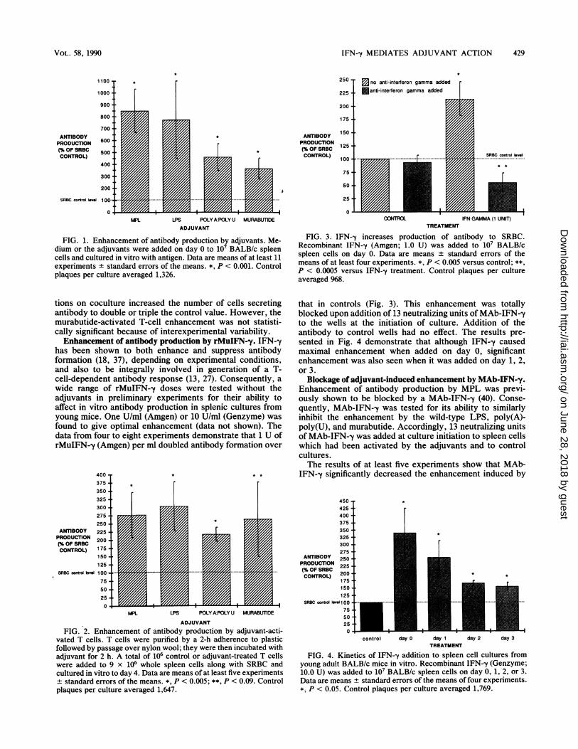
Enhancement of antibody production by adjuvants. Micro-bial products representing three different classes of com-pounds have been well documented as adjuvants to theimmune response. To verify this under our conditions, LPS,poly(A)-poly(U) complex, murabutide, and the nontoxicMPL were tested for their ability to enhance formation ofantibody to SRBC in splenic cultures from BALB/c mice.The mean number of PFC from at least 11 experiments isillustrated in Fig. 1. MPL and LPS enhanced antibodyformation to SRBC about sevenfold above that in controlsreceiving SRBC alone. Poly(A)-poly(U) and murabutide alsowere effective, but somewhat less so under these conditions,enhancing antibody formation about 3.5- and 2.5-fold, re-spectively.Enhancement of antibody production by adjuvant-activated
T cells. Our previous study with aging mice demonstratedthat T lymphocytes are important mediators of the adjuvantaction of a detoxified LPS, MPL (40). To determine whethera wild-type LPS and other adjuvants act in a like manner inyoung mice, we tested T cells that were treated with LPS,poly(A)-poly(U), and murabutide for their ability to elevateantibody formation on coculture. Control or adjuvant-stim-ulated T cells (106 cells) were added to unseparated spleencells (9 x 10⁶ cells) and incubated with antigen for 4 days.Results in Fig. 2 show the averaged data for 5 to 12experiments. Each of the adjuvant-stimulated T-cell popula-
INFECT. IMMUN.
on June 28, 2018 by guesthttp://iai.asm
.org/D
ownloaded from
IFN-y MEDIATES ADJUVANT ACTION 429
1100
1000
900
800
700'ANTIBODYPRODUCTION 600(% OF SRBC 500CONTROL)
400
300
200
SRBC control bvel 1 00-
0.
1ANTIBODY
PRODUCTION(% OF SRBCCONTROL)
I *ZIIZA. Z11M.~IZ
MPL LPS POLY A:POLY U MURABUTIDEADJUVANT
FIG. 1. Enhancement of antibody production by adjuvants. Me-dium or the adjuvants were added on day 0 to 107 BALB/c spleencells and cultured in vitro with antigen. Data are means of at least 11experiments + standard errors of the means. *, P < 0.001. Controlplaques per culture averaged 1,326.
tions on coculture increased the number of cells secretingantibody to double or triple the control value. However, themurabutide-activated T-cell enhancement was not statisti-cally significant because of interexperimental variability.Enhancement of antibody production by rMuIFN--y. IFN--y
has been shown to both enhance and suppress antibodyformation (18, 37), depending on experimental conditions,and also to be integrally involved in generation of a T-cell-dependent antibody response (13, 27). Consequently, awide range of rMuIFN-,y doses were tested without theadjuvants in preliminary experiments for their ability toaffect in vitro antibody production in splenic cultures fromyoung mice. One U/ml (Amgen) or 10 U/ml (Genzyme) wasfound to give optimal enhancement (data not shown). Thedata from four to eight experiments demonstrate that 1 U ofrMuIFN--y (Amgen) per ml doubled antibody formation over
400375350325300275250
ANTIBODY 225PRODUCTION 200(% OF SRBCCONTROL) 175
150 -
125-SRBC contral level 100 -
75 -
50-25 i
Ir
0o za i
{ |IA
MPL LPS POLY A:POLY U MURABUTiDE
ADJUVANT
FIG. 2. Enhancement of antibody production by adjuvant-acti-vated T cells. T cells were purified by a 2-h adherence to plasticfollowed by passage over nylon wool; they were then incubated withadjuvant for 2 h. A total of 106 control or adjuvant-treated T cellswere added to 9 x 10⁶ whole spleen cells along with SRBC andcultured in vitro to day 4. Data are means of at least five experiments+ standard errors of the means. *, P < 0.005; **, P < 0.09. Controlplaques per culture averaged 1,647.
250
225
200
175-
150 -
125 -
100 -
75.
50 -
25 -
0O
V no anti-interferon gamma added
fanti-interferon gamma added
..................
CONTROL
SRBC control level
M I V,1101MIA _IFN GAMMA (1 UNIT)
TREATMENT
-i
FIG. 3. IFN--y increases production of antibody to SRBC.Recombinant IFN-y (Amgen; 1.0 U) was added to 107 BALB/cspleen cells on day 0. Data are means + standard errors of themeans of at least four experiments. *, P < 0.005 versus control; **,P < 0.0005 versus IFN--y treatment. Control plaques per cultureaveraged 968.
that in controls (Fig. 3). This enhancement was totallyblocked upon addition of 13 neutralizing units of MAb-IFN--yto the wells at the initiation of culture. Addition of theantibody to control wells had no effect. The results pre-sented in Fig. 4 demonstrate that although IFN--y causedmaximal enhancement when added on day 0, significantenhancement was also seen when it was added on day 1, 2,or 3.
Blockage of adjuvant-induced enhancement by MAb-IFN-'y.Enhancement of antibody production by MPL was previ-ously shown to be blocked by a MAb-IFN--y (40). Conse-quently, MAb-IFN-y was tested for its ability to similarlyinhibit the enhancement by the wild-type LPS, poly(A)-poly(U), and murabutide. Accordingly, 13 neutralizing unitsof MAb-IFN--y was added at culture initiation to spleen cellswhich had been activated by the adjuvants and to controlcultures.The results of at least five experiments show that MAb-
IFN--y significantly decreased the enhancement induced by
450 -
425 t400-375 t350 i325 i300 i275 -
ANTIBODY 250 -
PRODUCTION 225 |(% OF SRBC 200 -CONTROL)20
175-1501255
SRBC control level 1 00 -
7550-25-0-
1.
control day 0 day 1 day 2 day 3TREATMENT
FIG. 4. Kinetics of IFN-y addition to spleen cell cultures fromyoung adult BALB/c mice in vitro. Recombinant IFN--y (Genzyme;10.0 U) was added to 107 BALB/c spleen cells on day 0, 1, 2, or 3.Data are means + standard errors of the means of four experiments.*, P < 0.05. Control plaques per culture averaged 1,769.
VOL. 58, 1990
I
;
-1
I
on June 28, 2018 by guesthttp://iai.asm
.org/D
ownloaded from

430 ODEAN ET AL.
ANTIBODY 400 -
PRODUCTION(% OF SRBCCONTROL) 300--
200--
SRBC control level 100 -
0IISRBC CONTROL UPS POLY A:POLY U MURABUTIDE
ADJUVANT
FIG. 5. Blocking of adjuvant enhancement by anti-IFN-y anti-body. Medium or adjuvants were added to BALB/c spleen cells onday 0 along with SRBC. Data are means of at least five experiments± standard errors of the means. *, P < 0.05 versus appropriatecontrol. Control plaques per culture averaged 1,320.
LPS (66%), poly(A)-poly(U) (48%), and murabutide (44%)(Fig. 5). Addition of MAb to control cultures did notsignificantly affect antibody production.Augmentation of IFN-y release by treatment with adjuvant.
Since IFN-y was implicated as a mediator released fromadjuvant-activated T cells and 13 U of MAb-IFN-y blockedapproximately half of the adjuvant-induced enhancement,we tested whether adjuvant-activated T cells released in-creased levels of IFN-,y compared with control T cells.Accordingly, T cells enriched by removal of adherent cellsand passage over nylon wool were activated for 2 h with theadjuvants, washed, and cultured for 48 h. The IFN--y titers inthe resultant supernatant fluids from four experiments areshown in Fig. 6. MPL, LPS, and murabutide induced signif-icant increases in IFN--y release by T cells, which averagedabout threefold higher than that in controls, while poly(A)-poly(U) doubled IFN--y levels.NK cells and IFN--y. NK cells, as well as T lymphocytes,
800 T
700
600 4MEAN PERCENTOF CONTROL(INTERFERONUNITS/ML)
500 4 i400 4300-
200-
control level 100
0. +IMPL LPS POLY A:POLY U MURABUTIDE
TREATMENT
FIG. 6. IFN--y release is augmented by 2-h adjuvant activation ofenriched T cells. Enriched T cells (107) were activated for 2 h withadjuvants, washed three times, and suspended in medium. After 48h, supernatants were collected and frozen until an enzyme-linkedimmunosorbent assay was performed. Data are means standarderrors of the means of four experiments. *, P < 0.05; **, P < 0.12.Control IFN units per milliliter averaged 1.10.
TREATMENT
FIG. 7. NK cells do not mediate the adjuvant action of MPL orLPS. Data are means of two experiments + standard errors of themeans. Control plaques per culture averaged 419.
have been shown to release IFN--y (7). NK cell-depletedspleen cell populations were tested for their responsivenessto MPL and LPS. There was no loss in the adjuvanticity ofMPL or LPS in two experiments when spleen cells weretreated with anti-asialo GM1 to remove NK cells (Fig. 7).This antibody removed essentially all NK cell activity whentested in our laboratory (data not shown).
DISCUSSION
The three adjuvants under study in this investigation arefrom diverse sources and are structurally dissimilar. Yet itappears that IFN-y release is integrally involved in theirstimulatory action. Although each most likely has a differentreceptor, the data suggest the interesting possibility that thetransmembrane signals activated by each adjuvant workthrough the same intracellular pathway and G-protein signaltransducer leading to IFN--y release.The results obtained in this study implicate IFN--y-pro-
ducing T cells (Thl) as initial target cells of the adjuvants. Tccells were ruled out as the source of IFN--y in previous workwith MPL (40), in which enhancement remained followingtreatment with monoclonal anti-Lyt2.2 antibody plus com-plement. The IFN--y released presumably increases la anti-gen (38) and/or interleukin-1 (IL-1) (3) release from macro-phages, leading to increased antibody production.Acceptance of this hypothesis is tempered with the knowl-edge that these immunomodulators are known to affectdirectly the release of IL-1 from macrophages (31, 33;unpublished observations). In addition, several investigatorshave reported IL-1-induced release of IL-2 as obligatory toIFN--y release (8, 9, 23, 41), leading to the rationale that theinitial target cell should be the macrophage. However, Thlclones are known to synthesize IL-2 and IFN--y but lackreceptors for IL-1 and do not respond to IL-1 (12, 20). Sinceall three adjuvants increase IFN--y release, the hypothesisfavored is that IFN--y is the initiating cytokine of the result-ant enhancement of antibody formation. It should also beemphasized that following a mitogenic stimulus, IFN-ymRNA production has been shown to be biphasic, withaccumulation peaking at 60 min and 20 h (11). The initialmRNA peak for IFN--y appeared simultaneously with that ofIL-2 mRNA, suggesting that the early IFN-,y release is notdependent on IL-2 (11). The late-appearing IFN--y is de-creased by exogenous IL-2 and antibody specific for the IL-2
INFECT. IMMUN.
on June 28, 2018 by guesthttp://iai.asm
.org/D
ownloaded from

IFN--y MEDIATES ADJUVANT ACTION 431
receptor (Tac) and thus depends on IL-2-IL-2 receptorinteraction (42).These observations offer an explanation for the recent
results of Tomai and Johnson (40). When MPL was given invivo 2 h prior to sacrifice, followed by transfer of isolatedcell populations to a Mishell-Dutton system, macrophageswere found capable of transferring MPL-induced enhance-ment. However, when MPL was added to a totally in vitrosystem, only T cells were found capable of transferring theadjuvant action. The explanation for this dichotomy may liein the probability that the 2-h interval following in vivoinjection was sufficient time for the adjuvant to activate Tcells to release IFN-y. The IFN--y then could instill inmacrophages the capability for continuation of the cascadeleading to increased numbers of PFC. The rapid induction ofIL-1 by IFN-y also may account for the inability of anti-IFN-y antibody to block completely the enhancing action,despite the use of high levels of antibody (data not shown).T cells have been implicated previously as being affected
by all three adjuvants used in this study. Thus, Allison andDavies (2), McGhee et al. (29), Nakano et al. (32), and Vogelet al. (43) all have reported the involvement of T cells in theadjuvant action of LPS. In addition, Baker et al. (4) andTomai and Johnson (40) have identified a T cell as the targetof the detoxified endotoxin, MPL. Bick and Johnson (5)reported secretion of a helper factor from T lymphocytesfollowing poly(A)-poly(U) stimulation which increased anti-body production. This followed earlier studies showing thatpoly(A)-poly(U) amplifies thymus cell functions (16). Morerecently, Hovanessian et al. (15) reported that poly(A)-poly(U) stimulation resulted in increased p67K kinase activ-ity in mice and humans, which has been shown to be amarker for the presence of IFN--y. Murabutide has also beenshown to act on T cells (24). These studies coupled with thedemonstration by Lei and Morrison (26) of LPS-bindingreceptors on murine splenocytes, including T cells, offerevidence for an initial adjuvant signal bestowed on T cells.Although NK cells are known to possess immunoregulatorycapability (1) and to secrete IFN-y, any role for NK cells isunlikely since enhancement by both MPL and LPS remainedafter NK cells were removed by in vitro treatment withanti-asialo GM1 antibody (Fig. 7).
Despite reports that IFN--y suppresses the antibody re-sponse (17, 18) and is required for the activity of naturalsuppressor cells (14), other studies have demonstrated apositive or perhaps even obligatory requirement for IFN--y.Thus, Sonnenfeld et al. (37) and Shalaby et al. (35) found thatIFN--y enhanced the antibody responses when added exog-enously. It was suggested (37) that the antibody enhance-ment was mediated by T lymphocytes while the observedimmunosuppression was mediated by B lymphocytes. Sev-eral investigators (13, 27) have shown that IFN--y is involvedin, if not required for, the normal generation of antibody byB cells. We could not conclude this from our experiments,however, even though the MAb-IFN--y used originated fromthe same hybridoma cell line as that used by Helman andWallace (13). When we added the antibody in vitro on day 0to spleen cell cultures with SRBC (without adjuvant) in 43experiments, the mean percentage ± standard error of themean relative to the control without antibody was 93 ± 9.This dose (13 U) of MAb-IFN--y was sufficient to signifi-cantly decrease adjuvant-induced enhancement of antibodyproduction (Fig. 5). An explanation of the different resultsmay be that high doses of antigen induce increased amountsof IFN-y, since Helman and Wallace (13) used approxi-mately 10-fold more SRBC than was used in our experi-
ments. The experiments by Langford et al. (22), which alsoshowed blocking of the antibody response to SRBC withMAb-IFN-y, used both a high SRBC dose (108 cells) and theintravenous route, which is generally more effective ingenerating antibody than the intraperitoneal route. Since wedid not stimulate with antigen at a high level in our SRBCcontrol group, it is probable that IFN--y was not beingproduced optimally and could then be induced by the adju-vants, resulting in increased numbers of PFC. This is sup-ported by the oft-noted evidence that adjuvant activity ismost obvious in models in which antibody synthesis is belownormal and raises the question whether the IFN--y levels area key controlling variable in those situations.While differing experimental conditions may explain in
part the opposing suppression and enhancement induced byIFN--y, no general concept has emerged as to the immuno-regulatory mechanism of this cytokine. Though the adju-vants seem to act via IFN--y released by T cells very early inthe response (1 to 2 h), IFN--y also significantly increasedantibody production when added late to cultures (Fig. 4). Insupport of this, Leibson et al. (28) added IL-X, which waslater identified as IFN-y (27), to purified B cells with IL-1and P388D1 supernatants on days 0, 1, and 2 of culture. Peakresponse was seen with IL-X (IFN--y) addition on day 1, buta significant increase over cultures with only IL-1 andP388D1 supernatants was seen with IL-X added on days 0and 2. A possible explanation of this observation comes fromthe results of Sidman et al. (36), which suggest that IFN--ymay be involved in the maturation of B cells to activeantibody production, which occurs late in the response.Thus, the observed biphasic release of IFN--y reported byGrabstein et al. (11) may have the following explanation.Early induction of IFN-y by a mitogen, adjuvant, or antigenresults in macrophage activation and increased levels of Iaantigen and IL-1. IL-1 increases IL-2 receptor expressionand IL-2 release, inducing additional IFN--y, which in turnaids in the maturation of B cells to immunoglobulin secre-tion.
ACKNOWLEDGMENT
This research was supported by Public Health Service grants Al25810 and AG 06198 from the National Institutes of Health.
LITERATURE CITED1. Abruzzo, L. V., and D. A. Rowley. 1983. Homeostasis of the
antibody response: immunoregulation by NK cells. Science222:581-585.
2. Allison, A. C., and A. J. S. Davies. 1971. Requirement ofthymus-dependent lymphocytes for potentiation by adjuvants ofantibody formation. Nature (London) 233:330-332.
3. Arenzana-Seisdedos, F., J. L. Virelizier, and W. Fiers. 1985.Interferons as macrophage-activating factors. III. Preferentialeffects of interferon--y on the interleukin-1 secretory potential offresh or aged human monocytes. J. Immunol. 134:2444-2448.
4. Baker, P. J., J. R. Hiernaux, M. B. Fauntleroy, B. Prescott, J. L.Cantrell, and J. A. Rudbach. 1988. Inactivation of suppressorT-cell activity by nontoxic monophosphoryl lipid A. Infect.Immun. 56:1076-1083.
5. Bick, P. H., and A. G. Johnson. 1977. Poly A:U-inducedsecretion of T-lymphocyte helper factors. Scand. J. Immunol.6:1133-1144.
6. Chedid, L., M. Parant, F. Audibert, G. Riveau, F. Parant, E.Lederer, J. Choay, and P. Lefrancier. 1982. Biological activity ofa new synthetic muramyl peptide adjuvant devoid of pyrogenic-ity. Infect. Immun. 35:417-424.
7. Djeu, J. Y., N. Stocks, K. Zoon, G. J. Stanton, T. Timonen, andR. B. Herberman. 1982. Positive self regulation of cytotoxicityin human natural killer cells by production of interferon upon
VOL. 58, 1990
on June 28, 2018 by guesthttp://iai.asm
.org/D
ownloaded from

432 ODEAN ET AL.
exposure to influenza and herpes viruses. J. Exp. Med. 156:1222-1234.
8. Epstein, L. B., M. J. Cline, and T. C. Merigan. 1971. PPD-stimulated interferon: in vitro macrophage-lymphocyte interac-tion in the production of a mediator of cellular immunity. Cell.Immunol. 2:602-613.
9. Farrar, W. L., H. M. Johnson, and J. J. Farrar. 1981. Regula-tion of the production of immune interferon and cytotoxic Tlymphocytes by interleukin 2. J. Immunol. 126:1120-1125.
10. Galanos, C., 0. Luderitz, and 0. Westphal. 1969. A new methodfor the extraction of R lipopolysaccharides. Eur. J. Biochem.9:245-249.
11. Grabstein, K., S. Dower, S. Gillis, D. Urdal, and A. Larsen.1986. Expression of interleukin 2, interferon--y and the IL-2receptor by human peripheral blood lymphocytes. J. Immunol.136:4503-4508.
12. Greenbaum, L. A., J. B. Horowitz, A. Woods, T. Pasqualini, E.Reich, and K. Bottomly. 1988. Autocrine growth of CD4' Tcells. Differential effects of IL-1 on helper and inflammatory Tcells. J. Immunol. 140:1555-1560.
13. Helman, S. W., and J. H. Wallace. 1989. Differential regulationof the immune response to SRBC by monoclonal antibodies tointerferon-y. Proc. Soc. Exp. Biol. Med. 191:55-59.
14. Holda, J. H., T. Maier, and H. N. Claman. 1988. Evidence thatIFN y is responsible for natural suppressor activity in GVHDspleen and normal bone marrow. Transplantation 45:772-777.
15. Hovanessian, A. G., Y. Riviere, L. Montagnier, M. Michelson, J.Lacour, and F. Lacour. 1982. Enhancement of interferon-medi-ated protein kinase in mouse and human plasma in response totreatment with poly A:poly U. J. Interferon Res. 2:209-215.
16. Johnson, A. G. Modulation of the immune system by syntheticpolynucleotides. 1979. Springer Semin. Immunopathol. 2:149-168.
17. Johnson, H. M., and S. Baron. 1976. The nature of the suppres-sive effect of interferon and interferon inducers on the in vitroimmune response. Cell. Immunol. 25:106-115.
18. Johnson, H. M., and B. A. Torres. 1983. Recombinant mouseinterferon--y regulation of antibody production. Infect. Immun.41:546-548.
19. Julius, M., E. Simpson, and L. Herzenberg. 1973. A rapidmethod for the isolation of functional thymus-derived murinelymphocytes. Eur. J. Immunol. 3:645-649.
20. Kaye, J., S. Gillis, S. B. Mizel, E. M. Shevach, T. R. Malek,C. A. Dinarello, L. B. Lachman, and C. A. Janeway, Jr. 1984.Growth of a cloned helper T cell line induced by a monoclonalantibody specific for the antigen receptor: interleukin 1 isrequired for the expression of receptors for interleukin 2. J.Immunol. 133:1339-1345.
21. Kennedy, J., and M. Axelrad. 1971. An improved assay forhemolytic plaque-forming cells. Immunology 20:253-261.
22. Langford, M. P., D. A. Weigent, T. S. Chan, H. M. Johnson, andG. J. Stanton. 1987. Antibodies to the carboxyl terminus ofmouse interferon-y neutralize its immunoregulatory and antivi-ral activities. J. Interferon Res. 7:95-101.
23. Le, J., J. X. Lin, D. Henriksen-DeStefano, and J. Vilcek. 1986.Bacterial lipopolysaccharide-induced interferon-gamma produc-tion: roles of interleukin 1 and interleukin 2. J. Immunol.136:4525-4530.
24. Leclerc, C., and F. R. Vogel. 1986. Synthetic immunomodula-tors and synthetic vaccines. Crit. Rev. Ther. Drug Carrier Syst.2:353-406.
25. Lefrancier, P., M. Derrien, X. Jamet, J. Choay, E. Lederer, F.Audibert, M. Parant, F. Parant, and L. Chedid. 1982. Apyro-genic, adjuvant-active N-acetyl-muramyl dipeptides. J. Med.
Chem. 25:87-90.26. Lei, M.-G., and D. C. Morrison. 1988. Specific endotoxic
lipopolysaccharide-binding proteins on murine splenocytes. I.Detection of lipopolysaccharide-binding sites on splenocytesand splenocyte subpopulations. J. Immunol. 141:996-1005.
27. Leibson, H. J., M. Gefter, A. Zlotnik, P. Marrack, and J. W.Kappler. 1984. Role of -y-interferon in antibody-producing re-sponses. Nature (London) 309:799-801.
28. Leibson, H. J., P. Marrack, and J. Kappler. 1982. B cell helperfactors. II. Synergy among three helper factors in the responseof T cell- and macrophage-depleted B cells. J. Immunol. 129:1398-1402.
29. McGhee, J. R., J. J. Farrar, S. M. Michalek, S. E. Mergenhagen,and D. L. Rosenstreich. 1979. Cellular requirements for lipopoly-saccharide adjuvanticity. A role for both T lymphocytes andmacrophages for in vitro responses to particulate antigens. J.Exp. Med. 149:793-807.
30. Mishell, R., and R. Dutton. 1967. Immunization of dissociatedspleen cell cultures in normal mice. J. Exp. Med. 126:423-442.
31. Morrison, D. C., and J. L. Ryan. 1979. Bacterial endotoxins andhost immune responses. Adv. Immunol. 28:293-451.
32. Nakano, M., T. Uchiyama, and K. Saito. 1973. Adjuvant effect ofendotoxin: antibody response to sheep erythrocytes in miceafter transfer of syngeneic lymphoid cells treated with bacteriallipopolysaccharide in vitro. J. Immunol. 110:408-413.
33. Oppenheim, J. J., A. Togawa, L. Chedid, and S. Mizel. 1980.Components of mycobacteria and muramyl dipeptide with adju-vant activity induce lymphocyte activating factor. Cell. Immu-nol. 50:71-81.
34. Qureshi, N., and K. Takayama. 1982. Purification and structuraldetermination of nontoxic lipid A obtained from the lipopoly-saccharide of Salmonella minnesota. J. Biol. Chem. 257:11808-11815.
35. Shalaby, M. R., P. K. Weck, E. Rinderknecht, R. N. Harkins,J. W. Frane, and M. J. Ross. 1984. Effects of bacteria-producedhuman alpha, beta and gamma interferons on in vitro immunefunctions. Cell. Immunol. 84:380-392.
36. Sidman, C. L., J. D. Marshall, L. D. Shulz, P. W. Gray, andH. M. Johnson. 1984. -y-Interferon is one of several direct B cellmaturing lymphokines. Nature (London) 309:801-803.
37. Sonnenfeld, G., A. D. Mandel, and T. C. Merigan. 1978. Timeand dosage dependence of immunoenhancement by murine typeII interferon preparations. Cell. Immunol. 40:285-293.
38. Steeg, P. S., R. N. Moore, H. M. Johnson, and J. J. Oppenheim.1982. Regulation of murine macrophage Ia antigen expressionby a lymphokine with immune interferon activity. J. Exp. Med.156:1780-1793.
39. Takayama, K., E. Ribi, and J. L. Cantrell. 1981. Isolation of anontoxic lipid A fraction containing tumor regression activity.Cancer Res. 41:2654-2657.
40. Tomai, M. A., and A. G. Johnson. 1989. Gamma interferonmediates the adjuvant action of a detoxified endotoxin. J. Biol.Response Modif. 8:643-661.
41. Torres, B. A., W. L. Farrar, and H. M. Johnson. 1982. Inter-leukin 2 regulates immune interferon (IFN gamma) productionby normal and suppressor cell cultures. J. Immunol. 128:2217-2219.
42. Vilcek, J., D. Henriksen-DeStefano, D. Siegel, A. Klion, R. J.Robb, and J. Le. 1985. Regulation of IFN -y induction in humanperipheral blood cells by exogenous and endogenously pro-duced interleukin 2. J. Immunol. 135:1851-1856.
43. Vogel, S. N., M. L. Hilfiker, and M. J. Caulfield. 1983. Endo-toxin-induced T lymphocyte proliferation. J. Immunol. 130:1774-1779.
INFECT. IMMUN.
on June 28, 2018 by guesthttp://iai.asm
.org/D
ownloaded from
![ORAL FAST DISSOLVING FILMS-AN INNOVATIVE DRUG DELIVERY … · 2019. 12. 10. · KEYWORDS: Oral dissolving film, Film forming polymer, Challenges, Evaluation. ORAL DISSOLVING FILMS[22,30,60,68]](https://img.pdfslide.us/doc/110x75/61296ff54735c37777083b50/oral-fast-dissolving-films-an-innovative-drug-delivery-2019-12-10-keywords.jpg)