Embed Size (px)
Citation preview
Histol Histopalhol (2000) 15: 825-834
001 : 10.14670/HH-15.825
http://www.hh.um.es
Histology and Histopathology
Cellular and Molecular Biology
Invited Review
The distribution of cholinergic neurons in the human central nervous system V. Oda and I. Nakanishi First Department of Pathology, Faculty of Medicine, Kanazawa University, Japan
Summary. Choline acetyltransferase (ChAT), the enzyme responsible for the biosynthesis of acetylcholine, is presently the most specific marker for identifying cholinergic neurons in the central and peripheral nervous systems. The present article reviews immunohistochemical and in situ hybridization studies on the distribution of neurons ex pressing ChAT in the human central nervous system. Neurons with both immunoreactivity and in situ hybridization signals of ChAT are observed in the basal forebrain (diagonal band of Broca and nucleus basalis of Meynert), striatum (caudate nucleus, putamen and nucleus accumbens), cerebral cortex, mesopontine tegmental nuclei (pedunculopontine tegmental nucleus , laterodorsal tegmental nucleus and parabigeminal nucleus), cranial motor nuclei and spinal motor neurons. The cerebral cortex displays regional and laminal differences in the distribution of neurons with ChAT. The medial seotal nucleus and medial habenular nucleus contain immunoreactive neurons for ChAT, which are devoid of ChAT mRNA signals. This is probably because there is a small number of cholinergic neurons with a low level of ChAT gene expression in these nuclei of human. Possible connections and speculated functions of these neurons are briefly summarized.
Key words: Choline acetyltransferase, Central nervous system, Human, Immunohi s tochemistry, In situ hybridization
Introduction
Neurons that synthesize and release acetylcholine for neurotransmission are referred to as cholinergic neurons . Cholinergic neurons in the mammalian central nervous system are thought to play an important role in fundamental brain functions, such as learning, memory ,
Offprint requests to: Dr. Yoshio Oda, MD, Associate professor, First Department of Pathology, Faculty of Medicine, Kanazawa University, 13-1 Takara-machi, Kanazawa, Ishikawa 920·8640, Japan , e·mail : yoda@med,kanazawa-u ,ac.jp
arousal, sleep and movement (Butcher and Woolf, 1986; Woolf, 1991). Selective loss of cholinergic neurons in the basal forebrain has been observed in Alzheimer's disease (Perry et ai., 1978 ; Whitehouse et ai., 1981). Amyotrophic lateral sclerosis and Huntington's disease a re other neurodegenerative di sorders in which cholinergic neurons are affected. Choline acetyltransferase (ChAT, acetyl CoA:choline O-acetyltransferase , EC 2.3.1.6), the enzyme responsible for the biosynthesis of acetylcholine, is presently the most specific indicator for monitoring the functional state of cholinergic neurons in the central and peripheral nervous systems. This enzyme is also a useful specific marker for identifying cholinergic neurons in the nervous system. During the past 20 years, seve ral polyclonal and monoclonal anti-ChAT antibodies have been developed and applied in anatomical analyses of the central cholinergic organization in mammalian species. More recently, following the successful cDNA cloning for ChAT, an in situ hybridization technique has been induced to detect ChAT mRNA in neurons for identifying authentic cholinergic neurons. The present article reviews immunohistochemical and in situ hybridization studies on the distribution of neuron s expressing ChAT in the human central nervous system.
Localization of neuronal cell body with ChAT immunoreactivity and in situ hybridization signals
Based on immunohistochemical studies of monkey and rodent brains , Mesulam (1988) proposed a nomenclature to describe ChAT-immonoreactive neurons which are aggregated in eight major groups: Chl-Ch8. Chi designates the ChAT-containing neurons associated with the medial septal nucleus , Ch2 is associated with the vertical nucleus of the diagonal band of Broca, Ch3 with the horizontal limb nucleus of the diagonal band of Broca , Ch4 with the nucleus basalis of the substantia innominata, ChS with the pedunculopontine nucleus of the rostal brain stem, Ch6 with the laterodorsa l tegmental nucleus of the rostal brain stem, Ch7 with the medial habenula, and Ch8 is associated with those neurons in the parabigeminal nucleus. In addition, the
826
Human central cholinergic neuron
striate nucleus, cerebral cortex, so me cranial nerve nuclei and the spinal cord gray matter also contain ChAT-immunoreactive neurons . This cl assifica tion is applicable in describing the human central cholinergic system. In human, ChAT-immunoreactive neurons are
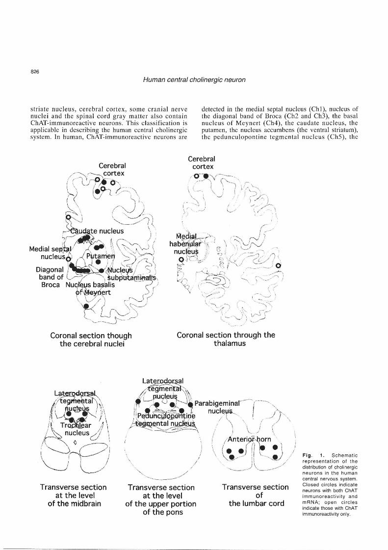
detected in the medial septal nucleus (ChI), nucleus of the diagonal band of Broca (Ch2 and Ch3), the basal nucleu s of Meynert (Ch4), the caudate nucleus, the putamen, the nucleus accumbens (the ventral striatum), the pedunculoponline tegm e ntal nucl eus (Ch5), th e
, .
Coronal section though the cerebral nuclei
Coronal section through the thalamus
Transverse section at the level
of the midbrain
'.
Transverse section at the level
of the upper portion of the pons
Transverse section of
the lumbar cord
Fig. 1. Schematic representation of the distribution of chOlinergic neurons in the human central nervous system, Closed circles indicate neurons with both ChAT immunoreactivity and mRNA; open circles indicate those with ChAT immunoreactivity only,
827
Human central cholinergic neuron
laterodorsal tegmental nucleus (Ch6), the medial habenular nucleus (Ch7), the parabigeminal nucleus (Ch8), some distinct areas of the cerebral cortex, some cranial nerve nuclei, and the anterior gray horn of the spinal cord. Furthermore, nucleu s subputaminalis (Ayala) , which has long been disregarded in morphological studies, contains ChAT-immunoreactive neurons (Simic et aI., 1999). Table 1 summarizes the results of immunohistochemical and in situ hybridization studies and Figure 1 shows a schematic representation of the distribution of neurons containing ChAT in the human central nervous system.
I. Magnocel/ular nuclei of the basal forebrain
Magnocellular nuclei of the basal forebrain are composed of the medial septal nucleus, the nucleus of the diagonal band and the basal nucleus of Meynert. These structures are tightly connected to each other (Ulfig, 1989). In addition, a recent histochemical and immunohistochemical study pointed out that the nucleus subputaminalis is also a component of the magnocellular complex (Simic et aI., 1999).
1. Medial septal nucleus (ChI)
The medial septal nucleus located in the septum verum forms the anteromost portion of the basal magnocellular complex. Large , ovoid neurons with vertical orientation are immunopositive for ChAT. The immunoreactivity of the neurons is relatively weak compared with that of neurons in other magnocellular complex areas such as the nucleus basalis of Meynert. In situ hybridization failed to detect positive signals for ChAT mRNA in the neurons. This is probably because there is a small number of cholinergic neurons with a
A
low level of ChAT gene expression in this nucleus (Kasashima et aI., 1998).
2. Diagonal band of Broca (Ch2 and Ch3)
The diagonal band of Broca is located from the ventromedial portion of the lateral septum (vertical limb of the diagonal band, Ch2) to the ventrorostromedial portion of the subcommissural region (horizontal limb of the diagonal band, Ch3) below Ch4. The boundary between the medial septal nucleus and the vertical limb of the diagonal band (Ch2) is not clearly defined, an area in which neurons are rather sparsely distributed. Sometimes, the two structures are categorized together as Ch1-2. The shape of Ch3 is ill-defined, where neurons are scattered randomly. ChAT-immunoreactive neurons in the diagonal band are large, polygonal (particularly in the vertical) or fusiform (particularly in the horizontal limb). An in situ hybridization study revealed that large neurons in this area contain hybridization signals for ChAT (Kasashima et aI., 1998).
3. Nucleus basalis of Meynert (Ch4)
The nucleus basalis of Meynert is a continuous plate of neurons, which occupies the sub lenticular region between the sagittal levels of the olfactory tubercle and the lateral geniculate nucleus . Based on a cytoarchitectural study of monkey brain (Mesulam et aI., 1983), Mesulam and Geula (1988) stated that the human Ch4 complex can also be subdivided into six sectors that occupy its anteromedial (Ch4am), anterolateral (Ch4al), anterointermediate (Ch4ai), intermediodorsal (Ch4id), intermedioventral (Ch4iv) and posterior (Ch4p) regions. Some other researchers used this classification in morphological studies of the human basal forebrain
B Fig. 2. Immunohistochemistry (A) and in situ mRNA hybridization (8. dark field) studies of ChAT in the nucleus basalis of Meynert. Most of the large neurons are labeled. x 87.5
828
Human central cholinergic neuron
Table 1. Regional distribution of neurons with choline acetyltransferase immunoreactivity and mRNA in the human central nervous system
IMMUNOHISTOCHEMISTRY IN SITU HYBRIDIZATION
Basal forebrain Medial septal nucleus Nucleus of diagonal band of Broca Nucleus basalis of Meynert Nucleus subputaminalis Amygdala
Basal ganglia Caudate nucleus Putamen Globus pall idus
Thalamus
Epithalamus Medial habenular nucleus Lateral habenular nucleus
Hippocampus
Enthorhinal cortex
Neocortex Brodmann's areas
1, 2, 3 (somatosensory) 4 (somatomotor) 6, 8, 9 (premotor) 17 (fi rst visual) 18 (secondary visual) 20,21 22 (secondary auditory) 23,31 (posterior half of cingulate) 24 (anterior half of cingulate) 39,40 41, 42 (first auditory) 46
Subthalamic nucleus
Substantia nigra
Red nucleus
Mesencephalon Pedunculopontine tegmental nucleus Laterodosal tegmental nucleus Parabigeminal nucleus Pontine nucleus
Cerebellum
Cranial nerve nuclei Oculomotor nucleus Trochlear nucleus
Spinal cord (anterior horn)
(Mufson et aI., 1989; Lehericy et aI. , 1993). However, a three-dimensional reconstruction study (Ulfig, 1989; Halliday et aI., 1993) revealed that these six subgroups were difficult to clearly distinguish from each other. Halliday e t al. (1993) divided the nucl e us by the surrounding structures into four subgroups; Ch4a, Ch4ac (corresponding to the nucleus subputaminali s, as described below), Ch4i, and Ch4p regions. Ch4a and Ch4ac are situated between the olfactory tubercle and the posterior edge of the anterior commisure; Ch4i, between the posterior edge of the anterior commisure and the ansa peduncularis; and Ch4p, posterior to the ansa peduncularis. The majority of neurons in Ch4 are
+ + + +
+ +
+
+
+ +
+
+
+
+
+ + +
+ +
+
+ + +
+ +
+
+
+
+
+ + +
+ +
+
large and polymorphic. Most of the large neurons are ChAT-immunopositive (80-90% according to the calculation by Mesulam et aI., 1988) and with RNA signals (Kasashima et aI., 1998) (Fig. 2). Their shapes are heterogeneous, ranging from fusiform to mUltipolar. Almost all neurons with ChAT immunoreactivity also express the nerve growth factor receptor (Mufson et aI., 1989).
4. Nucleus subputaminalis
Although the nucleus subputaminalis was first described many years ago by Ayala (1915), the nucleus
829
Human central cholinergic neuron
is unfamiliar to many scientists, including neuroanatomists. The nucleus subputaminalis is well developed in the human brain and located ventrolaterally to the putamen. This nucleus represents a rostrolateral extension of the basal magnocellular complex. Halliday et al. (1993) subgrouped this nucleus as Ch4ac. The current immunohistochemical study (Simic et a1., 1999) demonstrated that 80-90% of the cell bodies contained both ChAT and the vitamin-D-dependent calcium binding protein calbindin-D28k with the remainder of cell bodies showing positivity for nicotinamide adenine dinucleotide phosphate diaphorase.
II. Striatum
The striatum, consisting of the caudate nucleus, the putamen and the nucleus accumbens, is relatively homogenous in terms of cell types. There are three types of neurons: medium-sized spiny neurons (neurons with spiny dendrites); medium-sized aspiny neurons (neurons with aspiny dendrites); and large aspiny neurons. Large multipolar neurons, which correspond to large aspiny neurons, express ChAT (Hirsch et al., 1989; Mesulam et a I., 1992; Selden et aI., 1994; Holt et aI., 1996 ; Kasashima et al., 1998) (Fig. 3) or nicotinamide adenine dinucleotide phosphate diaphorase (Selden et al., 1994). ChAT-positive large neurons make up 1-2% of the total cell population in the striatum (Holt et al., 1996), and most of them are considered to be interneurons with intrinsic projections. The distribution of cholinergic neurons in the striatum is in a relatively even manner, whereas that of the ChAT-immunopositive neuropils is not uniform (Hirsch et al., 1989; Mesulam et al., 1992; Selden et al., 1994; Holt et al., 1996). Patches of weak ChAT immunoreactivity, called striosomes, are embedded in a strong ChAT-immunoreactive background, called a matrix. The irregular density of cholinergic fibers in the striatum may be related to the
A
compartmental organization for processing different types of information.
III. Cerebral cortex
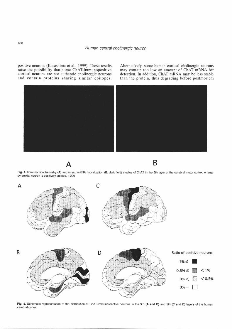
The existence of cholinergic neurons in the mammalian cerebral cortex has been a matter of controversy. Early immunohistochemical studies detected no cholinergic neurons in the cerebral cortex (Kimura et aI., 1980, 1981; Sofroniew et aI., 1982; Armstrong et aI., 1983). However, a considerable number of reports have more recently shown ChATimmunoreactive neurons present in the cerebral cortex of the rat (Houser et al., 1983; Levey et al., 1984; Ichikawa and Hirata, 1986; Eckenstein et ai, 1988; Kosaka et al., 1988; Umbriaco et al., 1994), cat (Stichel et al., 1987; Avendano et al., 1996), fetal monkey (Hendry et al., 1987), and human (Kasashima et aI., 1999). In situ hybridization studies demonstrated neurons possessing ChAT mRNA in the rat (Lauterborn et al., 1993) and human (Kasashima et al., 1998, 1999) cerebral cortices. The cholinergic neurons reported in the experimental animals are small fusiform or bipolar neurons, most of which are observed in layers lJ and Ill. In human , however, most of the cerebral ChAT-immunoreactive neurons are medium-sized or large pyramidal neurons located predominantly in layers III and V (Fig. 4) . The density of such neurons is higher in the motor and secondary sensory areas compared to other cortical areas (Fig. 5). No ChAT-immunoreactive neurons are found in the primary sensory areas. In situ hybridization studies (Kasashima et al., 1998, 1999) also revealed the presence of neurons with ChAT mRNA signals in the human cerebral cortex, though the number of neurons with the hybridization signals is much smaller than that of the immunoreactive neurons in each cortical region and the distribution of neurons demonstrating the ChAT mRNA is more restricted than that of the immuno-
B
Fig. 3. ChAT immunohistochemistry of the caudate nucleus (A) and in situ mRNA hybridization of the putamen (B, dark field). Large neurons are positively labeled in both areas. x 250
830
Human central cholinergic neuron
positive neurons (Kasashima et aI., 1999). These results rai se the possibility that some ChAT-immunopositive cortical neurons are not a uthe ntic cholinergic neurons and contain protein s sharing similar e pitope s.
A
Alternatively, some human cortical cholinergic neurons may contain too Iowan amount of ChAT mRNA for detection. In addition, ChAT mRNA may be less stable than the protein , thus degrading before pos tmort em
B Fig. 4. Immunohistochemistry (A) and in situ mRNA hybridization (B, dark field) studies of ChAT in the 5th layer of the cerebral motor cortex. A large pyramidal neuron is positively labeled. x 200
A c
B D Ratio of positive neurons
1%~ • 0.5%~ [11 <1%
0%< IDJ <0.5%
0%= 0
Fig. 5, Schematic representation of the distribution of ChAT-immunoreactive neurons in the 3rd (A and B) and 5th (C and 0) layers of the human cerebral cortex.
831
Human central cholinergic neuron
fixation. The development of monoclonal antibodies for human ChAT applicable to paraffin-embedded tissue sections may help to resolve this issue. Overall, particular areas of the human cerebral cortex contain a certain number of neurons that express ChAT.
IV. Medial habenular nucleus (Ch 7)
The medial habenular nucleus, one of the components of the epithalamus, contains small ChATimmunopositive neurons surrounded by a moderate density of immunoreactive fibers (Heckers et aI., 1992). However, an in situ hybridization study failed to demonstrate ChAT mRNA in this nucleus (Kasashima et aI. , 1998).
V. Mesopontine tegmental nuclei
1. Pedunculopontine nucleus (Ch5)
The ChS complex that consists of the compact portion (Ch5c) and the diffuse-interstitial portion (ChSd) (Mesulam et aI., 1989; Manaye et a!., 1999) is located in the ventromedial portion of the rostoral pontine tegmentum (Mizukawa et aI., 1986; Mesulam et aI., 1989; Manaye et aI., 1999). ChSc is surrounded by the superior cerebellar peduncle, lateral lemniscus and medial lemniscus. More than half of the large neurons are intensely ChAT-immunopositive in ChSc (Mesulam et aI., 1989; Manaye et aI., 1999). These neurons are polymorphic in shape (oval or triangular form). ChATpositive neurons belonging to Ch5d are embedded in the lateral lemniscus, superior cerebellar peduncle, and the
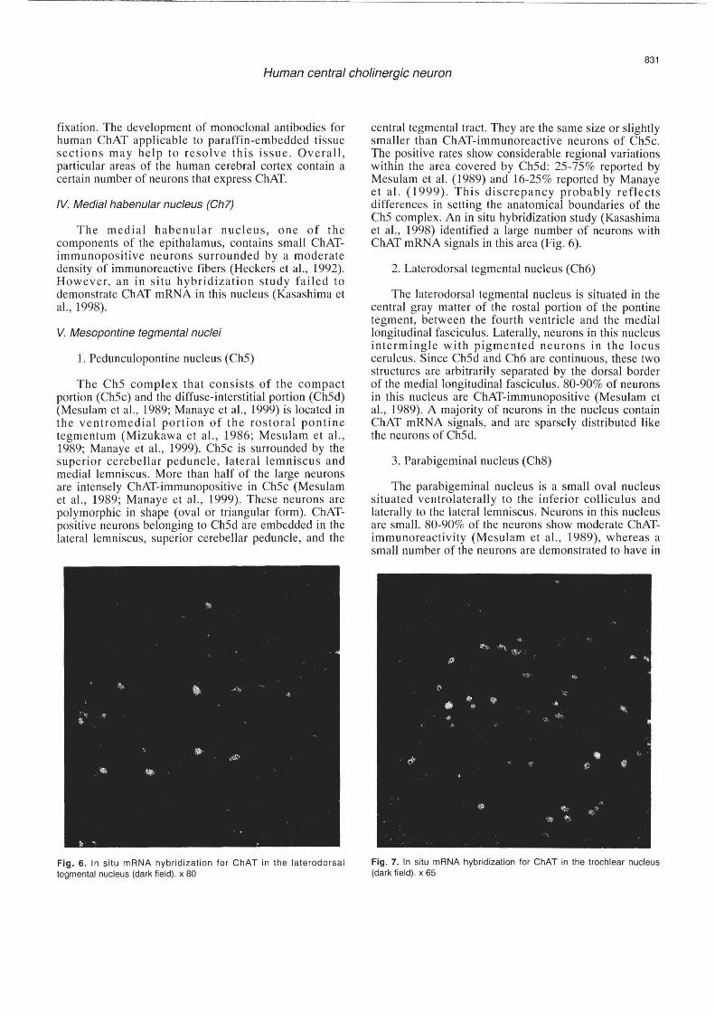
Fig. 6. In situ mRNA hybridization for ChAT in the laterodorsal tegmental nucleus (dark field). x 80
central tegmental tract. They are the same size or slightly smaller than ChAT-immunoreactive neurons of Ch5c. The positive rates show considerable regional variations within the area covered by Ch5d: 2S-7S% reported by Mesulam et al. (1989) and 16-2S% reported by Manaye et al. (1999). This discrepancy probably reflects differences in setting the anatomical boundaries of the ChS complex. An in situ hybridization study (Kasashima et aI., 1998) identified a large number of neurons with ChAT mRNA signals in this area (Fig. 6).
2. Laterodorsal tegmental nucleus (Ch6)
The laterodorsal tegmental nucleus is situated in the central gray matter of the rostal portion of the pontine tegment, between the fourth ventricle and the medial longitudinal fasciculus. Laterally, neurons in this nucleus intermingle with pigmented neurons in the locus ceruleus. Since ChSd and Ch6 are continuous, these two structures are arbitrarily separated by the dorsal border of the medial longitudinal fasciculus. 80-90% of neurons in this nucleus are ChAT-immunopositive (Mesulam et aI., 1989). A majority of neurons in the nucleus contain ChAT mRNA signals, and are sparsely distributed like the neurons of ChSd.
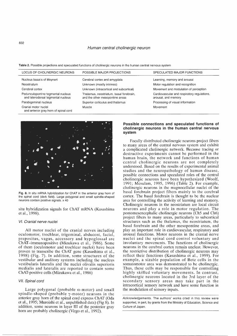
3. Parabigeminal nucleus (Ch8)
The parabigeminal nucleus is a small oval nucleus situated ventrolaterally to the inferior colliculus and laterally to the lateral lemniscus. Neurons in this nucleus are small. 80-90% of the neurons show moderate ChATimmunoreactivity (Mesulam et aI., 1989), whereas a small number of the neurons are demonstrated to have in
Fig. 7. In situ mRNA hybridization for ChAT in the trochlear nucleus (dark field) . x 65
832
Human central cholinergic neuron
Table 2. Possible projections and speculated functions of cholinergic neurons in the human central nervous system
LOCUS OF CHOLINERGIC NEURONS POSSIBLE MAJOR PROJECTIONS SPECULATED MAJOR FUNCTIONS
Nucleus basalis of Meynert
Neostriatum
Cerebral cortex and amygdala
Unknown (mostly intrinsic)
Learning , memory and arousal
Cerebral cortex
Pedunculopontine tegmental nucleus and laterodorsal tegmental nucleus
Unknown (intracortical and subcortical)
Thalamus. neostriatum, basal forebrain, and the other mesopontine areas
Motor regulation and recognition
Movement and modulation of perception
Cardiovascular and respiratory regulations, arousal, and memory
Parabigeminal nucleus
Cranial motor nuclei
Superior colliculus and thalamus
Muscle
Processing of visual information
Movement and anterior gray horn of spinal cord
.: • , . II!
• .. • t '.,
$~ • -Ii .. , .- • I ~
" j' , .1
• ~ • &} f
• I>
• • ". fl •
• " ~ • t
t • ~
.. .. •
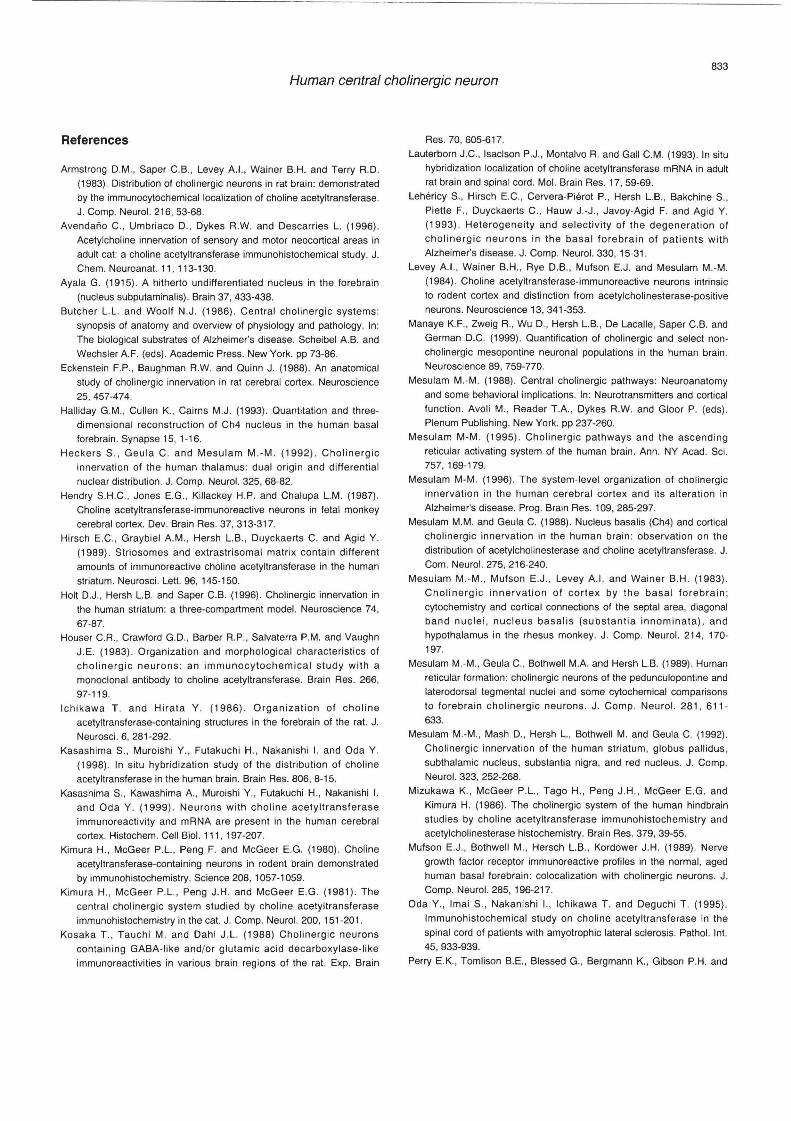
• Fig. 8. In situ mRNA hybridization for ChAT in the anterior gray horn of the spinal cord (dark field). Large polygonal and small spindle-shaped neurons contain positive signals. x 40
situ hybridization signals for ChAT mRNA (Kasashima et aI., 1998).
VI. Cranial nerve nuclei
All motor nuclei of the cranial nerves including oculomotor, trochlear, trigeminal, abducent, facial, prepositus, vagus, accessory and hypoglossal are ChAT-immunopositive (Mizukawa et aI., 1986). Some of them (oculomotor and trochlear nuclei) have been proven to transcribe the ChAT gene (Kasashima et aI., 1998) (Fig. 7). In addition, some st ructures of the vestibular and auditory systems including the nucleus vestibularis lateralis and the nuclei olivalis superioris medialis and lateralis are reported to contain some ChAT-positive cells (Mizukawa et aI. , 1986)
VII. Spinal cord
Large polygonal (probable a-motor) and small spindle-shaped (probable y-motor) neurons in the anterior gray horn of the spinal cord express ChAT (Oda et aI. , 1995; Muroishi et aI., unpublished data) (Fig 8). In addition, some neurons in layer 111 of the posterior gray horn are probably cholinergic (Virgo et aI., 1992) .
Possible connections and speculated functions of cholinergic neurons in the human central nervous system
Focally distributed cholinergic neurons proj ect fibers to many areas of the central nervous system and exhibit a complicated cholinergic network. Because tracing or destructive experiments cannot be performed in the human brain , the network and functions of human central cholinergic neurons are not completely understood. Based on the results of experimental animal studies and the neuropathology of human disease, possible connections and speculated roles of the central cholinergic neurons have been hypothesized (Woolf, 1991; Mesulam, 1995, 1996) (Table 2). For example, cholinergic neurons in the magnocellular nuclei of the basal forebrain project fibers mainly to the cerebral cortex. The basal forebrain is thought to be the nuclear area for controlling the activity of learning and memory. Cholinergic neurons in the neostriatum are local circuit neurons and playa rol e in motor regulation . The pontomesencephalic cholinergic neurons (Ch5 and Ch6) project fibers to many areas, particularly to subcortical s tructures such as the thalamus, the neostriatum , the basal forebrain and the other mesopontine areas, and play an important role in cardiovascular, respiratory and arousal functions. Motor neurons in the cranial nerve nuclei and the spinal cord control voluntary and involuntary movements. The functions of cholinergic neurons in the cerebral cortex remain unclear. However, the restrictive distribution of cholinergic neurons may reflect their functions (Kasashima et aI., 1999). For example, a sizable popUlation of Betz cells in th e somatomotor area was demonstrated to be cholinergic. Thus, these cells may be responsible for controlling highly skilled voluntary movements. In contrast, cholinergic neurons located in the 3rd layer of the secondary sensory areas may take part in the intracortical sensory network and have some function in the modulation of sensory inputs.
Acknowledgements. The authors ' works cited in this review were
supported, in part, by grants from the Ministry of Education , Science and
Culture of Japan.
833
Human central cholinergic neuron
References
Armstrong D.M ., Saper C.B., Levey A.I. , Wainer B.H. and Terry A.D . (1983) . Distribution of cholinergic neurons in rat brain: demonstrated
by the immunoCy1ochemical localization of choline acetyltransferase. J. Compo Neurol. 216, 53-68.
Avendano C., Umbriaco D., Dykes R.W. and Descarries L. (1996). Acetylcholine innervation of sensory and motor neocortical areas in adult cat: a choline acetyltransferase immunohistochemical study. J. Chem. Neuroanat. 11 , 113-130.
Ayala G. (1915). A hitherto undifferentiated nucleus in the forebrain (nucleus subputaminalis). Brain 37, 433-438.
Butcher L.L . and Woolf N.J. (1986) . Central chOlinergi c systems: synopsis of anatomy and overview of physiology and pathology. In: The biological substrates of Alzheimer's disease. Scheibel A.B. and
Wechsler AF. (eds) . Academic Press. New York. pp 73-86. Eckenstein F.P. , Baughman R.W. and Quinn J. (1988). An anatomical
study of cholinergic innervation in rat cerebral cortex. Neuroscience 25,457-474.
Halliday G.M., Cullen K. , Cairns M.J. (1993). Quantitat ion and threedimensional reconstruction of Ch4 nucleus in the human basal forebrain . Synapse 15, 1-16.
Heck ers S ., Geu la C. and Mesulam M.-M . (1992) . Cholinergic innervation of th e human thalamus : dual origin and diffe rential nuclear distribution. J. Compo Neurol. 325, 68-82.
Hendry S.H.C., Jones E.G., Killackey H.P. and Chalupa L.M. (1987) .
Choline acetyltransferase-immunoreactive neurons in fetal monkey cerebral cortex. Dev. Brain Res. 37, 313-317.
Hirsch E.C., Graybiel A.M. , Hersh L.B ., Duyckaerts C. and Agid Y.
(1989). St riosomes and extrastrisomal matrix conta in different amounts of immunoreactive choline acetyltransferase in the human
striatum. Neurosci. Lett. 96, 145-150. Holt D.J., Hersh L.B. and Saper C.B. (1996). Cholinergic innervation in
the human striatum: a three-compartment model. Neuroscience 74 ,
67-87. Houser C.A. , Crawtord G.D., Barber R.P., Salvaterra P.M. and Vaughn
J.E. (1983). Organization and morphological characteristics of chOlin ergic neurons : an immunocytochemical study with a monoclonal antibody to choline acetyltransferase. Brain Res. 266,
97-119. Ichikawa T . and Hirata Y . (1986) . Organization of choline
acetyltransferase-contain ing structures in the forebrain of the rat. J.
Neurosci. 6, 281-292. Kasash ima S. , Muroishi Y., Futakuchi H., Nakanishi I. and Oda Y.
(1998) . In situ hybridization study of the distribution of choline acetyltransferase in the human brain. Brain Res. 806, 8-15.
Kasashima S., Kawashima A. , Muroishi Y. , Futakuchi H., Nakanishi I.
and Oda Y. (1999) . Neurons with choline acetyl transferase immunoreactivity and mRNA are present in the human cerebral cortex. Histochem. Cell BioI. 111 , 197-207.
Kimura H., McGeer P.L. , Peng F. and McGeer E.G. (1980). Choline
acetyltransferase-containing neurons in rodent brain demonstrated by immunohistochemistry. Science 208, 1057-1059.
Kimura H., McGeer P.L., Peng J.H . and McGeer E.G. (1981) . The central cholinergic system studied by choline acetyltransferase
immunohistochemistry in the cat. J. Compo Neurol. 200, 151 -201. Kosaka T., Tauchi M. and Dah l J.L. (1988) Cholinerg ic neurons
con taining GABA-like and/or glutamic acid decarboxylase-like
immunoreactivities in various brain reg ions of the rat. Exp. Brain
Res. 70, 605-617.
Lauterborn J.C., Isaclson P.J. , Montalvo R. and Gall C.M. (1993). In situ
hybridization localization of cho line acetyltransferase mRNA in adult rat brain and spinal cord. Mol. Brain Res. 17, 59-69.
Lehericy S., Hirsch E.C., Cervera-Pierot P., Hersh L.B. , Bakchine S., Piette F., Duyckaerts C., Hauw J.-J ., Javoy-Agid F. and Agid Y. (1993) . Heterogeneity and selectiv ity of the degeneration of cholinergic neurons in the basal forebrain of patients with Alzheimer's disease. J. Compo Neurol. 330, 15-31 .
Levey A.I. , Wainer B.H., Rye D.B., Mufson E.J. and Mesulam M.-M.
(1984). Choline acetyltransferase-immunoreactive neurons intrinsic to rodent cortex and distinction from acetylcholinesterase-positive neurons. Neuroscience 13, 341 -353.
Manaye K.F. , Zweig R., Wu D., Hersh L.B., De Lacalle, Saper C.B. and German D.C. (1999) . Quantification of cholinergic and select noncholinergic mesopontine neuronal populations in the human brain . Neuroscience 89, 759-770.
Mesulam M.-M. (1988). Central cholinergic pathways: Neuroanatomy
and some behavioral implications. In: Neurotransmitters and cortical function. Avoli M., Reader T.A., Dykes R.w. and Gloor P. (eds). Plenum Publishing. New York. pp 237-260.
Mesulam M-M . (1995). Cholinergic pathways and the ascend ing reticu lar activating system of the human brain. Ann . NY Acad . Sci. 757, 169-179.
Mesulam M-M . (1996). The system-level organization of cholinergic innervation in the human cerebral cortex and its alteration in
Alzheimer's disease. Prog. Brain Res. 109, 285-297. Mesulam M.M. and Geula C. (1988) . Nucleus basalis (Ch4) and cortical
chol inergic innervation in the human brain : observation on the distribution of acetylcholinesterase and choline acetyltransferase. J. Com. Neurol. 275, 216-240.
Mesulam M.-M., Mufson E.J., Levey A.I. and Wainer B.H. (1983). Cholinergic innervation of cortex by the basa l forebrain ; Cy10chemistry and cortical connections of the septal area, diagonal band nuclei , nucleus basalis (substant ia innominata) , and hypothalamus in the rhesus monkey. J. Compo Neurol. 214, 170-197.
Mesulam M.-M., Geula C. , Bothwell MA and Hersh L.B. (1989) . Human reticular formation: cholinergic neurons of the pedunculopontine and laterodorsal tegmental nuclei and some cy10chemical comparisons to forebrain cholinerg ic neurons . J. Comp o Neurol. 281 , 611 -
633. Mesulam M.-M ., Mash D., Hersh L., Bothwell M. and Geula C. (1992).
ChOlinergic innervation of the human striatum, globus pallid us , subthalamic nucleus, substantia nigra, and red nucleus. J. Compo Neurol. 323, 252-268.
Mizukawa K., McGeer P.L., Tago H. , Peng J.H., McGeer E.G. and
Kimura H. (1986). The cholinergic system of the human hindbrain studies by choline acetyltransferase immunohistochemistry and acetylcholinesterase histochemistry. Brain Res. 379, 39-55.
Mufson E.J., Bothwell M., Hersch L.B. , Kordower J.H. (1989). Nerve growth factor receptor immunoreactive profiles in the normal, aged human basal forebrain : colocalization with cholinergic neurons. J. Compo Neurol. 285,196-217.
Oda Y., Imai S., Nakan ishi I. , Ichikawa T. and Deguchi T. (1995) .
Immunohistochemical study on choline acetyltransferase in the spinal cord of patients with amyotrophic lateral sclerosis. Pathol. Int. 45, 933-939.
Perry EX, Tomlison B.E. , Blessed G., Bergmann K. , Gibson P.H. and
834
Human central cholinergic neuron
Perry R.H. (1978). Correlation of cholinergic abnormalities with
senile plaques and mental test scores in senile dementia. Br. Med. J. 25, 1457-1459.
Selden N., Geu la C., Hersh l . and Mesulsm M.-M. (1994) . Human
striatum: chemoarchitecture of the caudate nucleus, putamen and ventral striatum in health and Alzheimer's disease. Neuroscience 60, 621-636.
$imic G, Mrzl jak l., Fucic A., Winblad B., lovric H. and Kostovic I. (1999) . Nucleus subputaminalis (Ayala) : the still disregarded magnocellular component of the basal forebrain may be human specific and connected with the cortical speech area. Neuroscience 89, 73-89.
Sofroniew M.V., Eckenstein F., Thoenen H. and Cuello A.C. (1982) . Topography of choline acetyltransferase-containing neurons in the
forebrain of the rat. Neurosci. lett. 33, 7-12. Stichel C.C., Dolabela de Lima A. and Singer W. (1987). A search for
choline acetyltransferase-like immunoreactivity in neurons of cat striate cortex. Brain Res. 405, 395-399
Ulfig N. (1989) . Configulation of the magnocellular nuclei in the basal forebrain of the human adult. Acta Anal. 134, 100-105.
Umbriaco D., Watkins K.C., Descarries l ., Cozzari C. and Hartman BK (1994) . Ultrastructual and morphometric features of the acetylcholine
innervation in adult rat parietal cortex: an electron microscopic study in serial sections. J. Compo Neurol. 348, 351-373.
Virgo l ., de Belleroche J ., Rossi M. and Steiner T .J . (1992) . Characterisation of the distribution of choline acetyltransferase messenger RNA in human spinal cord and its depletion in motor
neuron disease. J. Neurol. Sci. 112, 126-132. Whitehouse P.J., Price D.L. , Clark AW. , Coyle J.T. and Delong M.R.
(19 81) . Alzheimer's disease: Evidence for selective loss of
chOlinergic neurons in the nucleus basalis. Ann . Neurol. 10, 122-126.
Woolf N.J. (1991). Cholinergic systems in mammalian brain and spinal
cord. Prog. Neurobiol. 37, 475-524.
Accepted January 28, 2000