Embed Size (px)
Citation preview

INVITED REVIEW
Morphologic and Functional Features of the Canine Cruciate
Ligaments
HILDE DE ROOSTER, DVM, PhD, TANYA DE BRUIN, DVM, and HENRI VAN BREE, DVM, PhD, Diplomate ECVS & ECVDI
Objective—To review the gross, microscopic, and functional anatomy of the cranial cruciateligament (CCL) in dogs.Study Design—Literature review.Methods—Reports of the anatomy and function of the cruciate ligaments in dogs were retrieved bysearch of the 1975–2005 PubMed database.Results—The CCL has an important biomechanical function resisting cranial drawer, hyperexten-sion, and internal rotation and acts to fine tune and guide the stifle through its rolling and slidingmotion. It has a complex architecture, and distinct geographic regions within the ligament havedifferent functional roles depending on the angle and loading conditions. Collagen type I is the maincomponent of the extracellular matrix; the fibrils have a crimped structure. The cruciate ligamentsare almost completely covered by synovium, protecting them from synovial fluid. Cruciate bloodsupply is mainly of soft tissue origin. The intraligamentous network is relatively limited whereas thecore of the middle third of the CCL is even less well vascularized. Neurohistologic studies are verylimited in the dog. Various mechanoreceptors and proprioceptive receptors have been identifiedwithin the substance of the cruciate ligaments.Conclusions—CCL structural characteristics play an important part in its complex behaviour withthe crimped pattern of the collagen fibrils being an important determinant of its biomechanicalproperties. In contrast to reports of managing CCL rupture, there are few reports describing themicroanatomy and neurovascular morphology of the cruciate ligaments.Clinical Relevance—Cruciate disease is likely multi-factorial. Improved understanding of CCLdegradation leading to CCL rupture is critical to development of new diagnostic tests for cruciatedisease in dogs. Appropriate intervention during the early stages of disease process might preserveCCL structural properties by preventing further collagen degradation. Accurate knowledge offunctional and fiber bundle anatomy is imperative for reconstruction and restoration of normalstifle joint physiology. Reconstructive goals should alleviate existing instability and mimic normalkinematics. Knowledge of the exact function of the CCL in the neuromuscular control around thestifle joint could possibly explain osteoarthritis progression after CCL damage.r Copyright 2006 by The American College of Veterinary Surgeons
INTRODUCTION
ONE OF the most complex joints, the stifle joint,contains the cruciate ligaments that are required for
craniocaudal joint stability. Morphologically and func-tionally complex, the cruciate ligaments are dynamicstructures, strongly connecting the femur to the tibia. In
early medical literature, the cruciate ligaments were calledcrucial ligaments because of their crossed arrangement.1
Subsequently, the crucial role of the cruciate ligaments tostifle joint kinematics of the stifle joint has been appre-ciated.2
The distal femoral intercondylar notch is almost com-pletely filled by the cruciate ligaments and some fat. The
Address reprint request to Hilde de Rooster, DVM, PhD, Department of Small Animal Medicine and Clinical Biology, Faculty of
Veterinary Medicine, Salisburylaan 133, 9820 Merelbeke, Belgium. E-mail: [email protected].
Accepted October 2005
From the Departments of Small Animal Medicine and Clinical Biology; and Medical Imaging of Domestic Animals, Faculty of
Veterinary Medicine, Ghent University, Merelbeke, Belgium.
r Copyright 2006 by The American College of Veterinary Surgeons
0161-3499/06
doi:10.1111/j.1532-950X.2006.00221.x
769
Veterinary Surgery
35:769–780, 2006
cranial cruciate ligament (CCL) and the caudal cruciateligament (CaCL) both attach to the intercondyloid areaof the tibia.3–6 Although the length of both canine cru-ciate ligaments is nearly equal, their distal attachmentpoints on the tibia are separated by almost twice the dis-tance of their femoral origins.7 Because of their anatomyand spatial arrangement, the cruciate ligaments provideprimary ligamentous support for craniocaudal and axialstability of the stifle joint throughout the functional rangeof motion.5 The CCL controls cranial drawer motion,whereas the CaCL acts as a major stabilizer against cau-dal drawer motion. Furthermore, the CCL is consideredto fine-tune normal stifle joint kinematics.
Published reports have primarily focused on the CCL,seemingly because it is the most vulnerable and importantligament of the stifle joint.3,8 Its morphologic features andrelationships with other joint structures have been studiedextensively and comparative studies reinforce the import-ance of the CCL in various mammals. Although speciesdifferences in CCL anatomy, physiology, and biome-chanics are minor,9 there are nevertheless differences andinformation should clearly reference the species source.Unfortunately, this distinction has not always been clear-ly stated in veterinary reports, leading the less criticalreader to believe that much more is known about caninecruciate ligaments than is actually so. Understanding thenormal anatomy of the stifle joint, and in particular thenormal cruciate anatomy, is essential for diagnosis andrational treatment of CCL rupture.
FIBER BUNDLE ANATOMY
The cruciate ligaments are not just a single-strandconfiguration of longitudinally orientated collagen fi-bers.5 In humans and dogs, they contain twisted collag-enous fascicles and fiber bundles that are subdividedinto fascicles, subfascicular units, fibers, and fibrils.10,11
The canine CCL and CaCL can each be divided into 2functional components because they have individualattachment zones.5
The CCL originates on the axial aspect of the lateralfemoral condyle, very close to the articular margin. Itextends diagonally across the joint space and attaches tothe cranial intercondyloid area of the tibial plateau.3,5,12
The proximal attachment site is bordered cranially by thecranial meniscotibial ligament of the medial meniscus andcaudally by the cranial meniscotibial ligament of the lat-eral meniscus.4,13,14 The canine CCL is the narrowest inits mid region and fans out proximally and distally.13 Itsshape changes through the normal range of motion of thestifle joint,5,13 and the decrease in cross-sectional area isalso the greatest at the mid region when forces are act-ing.15 CCL length is positively correlated with canine
body weight; taking the average length of its cranial andcaudal borders, the mean length of the canine CCL hasbeen reported as 13.5–18.7mm.16–18
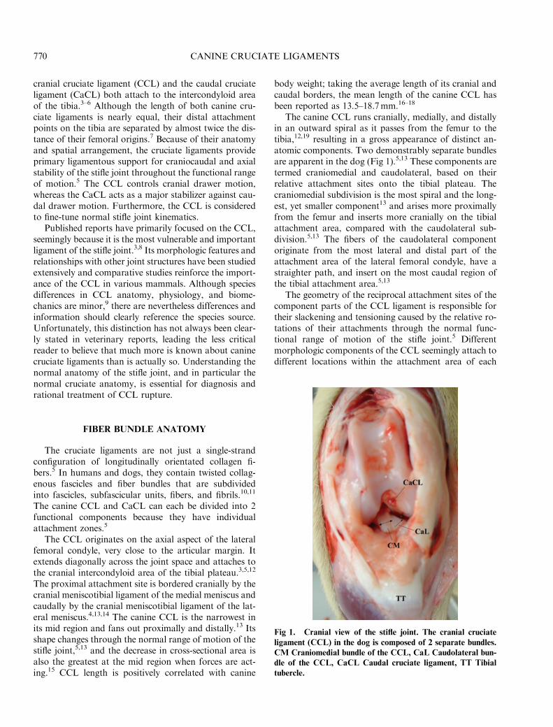
The canine CCL runs cranially, medially, and distallyin an outward spiral as it passes from the femur to thetibia,12,19 resulting in a gross appearance of distinct an-atomic components. Two demonstrably separate bundlesare apparent in the dog (Fig 1).5,13 These components aretermed craniomedial and caudolateral, based on theirrelative attachment sites onto the tibial plateau. Thecraniomedial subdivision is the most spiral and the long-est, yet smaller component13 and arises more proximallyfrom the femur and inserts more cranially on the tibialattachment area, compared with the caudolateral sub-division.5,13 The fibers of the caudolateral componentoriginate from the most lateral and distal part of theattachment area of the lateral femoral condyle, have astraighter path, and insert on the most caudal region ofthe tibial attachment area.5,13
The geometry of the reciprocal attachment sites of thecomponent parts of the CCL ligament is responsible fortheir slackening and tensioning caused by the relative ro-tations of their attachments through the normal func-tional range of motion of the stifle joint.5 Differentmorphologic components of the CCL seemingly attach todifferent locations within the attachment area of each
Fig 1. Cranial view of the stifle joint. The cranial cruciate
ligament (CCL) in the dog is composed of 2 separate bundles.
CM Craniomedial bundle of the CCL, CaL Caudolateral bun-
dle of the CCL, CaCL Caudal cruciate ligament, TT Tibial
tubercle.
770 CANINE CRUCIATE LIGAMENTS

bone. Reciprocal tension and thus functional differenceoccur because their individual attachment sites rotate andtranslate relative to each other.5 In stifle extension, thelong axis of the CCL is aligned with the long axis of thefemur. The femoral attachments of both craniomedialand caudolateral components are almost perpendicular tothe joint surface and both are taut.13 In flexion, thecraniomedial component of the CCL curves and twistsaround the caudolateral component while the femoralattachment site moves distally and caudally.5,13 This re-orientation of femoral attachment sites during stifle flex-ion results in an increased distance between the sites offemoral origin and tibial attachment of the craniomedialcomponent, causing tension in this component duringflexion. The relative relaxation of the fibers of thecaudolateral component can be explained by the sameprinciples, because the bone attachment sites move closertogether as the stifle is flexed; thus, the caudolateralcomponent is slack in flexion.5,13
The canine CaCL is slightly longer and broader thanthe CCL.3–6,20 Even its collagen fibrils are thicker com-pared with the CCL.21 The total midsection diameter ofCaCL is the smallest as it fans out from the center, mak-ing the femoral and, to a lesser extent, the tibial attach-ments larger.4 The CaCL also has 2 componentsalthough they are less distinct and often inseparable.13,20
The cranial component is larger than the caudal compo-nent.22 The restraining effect of both components of theCaCL also varies with stifle position, and they performreciprocal functions at different angles of flexion becauseof the location of their attachment sites.5,20 Similar to theCCL, the geometry of the femoral attachment is largelyresponsible for ligament tensioning13; the cranial compo-nent is taut in flexion and loose in extension, whereasthese states are reversed for the caudal component.5,20
FUNCTIONAL ANATOMY
There is a ranked hierarchy of structures neutralizingspecific forces acting on the stifle joint and resisting dif-ferent kinds of joint laxity. Stifle function is complement-ed by static support from a complex (passive) restrainingsystem consisting of bony and musculotendinous struc-tures, menisci, and several ligaments. Furthermore, mus-cular forces and joint compression contribute to jointstability.23,24 The cruciate ligaments have specific func-tions that are directly related to their anatomic locationsand orientations within the stifle joint. Although the mainfunctions of other intra-articular and peri-articular struc-tures and ligaments differ from those of the cruciate lig-aments, they act complementary as constraints of stiflejoint motion in various planes.14,25
The CCL has to provide a stabilizing effect of the tibiaon the femur throughout the whole range of motion,resisting forces that would cause the tibia to translatecranially relative to the femur and, to a lesser degree,resisting forces that would cause tibial rotation duringflexion of the stifle joint.5,22,26 In dogs, both cruciate lig-aments have 2 components that behave independentlyand differently from each other throughout loading.5
Every change in joint angle alters the tension in the sep-arate bands as some fibers are stressed and others are not.This feature contributes to the CCL’s ability to withstandthe multi-axial stresses of normal function and range ofmotion. Most assessments of ligamentous function havebeen based on the changes in laxity observed after se-quential cutting of selected ligaments in cadaveric humanknees.27–30 The actual proportion of their combined con-tributions to sustaining load varies with the angle of stifleflexion. With the stifle in extension, the entire CCL is tautand thus both components limit cranial translation of thetibia relative to the femur.5,13 Because of combined in-teractions, an isolated lesion of a component part of theCCL does not necessarily provoke clinically detectableinstability.27,28 In the human knee, the CCL is the solestructure to limit cranial drawer motion near full exten-sion of the joint, as tension in the hamstring muscles lacksto provide extra restraint.31 It is probably much less so inthe canine stifle joint where maximal extension is far lessthan 1801. The craniomedial component of the CCL istaut during the whole range of stifle motion and is themajor contributor to craniocaudal stability in stifle flex-ion.8,26 The relaxed caudolateral component only acts asa weak secondary restraint to this unidirectional cranialtranslating force and in fact, only contributes when thecraniomedial band is damaged or severely stretched.Other joint structures seemingly contribute less to cranio-caudal stability during flexion.18
In subtle balance with the capsular structures, the col-lateral ligaments, muscles, the condylar geometry, andjoint surface contact, the cruciate ligaments control andproduce rotation of the tibia relative to the fe-mur.3,8,20,22,29,32 An increased angle of stifle flexion is ac-companied by increased internal rotation of the tibia ifunrestricted.8,14,32,33 Part of the valgus load is trans-formed into an axial rotatory force as the lateral collat-eral ligament begins to relax.22,34 As the stifle flexes, thecruciate ligaments are not only wrapped upon each otherbut also spiral on themselves.3,5,22 The higher strain in theligaments also limits the amount of normal internal ro-tation of the tibia on the femur.5,12,20,22 As the stifle ex-tends, the lateral collateral ligament tightens and thelateral femoral condyle moves cranially, causing externalrotation of the tibia. This motion has classically beendescribed as the screw-home mechanism.32 In extension,the medial and lateral collateral ligaments become the
771DE ROOSTER, DE BRUIN, AND VAN BREE

primary restraints of rotation, and the cruciate ligamentsprovide only a secondary check from the tension in bothligaments.3,12,17 No singular limiting effect on externalrotation is provided by the cruciate ligaments in dogs, noteven as secondary restraint structures.5,20,32 By externalrotation of the tibia, the cruciate ligaments start to un-twist, and strain decreases.8,12,32
Axial tibial rotation of the canine stifle joint is coupledwith varus–valgus rotation.34 In a stable stifle joint, thecollateral ligaments are considered the primary ligament-ous structures providing sideways restraint when stiflejoint motion is restricted. In fact, they share their func-tion with other joint structures and ligaments.32,34 Bothcruciate ligaments together are important secondary re-straints against varus and valgus angulation. Stresses onthe cruciate ligaments during medial and lateral openingof the joint space generally increase slightly with the de-gree of flexion of the stifle joint, as the collateral liga-ments begin to relax as the stifle joint is flexed.32,34 Inhumans and dogs, the cruciate ligaments become primaryrestraints if there is loss of collateral ligament sup-port.28,32 In the fully hyperextended human knee, thecruciate ligaments can prevent joint opening by them-selves.28 When varus forces act, the CCL has to sustainlarger strains than the CaCL although these forces arestill much lower than those sustained by the lateral col-lateral ligament.28 For medial restraint (valgus force), therelative contribution of the CaCL becomes greater withincreases in flexion angle.28,34
Overextension is prevented by tension in the cruciateligaments,3 where the CCL acts as the primary restraintwith both the craniomedial and the caudolateral compo-nents taut at full extension.5,13 The caudolateral com-ponent of the CCL is under the greatest tension inextension,13 and is thus the primary contributor to re-straining hyperextension. The slightly longer caudal com-ponent of the CaCL can only be considered a secondaryrestraint.3,5,22
Hyperflexion of the stifle normally will not occur be-cause of contact between the thigh muscles and the gas-trocnemius muscle.3 The cruciate ligaments spiral onthemselves, and they are naturally twisted upon eachother when the stifle is flexed.3,5,22 During the stancephase, the angle of the stifle joint is �1401 in dogs. Thetwist of both cruciate ligaments upon each other preventsstifle collapse during stance.5 Quadriceps contractionwould cause cranial tibial subluxation at this flexionangle but by loading of an intact CCL this is prevented.24
Stifle joint stability in humans and dogs is dynamic asfar as muscle control (active forces) is concerned.4,23–25
On the cranial aspect of the stifle joint, the quadricepsmuscle and the patellar tendon provide support. Thepopliteal muscle as well as the hamstring muscles andgastrocnemius provide additional support caudally.35 In
cats, through CCL-muscle reflexes, direct loading of theCCL causes quadriceps muscle inhibition and simultan-eously increases hamstring muscle activity to reduce CCLloading.36 Joint compression and muscle actions greatlycontribute to joint stabilization.24 Cranial tibial thrust isgenerated during weight bearing by the slope of the tibialplateau and by tibial compression because of musculo-tendinous attachments such as the tendon of the bicepsfemoris.25,33 According to Slocum and Devine, the CCLis only a backup mechanism for control of cranial pro-jection of the proximal aspect of the tibia as a dog walks,and experiences no stresses as long as the cranial tibialthrust is effectively opposed by the caudal pull of thebiceps femoris and hamstring muscle group.23,26 Onlywhen these active muscle forces are insufficient to coun-teract cranial translation of the tibia will the CCL providethe first passive restraint. There is no perfect balance atall times and because of the functional cranial to caudalslope of the tibial plateau, the CCL must intermittentlyresist cranial tibial thrust.26,33 A lesser tibial plateau slopeis thought to account for less CCL strain because cranialtranslation is prevented by decreasing cranial tibialthrust.23,26,37–39 However, a more recent study reportedthat the standing tibial plateau angle is not significantlydifferent from a plane parallel to the ground in mostdogs; furthermore, it failed to demonstrate differences inangles between dogs with and without risk of cruciatedisease.40
In contrast to the individual functions of the CCL,selective transection of only one component of the CaCLdoes not result in caudal drawer in the canine stifle jointfor any joint angle.5,20 The importance of the CaCL instability of the canine stifle joint is far less than that of theCCL although its exact contribution remains unre-solved.41 The CaCL is usually larger than the CCL;therefore, it seems unlikely that in the dog it does nothave some important function. Prevention of caudal dis-placement of the tibia on the femur seems the onlyprimary role of the CaCL.5,8 In a flexed stifle joint, thecranial component of the CaCL is a primary restraintagainst caudal instability because of looseness of the col-lateral ligaments in this joint position.20,22,32
MICROANATOMY AND ULTRASTRUCTURE
Both cranial and caudal cruciate ligaments are coveredby a fairly uniform fold of synovial membrane whichincompletely divides the stifle joint in the sagittalplane.6,42 This synovial tissue continues over the hornsof the menisci.43 These enveloping epiligamentous mem-branes consist mainly of dense connective tissue, smallfibroblasts, and some adipocytes;13,44 an intima and athin sub-intimal layer can be distinguished. The intima is
772 CANINE CRUCIATE LIGAMENTS

a single layer of synoviocytes and the subintimal layer isareolar tissue containing small vascular structures.17
Compared with the cruciate ligaments, the envelopingsynovial membrane is relatively cellular.13 Synovial liningdoes not occur on the surfaces in direct contact with theother cruciate ligament.17 This synovial envelope makesthe cruciate ligaments extrasynovial structures, protectedfrom the degradative effects of the synovial environment,even though they are intra-articular.5,6 The synovial en-velope covering the CCL originates caudally at the inter-condylar notch and extends to the cranial aspect of thetibial attachment.45 At that point, the epiligamentoustissue communicates with a fold of the distal joint cap-sule.45 The CaCL is ensheathed by 2 folds of synovialmembrane. The cranial envelope originates proximallyfrom the cranial aspect of the joint capsule, while distallythe caudal fold originates from the caudal aspect of thejoint capsule.45
Grossly, the canine cruciate ligaments each have 2components.5,13 This uncomplicated and distinct subdiv-ision does not carry through to their intricate microar-chitecture.13 Each ligament component is amultifascicular structure, and contains many wavy fas-cicular subunits. The fascicles located at the periphery ofthe CCL appear to follow a spiral path of wavinessaround the fascicle axis.11,30,46 In the canine CCL, there isa great variability in the elliptical-shaped fascicle size, asfascicles may be composed of 1–10 subfascicles, subdiv-ided by loose endoligamentous tissue.11,13 The subfasci-cles contain bundles of collagen fibers. Each fiber bundleis not oriented such that it is isometric during stifle jointmotion.30 Every subtle 3-dimensional change in stiflejoint position recruits fibers differently.47 Individual fiberschange length by straightening their crimp as they arerecruited into tension. This change is not visible at a grossanatomic level, but is confirmed by histologic assess-ment.11,30 At the osseous attachment sites of the CCL, thecollagen fibers are not arranged entirely parallel to thelongitudinal axis of the ligament and, especially in young-er specimens, columns of chondroid cells do penetrateinto the CCL (Fig 2B).12,45 Where the CCL and CaCLare in contact, the collagen fibers are more dense andoriented tangential to the surface instead of parallel to thelong axis.17 Those fibers maintain an orientation tangen-tial to the surface of contact, even when the cruciate lig-aments start to twist about each other.17 Fibers areformed by fibrils, which are composed of organization ofrepeated collagen subunits.13,17,44,45 Collagen fibrilmorphology and architecture is also characterized byuniform crimp parallel to the long axis of the fascicle (Fig2A).48,49 The internal collagen fibrils are nearly straight,whereas the fibrils undergo a maximum crimp at the fas-cicular periphery.12,45 The collagen fibrils are the smallestvisible structures on electron microscopy.10,11,50
Ultrastructurally, the CCL is a heterogenic compositestructure formed by an extracellular matrix composed ofmacromolecules with highly specific arrangements andinteractions.44,51 Collagen is the chief macromoleculeprevalent in the framework of the CCL. Type I collagencomprises 490% of the collagen content of the CCL,with the remainder being type III collagen.52,53 The mol-ecules are produced by the fibroblasts in the loosesupporting connective tissue. The cells are present inlong parallel columns between the collagen fibers, theiraxes parallel to the surrounding collagen fibers. Neuro-vascular components follow the same longitudinal orien-tation.13,42,45,46 Besides fibroblasts, the cell populationalso consists of various stages of chondrocyte-likecells.17,49,54
MICROVASCULAR SUPPLY
The major vascular contribution to the center of thestifle joint occurs from branches of the middle genicularartery,55–57 which arises from the popliteal artery, pene-trates the caudal joint capsule, and passes craniodistallyto the fossa intercondylaris, running cranially betweenthe cruciate ligaments (Fig 3).58 The vascular structuresto the proximal part of the CCL are more numerous and
Fig 2. Normal cranial cruciate ligament (CCL) of a 4-month-
old Riezenschnauzer, harvested at its tibial attachment site
(H&E stain). (A) Along the CCL, dense collagen is aligned
parallel to the longitudinal axis of the ligament (bar¼ 100 lm).
The collagen fibers have a recurrent crimped pattern. (B) At
the osseous attachment site of the CCL, the collagen fibers are
not arranged entirely parallel to the long axis of the ligament.
Columns of chondroid cells (arrow) do penetrate into the CCL
(bar¼ 100 lm).
773DE ROOSTER, DE BRUIN, AND VAN BREE

have a larger diameter compared with those on the tibialside.12,45 Most of these vascular structures originate froma branch of the middle genicular artery and from somebranches of the distal genicular arteries.55–57,59,60 Also forthe CaCL most of the vessels originate from branches of
the middle genicular artery and from some branches ofthe other genicular arteries.55,57 The cruciate ligamentsare also nourished by passive permeation from the syn-ovial fluid.61,62
The blood supply to both cruciate ligaments is pre-dominantly of soft tissue origin; the contribution fromthe osseous attachments is negligible.42,56,57,63,64 The in-frapatellar fat pad and the well-vascularized synovialmembranes that form an envelope around the cruciateligaments are the most important sources of vessels andthe major pathway for delivery of nutrients.42,45,59,61,63,64
The synovial vessels arborize into a finely meshed net-work of epiligamentous vessels that ensheath the cruciateligaments throughout their entire length (Figs 4 and5).42,64 In general, the vascular arrangement and struc-tural characteristics of the vasculature inside the CaCLand the CCL are similar.42,45,64 In the inner part of thecruciate ligaments, around and along the bundles of col-lagen fibers, an endoligamentous vascular network cours-es in the supporting connective tissue.42,45 The largervessels, usually one artery accompanied by two veins,mainly course in a longitudinal direction both proximallyand distally and lie parallel to the collagen fascicles.45
Some of them have a tortuous path in the interfascicularareolar tissue.57 Only small capillaries branching fromthe longitudinal endoligamentous vessels, running in atransverse direction, encircle the collagen bundles.45 Thecore of the midportion of the CCL is less well vascu-larized compared with the remainder of the liga-ment.12,17,42,45,59,63
Anastomoses exist between extra- and intraligament-ous blood networks.42,45,64 Epiligamentous vessels pene-trate transversely into the cruciate ligaments (Fig 5 A).64
Their branches ramify and anastomose with the endo-
Fig 3. Caudal view of the major blood supply to the stifle
joint in the dog. (1) Femoral artery, (2) popliteal artery, (3)
descending genicular artery, (4) proximal medial genicular
artery, (5) middle genicular artery, (6) cranial tibial artery,
(7) caudal tibial artery.
Fig 4. Superficial vascularization of normal cruciate ligaments in the dog. (A) Macroscopic view after injection of latex in a
canine cadaver specimen. (B) Arthroscopic view of a stifle joint in a normal dog. (1) Cranial cruciate ligament (2) caudal cruciate
ligament (3) lateral femoral condyle, (4) tibial plateau. Arrow, artery originating from infrapatellar fat pad.
774 CANINE CRUCIATE LIGAMENTS

ligamentous vessels. There are numerous endosteal ves-sels at the ligamentous–osseous junctions; however, com-munications with intrinsic endoligamentous vessels arequite poor, especially at the tibial attachment of the CCLwhere most of the endosteal vessels seem to terminate insubchondral loops instead of crossing the ligamentous–osseous junction.42,45,61,64,65 A number of endosteal ves-sels communicate with the epiligamentous vascular net-work overlying the CCL.45 Vessels from the meniscianastomose with the epiligamentous vascular plexus.45
The web-like network of epiligamentous and synovialvessels surrounding the CaCL appear to be slightly moreextensive than the vascular plexus around the CCL;42,59,63
however, the CaCL does not have a more abundant in-trinsic vascular supply than the CCL.10,56,57,60 Endostealvascular communications are present proximally and dis-tally, although they are rare.45,65
INNERVATION
Three major articular nerves arise from the saphenousnerve, tibial nerve, and common peroneal nerve to in-
nervate the periarticular tissues of the canine stifle joint(Fig 6).66 In each of the studied species, the main trunk ofthe nerve bundles is found at the femoral end of the cru-ciate ligaments, an area that becomes strained only athigh loads.10,66–68 Other nerves may contribute afferentfibers to a variable extent to the cruciate ligaments.66,69
In dogs, the medial articular nerve, which branchesfrom the saphenous nerve in the mid thigh region, is thelargest supply to the stifle joint. Some of its branchescourse through the infra-patellar fat pad to terminatewithin the proximal or distal attachments of the cruciateligaments or within the meniscal horns.66 Other branchesof the medial articular nerve pass cranially through thejoint capsule to supply an extensive innervation of thefemoral attachment of the CaCL.66 The caudal articularnerve is variably present in dogs. Its branches arise eitherdirectly from the tibial nerve or from a muscular branchof the tibial nerve.66 The caudal articular nerve runs tothe caudal aspect of the joint capsule, where it may com-municate with branches of the medial articular nerve.66
The lateral articular nerve branches from the commonperoneal nerve at the level of the fibular head, deep to thebiceps femoris muscle, and supplies the lateral aspect ofthe stifle joint.66
Nerves of differing sizes are located in the richly vas-cularized synovial tissue covering the cruciate liga-ments.46,70 From this peripheral synovium, axonsradiate toward the center of the ligaments.70 Within thecruciate ligaments, most nerves course along the epiliga-mentous and endoligamentous blood vessels in the inter-fascicular areolar spaces. It is believed that their functionis primarily associated with autonomic nervous regula-tion of blood flow10,46 and pain perception.71
In 1992, the first neurohistologic studies of the caninecruciate ligaments were published.70,72 Various types ofsensory nerve endings (receptors and free nerve endings)were identified in the middle of the ligaments, well be-neath the synovial sheath.70 In the dog, the highest num-ber of mechanoreceptors was found in the proximal thirdof the CCL, and the lowest in the distal third.71 Thisfinding is in contrast to human and feline CCLs where themiddle third of the cruciate ligaments has less sensoryendings.69,73–75
The sensory network of the cruciate ligaments has animportant role in the neurosensory system around thestifle joint, providing information about joint movementand position as well as noxious events.73,76 Mechanore-ceptors located near the surface of the cruciate ligamentsrespond to longitudinal extension and deformation of theligament.72,77,78 Mechanoreceptors within the substanceof the cruciate ligament activate local reflex patterns toprotect the ligament from tearing and warn against pos-sible joint damage.69,75 A CCL-muscle reflex has beenreported in cats, dogs, and humans.46,68,73,75,76,79 By
Fig 5. Normal cranial cruciate ligament (CCL) of an adult
dog (H&E stain). (A) The CCL is ensheathed by epiligament-
ous vessels (bar¼ 100lm). (B) The well-vascularized synovial
membrane (SM) forms an envelope over the CCL
(bar¼ 100 lm). (1) Epiligamentous vessels, (2) Anastomosis
between epiligamentous and endoligamentous vessels, (3) hypo-
vascular zone, (4) synovial vessels.
775DE ROOSTER, DE BRUIN, AND VAN BREE

reflex arches, periarticular muscle groups are triggered tocontract to avoid ligamentous injury by extremes of mo-tion.46,68,73,75,76 It is not a matter of conscious perception,but rather a reaction to mechanically evoked electricalsignals. By this potent reflex, the sensory system of thecruciate ligaments is able to contribute to the functionalstability of the joint by modifying the stiffness of thesurrounding muscles.
Based on limited information acquired from humans,the synovia seem to serve as primary pain receptors.75
The cruciate ligaments themselves are considered rela-tively insensitive to pain, although some sensation of painmight also be transmitted by a small population of freenerve endings that ramify in the cruciate ligaments.46,78
Comparable information for cats or dogs was not iden-tified.
PATHOGENESIS OF CCL DISEASE
The CCL is a critical stabilizer of the stifle joint, andCCL rupture is the most common cause of stifle jointlameness in dogs. CCL rupture can occur after trauma;
however, in most dogs, mid-substance rupture occursunder conditions of normal loading because of pre-exist-ing progressive fatigue and is often bilateral. Becausedogs in the early phase of cruciate disease might have astable stifle joint on palpation, the condition is not readilydiagnosed.
With extensive knowledge of the normal microanato-my, subtle changes might be detectable in the early phaseof cruciate disease in dogs, facilitating selection of ap-propriate medical (or surgical) treatments. An accurateunderstanding of the fiber bundle and functional anato-my is imperative if reconstructions are to restore normalstifle joint physiology. The goal of reconstructive meth-ods should not only be to alleviate the existing instabilityof the unstable stifle joint but also to mimic normal kin-ematics as closely as possible. In addition to its biome-chanical function as a primary restraint to cranialtranslation, the proprioceptive functions of the normalCCL are undoubtedly important for prevention of jointdamage as well as for postoperative rehabilitation. Onlythe first step toward understanding the role of mechano-receptors in proprioception is made by identifying theirdistribution in the canine CCL, and further research is
Fig 6. Major nerve supply to the stifle joint in the dog. (A) Medial view, (B) lateral view, (1) saphenous nerve, (2) medial articular
nerve, (3) posterior articular nerve (4) common peroneal nerve, (5) tibial nerve, (6) lateral articular nerve.
776 CANINE CRUCIATE LIGAMENTS

certainly needed. Knowledge of the exact function of theCCL in neuromuscular control around the stifle jointcould possibly explain the progression of OA after CCLdamage.
In the search for the mechanisms for pathologic CCLrupture, most studies have focused on the affected stiflejoint after CCL rupture. This is a major limitation whenexamining degenerative and immunologic phenomenainvolved in CCL degeneration because these joints havereached the end stage of the disease. Prospective long-term studies in the contralateral joint of unilaterallyaffected dogs may bypass this flaw.
Cruciate disease is likely to be multi-factorial with nosingle factor accounting for all aspects of its progression.Furthermore, pathogenesis may differ for different sub-groups of cruciate patients. The classic picture of old,sedentary patients is more and more complemented withthat of young, active large-breed dogs.
The loads experienced by the CCL may be influencedby several factors. It has been suggested that steep tibialplateau angles predispose to CCL rupture. Nevertheless,many dogs with a steep angle do not seem to developcruciate disease.40 It is known that structural propertiesof the CCL are affected by age, more commonly inheavier dogs where onset of the degenerative changesoccurs earlier.12,17
Dogs with CCL rupture typically have inflammatorychanges in the synovial membrane and the CCL epiliga-ment, as well as in the synovial fluid.48 Although furtherstudies are needed to evaluate whether this inflammationprecedes actual CCL rupture, Muir et al49 recently hy-pothesized that the inflammation develops in the earlyphase of cruciate disease and before the development ofstifle instability. It has also been shown recently that theepiligamentous synovial membrane reveals many smallholes that would allow infiltration of the CCL by syn-ovial fluid.64
Joint inflammation, mechanical loading, ligamentmicroinjury, and ischemia may influence cellular metab-olism, resulting in matrix changes. Progressive mechan-ical overload diminishes the typical crimped structure ofthe collagen fibrils seen in intact CCLs, and further tensileloading causes disruption of the ligament fascicles.48,80
Increased collagen remodeling predisposes the CCL toincreased laxity leading to progressive OA.
It has been postulated that vascular mechanisms areimportant in the pathophysiology of cruciate disease. Theblood supply to the core region of the CCL is alreadymarginal and tissue hypoxia because of microinjury fur-ther weakens the midsubstance of the ligament. Extensivetissue repair processes in response to hypoxia occur in theepiligamentous tissue, which fails to bridge the site ofinjury.48 A microvascular study in canine CCLs providedevidence that the blood flow in the CCL is affected by
disturbance of joint fluid and that the blood–CCL barrierof the endothelium is not very effective.64
Immune-mediated phenomena may play a role in CCLdegradation. Several studies support the hypothesis thatthe inflammation within the synovium found in dogs withCCL rupture is, at least in part, immune mediated.49,81,82
Because intact cruciate ligaments are in fact extra-syn-ovial because of a protective layer of synovium, collagentype I is normally obscured from immunologic surveil-lance and therefore has the potential to act as a self-antigen when it is exposed after microinjury. Antibodiesto collagen type I have been found in the synovial fluid ofdogs with CCL rupture, but it remains to be elucidatedwhether they are not just a secondary phenomenon.81
The search for collagen reactive T-cells as a proof ofcellular immune reaction is ongoing.83
There is clinical evidence for major underlying bio-chemical processes. Collagen might be weakened by bi-ochemical factors such as collagenases and gelatinases.Expression of the matrix metalloproteinases (MMPs)that are able to degrade collagen has been studied in dogswith cruciate disease.82 MMP targeting may offer a newtherapeutic approach to canine cruciate disease. Anti-in-flammatory medical therapy may possibly slow downfurther degeneration of the CCL, ameliorate joint deg-radation, and eventually prevent rupture in the contra-lateral stifle joint.
REFERENCES
1. Palmer I: On injuries of the ligaments of the knee joint. A
clinical study. Acta Chir Scand 53(Suppl) 1938
2. Arnoczky SP, Brinker WO: The functional anatomy of the
knee: a comparative analysis between the dog and man, in
Proceedings Comparative aspects on hip and knee joint le-
sions in dog and man, Uppsala Sweden, 16–19 September
1992, pp 25–29
3. Singleton WB: The diagnosis and surgical treatment of some
abnormal stifle conditions in the dog. Vet Rec 69:1387–
1394, 1957
4. Rudy RL: Stifle joint, in Archibald J (ed): Canine Surgery.
Santa Barbara, CA, American Veterinary Publications Inc,
1974, pp 1104–1115
5. Arnoczky SP, Marshall JL: The cruciate ligaments of the ca-
nine stifle: an anatomical and functional analysis. Am J Vet
Res 38:1807–1814, 1977
6. Evans HE, Christensen GC: Ligaments and joints of the pel-
vic limb, in Evans HE, Christensen GC (eds): Miller’s
Anatomy of the Dog (ed 2). Philadephia, PA, Saunders,
1979, pp 225–268
7. Badoux DM: The geometry of the cruciate ligaments in the
canine and equine knee joint, a Tchebychev mechanism.
Acta Anat 119:60–64, 1984
8. Arnoczky SP, Marshall JL: Pathomechanics of cruciate and
meniscal injuries, in Bojrab MJ (ed): Pathophysiology of
777DE ROOSTER, DE BRUIN, AND VAN BREE

Small Animal Surgery. Philadelphia, PA, Lea & Febiger,
1981, pp 590–603
9. Radford WJP, Amis AA, Stead AC: The ovine stifle as a
model for human cruciate ligament surgery. Vet Comp
Orthop Traumatol 9:134–139, 1996
10. Arnoczky SP: Anatomy of the anterior cruciate ligament.
Clin Orthop 172:19–25, 1983
11. Yahia L-H, Drouin G: Microscopical investigation of canine
anterior cruciate ligament and patellar tendon: collagen
fascicle morphology and architecture. J Orthop Res 7:243–
251, 1989
12. Zahm H: Die Ligamenta decussata in gesunden und arthr-
otischen Kniegelenk des Hundes. Kleintierprax 10:38–47,
1965
13. Heffron LE, Campbell JR: Morphology, histology and func-
tional anatomy of the canine cranial cruciate ligament. Vet
Rec 102:280–283, 1978
14. Robins GM: The canine stifle. Anatomy, function and kine-
siology, in Whittick WG (ed): Canine Orthopaedics. Phi-
ladelpia, PA, Lea & Febiger, 1990, pp 693–702
15. Alm A, Ekstrom H, Stromberg B: Tensile strength of the
anterior cruciate ligament in the dog. Acta Chir Scand
445(Suppl): 15–23, 1974
16. Dorlot J-M, Ait Ba Sidi M, Tremblay GM, et al: Load
elongation behavior of the canine anterior cruciate liga-
ment. Trans ASME 120:190–193, 1980
17. Vasseur PB, Pool RR, Arnoczky SP, et al: Correlative bio-
mechanical and histologic study of the cranial cruciate
ligament in dogs. Am J Vet Res 46:1842–1854, 1985
18. Wingfield C, Amis AA, Stead AC, et al: Cranial cruciate
stability in the Rottweiler and racing Greyhound: an in
vitro study. J Small Anim Pract 41:193–197, 2000
19. Haut RC, Little RW: Rheological properties of canine an-
terior cruciate ligaments. J Biomech 2:289–298, 1969
20. Harari J: Caudal cruciate ligament injury. Vet Clin North
Am: Sm Anim Pract 23:821–829, 1993
21. Brunnberg L: Klinische Untersuchungen zu Atiologie und
Pathogenese der Ruptur des Ligamentum cruciatum cra-
niale beim Hund. 2. Mitteilung: Zur Atiologie und Diag-
nose der Ruptur des Ligamentum cruciatum craniale beim
Hund. Kleintierprax 34:445–449, 1989
22. Hulse DA, Shires PK: The stifle joint, in Slatter DH (ed):
Textbook of Small Animal Surgery. Philadelphia, PA,
Saunders, 1985, pp 2193–2235
23. Slocum B, Devine T: Tibial plateau leveling osteotomy
for repair of cranial cruciate ligament rupture in the
canine. Vet Clin North Am: Sm Anim Pract 23:777–795,
1993
24. Korvick DL, Pijanowski GJ, Schaeffer DJ: Three-dimension-
al kinematics of the intact and cranial cruciate ligament-
deficient stifle of dogs. J Biomech 27:77–87, 1994
25. Markolf KL, Bargar WL, Shoemaker SC, et al: The role of
joint load in knee stability. J Bone Joint Surg (Am)
63A:570–585, 1981
26. Slocum B, Devine T: Algorithm for diagnosis and treatment
of the stifle for cranial cruciate ligament rupture, in Bojrab
MJ (ed): Current Techniques in Small Animal Surgery.
Baltimore, MD, Williams & Wilkins, 1998, pp 1187–1193
27. Furman W, Marshall JL, Girgis FG: The anterior cruciate
ligament. A functional analysis based on postmortem stud-
ies. J Bone J Surg (Am) 58-A:179–185, 1976
28. Grood ES, Noyes FR, Butler DL, et al: Ligamentous and
capsular restraints preventing straight medial and lateral
laxity in intact human cadaver knees. J Bone J Surg (Am)
63-A:1257–1269, 1981
29. Fukubayashi T, Torzilli PA, Sherman MF, et al: An in vitro
biomechanical evaluation of anterior–posterior motion of
the knee. Tibial displacement, rotation, and torque. J Bone
J Surg (Am) 64-A:258–264, 1982
30. Amis AA, Dawkins GPC: Functional anatomy of the anter-
ior cruciate ligament. Fiber bundle actions related to liga-
ment replacements and injuries. J Bone J Surg (Br)
73-B:260–267, 1991
31. More RC, Markolf KL: Measurement of stability of the knee
and ligament force after implantation of a synthetic anter-
ior cruciate ligament. In vitro measurement. J Bone J Surg
(Am) 70A:1020–1031, 1988
32. Vasseur PB, Arnoczky SP: Collateral ligaments of the canine
stifle joint: anatomic and functional analysis. Am J Vet Res
42:1133–1137, 1981
33. Slocum B, Devine T: Cranial tibial thrust: a primary force in
the canine stifle. J Am Vet Med Assoc 183:456–459, 1983
34. Monahan JJ, Grigg P, Pappas AM, et al: In vivo strain pat-
terns in the four major canine knee ligaments. J Orthop
Res 2:408–418, 1984
35. Aron DN: Traumatic dislocation of the stifle joint: treatment
of 12 dogs and one cat. J Am Anim Hosp Assoc 24:333–
340, 1988
36. Solomonow M, Baratta R, Zhou BH, et al: The synergistic
action of the anterior cruciate ligament and thigh muscles
in maintaining joint stability. Am J Sports Med 15:207–
213, 1987
37. Morris E, Lipowitz AJ: Comparison of tibial plateau angles
in dogs with and without cranial cruciate ligament injuries.
J Am Vet Med Assoc 218:363–366, 2001
38. Warzee CC, Dejardin LM, Arnoczky SP, et al: Effect of tibial
plateau leveling on cranial and caudal tibial thrust in ca-
nine cranial cruciate deficient stifles: an in vitro experimen-
tal study. Vet Surg 30:278–286, 2001
39. Reif U, Hulse DA, Hauptman JG: Effect of tibial plateau
leveling on stability of the canine cranial cruciate-deficient
stifle joint: an in vitro study. Vet Surg 31:147–154, 2002
40. Wilke VL, Conzemius MG, Besancon MF, et al: Comparison
of tibial plateau angle between clinically normal Grey-
hounds and Labrador Retrievers with and without rupture
of the cranial cruciate ligament. J Am Vet Med Assoc
211:1426–1429, 2002
41. Arnoczky SP: Surgery of the stifle—the cruciate ligaments
(Part I). Compend Contin Educ Pract Vet 2:106–116, 1980
42. Arnoczky SP, Rubin RM, Marshall JL: Microvasculature of
the cruciate ligaments and its response to injury. An ex-
perimental study in dogs. J Bone J Surg 61A:1221–1229,
1979
43. Arnoczky SP, Warren RF: The microvasculature of the men-
iscus and its response to injury. An experimental study in
the dog. Am J Sports Med 11:131–141, 1983
778 CANINE CRUCIATE LIGAMENTS

44. Jackson DW, Heinrich JT, Simon TM: Biologic and synthetic
implants to replace the anterior cruciate ligament. Arthro-
scopy 10:442–452, 1994
45. Alm A, Stromberg B: Vascular anatomy of the patellar and
cruciate ligaments. A microangiographic and histologic in-
vestigation in the dog. Acta Chir Scand 445(Suppl): 25–35,
1974
46. Kennedy JC, Weinberg HW, Wilson AS: The anatomy and
function of the anterior cruciate ligament. As determined
by clinical and morphological studies. J Bone J Surg (Am)
56-A:223–235, 1974
47. Butler DL, Guan Y, Kay MD, et al: Location-dependent
variations in the material properties of the anterior cruciate
ligament. J Biomech 25:511–518, 1992
48. Hayashi K, Frank JD, Dubinsky C, et al: Histologic changes
in ruptured canine cranial cruciate ligament. Vet Surg
32:269–277, 2003
49. Muir P, Schamberger GM, Manley PA, et al: Localization of
Cathepsin K and Tartrate-resistant acid phosphatase in
synovium and cranial cruciate ligament in dogs with cru-
ciate disease. Vet Surg 34:239–246, 2005
50. Danylchuk KD, Finlay JB, Krcek JP: Microstructural or-
ganisation of human and bovine cruciate ligaments. Clin
Orthop Rel Res 131:294–298, 1978
51. Figgie HE, Bahniuk EH, Heiple KG, et al: The effects of
tibial–femoral angle on the failure mechanics of the canine
anterior cruciate ligament. J Biomech 19:89–91, 1985
52. Amiel D, Frank C, Harwood F, et al: Tendons and ligaments:
a morphological and biochemical comparison. J Orthop
Res 1:257–265, 1984
53. Wildey GM, McDevitt CA: Matrix protein mRNA levels in
canine meniscus cells in vitro. Arch Biochem Biophys
353:10–15, 1998
54. RiitanoMC, Pfister H, Engelhardt P, et al: Effects of stimulus
with proinflammatory mediators on nitric oxide produc-
tion and matrix metalloproteinase activity in explants of
cranial cruciate ligaments obtained from dogs. Am J Vet
Res 63:1423–1428, 2002
55. Muller A: Topographisch-anatomische Grundlagen zu den
Kniegelenkoperationen des Hundes. Inaugural-Disserta-
tion, Universitat Zurich, 1968.
56. Marshall JL, Arnoczky SP, Rubin RM, et al: Microvascu-
lature of the cruciate ligaments. Phys Sports Med 7:87–91,
1979
57. Scapinelli R: Studies on the vasculature of the human knee
joint. Acta Anat 70:305–331, 1968
58. de Vos NR, Simoens PJ: Angiologia, in Schaller O (ed): Il-
lustrated Veterinary Anatomical Nomenclature. Stuttgart,
Germany, Ferdinand Enke Verslag, 1992, pp 322–325
59. Tirgari M: The surgical significance of the blood supply of the
canine stifle joint. J Small Anim Pract 19:451–462, 1978
60. Arnoczky SP: Blood supply to the anterior cruciate ligament
and supporting structures. Orthop Clin North Am 16:15–
28, 1985
61. Whiteside LA, Sweeney RE: Nutrient pathways of the cru-
ciate ligaments. An experimental study using the hydrogen
wash-out technique. J Bone Joint Surg (Am) 62-A:1176–
1180, 1980
62. Skyhar MJ, Danzig LA, Hargens AR, et al: Nutrition of the
anterior cruciate ligament. Effects of continuous passive
motion. Am J Sports Med 13:415–418, 1985
63. Rubin RM, Marshall JL: Vascular anatomy of the cruciate
ligaments in the dog—normal and injured states. Trans
22nd Annual Meeting of Orthop Res Soc 1:148, 1976
64. Kobayashi S, Baba H, Uchida K, et al: Microvascular
system of anterior cruciate ligament in dogs. J Orthop 24:
1509–1520, 2006.
65. Clancy WG, Narechania RG, Rosenberg TD, et al: Anterior
and posterior cruciate ligament reconstruction in rhesus
monkeys. J Bone Joint Surg (Am) 63-A:1270–1284, 1981
66. O’Connor BL, Woodbury P: The primary articular nerves to
the dogs knee. J Anat 134:563–572, 1982
67. Kennedy JC, Alexander IJ, Hayes KC: Nerve supply of the
human knee and its functional importance. Am J Sports
Med 10:329–335, 1982
68. Schultz RA, Miller DC, Kerr CS, et al: Mechanoreceptors in
human cruciate ligaments. A histological study. J Bone J
Surg (Am) 66-A:1072–1076, 1984
69. Krauspe R, Schmidt M, Schaible H-G: Sensory innervation
of the anterior cruciate ligament. J Bone J Surg (Am)
74-A:390–397, 1992
70. Yahia LH, Newman NM, St Georges M: Innervation of the
canine cruciate ligaments. A neurohistological study. Anat
Histol Embryol 21:1–8, 1992
71. Arcand MA, Rhalmi S, Rivard C-H: Quantification of mech-
anoreceptors in the canine anterior cruciate ligament. Int
Orthop 24:272–275, 2000
72. Goertzen M, Gruber J, Dellmann A, et al: Neurohistological
findings after experimental anterior cruciate ligament allo-
graft transplantation. Arch Orthop Trauma Surg 111:126–
129, 1992
73. Johansson H, Sjolander P, Sojka P: A sensory role for the
cruciate ligaments. Clin Orthop Rel Res 268:161–178, 1991
74. Freeman MAR, Wyke B: The innervation of the knee joint.
An anatomical and histological study in the cat. J Anat
101:505–532, 1967
75. Biedert RM, Stauffer E, Friederich NF: Occurrence of free
nerve endings in the soft tissue of the knee joint. A
histologic investigation. Am J Sports Med 20:430–433,
1992
76. Miyatsu M, Atsuta Y, Watakabe M: The physiology of
mechanoreceptors in the anterior cruciate ligament. An
experimental study in decerebrate-spinalised animals.
J Bone J Surg (Br) 75-B:653–657, 1993
77. Boyd IA, Roberts TDM: Proprioceptive discharges from
stretch-receptors in the knee-joint of the cat. J Physiol
122:38–58, 1953
78. Schutte MJ, Dabezies EJ, Zimmy ML, et al: Neural anatomy
of the human anterior cruciate ligament. J Bone J Surg
(Am) 69-A:243–247, 1987
79. Gomez-Barrena E, Nunes A, Martinez-Moreno E, et al:
Neural and muscular electric activity in the cat’s knee. Acta
Orthop Scand 68:149–155, 1997
80. Hayashi K, Frank JD, Hao Z, et al: Evaluation of ligament
fibroblast viability in ruptured cranial cruciate ligament of
dogs. Am J Vet Res 64:1010–1016, 2003
779DE ROOSTER, DE BRUIN, AND VAN BREE
81. De Rooster H, Cox E, van Bree H: Prevalence and relevance
of antibodies to type-I and -II collagen in synovial fluid of
dogs with cranial cruciate ligament damage. Am J Vet Res
61:1456–1461, 2000
82. Muir P, Danova NA, Argyle DJ, et al: Collagenolytic pro-
tease expression in cranial cruciate ligament and stifle syn-
ovial fluid in dogs with cranial cruciate ligament rupture.
Vet Surg 34:482–490, 2005
83. de Bruin T, de Rooster H, van Bree H, Cox E: Lymphocyte
proliferation to collagen type I occurs in dogs with cranial
cruciate disease. Submitted for publication.
780 CANINE CRUCIATE LIGAMENTS