Embed Size (px)
Citation preview


ARTICLE IN PRESS
0306-4565/$ - se
doi:10.1016/j.jth
�Tel.: +1 508
E-mail addr
Journal of Thermal Biology 31 (2006) 67–81
www.elsevier.com/locate/jtherbio
Invited Review
The thermoregulatory consequences of heat stroke:Are cytokines involved?
Lisa R. Leon�
US Army Research Institute of Environmental Medicine, Thermal and Mountain Medicine Division, Natick, MA 01760-5007, USA
Accepted 22 November 2005
Abstract
The thermoregulatory changes induced by prolonged heat exposure consist of hyperthermia in response to direct heat exposure, and a
biphasic response consisting of hypothermia followed by ‘‘fever’’, which develops during long-term recovery. This review discusses the
importance of these thermoregulatory responses for prediction of heat stroke morbidity and mortality and the potential role of
endogenous cytokines in the regulation of these responses. Current data suggest that the magnitude and duration of hypothermia is
directly related to severity of the heat insult, whereas ‘‘fever’’ is a biomarker of the systemic inflammatory response syndrome (SIRS) that
ensues during heat stroke recovery. Correlation studies showing elevated cytokine concentrations in human and animal heat stroke
models suggest an adverse role of these substances in heat-induced SIRS, although few neutralization studies have been conducted to
support this hypothesis. Preliminary results from cytokine and cytokine receptor knockout mice suggest that cytokines may not be
involved in the thermoregulatory responses to heat stroke, but in some cases appear to have a protective, permissive role for heat stroke
survival.
Published by Elsevier Ltd.
Keywords: Heat stress; Hyperthermia; Hypothermia; Fever; Interleukin-1; Interleukin-6; Tumor necrosis factor; Radiotelemetry; Gene knockout; Sepsis
Contents
1. Introduction . . . . . . . . . . . . . . . . . . . . . . . . . . . . . . . . . . . . . . . . . . . . . . . . . . . . . . . . . . . . . . . . . . . . . . . . . . . . . . . . . 67
2. Cytokines and heat stroke . . . . . . . . . . . . . . . . . . . . . . . . . . . . . . . . . . . . . . . . . . . . . . . . . . . . . . . . . . . . . . . . . . . . . . . 68
3. The hyperthermic response to heat exposure . . . . . . . . . . . . . . . . . . . . . . . . . . . . . . . . . . . . . . . . . . . . . . . . . . . . . . . . . . 69
4. Factors that influence the thermoregulatory response . . . . . . . . . . . . . . . . . . . . . . . . . . . . . . . . . . . . . . . . . . . . . . . . . . . . 70
5. The time-intensity relationship of heat exposure . . . . . . . . . . . . . . . . . . . . . . . . . . . . . . . . . . . . . . . . . . . . . . . . . . . . . . . . 71
6. Ambient temperature effects on heat stroke outcome . . . . . . . . . . . . . . . . . . . . . . . . . . . . . . . . . . . . . . . . . . . . . . . . . . . . 73
7. Hypothermia as a biomarker of heat severity . . . . . . . . . . . . . . . . . . . . . . . . . . . . . . . . . . . . . . . . . . . . . . . . . . . . . . . . . . 73
8. ‘‘Fever’’ characteristics are independent of heat severity . . . . . . . . . . . . . . . . . . . . . . . . . . . . . . . . . . . . . . . . . . . . . . . . . . 74
9. Altered heat stress responses in gene knockout mice . . . . . . . . . . . . . . . . . . . . . . . . . . . . . . . . . . . . . . . . . . . . . . . . . . . . . 75
10. Conclusions and perspective . . . . . . . . . . . . . . . . . . . . . . . . . . . . . . . . . . . . . . . . . . . . . . . . . . . . . . . . . . . . . . . . . . . . . . 77
Acknowledgements . . . . . . . . . . . . . . . . . . . . . . . . . . . . . . . . . . . . . . . . . . . . . . . . . . . . . . . . . . . . . . . . . . . . . . . . . . . . . . . . 79
References . . . . . . . . . . . . . . . . . . . . . . . . . . . . . . . . . . . . . . . . . . . . . . . . . . . . . . . . . . . . . . . . . . . . . . . . . . . . . . . . . . . . . . 79
e front matter Published by Elsevier Ltd.
erbio.2005.11.023
233 4862; fax: +1 508 233 5298.
ess: [email protected].
1. Introduction
Heat stroke is a life-threatening illness clinically diag-nosed as core temperature (Tc) in excess of 41.0 1C,

ARTICLE IN PRESS
Fig. 1. The heat illness continuum is best viewed as a continuum with symptoms of increasing severity appearing as one progresses from heat cramps, to
heat exhaustion and ultimately heat stroke, the most deleterious condition. Note the specific core temperature (Tc) values associated with heat exhaustion
and heat stroke, despite wide T c variability observed in human and animal studies.
L.R. Leon / Journal of Thermal Biology 31 (2006) 67–8168
presence of hot, flushed dry skin and central nervoussystem (CNS) abnormalities, such as delirium, seizures,and coma (Petersdorf, 1994). Heat stroke represents themost serious condition of the heat illness syndrome, whichis typically viewed as a continuum of increasing severity(Fig. 1). The use of specific, well-defined symptoms (e.g., aspecific T c value of 41.0 1C) to define heat stroke suggestsdetailed understanding of the mechanisms mediating thedebilitating effects of this syndrome. However, the etiologyof the heat stroke syndrome is poorly understood and themechanisms mediating the adverse consequences of thiscondition remain unknown. Cooling therapy is currentlythe most effective strategy for the prevention of heat injury,but despite this treatment, �30% of heat stroke survivorsincur permanent neurological damage (Dematte et al.,1988). Thus, future research is required to develop moreeffective preventive and treatment strategies.
The thermoregulatory changes induced by prolongedheat exposure consist of two main responses: (1) animmediate hyperthermic response to direct heat exposure,and (2) a biphasic response consisting of hypothermiafollowed by ‘‘fever’’, which develops during long-termrecovery. This review discusses the importance of thesethermoregulatory responses as predictors of heat strokemorbidity and mortality. Data showing altered heat stress
responses in gene knockout mice are presented as theyrelate to a putative role for endogenous cytokines in theregulation/modulation of heat-induced Tc responses andoutcome.
2. Cytokines and heat stroke
In the late 1980s to early 1990s, several studies wereconducted during the annual pilgrimage to Mecca (theHajj) in an effort to characterize peripheral cytokinedisturbances in heat stroke patients. Several studies at theHajj determined circulating cytokine concentrations at thetime of clinical presentation and following cooling therapy.At the time of admission, elevations in circulatingconcentrations of interleukin (IL)-1a, IL-1b, IL-1 receptorantagonist (IL-1ra), IL-6, soluble IL-6 receptor (sIL-6R),IL-10, interferon (IFN)g, tumor necrosis factor (TNF)a,and soluble TNF receptors (sTNFR60 and sTNFR80) areobserved (Bouchama et al., 1991, 1993, 2000; Hammamiet al., 1997; Hashim et al., 1997). In some cases, only30–40% of patients show increased concentration of aparticular cytokine (e.g., IL-1b and IL-10; Bouchama et al.,1993, 2000), whereas other cytokines, such as IL-6, aresignificantly elevated in 100% of patients (Bouchama et al.,1993). IL-6 shows the highest correlation with mortality

ARTICLE IN PRESSL.R. Leon / Journal of Thermal Biology 31 (2006) 67–81 69
and neurological symptoms, implicating it as a potentialtherapeutic target for heat stroke prevention/treatmentstrategies (Bouchama et al., 1993; Hammami et al., 1997;Hashim et al., 1997). Although attempts to correlate IL-6with T c at admission have been unsuccessful, this may be aconsequence of variability in presentation times and a wideT c range between patients. Thus, a role for IL-6 in theregulation of T c during the heat stroke syndrome has notbeen demonstrated.
Soluble IL-6R, sTNFR60 and sTNFR80 concentrationsare significantly elevated at 24 h post-cooling in one study(Hammami et al., 1997). It is currently unclear if cooling isa consequence of soluble receptor function (i.e., inhibitionof endogenous cytokine actions) in this study. Animportant aspect of cytokine analysis in heat strokeresearch is to understand the relationship of endogenouslevels of a cytokine to its soluble receptor (or naturalantagonists), such as exists for TNF and IL-6. In the studyby Hammami et al. (1997), TNFa and b concentrationswere undetectable at time of clinical admission, whereassTNFR60 and sTNFR80 concentrations were significantlyelevated above controls. The inability to detect circulatingTNF concentrations may be the result of localizedproduction (not detectable in serum samples) or theneutralizing activity of the sTNFRs, the latter of whichmay interfere with assay detection of the cytokine.Interestingly, heat stroke survivors had higher sTNFRconcentrations than non-survivors, suggesting a potentialdetrimental effect of TNF in this syndrome; however, thesmall sample size ðN ¼ 3Þ in this study precludes adefinitive conclusion as to the role of these receptors andendogenous TNF in human heat stroke mortality. As willbe discussed in more detail at the end of this review, studiesfrom TNF receptor knockout mice suggest that endogen-ous TNF has beneficial permissive actions for heat strokesurvival.
Animal experimentation has typically relied on passive,rather than exertional heat exposure to study the role ofcytokines (Adolph, 1947; Gathiram et al., 1987; Hall et al.,2001; Hubbard et al., 1976, 1977; Lord et al., 1984;Romanovsky and Blatteis, 1996; Wilkinson et al., 1988;Wright, 1976; Wright et al., 1977). Elevated circulatingconcentrations of IL-1, IL-6, IL-8, IL-10, TNF andgranulocyte colony stimulating factor have been observedfollowing localized or whole body hyperthermia inprimates (Bouchama et al., 2005), rabbits (Lin et al.,1994), mice (Neville and Sauder, 1988; Leon et al., in press)and rats (Chiu et al., 1995, 1996; Haveman et al., 1996; Linet al., 1997; Liu et al., 2000). As shown with humanexertional heat stroke, IL-6 is correlated with heat strokeseverity in a primate model of passive heat stroke(Bouchama et al., 2005). Similarities in cytokine responsesbetween passive (animal) and exertional (human) cases ofheat stroke indicate the appropriateness of animal modelsfor the study of heat stroke responses, although exertionalheat stroke models need to be developed.
Several attempts have been made to correlate cytokinechanges with different aspects of the heat stroke syndrome,such as hyperthermia and heat severity. It is typicallydifficult to find a strong correlation between cytokine levelsand T c in human heat stroke cases, due to differences inclinical treatment strategies and presentation times. Forexample, weak correlations between reported T c andcytokine values are common as patients are treated withdifferent cooling regimens and durations (Bouchama et al.,1993, 2000; Hammami et al., 1997; Hashim et al., 1997;Sonna et al., 2004). However, in one study the ability tocool patients from 40 to 38 1C was dependent on the serumlevel of IL-1b (Chang, 1993). This is the only reportimplicating endogenous IL-1b in the control of Tc
responses in heat stroke patients. This seems rathersurprising since several of the cytokines implicated in heatstroke pathophysiology are known regulators of T c inhealth and disease (Kluger, 1991; Leon, 2004). On the otherhand, lack of correlation between cytokine levels and Tc
may be due to tissue rather than circulating concentrationsbeing important in the mediation of the responses. It isanticipated that the application of transgenic, genomicand/or proteomic technologies to the study of heat strokeresponses will aid in our understanding of tissue-specificcytokine changes and its relation to the morbidity andmortality of this syndrome.The anterior hypothalamus is thought to be the main
integration site of afferent sensory information for Tc
homeostasis. Lin et al. (1994) reported increased plasmaand hypothalamic IL-1b levels in heat stroked rabbits,which to my knowledge is the only study to determinecytokine concentrations at a particular tissue site in anexperimental model of heat stroke. While systemic injec-tion of the IL-1ra (a naturally occurring antagonist ofendogenous IL-1 actions) attenuated the rectal temperatureresponse to heat exposure, the effectiveness of thistreatment following administration directly into the hy-pothalamus was not tested (Lin et al., 1994). In addition, itis unclear if attenuation of hyperthermia was an indirectresponse following a reduction in cardiovascular strain orvice versa; thus, the specific role of endogenous IL-1b (andother cytokines) in Tc changes induced by heat exposureand/or recovery remains unknown. Given the large volumeof data implicating a variety of cytokines in T c control andheat stroke pathophysiology, it is surprising that moreexperimental data have not been provided in this area.
3. The hyperthermic response to heat exposure
An elevation of Tc above 41.0 1C (often referred to asfever or hyperpyrexia) is the most widely recognized heatstroke symptom. The basis for a specific Tc cut-off value of41.0 1C is not readily apparent, but may reflect an attemptto dissociate the degree of hyperthermia observed in heatstroke from that of infection, in which fevers rarely exceed41.0 1C (Dubois, 1949).

ARTICLE IN PRESSL.R. Leon / Journal of Thermal Biology 31 (2006) 67–8170
Tc varies dramatically in heat stroke patients with rangesof 41–42 1C commonly observed, but values as high as�47 1C reported (Bouchama et al., 1993; Chang, 1993;Hammami et al., 1997; Hashim et al., 1997; Lu et al., 2004;Sonna et al., 2004). Austin and Berry (1956) reported T c
values ranging from 38.5 1C to 44.0 1C in heat strokepatients, with 10% of the mortalities occurring below41.1 1C. Thus, in this study many patients did not meet theclinical Tc criterion of heat stroke. Large variability in T c
values may be due to several factors, including: (1)differences in the time of clinical presentation such thatpatients’ temperature is obtained at varying stages of heatstroke progression and treatment (i.e., hyperthermia vs.cooling), (2) individual differences in the critical thermalmaximum (CTM) associated with heat stroke collapse and/or mortality, and (3) the site of the temperature measure-ment. Predisposing factors such as medications, infectionsand cardiovascular disease may enhance susceptibility toheat stroke, which might manifest as a lower CTM prior tocollapse.
CTM is defined as the minimum T c that is lethal to anorganism (Cowles and Bogert, 1944; Hutchison, 1961).Wide CTM values are reported for monkeys (�44.5 1C;Gathiram et al., 1987), dogs (37.7–41.1 1C; Drobatz andMacintire, 1996), sheep (43.7–44.0 1C; Hales et al., 1987),rats (40.4–45.4 1C; DuBose et al., 1983; Hubbard et al.,1976; Lord et al., 1984; Ohara et al., 1975; Wright et al.,1977), mice (42.7 1C and �44–45 1C; Leon et al., 2005;Wright, 1976), and salamanders (�33 1C; Hutchison andMurphy, 1985). Adolph (1947) determined the CTM in cats(�43.5 1C), dogs (41.7 1C), and rats (42.5 1C) and proposeddifferences in tissue susceptibility as responsible for CTMvariability between species, although tissue injury was notmeasured in this study. Similarly, Malamud et al. (1946)proposed direct thermal injury to the thermoregulatorycenters of the brain as the primary mechanism of mortality,despite an inability to detect hypothalamic injury atautopsy of 125 fatal cases of heat stroke. The ability toinduce T c of 41.6–42.0 1C in humans (within the reportedCTM range) with no adverse clinical effects illustrates theinability to rely on a specific CTM for tissue injurypredictions (Bynum et al., 1978). Similarly, rectal tempera-ture of 41.9 1C has been recorded in competitive runnersshowing no adverse clinical signs of heat injury, suggestingthat this level of elevated Tc is tolerable in humans (Maronet al., 1977). Of course, runners may be acclimatized toelevated T c due to repeated exposure during intensivetraining regimens.
Methodological inconsistencies between studies mayaccount for variability of CTM values. One of the mostdramatic differences between studies is the ambienttemperature (Ta) used to induce heat stroke. The Ta usedin animal studies ranges from 38.6–59.4 1C, making studycomparisons difficult (Adolph, 1947; DuBose et al., 1983;Gathiram et al., 1987; Hubbard et al., 1977; Leon et al.,2005; Ohara et al., 1975; Wright, 1976, 1977). Thephysiological relevance of 59.4 1C is questionable since
this Ta is not routinely encountered in nature (Adolph,1947). Similarly, the majority of these studies exposedanimals to pre-heated environmental chambers (Adolph,1947; DuBose et al., 1983; Gathiram et al., 1987;Heidemann et al., 2000; Hubbard et al., 1977; Ohara etal., 1975; Wright, 1976; Wright et al., 1977), whichrepresents a heat ‘‘shock’’ rather than heat ‘‘stress’’paradigm (Leon et al., 2005). In vitro studies have alsoused heat shock paradigms (e.g., 42–43 1C water bathexposure for 1 h) to examine responses in different celltypes (D’Souza et al., 1994; Watanabe et al., 1997, 1998).Heat stroke severity is influenced by the rate of coreheating such that rapid exposure to a pre-heated chambermay not provide sufficient lag time for the thermoregula-tory system to sense and respond to a dramatic shift in Ta
before T c reaches lethal levels (Flanagan et al., 1995;Hutchison, 1961). Thus, the physiological relevance of theheat shock paradigm is unclear.
T c values of heat stroke patients (and animals) will differdepending on the site of measurement. In humans,esophageal temperature is the most accurate and respon-sive to changes in blood temperature, although instrumen-tation may not be feasible in severely injured, unresponsivepatients. Rectal temperature has a slower response rate andgives slightly higher readings than esophageal temperature(Bynum et al., 1978). Oral temperature is rapidly measured,but may provide inaccurate (low) readings due tohyperventilation in the heat stroke patient (Cole, 1983).The recent development of remote T c sensing by radio-telemetry is a powerful technique that is applicable to bothhuman and animal studies of heat stress (Leon et al., 2005;O’Brien et al., 1998). In humans, T c may vary as the pill isswallowed and travels through the gastrointestinal tract. Inanimal models, the use of radiotelemetry has significantlyimproved experimental design by permitting an assessmentof rapid and long-term Tc changes in conscious, freelymoving animals (Leon et al., 2005). It is anticipated thatthese advances in T c monitoring will significantly improveour ability to model heat stroke responses in experimentaltest species, permitting more detailed analysis of alterationsin thermoregulatory mechanisms occurring under heatstroke conditions.
4. Factors that influence the thermoregulatory response
The Tc profile displayed during heat exposure inexperimental animals is characterized as a triphasichyperthermic curve (Fig. 2). Characteristics of this curveinclude a rapid increase in Tc (from baseline to thermo-regulatory equilibrium), establishment of an equilibriumplateau (i.e., a slower Tc rise), and a final rapid progressionthat ensues, following thermoregulatory breakdown, lead-ing to T c;Max (may or may not represent the CTM). This Tc
pattern is predictable and highly reproducible betweenspecies, although individual variability exists in the time toreach T c;Max (Leon et al., 2005; Ohara et al., 1975). Oharaet al. (1975) performed segmental analysis of the three

ARTICLE IN PRESS
34.0
36.0
38.0
40.0
42.0
0 20 40 60 80 100 120 140 160 180 200 220 240
Time (Minutes)
Tc
(°C
)Equilibrium Plateau
ThermoregulatoryEquilibrium
ThermoregulatoryBreakdown
Tc , Max
Fig. 2. Core temperature (T c) data from a male C57BL/6J mouse showing the typical triphasic hyperthermic curve during heat exposure. Ambient
temperature during heat exposure was 39:5� 0:2 1C. Tc were collected at 1-min intervals using the intraperitoneal implantation of a radiotelemetry device.
Time 0 represents the start of heat exposure. Adopted from Ohara et al. (1975).
L.R. Leon / Journal of Thermal Biology 31 (2006) 67–81 71
phases of this Tc profile, showing that the greatest inter-individual variability exists in the slope of the equilibriumplateau. The equilibrium plateau represents the periodduring which physiological and behavioral reflexes arestimulated to enhance heat loss and reduce heat gain. Inanimals, typical responses include tail vasodilatation toshunt core blood to the skin for dry heat loss (the tail is animportant thermoregulatory organ in rodents) and thespreading of saliva and/or urine onto highly vascularizedbody surfaces to enhance evaporative cooling (rats do notsweat; Elmer and Ohlin, 1970; Hainsworth, 1967; Oharaet al., 1975). High humidity inhibits core cooling as itattenuates evaporation of saliva (or sweat in humans) fromthe body surface, resulting in enhanced mortality (Fur-uyama, 1982). In rats, the degree of saliva spreading duringthe equilibrium plateau is directly related to straindifferences in heat tolerance, suggesting a genetic influenceon this response (Furuyama, 1982). In humans, racial,geographic and sex differences in heat tolerance have beennoted (Carter et al., 2005; Stallones et al., 1957). While it isassumed that differences in acclimatization profiles mayaccount for heat tolerance differences, it is also expectedthat genetic factors will eventually be identified to accountfor differences between human populations.
In rodents, hydration level is the most significant factorimpacting temperature homeostasis during the equilibriumplateau (Stricker and Hainsworth, 1970). Rats with accessto water during heat exposure show increased thermo-tolerance due to behavioral spreading of water (rather thansaliva) on the body surface (Stricker and Hainsworth,1970). Surgical removal or ligation of rat salivary glandssharply elevates T c and reduces the duration of the plateau(Hainsworth, 1967; Horowitz et al., 1983; Stricker andHainsworth, 1970). Similarly, dehydration is expected tolimit salivary gland secretion and urine volume, thusinhibiting salivary cooling mechanisms. Forty-eight hour
dehydrated rats show a significant reduction in duration ofthe equilibrium plateau compared to controls (Wrightet al., 1977). Interestingly, behavioral spreading of saliva isinhibited in dehydrated rats at non-lethal T c (o40 1C) as aproposed method of conserving fluid stores until lethal Tc
are encountered (Stricker and Hainsworth, 1970). Theenhanced release of antidiuretic hormone (ADH) inresponse to dehydration has been proposed as a mechan-ism for this response (Junqueira et al., 1967; Stricker andHainsworth, 1970).In mammals and poikilotherms, a temporal pattern of
heat susceptibility and hydration state related to thecircadian cycle has been noted (Hutchison, 1961; Wrightet al., 1977). CTM is significantly lower in the afternoon(12–16 h) compared to morning (8–12 h) and evening(16–20 h) in rats (Wright et al., 1977). It is important tonote that heat stress experiments are typically performedduring the inactive (lights on or daytime) period in rodents(Adolph, 1947; Dubose et al., 1983; Hubbard et al., 1976,1977; Leon et al., 2005; Lord et al., 1984; Ohara et al.,1975; Romanovsky and Blatteis, 1996; Wright, 1976;Wright et al., 1977). It is not known if T c responses willdiffer if heat stress is initiated during the active (lights offor nighttime) period in nocturnal species. Previous datashowing a direct correlation between high baseline T c andmortality (Hubbard et al., 1976) would suggest that heatstroke susceptibility will be elevated during the nocturnalperiod of rodents (when baseline Tc and activity areelevated), but this hypothesis has not been directly tested.Additionally, a study of rodent heat stress responses duringthe nocturnal (active) period would more closely simulatethe human condition since elevated ambient temperaturesare typically encountered during the day when humans aremost active.

ARTICLE IN PRESSL.R. Leon / Journal of Thermal Biology 31 (2006) 67–8172
5. The time-intensity relationship of heat exposure
The fact that the highest mortality rates are observed atleast 24 h after the onset of a heat wave suggests thatduration and intensity of heat exposure (i.e., the time-intensity relationship) is an important factor to considerfor heat injury prediction; it is expected that attempts tolimit either of these variables will significantly improve heatstroke outcome (Dematte et al., 1988; Kark et al., 1996;Naughton et al., 2002; Ramlow and Kuller, 1990).
The mathematical concept of thermal area (TA) has beenused to evaluate thermal load imposed by heat exposurewith post hoc analysis of rodent Tc curves demonstrating acorrelation between TA and the morbidity and mortality ofheat stroke (Flanagan et al., 1995; Hubbard et al., 1977;Leon et al., 2005). TA (expressed in degree–minutes)represents the area under the T c curve and is calculated asP
{(time intervals in minutes) � (T c in 1C above CTM atthe start of the intervalþ T c in 1C above CTM at the endof the interval)} (Fig. 3). A CTM value of 40.4 1C hastraditionally been used for TA calculations as it representsthe lowest T c at which death has been observed in a ratheat stroke model (Dubose et al., 1983; Flanagan et al.,1995; Hubbard et al., 1976). However, the universalapplication of this CTM for TA calculations to all speciesmay be inappropriate for several reasons. First, CTMvaries with the methodology used to induce a thermal load(e.g., heat shock vs. heat stress paradigms) and showsconsiderable individual and species variability, as pre-viously described. Second, the CTM value of 40.4 1C wasbased on an exhaustive heat stroke model in rats, whereasmany models and instances of heat stroke are of a passivenature (Chiu et al., 1995, 1996; Haveman et al., 1996; Kaoand Lin, 1996; Leon et al., 2005; Lin et al., 1994, 1997; Liu
28.00 120 240 360
32.0
36.0
40.0
44.0
Time
Tc (°
C)
Hypot
Hypoth
Ascending TA Descending T
Total TA
Tc =
Fig. 3. Typical core temperature (Tc) response of a male C57BL/6J mouse dur
25� 2 1C (240–840min). The T c response during heat exposure is depicted by a
depth and duration is directly related to heat severity (Leon et al., 2005). Chara
thermal load, which is calculated as thermal area (TA; see text for details). As
cooling (heat loss). TA is calculated for all T c values that are greater than hea
that Tco34:5 1C, which represents the lowest baseline Tc observed in an undist
value observed in a heat stressed mouse during recovery. Tc was collected at
device. Time 0 represents the start of heat exposure.
et al., 2000; Romanovsky and Blatteis, 1996; Wilkinson etal., 1988; Wright, 1976; Wright et al., 1977). Third, notedspecies (Hutchison, 1961), strain (Furuyama, 1982),seasonal (Hoar, 1955; Hutchison, 1961), circadian (Hutch-ison, 1961; Kosh and Hutchison, 1968; Wright et al., 1977),photoperiod (Hutchison, 1961), geographic (Carter et al.,2005; Hutchison, 1961) and sex effects (Aoki et al., 1998;Carpenter and Nunneley, 1988; Furuyama, 1982; Lublinet al., 1995; Ohara et al., 1975; Mehnert et al., 2002) onheat susceptibility suggest that considerable variability ofCTM will exist between studies depending on one or all ofthese factors. For example, the lowest observed CTM inpassively heat stressed mice is 40.7 1C (unpublishedobservations). Whether this value is species-specific, acondition of the ambient temperature used to induce heatstress (39.5 1C), a strain effect (C57BL/6J) or indicative of adifference in the heating rate between exhaustive andpassive heat models is unknown.The most accurate method for applying TA calculations
to heat stroke responses is to determine the CTM for eachspecies and experimental condition under study. Animalmodels of heat stroke are often confounded by experi-mental techniques (e.g., restraint and/or anesthesia), whichaffect T c homeostasis. Thus, CTM determinations areessential under a variety of experimental conditions toaccurately determine the effects of these methodologiesversus that of heat exposure on experimental outcome (i.e.,mortality). However, ethical concerns regarding the use ofmortality as a study endpoint typically prohibits thisapproach, and it is a time-consuming proposition. As analternative, it is suggested that TA be calculated for allpoints at which Tc is greater than the Ta used to imposeheat stress (Leon et al., 2005). It is anticipated that thisapproach will help to standardize TA determinations
480 600 720 840
(Minutes)
hermia Duration
ermia Depth
Tc = 34°C
A
Ta
ing heat exposure at Ta of 39:5� 0:2 1C (0–240min) and recovery at Ta of
triphasic hyperthermic curve and during recovery as hypothermia, whose
cterization of the triphasic hyperthermic curve is provided by an analysis of
cending TA assesses thermal load (heat gain) and descending TA assesses
t exposure Ta (T c ¼ Ta). Hypothermia duration represents the total time
urbed mouse (Leon et al., 2005). Hyopthermia depth is the lowest 1-min Tc
1-min intervals using the intraperitoneal implantation of a radiotelemetry

ARTICLE IN PRESSL.R. Leon / Journal of Thermal Biology 31 (2006) 67–81 73
between studies that are using different ambient tempera-tures for heat exposure. For the T c curve depicted in Fig. 3,TA was calculated for all time points at which T c4Ta; acorrelation between TA and heat severity was detected andsupports results from earlier studies in which the CTM of40.4 1C was used (Hubbard et al., 1976, 1977; Leon et al.,2005).
6. Ambient temperature effects on heat stroke outcome
The breakdown of TA into its ascending and descendingaspects allows differences in heat gain (ascending TA)versus heat loss (descending TA) mechanisms to bedetected between individuals or animal populations(Fig. 3; Leon et al., 2005). The Ta during heat exposurehas a direct impact on ascending TA as the rate of heatingis increased with elevations in Ta. Perhaps contrary toexpectation, a high rate of heating can induce enhancedmortality despite core temperature remaining elevatedabove a critical level for a shorter period of time. Forexample, exercise-induced exhaustion in rats inducesgreater mortality at a lower T c;Max (�0.4 1C difference)than passive heat exposure (Hubbard et al., 1978). Thisrelationship holds true even under conditions in which TAis maintained constant between exhaustive and passive heatstress groups, indicating a direct effect of the rate of heatgain (enhanced under exercise conditions) on thermalinjury. The rate of heating has been shown to have adirect impact on hyperthermic cytotoxicity (Herman et al.,1981; Hubbard et al., 1978), tissue damage, as assessed byserum levels of creatine kinase, lactate dehydrogenase andaspartate aminotransferase (Manjoo et al., 1985), and heatshock protein expression (Flanagan et al., 1995).
The development of heat mitigation techniques toenhance core cooling is based on the premise that the rateof heat loss has the largest impact on heat stroke outcome(Wyndham, 1966). The impact of recovery Ta (whichaffects cooling rate) on heat stroke mortality may beassessed by a determination of the descending TA or rateof heat loss (Fig. 3). Due to the large surface area to bodymass ratio (SA:Mb) of small rodents, Ta has a significantimpact on the rate of heat exchange with the environment.In mice, heat stroke survival is significantly enhanced withdecreases in recovery Ta (Leon et al., 2005; Wilkinson etal., 1988; Wright, 1976). The thermoneutral zone (TNZ;corresponds to minimal metabolic rate) of rats is 25–28 1Cwhereas mice prefer Ta ranges of 30–35 1C (Gordon, 1993;Leon, 2005). As expected from the curve shown in Fig. 3,an enhanced rate of core cooling during recovery at a coolTa (i.e., below the TNZ) significantly reduced descendingTA and enhanced survival (Leon et al., 2005). Mice heatstressed to the CTM of 42.7 1C showed an increase indescending TA and mortality during recovery at Ta of30 1C compared to 25 1C (100 vs. 8% mortality, respec-tively; Leon et al., 2005). As might be expected, the coolingrate of mice housed at 30 1C was significantly greater(0:10� 0:01 1C) than that at 25 1C (0:06� 0:01 1C; ANO-
VA, P ¼ 0:002). Similarly, previous studies in humans haveindicated that decreasing T c to o39.8 1C within 30min ofpresentation significantly decreases mortality (Dematteet al., 1988).There are several pre-disposing factors (e.g., cardiovas-
cular deficiency, diuretics use) that affect thermotolerancein animal and human populations. In many cases, thesefactors have been shown to have a direct impact on thethermoregulatory response to direct heat exposure, buttheir effects on the pathophysiological responses duringrecovery remain unknown. It is hypothesized that thermo-regulatory efficiency during heat exposure and recoverywill have the most immediate impact on heat strokerecovery. However, despite appropriate behavioral andphysiological adjustments, organisms will continue tocollapse from heat exposure. Thus, advances in treatmentstrategies will be required to limit heat stroke mortalityduring the hours, days, weeks, and months of recovery. It isanticipated that an understanding of the thermoregulatorychanges occurring during recovery will shed light on thephysiologic condition of heat stroke victims and provideimportant feedback in terms of treatment efficacy and re-establishment of homeostasis. Discussion is providedbelow on the usefulness of T c recovery changes asbiomarkers of heat stroke severity.
7. Hypothermia as a biomarker of heat severity
While the triphasic hyperthermic response to heatexposure is well-defined, the T c response displayed duringlong-term (i.e., 424 h) recovery has received less attention.This is rather surprising since the magnitude, duration anddirection of T c changes displayed during recovery mayprovide information regarding severity and etiology of theinitial heat insult. In experimental animals, hypothermia isthe predominant heat stress recovery response (Fig. 3).Heat-induced hypothermia is the term used to define theseemingly paradoxical decrease of Tc below baseline levelsduring recovery (Romanovsky and Blatteis, 1996).Although the depth and duration of hypothermia variesbetween studies, heat-induced hypothermia has beenobserved in cats (Adolph, 1947), guinea pigs (Adolph,1947; Romanovsky and Blatteis, 1996), mice (Leon et al.,2005; Wilkinson et al., 1988; Wright, 1976), rats (Lord etal., 1984) and salamanders (Hutchison and Murphy, 1985).Regardless of the experimental conditions, hypothermia of41:0 1C is commonly observed. In some cases, hypother-mia is quite profound such that T c is regulated only a fewdegrees above Ta (Leon et al., 2005; Wilkinson et al., 1988;Wright, 1976). In mice, the depth (�1.0–5.0 1C) andduration (�1–24 h) of hypothermia is directly related toseverity of the heat insult (Leon et al., 2005; Wilkinson etal., 1988). Thus, the characteristics of the hypothermicresponse, as depicted in Fig. 3, serve as powerfulbiomarkers of heat morbidity and mortality. For example,a comparison of individual responses to heat stressindicates that animals experiencing the longest duration

ARTICLE IN PRESSL.R. Leon / Journal of Thermal Biology 31 (2006) 67–8174
of heat exposure show the largest depth and duration ofheat-induced hypothermia during recovery (Leon et al.,2005). Furthermore, post-hoc analysis of mouse T c curvesindicates that mice that are unable to transition out ofhypothermia (i.e., re-warm) within 765min of recovery donot survive (Leon et al., 2005). If a similar relationshipexists between heat severity and hypothermic character-istics for humans, this could have important implicationsfor the timing of clinical treatment strategies. However, tomy knowledge, hypothermia has not been reported inhuman heat stroke cases, which may be due to body scalingissues (significantly smaller SA:Mb compared to smallrodents) or clinical interventions that have masked theresponse.
As previously described, recovery Ta can have asignificant impact on cooling rate and heat susceptibility.In mice, heat stress recovery at Ta of 30 1C (within theTNZ), which prevents hypothermia development, isassociated with enhanced intestinal damage and mortalitywithin �2 h of recovery (Leon et al., 2005; Wilkinson et al.,1988). This response is correlated with significantly greaterdescending TA in mice that recovered at Ta of 30 1Ccompared to 25 1C (Leon et al., 2005). Thus, heat strokesurvival in small rodents is dependent on recovery Ta,which in this case appears to be due to effects on the rate ofcore cooling. A correlation between hypothermia develop-ment and prevention of tissue injury suggests that coolingof heat stroke patients to a hypothermic level (i.e., T c
o37 1C) may be beneficial. Further support for thiscontention is provided by the use of induced hypothermia,in which Tc is physically decreased using cooling blanketsor other methods, as a protective measure duringcardiopulmonary bypass surgery and as treatment forcerebral ischemia and stroke (Dietrich and Kuluz, 2003;Marion et al., 1997). To the best of my knowledge, theeffect of induced hypothermia on heat stroke outcome hasnot been tested. The realization that hypothermic treat-ment would be more efficacious if regulated, rather thanforced reductions in Tc were implemented suggests thatfurther studies are required to determine the regulatednature of hypothermia under injurious conditions (Gor-don, 2001). Furthermore, if cytokines are regulators/modulators of heat-induced hypothermia or their produc-tion is influenced by hypothermia, as previously describedfor bacterial infections (Arons et al., 1999; Fairchield et al.,2004), they may represent once class of substances thatcould be targeted to induce hypothermia in a regulatedfashion and minimize tissue injury in heat stroke patients.
An intriguing question is whether the mechanism(s) ofheat-induced hypothermia is active or passive in nature.There is controversy regarding the regulated nature ofheat-induced hypothermia; it is unclear if this T c responserepresents a survival mechanism aimed at reducingmetabolic demands or an unregulated response due tothermal injury of critical tissues involved in Tc home-ostasis. Hypothermia may be beneficial due to a reductionof free radical production at low T c. Based on the Q10
effect (i.e., the factor by which biochemical reaction rate ischanged for each 10 1C change in temperature), onepredicts that for each 1 1C decrease in Tc, there will be a�10% decrease in tissue metabolic requirements (and asubsequent inhibition of the production of harmful tissueend products). The widespread occurrence of regulatedhypothermia argues in favor of its function as a survivalmechanism.The first studies to examine the regulated nature of heat-
induced hypothermia were conducted by Hutchison andMurphy (1985) in salamanders, which are reliant on thebehavioral selection of Ta to regulate T c. Following heatexposure to the CTM or sub-CTM, heat stressed sala-manders selected warmer ambient temperatures comparedto controls, which was hypothesized to be a direct result ofthermal damage to the hypothalamic controlling centers(Hutchison and Murphy, 1985). The selection of coolertemperatures during the three remaining days of recoverywas interpreted as evidence of either (1) a disruption ofhomeostatic processes due to heat injury to CNS centers or(2) an adaptive thermoregulatory survival mechanism.Unfortunately, a heat clamp experiment, in which themortality rate of heat stressed salamanders was assessed ina thermal gradient that prevented the selection of coolerambient temperatures, was not used to directly test thesehypotheses.Active mechanisms of hypothermia development may
include reversible metabolic inhibition and/or a decrease inthe thermal setpoint. The ability of naltrexone (a wide-spectrum opioid-receptor antagonist) to blunt heat-inducedhypothermia in guinea pigs implicates endogenous opioidsin the regulation of this response (Romanovsky andBlatteis, 1996). The proposed effect of endogenous opioidsis thought to be through the inhibition of metabolism,although this hypothesis has not been directly tested(Romanovsky and Blatteis, 1996). Reversible metabolicdepression is a common survival mechanism used by smallrodents to diminish metabolic demands under conditionsof resource scarcity, tissue injury and infection. Passivemechanisms of hypothermia development may includeautonomic failure that results in peripheral vasodilation inthe absence of compensatory heat gain, or widening of theinter-threshold zone such that a dead band or poikilother-mic type of Tc control is manifest (Romanovsky andBlatteis, 1996). A widening of the inter-threshold zone forthermal effector activation (i.e., Tc becomes dependent onTa) has been observed during recovery from hyperthermiaand bacterial infection (i.e., sepsis) in rodents (Romanovs-ky and Blatteis, 1996; Romanovsky et al., 1996). It isunclear if this is a universal mechanism of injury-inducedhypothermia development or is specific to rodents.
8. ‘‘Fever’’ characteristics are independent of heat severity
Current data suggest that heat stroke pathophysiologicresponses are the result of a systemic inflammatoryresponse syndrome (SIRS) that ensues following thermal

ARTICLE IN PRESS
39.0
38.0
37.0
36.0
35.0
34.0
33.0-2 0 2 4 6 8 10 12 14 16 18 20 22 24 26 28 30 32 34 36 38 40 42 44
Time Post CLP (Hours)
Hypothermia
Fever
B6129F2 Sham (n=9)B6129F2 CLP (n=7)
Tem
pera
ture
(oC
)
Fig. 4. Typical core temperature (T c) response observed in male mice
during 48 h of recovery from cecal ligation and puncture (CLP; sepsis).
This biphasic Tc response is similar in characteristics (hypothermia
preceding fever) and time course to that observed during heat stroke
recovery. T c was collected at 5-min intervals using the intraperitoneal
implantation of a radiotelemetry device; data are presented as 1 h averages
for ease of presentation. Black horizontal bars represent lights-off period
on a 12:12 h L:D cycle (lights on at 0700 h). Arrow at time 0 indicates time
of CLP and at time 24 h represents time of weighing for BW measurement
(data not shown).
L.R. Leon / Journal of Thermal Biology 31 (2006) 67–81 75
injury, rather than a direct effect of heat exposure, per se(Bouchama and Knochel, 2002). In humans, anecdotalevidence suggests that fever is a symptom of heat stroke,persisting for 7–14 days following clinical presentation(Attia et al., 1983; Austin and Berry, 1956; Malamud et al.,1946). The occurrence of this thermoregulatory response atlate stages of the heat stroke syndrome suggests that fever,which is a tightly controlled physiologic response to stress,is more directly related to the complications ensuing afterheat exposure, than to the initial heat insult. Although‘‘fever’’ is reported immediately upon admission in manyclinical heat stroke cases, this likely represents thehyperthermic response to direct heat exposure rather thana true fever. Fever is defined as a regulated increase in thehypothalamic thermal setpoint and is observed in responseto several stimuli including bacterial infection, stress andtissue inflammation (Kluger, 1991). Fever results from thecoordinated action of behavioral and physiological me-chanisms that increase heat production and decrease heatloss to raise Tc to a new elevated level. The presence oftissue injury (Bouchama et al., 2005; Dematte et al., 1988;Lu et al., 2004; Malamud et al., 1946), cytokines(Bouchama et al., 1991, 1993, 2005) and endotoxemia(Graber et al., 1971) in heat stroke suggests that thepersistence of fever beyond the initial day of heat exposure(and clinical admission) may be a result of heat-inducedSIRS. This long-term occurrence of fever may also accountfor it not being widely recognized as a heat stroke recoveryresponse in animal studies. Due to reliance on rectalprobes, restraint and/or anesthesia for Tc measurements inanimals, thermoregulatory responses to heat stress havetypically not been examined across multiple circadiancycles (Bouchama et al., 2005; Lin et al., 1994, 1997). Theadvent of radiotelemetry has overcome this experimentallimitation and shown that a ‘‘fever-like’’ elevation isobserved from �24–36 h following heat exposure in mice(Leon et al., 2005). Thus, mice develop a biphasicthermoregulatory response during heat stress recovery thatconsists of an initial, profound hypothermia (�6–7 1Cbelow baseline) followed by a fever-like elevation thefollowing day (Leon et al., 2005). Interestingly, thisbiphasic Tc response is remarkably similar to that observedin response to sepsis (Fig. 4), suggesting that endotoxemiamay be one of several stimuli inducing this response;elevated endotoxin levels have been reported in human andanimal models of heat stroke (Bouchama et al., 1991;Gathiram et al., 1987). Evidence in favor of the heat-induced T c elevation being a true fever, similar to thatobserved during sepsis, include the observations that it isdisplayed only during the day (inactive period; Leon et al.,2005) and is associated with elevated plasma levels of IL-6(Leon et al., submitted), a recognized endogenous pyrogen(Kluger, 1991).
Mice show virtually identical increases in the fever-likeresponse (�1.0–1.5 1C) irrespective of heat severity (Leonet al., 2005). This is in contrast to hypothermia, which isdirectly related to heat severity in mice, as previously
described (Leon et al., 2005; Wilkinson, et al., 1988). Thus,in contrast to hypothermia, heat-induced ‘‘fever’’ is not areliable biomarker of heat severity. Interestingly, mice thatdo not recover from hypothermia to develop ‘‘fever’’,succumb to heat stroke (Leon et al., 2005). It is unclear ifthis is indicative of a protective function of fever in the heatsyndrome or a debilitating effect of prolonged hypother-mia. It would be of interest to administer antipyretic drugs,such as NSAIDS, in mice to determine the regulated natureof this fever-like phase and determine its importance forsurvival.
9. Altered heat stress responses in gene knockout mice
The study of cytokine-mediated heat stroke responseshas been limited by technical difficulties, such as a lack ofcommercial availability of cytokine antibodies and/orantagonists. The recent advent of gene knockout technol-ogy provides a technique by which the efficacy of cytokineneutralization on heat stroke outcome measures can beexamined in vivo. Gene knockout mice are geneticallyengineered to lack a functional gene in every tissue of thebody and essentially function as ‘‘chronic protein neutra-lization systems’’ (Sigmund, 1993). There is wide commer-cial availability of cytokine and cytokine receptorknockout mice, many of which have been used for thestudy of infectious and inflammatory syndromes. While thedevelopment of functional redundancy is an importantconcern with the use of these models in physiologicalresearch (i.e., the redundant/pleiotropic properties ofcytokines may allow developmental redundancy to com-pensate for a missing gene’s action), these models alsoprovide several methodological advantages over traditional
ARTICLE IN PRESSL.R. Leon / Journal of Thermal Biology 31 (2006) 67–8176
techniques (reviewed in Leon, 2005). As will be described inmore detail below, my laboratory recently characterizedheat stroke responses in cytokine and cytokine receptorknockout mice and showed altered heat stress responsesthat were unexpected based on previous data that had beenprovided from correlation studies.
As previously described, high IL-6 levels correlate withheat stroke morbidity and mortality in patients and animalmodels, yet protective effects of IL-6 neutralization havenot been experimentally verified (Bouchama et al., 1993,2005; Hammami et al., 1997; Hashim et al., 1997). Using a
Time Post H
-120 0
120
240
360
480
600
720
840
960
1080
1200
-120 0
120
240
360
480
600
720
840
960
1080
Tc (°
C)
27.0
30.0
33.0
36.0
39.0
42.0
Tc (°
C)
27.0
30.0
33.0
36.0
39.0
42.0
Tc, Max
Tc, Max
Thermal Area (°C.Min)
Ascending TADescending TA
Hypothermia Depth (°C)
Survival (%)
C5(
275242
29
33
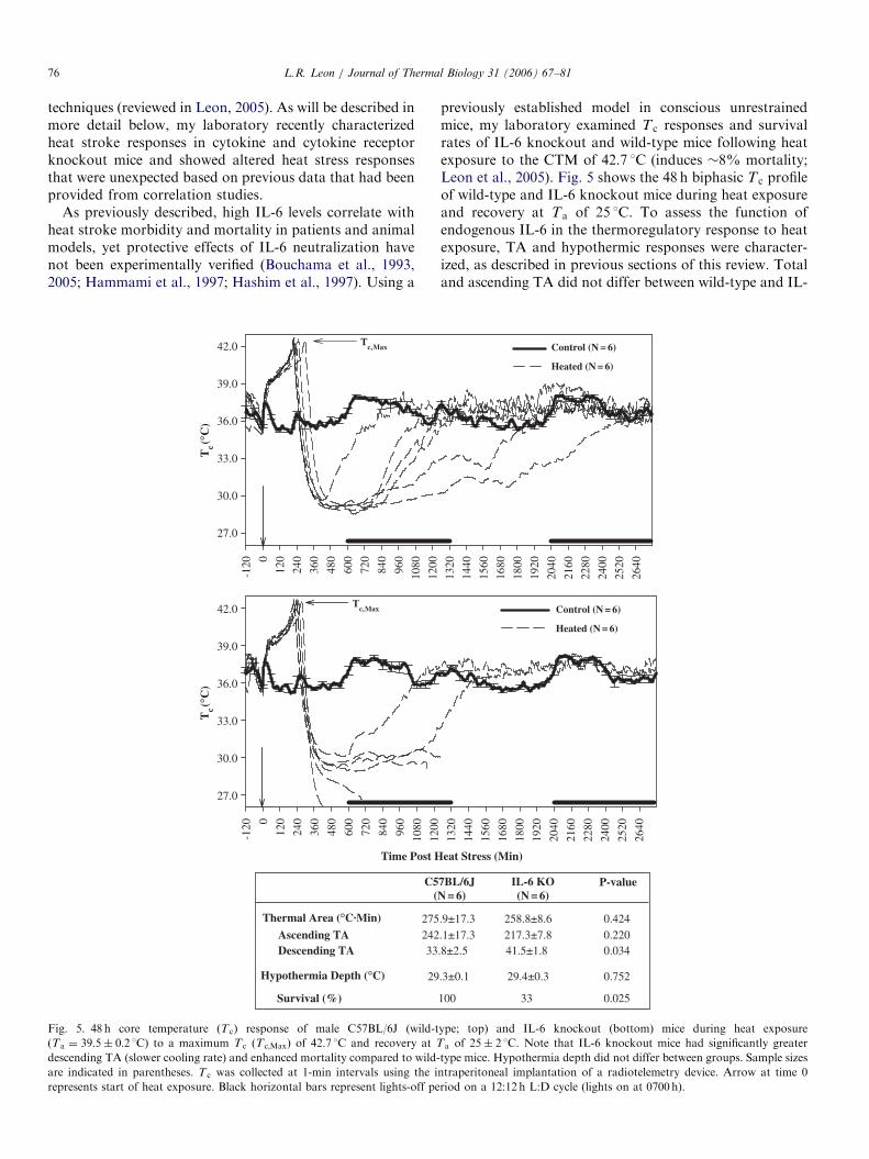
Fig. 5. 48 h core temperature (T c) response of male C57BL/6J (wild-t
(Ta ¼ 39:5� 0:2 1C) to a maximum T c (Tc;Max) of 42.7 1C and recovery at T
descending TA (slower cooling rate) and enhanced mortality compared to wild-
are indicated in parentheses. T c was collected at 1-min intervals using the i
represents start of heat exposure. Black horizontal bars represent lights-off pe
previously established model in conscious unrestrainedmice, my laboratory examined T c responses and survivalrates of IL-6 knockout and wild-type mice following heatexposure to the CTM of 42.7 1C (induces �8% mortality;Leon et al., 2005). Fig. 5 shows the 48 h biphasic T c profileof wild-type and IL-6 knockout mice during heat exposureand recovery at Ta of 25 1C. To assess the function ofendogenous IL-6 in the thermoregulatory response to heatexposure, TA and hypothermic responses were character-ized, as described in previous sections of this review. Totaland ascending TA did not differ between wild-type and IL-
eat Stress (Min)
1320
1440
1560
1680
1800
1920
2040
2160
2280
2400
2520
2640
1200
1320
1440
1560
1680
1800
1920
2040
2160
2280
2400
2520
2640
Control (N = 6)
Heated (N = 6)
Control (N = 6)
Heated (N = 6)
7BL/6JN = 6)
IL-6 KO(N = 6)
P-value
.9±17.3
.1±17.3
.3±0.1
100
.8±2.5
258.8±8.6 0.424
0.0340.220
0.752
0.025
217.3±7.8
29.4±0.3
33
41.5±1.8
ype; top) and IL-6 knockout (bottom) mice during heat exposure
a of 25� 2 1C. Note that IL-6 knockout mice had significantly greater
type mice. Hypothermia depth did not differ between groups. Sample sizes
ntraperitoneal implantation of a radiotelemetry device. Arrow at time 0
riod on a 12:12 h L:D cycle (lights on at 0700 h).
ARTICLE IN PRESSL.R. Leon / Journal of Thermal Biology 31 (2006) 67–81 77
6 knockout mice, indicating that thermoregulatory me-chanisms to direct heat exposure were not altered in theabsence of IL-6 actions. This was not a particularlysurprising finding since increased circulating IL-6 levelswere not detectable in heat stressed mice at Tc;Max (Leon etal., in press). However, analysis of the cooling phase ofrecovery indicated that IL-6 knockout mice had asignificantly greater descending TA than their wild-typecontrols (Fig. 5; Student’s t-test, P ¼ 0:034). Thus, IL-6knockout mice did not dissipate core heat to the environ-ment as effectively as wild-type mice during recovery (it isimportant to note that body weight did not differ betweengenotypes). It is hypothesized that differences in coolingrate were responsible for enhanced mortality in the IL-6knockout compared to wild-type mice within 24 h ofrecovery (see Table associated with Fig. 5; 33 vs. 100%,respectively). While these data do not refute the hypothesisthat elevated IL-6 levels are detrimental to heat strokesurvival (as suggested by correlation studies), they aresuggestive of a permissive effect of IL-6 that is required forheat stroke recovery. Similar results are reported for IL-6knockout mice in the sepsis syndrome (Leon et al., 1998).Note that hypothermia depth was virtually identicalbetween wild-type and IL-6 knockout mice (29:3� 0:1 vs.29:4� 0:3 1C, respectively) suggesting a lack of involve-ment of endogenous IL-6 in the regulation of this T c
response. Similarly, although only 2 animals survivedthrough 48 h of observation, the fever-like response from�24–36 h appeared to remain intact in IL-6 knockoutsurvivors; thus, IL-6 may not be regulating/modulating thisT c response either, although a larger sample size is requiredto statistically verify this conclusion. It would be interest-ing to conduct a feedback experiment in which IL-6 isadministered to knockout mice during different stages ofheat stroke recovery to determine (1) the endogenous levelof IL-6 that is required for survival (i.e., baseline orelevated levels), and (2) the time point during recovery atwhich IL-6 actions are beneficial. Interestingly, the contra-diction between the results from IL-6 knockout mice andthose reported in correlation studies suggest that IL-6 mayhave both pro- and anti-inflammatory properties, whichare dependent on the cytokine milieu in which it functions.Thus, future studies should be directed at determiningchanges in circulating levels of other cytokines influencedby heat exposure and IL-6 to determine how the absence ofthis cytokine has altered the balance of others mediators insurvivors and non-survivors of this syndrome.
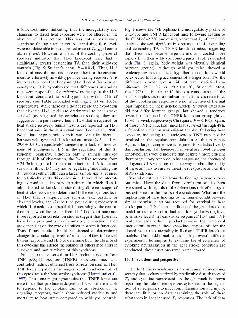
Similar to that observed for IL-6, preliminary data fromTNF p55/p75 receptor (TNFR) knockout mice alsocontradict findings obtained from correlation studies. HighTNF levels in patients are suggestive of an adverse role ofthis cytokine in the heat stroke syndrome (Hammami et al.,1997). Thus, one might hypothesize that TNFR knockoutmice (mice that produce endogenous TNF, but are unableto respond to the cytokine due to an absence of thesignaling receptors) would show reduced morbidity andmortality to heat stress compared to wild-type controls.
Fig. 6 shows the 48 h biphasic thermoregulatory profile ofwild-type and TNFR knockout mice following heating tothe CTM of 42.7 1C and during recovery at Ta of 25 1C. TAanalysis showed significantly decreased total, ascendingand descending TA in TNFR knockout mice, suggestingthat these mice became hyperthermic and cooled morerapidly than their wild-type counterparts (Table associatedwith Fig. 6; again, body weight was virtually identicalbetween groups). Although wild-type mice showed atendency towards enhanced hypothermia depth, as wouldbe expected following accruement of a larger total TA, thedifference between groups did not reach statistical sig-nificance (28:7� 0:2 vs. 29:2� 0:3 1C, Student’s t-test,P ¼ 0:275). It is unclear if this is a consequence of thesmall sample sizes or an indication that the characteristicsof the hypothermic response are not indicative of thermalload imposed on these genetic models. Survival rates alsodid not differ between groups, but showed a tendencytowards a decrease in the TNFR knockout group (40 vs.100% survival, respectively; Chi square, P ¼ 0:100). Again,of those TNFR knockout mice that survived through 48 h,a fever-like elevation was evident the day following heatexposure, indicating that endogenous TNF may not beinvolved in the regulation/modulation of this response.Again, a larger sample size is required to statistical verifythis conclusion. If differences in survival are noted betweengenotypes, this would indicate that despite a more efficientthermoregulatory response to heat exposure, the absence ofendogenous TNF actions in some way inhibits the abilityof these animals to survive direct heat exposure and/or theSIRS syndrome.Several questions arise from the findings in gene knock-
out mice. Have the data from correlation studies beenoverstated with regards to the deleterious role of endogen-ous cytokines in the heat stroke syndrome? What are theimplications of these findings to the human condition—aresimilar permissive actions required for survival in heatstroke patients? Is this a condition of the gene knockoutmodel or indicative of a dual role for cytokines (high vs.permissive levels) in heat stroke responses? IL-6 and TNFmodulate each other’s production—are the reciprocalinteractions between these cytokines responsible for thealtered heat stroke mortality in IL-6 and TNFR knockoutmodels? Until additional studies using several differentexperimental techniques to examine the effectiveness ofcytokine neutralization in the heat stroke condition areconducted, these questions remain unanswered.
10. Conclusions and perspective
The heat illness syndrome is a continuum of increasingseverity that is characterized by predictable disturbances ofTc and cytokine homeostasis. Although much is knownregarding the role of endogenous cytokines in the regula-tion of T c responses to infection, inflammation and injury,there are little or no data examining the role of thesesubstances in heat-induced Tc responses. The lack of data

ARTICLE IN PRESS
-120 0
120
240
360
480
600
720
840
960
1080
1200
1320
1440
1560
1680
1800
1920
2040
2160
2280
2400
2520
2640
-120 0
120
240
360
480
600
720
840
960
1080
1200
1320
1440
1560
1680
1800
1920
2040
2160
2280
2400
2520
2640
Tc
(°C
)
27.0
30.0
33.0
36.0
39.0
42.0
Tc
(°C
)
27.0
30.0
33.0
36.0
39.0
42.0
Time Post Heat Stress (Min)
Control (N = 5)Heated (N = 5)
Control (N = 4)Heated (N = 4)
Tc, Max
Tc, Max
Thermal Area (°C.Min)
Ascending TADescending TA
Hypothermia Depth (°C)
Survival (%)
B6129F2(N = 4)
TNFR KO(N = 5)
P-value
455.8±29.9408.4±2.3
28.7±0.2
100
47.5±6.6
327.6±31.8 0.024*
0.010*0.044*
0.275
0.100
301.3±31.6
29.2±0.3
40
26.3±0.8
Fig. 6. 48 h core temperature (T c) response of male B6129F2 (wild-type; top) and TNFR knockout (bottom) mice during heat exposure
(Ta ¼ 39:5� 0:2 1C) to a maximum Tc (T c;Max) of 42.7 1C and recovery at Ta of 25� 2 1C. Note that TNFR knockout mice had significantly lower
total, ascending (thermal load) and descending TA (slower cooling rate) compared to wild-type mice. Hypothermia depth and survival did not differ
between groups. Sample sizes are indicated in parentheses. T c was collected at 1-min intervals using the intraperitoneal implantation of a radiotelemetry
device. Arrow at time 0 represents start of heat exposure. Black horizontal bars represent lights-off period on a 12:12 h L:D cycle (lights on at 0700 h).
L.R. Leon / Journal of Thermal Biology 31 (2006) 67–8178
in this area is perplexing for several reasons. First,hypothermia and fever represent predictable responses toheat exposure in rodents and this biphasic Tc responseresembles that observed in the endotoxemic/sepsissyndrome, for which a role for several cytokines (e.g., IL-1, IL-6, IL-10, TNF) has been strongly implicated. Thus, itis anticipated that much knowledge can be gained fromapplying our current understanding of endotoxemicpathophysiology to the study of heat stroke. Second,hypothermia appears to be a sensitive biomarker of heatseverity. This may have important implications in theclinical setting since (a) details regarding the initial heatinsult are rarely known, and (b) induced hypothermia mayhave currently unrecognized protective effects that could beutilized as a clinical treatment strategy. Clearly, theregulated nature of heat-induced hypothermia and thebeneficial effect of the fever-like response need to be
determined. Third, the efficacy of cytokine neutralizationon heat stroke morbidity and mortality has not beenextensively investigated. Currently, IL-1 is the onlycytokine whose neutralization has been examined for itsbenefit in heat stroke outcome, showing a protective effect(Chiu et al., 1995; Liu et al., 2000). Yet, the mechanism(s)of protection remain unknown. How is the thermoregula-tory profile altered with this treatment? Is the beneficialeffect a direct thermal effect or due to interactions betweenchanges in thermal load and cardiovascular strain?Unfortunately, current data implicating cytokines in heatstroke responses are mainly from correlation studiesshowing elevated plasma levels in heat stroke patientsand experimental animal models. Correlation data fall farshort of revealing the mechanisms of cytokine action, assuggested from the results obtained in IL-6 and TNFRknockout mice. Furthermore, cytokine determinations

ARTICLE IN PRESSL.R. Leon / Journal of Thermal Biology 31 (2006) 67–81 79
have typically been performed at end-stage heat stroke,such that the role of these substances in progression andlong-term recovery from heat injury is poorly understood.While the thermoregulatory changes induced by heatexposure represent only one physiological aspect of thiscomplex syndrome, T c is extremely labile to environmentalperturbation and is simple to measure, thus providingrapid and powerful information regarding homeostaticbalance of the individual. It is anticipated that moredetailed understanding of the role of cytokines in thehypothermic and fever-like responses to heat stroke willprovide important insight into the role of these substancesin the complex etiology of the long-term consequences ofthis syndrome.
Acknowledgements
I thank A.M. Bastille, M. Blaha, J. Castor, and L.D.Walker for their technical assistance with the gene knock-out studies. D.A. Dubose and M.N. Sawka providedinvaluable intellectual contributions through numerousdiscussions concerning heat stroke pathophysiology.
The opinions or assertions contained herein are theprivate views of the authors and are not to be construed asofficial or as reflecting the views of the Army or theDepartment of Defense.
In conducting the research described in this report, theinvestigators adhered to the ‘‘Guide for Care and Use ofLaboratory Animals’’ as prepared by the Committee onCare and Use of Laboratory Animals of the Instituteof Laboratory Animal Resources, National ResearchCouncil.
Citations of commercial organizations and trade namesin this report do not constitute an official Department ofthe Army endorsement or approval of the products orservices of these organizations.
References
Adolph, E.F., 1947. Tolerance to heat and dehydration in several species
of mammals. Am. J. Physiol. 151, 564–575.
Aoki, K., Kondo, N., Shibasaki, M., Takano, S., Katsuura, T., 1998.
Circadian variation in skin blood flow responses to passive heat stress.
Physiol. Behav. 63, 1–5.
Arons, M.M., Wheeler, A.P., Bernard, G.R., Christman, B.W., Russell,
J.A., Schein, R., Summer, W.R., Steinberg, D.P., Fulkerson, W.,
Wright, P., Dupont, W.D., Swindell, B.B., 1999. Effects of ibuprofen
on the physiology and survival of hypothermic sepsis. Ibuprofen in
sepsis study group. Crit. Care Med. 27, 699–707.
Attia, M., Khogali, M., El-Khatib, G., Mustafa, M.K.E., Mahmoud,
M.A., Eldin, A.N., Gumaa, K., 1983. Heat-stroke: an upward shift of
temperature regulation set point at an elevated body temperature. Int.
Arch. Occup. Environ. Health 53, 9–17.
Austin, M.G., Berry, J.W., 1956. Observations on one hundred cases of
heatstroke. J. Am. Med. Assoc. 161, 1525–1529.
Bouchama, A., Knochel, J.P., 2002. Heat stroke. N. Engl. J. Med. 346,
1978–1988.
Bouchama, A., Parhar, R.S., el-Yazigi, A., Sheth, K., al-Sedairy, S., 1991.
Endotoxemia and release of tumor necrosis factor and interleukin 1
alpha in acute heatstroke. J. Appl Physiol. 70, 2640–2644.
Bouchama, A., al-Sedairy, S., Siddiqui, S., Shail, E., Rezeig, M., 1993.
Elevated pyrogenic cytokines in heatstroke. Chest 104, 1498–1502.
Bouchama, A., Hammami, M.M., Al Shail, E., De Vol, E., 2000.
Differential effects of in vitro and in vivo hyperthermia on the
production of interleukin-10. Intensive Care Med. 26, 1646–1651.
Bouchama, A., Roberts, G., Al Mohanna, F., El-Sayed, R., Lach, B.,
Chollet-Martin, S., Ollivier, V., Al Baradei, R., Loualich, A., Nakeeb,
S., Eldali, A., de Prost, D., 2005. Inflammatory, hemostatic, and
clinical changes in a baboon experimental model for heatstroke.
J. Appl. Physiol. 98, 697–705.
Bynum, G.D., Pandolf, K.B., Schuette, W.H., Goldman, R.F., Lees, D.E.,
Whang-Peng, J., Atkinson, E.R., Bull, J.M., 1978. Induced hyperther-
mia in sedated humans and the concept of critical thermal maximum.
Am. J. Physiol. 235, R228–R236.
Carpenter, A.J., Nunneley, S.A., 1988. Endogenous hormones subtly alter
women’s response to heat stress. J. Appl. Physiol. 65, 2313–2317.
Carter, R., Cheuvront, S.N., Williams, J.O., Kolka, M.A., Stephenson,
L.A., Amoroso, P.J., Sawka, M.N., 2005. Epidemiology of hospita-
lizations and deaths from heat illness in soldiers from 1980 through
2002. Med. Sci. Sport. Exerc. 37, 1328–1334.
Chang, D.M., 1993. The role of cytokine in heat stroke. Immunol. Invest.
22, 553–561.
Chiu, W.-T., Kao, T.-Y., Lin, M.-T., 1995. Interleukin-1 receptor
antagonist increases survival in rat heatstroke by reducing hypotha-
lamic serotonin release. Neurosci. Lett. 202, 33–36.
Chiu, W.T., Kao, T.Y., Lin, M.T., 1996. Increased survival in
experimental rat heatstroke by continuous perfusion of interleukin-1
receptor antagonist. Neurosci. Res. 24, 159–163.
Cole, R.D., 1983. Heat stroke during training with nuclear, biological,
and chemical protective clothing: case report. Mil. Med. 148,
624–625.
Cowles, R.B., Bogert, C.M., 1944. A preliminary study of the
thermal requirements of desert reptiles. Bull. Am. Mus. Nat. Hist.
83, 265–296.
Dematte, J.E., O’Mara, K., Suescher, J., Whitney, C.G., Forsythe, S.,
McNamee, T., Adiga, R.B., Ndukwu, I.M., 1988. Near-fatal heat
stroke during the 1995 heat wave in Chicago. Ann. Intern. Med. 129,
173–181.
Dietrich, W.D., Kuluz, J.W., 2003. New research in the field of stroke:
therapeutic hypothermia after cardiac arrest. Stroke 34, 1051–1053.
Drobatz, K.J., Macintire, D.K., 1996. Heat-induced illness in dogs: 42
cases (1976–1993). J. Am. Vet. Med. Assoc. 209, 1894–1899.
D’Souza, S.D., Antel, J.P., Freedman, M.S., 1994. Cytokine induction of
heat shock protein expression in human oligodendrocytes: an
interleukin-1-mediated mechanism. J. Neuroimmunol. 50, 17–24.
Dubois, E.F., 1949. Why are fevers over 1061F rare? Am. J. Med. Sci. 217,
361–368.
Dubose, D.A., Basamania, K., Maglione, L., Rowlands, J., 1983. Role of
bacterial endotoxins of intestinal origin in rat heat stress mortality. J.
Appl. Physiol. 54, 31–36.
Elmer, M., Ohlin, P., 1970. Salivary glands of the rat in a hot
environment. Acta Physiol. Scand. 79, 129–132.
Fairchield, K.D., Singh, I.S., Patel, S., Drysdale, B.E., Viscardi, R.M.,
Hester, L., Lazusky, H.M., Hasday, J.D., 2004. Hypothermia prolongs
activation of NF-kB and augments generation of inflammatory
cytokines. Am. J. Physiol. 287, C422–C431.
Flanagan, S.W., Ryan, A.J., Gisolfi, C.V., Moseley, P.L., 1995. Tissue-
specific HSP70 response in animals undergoing heat stress. Am. J.
Physiol. 268, R28–R32.
Furuyama, F., 1982. Strain difference in thermoregulation of rats
surviving extreme heat. J. Appl. Physiol. 52, 410–415.
Gathiram, P., Gaffin, S.L., Brock-Utne, J.G., Wells, M.T., 1987. Time
course of endotoxemia and cardiovascular changes in heat-stressed
primates. Aviat. Space Environ. Med. 58, 1071–1074.
Gordon, C.J., 1993. Temperature Regulation in Laboratory Rodents.
Cambridge University Press, New York.
Gordon, C.J., 2001. The therapeutic potential of regulated hypothermia.
Emerg. Med. J. 18, 81–89.

ARTICLE IN PRESSL.R. Leon / Journal of Thermal Biology 31 (2006) 67–8180
Graber, C.D., Reinhold, R.B., Breman, J.G., Harley, R.A., Hennigar,
G.R., 1971. Fatal heat stroke. Circulating endotoxin and gram-
negative sepsis as complications. J. Am. Med. Assoc. 216, 1195–1196.
Hainsworth, F.R., 1967. Saliva spreading, activity, and body temperature
regulation in the rat. Am. J. Physiol. 212, 1288–1292.
Hales, J.R., Khogali, M., Fawcett, A.A., Mustafa, M.K., 1987.
Circulatory changes associated with heat stroke: observations in an
experimental animal model. Clin. Exp. Pharmacol. Physiol. 14,
761–777.
Hall, D.M., Buettner, G.R., Oberley, L.W., Xu, L., Mattes, R.D., Gisolfi,
C.V., 2001. Mechanisms of circulatory and intestinal barrier dysfunc-
tion during whole body hyperthermia. Am. J. Physiol. 280,
H509–H521.
Hammami, M.M., Bouchama, A., Al-Sedairy, S., Shail, E., Al Ohaly, Y.,
Mohamed, G.E., 1997. Concentrations of soluble tumor necrosis
factor and interleukin-6 receptors in heatstroke and heatstress. Crit.
Care Med. 25, 1314–1319.
Hashim, I.A., Al-Zeer, A., Al-Shohaib, S., Al-Ahwal, M., Shenkin, A.,
1997. Cytokine changes in patients with heatstroke during pilgrimage
to Makkah. Mediators Inflammation 6, 135–139.
Haveman, J., Geerdink, A.G., Rodermond, H.M., 1996. Cytokine
production after whole body and localized hyperthermia. Int. J.
Hyperthermia 12, 791–800.
Heidemann, S.M., Lomo, L., Ofenstein, J.P., Samaik, A.P., 2000. The
effect of heat on cytokine production in rat endotoxemia. Crit. Care
Med. 28, 1465–1468.
Herman, T.S., Gerner, E.W., Magun, B.E., Stickney, D., Sweets, C.C.,
White, D.M., 1981. Rate of heating as a determinant of hyperthermic
cytotoxicity. Cancer Res. 41, 3519–3523.
Hoar, W.S., 1955. Seasonal variation in the resistance of goldfish to
temperature. Trans. Roy. Soc. Can. 49, 25–34.
Horowitz, M., Argov, D., Mizrahi, R., 1983. Interrelationships between
heat acclimation and salivary cooling mechanism in conscious rats.
Comp. Biochem. Physiol. A 74, 945–949.
Hubbard, R.W., Matthew, W.T., Linduska, J.D., Curtis, F.C., Bowers,
W.D., Leav, I., Mager, M., 1976. The laboratory rat as a model
for hyperthermic syndromes in humans. Am. J. Physiol. 231,
1119–1123.
Hubbard, R.W., Bowers, W.D., Matthew, W.T., Curtis, F.C., Criss,
R.E.L., Sheldon, G.M., Ratteree, J.W., 1977. Rat model of acute
heatstroke mortality. J. Appl. Physiol. 42, 809–816.
Hubbard, R.W., Matthew, W.T., Criss, R.E., Sils, I., Mager, M., Bowers,
W.D., Wolfe, D., 1978. Role of physical effort in the etiology of rat
heatstroke injury and mortality. J. Appl. Physiol. 45, 463–468.
Hutchison, V.H., 1961. Critical thermal maxima in salamanders. Physiol.
Zool. 34, 92–125.
Hutchison, V.H., Murphy, K., 1985. Behavioral thermoregulation in the
salamander Necturus maculosus after heat shock. Comp. Biochem.
Physiol. 82A, 391–394.
Junqueira, L.C.U., Fava-de-Moraes, F., Toledo, A.M.S., 1967. Action
of vasopressin on salivary secretion. Acta Physiol. Latinoamer. 17,
36–41.
Kao, T.Y., Lin, M.T., 1996. Brain serotonin depletion attenuates
heatstroke-induced cerebral ischemia and cell death in rats. Am. J.
Physiol. 80, 680–684.
Kark, J.A., Burr, P.Q., Wenger, C.B., Gastaldo, E., Gardner, J.W., 1996.
Exertional heat illness in Marine corps recruit training. Aviat. Space
Environ. Med. 67, 354–360.
Kluger, M.J., 1991. Fever: role of pyrogens and cryogens. Physiol. Rev.
71, 93–127.
Kosh, R.J., Hutchison, V.H., 1968. Daily rhythmicity of temperature
tolerance in the eastern painted turtle, Chrysemys picta picta. Copiea 2,
244–246.
Leon, L.R., 2004. Hypothermia in systemic inflammation: role of
cytokines. Front. Biosci. 9, 1877–1888.
Leon, L.R., 2005. The use of gene knockout models in thermoregulation
studies. J. Thermal Biol. 30, 273–288.
Leon, L.R., White, A.A., Kluger, M.J., 1998. Role of IL-6 and TNF in
thermoregulation and survival during sepsis in mice. Am. J. Physiol.
275, R269–R277.
Leon, L.R., DuBose, D.A., Mason, C.D., 2005. Heat stress induces a
biphasic thermoregulatory response in mice. Am. J. Physiol. 288,
R197–R204.
Leon, L.R., Blaha, M.D., Dubose, D.A. Time course of cytokine,
corticosterone and tissue injury responses in mice during heat strain
recovery. J. Appl. Physiol., in press.
Lin, M.T., Kao, T.Y., Su, C.F., Hsu, S.S., 1994. Interleukin-1bproduction during the onset of heat stroke in rabbits. Neurosci. Lett.
174, 17–20.
Lin, M.T., Liu, H.H., Yang, Y.L., 1997. Involvement of interleukin-1
receptor mechanisms in development of arterial hypotension in rat
heatstroke. Am. J. Physiol. 273, H2072–H2077.
Liu, C.-C., Chien, C.-H., Lin, M.-T., 2000. Glucocorticoids reduce
interleukin-1b concentration and result in neuroprotective effects in rat
heatstroke. J. Physiol. 527, 333–343.
Lord, P.F., Kapp, D.S., Hayes, T., Weshler, Z., 1984. Production of
systemic hyperthermia in the rat. Eur. J. Cancer Clin. Oncol. 20,
1079–1085.
Lu, K.-C., Wang, J.-Y., Lin, S.-H., Chu, P., Lin, Y.-F., 2004. Role of
circulating cytokines and chemokines in exertional heatstroke. Crit.
Care Med. 32, 399–403.
Lublin, A., Wolfenson, D., Berman, A., 1995. Sex differences in blood
flow distribution of normothermic and heat-stressed rabbits. Am. J.
Physiol. 268, R66–R71.
Malamud, N., Haymaker, W., Custer, R.P., 1946. Heat stroke. Mil. Surg.
99, 397–449.
Manjoo, M.F., Burger, F.J., Kielblock, 1985. A relationship between heat
load and plasma enzyme concentration. J. Therm. Biol. 10, 221–225.
Marion, D.W., Penrod, L.E., Kelsey, S.F., Obrist, W.D., Kochanek, P.M.,
Palmer, A.M., Wisniewski, S.R., DeKosky, S.T., 1997. Treatment of
traumatic brain injury with moderate hypothermia. N. Engl. J. Med.
336, 540–546.
Maron, M.B., Wagner, J.A., Horvath, S.M., 1977. Thermoregulatory
responses during competitive marathon running. J. Appl. Physiol. 42,
909–914.
Mehnert, P., Brode, P., Griefahn, B., 2002. Gender-related difference in
sweat loss and its impact on exposure limits to heat stress. Intern. J.
Indust. Ergonom. 29, 343–351.
Naughton, M.P., Henderson, A., Mirabelli, M.C., Kaiser, R., Wilhelm,
J.L., Kieszak, S.M., Rubin, C.H., McGeehin, M.A., 2002. Heat-
related mortality during a 1999 heat wave in Chicago. Am. J. Prev.
Med. 22, 221–227.
Neville, A.J., Sauder, D.N., 1988. Whole body hyperthermia (41–42 1C)
induces interleukin-1 in vivo. Lymphokine Res. 7, 201–206.
O’Brien, C., Hoyt, R.W., Buller, M.J., Castellani, J.W., Young, A.J.,
1998. Telemetry pill measurement of core temperature in humans
during active heating and cooling. Med. Sci. Sports Exerc. 30, 468–472.
Ohara, K., Furuyama, F., Isobe, Y., 1975. Prediction of survival time of
rats in severe heat. J. Appl. Physiol. 38, 724–729.
Petersdorf, R.G., 1994. Hypothermia and hyperthermia. In: Isselbacher,
K.J., Braunwald, E., Wilson, J.D., Martin, J.B., Fauci, A.S., Kasper,
D.L. (Eds.), Harrison’s Principles of Internal Medicine. McGraw-Hill,
Health Professions Division, New York.
Ramlow, J.M., Kuller, L.H., 1990. Effect of the summer heat wave of 1988
on daily mortality in Allegheny County, PA. Public Health Rep. 105,
283–289.
Romanovsky, A.A., Blatteis, C.M., 1996. Heat stroke: opioid-mediated
mechanisms. J. Appl. Physiol. 81, 2565–2570.
Romanovsky, A.A., Shido, O., Sakurada, S., Sugimoto, N., Nagasaka, T.,
1996. Endotoxin shock: thermoregulatory mechanisms. Am. J.
Physiol. 270, R693–R703.
Sigmund, C.D., 1993. Major approaches for generating and analyzing
transgenic mice. Hypertension 22, 599–607.

ARTICLE IN PRESSL.R. Leon / Journal of Thermal Biology 31 (2006) 67–81 81
Sonna, L.A., Wenger, C.B., Flinn, S., Sheldon, H.K., Sawka, M.N., Lilly,
C.M., 2004. Exertional heat injury and gene expression changes: a
DNA microarray analysis study. J. Appl. Physiol. 96, 1943–1953.
Stallones, R.A., Gauld, R.L., Dodge, H.J., Lammers, T.F.M., 1957. An
epidemiological study of heat injury in Army recruits. A.M.A. Arch.
Ind. Health 15, 455–465.
Stricker, E.M., Hainsworth, F.R., 1970. Evaporative cooling in the rat:
effects of dehydration. Can. J. Physiol. Pharmacol. 48, 18–27.
Watanabe, N., Tsuji, N., Akiyama, S., Sasaki, H., Okamoto, T.,
Kobayashi, D., Sato, T., Hagino, T., Yamauchi, N., Niitsu, Y.,
Nakai, A., Nagata, K., 1997. Induction of heat shock protein 72
synthesis by endogenous tumor necrosis factor via enhancement of the
heat shock element-binding activity of heat shock factor 1. Eur. J.
Immunol. 27, 2830–2834.
Watanabe, N., Tsuji, N., Akiyama, S., Sasaki, H., Okamoto, T.,
Kobayashi, D., Sato, T., Hagino, T., Yamauchi, N., Niitsu, Y.,
1998. Endogenous tumour necrosis factor regulates heat-inducible heat
shock protein 72 synthesis. Int. J. Hyperthermia 14, 309–317.
Wilkinson, D.A., Burholt, D.R., Shrivastava, P.N., 1988. Hypothermia
following whole-body heating of mice: effect of heating time and
temperature. Int. J. Hyperthermia 4, 171–182.
Wright, G.L., 1976. Critical thermal maximum in mice. J. Appl. Physiol.
40, 683–687.
Wright, G., Knecht, E., Wasserman, D., 1977. Colonic heating pattern and
the variation of thermal resistance among rats. J. Appl. Physiol. 43, 59–64.
Wyndham, C.H., 1966. A survey of research initiated by the chamber of
mines into clinical aspects of heat stroke. Proc. Mine Med. Off. Assoc.
46, 68–80.