Embed Size (px)
Citation preview

Investigations in Neurology
Professor Bruce CampbellNeurologist, Head of Stroke, Interim Head of Neurology
Royal Melbourne Hospital

Scenarios
• ?stroke– CT, carotid US/CTA, ECG, echo, bloods, MRI and advanced CT, DSA, SAH algorithm
• ?MS– MRI, CSF(inc pressure), VEP, OCT, NMO, JC Abs
• ?Seizure– EEG – focal, generalized, MRI – HS, SOL, HLA B1501
• ?neuropathy– NCS, fBSL/OGTT, B12, B1, B6, T4/TSH, SPEP, vasculitic, HNPP PMP22 gene, drugs, biopsy
• ?GBS (acute flaccid paralysis)– FVC, NCS, CSF, GM1/GQ1b etc, serology
• ?myasthenia– Rep Stim, SFEMG, AChR Abs, MuSK Abs, CT chest, antiSM Abs, Tensilon, Ice
• ?myopathy/MND– CK, “LFT”, vasculitic, Vit D, T4/TSH, electrolytes, EMG, biopsy
• Miscellaneous/movement disorders– Cu/caeruloplasmin, acanthocytes, NOTCH3, TA Bx, 14-3-3/PrP/DWI, antiGAD, NMDA, VGKC

?Stroke
• Non-contrast CT brain – rate-limiting step for treatment
• Loss of grey-white differentiation (especially caudate, lentiform, insula)
• Hyperdense artery (when asymmetrical) = acute clot
• CT angiography now regarded as standard – large vessel occlusion for clot retrieval– helps improve diagnostic certainty/risk stratify mild stroke
• CT perfusion required for thrombectomy >6h, thrombolysis >4.5h• helps improve diagnostic certainty vs mimics/risk stratify mild
stroke/prognosticate e.g. large core/plan e.g. hemicraniectomy→ recommended for all suspected stroke

Non-contrast CT Brain
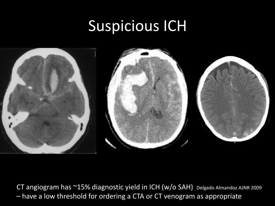
Suspicious ICH
CT angiogram has ~15% diagnostic yield in ICH (w/o SAH) Delgado Almandoz AJNR 2009
– have a low threshold for ordering a CTA or CT venogram as appropriate


but more common:
72yo F left hemiparesis and dysarthria


5mm thick
STANDARD 5mm
1mm thick
THIN reformat 1mm
“partial
volume
effect”

• Principles of CT perfusion
Delayed TTP= collateral territory
time
Area ≈ 0
con
cen
trat
ion
time
Area = CBV
TTPco
nce
ntr
atio
n
CBV
Low CBV = likely irreversibly damaged
CBF TTP

CTP pattern = occlusion site

artefacts (non-arterial territory)

Lacunar infarction
MTT CBF DWI
Cao et al JOCN 2016
requires
excellent image
quality (dose)
and sharp eyes!

TTP
CBV
“mild stroke”

CT perfusion imaging –beyond diagnosis to tissue viability & late-window reperfusion
“How much blood supply”(severely reduced ≈ dead)* time to reperf & grey vs white matter
“How delayed is the blood supply”(severely delayed ≈ at risk)

CTA CTP
Dynamic CTA

Migraine

Post-seizure hyperperfusion
reduced Tmax
increased CBF

Post-reperfusion hyperperfusion
reduced Tmax
increased CBF

Stroke: mechanism determines prevention
Aetiology:
• Carotids – CTA or Doppler US
• US – look for increased velocity/spectral broadening
• >70% symptomatic stenosis→ endarterectomy consider for 50-70% stenosis (not if occluded)
• ECG ?AF, evidence for big old infarct (mural thrombus)
• TTE/TOE ?vegetation, mural thrombus, PFO +/- ASAaortic arch atheroma,

NORMAL ABNORMAL
Increased velocity and “spectral broadening”
Tapered occlusion, above bifurcation = dissection
Stenosis at bifurcation = atherosclerosisCTA

Young stroke aetiology:
• Carotids – prefer CTA ?dissection
• TTE/TOE ?vegetation, mural thrombus, PFO +/- ASAaortic arch atheromaTTE with bubbles – better Valsalva to detect shunt, TOE for valves
• Vasculitic & thrombophilic bloods
• “vasculitic”: ANA, ENA, RF, ANCA, C3/4,
• “thrombophilic” – FBE (thrombocytosis, polycythemia) antiphospholipid (anticardiolipin IgG, anti β2glycoprotein, lupus inhibitor), protein C, S, antithrombin, factor V Leiden and prothrombin mutations, ?homocysteine
• ?Fabry’s testing (dried blood spot)
Stroke: mechanism determines prevention

Diffusion MRI
DWI ADCDiffusion-weighted imaging Apparent diffusion co-efficient
Always check that the bright area on DWI is “true” restriction ie dark on ADCvs “T2 shine-through”
“TIA” but DWI +ve = strokeHigh risk recurrence
T1 fat saturated image -
Methemoglobin in arterial wall = bright→ Dx = dissection

groin puncture 19.15Pre (19.21) Post (19.40)
Advanced imaging: Digital subtraction angiogram mostly for treatment, occasionally if suspect dissection/vascular malformation and CTA equivocal
thrombectomy target territory:ICAM1 MCAproximal M2 MCABasilar

?subarachnoid hemorrhage
• Non-con CT• If normal → LP for xanthochromia (foil - light protected)
• CTA if SAH confirmed (otherwise you find lots of incidental aneurysms)
• DSA if looks treatable via endovascular or if CTA equivocal
• Thunderclap headache without SAH?– Reversible cerebral vasoconstriction
dissection, CVST, colloid cyst?
– Exertional headache

Dissemination in space = at least 2 lesions in different “high specificity” regionsDissemination in time = 2nd scan with a new lesion (at any time) or a single scan with both enhancing and non-enhancing lesions or OCBs
*the symptomatic lesion is now counted towards “dissemination in space” NB periventricular lesions less specific – MAGNIMS 2016 criteria required ≥3…
NB 2 clinical attacks separated in space and time still counts as clinically definite MS
PPMS = 1yr of disease progression & 2/3 of:Dissemination in space of cord, brain or oligoclonal bands
?MS
“high specificity” white matter lesion locations:• Periventricular• Juxtacortical “U-fibre”/cortical• Infratentorial• Spinal cord*
2017 revision – McDonald Criteria
Thompson et al Lancet Neurol 2018

Dissemination in space = at least 2 lesions in different “high specificity” regionsDissemination in time = 2nd scan with a new lesion (at any time) or a single scan with both enhancing and non-enhancing lesions or OCBs
*the symptomatic lesion is now counted towards “dissemination in space” NB periventricular lesions less specific – MAGNIMS 2016 criteria required ≥3…
NB 2 clinical attacks separated in space and time still counts as clinically definite MS
PPMS = 1yr of disease progression & 2/3 of:Dissemination in space of cord, brain or oligoclonal bands
?MS
“high specificity” white matter lesion locations:• Periventricular• Juxtacortical “U-fibre”/cortical• Infratentorial• Spinal cord*
“the McDonald criteria were not developed to differentiate multiple sclerosis from other conditions but to identify multiple sclerosis or a high likelihood of the disease in patients with a
typical clinically isolated syndrome once other diagnoses have been deemed unlikely”
Easier to diagnose MS earlier – starting treatment separate decisionwe need to consider the likely disease activity vs burden of therapy
2017 revision – McDonald Criteria
Thompson et al Lancet Neurol 2018

Gadolinium Enhancing lesions
Coronial T1 sequence with GadoliniumNew lesions usually enhance for <1 month. With concern about gadolinium accumulation,diffusion restriction often used as a surrogate for active lesions

?MS
CSF
• a few lymphocytes common (usually <50)
• unmatched oligoclonal bands (paired serum)
• present in >90% clinically definite MS
• differential: vasculitis, CNS infection, paraneoplastic
serum
CSF

?MS
Visual Evoked Potentials (VEP)
?MS
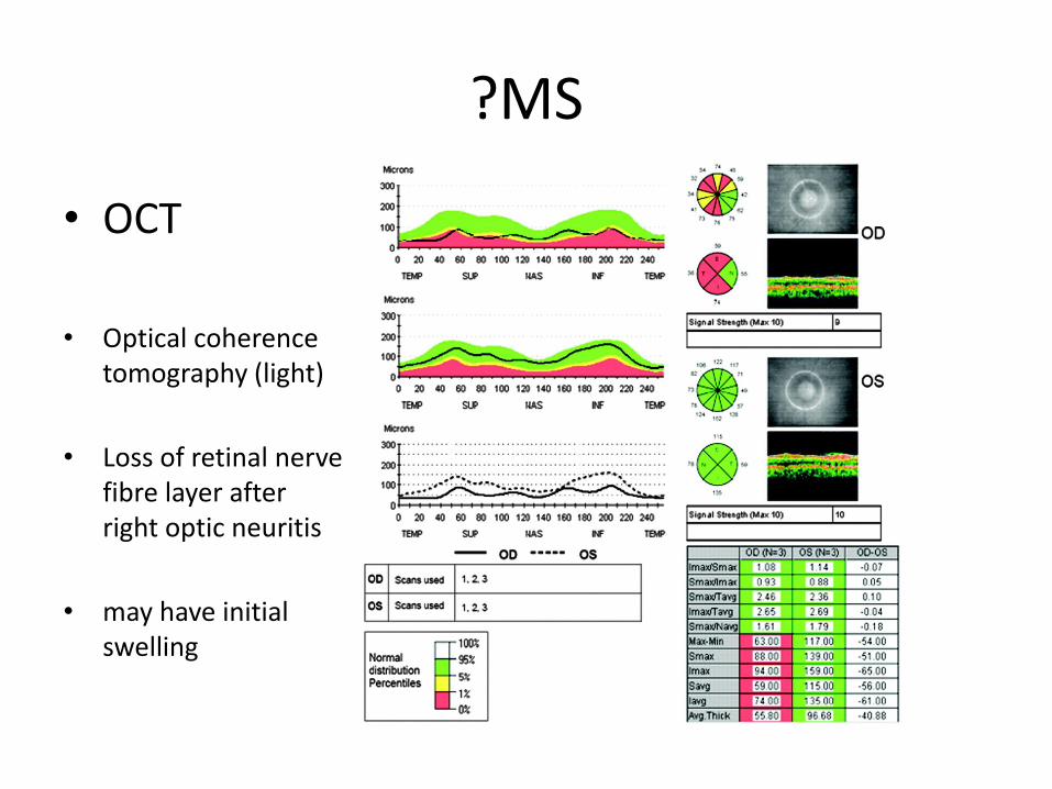
• OCT
• Optical coherence tomography (light)
• Loss of retinal nerve fibre layer after right optic neuritis
• may have initial swelling

?MS
• Consider other inflammatory diseases:– Neuromyelitis optica (anti-aquaporin4 “NMO” Abs)
• longitudinally extensive transverse myelitis (LETM) ≥3 vertebral segments
– anti-MOG antibody syndrome • isolated optic neuritis 55%, half bilateral; TM (often LETM) 18%; ADEM 18%
– Sarcoid, Behçet’s
• Many newer MS treatments are immunosuppressive– Consider immunization status and viral status similar to pre-transplant
etc
– VZV vax
• Serology for JC virus if considering natalizumab(PML risk stratification)

?seizure
• Hx (especially from a witness) is the best test – use the telephone if no witness present!
• EEG
• MRI
• (PET/SPECT – hypometabolism in lesion at rest but hypermetabolic during seizure)
• Pharmacogenomics– Carbamazepine, Stevens-Johnson Syndrome, HLA B1502

Epilepsy - MRI

Electroencephalography - EEG
General points:
• Even numbers = right hemisphere, odd = left
• F = frontal, T = temporal, C = central, O = occipital
• Normal = “alpha” 8-12Hz, attenuates with eyes open
• Abnormalities – generalized slowing (“encephalopathic”)
– focal slowing (intermittent or persistent)
– epileptiform discharges – focal or generalized
– triphasic waves
– burst-suppression

Eye blink(normal)
Normal EEG
9 per sec = 9Hz (“alpha”)
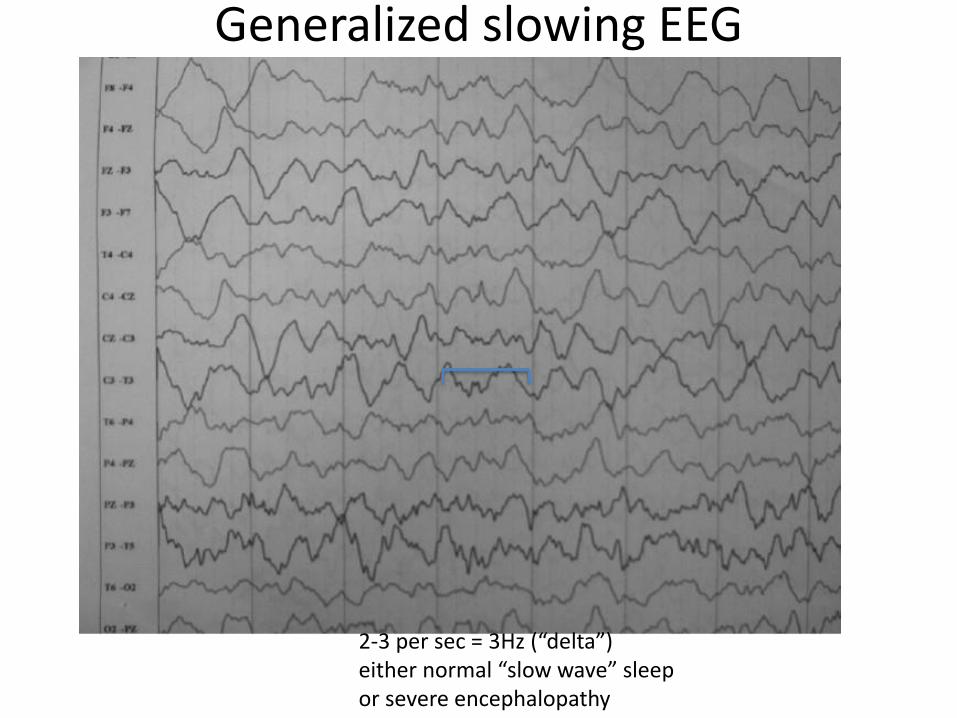
Generalized slowing EEG
2-3 per sec = 3Hz (“delta”)either normal “slow wave” sleep or severe encephalopathy

Generalized slowing EEG
Background 5-7Hz (“theta”) – encephalopathicPlus “triphasic waves” (boxes) seen in metabolic encephalopathy and CJD

Focal slowing EEG
Left hemisphere (odd numbers) = slow, right = normalNonspecific – any structural or functional lesion (in this case stroke – note the AF on rhythm strip)

Focal epileptiform discharge
Left frontal epileptiform discharge
Spike and slow waveStands out from backgroundHas “field” (appears in >1 electrode)Phase reverses at F7 (localization)

Periodic lateralized epileptiform discharge (PLED)
Right hemisphere, periodic (~1sec in this case) epileptiform dischargeNonspecific severe injury eg stroke, encephalitis, tumour etc, no need for anticonvulsant

Generalized epileptiform discharge
“childhood absence epilepsy”Normal background then a run of 3Hz “spike and wave” bilateral but doesn’t have to be same in all leads clinical correlate = absence seizure
“Juvenile myoclonic epilepsy”Isolated jerks or 4-6Hz spike and wave

Generalized epileptiform discharge?Generalized Periodic Epileptiform Discharges (GPEDs) – often seen post-cardiac arrestIn post-anoxic injury no benefit from anticonvulsants, generally poor prognosis
Burst-suppression with GPEDs–post-cardiac arrestor deep induced coma

Seizure
Right temporal focal seizureVarying morphologies but the key is evolution in frequency and amplitude over timeIf generalizes → can only see muscle artefact

?Neuropathy
• Generalized peripheral neuropathy:Common causes: DM, EtOH, vitamins, paraproteins, vasculitic, drugs, hereditary
• Bloods: fBSL/OGTT, U&E, Vit B12, T4/TSH, SPEP, vasculitic• Nerve conduction studies:
reduced velocity, dispersion +/- delayed “F-waves”= demyelinatingreduced amplitude = axonal but focal “block” (>50% drop in amplitude) = demyelinating
• Sural nerve biopsy – rarely needed. Consider if suspect amyloid, sarcoid, vasculitis
• Mononeuropathy:usually compressive (occasionally vasculitic – painful): median/ulnar/peroneal→ nerve conduction studies– if several pressure palsies consider HNPP (Hereditary neuropathy with pressure palsies, autosomal dominant – PMP22 gene test)
• More complex neuropathies – CIDP, MMN (anti-GM1 in 60-80%), brachial/lumbosacral plexopathy, radiculopathy→nerve conduction studies/EMG, MRI of spine/plexus

Normal Motor Conduction
Demyelinating Neuropathy

Normal F-waves
Prolonged F-waves

Focal Conduction Block

Multifocal Motor Neuropathy (MMN) – motor conduction block

Acute flaccid paralysis
• dDx GBS, cord compression/transverse myelitis, myasthenia, botulism, porphyria
• MRI spine if can’t exclude cord compression clinically• CSF “albuminocytologic dissociation” in GBS (inflammation/cells
outside dura in nerve roots, spill-over increased protein) exception = HIV-related GBS has increased cells– elevated protein – non-specific: inflammation, diabetes, blocked spinal
CSF flow
• NCS/EMG – take time to become abnormal (major early abNsuggests actually CIDP)– Delayed/loss of F-waves, later slowing
• Bloods - ganglioside Abs: GM1 (GBS, also MMN, MND), GQ1b (Miller-Fisher)
• Don’t forget to monitor FVC (respiratory failure risk)

?Myasthenia
• Repetitive stimulation EMG (2-3Hz) - decrement
• Single Fibre EMG – “jitter” – best sensitivity (provided tested in a weak muscle)
• Tensilon (edrophonium) test
• ice test (for ptosis)
• Acetylcholine receptor antibodies
• Anti-MuSK Abs (~50% of AChR negative myasthenia)
• CT Chest, anti-striated muscle Abs - ?thymoma

Dx of Myasthenia Gravis
• Repetitive Nerve Stimulation
– Sens 48-76%
– Hand > Shoulder > Facial mm
– Proper technique
• Single Fibre EMG
– Sens.60-89 %
– EDC > Facial mm.
– Weak muscle: Sens. 99%
variation in delay between 2 fibres from same muscle unit = “jitter” (variable NMJ transmission)

?Motor neuron disease• anterior horn cell degeneration• key = mix of upper and lower motor neuron signs (“ALS”)
(c-spine can also do this but LMN signs would be exclusively “above” the UMN signs)Some MND presentations can be virtually pure UMN (“PLS”) or LMN (“PMA” 4%)
• usually starts in one limb or bulbar and spreads• dDx – always exclude cervical spine disease and MMN
- sensory, sphincter, autonomic, visual abnormalities are not consistent with MND
• El-Escorial criteria: 4 “regions”: bulbar, cervical, thoracic, lumbosacral“definite MND” = UMN+LMN in bulbar +1 spinal or 3 spinal
• CK (often mildly elevated – non-specific)anti-GM1 (can be mildly raised in ~15%, high titre suggests alternative Dx)
• NCS/EMG – fibrillations (active denervation)– large polyphasic motor units (neuropathic reinervation)– exclude sensory involvement, repeat in 6 months to assess progression/spread
• RFT - FVC, MIP
• if young onset or family history consider genetics (and overlap with fronto-temporal dementias)
ALS – amyotrophic lateral sclerosis, PLS – primary lateral sclerosis, PMA – progressive muscular atrophy

?Myopathy
• Usually proximal-predominant– Inflammatory (poly/dermatomyositis, inclusion body myositis): consider underlying malignancy if
DM/PMIBM is more degenerative than truly inflammatory – quads & finger flexors
– Metabolic (electrolytes, thyroid, parathyroid, Vit D)
– Hereditary – patterns eg fascio-scapulohumeral or limb-girdle
– Myotonia, periodic paralysis
• Bloods: CK (AST/ALT!), T4/TSH, Vit D, electrolytes, vasculiticCK very high in inflammatory myopathy eg 5-50x ULN (also elevated LD)
• EMG: next slide
• MRI muscles – can guide site of biopsy
• Biopsy: suspected inflammatory myopathy or mitochondrial disease sample an affected but not end-stage muscle (?use MRI), away from EMG needle sites– PM: focal endomysial (ie inside muscle) infiltration by mononuclear cells (mostly CD8+ T lymphocytes
and macrophages), capillary obliteration, endothelial cell damage, increased connective tissue
– DM: mixed B- and T-cell perivascular inflammatory infiltrate, perifascicular muscle fiber atrophy
– IBM: inflammatory cells + inclusion bodies
– Mitochondrial: COX negative fibres, "Ragged Red Fibers" – clumps of diseased mitochondria accumulate in the subsarcolemmal region of the muscle fiber (modified Gömöri trichrome stain) also raised lactate
COX = cytochrome oxidase

Electromyography - EMG
• Spontaneous activity – fibrillations, positive sharp waves– Denervation– Inflammatory myopathy
• Neurogenic (Re-innervation – high amplitude and duration, polyphasic)
• Myopathic (low amplitude and duration)• Myotonia “dive-bomber” sound

Miscellaneous
• Rapidly progressive cognitive issues– ?CJD – Diffusion MRI, CSF 14-3-3 (non-specific)
direct PrP detection“RT-QUIC” assay, EEG
• +/- seizure, psychosis, sleep-wake disturbance, movement disorder:– ?NMDA Abs, Ix for teratoma– ?limbic encephalitis – paraneoplastic, VGKC Abs – now subclassified:
Abs to extracellular domains of leucine-rich glioma-inactivated 1 (LGI1) and contactin-associated protein-like 2 (CASPR2)

Miscellaneous
• Migraine, strokes, dementia with this MRI:
• CADASIL Cerebral Autosomal Dominant ArteriopathySubcortical Infarcts & Leucoencephalopathy
– NOTCH3 gene mutation– Skin biopsy for electron microscopy
(osmophilic granules)
Extensive white matter disease with anterior temporal involvement

Miscellaneous neuro investigations
– Temporal artery biopsy
– Anti-GAD – Stiff person syndrome (spasms and rigidity)
– Cu/caeruloplasmin – Wilson’s (dystonia/parkinsonism, cognitive impairment)
– Blood film – acanthocytes: neuroacanthocytosis (chorea, parkinsonism, cognitive impairment, seizures)

Spot Quiz
Grave’s ophthalmopathy -Inferior/medial rectus often worst affected

top left – subtle left basal ganglia loss of grey-white differentiation, top right established infarct 24h later, bottom = left MCA hyperdense thrombus

top left – non-contrast CT hyperdense sagittal sinus, top right CT Venogram “empty delta” lack of filling in sagittal sinus,
lower panel = gadolinium-enhanced MRI, lack of filling in sagittal and transverse sinuses

left lacunar infarct with low ADC

MS: top left - Dawson’s fingers, bottom left - open C enhancement, middle – c-spine and medulla lesions(not longitudinally extensive), right – juxtacortical u-fibre lesion + periventricular

left – malignant cord compression; right disc herniation causing cord compression

HSV encephalitis

Questions?
Blood tests
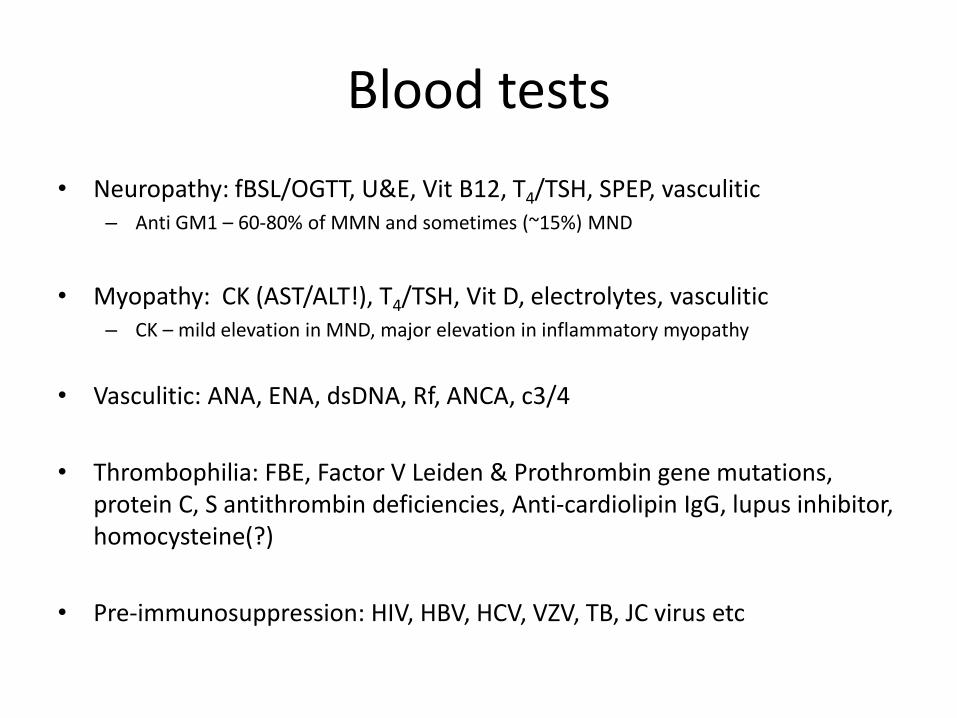
• Neuropathy: fBSL/OGTT, U&E, Vit B12, T4/TSH, SPEP, vasculitic– Anti GM1 – 60-80% of MMN and sometimes (~15%) MND
• Myopathy: CK (AST/ALT!), T4/TSH, Vit D, electrolytes, vasculitic– CK – mild elevation in MND, major elevation in inflammatory myopathy
• Vasculitic: ANA, ENA, dsDNA, Rf, ANCA, c3/4
• Thrombophilia: FBE, Factor V Leiden & Prothrombin gene mutations, protein C, S antithrombin deficiencies, Anti-cardiolipin IgG, lupus inhibitor, homocysteine(?)
• Pre-immunosuppression: HIV, HBV, HCV, VZV, TB, JC virus etc

Antibodies
• Anti-neuronal – Hu, Yo, Ri,
• VGKC - (LGI1 and CASPR2)
• NMDA
• GAD (stiff person syndrome)
• Ganglioside– GM1 – GBS, MND…
– GQ1b – Miller Fisher (ataxia, arreflexia, ophthalmoplegia)
• NMO (anti-aquaporin4 antibodies) - ~70% sensitive for neuromyelitis optica
• anti-MOG (myelin)
• JC virus serology ~50% of patients exposed – risk stratification for natalizumab PML risk

CSF
• generally image before LP – safety, leptomeningeal enhancement after LP can confuse interpretation
• opening pressure: N<20cm H2O, abN>25cm H2O (careful in obese/compressed abdomen)
• biochem– “albuminocytologic dissociation” – GBS (inflammation outside dura in
nerve roots, spill-over increased protein)
– elevated protein – non-specific: inflammation, diabetes, blocked spinal CSF flow
• Cells – lymphocytes, PMNs
• Cytology (spun down) - ?malignancy (larger volume 3x

CSF - special tests
• xanthochromia – spectrophotometry for bilirubin etc –extra, light protected tube, not sent in vacuum tube system
• unmatched oligoclonal bands (= found in CSF but not serum ie CNS-restricted immune process)
• 14-3-3 – non-specific marker of neuronal death (CJD but many other conditions too)
• JC virus PCR on CSF for Dx of PML• VZV IgG for post-zoster CNS vasculitis etc (better than PCR)

Nerve/Muscle biopsy
• Sural nerve biopsy rarely required• Perhaps if suspect:
– Vasculitis– Amyloidosis– Sarcoidosis– HNPP (tomaculous neuropathy) but use PMP22 gene test…– Leprosy– Tumour infiltration– Inherited (some)
• Muscle biopsy – suspected inflammatory myopathy or mitochondrial disease