Embed Size (px)
Citation preview
Spectroscopy 25 (2011) 113–122 113DOI 10.3233/SPE-2011-0498IOS Press
Investigation of the mechanism of binding ofthiacloprid to human serum albumin usingspectroscopic techniques and molecularmodeling methods
Chuanxian Wang a, Qinghua Chu a, Changyun Chen b,∗ and Zhao Bo c
a Shanghai Exit-Entry Inspection and Quarantine Bureau, Shanghai, Chinab School of Biochemical and Environmental Engineering, Nanjing Xiaozhuang University, Nanjing,Chinac School of Chemical and Material Science, Nanjing Normal University, Nanjing, China
Abstract. Fluorescence spectroscopy, UV absorption, circular dichroism (CD) spectroscopy and molecular modeling methodswere used to characterize the binding properties of thiacloprid (TL) with human serum albumin (HSA) at molecular level underphysiological conditions. The fluorescence intensity of HSA decreased regularly with the gradually increasing concentration ofthiacloprid. The binding constant K at three different temperatures (290, 300 and 310 K) were 3.07, 2.74 and 1.35 × 10⁴ M−1,respectively, for TL–HSA interaction have been calculated from the relevant fluorescence data. CD spectroscopic measure-ments have shown that the secondary structures of the protein have been changed by the interaction of thiacloprid with HSA.Furthermore, the study of molecular modeling indicated that thiacloprid could be located on the surface of the binding pocketof subdomains IIA in HSA. The hydrophobic interaction was the major acting force and there are H-bonds and electrostatic in-teractions between TL and HSA, which is in good agreement with the results from the experimental thermodynamic parameters(the enthalpy change ΔH0 and the entropy change ΔS0 were calculated to be −20.378 kJ/mol and 16.328 J/mol K accordingto the Van’t Hoff equation).
Keywords: Binding mechanism, human serum albumin, thiacloprid, spectroscopic techniques, molecular modeling
1. Introduction
Chemicals released from agriculture or industry may potentially develop toxic effects in the environ-ment and ecological systems. Among them, pesticides are actively applied and globally used for cropcontrol and to prevent damage to plants, animals, humans or aliments. Thiacloprid (structure shown inFig. 1), a relatively new neonicotinoid insecticide, ((Z)-3-(6-chloro-3-pyridylmethyl)-1,3-thiazolidin-2-ylidenecyanamide) has been produced by Bayer Crop Science since 2003. It acts as an agonist on thenicotinoid acetylcholine receptor (nAChR). Neonicotinoids are systemic insecticides and enter the bodyvia contact and ingestion. They act on the central nervous system of the insects and are mainly usedin pest management against biting and sucking insects. At physiological pH levels, neonicotinoids arenot protonated. As a result of this, they have little affinity to the nAChR of vertebrates. Therefore, they
*Corresponding author: Changyun Chen, School of Biochemical and Environmental Engineering, Nanjing XiaozhuangUniversity, Nanjing 211171, China. E-mail: [email protected].
0712-4813/11/$27.50 © 2011 – IOS Press and the authors. All rights reserved

114 C. Wang et al. / Investigation of the mechanism of binding of thiacloprid to human serum albumin
generally show low acute and chronic toxicity to mammals, birds and fish. However, among neonicoti-noids investigated so far, thiacloprid was found to exhibit a comparatively high level of acute toxicity tofish [14].
Human serum albumins have been one of the most studied proteins for many years. They are the mostabundant of proteins in blood plasma, accounting for about 60% of the total protein corresponding to aconcentration of 42 g/l and provide about 80% of the osmotic pressure of blood [1,3]. Albumins havebeen used as a model protein for many and diverse biophysical and physicochemical studies. They playan important role in the transport and deposition of a variety of endogenous and exogenous substancesin blood [21]. The globular protein consists of a single polypeptide chain of 585 amino acid residues andhas many important physiological functions [2]. The globular protein is composed of three structurallysimilar domains (I, II and III), each containing two subdomains (A and B) [5,15].
Since the wide spread use of thiacloprid has led to potential toxicological risk to public health, it isnecessary to investigate the interaction of thiacloprid with protein, as it can illustrate the nature of TL–protein complex in vitro and provide important insight into the interaction of the physiologically impor-tant protein HSA with thiacloprid, which has led to potential toxicological risk to public health [11].However, so far, none of the investigations were done by determining the TL–HSA binding constantsand the effects of thiacloprid complexation on the protein structure in detail. For a better understanding,the study of interaction of thiacloprid with HSA is much needed. Parameters such as mode of interac-tion, binding constant and number of binding sites are important, these investigations may provide someimportant theoretic information for the improvement of the metabolism and distribution of thiaclopridin life science, biologic technology, medicine, molecular functional design. To characterize these inter-actions on the molecular level, optical techniques have become valuable tool because these methods aresensitive and relatively easy to use. As an assistant method, molecular modeling was also employed toexplain the binding of thiacloprid to HSA. The results of molecular modeling and the thermodynamicparameters obtained suggest that thiacloprid binds to the hydrophobic cavity of human serum albuminand the hydrophobic interaction is the predominant intermolecular force stabilizing the complex. Thisstudy provides a molecular basis for elucidating the mechanism of pesticide acting and predicting un-favorable pesticide interaction. It also provides some basic information of the effect on the function ofproteins which reacted with thiacloprid.
2. Materials and methods
2.1. Materials
Human serum albumin (HSA) was purchased from Sigma Chemical Company. It was used withoutfurther purification and its molecular weight was assumed to be 66,500. HSA (1 µM) solutions wereprepared in (pH 7.4) Tris-HCl buffer solution, and HSA stock solutions were kept in the dark at 277 K.Thiacloprid (grade) was obtained from the National Institute for Control of Pharmaceutical and Bio-products, China. The stock solution was prepared in methanol because thiacloprid easily dissolve inthe methanol. The concentration of thiacloprid is 5 × 10⁻³ mol/l. All the stock solutions were then di-luted with buffer solution to obtain the actual assay solutions. NaCl (analytical grade, 1.0 mol/l) solutionwas used to maintain the ion strength at 0.1. The buffer (pH 7.4) consists of Tris (0.2 mol/l) and HCl(0.1 mol/l). The pH was checked with a suitably standardized pH meter (pHS-3C). The thiacloprid wastitrated by micro-injector. All reagents were of analytical reagent grade and distilled water was usedthroughout the experiments.

C. Wang et al. / Investigation of the mechanism of binding of thiacloprid to human serum albumin 115
2.2. Apparatus and methods
2.2.1. UV-absorbance titrationUV absorption spectra were recorded on an UV-2450 spectrophotometer. Quartz cuvettes with a path
length of 1 cm were used. The absorbance titration were performed by keeping the concentration ofHSA (1.0 µM) constant while varying the concentration of thiacloprid (0–25.0 µM). An equal amountof thiacloprid was titrated into the reference cell at the same time.
2.2.2. Fluorescence titrationFluorescence spectra was recorded on a LS 50B spectrofluorimeter (Perkin–Elmer, USA). A slit width
of 5 nm/5 nm was typically used 3.0 ml solution containing appropriate concentration of HSA (1.0 µM)was titrated manually by successive additions of 5 × 10⁻³ M solution of thiacloprid (to give a finalconcentration of 0–35 µM). The total content of titration was not more than 3.5% of the volume of theprotein. The pH measurements were carried out on a PHS-3C Exact Digital pH meter. Protein mixedwith the thiacloprid was maintained the full 10 min before the measurement for the sufficient reaction.The emission spectra were obtained in the rang from 300 to 500 nm, with the excitation wavelength at280 nm and the emission at 352 nm. All experiments were measured at different temperature 290, 300and 310 K, respectively.
2.2.3. Circular dichroism spectroscopyCircular dichroism were performed on a Jasco-20 automatic recording spectropolarimeter (Japan),
using a 2-mm quartz cell at room temperature, and the speed of scanning was 20 nm/min. The spectrawere recorded in the far-UV region which range from 200–250 nm. The protein concentration used in ourexperiment was 1.0 × 10⁻⁶ M. And the buffer solution was selected as the blank and was automaticallysubtracted during scanning.
2.2.4. Molecular modelingProtein-ligand docking study was performed using Discovery Studio 2.1 software where the pH is
set to 7.4. The crystal structure of HSA (PDB entry code 1h9z) was downloaded from the Protein DataBank [16] and the potential of the 3D structure of HSA was assigned to the CHARMm force field. Thestructure of thiacloprid was built in Discovery Studio 2.1 and optimized by applying the CHARMmforce field. During the docking process, a maximum of 10 conformers have been chosen for the finalenergy minimization. The conformation with the lowest energy was selected for final analysis.
3. Results and discussion
3.1. UV-absorbance spectra of HSA with thiacloprid
UV/vis absorption measurement is a simple and effective method in detecting complex formation.The UV absorption spectrum of HSA in the absence and presence of thiacloprid are shown in Fig. 1.The complex of TL–HSA is evident from UV–vis absorption spectral data. HSA has strong absorbancewith a peak at 204 nm and the absorbance of HSA decrease with the addition of thiacloprid. The peakposition of HSA has a little red shift. The results indicated that an interaction occurred between thia-cloprid and HSA. In addition, the absorbance values (280 nm) of the system are very low (<0.05) as inFig. 1A. Therefore, inner filter effects and re-absorption were negligible [12]. This result indicates thatthe probable quenching mechanism is due to the formation of the complex between thiacloprid and HSA.

116 C. Wang et al. / Investigation of the mechanism of binding of thiacloprid to human serum albumin
3.2. Analysis of fluorescence quenching of HSA by thiacloprid
Fluorescence measurements were carried out to investigate whether thiacloprid interacts with HSA.Figure 2 shows the fluorescence emission spectra of HSA in the absence and presence of thiacloprid.HSA has a strong fluorescence emission peak at 352 nm on excitation at 280 nm. Gradual addition of thi-acloprid to the HSA solution leads to a small bathochromic shift of the emission maximum accompanyby an decrease of fluorescence intensity. The observations reflect that the microenvironment around thefluorophore in the protein solution is quite different from that of pure aqueous phase. The changes in thefluorometric behavior of the fluorophore with the addition of thiacloprid in HSA solution can be ratio-nalized in terms of binding of the pesticide with the protein leading to a more polar microenvironmentaround the fluorophore. A red shift in the fluorescence maximum also suggests an enhancement in thepolarity of the microenvironment.
The static and dynamic quenching is distinguished by the results at different temperatures [4]. Highertemperature results in faster diffusion and larger amounts of collisional quenching and will typically
Fig. 1. UV absorption spectrum obtained in Tris-HCl buffer solution (pH 7.4): (a) the absorption spectrum of HSA,CHSA = 1.0 µM; (b)–(f) TL–HSA, 1 µM HSA in the presence of 1.0, 2.0, 4.0, 7.0, 8.0 µM TL, (g) the absorption spectrum of1.0 µM TL.
Fig. 2. Fluorescence emission spectra at pH 7.4: (a) 1.0 µM HSA; (b)–(h), respectively, 1.0 µM HSA in the presence of 5.0, 10,15, 20, 25, 30, 35 µM TL.

C. Wang et al. / Investigation of the mechanism of binding of thiacloprid to human serum albumin 117
Fig. 3. The Stern–Volmer curves for the binding of TL with HSA at different temperatures. Plot of F0/F − 1 against [Q] forthe TL–HSA: (Q) 290 K; (") 300 K; (�) 310 K; HSA = 1.0 µM.
leads to the dissociation of weakly bound complexes. Therefore, the quenching constant increases withincreasing temperature for dynamic quenching. However, it decreases with increase in temperature forstatic quenching. The possible quenching mechanism can be interpreted by fluorescence quenching spec-tra of the protein and the F0/F − 1–[Q] (Stern–Volmer) curves of HSA with thiacloprid at differenttemperatures 290, 300 and 310 K as shown in Fig. 3. It can be found from Fig. 3 that the Stern–Volmerplots were linear and the slopes decreased with increasing temperature, which is consistent with thestatic type of quenching mechanism. According to Eq. (1):
F0/F = 1 + Kqτ0[Q] = 1 + KSV[Q], (1)
where F0 and F are the fluorescence intensities of HSA in absence and presence of quencher, respec-tively, KSV is the Stern–Volmer quenching constant and [Q] is the concentration of quencher (thia-cloprid). Quenching constant KSV can be calculated from Fig. 3 and their values are 3.304 × 104,2.672 × 104, 1.911 × 10⁴ l/mol at 290, 300 and 310 K, respectively. These results indicate that theprobable quenching mechanism of fluorescence of HSA by thiacloprid is a static quenching processand can predict that a complex formed between thiacloprid and HSA. The interaction was decreasedwhen the temperature rose, thus the quenching efficiency of thiacloprid to HSA was reduced when theexperimental temperature rose.
3.3. Determinations of binding constants and the number of binding site
For static quenching, the relationship between fluorescence quenching intensity and the concentrationof quenchers can be described by the binding constant formula [23]:
log(F0 − F )/F = log Kb + n log[Q], (2)
where Kb is the binding constant and n is the number of binding sites. After the fluorescence quenchingintensities were measured, the double-logarithm algorithm was assessed by Eq. (2). The binding con-stants (Kb) and the number of binding sites (n) presents in Table 1. The result illustrates that there is astrong binding force between thiacloprid and HSA, and the number of binding sites is independent oftemperature from 290 to 310 K. The obtained value n = 0.988 ± 0.02 indicates that almost one mole-cule of thiacloprid binds to a HSA molecule and it corresponds to the high affinity binding site. It also

118 C. Wang et al. / Investigation of the mechanism of binding of thiacloprid to human serum albumin
shows that the values of Kb decrease with the rising temperature, which is in good agreement with thetrend of KSV as mentioned above. It implies that an unstable complex may be formed in the bindingreaction and the complex would possibly be dissociated partly when the temperature increases.
3.4. Thermodynamic analysis and determination of the molecular forces
The molecular forces between a small molecular substance and macromolecule mainly include hydro-gen bond, van der Waals force, electrostatic force, hydrophobic interaction force and so on. Consideringthe dependence of binding constant on temperature, a thermodynamic process was deemed to be re-sponsible for the formation of the complex. On this purpose, the temperature dependence of the bindingconstant was studied. In this section, the association constants obtained by the Stern–Volmer was usedto calculate the thermodynamic parameters. Thermodynamic parameters were calculated based on thetemperature dependence of the quenching constant for TL–HSA binding.
The temperatures chosen were 290, 300 and 310 K. By plotting the quenching constants according toVan’t Hoff equation, the thermodynamic parameters were determined from a linear Van’t Hoff plot andlisted in Table 2. It is clear from the values of standard entropy changes (ΔS0) and standard enthalpychanges (ΔH0) that the binding of thiacloprid to HSA is an exothermic process accompanied by a posi-tive value of ΔS0 and a negative value of ΔG0. The binding process is always spontaneous as evidencedby the negative sign of ΔG0 value. For pesticide–protein interaction, Ross and Subramanian [17] havecharacterized the sign and magnitude of the thermodynamic parameters associated with these variouskinds of interactions that play an important role in the protein association process. From the point of theview, a positive ΔH0 and ΔS0 value is associated with hydrogen bonding interaction. The negative ΔH0
and ΔS0 values are associated with hydrogen bonding and van der Waals interaction in low dielectricmedium. Specific electrostatic interaction between ionic species in an aqueous solution is characterizedby positive ΔS0 value and very small negative ΔH0 value, almost zero [18]. The negative ΔH0 andpositive ΔS0 values are associated with hydrophobic force and electrostatic force interaction. Thus it isdifficult to interpret the thermodynamic parameters of TL–HSA interaction with a single intermolecularforce. The interaction force between small molecules and proteins is not only a single force in general.May be there are variety of forces existing in the interaction forces, which contains the electrostaticforce. It is believed that the hydrophobic small pharmacological molecule cuttage grafting to proteinmolecules within the hydrophobic region, when a small pharmacological molecule combine with pro-teins. The solubility of thiacloprid in water is very small, so thiacloprid is belonging to hydrophobic
Table 1
The binding constant Kb and the number of binding site n at different temperatures
T (K) Linear equation R 104KA (M−1) n
290 log[(F0 − F )/F ] = 4.4869 + 0.9936 log[Q] 0.9999 3.07 0.9936300 log[(F0 − F )/F ] = 4.4374 + 1.0036 log[Q] 0.9997 2.74 1.0036310 log[(F0 − F )/F ] = 4.1289 + 0.9667 log[Q] 0.9998 1.35 0.9667
Table 2
Thermodynamic parameters of TL–HSA
T (K) ΔG0 (kJ/mol) ΔS0 (J/(mol · K)) ΔH0 (kJ/mol)290 −25.113300 −25.276 16.328 −20.378310 −25.439

C. Wang et al. / Investigation of the mechanism of binding of thiacloprid to human serum albumin 119
small molecule. The drug molecule embedded in the internal hydrophobic region of protein moleculesmay be responsible for the fluorescence quenching [8,13]. Therefore, the binding of thiacloprid to HSAmight involve hydrophobic interaction strongly as evidenced by the positive value of ΔS0 although therole of electrostatic interaction cannot be excluded. From molecular modeling we found that hydrogenbonding force also plays a major role.
3.5. Changes of the HSA secondary structure induced by thiacloprid binding from CD
To obtain more information on the binding of thiacloprid to HSA, CD spectroscopy was used to studythe structure of HSA and TL–HSA complex. CD, a sensitive technique to monitor the conformationalchange in the protein.The CD spectra of HSA and TL–HSA complex were shown in Fig. 4. As can beseen from Fig. 4, the CD spectra of HSA exhibits two negative bands in the ultraviolet region at 208and 223 nm, which is a characteristic of α-helical structure of protein. The reasonable explanation isthat the negative peaks between 208–209 and 222–223 nm are both contributed by n → π∗ transferfor the peptide bond of α-helical [22]. The addition of thiacloprid to HSA leads to a decrease in theellipticity without significant shift of the peaks, indicating that the binding of thiacloprid to HSA inducesa decrease in the α-helical content of HSA. However, the CD spectra of HSA in the presence and absenceof thiacloprid were similar in shape, which means the structure of HSA was also predominated byα-helix after the binding of thiacloprid [20]. The binding of thiacloprid to HSA causes an decrease withnegative Cotton effect in band intensity, indicating the considerable changes in the protein secondarystructure with the decrease of α-helical structure, and it may be the result of the formation of TL–HSAcomplex. The CD result was expressed as MRE (Mean Residue Ellipticity) in deg · cm2 · dmol−1, whichis defined as:
MRE = θobs/(10Cpnl),
where θobs is the CD in millidegree, n is the number of amino acid residues (585), l is the path length ofthe cell (1 cm) and Cp is the molar concentration of HSA. The α-helical contents of free and combinedHSA were calculated from the MRE values at 208 nm using the equation [7]:
α-helix (%) = {(−MRE208 − 4000)/(33,000 − 4000)} × 100. (3)
Fig. 4. CD spectra of the HSA–TL system. (a) 1.0 × 10⁻⁶ mol/l HSA; (b) 1.0 × 10⁻⁶ mol/l HSA in the presence of1.0 × 10−6 mol/l TL; (c) 1.0 × 10⁻⁶ mol/l HSA in the presence of 1.0 × 10⁻⁵ mol/l TL.
120 C. Wang et al. / Investigation of the mechanism of binding of thiacloprid to human serum albumin
The calculated results exhibit an decrease of α-helical structure from 49.69 to 48.88% at molar ratioof TL/HSA of 1:1 and from 49.69 to 29.86% at molar ratio of TL/HSA of 10:1. The results reveal thatthe binding of thiacloprid to HSA has changed the secondary structure of HSA. The percentage α-helixstructure of HSA decreased indicated that thiacloprid bound with the amino acid residue of the mainpolypeptide chain of HSA and destroyed their hydrogen bonding networks [9].
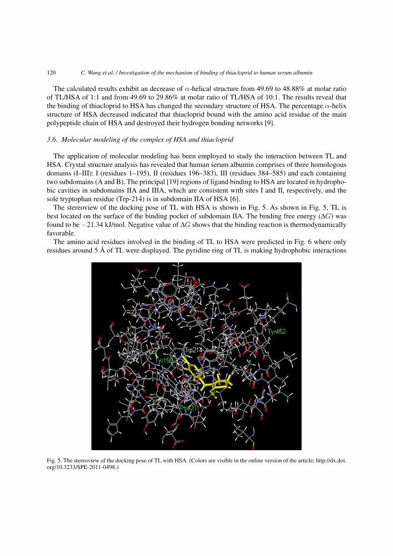
3.6. Molecular modeling of the complex of HSA and thiacloprid
The application of molecular modeling has been employed to study the interaction between TL andHSA. Crystal structure analysis has revealed that human serum albumin comprises of three homologousdomains (I–III): I (residues 1–195), II (residues 196–383), III (residues 384–585) and each containingtwo subdomains (A and B). The principal [19] regions of ligand binding to HSA are located in hydropho-bic cavities in subdomains IIA and IIIA, which are consistent with sites I and II, respectively, and thesole tryptophan residue (Trp-214) is in subdomain IIA of HSA [6].
The stereoview of the docking pose of TL with HSA is shown in Fig. 5. As shown in Fig. 5, TL isbest located on the surface of the binding pocket of subdomain IIA. The binding free energy (ΔG) wasfound to be −21.34 kJ/mol. Negative value of ΔG shows that the binding reaction is thermodynamicallyfavorable.
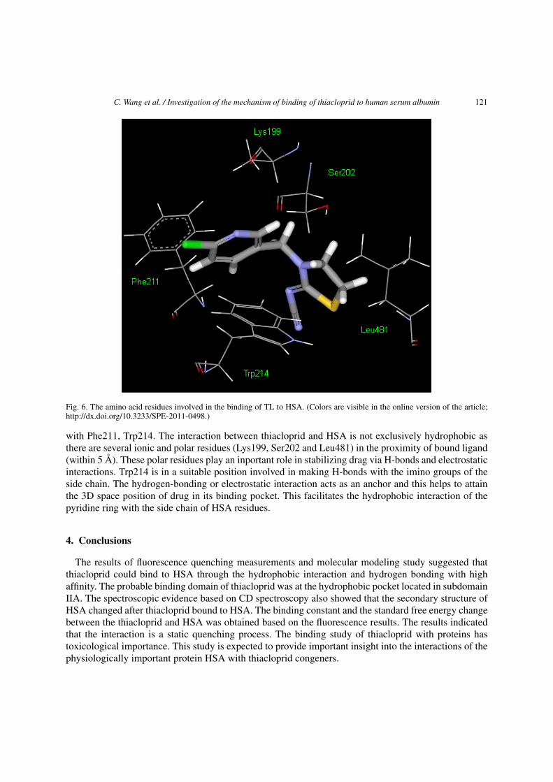
The amino acid residues involved in the binding of TL to HSA were predicted in Fig. 6 where onlyresidues around 5 Å of TL were displayed. The pyridine ring of TL is making hydrophobic interactions
Fig. 5. The stereoview of the docking pose of TL with HSA. (Colors are visible in the online version of the article; http://dx.doi.org/10.3233/SPE-2011-0498.)
C. Wang et al. / Investigation of the mechanism of binding of thiacloprid to human serum albumin 121
Fig. 6. The amino acid residues involved in the binding of TL to HSA. (Colors are visible in the online version of the article;http://dx.doi.org/10.3233/SPE-2011-0498.)
with Phe211, Trp214. The interaction between thiacloprid and HSA is not exclusively hydrophobic asthere are several ionic and polar residues (Lys199, Ser202 and Leu481) in the proximity of bound ligand(within 5 Å). These polar residues play an inportant role in stabilizing drag via H-bonds and electrostaticinteractions. Trp214 is in a suitable position involved in making H-bonds with the imino groups of theside chain. The hydrogen-bonding or electrostatic interaction acts as an anchor and this helps to attainthe 3D space position of drug in its binding pocket. This facilitates the hydrophobic interaction of thepyridine ring with the side chain of HSA residues.
4. Conclusions
The results of fluorescence quenching measurements and molecular modeling study suggested thatthiacloprid could bind to HSA through the hydrophobic interaction and hydrogen bonding with highaffinity. The probable binding domain of thiacloprid was at the hydrophobic pocket located in subdomainIIA. The spectroscopic evidence based on CD spectroscopy also showed that the secondary structure ofHSA changed after thiacloprid bound to HSA. The binding constant and the standard free energy changebetween the thiacloprid and HSA was obtained based on the fluorescence results. The results indicatedthat the interaction is a static quenching process. The binding study of thiacloprid with proteins hastoxicological importance. This study is expected to provide important insight into the interactions of thephysiologically important protein HSA with thiacloprid congeners.

122 C. Wang et al. / Investigation of the mechanism of binding of thiacloprid to human serum albumin
Acknowledgements
This work was supported by the Nanometer Technology Special Project (No. 1052NM06700), andthe Technology Standard Project (No. 08DZ0505000) of Shanghai Municipal Science and TechnologyCommission, and High-tech Industrialization Project (JHZD09-18) of Jiangsu Education Department.
References
[1] J.R. Brown and P. Shockley, Serum albumin: structure and characterization of its ligand binding sites, in: Lipid ProteinInteractions, Vol. 1, P.C. Jost and O.H. Griath, eds, Wiley, New York, 1982, pp. 26–68.
[2] D.C. Carter, X.M. He, S.H. Munson, P.D. Twigg, K.M. Gernert, M.B. Broom and T.Y. Miller, Three-dimensional structureof human serum albumin, Am. Assoc. Adv. Sci. 244(4909) (1989), 1195–1198.
[3] D.C. Carter and J.X. Ho, Structure of serum albumin, Adv. Protein Chem. 2(45) (1994), 153–203.[4] G.Z. Chen, X.Z. Huang, J.G. Xu, Z.Z. Zheng and Z.B. Wang, The Methods of Fluorescence Analysis, 2nd edn, Science
Press, Beijing, 1990, p. 50.[5] S. Curry, P. Brick and N.P. Frank, Atomic structure and chemistry of human serum albumin, Biochem. Biophys. Acta
13(1441) (1999), 269–272.[6] S. Curry, H. Mandelkow, P. Brick and N. Franks, Crystal structure of human serum albumin complexed with fatty acid
reveals an asymmetric distribution of binding sites, Nat. Struct. Biol. 5 (1998), 827–835.[7] H. Gao, Y.N. Wang, Y.G. Fan and J.B. Ma, Interactions of some modified mono- and bis-β-cyclodextrins with bovine
serum albumin, Bioorg. Med. Chem. 14 (2006), 131–137.[8] B. Huang, G.L. Zou and Y.M. Yang, Studies on the interaction between adriamy and bovine serun albumin, Acta Chim.
Sinica 60(10) (2002), 1867–1871.[9] J. Kang, Y. Liu, M.-X. Xie, S. Li, M. Jiang and Y.D. Wang, Interactions of human serum albumin with chlorogenic acid
and ferulic acid, Biochim. Biophys. Acta 1674 (2004), 205–214.[10] J.R. Lakowicz, Principles of Fluorescence Spectroscopy, Plenum Press, New York, 1983, pp. 257–295.[11] J. Li, X. Liu, C. Ren, J. Li, F. Sheng and Z. Hu, In vitro study on the interaction between thiophanate methyl and human
serum albumin, J. Photochem. Photobiol. B 94 (2009), 158–163.[12] J. Liu, J.-N. Tian, J. Zhuang, Z. Hu and X. Chen, Interaction of magnololl with bovine serum albumin: a fluorescence-
quenching study, Anal. Bioanal. Chem. 376 (2003), 864–867.[13] X.F. Liu, Y.M. Xia, Y. Fang et al., Interaction between natural pharmaceutical homologued of coumarin and bovine serum
albumin, Acta Chim. Sinica 64(16) (2004), 1484–1490.[14] R. Osterauer and H.-R. Köhler, Temperature-dependent effects of the pesticides thiacloprid and diazinon on the embryonic
development of zebrafish (Danio rerio), Aquat. Toxicol. 86 (2008), 485–494.[15] T. Peters, Serum albumin, Adv. Protein Chem. 37 (1985), 161–242.[16] I. Petitpas, A.A. Bhattacharya, S. Twine, M. East and S. Curry, Crystal structure analysis of warfarin binding to human
serum albumin: anatomy of drug site I, J. Biol. Chem. 276(25) (2001), 22804–22809.[17] P.D. Ross and S. Subramanian, Thermodynamics of protein association reactions: forces contributing to stability, Bio-
chemistry 20 (1981), 3096–3102.[18] I. Sjoholm, B. Ekman, A. Kober, I. Ljungstedt-Pahlman, B. Seiving and T. Sjodin, Binding of drugs to human serum
albumin: XI. The specificity of three binding sites as studied with albumin immobilized in microparticles, Mol. Pharmacol.16(3) (1979), 767–777.
[19] G. Sudlow, D.J. Birkett and D.N. Wade, Further characterization of specific drug binding site on human serum albumin,Mol. Pharmacol. 12 (1976), 1052–1061.
[20] S. Sugio, A. Kashima, S. Mochizuki, M. Noda and K. Kobayashi, Crystal structure of human serum albumin at 2.5 Åresolution, Protein Eng. 12(6) (1999), 439–446.
[21] K. Yamasaki, T. Maruyama, U. Kragh-Hansen and M. Otagiri, Characterization of site I on human serum albumin: conceptabout the structure of a drug binding site, Biochem. Biophys. Acta 1295 (1996), 57–147.
[22] P. Yang and F. Gao, The Principle of Bioinorganic Chemistry, Science Press, Beijing, 2002, pp. 322–342.[23] Q.J. Zhou, J.F. Xiang, Y.L. Tang, J.P. Liao, C.Y. Yu, H. Zhang, L. Li, Y.Y. Yang and G.Z. Xu, Investigation on the
interaction between a heterocyclic aminal derivative, SBDC, and human serum albumin, Colloids Surf. B 61 (2008),75–80.

Submit your manuscripts athttp://www.hindawi.com
Hindawi Publishing Corporationhttp://www.hindawi.com Volume 2014
Inorganic ChemistryInternational Journal of
Hindawi Publishing Corporation http://www.hindawi.com Volume 2014
International Journal ofPhotoenergy
Hindawi Publishing Corporationhttp://www.hindawi.com Volume 2014
Carbohydrate Chemistry
International Journal of
Hindawi Publishing Corporationhttp://www.hindawi.com Volume 2014
Journal of
Chemistry
Hindawi Publishing Corporationhttp://www.hindawi.com Volume 2014
Advances in
Physical Chemistry
Hindawi Publishing Corporationhttp://www.hindawi.com
Analytical Methods in Chemistry
Journal of
Volume 2014
Bioinorganic Chemistry and ApplicationsHindawi Publishing Corporationhttp://www.hindawi.com Volume 2014
SpectroscopyInternational Journal of
Hindawi Publishing Corporationhttp://www.hindawi.com Volume 2014
The Scientific World JournalHindawi Publishing Corporation http://www.hindawi.com Volume 2014
Medicinal ChemistryInternational Journal of
Hindawi Publishing Corporationhttp://www.hindawi.com Volume 2014
Chromatography Research International
Hindawi Publishing Corporationhttp://www.hindawi.com Volume 2014
Applied ChemistryJournal of
Hindawi Publishing Corporationhttp://www.hindawi.com Volume 2014
Hindawi Publishing Corporationhttp://www.hindawi.com Volume 2014
Theoretical ChemistryJournal of
Hindawi Publishing Corporationhttp://www.hindawi.com Volume 2014
Journal of
Spectroscopy
Analytical ChemistryInternational Journal of
Hindawi Publishing Corporationhttp://www.hindawi.com Volume 2014
Journal of
Hindawi Publishing Corporationhttp://www.hindawi.com Volume 2014
Quantum Chemistry
Hindawi Publishing Corporationhttp://www.hindawi.com Volume 2014
Organic Chemistry International
ElectrochemistryInternational Journal of
Hindawi Publishing Corporation http://www.hindawi.com Volume 2014
Hindawi Publishing Corporationhttp://www.hindawi.com Volume 2014
CatalystsJournal of