Embed Size (px)
Citation preview

BearWorks BearWorks
MSU Graduate Theses
Spring 2018
Investigation of the Homologs Rad51 and Dmc1 Role in Cell Investigation of the Homologs Rad51 and Dmc1 Role in Cell
Division and Homologous Recombination Division and Homologous Recombination
Amaal Abulibdeh Missouri State University, [email protected]
As with any intellectual project, the content and views expressed in this thesis may be
considered objectionable by some readers. However, this student-scholar’s work has been
judged to have academic value by the student’s thesis committee members trained in the
discipline. The content and views expressed in this thesis are those of the student-scholar and
are not endorsed by Missouri State University, its Graduate College, or its employees.
Follow this and additional works at: https://bearworks.missouristate.edu/theses
Part of the Bioinformatics Commons, Cell Biology Commons, and the Molecular Biology
Commons
Recommended Citation Recommended Citation Abulibdeh, Amaal, "Investigation of the Homologs Rad51 and Dmc1 Role in Cell Division and Homologous Recombination" (2018). MSU Graduate Theses. 3269. https://bearworks.missouristate.edu/theses/3269
This article or document was made available through BearWorks, the institutional repository of Missouri State University. The work contained in it may be protected by copyright and require permission of the copyright holder for reuse or redistribution. For more information, please contact [email protected].

INVESTIGATION OF THE HOMOLOGS RAD51 AND DMC1 ROLE IN CELL
DIVISION AND HOMOLOGOUS RECOMBINATION
A Master's Thesis
Presented to
The Graduate College of
Missouri State University
TEMPLATE
In Partial Fulfillment
Of the Requirements for the Degree
Master of Science, Cell and Molecular Biology
By
Amaal A. Abulibdeh
May 2018

ii
INVESTIGATION OF THE HOMOLOGS RAD51 AND DMC1 ROLE IN CELL
DIVISION AND HOMOLOGOUS RECOMBINATION.
Biomedical Sciences
Missouri State University, May 2018
Master of Science
Amaal A. Abulibdeh
ABSTRACT
RecA-like proteins homologs Rad51 and Dmc1 (disruption of meiotic control) promote
recombination between homologous chromosomes by repairing programmed DNA
Double-Strand Breaks (DSBs). Dmc1 is a Recombinase involved in meiosis-specific
repair of DSBs, whereas Rad51 has been found to be involved in meiotic and non-meiotic
DSBs repair. Previous studies showed that when RAD51 is overexpressed,
interhomologous recombination still occurs even when DMC1 is knocked out. Dmc1 and
Rad51 have not been fully characterized in the ciliate Tetrahymena thermophila. In order
to more fully investigate the role of Rad51 and Dmc1 in Homologous Recombination
Repair (HHR), this work focuses on using a model organism, T. thermophila, to further
elucidate the contribution of Rad51 and Dmc1 in DNA repair following various
genotoxic stressors (H2O2, MMS, and UV radiation). Bioinformatics was used to
illustrate the extensive conservation of the Rad51 and Dmc1 homologs in various
organisms and between one another. Expression of RAD51 and DMC1 was shown to be
altered following exposure to H2O2, MMS, and UV radiation, and that the RAD51
expression was significantly higher than Dmc1 expression levels following all DNA
damaging agents. Localization studies using Green fluorescent protein (GFP) and Red
fluorescent protein (RFP) tagged to RAD51 or DMC1 and introduced back into T.
thermophila revealed that Rad51 does not localize to the micronucleus or macronucleus
following exposure to MMS. Tagging revealed that Dmc1 may localize in the
micronucleus without DNA damage but does not localize after MMS treatment. Both
proteins showed localization outside the nuclei, suggesting expression of the tagged
Rad51 and Dmc1 in T. thermophila.
KEYWORDS: Rad51, Dmc1, Homologous Recombination, DNA repair, Tetrahymena
thermophila.
This abstract is approved as to form and content
_______________________________
Joshua J. Smith, PhD
Chairperson, Advisory Committee
Missouri State University

iii
INVESTIGATION OF THE HOMOLOGS RAD51 AND DMC1 ROLE IN CELL
DIVISION AND HOMOLOGOUS RECOMBINATION.
By
Amaal A. Abulibdeh
A Masters Thesis
Submitted to the Graduate College
Of Missouri State University
In Partial Fulfillment of the Requirements
For the Degree of Master of Sciences, Cell and Molecular Biology
May 2018
Approved:
_______________________________________
Joshua J. Smith, PhD
_______________________________________
Colette M. Witkowski, PhD
_______________________________________
Amanda C. Brodeur, MD, PhD
_______________________________________
Julie Masterson, PhD: Dean, Graduate College
In the interest of academic freedom and the principle of free speech, approval of this thesis indicates the
format is acceptable and meets the academic criteria for the discipline as determined by the faculty that
constitute the thesis committee. The content and views expressed in this thesis are those of the student-
scholar and are not endorsed by Missouri State University, its Graduate College, or its employees.

iv
ACKNOWLEDGEMENTS
First, I would like to express my sincere gratitude to my advisor Dr. Joshua J.
Smith for the continuous support of my study, for his patience, motivation, and immense
knowledge. His guidance helped me in all the time of research and writing of this thesis. I
could not have imagined having a better advisor and mentor for my master study.
Besides my advisor, I would like to thank the rest of my thesis committee: Dr.
Amanda C. Brodeur, for her insightful comments and encouragement, and Dr. Colette M.
Witkowski for steering me in the right direction whenever I needed it.
I would like to thank the members of Smith Lab for their help during the research
that went into this Thesis. I would especially like to thank Jeremy Tee for designing GFP
and RFP epitope tags.
Finally, I must express my very profound gratitude to my parents and to my
husband, Amjed, for providing me with unfailing support and continuous encouragement
throughout my years of study. This accomplishment would not have been possible
without them.

v
TABLE OF CONTENTS
Introduction ..........................................................................................................................1
The Pathways of Double-Strand DNA Breaks ........................................................1
Non-Homologous End Joining ................................................................................3
Homologous Recombination Repair ........................................................................6
Eukaryotic Homologs of RecA: Rad51 and Dmc1 ..................................................9
Hop2-Mnd1 Complex ............................................................................................14
Tetrahymena thermophila as a Model Organism...................................................15
Meiosis in Tetrahymena thermophila ....................................................................17
The Recombinase Proteins and Hop2-Mnd2 Complex in
Tetrahymena thermophila ......................................................................................19
Purpose Statement ..................................................................................................22
Materials and Methods .......................................................................................................23
Tetrahymena thermophila Strains and Growth Conditions ...................................23
Cryopreservation of Tetrahymena thermophila .....................................................23
LR Clonase™ Reaction .........................................................................................25
Electroporation Transformation of E.coli ..............................................................25
Midiprep DNA Purification ...................................................................................26
Biolistic Transformation of Tetrahymena thermophila .........................................27
Bioinformatics........................................................................................................29
qRT-PCR................................................................................................................30
Damage Treatment for RNA Extraction ................................................................32
Fluorescence Microscopy ......................................................................................33
Protein Isolation .....................................................................................................33
Results ...............................................................................................................................35
Bioinformatics........................................................................................................35
RAD51 and DMC1 Plasmid Purification and Transformation into
Tetrahymena thermophila ......................................................................................38
Primer Optimization...............................................................................................49
RAD51 and DMC1 DNA Damage Expression after Various Damaging Agents 53
Fluorescent Microscopy of Rad51 and Dmc1 .......................................................54
Rad51 and Dmc1 Localization in Response to DNA Damage ..............................65
Discussion ..........................................................................................................................68
Bioinformatics........................................................................................................68
Expressions of RAD51 and DMC1 Following DNA Damage in Tetrahymena
thermophila ............................................................................................................71
Epitope and Fluorescent Tagged Rad51 and Dmc1 Expression and Localization
................................................................................................................................73
Future Directions ...................................................................................................74

vi
References ..........................................................................................................................78
Appendices .........................................................................................................................86
Appendix A. Tetrahymena Rad51 and Dmc1 Epitope Tags Constructs ...............86
Appendix B. T-COFFEE Alignment and T. thermophila Hop2-Mnd1 Proteins ...97

vii
LIST OF TABLES
Table 1. T. thermophila Stains... ........................................................................................24
Table 2. Quantitative RT-PCR Primers... ..........................................................................32
Table B1. T. thermophila Hop2-Mnd1 homologs... ........................................................106

viii
LIST OF FIGURES
Figure 1. Schematic of Non-Homologous End Joining Pathway ........................................5
Figure 2. The main steps of the DNA DSBs repair pathway of HRR .................................8
Figure 3. Comparison of Rad51 Model with Dmc1 Structure and Schematic
Representation of the Domain Organization of the RecA/ Rad51 Recombinase Family ..12
Figure 4. The Tetrahymena thermophila life cycle ...........................................................18
Figure 5. Overexpression of RAD51 Causes an Amacronuclear Cell Phenotype..............21
Figure 6. Protein Domains in DMC1 and RAD51 .............................................................40
Figure 7. Unweighted Pair Group Method with Arithmetic (UPGMA) Bootstrapped
Phylogenetic tree for possible T.thermophila Rad51 and Dmc1 homologs ………….... 41
Figure 8. Tetrahymena thermophila RNAseq data of RAD51 and DMC1 ........................42
Figure 9. Microarray Expression Profiling Data for RAD51 and DMC1 ..........................43
Figure 10. Confirmation Digests for GFP, RFP, and HA Constructs of RAD51 and DMC1
............................................................................................................................................44
Figure 11. Confirmation Digests for mCherry, GFP, FH6, and HA of RAD51 and DMC1
Tagged Constructs .............................................................................................................45
Figure 12. Expected Band Sizes Observed in Restriction Enzyme Digestion of RFP-
DMC1 Construct ................................................................................................................47
Figure 13. Restriction digest with KpnI and SacI to linearize DNA constructs for
transformation into Tetrahymena thermophila ..................................................................48
Figure 14. Confirmation of RAD51 and DMC1 primers in GoTaq PCR. ..........................51
Figure 15. Melt and amplification curves for RAD51 and DMC1 primers. .......................51
Figure 16. Confirmation of SsoFast Evagreen qRT-PCR products. ..................................52
Figure 17. qRT-PCR expression profile analysis of the transcription of Rad51 in response
to H2O2 treatment ...............................................................................................................55
Figure 18. qRT-PCR expression profile analysis of the transcription of Dmc1 in response
to H2O2 treatment ...............................................................................................................56
ix
Figure 19. qRT-PCR expression profile analysis of the transcription of Rad51 and Dmc1
in response to H2O2 treatment. ...........................................................................................57
Figure 20. qRT-PCR expression profile analysis of the transcription of Rad51 in response
to MMS treatment ..............................................................................................................58
Figure 21. qRT-PCR expression profile analysis of the transcription of Dmc1 in response
to MMS treatment ..............................................................................................................59
Figure 22. qRT-PCR expression profile analysis of the transcription of Rad51 and Dmc1
in response to MMS treatment ...........................................................................................60
Figure 23. qRT-PCR expression profile analysis of the transcription of Rad51 in response
to UV treatment..................................................................................................................61
Figure 24. qRT-PCR expression profile analysis of the transcription of Dmc1 in response
to UV treatment..................................................................................................................62
Figure 25. qRT-PCR expression profile analysis of the transcription of Rad51 and Dmc1
in response to UV treatment. .............................................................................................63
Figure 26. Fluorescent Microscopy images of Rad51 and Dmc1. .....................................64
Figure 27. Dmc1 does not localize to nucleus following MMS treatment. .......................66
Figure 28. Rad51 does not localize to nucleus following MMS treatment. ......................67

x
LIST OF APPENDICES’ FIGURES
Figure A1. pENTR-RAD51 construct map. .....................................................................87
Figure A2. pENTR-DMC1 plasmid map. ..........................................................................88
Figure A3. pBM2HA-RAD51 plasmid map. .....................................................................89
Figure A4. pBM2HA-DMC1 plasmid map. ......................................................................90
Figure A5. pBMFH6-RAD51 plasmid map. ......................................................................91
Figure A6. pBMFH6-DMC1 plasmid map. .......................................................................92
Figure A7. pBMGFP-DMC1 plasmid map. .......................................................................93
Figure A8. pBMGFP-RAD51 plasmid map. ......................................................................94
Figure A9. pBMRFP-RAD51 plasmid map. ......................................................................95
Figure A10. pBMRFP-DMC1 plasmid map. .....................................................................96
Figure B1. T-COFFEE Alignment of DMC1. ................................................................101
Figure B2. T-COFFEE Alignment of RAD51. ................................................................105

1
INTRODUCTION
The Pathways of Double-Strand DNA Breaks
Deoxyribonucleic acid (DNA) is the genetic material stored within the nucleus of
the cell, and the preservation of this genetic information requires not only the accuracy of
its copying during DNA replication, but also the availability of multiple DNA repair
processes to cope with any loss of genomic material. Unlike any other molecules, DNA
requires a single strand of DNA as a template and a region of a few base pairs long on a
double-strand of DNA to start the synthesis of a new strand. Once DNA starts forming a
new strand, it is partially broken down to monomers and the other strand is used as a
template to make a new copy of double-strand of DNA (Aerssens et al., 2001). DNA is
continually exposed to the endogenous and exogenous DNA damaging agents, leading to
mitotic cell death, permanent cell cycle arrest, and changes that lead to carcinogenesis
through (translocations, inversions, deletions), or consequently induction of apoptosis.
Damage to DNA can result from either physical mutagens that cause covalent
modifications between neighboring pyrimidine nucleotides resulting in pyrimidine
dimmers: (6-4) pyrimidine photoproducts, and cyclobutane pyrimidine dimmers (CPDs),
or chemical mutagens (alkylating and oxidizing agents) that intercalate or covalently bind
to DNA to produce a specific mutational signature (Hakem, 2008; Helleday et al., 2014;
Rothkamm et al., 2003). Mechanisms of DNA repair provide high fidelity and genome
integrity (Waters, 2006). Probably one of the most severe type of DNA damage is
double-strand breaks (DSBs) that result in loss and rearrangement of genomic sequence.
Exogenous agents such as ionizing radiation and chemotherapeutic drugs generate
reactive oxygen species and mechanical stress on the chromosomes. Sometimes, when
the DNA replication forks face single strand breaks during initiation of the recombination

2
between homologous chromosomes, DSBs occur, leading to loss/amplification of
chromosomal material or to tumorigenesis, if the deleted chromosomal region encodes a
tumor suppressor protein (Khanna and Jackson, 2001).
DSBs are initiated when the two complementary sequences of the DNA double
helix are broken at sites that are close enough to one another, and base paring is
insufficient to keep the two strands of DNA together. Consequently, the two DNA ends
generated by DSBs perform a perilous recombination with other sites in the genome. In
addition to interfering with transcription or replication of genes, they are disrupted in this
process, leading to hybrid proteins or inappropriate activation of genes. One cellular
response to DSBs is activation of the DNA repair proteins ataxia-telangiectasia mutated
(ATM), which is one of these crucial molecules recruited to the site of DNA double-
strand breaks (Maréchal, 2013). Once DSBs occur, ATM phosphorylates downstream
substrates such as p53, BRCA1, and NBS1, causing multiple effects on the DNA repair
process. ATM deficiency leads to the development of cancer and neurodegenerative
syndrome called Ataxia Telangiectasia (A-T), resulting in hypersensitivity to ionizing
radiation and chemical agents that yield DNA DSBs.
Two major DNA repair pathways have evolved to cope with DSBs. DSBs are
repaired by either Non-Homologous End-Joining (NHEJ) or Homologous Recombination
Repair (HRR). Both these pathways are very distinct from one another and function in
different ways to effect DNA double-strand breaks repair.

3
Non-Homologous End Joining
Non-Homologous End Joining is the simplest mechanism that is used at different
points of the cell cycle when sister chromatids are not available to be used as homologous
recombination templates. It rejoins two ends of a broken DNA molecule without the
requirement for homologous sequences between the ends. It involves XRCC4-LIG4
complex, DNA-dependent protein kinase (DNA-PK) holoenzyme of catalytic subunit
DNA-PKc, and DNA end-binding heterodimer Ku70-Ku80 (Davis, 2013). The basic
mechanism of NHEJ is described in Figure 1. The core component of NHEJ is the Ku
protein, a heterodimer of two subunits called Ku70 and Ku80. It binds the ends of a
broken DNA strand for repair, protecting them from degradation. The mammalian Ku
protein forms a complex with the DNA dependent protein kinase (DNA-PKcs); the
serine/threonine kinase is activated in the presence of the DNA ends. Ku protein recruits
DNA-PKcs to the broken DNA strands, which together form the DNA-PK holoenzyme.
Two relative proteins of DNA-PKcs are implicated in responses to DNA damage such as
ATM and ATR. Those proteins are kinases and physically recruited to the site of DNA
damage. ATM binds to the DNA and phosphorylates p53 protein in response to DSBs,
and ATR phosphorylates the p53 in the same fashion. DNA-PKcs phosphorylates
proteins bound to the DNA around the break. Then a complex of MRE11, XRS2 and
RAD50, which possesses exonuclease, endonuclease and unwinding activity, localize to
site of DNA double-strand breaks in mammalian. The human Mre11 protein has nuclease
activity and Nbs1 seems to replace Xrs2. This complex trims the DNA ends to create
single-stranded overhangs before they can be rejoined. Another factor possesses
hydrolytic activity called Artemis as shown in Figure 1B, can process DNA DSBs before
NHEJ occurs (Jackson, 2002; Featherstone et al., 1999). Finally, ligase IV stimulates
4
DNA end-ligation, which functions in a tight complex with protein XRCC4 (Ramsden et
al., 1998). XRCC4 is a substrate for DNA-PKcs that might regulate the activity of the
ligase.

5
Figure 1: Schematic of Non-Homologous End Joining Pathway. NHEJ requires
several factors to rejoin the two broken ends of the DNA after inducing DSBs. (A) DSB
recognition, a heterodimer of two subunits (Ku70 and Ku80) that quickly binds to free
ends to recruit DNA-PKcs; (B) Processing of DSB ends, Ku recruits XRCC4 along with
DNA ligase IV, and DNA-PKcs-mediated phosphorylation of XRCC4 may influence its
activity. The MRE11-RAD50-XRS2 complex contains xo- and endo-nuclease and
helicase activity that processes the DNA ends before ligation. Complex DNA damage
may be processed via the DNA-PKcs-mediated recruitment of the nuclease Artemis; (C)
Sealing of DSB, ligase IV then brings about the physical relegation of the DNA ends. In
many cases, NHEJ may also require the actions of a DNA polymerase(s) (Jackson, 2002).

6
Homologous Recombination Repair
The homologous recombination repair (HRR) pathway maintains genomic
integrity during both meiosis and mitosis, and it provides a template-dependent repair and
is tolerance of complex DNA damage including DNA gaps. The HRR pathway is
activated when the cell is in the late S/G2 phase, and the template has recently been
duplicated. This mechanism requires the damaged chromosome to enter synapsis with an
undamaged DNA strand, which shares extensive sequence homology. The HRR pathway
can be divided into three steps. In the first step, presynaptic, multiple nucleases resect
both ends of a DNA double-strand break to generate 3' ssDNA overhangs and enable the
Rad51 recombinase nucleoprotein filament to search for homology as illustrated in
Figure 2A. The second step is synapsis where the formation of a D loop takes place, and
the invading strand serves as a primer for DNA synthesis (Figure 2B). During the third
step, a postsynaptic step, the intact DNA structure is restored (Figure 2D). An essential
event in the presynaptic step is the nucleolytic resection of the DNA DSBs in the 5' to 3'
by a complex containing Rad50, Mre11 and Xrs2 (NBS1 in human) and the complex is
responsible for sensing DNA breaks, activating the checkpoint, and controlling the end
resection (Sebesta et al., 2016). Also, the replication protein A (RPA) binds to 3' ssDNA
overhangs for nucleation of the Rad51 recombinase. There are different recombination
mediators that are required to load Rad51 onto ssDNA tails such as Rad52, Rad54, and
Rad55-Rad57 complex (Gasior, 1998; Sung P, 1997; Ogawa et al., 1993).
Notably, Rad52 binds to DNA DSBs leading to competition with Ku for DNA
ends that may determine which one of the two DNA DSBs repair pathways is applied
(Lieber MR. 2010). Rad52 helps in binding of the DNA and Rad51 as well as RPA
through different domains (Seong et al., 2008). After formation of the Rad51

7
nucleoprotein filament, nucleofilament interacts with an undamaged DNA molecule to
search for homologous sequences within the genome during the synapsis step. Once
homology is found, the transient structure known as the D-loop is formed.
Before any extension of the D-loop by replication factors, Rad54 translocase
should free the 3'-OH of the invading strand to prime DNA synthesis off the template
duplex DNA. Next, the replication factory C (RFC) clamp loader loads PCNA onto the
D-loop to allow DNA polymerase δ to extend the D-loop (Li and Heyer, 2008; Li and
Heyer 2009). Some studies have shown that other polymerases are involved in HRR such
as polymerase η and polymerase κ, which function in the DNA repair by translesion
synthesis (Sebesta et al., 2011). Finally, the 3' end of the damaged DNA is extended by a
DNA polymerase that copies the information from the undamaged DNA strand, and later
the ends are ligated by DNA ligase I as shown in Figure 2 (Jackson SP, 2002). During
migration, Holliday junctions (HJs) allow a branch migration process to occur where the
strands move through the junction point. After migration, HJs are resolved by cleavage or
ligation to yield two intact DNA helixes.

8
Figure 2. The main steps of the DNA DSBs repair pathway of HRR. Upon DNA
damage, the free ends of a DSB are first processed by an exonuclease. (A) The first step,
presynaptic, the nucleolytic resection of the DNA DSBs in the 5' to 3' by a complex
containing Rad50, Mre11 and Xrs2 takes place to generate 3' ssDNA overhangs; (B)
RPA binds to 3' ssDNA overhangs for nucleation of the Rad51 recombinase;
recombination mediators are required to load Rad51 onto ssDNA tails such as Rad52,
Rad54, and Rad55-Rad57 complex for Rad51 nucleoprotein filament; (C) The second
step is synapsis. Rad51 nucleoprotein filament searches for homologous sequence within
an undamaged DNA sequence and D loop is formed; invading strand serves as a primer
for DNA synthesis and the 3' end of the damaged DNA is extended by a DNA
polymerase; (D) During the third step, postsynaptic, the intact DNA structure is restored,
and gaps are filled with DNA ligase I. Holliday junctions allow a branch migration
process to occur and then resolved by cleavage to yield an intact DNA helix (Jackson SP,
2002).

9
Eukaryotic Homologs of RecA: Rad51 and Dmc1
Rad51 and Dmc1 recombinases are the eukaryotic homolog of Escherichia coli
RecA strand transfer enzyme that can promote the DNA double-strand breaks repairing
through homologous recombination by catalyzing efficient homologous pairing
(Dresser,1997). The yeast and the human Rad51 and Dmc1 proteins are closely related to
RecA at the amino acid level (Appendix B - Figures B1 & B2), where human Rad51 and
Dmc1 share about 54% of their amino acids (Masson et al., 2001). It is unknown why
eukaryotic cells possess two RecA homologs. However, there are obvious differences in
their expression profiles. Some co-localization studies show the appearance of
Rad51/Dmc1 foci in meiosis of yeast coinciding with the presence of DNA DSBs
(Bishop, 1994). The assembly of Rad51 foci in yeast requires different proteins such as
Rad55, Rad52, and Rad57 (Gasior et al., 1998). In addition, it was found that Dmc1 foci
are detectable in rad51 mutant during meiotic prophase, and Rad51 foci are normal in
dmc1 mutants, indicating that the assembly of Rad51 protein on the chromosomes is
independent of the DMC1 function.
The Dmc1-independent assembly of Rad51 is also seen in a dmc1 knockout
mouse (Bishop, 1998; Pittman et al., 1998). These findings led to a conclusion that Rad51
and Dmc1 function independently, rather than forming a heteromeric nucleoprotein
filament containing both proteins at DNA DSBs site. However, other studies have been
proposed that the budding yeast rad51 mutant is defective in Dmc1-focus formation
(Shinohara et al., 1997), indicating that Rad51 promotes Dmc1-assembly. The presence
of several Rad51 paralogs in higher eukaryotes with weaker homology to the catalytic
domain of Rad51 such as XRCC2 and XRCC3 suggests that some of these factors may
interact directly with Rad51 and help in the assembly of Rad51 nucleoprotein filament

10
(Liu N, 2002). Studies on mouse cells that lack either XRCC2 or XRCC3 have showed a
reduction in the rate of HRR as seen in cells lacking Rad51.
In mitotic cells, Rad51 is required to be oriented towards inter-sister
recombination (Masson et al., 2002), but meiotic recombination is dependent upon both
Rad51 and Dmc1 where Dmc1 might help to direct Rad51 towards inter-homologous
recombination (Schwacha et al.,1997), while Rad51 promotes the formation of Dmc1-
ssDNA filaments (Cloud, 2012). A previous study in yeast has found that the meiotic
phenotypes of the rad51 and dmc1 single mutants appear to be similar in which both
accumulate DNA DSBs to levels higher than normal (Bishop et al., 1992; Schwacha et
al., 1997), and they exhibit a significant reduction homologous pairing and delayed
synapsis (Rockmill et al., 1995). Human Rad51 and Dmc1 proteins are with DNA-
dependent ATPase activity and possess the ability to promote homologous DNA pairing
and strand reactions in vitro (Tanaka et al., 2002).
Interestingly, the three proteins, Rad51, Dmc1, and RecA share two highly
conserved motifs, Walker A and Walker B for ATP binding and hydrolysis (Chang et al.,
2015). This indicates the significant functional similarities of the two proteins (Rad51
and Dmc1) to RecA. However, Rad51 and Dmc1 have an additional N-terminal region
that is absent in RecA, and they lack an extended C-terminal region found in RecA
(Figure 3C; Yu et al., 2001).
The difference between the two recombinases Rad51 and Dmc1 is still not well
understood. These proteins share sequence, structural homology, and a close functional
relationship, but they have some differences in the location of expression and also the
structure itself. The biochemical analysis of Rad51 is well advanced, but much less is
known about the Dmc1 protein. Electron microscopic observations of human Rad51

11
revealed that it forms helical filaments in ssDNA in the presence of ATP, which carries
out a strand exchange reaction (Okorokov, 2010). Like Rad51, RecA binds DNA to form
helical nucleoprotein filaments. Surprisingly, Dmc1 forms an octameric ring structure (an
eight-subunit ring with a central hole) on the DNA, and these rings are often found to
form short filaments composed of stacked rings (Figure 3A-B). The biological
significance of these structures remains to be elucidated. DNA passes through the central
channel of Dmc1, and the rings may open to encircle the DNA.
Although much emphasis is often placed on the deleterious effects of DSBs, they
are not always harmful to the cell. Elevated levels of Rad51 expression could be
beneficial where DSBs occurs naturally. Also, meiotic recombination is involved in
genetic diversity and potential evolutionary. On the other hand, the opposite of that
statement is true. Some human cancer types exhibit overexpression of RAD51 to very
high levels in the absence of DNA damage agents, suggesting that overexpression of
Rad51 may support the cancer development in tumor cells (Li et al., 2017).
Overexpression of Rad51 will cause lower homologous recombination efficiency
and reduced viability (Kim et al., 2001). Tumors with high level of a Rad51 expression
exhibit serious pathologic features (Qiao et al., 2005). Conversely, the expression of
Rad51 is reduced in some sporadic cancer cells. On the other hand, nothing reported
about DMC1 expression happens to be related to cancer. Indeed, a physical analysis
revealed that rad51 and dmc1 mutants accumulate DSBs (Shinohara et al.,1992). In
addition, in S. cerevisiae, dmc1 mutation triggers cell cycle arrest and a reduction in

12
Figure 3. Comparison of Rad51 Model with Dmc1 Structure and Schematic
Representation of the Domain Organization of the RecA/ Rad51 Recombinase
family. (A) The Octameric Ring Structure of Dmc1. (B) The helical filament structure
of Rad51. (C) Schematic representation of the domain organization Rad51/Dmc1 and
RecA. The N-terminal domain (Nt) of Rad51/Dmc1 is in orange; the C-terminal domain
of RecA is in blue and the core domain is in green. RadA is RecA protein homolog from
the archaeon Sulfolobus solfataricus (Reymer et al., 2009; Okorokov et al., 2010)

13
chromosome synapsis (Bishop et al.,1992). In mice, a dmc1 knockout is viable, but
unable to reproduce since the reproductive organs are smaller than normal (Yoshida et
al.,1998). This is expected because Dmc1 protein takes place at the time of recombination
of the homology search, so absence of Dmc1 will result in the absence of the interaction
between non-homologous strands. Meiotic arrest occurs in both rad51 and dmc1 mutants
but more largely is in dmc1 mutant. Placing Rad51 mutation into dmc1 strain weaken the
arrest phenotype in the same level rad51 single mutant does (Shinohara et al.,1997).
These results raise the possibility that Rad51 is required for persistent meiotic arrest in a
dmc1 mutant.
Since both Rad51 and Dmc1 are involved in HRR, rad51 mutants are much
more sensitive to DNA-damaging agents such as Methyl Methanesulfonate (MMS). In
this study, the expression of Rad51 was induced by introducing the cells into MMS agent
to damage the DNA. Tumor-associated variants in human RAD51 have a change in the
catalytic activities of the Rad51-DNA filaments, and therefore it affects the efficacy of
HRR and promote the genomic instability (Ristic et al.,2005). The exact cause of Rad51
overexpression is not known, but there are important clues. For example, the wild-type
p53 protein directly interacts with the Rad51 protein, suppressing the transcriptional
regulation of RAD51 (Buchhop et al., 1997). Considering this, tumors suppress the
function of p53 and hence upregulate RAD51 expression. Also, many factors such as a
transcriptional activator protein (AP2) in combination with p53 down-regulates RAD51
transcription (Hannay et al., 2007). In addition to p53 interaction, Rad51 interacts with
peptides derived from Brca2 but no direct interaction has been reported with Brca1
(Mizuta et al., 1997; Sharan et al.,1997). A recent work has found a strong links between
HRR and the breast cancer susceptibility proteins (Brca1 and Brca2) and loss of function

14
of either one will reduce the efficiency of accurate homology directed DNA repair. The
main defect between the interaction of Rad51 and Brca2 and the influence of these tumor
suppressors exert over RAD51 activity lead to genome instability in these cell line in
Rad51-mediated DNA repair systems (Tarsounas et al.,2004; Jasin M, 2002). Moreover,
there is evidence that oncogenic fusion tyrosine kinase BCR/Abl, the result of
translocation, increases the RAD51 expression (Slupianek et al., 2001), and maybe c-AbI
is involved in up-regulating RAD51 transcription (Choudhury et al., 2009).
Interestingly, the heterodimeric Hop2-Mnd1 complex are required for normal
progression of meiotic recombination (Petukhova al et.,2003), and mutations in Hop2
have been found in early onset familial breast and ovarian cancer patients (Peng et al.,
2013). In general, better understanding of how RAD51 expression is up-regulated should
be useful in the analyses of primary tumors and help to determine potential treatment
modalities.
Hop2-Mnd1 Complex
The heterodimeric Hop2-Mnd1 complex is a conserved recombinase cofactor that
stabilizes the presynaptic filament. It promotes the capture of the double-stranded DNA
partners by the recombinase filament to assemble the synaptic complex (Chen et al.,
2004; Bugreev et al., 2014). The mammalian Hop2-Mnd1 complex physically interacts
with Rad51 to stabilize their function in mediating homologous pairing between the
recombining DNA molecules to form the D-loop. However, it appears to interact with
Dmc1 in S. pombe (Ploquin et al., 2007). The X-ray scattering analysis revealed that
Hop2-Mnd1 complex is a V-shaped molecule that regulates ATP and DNA binding by
Rad51 and Dmc1. Recent work has provided an evidence for the existence of three

15
distinct DNA binding domains in Hop2-Mnd1 complex. It binds to dsDNA preferentially
over ssDNA (Pezza et al., 2006). Specifically, N-terminal region of Hop2 and Mnd1
prefers dsDNA, but the C-terminal region of Hop2 has a preference for ssDNA (Zhao et
al., 2014). Based on new studies, Hop2 can bind DNA, but only Mnd1 seems to interact
with hRad51 once HRR is elevated (Chi et al., 2007). Genetic studies in S. cerevisiae
have found that hop2 mutants arrest in the meiotic prophase, DSBs are not repaired, and
more frequently synapsed with a non-homologous counterpart (Leu et al., 1998).
Similarly, mnd1 mutants arrest before the first meiotic division and confers very similar
phenotypes to that of dmc1 mutants (Henry et al., 2006). Both hop2 and dmc1 mutants
accumulate unrepaired DSBs and show strong prophase arrest. Importantly, mutations in
hop2 have been found in different types of cancer such as early onset familial breast
(Peng et al., 2013) and XX ovarian dysgensis (Zangen et al.,2011; Zhao, et al.,2015).
Tetrahymena thermophila as a Model Organism
Tetrahymena thermophila is a free-living unicellular eukaryote that belongs to the
ciliated protozoa (Eisen, J.A. 2006). It grows rapidly to high density over a wide range
scale; it has locomotory and oral cilia that organized into membranelles to sweep food
particles into oral cavity (Peterson et al.,2002). It is greatly used in research because it
possesses special advantages for the study of regulated secretion, ciliary motility,
chromatin function, and regulation (Orias, 2000). In addition to that, it is easily
manipulated by genetic techniques such as, epitope tagging under the cadmium-induced
promoter MTT (Shang at el.2002) by inserting transgenes into the non-essential BTU1
locus, transformation, knockout, knock in gene, suppression and inducible gene
expression (Eisen J.A.2006). Homologous recombination allows any region of the

16
genome to be targeted for manipulation. The reason behind using T. thermophila as a
model in this project is that it exhibits nuclear dimorphism in which each cell has two
nuclei, the micronucleus (MIC) and the micronucleus (MAC). The MIC is a germline that
passes the genetic information by conjugation in T. thermophila life cycle, and it is in the
form of heterochromatin containing five pairs of chromosomes, and therefore it is silent,
except during meiosis. However, the DNA of the MAC is in the form of euchromatin
consisting of approximately 180 chromosomes (Orias, 2012), transcriptionally active, and
it divides amitotically. Studying Rad51 and Dmc1 in T. thermophila has a great
advantage for being Dmc1 a meiosis-specific and only expressed in the micronucleus
during conjugation, and Rad51 is expressed in both MAC and MIC. The life cycle of
Tetrahymena consists of an alternation of haploid and diploid stages with the reference to
the germline. Conjugation is the sexual stage of the Tetrahymena life cycle where two
starved cells pair of complementary mating type form a junction for exchanging genetic
information shown in Figure 4. Then, it is followed by micronuclear meiosis, producing
four haploid nuclei, but only one of them is functional and three of four haploid nuclei
are degraded. This is the stage at which homologous meiotic recombination occurs and
Dmc1 localizes to this structure. During mitosis, the functional micronuclei undergo
mitosis producing two gamete pronuclei. Gamete nuclei are exchanged and fused to form
the zygote nucleus, which undergoes two mitosis rounds. This is the stage at which site-
specific DNA rearrangements and mating type determination occur in the MAC. The
nuclei produced by mitosis differentiate into new micronuclei and macronuclei.
Exconjugant separation occurs, in which the old macronucleus and one of the two new
micronuclei are destroyed. Then each exconjugants undergoes the first postzygotic cell

17
division generating four karyonide cells, each consists of both new MAC and MIC.
Finally, these cells continue multiplication by binary fission (Orias, 2012).
Meiosis in Tetrahymena thermophila
Unlike many other eukaryotes, meiosis in Tetrahymena occurs in MIC, whereas
the MAC degenerates and a new MAC is recreated from the MIC. Meiosis doesn't
involve synaptonemal complex (SC), and it elongates the nuclei during prophase to 50
fold, twice the length of the cell. All the centromeres are arranged at one end of the
nucleus, and the telomeres gather at the opposite end (Wolfe et al.1976; Loidl and
Scherthan.2004). This meiotic bouquet arrangement (crescent; Ray, 1956) promotes
homologous pairing and crossing over (Wolfe et al.1976). Reaching MICs their maximal
elongation during DSBs is initiated by nuclease Spo11 protein that is removed at 5’ end
and 3' ssDNA overhangs must be generated. Shortening, widening the DNA, and limiting
the homology search to essentially one-dimensional space are required to perform
homologous meiotic recombination in a few hours. Chiasmata is needed for separation of
meiotic bivalents to avoid aneuploidy. A defect in chiasma structure will result in the
separation of bivalents into univalents due to the deficiency in HOP2 function. In general,
completion of meiosis prophase takes around 3.5 hours.

18
Figure 4. The Tetrahymena thermophila life cycle. Vegetatively growing cells
reproduce asexually. Conjugation is the sexual stage of the Tetrahymena life cycle where
two starved cells pair of complementary mating type form a junction for exchanging
genetic information.in the first step, the micronuclear meiosis produces four haploid
nuclei (HRR occurs at this step), but only one of them is functional and others are
degraded. The functional micronuclei undergo mitosis producing two gamete pronuclei.
Gamete nuclei are exchanged and fused to form the zygote nucleus, which undergoes two
mitosis rounds. The nuclei produced by mitosis differentiate into new micronuclei and
macronuclei. Exconjugant separation occurs; the old macronucleus and one of the two
new micronuclei are destroyed. Then each exconjugants undergoes the first postzygotic
cell division generating four karyonide cells, each consists of both new MAC and MIC.
Finally, these cells continue multiplication by binary fission (Orias, 2012).

19
The Recombinase Proteins and Hop2-Mnd2 Complex in Tetrahymena thermophila
T. thermophila Rad51 cDNA has 996 base pairs coding for a protein of 331 amino
acids with a mass of 36.3 kDa, whereas the sequence of T. thermophila Dmc1 cDNA has
1,071 base pairs coding a protein of 356 amino acids with a mass of 37 kDa
(Tetrahymena Genome Database, http://www.ciliate.org) (Stover et al.2006).
Immunostaining and protein tagging demonstrated that numerous DSB-dependent Dmc1
foci is formed on chromatin in elongating prophase I of meiosis, whereas weak Rad51
foci appear only in shortening nuclei after maximal elongation (Howard et al.,2011).
Localization and nuclear elongation begin 2 hours after meiosis induction, and this
explains the reason behind the presence of Dmc1 foci peaks at that time. Similarly,
Rad51 expression peaks during prezygotic development in conjugating Tetrahymena
(Marsh et al., 2000). A proximity ligation assay detected large amounts of Dmc1 protein
signals with meiotic nuclei, whereas Rad51 protein signals was more common in somatic
nuclei, not detected on meiotic chromatin (Howard et al., 2011). This supports the
hypothesis that Dmc1 is meiosis-specific because it localizes to the micronucleus during
conjugation. However, Rad51 localizes to the micronucleus (Smith et al., 2004). In the
absence of Dmc1, efficient Rad51-dependent repair takes place via the sister chromatid,
but the chromosomes remain univalent, suggesting minimal Rad51 protein is required for
the repair of meiotic DSBs and homologous crossover does not occur. Also, the inter-
homolog repair deficit in dmc1 mutant meiosis is consistent with a requirement of Dmc1
to homolog between recombination partners. Basically, Dmc1 is more efficient than
Rad51 in searching similar but non-identical DNA sequences at DSBs (Lee et al., 2015).
In response to treatment with MMS, Rad51 protein levels increased followed by
localization in the macronucleus (Campbell and Romero.1998). In the absence of Rad51,
20
chromosomes of metaphase meiosis I were fragmented and pulsed-field gel
electrophoresis exhibited that DNA is permanently broken (Howard et al.,2011). In
contrast, Dmc1 foci form independently of Rad51. Dmc1 nucleoprotein filaments can be
formed without the participation of Rad51, but they are inefficient for strand exchange,
therefore, Rad51 is required for the repair of meiotic DSBs (Brown et al.,2015).
The phenotypic of rad51 knockout in the developing macronuclei displays an
increase in the cell mass and macronucleus volume, greater than wild-type cells. The
absence of Rad51 in progeny cells prevents the initiation of first vegetative division and
leads to developmental arrest (Marsh et al., 2001). Also, a phenotype of overexpression
Rad51 results in cells without macronuclei due to the defect in the initiation of
macronuclear elongation (Figure 5; Dr. Smith J, Unpublished Data). These results
support the idea that Rad51 participate in a cell cycle progression and inhibition of
micronuclear elongation during DNA damage.
Hop2 (for homologous pairing 2; also known as TBPIP, and as PSMC3IP in
mammals; Neale et al., 2006) binds as a complex with Mnd1 and enhances the processing
of meiotic DSBs. It was reported that Hop2-Mnd1 complex stabilizes the Rad51 and
Dmc1-ssDNA nucleoprotein filaments and enhance their ability to invade duplex DNA
(Chi et al., 2007). In Tetrahymena, meiotic Hop2 protein is specific for Dmc1
nucleoprotein filaments, and the ubiquitous version of Hop2 protein functions with
Rad51 in inter-sister repair. Since Hop2-Mnd1 is involved in meiotic recombination, any
mutation in HOP2 will cause severe pairing defects. In the absence of meiotic Hop2, the

21
Figure 5. Overexpression of RAD51 causes an amacronuclear cell phenotype. (A)
RT-PCR for CU522 (btu1-1), btu1-1R (RAD51 overexpression), btu1-Luc (Luciferase
overexpression) with and without reverse transcriptase (AMV). No product was detected
for btu1-1R, btu1-Luc showing that 100% of the btu1-1 allele was assorted away. None
of the samples without AMV showed any amplification. (B) Graph of the percentage of
amacronucleate cells observed at 25°C, 30°C, and 35°C. The amacronucleate phenotype
was tested for in bRAD51 (RAD51 driven by RAD51 promoter), btu1-Luc, btu1-1R, and
btu1-1R-Neo (btu1-1R disrupted with Neo cassette) (Unpublished data).

22
early meiotic development of MICs was normal, and DSBs are repaired normally.
However, chromosomes remain univalent at metaphase I (Mochizuki K. 2008). The
protein family database (Pfam) has reported two Tetrahymena Hop2 homologs and two
Mnd1 homologs. My goal is to confirm this hypothesis using bioinformatics data.
Purpose Statement
The purpose of this study is to investigate the homologs Rad51 and Dmc1role in
homologous recombination repair. The results of previous studies initially indicated that
Dmc1 does not localize to either micronucleus or macronucleus after treatment with MMS.
However, the expression of Dmc1 increases after the treatment and may not be involved in
the actual repair process. It probably plays role in regulating RAD51 expression levels,
therefore this study aims to further characterize the relationship between Dmc1 and Rad51
to better understand the factors that are involved in regulating their expression during DNA
repairing damage. First, the amino acid sequences of Dmc1 and Rad51 homologs in T.
thermophila and different higher and lower organisms were analyzed using bioinformatics
techniques to determine the functional conservation. Quantitative Reverse Transcriptase
Polymerase Chain Reaction (qRT-PCR) was used to explore DMC1 and RAD51 expression
levels in response to DNA damage caused by MMS, H2O2, and UV. Transformation of
DMC1 and RAD51 genes with different epitope tags (2HA, Flag) into T. thermophila was
done to perform western-blot analysis. Localization studies of Dmc1 and Rad51 in
response to MMS damage was performed using fluorescence microscopy using Green/Red
Fluorescent Protein (GFP/RFP) Tags. These results will provide insight into the
relationship between Dmc1 and Rad51 and their role in DNA repair in T. thermophila.

23
MATERIAL AND METHODS
Tetrahymena thermophila Strain and Growth Conditions
Tetrahymena thermophila strains CU522 and CU725 (T. thermophila Stock Center,
Cornell University) containing the mutant btu-1 gene (K350M) that confers paclitaxel
sensitivity, were grown in 2% PPYS (2% bacto proteose peptone, 0.2% bacto yeast
extract, 0.1% sequestrene) with 1x Penicillin/Steptomycin/Fungizone (PSF) (Thermo
Scientific HyClone, Logan, UT, Cat #SV30079.01) at 30 o C incubator without shaking.
Further details on each strain and construct used for this project are noted below in
Table 1.
Cryopreservation of Tetrahymena thermophila
RAD51 and DMC1 constructs were grown in 20 mL of 2% PPYF and 1x PSF in a
30 o C shaking incubator. The cells were counted using a hemacytometer and diluted to
2x10⁵ cells/mL. Then, 10 mL of the culture was centrifuged at 3,000 rpm for 3 min
(Marathon 21000R, Fisher Scientific). Pallets were resuspended in 10 mL of 10 mM Tris-
HCL pH 7.5 and starved at room temperature for 3 days. Cells were centrifuged again at
3,000 rpm for 3 min (Marathon 21000R, Fisher Scientific) and resuspended in 2 mL of
DMSO solution [12.1 mL 10 mM Tris-HCL pH 7.5, 1.9 mL dimethyl sulfoxide
(DMSO)]. Immediately, 300 μL was aliquoted into cryovials and allowed equilibrate at
RT for 30 min. The vials were placed in a Nalgene 5100 Cryo 1 o C freezing container
(Cat.# 5100-0001) and stored overnight at -80 o C . The next day, the vials were placed in
the liquid nitrogen to be used for further studies

24
Table 1: T. thermophila Stains.
Name Genotype Phenotype Description
CU522 MIC: mpr1-1/mpr1-1, btu1-
1::btu1-1M350K/btu1-1::btu1-
1M350K. MAC: btu1-1::btu1-
1M350K
6-methylpurine
resistant,
paclitaxel
sensitive,
vinblastine
resistant
BTU1 mutant used for
transformation of epitope
tagging constructs
CU725 MIC: chx1-1/chx1-1
btu1-1::btu1-1M350K/btu1-
1::btu1-1M350K MAC: btu1-
1::btu1-1M350K
Cyclohexamide,
paclitaxel
sensitive,
vinblastine
resistant
BTU1 mutant used for
transformation of epitope
tagging constructs
2HA-
RAD51
MAC: btu1-1M350K ::2HA-
RAD51
Paclitaxel
resistance
Expresses 2HA-tagged
RAD51, CU522
background
2HA-
DMC1
MAC: btu1-1M350K ::2HA-
DMC1
Paclitaxel
resistance
Expresses 2HA-tagged
DMC1, CU522
background
FH6-
RAD51
MAC: btu1-1M350K :: FH6-
RAD51
Paclitaxel
resistance
Expresses FH6-tagged
RAD51, CU725
background
FH6-
DMC1
MAC: btu1-1M350K :: FH6-
DMC1
Paclitaxel
resistance
Expresses FH6-tagged
DMC1, CU725
background
GFP-
RAD51
MAC: btu1-1M350K :: GFP-
RAD51
Paclitaxel
resistance
Expresses GFP-tagged
RAD51, CU725
background
GFP-
DMC1
MAC: btu1-1M350K :: GFP-
RAD51
Paclitaxel
resistance
Expresses GFP-tagged
DMC1, CU725
background
RFP-
RAD51
MAC: btu1-1M350K :: RFP-
RAD51
Paclitaxel
resistance
Expresses RFP-tagged
RAD51, CU522
background
RFP-
DMC1
MAC: btu1-1M350K :: RFP-
RAD51
Paclitaxel
resistance
Expresses RFP-tagged
DMC1, CU522
background

25
LR Clonase™ Reaction
LR Clonase™ II enzyme mix (Invitrogen, Cat. #11791100) was made to generate
an expression clone between an entry clone and a destination vector shown in Figure s.4-
13. pMTFH6-GTW, pBM2HA-GTW (Washington University in St. Louis), pBMRFP-
GTW, and pBMGFP-GTW (constructed by Jeremy Tee, Missouri State University,
Springfield, MO) were used as a destination vector to insert pENTR-RAD51 and
pENTR-DMC1 into the vector. Each reaction of 5x solution contained: 2 µL/150 ng
PENTR-plasmid, 1 µL destination vector (200 ng/µL of HA and FLAG; 400 ng/µL of
RFP and GFP); and brought to 4 µL total volume with nuclease-free water. Then 1 μL of
LR Clonase™ II enzyme mix to the reaction was added and kept overnight at room
temperature. The next day, 0.5 μL 2μg/μL Proteinase K solution was added to terminate
the reaction and incubated at 37 ° C for 30 min.
Electroporation Transformation of E. coli
LR clones reactions were transformed into electrocompetent E. coli cells
(DH10B). A mix of 50 µL of DH10B cells and 1 µL of the LR clonase reaction were
placed to a chilled electroporation cuvette (Fisher). Samples were electroporated at the
following setting: 2.5 kV voltage, 200 ohms resistance, and 25 μF capacitance (BIO-
RAD Gene Pulser II Electroporation System.) Recording time was between (4-5 ms);
Cells were transferred into 1 mL of LB media and allowed to recover for one hour at 37
˚C. After recovery, 100 μL of cells were plated onto LB-Amp plate (1% w/v bacto-
tryptone, 1% NaCl, 0.5% yeast extract in water with 100 μg/mL ampicillin) and allowed
to grow overnight at 37 °C. The next day, different colonies were picked and surviving

26
colonies were screened by plasmid isolation and restriction enzyme digests for presence
of DMC1 or RAD51.
Midiprep DNA Purification
Positive constructs were selected to perform a Midiprep DNA isolation. Media
(25 mL) of LB+AMP (1% w/v bacto-tryptone, 0.5% yeast extract, 1% NaCl in water with
100 μg/mL ampicillin)was inoculated with E. coli DH10B cells expressing the FH6,
RFP,2HA, GFP epitope tags fused to both DMC1 and RAD51 genomic DNA (gDNA)
sequence (see Appendix A for construct maps). The culture was placed in the 37°C
shaking incubator (220 rpm) to grow overnight. Samples were centrifuged at 6,000 rpm
for 10 min (Marathon 21000R, Fisher Scientific) and the supernatant was removed; cells
were resuspended in 3.5 mL of Sucrose Lysis Buffer(8% sucrose, 0.5% Triton X-100, 50
mM EDTA, 10 mM Tris pH 8.0 in water) and 250 μL of 10 mg/mL Lysozyme were added
to microcentrifuge tubes. Cells were incubated at room temperature for 5 min and were
then placed in 99˚C water for one minute. The samples were then centrifuged for 15 min.
The pellets of cell debris were removed and 400 μL of 3 M NaOAc (pH 5.1), 2.2 mL of
Isopropanol were added. Next, plasmid DNA was allowed to precipitate at RT for 5 min
followed by centrifugation at 13300 rpm for 10 min. The supernatant was removed and
the pellets were washed with 1 mL of 70% Ethanol and allowed to air dry before being
resuspended in 300 μL of 1x TE (pH 8.0). Purified sample was treated overnight with
RNase A (10 mg/mL) (1 μL per 100 μL sample). An equal volume of
Phenol:Chloroform: Isoamyl alcohol (25:24:1) extraction to an aqueous solution of lysed
cells was added and vortexed vigorously to mix the phases. Then the samples were
centrifuged for 5 min at 13,000 rpm (Spectrafuge 24D, Labnet International) followed by

27
pipetting off the top aqueous layer and transferred to a clean microcentrifuge. The sample
was mixed with1/10th volume of 3 M sodium acetate and 2.5 times the total volume of
100 % chilled ethanol were added, mixed by inversion, and incubated overnight at -20°C.
The following day, extra salt was washed after 10 min of centrifugation with 1 mL of
70% ethanol and centrifuged again for 10 min at 4°C (Spectrafuge 24D, Labnet
International). The dried pellet was dissolved with 150 μL of nuclease-free water. The
purified plasmid was quantified by a NanoDrop 2000 Spectrophotometer (Thermo
Scientific, Waltham, MA) which was diluted to between 2 to 3 μg/μL for all samples.
Then DNA was confirmed by restriction enzyme digestion as follows: 0.5 μL specified
restriction endonuclease, 2 μL SmartCut Buffer, and 2 μL purified plasmid DNA were
combined and brought to 20 μL total volume with nuclease-free water. After incubation
2 hours to overnight in a 37°C water bath, 10X RNase Sample Dye (2 μL of 10X for a
final concentration 1X) was added to 20 μL reaction. 1% agarose gel with 5 μg/mL
Ethidium bromide (EtBr) was run at 120 V for 45 min. The visualized DNA fragments
(Fotodyne Incorporated; Gel Logic 200 Imaging System, Kodak) was compared with the
predicted band size on gel using SnapGene program.
Biolistic Transformation of Tetrahymena thermophila
The DNA constructs were linearized with restriction enzymes KpnI and SacI;
each reaction had (2.5 μL SacI, 2.5 μL KpnI, 100 μg purified plasmid, 20 μL SmartCut
and all were brought to 200 μL total volume with nuclease-free water.). The reactions
then were incubated overnight in water bath at 37°C. The next day, Phenol: Chloroform:
Isoamyl alcohol (25:24:1) extraction and ethanol precipitation was performed to
precipitate the purified constructs and incubated overnight at -20°C. The pellet was

28
precipitated by removing ethanol and 10 μL Tris-EDTA pH 8.0 (TE) was added. The
reaction was diluted to a final concentration of 2.0 μg/μL and confirmed the linearization
using 1% agarose gel. Tetrahymena strains CU522 and CU725 were grown in 100 mL
cultures (2% PPYF with 1x PSF) to a density between 1-3x10⁵ cells/mL. Cells were
centrifuged at 3,000 rpm for 3 min (Marathon 21000R, Fisher Scientific) and the media
was removed and replaced with 10 mM Tris-HCl pH 7.5, and were allowed to starve at
30˚C for 18 hours without shaking. The starved cells were counted using a
hemacytometer and centrifuged at 3,000 rpm for 3 min (Marathon 21000R, Fisher
Scientific). The pellet was resuspended in ~2 mL of 10 mM HEPES (pH 7.5) to a density
of 1x10⁷ cells/mL. The starved cells were placed onto a Petri dish (100 mm diameter) that
contained a presoaked a sterile whatman 114 filter paper with 2 mL of 10 mM HEPES
(pH 7.5). The linearized plasmid constructs (2 μL) for transformation were coated onto
1 μm gold beads (25 μL of 1.5 mg of beads in 50% glycerol) with the addition of 25 μL
of 2.5 M CaCl2 and 10 μL of 100 mM spermidine. The mix was then vortexed at 4˚C for
30 min before being centrifuged briefly for 5 seconds at 13300 RPM (Spectrafuge 24D,
Labnet International). The supernatant was removed and the beads were washed with 100
μL of 70% ethanol once followed by a wash with 100 μL of 100% ethanol. Finally, the
beads were suspended back into 25 μL of 100% ethanol and were then added to a
macrocarrier and allowed to dry.
Constructs were introduced into T. thermophila biolistically with the BioRad
Gene GunTM, using a pressure of 900 psi and a vacuum of 27 mm Hg according to
manufacturer's instructions. Previously, a steel macrocarrier holder, yellow plastic
macrocarrier, a metal stopping screens, and the red plastic cap were sterilized by 100 %
ethanol in a laminar flow hood. The plastic rupture disks were sterilized using 100%

29
isopropanol. The DNA-bead mixture (25 μL) was allowed to dry onto the marocarrier.
The parts of the gen gun were assembled and the DNA was shot into T. thermophila cells.
After transformation, the cells on the filter paper were transferred into a flask containing
50-mL of pre-warmed 2% PPYF with 1X PSF and then incubated at 30 °C for 6 hr.
without shaking. The cells were treated with 20 μM Paclitaxel (Pac; LKT Laboratories).
Then they were plated onto three 96-well plates at 100 μL/well and incubated at 30 °C in
humidity chamber. After 7 days of Pac selection for transformants, wells with growth
were re-plated into new 2% PPYF with 1X PSF in 48-well plates (500 μL/well) in 20 μM
Paclitaxel containing meeting. Then a second round of selection in 24-well plates (1.0
mL/well) in 40 μM Paclitaxel containing media was performed. Cells that were able to
grow at 40 μM Paclitaxel were then selected for experiments and 10-mL 1% PPYS stock
tubes with 1X PSF were started in 15-mL conical tubes.
Bioinformatics
The protein sequences of Rad51 (TTHERM_00142330), Dmc1
(TTHERM_00459230). Hop2a (TTHERM_00794620), Hop2b (TTHERM_01190440),
Mnd1(TTHERM_00300660), and Mndp1 (TTHERM_00382290) in T. thermophila were
retrieved from the Tetrahymena Genome Database (http://ciliate.org;Stover et al, 2006).
Those sequences of proteins were compared with similar proteins from other species
using NCBI protein database. An EXPASY Proteomics Tools Prosite database
(http://www.prosite.espasy.org) was used to obtain the functional domains in the original
protein and homologs sequences. The T-COFFEE database analysis
(http://www.tcoffee.vitalit.ch/apps/tcoffee/do:regular) was used to align sequences and
analyze the conserved domains among the homologs. CLUSTALW

30
(http://www.genome.jp/tools-bin/clustalw) was used to align all selected sequences and
obtained clustalw.aln file, which uploaded to construct phylogenetic tree using Mega7.0
program (http://www.megasoftware.net). Unweighted Pair Group Method with
Arithmetic Mean (UPGMA) was the evolutionary tree used predicting the likelihood of
branch formation based on 500 replicates
qRT-PCR
To determine the expression of RAD51 and DMC1, gene sequences were
obtained from the Tetrahymena Genome Database (http://www.ciliate.org;Stover et al.
2006). Primers were designed previously using the Primer3 program, ordered from
Integrated DNA Technologies (Coralville, IA) and then reconstituted in nuclease free-
water to prepare a 200 µM stock primers and a 20 µM working stock. Two primer sets
for both RAD51 and DMC1 were previously ordered. The RAD51-2 primers spans the
second intron of RAD51, but RAD51-1 spans the first intron. The DMC1-1 primers span
the second intron of DMC1), and the DMC1-2 span the third and the fourth introns. The
sequences of all primers are given in Table.2.
To check which of RAD51 primers work, quantitative real-time PCR (qRT-PCR)
was used in a MiniOpticon Real Time PCR system (Bio-Rad) using (10 µL 2x Ssofast
EvaGreen supermix (Bio-Rad, Hercules, Ca, Cat. #172-5200), 0.5 µL of each forward
and reverse primers, 1.0 µL T. thermophila cDNA or gDNA, and brought to 20 µL total
volume with nuclease free-water). The optimal annealing temperature for both primers
was determined to be 56 °C (A. Maltzman MSCMB Thesis,). The reactions were run on a
thermocycler according to the following protocol: 98°C for 2 min; 98°C for 5 sec; 56°C
for 20 sec; go back to step 2, repeat 39 times; 56°C for 10 sec; 95°C; 4°C forever. The

31
results were analyzed and confirmed the primers using the Bio-Rad CFX Manager
program. PCR products were run on a 1.5% agarose gel and visualized using a UV
transillumiator to verify the amplification of both RAD51 and DMC1.
The complementary DNA (cDNA) was prepared for qRT-PCR, wild-type CU428 was
treated with 10 mM Methyl Methanesulfonate (MMS), 1 mM hydrogen peroxide (H2O2),
or 100 J/m2 ultraviolet light (UV) and allowed to recover for 0-4 hrs. A Reverse
Transcriptase cocktail was made (Qiagen RNeasy Mini Kit. Valencia, CA, Cat. #74104)
per reaction (4 µL 5x an Avian Myeloblastosis Virus (AMV), 4 µL 25 mM MgCl2, 2 µL
10 mM dNTPs, 1 µL RNasin, 1 µL 7.5 U/µL AMV RT, 2 µL 50 µM Oligo dTVN, 4 µL
RNase-free H2O, 2 µL total RNA), then put in a thermocycler as following: 42°C for 25
min; 99 °C for 5 min; 4 °C for 5 min. Samples was stored at -20 °C after adding equal
volume of RNase free-water (20 µL).
The relative expression of RAD51 and DMC1 was estimated in response to
MMS, UV, and H2O2 at 0, 1, 2, 3, and 4 hrs. Untreated samples were used as control, and
histone heterochromatin protein 1 (HHP1) housekeeping gene primers were used to
normalize each treatment sample expression levels and treatments were made relative to
the untreated samples. Actin 1 (ACT1) primers were used for a standard curve of known
amounts of genomic DNA from 0.1-1000 ng to construct a standard curve so Starting
Quantity values could be obtained for each run in order to be able to compare runs done
at different times with different batches of SsoFast Evagreen Master Mix.

32
Table 2: Quantitative RT-PCR Primers
Target
Primer sequence (5’-3’)
DMC1-1
Forward: GAATAGAGTCTCAAAGCATAACAG
Reverse: TATTCACCCTCCATTCCGTAGTG
DMC1-2
Forward: GCTGAATTTAATATCGCAGTG
Reverse: TACAAATAAGGTGAATCAACCAGC
RAD51-1
Forward: TTGAAACAGGCTCTCTCACTG
Reverse: CATTCGGATTACATCCTCAAGAAT
RAD51-2
Forward: CTGCAGCTGAATACTATGTAAAGAGA
Reverse: ATCCTTCACCACCACCCTTT
HHP1
Forward: TTAGCAATGATAAACCTTCAGAC
Reverse: TGTGTAAAGAGATTTTCCATC
ACT1
Forward: TGAATTAAAGGCTTACAAGGAATC
Reverse: CACACTTCATGATAGAGTTGAAGG
Damage Treatment for RNA Extraction
Positive transformants were grown in 10 mL media with 2% PPYF and 1x PSF,
diluted to 1x10⁵ cells/mL, and treated with 10 mM MMS. The cells were incubated in the
shaking incubator at 30oC at 100 rpm until the RNA isolation time points (1, 2, and 3
hours) from both the treated and untreated cells. At each time-point, the cells were
centrifuged at 3,000 rpm for 3 minutes (Marathon 21000R, Fisher Scientific), media was
decanted and cells were resuspended in 600 µL of lysis buffer containing 10 µL Beta-
mercaptoethanol (βME) per 1 mL of RNeasy Lysis Buffer (RLT). A sterile 70% ethanol
(600 µL) was added to homogenized cells and moved to a spin column placed in a 2 mL
collection tube, followed by centrifugation at 13,300 rpm for 30 sec. (Spectrafuge 24D,

33
Labnet International) and flow through was discarded. Next, column was washed once
with 700 μL of Buffer RW1 followed by two washes with500 μL Buffer RPE. Column
was centrifuged at 13,300 rpm for 2 min. The spin column was placed in a new collection
tube followed by centrifugation at full speed for 1 minute and 50 μL of nuclease-free
water was placed and allowed to be incubated at RT for 2 min. The samples were
centrifuged at maximum speed for 1 minute and saved with flow through on at -80 oC.
Fluorescence Microscopy
GFP and RFP transformants were prepared by placing them in 10 mL of 2%
PPYF, 1X PSF at 30 o C. Cadmium chloride (CdCl2 1.5 μg/mL) induced the cells and
incubated at 30°C for 2 hrs. The cells were treated with 10 mM MMS for 1 to 4 hrs. at
each time point, 1 mL of the cells was removed and centrifuged at 3,000 rpm for 3 min,
and media was decanted away and later cells were transferred to microcentrifuge tubes.
The cells were resuspended in 0.5 mL 10 mM Tris-HCL (pH 7.5) and 1 μL of 1 mg/mL
6-diamidino-2-phenylidole (DAPI) (3.7% formaldehyde, 0.1 μ/mL DAPI) was added to
stain the cells for 15 min at RT. The samples were centrifuged at 3,00 rpm for 3 min and
the supernatant was removed. Highly concentrated cells (2 μL) and 3 μL of 2%
methylcellulose were placed on a glass microscope slide. The cells were visualized under
1000x magnification with oil immersion on an Olympus BX60 Fluorescence Microscope.
Protein Isolation
FLAG and 2HA transformants were grown in 5 mL of 2% PPYF and 1x PSF in a
30 °C shaking incubator overnight and treated with 1.5 µg/mL CdCl2 for 2 hrs. The
cultures were centrifuged at 3,000 rpm for 3 min (Spectrafuge 24D, Labnet

34
International), decanted supernatant, and resuspended in 5 mL of 10mMTris-HCL (pH
4.5). Cells were centrifuged, decanted supernatant, and resuspended in 1 mL of 10 mM
Tris-HCL (pH7.4). Cells were transferred to a 1.5 mL microcentrifuge tube and spun at
5,000 rpm for 2 min at 4°C (Spectrafuge 24D, Labnet International) . The supernatant
was removed and 600 µl of Breaking Buffer (350 mM NaCl, 40 mM HEPES (pH 7.5),
1% TritonX-100, 10% glycerol, 1 mM DTT and water to 100 mL total volume) with 1x
Protease inhibitors (Roche, Mannheim, Germany, Cat. #11873580001) was added. The
lysed cells were vortexed for 1 min at 4 °C, then centrifuged at 13,000 for 15
min(Spectrafuge 24D, Labnet International). The supernatant was removed and protein
extracts were quantified using Bio-Rad 500-0006 Protein Assay. A standard curve was
made using bovine serum albumin (BSA) standards to determine the concentration of
protein in each sample.10 mg/mL BSA stock (Hercules, CA, Cat. #500-0002) was diluted
to 100 μg/mL in water (2μl 10 mg/mL BSA and 198 μl water). The diluted BSA was
diluted to make 10, 8, 4, 2, and 0 μg/mL standards; 5 μl of each dilution was added to 795
μL water and 200 μL Bio-Rad protein assay reagent (Hercules, CA, Cat. #500-0006). The
absorbance of the solution was measured at wavelength of 595 nm and standard curve
was created. Protein samples were diluted 1:5 to 1:25 and 5 µL of protein extract was
used to measure the absorbance as above. The concentration of each extract was
determined by linear regression using standard curve and analyzed by western blot.

35
RESULTS
Bioinformatics
The first step in the analysis of Rad51 and Dmc1 sequences is to search the
protein databases for similar sequences. A high degree of similarity score across the
entire sequence set within a given alignment indicates structural and functional
importance of that gene. Rad51 and Dmc1 proteins are very well conserved among
various species. The protein sequences of Rad51 and Dmc1 homologs were aligned using
T-COFFEE to identify the most highly conserved amino acid residues (Appendix B).
Multiple sequence alignment for Rad51 homologs shows 92% similarity. Individual
species' scores ranged from ninety-three to seventy-one. Alignment of the Dmc1
homologs has an overall score of 90.4%, with scores ranging from ninety-one to seventy-
two. As a result, the similarity, substitutions of an amino acids with similar properties
(e.g. acidic amino acids), among Rad51 homologs is higher than Dmc1 homologs
(Appendix B - Figures B1 & B2). S. cerevisiae Rad51 has high similarity sequence of
93% compared to other species. Also, it is found that T. thermophila Rad51 sequence
shows homology of 92% with Rad51 homology of H. sapiens, M. musculus, D. rerio, X.
laevis. D. melanogaster, and Paramecium. However, X. laevis Dmc1 shows 91%
similarity, and the other homologs received very good score between 87 to 90%,
confirming the highly conserved nature of the Rad51 and Dmc1.
To further support T-COFFEE alignment results, T. thermophila Dmc1 has 42%
identity (refers to the number of amino acids which are exactly conserved), and 61%
similarity to T. thermophila Rad51. T. thermophila Dmc1 has 43% identity and 64%
similarity to H. sapiens Dmc1 with an E-value: 1e-92 (the lower the E-value, the more

36
probably it is a homolog). Additionally, it has 40% identity and 53% similarity to E. coli
RecA with a very low E-value: 0.018 (Altschul et al.2005).
T. thermophila Rad51 and Dmc1 proteins along with the potential homologs were
evaluated to determine if they contained similar domains utilizing the ExPASy
Proteomics Tool PROSITE. All the homologs were found to possess the same conserved
RECA2 and RECA3 domains (Figure 6). RECA-2 domain is for ATP binding and
hydrolysis, located in the N-terminal part of the Rad51 and Dmc1 proteins, whereas
RECA3 domain is for nucleotide binding, located in the C-terminal region. Continually,
T-COFFEE revealed the presence of Walker A and Walker B motifs (black box) in all
Rad51 and Dmc1 homologs (Appendix B - Figures B1 and B2). The E. coli sequence
GPESSGKT matches the consensus sequence of amino acids (G/A) XXXXGK(T/S) for
the Walker A motif (also called the P-loop or phosphate binding loop), where X is any
amino acid (Koonin et al., 1995). Another nucleotide binding motif, the Walker B that is
characterized by ZZZZD/E, where is Z is a hydrophobic amino acid followed by an
acidic residue (usually aspartate) (Koonin, 1993; Koonin, 1993b; Leipe et al., 2002). The
Walker A and Walker B motifs are found in the RECA2 domain at a highly conserved
residue of the N-terminal region of Rad51 and Dmc1 proteins, providing a possible
explanation for the regulation of DNA binding by phosphorylation within the N-terminal
domain.
Given the high level of conservation of both proteins, phylogenetic tree was
constructed to confirm the homology of Rad51 and Dmc1 and all homologs (Figure 7). In
the majority of the organisms looked at Rad51 and Dmc1 branch closely together (Homo
sapiens, Mus musculus, Xenopus laevis, Arabidopsis thaliana, Drosophila melanogaster,
Dictyostelium discoideum, Paramecium tetraurelia, Caenorhabditis elegans) showing the

37
high conservation of the two RecA protein paralogs in those species. For some other
organisms the Dmc1 sequences have diverged significantly from their Rad51 paralogs,
which can be seen in by the distant branching clade of four Dmc1 homologs (Danio
rerio, Saccharomyces cerevisiae, Schizosaccharomyces pombe, and Zea mays). This is
also to a little lesser extent true for T. thermophila Dmc1 that is closer to the main
eukaryotic Rad51/Dmc1 clade but still branches off away from the main group and the T.
thermophila Rad51 homolog (Figure 7). Additionally, E. coli RecA was used to root the
tree and show the relative divergence of the Dmc1 and Rad51 paralogs in eukaryotes.
RNAseq of RAD51 shows it contains two introns unlike DMC1 that are composed
of four introns (Figure 8). RNAseq coverage and transcript data indicated that there are
three exons in RAD51, and five exons in DMC1. The RNAseq coverage for DMC1
displayed some coverage over the area of the second intron, where it should not be
present.
Further information was collected to identify the expression patterns for RAD51
and DMC1 using microarray data obtained from the Tetrahymena Functional Genome
Database (http://tfgd.ihb.ac.cn, Figure 9). Based on microarray expression profiling data,
DMC1 is not expressed in T. thermophila during logarithmic growth and starved
condition, but it starts to increase drastically at 2 to 4 hours during conjugation (Figure
9B), and then decrease during conjugation with a later secondary peak between 12-16
hours. However, RAD51 expression profile has more expression during logarithmic
growth and a broad increase during starvation peaking at 9 hours (Figure 9A). During
conjugation RAD51 follows the same pattern as DMC1 peaking around 4 hours and later
around 14 to 16 hours. Notably, RAD51 is expressed in all conditions, where it peaks and
drops at different time points, but DMC1 is expressed only during conjugation.

38
Apparently, this expression pattern reveals that expression of DMC1 is specific for
conjugation of the cell cycle.
Following the Rad51 and Dmc1 analysis, Hop2 and Mnd1 proteins were
investigated in T. thermophila. Hop2 and Mnd1 are meiosis proteins that act together in a
complex to stabilize Rad51 and Dmc1 activity. Unlike other species studied thus far, T.
thermophila has two HOP2 paralogs, of which TTHERM_01190440 (HOPP2) protein
has the best e-value, 1e-10 (Appendix B - Table B1). The second Hop2 paralog (HOP2),
TTHERM_00794620, had a lower e-value, 5e-05 (Appendix B - Table B1), but the
microarray expression data mimicked more that of DMC1 (data not shown). In
accordance with the presence of two Tetrahymena Hop2 homologs, two Mnd1 homologs
were also found, TTHERM_00300659 (MND1; once TTHERM_00300660 but
reannotation split into two genes) that has a role in meiotic pathway and
TTHERM_00382290 (MNDP1), which is a ubiquitously expressed (Appendix B - Table
B1 and data not shown). This raises the possibility that a meiotic and a ubiquitous
Mnd1p-Hop2p complex exists.
RAD51 and DMC1 Plasmid Purification and Transformation into Tetrahymena
thermophila
To verify that the constructs were successfully containing DMC1 and RAD51
genes, they were digested with restriction enzymes and run on agarose gel to observe the
predicted fragment sizes using SnapGene program (Appendix A - Figures A1-10). E. coli
expressing a plasmid containing FLAG-His6 epitope tag fused to the DMC1 genomic
DNA (gDNA) and plasmid expressed 2HA tag fused to either DMC1-cDNA or DMC1-
gDNA were gifted as a frozen glycerol stocks (E. Gallichotte, Joint Science Department

39
of Claremont McKenna, Pitzer, and Scripps Colleges, Claremont, CA). They are under
inducible Metallothionein (MTT) promoter and a selectable marker for Pac resistance. A
plasmid containing GFP epitope tag fused to the DMC1 was obtained from collaborators
(Dr. Emily Wiley, Claremont College, Claremont, CA), and GFP-RAD51 and FH6-
RAD51 constructs were prepared by former MSU Cell and Molecular Biology
undergraduate (Gregory R. Fuller). SnapGene program used to predict the expected DNA
fragments to confirm the constructs. These constructs were thawed from liquid nitrogen
container. After two attempts of thawing the cells, they were found dead. The E. coli
contain the constructs were frozen in glycerol at - 80°C, so they were allowed to grow
overnight in shaking incubator at 37°C. The purified DNA was digested with appropriate
restriction enzymes to check for the presence of the correct restriction fragment length
polymorphism (RFLP) for the constructs. FH6-DMC1 and 2HA-DMC1 constructs were
confirmed as predicted (Figure 10). The other constructs of GFP-RAD51, GFP-DMC1,
FH6-RAD51, and 2HA-DMC1 did not match with the predicted RFLP patterns (Figure
11). LR Clonase reaction and electroporation of E. coli cells (DH10B) was done to create
an expression vector of GFP-DMC1, GFP-RAD51, RFP-DMC1, RFP-RAD51, but
multiple attempts of transformation were unsuccessful (Figure 11). Due to the large
plasmid size of pBMTTmCherry-GTW and pBMTTGFP-GTW there was some
complications in getting fused DMC1 and RAD51 occurred. The pENTR-RAD51 and
pENTR-DMC1 were purified Fresh and the LR Clonase was done using new destination
vectors, pBMGFP-GTW and pBMRFP-GTW (provided by Jeremy Tee, Cell and
Molecular Biology undergraduate in Dr. Josh Smith’s lab) and pBMFH6-GTW and
pBM2HA-GTW (provided by Dr. Josh Smith, Missouri State University) were fused to
either RAD51 or DMC1 (Figure 10). Plasmid isolation and restriction enzyme digestion

40
revealed the predicted RFLPs indicating that the plasmids were correctly constructed
(Figure 10). Further LR Clonase reaction was preformed to fuse RFP epitope tag to
Figure 6. Protein Domains in Dmc1 and Rad51. PROSITE images showing functional
domains RECA-2 (grey) and RECA-3 (green) in the Dmc1 and Rad51 homologs from
various species, and the RecA protein in Escherichia coli. The size of the protein (amino
acids; aa) was also listed for each homolog.

41
Figure 7. Unweighted Pair Group Method with Arithmetic (UPGMA) Bootstrapped
Phylogenetic tree for possible T.thermophila Rad51 and Dmc1 homologs. Tree was
constructed using UPGMA method in the MEGA7.0 program with 500 bootstrap
replicates. The scientific species names are abbreviated: T.thermophila (T.t),
Schizosaccharomyces prombe (S.p), Drosophila melanogaster (D.m), Danio rario (D.r),
Saccharomyces cerevisiae (S.c), Arabidopsis thaliana (A.t), Zea mays (Z.m),
Caenorhabditis elegans (C.e), Xenopus laevis (X.l), Dictyostelium discoideum (D.d),
Homo sapiens (H.s), Mus musculus (M.m), Paramecium tetraurelia (P).

42
Figure 8. Tetrahymena thermophila RNAseq data of RAD51 and DMC1.
Estimated coverage of each gene is displayed in red. (A) RAD51 RNAseq expression
data. (B) DMC1 RNAseq expression. Predicted annotation of gene sequence is displayed
in black above red coverage. The red peaks indicates the presence of RNA expression
and corresponds with coding regions and stay within the boundaries of the exons. The
region between the exon is the intron and no RNAseq coverage should be observed in
this region if a true intron. The coverage was assembled using the annotated genome to
predict the RNA transcript for each gene (Blue). The RNAseq data was retrieved using
TetraFGD RNAseq database.

43
Figure 9. Microarray Expression Profiling Data for RAD51 and DMC1.
Expression profile obtained from two independent experiments, displayed by blue and
red lines. Each done in duplicated or triplicate for the points shown. For growing cells, L-
l, L-m and L-h correspond respectively to ~1X10⁵ cells/mL, ~3.5X10⁵cells/mL and
~1*106 cells/mL. For starvation, ~2X10⁵ cells/ml were collected at 0 to 24 hours) referred
to as S-0 toS-24. For conjugation, equal volumes of B2086 and CU428 cells were mixed,
and samples were collected at 0 to 18 hours after mixing (referred to as C-0 to C-18). (A)
The expression profile for RAD51; (B) DMC1 microarray expression. (Xiong et al.,
2011).

44
Figure 10. Confirmation digests for GFP, RFP, and HA constructs of RAD51 and
DMC1. SnapGene program used to predict resulting fragment sizes. Purified samples
were digested with restriction enzymes at 37°C overnight and run on 1% agarose gel at
100 V for 58 min. Purified pENTR-DMC1 plasmid was digested with NsiI (predicted
fragment sizes: 2892 bp, 784 bp, 266 bp); pENTR-RAD51 was digested with EcoRI
(predicted fragment sizes: 3485 bp, 390 bp); 2HA-RAD51, RFP-RAD51, GFP-RAD51
plasmids were digested with PstI and BamHI with (predict fragment sizes: 6351 bp, 1524
bp;6440 bp, 2034 bp; 6435 bp, 2013 bp respectively). The purified RFP-DMC1 plasmid
was digested with SphI (predict fragment sizes: 7249 bp, 1292 bp); GFP-DMC1 digested
with HindIII (predict fragment sizes: 7436 bp, 901 bp, 178 bp), 2HA-DMC1 digested
with BamHI (predict fragment sizes: 6351 bp, 1524 bp), FH6-RAD51 digested with PstI
and SpeI (predict fragment sizes: 3211 bp, 2810 bp, 1992 bp), and FH6-DMC1 digested
with SpeI to predict (predict fragment sizes: 3211 bp, 2575 bp, 2080 bp).The asterisk
denotes to the sample that was transformed into T. thermophila for further
experimentation and the correct bands were observed.

45
Figure 11. Confirmation digests for mCherry, GFP, FH6, and HA of RAD51 and
DMC1 tagged constructs. SnapGene program was used to predict resulting fragment
sizes. Purified samples were digested with restriction enzymes at 37 °C overnight and run
on 1% agarose gel at 100 V for 50 min. Purified PENTR-DMC1 plasmid was digested
with NsiI and predicted fragment size was (2892 bp, 784 bp, 266 bp); purified GFP-and
GFP cDNA of DMC1 were digested with EcoR1 and predicted fragment sizes (7747 bp,
2561 bp) and (7681 bp, 2346 bp) respectively; purified 2HA and 2HA-cDNA of DMC1
were digest with BamHI and predicted fragments were (6351 bp, 1524 bp) and (6135 bp,
1458 bp) respectively; purified FH6-RAD51 was digested with SpeI and PstI to predict
fragment sizes (3211 bp, 2810 bp, 1992 bp); GFP-RAD51 digested with PstI to predict
fragment sizes (6872 bp, 2347 bp, 736 bp ); mCherry-RAD51 digested with PstI and
Bg1II to get (6692 bp, 3362 bp) and mCherry-DMC1 digested with SphI to predict (6870
bp, 2591 bp, 658 bp). Correct bands were not observed for any construct.

46
DMC1 (Figure 10). A separate electroporation for RFP-DMC1 was repeated multiple
times and successfully confirmed clones were identified (Figure 12). Constructs were
purified and quantified for transformation into T. thermophila. Linearization of the
plasmid prior transformation is a required step to release the sequence containing the tag,
MTT promoter, and either RAD51 or DMC1 genes, flanked by the BTU5' and BTU3'-
NTS that allow for homologous recombination into the MAC btu-1 (K350M) locus of T.
thermophila. Purified constructs were linearized with the restriction enzymes KpnI and
SacI (Figure 13). FH6-RAD51, FH6-DMC1, GFP-RAD51, and GFP-DMC1 were
transformed into T. thermophila CU725 strain, and 2HA-RAD51, 2HA-DMC1, GFP-
RAD51, and RFP-DMC1 were transformed into T. thermophila CU522 strain. GFP and
RFP tags were used for localization studies; FH6 and HA tags were used for western blot
analysis.

47
Figure 12. Expected band sizes observed in restriction enzyme digestion of RFP-
DMC1 construct. SnapGene program was used to predict resulting fragment sizes.
Purified samples were digested with SphI at 37 °C overnight and run on 1% agarose gel
at 100 V for 58 min. the predicted fragment sizes (7249 bp, 1292 bp);The asterisk
denotes the correct sample that was transformed into Tetrahymena.

48
Figure 13. Restriction digest with KpnI and SacI to linearize DNA constructs for
transformation into T. thermophila. The DNA samples were purified by phenol:
chloroform extraction and ethanol precipitation and run on 1% agarose gel at 115 V. l kb
ladder used to estimate the fragment’s sizes. SnapGene was used to predict resulting
fragment sizes of 2HA-RAD51 (4645 bp, 2876 bp), 2HA-DMC1 (4999 bp, 2876 bp),
FH6-RAD51 (4637 bp, 2876 bp), FH6-DMC1 (4990 bp, 2876 bp), GFP-RAD51 (5572
bp, 2876 bp), GFP-DMC1 (5639 bp, 2876 bp), RFP-RAD51 (5598 bp, 2876 bp), and
RFP-DMC1 (5665 bp, 2876 bp).

49
Primer Optimization
To determine if RAD51 and DMC1 have a role in DNA damage repair processes,
primers were designed for qRT-PCR and run with GoTaq PCR in order to identify their
optimum annealing temperature. These primers had to be tested to determine their
specificity and optimal annealing temperature before they could be used experimentally.
The optimal annealing temperatures of the primer sets were determined to be 56°C
(Table.2) and the results of the GoTaq RT-PCR product specificity and product size was
observed by agarose gel electrophoresis (Figure 14).
Two sets of DMC1 primers were designed: DMC1-1spans the first intron and
DMC1-2 spans the third and the fourth introns. The optimal annealing temperatures for
all primers was determined previously in the lab as 56°C. Using GoTaq PCR, gDNA and
cDNA of DMC1-2 are amplified to produce the same product sizes (Figure 14). DMC1-1
primer set shows amplification of the gDNA and cDNA at the predicted sizes. RAD51-1
primer set spans the first intron and RAD51-2 spans the second intron. Both primer sets
showed predicted amplification (Figure 14). Rad51-1 shows two bands in gDNA lane;
one band has the same size as gDNA and the band is the same size of cDNA (Figure 14).
The primers were then tested in qRT-PCR using the same annealing temperature
of 56°C using SsoFast Evagreen (SybrGreen PCR mix). Melt curve of DMC1-1 shows
two peaks (Figure 15), but DMC1-1 shows one sharp peak. Also, RAD51-2 is amplified
to two peaks and with a single peak for RAD51-1 primer set. The resulted products were
run on the gel to check the amplification of the gDNA and cDNA of all primers (Figure
16). RAD51-1 and DMC1-2 primer sets were chosen for all subsequent qRT-PCR
experiments. Based on RNAseq expression data for DMC1 (Figure 8), DMC1-2 maybe

50
amplifying an alternatively spliced form of it in which the first intron is not spliced out of
the mRNA.
Figure 14. Confirmation of RAD51 and DMC1 primers in GoTaq PCR.
T. thermophila gDNA and cDNA amplified using RAD51 and DMC1 primer sets at 56°C.
the expected gDNA product sizes for DMC1-1, DMC1-2, RAD51-1, and RAD51-2 were
281 bp, 334 bp, 335 bp, and 340 bp respectively. The expected cDNA product sizes for
DMC1-1, DMC1-2, RAD51-1, and RAD51-2 were 202 bp, 197 bp, 204 bp, and 210 bp
respectively. NOTE: RAD51-1 amplifies an additional band in the gDNA and gDNA and
cDNA of DMC1-2 have the same size.

51
Figure 15. Melt and amplification curves for RAD51 and DMC1 primers. (A)
amplification curve for all reactions. (B). The melt peak of the qRT-PCR reactions were
analyzed. A single, sharp peak indicates a single product, and two peaks indicates two
products. Red corresponds to DMC1-1 with one peak; purple corresponds to DMC1-2
with two peaks; black represents RAD51-1 with one product; blue represents RAD51-2
with two peaks. The horizontal green bar is representative of the threshold. Each melt
peak is representative of all qRT-PCR performed using indicated primer and each peak is
composed of triplicates.

52
Figure 16. Confirmation of SsoFast Evagreen qRT-PCR products. Products from
qRT-PCR run on a 1.5% agarose gel with EtBr. T. thermophila gDNA and cDNA
amplified using RAD51 and DMC1 primer sets at 56°C. The expected gDNA product
sizes for DMC1-1, DMC1-2, RAD51-1, and RAD51-2 were 281 bp, 334 bp, 335 bp, and
340 bp respectively. The expected cDNA product sizes for DMC1-1, DMC1-2, RAD51-
1, and RAD51-2 were 202 bp, 197 bp, 204 bp, and 210 bp respectively. Both RAD51
primers amplify as expected. RAD51-1 DMC1-2 primer sets were used for all subsequent
qRT-PCR experiments. NOTE: DMC1-2 primer set amplifies additional band in the
cDNA lane (green arrow).

53
RAD51 and DMC1 DNA Damage Expression After Various Damaging Agents
In order to determine the potential functional role of Rad51 and Dmc1 in DNA
damage repair processes, quantitative real-time polymerase chain reaction (qRT-PCR)
was conducted using cDNA, which was generated from the CU428 strain. These RNAs
were treated every hour for four hours with either 100 J/m2 of UV radiation, 10 mM
Methyl Methanesulfonate (MMS), or 0.5 mM Hydrogen Peroxide (H2O2). UV radiation
was utilized to induce CPD’s and (6-4) photoproducts, which are repaired by Nucleotide
Excision Repair (NER). To induce Base Excision Repair (BER), H2O2 was used to
stimulate the damage of individual bases on the DNA, which leads to stall the replication.
The MMS treatment was used to induce DSBs, which can be repaired through different
repair mechanisms, specifically HRR that utilize Rad51 and potentially its paralog Dmc1.
The expression of Rad51 and Dmc1 following exposure to genotoxic stressors was
quantified from the results of the qRT-PCR (Figures 17-25).
In response to H2O2 damage, both homologs show an induced expression (Figures 17-
19). RAD51 expression peaks at 2 hours after treatment with H2O2 to approximately 9-
fold (Figure 17). This pattern seems similar for DMC1 peaking the most 3 hours after
treatment with H2O2 to approximately 23-fold (Figure 18). For DMC1 there is a greater
induction of expression that is slightly later than RAD51 and a more rapid decrease in the
expression at 4 hours after treatment (Figure 19).
Under MMS treatment, the homologs exhibit the same pattern of expression, with
increased expression during the three and four-hour time points (Figure 20-22). RAD51
expression dramatically increases one hour after MMS treatment to 65-fold and continue
to remain increased even four hours after treatment (Figure 20). This pattern for DMC1,
although the degree of the increase is much smaller, increases about 2-fold after 3 hours

54
and remains elevated even at 4 hours (Figure 21). When RAD51 and DMC1 expression
levels are compared under MMS treatment, RAD51 expression is significantly more
induced than DMC1 (Figure 22). After UV treatment, RAD51 and DMC1 levels are
obviously similar in terms of their increasing and decreasing patterns of expression
(Figure 23-25). Expression of RAD51 peaked two to three hour after UV treatment to
around 15-fold and then decreased to 6-fold at four hours after treatment (Figure 23).
Expression of DMC1 followed a similar patter increasing approximately 6-fold at three
hours, and decreases to about 4-fold four hours after UV treatment (Figure 24).
Fluorescent Microscopy of Rad51 and Dmc1
Localization study have shown that Dmc1 localizes to the micronucleus during
meiosis, when crossing-over takes place (Howard et al., 2011). T. thermophila is a good
organism to track the expression of Dmc1 and Rad51 during DSBs. If they were involved
in DNA repair, they would localize to the macronucleus and/or micronucleus. The GFP
and RFP tags were used to observe Rad51 and Dmc1 localization within living T.
thermophila under fluorescent microscope (Figure 26). Nucleic acids were stained using
DAPI to reveal the relative locations of macronucleus and micronucleus. The GFP and
RFP tagged Rad51 and Dmc1 reveal that both proteins are not localizing to the
macronucleus in undamaged cells. Merged images of GFP and RFP-Rad51 show their
presence out of the nuclei (Figure 26). GFP and RFP-Dmc1 did not localize to the
macronucleus but may be present in the micronucleus but due to the cell moving merging
of the image with DAPI was not conclusive (Figure 26). Phase images were compared
with the DAPI and GFP/RFP tagged cells; the shape and the size of the cells expressing
GFP and RFP tagged proteins are normal.

55
Figure 17: qRT-PCR expression profile analysis of the transcription of Rad51 in
response to H2O2 treatment. Samples were treated with 10Mm H2O2; striped represents
H2O2 treatment at time points. All samples were normalized to expression levels of the
housekeeping gene HHP1 and set relative to an untreated cDNA control with the same
primers. Values represent mean of five trials with error bars of ±SEM. *p<0.05, **p<0.01,
***p<0.001 vs. untreated samples as measured by two-tailed, paired sample t-test.
0.0
2.0
4.0
6.0
8.0
10.0
12.0
NO H2O2 0 1 2 3 4
No
rma
lize
d R
ela
tiv
e F
old
Exp
ress
ion
Hours After H2O2 Treatment
**
**
* *

56
Figure 18: qRT-PCR expression profile analysis of the transcription of Dmc1 in
response to H2O2 treatment. Samples were treated with 10Mm H2O2; gray represents
H2O2 treatment at time points. All samples were normalized to expression levels of the
housekeeping gene HHP1 and set relative to an untreated cDNA control with the same
primers. Values represent mean of five trials with error bars of ±SEM. *p<0.05, **p<0.01,
***p<0.001 vs. untreated samples as measured by two-tailed, paired sample t-test.
0.0
5.0
10.0
15.0
20.0
25.0
30.0
35.0
no H2O2 0 1 2 3 4
No
rma
lize
d R
ela
tiv
e F
old
Exp
ress
ion
Hours After H2O2 Treatment
*
*

57
Figure 19. qRT-PCR expression profile analysis of the transcription of Rad51 and
Dmc1 in response to H2O2 treatment. Samples were treated with 10Mm H2O2; striped
represents Rad51; gray represents Dmc1 expression levels at different time points. All
samples were normalized to expression levels of the housekeeping gene HHP1 and set
relative to an untreated cDNA control with the same primers. Values represent mean of five
trials with error bars of ±SEM. *p<0.05, **p<0.01, ***p<0.001 vs. untreated samples as
measured by two-tailed, paired sample t-test.

58
Figure 20. qRT-PCR expression profile analysis of the transcription of Rad51 in
response to MMS treatment. Samples were treated with MMS; striped represents Rad51at
different time points. All samples were normalized to the expression levels of the
housekeeping gene HHP1 and set relative to an untreated cDNA control with the same
primers. Values represent mean of six trials with error bars of ±SEM. *p<0.05, **p<0.01,
***p<0.001 vs. untreated samples as measured by two-tailed, paired sample t-test.
0
10
20
30
40
50
60
70
80
90
100
NO MMS 0 1 2 3 4
Norm
ali
zed
Rel
ati
ve
Fo
ld E
xp
ress
ion
Conditions Under MMS Treatment
***
***
**

59
Figure 21. qRT-PCR expression profile analysis of the transcription of Dmc1 in
response to MMS treatment. Samples were treated with MMS; black represents Dmc1 at
different time points. All samples were normalized to the expression levels of the
housekeeping gene HHP1 and set relative to an untreated cDNA control with the same
primers. Values represent mean of six trials with error bars of ±SEM. *p<0.05, **p<0.01,
***p<0.001 vs. untreated samples as measured by two-tailed, paired sample t-test.
0.00
0.50
1.00
1.50
2.00
2.50
3.00
NO MMS 0 1 2 3 4
No
rma
lize
d R
ela
tiv
e F
old
Exp
ress
ion
Conditions After MMS Treatment
**
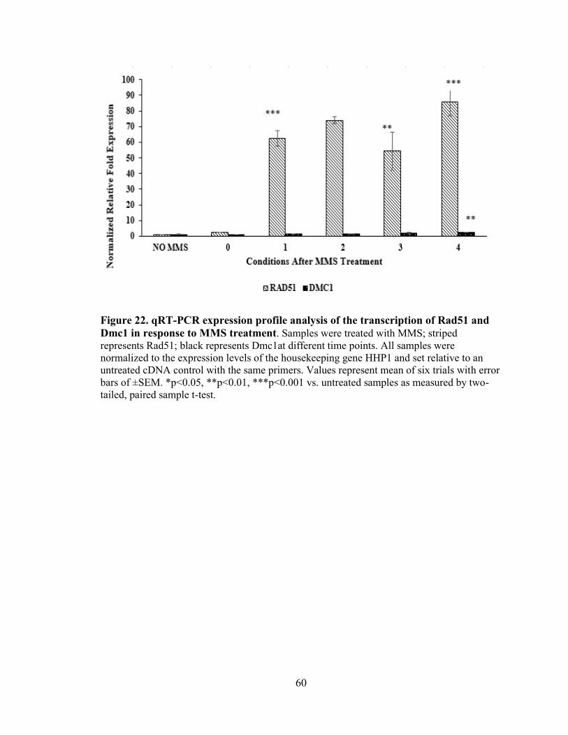
60
Figure 22. qRT-PCR expression profile analysis of the transcription of Rad51 and
Dmc1 in response to MMS treatment. Samples were treated with MMS; striped
represents Rad51; black represents Dmc1at different time points. All samples were
normalized to the expression levels of the housekeeping gene HHP1 and set relative to an
untreated cDNA control with the same primers. Values represent mean of six trials with error
bars of ±SEM. *p<0.05, **p<0.01, ***p<0.001 vs. untreated samples as measured by two-
tailed, paired sample t-test.

61
Figure 23. qRT-PCR expression profile analysis of the transcription of Rad51 in
response to UV treatment. Samples were treated with UV; white represents Rad51at
different time points. All samples were normalized to the expression levels of the
housekeeping gene HHP1 and set relative to an untreated cDNA control with the same
primers. Values represent mean of six trials with error bars of ±SEM. *p<0.05, **p<0.01,
***p<0.001 vs. untreated samples as measured by two-tailed, paired sample t-test.

62
Figure 24. qRT-PCR expression profile analysis of the transcription of Dmc1 in
response to UV treatment. Samples were treated with UV; striped represents Dmc1 at
different time points. All samples were normalized to the expression levels of the
housekeeping gene HHP1 and set relative to an untreated cDNA control with the same
primers. Values represent mean of six trials with error bars of ±SEM. *p<0.05, **p<0.01,
***p<0.001 vs. untreated samples as measured by two-tailed, paired sample t-test.

63
Figure 25. qRT-PCR expression profile analysis of the transcription of Rad51 and
Dmc1 in response to UV treatment. Samples were treated with UV; white represents
Rad51; striped represents Dmc1 at different time points. All samples were normalized to
the expression levels of the housekeeping gene HHP1 and set relative to an untreated
cDNA control with the same primers. Values represent mean of six trials with error bars
of ±SEM. *p<0.05, **p<0.01, ***p<0.001 vs. untreated samples as measured by two-
tailed, paired sample t-test.

64
Figure 26. Fluorescent Microscopy images of Rad51 and Dmc1.The cells were stained
with DAPI to view the nucleus. (A) Cells containing GFP-Dmc1. (B) Cells containing
RFP-Dmc1. (C) Cells containingGFP-Rad51. (D) Cells containing RFP-Rad51. Rad51
and Dmc1 were distributed within the cytoplasm.

65
Rad51 and Dmc1 Localization in Response to DNA Damage
Localization of GFP-Rad51 and Dmc1 was observed after MMS treatment
(Figure 27 & 28). Rad51 and Dmc1 expression is very sensitive to MMS. A
concentration of 10 mM MMS was used to treat tagged cells for 1, 2, 3, and 4 hours.
After one hour of MMS treatment, GFP-Dmc1 did not localize to either nucleus; it
disappeared and GFP fluorescent was not expressed (Figure 27). Two hours later, GFP-
Dmc1 started to show without localization to the nuclei; localization of GFP-Rad51 was
seen throughout the cytoplasm. The pattern of localization for GFP-Dmc1 and GFP-
Rad51 did not change when they were treated with MMS after three hours (Figures 27 &
28). The distribution of GFP-Dmc1 and GFP-Rad51 were observed throughout the
cytoplasm after four hours of MMS treatment (Figures 27 & 28).

66
Figure 27. Dmc1 does not localize to nucleus following MMS treatment. Cells
containing GFP-Dmc1 at log phase of growth (1 x 10⁵ cells/mL). Then, Cells containing
GFP-Dmc1 were treated with 10 mM MMS for 1-4 hours. In all panels cells were stained
with DAPI and imaged at 1000x with oil immersion

67
Figure 28. Rad51 does not localize to nucleus following MMS treatment. Cells
containing GFP-Rad51 at log phase of growth (1 x 10⁵ cells/mL). Then, Cells
containing GFP-Rad51 were treated with 10 mM MMS for 1-4 hours. In all panels cells
were stained with DAPI and imaged at 1000x with oil immersion.

68
DISCUSSION
Eukaryotic cells contain two recombinases paralogs, Rad51 and Dmc1 that are
closely related to the bacterial RecA protein. RecA is a part of a DNA-dependent ATPase
superfamily of proteins, which bind DNA and maintain genomic stability. RecA is the
recombinase required for repair of DSBs in prokaryotic cells and Rad51 possesses a
similar recombination function in eukaryotic cells. In eukaryotic cells they go through a
specialized cell division for sexual reproduction and thus contain a meiosis-specific
recombinase called Dmc1 that is used during non-sister chromatid DSB exchange
(crossing-over), which happens in meiotic prophase I. The goal of this project was
characterize the T. thermophila RecA homologs, Rad51 and Dmc1, and investigate their
role in the response to DNA damaging agents.
Bioinformatics
Phylogenetic analysis of Rad51 and Dmc1 homologs revealed that both are highly
conserved although in some organisms including T. thermophila the Dmc1 protein is
more divergent away from the other eukaryotic RecA homologs (Figure s 7, B1, & B2).
he phylogenetic tree identified that Rad51 and Dmc1 homologs were RecA family
proteins (Figure 7). Some organisms have a more divergent Dmc1 while others the two
paralogs were highly conserved and examples of both a divergent and highly conserved
Rad51/Dmc1 was seen in examples across the four different eukaryotic kingdoms
(Plantae, Fungi, Animalia, and Protista). This finding may help in identifying the unique
characteristics and functions that these paralogs have in their role in the cell. In some
paralogs there may be a loss of redundancy in function when the other is missing or a

69
separation of the functional roles. Studying the roles of Dmc1 and Rad51 in both P.
tetraurelia T. thermophila both complex single-cell protozoa could elucidate the purpose
for the divergence.
In addition to the similarity between Rad51 and Dmc1 at the whole protein level,
they also were highly conserved within their functional domains and motifs and all RecA
homologs contained two conserved ATPase domains (RECA2 and RECA3; Figure 6).
The RECA2 domains is located in the N-terminal region and required for ATP hydrolysis
and the RECA3 is located in the C-terminal region required for ATP binding. All RecA
homologs also contained the highly conserved Walker A and Walker B motifs located in
the RECA2 domain of both Rad51 and Dmc1 paralogs (Appendix B - Figures B1 & B2).
The T. thermophila Dmc1 scored 87%, and T. thermophila Rad51 scored 92% similarity
in their overall alignment further suggesting that Dmc1 may have a unique function in the
cell.
Expression of RAD51 and DMC1 was observed through three methods, which
included RNAseq, expression microarray, and qRT-PCR. The microarray data for
RAD51 indicated a ubiquitous basal expression during vegetative growth with induced
expression during conjugation and starvation conditions (Figure 9A). Interestingly,
DMC1 was only expressed during the conjugation and emphasizes the role of Dmc1 has
in meiosis (Figure 9B). Rad51 could function in both meiosis and mitosis, which has
been documented in previous experiments where RAD51 overexpression causes the
macronucleus to not divide creating amacronucleate cells (Figure 5). Expression of both
RAD51 and DMC1 increased in a bimodal pattern during conjugation, peaking between
2-4 hours corresponding to meiotic prophase I when crossing-over occurs and later
between 12-16 hours during the time when the new macronuclei are developing from

70
zygotic micronuclei (micronuclear-limited sequences are eliminated, chromosomal
fragmentation of 5 MIC chromosomes to 218 MAC chromosomes, and duplication of
each MAC chromosome to 45 copies (2N to 45C). This suggests either an overlapping
function, specific recombinational roles, or a complex interaction of Rad51 and Dmc1 in
T. thermophila conjugation. Previously published work on the knockouts of RAD51 and
DMC1 show deletion of either will halt conjugation in meiotic prophase but only removal
of RAD51 has a cell division phenotype causing arrest of micronuclear mitosis creating
amicronucleate cells (Howard-Till et al., 2011; Marsh et al., 2000; Marsh et al., 2001).
This suggests that the role is either recombinationally specific or a complex interaction
between Rad51 and Dmc1.
Figure 8 shows the predicted annotation of RAD51 to contain three exons and two
introns, and DMC1 to contain five exons and four introns and the RNAseq coverage data
confirms the RAD51 exons and introns (Figure 8A). The RNAseq coverage data for
DMC1 confirms the exons and introns but there is a low level of coverage detected over
the second intron (Figure 8B) and this could explain the multiple products produced from
DMC1-2 primers over this intron (Figure 16).
In addition to Rad51 and Dmc1 homologs of Hop2 and Mnd1 proteins that are
found to interact with each other and Rad51 and Dmc1 to promote efficient homologous
recombination were identified and bioinformatics analysis conducted. The Hop2 and
Mnd1 proteins families are typically represented by one homolog in most eukaryotes, but
in T. thermophila two HOP2 and two MND1 homologs were found (Appendix B - Table
B1 and data not shown). The meiosis-specific expression of HOP2 and the more
ubiquitous expression of HOPP2 which peaks also during starvation and conjugation has
been experimentally confirmed through RT-PCR (Mochizuki et al.,2008). There is a

71
higher ubiquitously expression of MNDP1 whereas MND1 is only expressed at very low
levels in the cell (data not shown). Since Hop2 and Mnd1 act as a complex together in
Dmc1 and Rad51 homologous pairing The linked patterns of expression could indicate
that Hop2 functions with Dmc1 while Hopp2 functions with Rad51 or that a specific
Hop2-Mnd1 complex interacts with Rad51 during mitosis and DNA repair while another
Hop2-Mnd1 complex functions during conjugation.
Expression of RAD51 and DMC1 Following DNA Damage in T. thermophila.
Analysis of DMC1 and RAD51 PCR products from two sets of primers for each
gene (each spanning across an intron) showed amplification of proper size product when
using genomic DNA as the template (Figure 14 & 16). Both RAD51-1 and RAD51-2
showed the correct size PCR product using cDNA as the template, but RAD51-1 primers
amplified a little better with the SsoFast Evagreen qPCR mix (Figure 16) so was used for
all of the DNA damage expression experiments. Mixed results were seen with the DMC1
primer sets when cDNA was used as the template. When the Gotaq PCR master mix was
used a single band the correct size corresponding to the cDNA size was only seen with
the DMC1-1 primer set and the DMC1-2 primers showed a product the same size as the
genomic DNA (Figure 14). When the SsoFast Evagreen qPCR master mix was used
DMC1-1 primers yielded a band that corresponded to the size of genomic DNA while
DMC1-2 primers yielded two bands one corresponding to the size for cDNA and the
other had the same size as gDNA (Figures 15 & 16). Since cDNA from vegetatively
growing cells was used the lack of template cDNA may have caused the amplification of
any genomic DNA contamination or in the case of the products in DMC1-2 there may be
an alternatively spliced form that includes the second intron being that RNAseq showed

72
some coverage over that intron (Figure 8). The reasoning for a genomic DNA sized band
with DMC1-1 primers is most likely genomic DNA contamination since it was not seen
with GoTaq but was with qPCR which has more cycles in the PCR protocol. If alternative
splicing occurs leaving the second intron in the cDNA a truncated protein of 194 amino
acids instead of 356 would be made and it would only have a truncated portion of the
RECA2 domain and lack the RECA3 domain (this would render it nonfunctional as a
DNA-dependent ATPase. The activity of this form of Dmc1 is unknown, but it could be
used as a decoy receptor to bind away proteins that can interact with both Rad51 and
Dmc1 to turn off the recombination activity at the later part of DNA repair or when not
needed during meiosis. Since the DMC1-2 set of primers gave a band at the predicted
size in cDNA (even though other products were present) this set of primers were used for
DMC1 expression analysis after various DNA damaging agents.
Further qRT-PCR experiments were conducted to identify the expression patterns
of RAD51 and DMC1 in response to DNA damaging agents. The qRT-PCR expression
data revealed an increased expression of RAD51 and DMC1 in response to DNA damage
by UV irradiation (Figure 25). The expression continued to increase as the time went on,
but it dropped down at the 4-hour time point. The similarity of the expression of Rad51
and Dmc1 after UV treatment, peaking to 17-fold for Rad51 and 6.5-fold for Dmc1 at the
3-hour time point, strongly suggested both Rad51 and Dmc1 have a functional role in the
UV induced DNA repair process (Figure 25). UV irradiation causes the production of
DNA adducts, which can result in DSBs that could be repaired by homologous
recombination (Rolfsmeier M et al.,2010). This explains the increased expression after
two hours of treatment. Interestingly, MMS is known to cause DBSs that are repaired by
HHR, which similar showed only a large induction of the expression of RAD51 (70-fold

73
induction) while after 1-hour treatment DMC1 only reached 2-fold induction and that was
not until 4-hours after treatment (Figure 22). The drastic difference in expression
illustrates that Rad51 and not Dmc1play a significant role in HRR DNA repair from
damaged caused by MMS (Figure 22). Additionally, a similar pattern of expression was
observed for RAD51 after treatment with H2O2 as was with UV but DMC1 expression
was unique with a slight increase 2-hours after treatment and a drastic spike inducing 25-
fold 3-hours after with a subsequent decrease back down 4-hours after treatment with
H2O2 (Figure 19). Hydrogen peroxide results in radical damage and there may either be a
role for both of these RecA homologs in the repair or it may be related to a cell cycle
specific expression since with both UV and H2O2 the cells may arrest preventing
progression through division and this increase is just a result of cell synchronization.
Analysis of the qRT-PCR data shows that DMC1 induction after DNA damage is always
delayed compared to RAD51 expression (Figures 19, 22, & 25) and it is possible that
Dmc1 acts differently to stabilize the DNA repair done by Rad51 or as a mechanism to
turn of the Rad51 repair by competing for interacting proteins like the Hop2-Mnd1
complex.
Epitope and Fluorescent Tagged Rad51 and Dmc1 Expression and Localization
T. thermophila transformants containing 2HA-Rad51, 2HA-Dmc1, FH6-Rad51,
and FH6-Dmc1 were made, and Total protein extracts were isolated from multiple
transformants to be used to confirm the expression of the epitope tagged Rad51 and
Dmc1 through western blot analysis. This experiment was not completed and isolated
protein extracts were stored at –80° C for future research. Transformation of GFP-Rad51,
RFP-Rad51, RFP-Dmc1, and GFP-Dmc1 into T. thermophila was completed and

74
expression of the GFP and RFP tagged Rad51 and Dmc1 was confirmed by fluorescent
microscopy in growing cells and all showed fluorescence in the cytoplasm confirming
positive expression of the fusion proteins (Figure 26). The GFP expression was easier to
visualize so GFP-Rad51 and GFP-Dmc1 transformants were treated with MMS to
observe if there was a change in localization following DSBs (Figure 28). Transformants
expressing GFP/RFP-Rad51 and GFP/RFP-Dmc1 exhibited the fluorescence protein in
the cytoplasm and not within the macronucleus but only Dmc1 in the micronucleus
(Figure 26). The localization in the micronucleus could not be confirmed by merging the
DAPI with the GFP/RFP because even when the cells were not moving the organelles
were still moving around in the cell slightly distorting the data between pictures. In
response to MMS damage, Rad51 and Dmc1 did not localize to either the macronucleus
or the micronucleus. After one hour of MMS treatment, Dmc1 was not detectible, but
Rad51 showed a slight expression. It is known that Rad51 localizes to the nuclei to repair
the DSBs caused by MMS (Campbell & Romero, 1998) but, localization was not
observed. This may have occurred due to the high concertation of MMS, 10 mM MMS,
which is known to turn off the MTT1 promoter which is driving the inducible expression
of the fluorescently tagged proteins. Another possibility is that the tag interferes with the
normal function and thus does not localize or that it takes a longer time to localize to the
nuclei then previously documented.
Future Directions
More research will be needed to find the interplay role between Rad51 and Dmc1.
Rad51 and Dmc1 were tagged with Flag and HA epitope tags and the total protein were
isolated from the cells to run western blot experiment. Western blot analysis could be

75
performed to confirm the tagged HA-Rad51 and HA-Dmc1 proteins. Once, they are
confirmed, an immunoprecipitation could be utilized to identify proteins interacting with
Rad51 and Dmc1. Pulling down the interacting proteins can help to elucidate the factors
that control the activity of Rad51 and Dmc1 during the DNA damage process and to even
determine if Rad51 can form a heteronucleofilament with Dmc1 during DNA repair, cell
cycle, and conjugation. In addition, it would be beneficial to create HA-Hop2/HA-Hopp2
and FH6-Mnd1/FH6-Mndp1 constructs to further characterize their roles in DSB repair
and interactions with Rad51 and Dmc1 and to determine which of the Hop2 and Mnd1
homologs interact with one another, Rad51, and Dmc1.As the localization studies were
inconclusive, repeating fluorescent microscopy analysis with GFP-Rad51/RFP-Rad51
and GFP-Dmc1/RFP-Dmc1will help to visualize any actual localization for these
proteins. To improve the likelihood of success, it would be ideal to decrease the
concentration of MMS from 10 mM to 5 mM instead and look at the localization between
1-6 hours to conclude if localization can be found in either nucleus. In addition, it would
be beneficial to treat the cells with UV and H2O2 to track any localization of Rad51 and
Dmc1 to the nuclei. Expression profiles showed an increase in RAD51 and DMC1
expression in response to UV and H2O2 treatment and were much higher for DMC1 with
these two damaging agents compared to MMS. Expression of RAD51 and DMC1 using
qRT-PCR could be done on synchronized cells or starved cells to elucidate the role of
Rad51 and Dmc1 in cell cycle progression and mitosis as well as stress related to
starvation. Microarray expression data showed that RAD51 was expressed through all cell
conditions where as DMC1 was only expressed during conjugation (Figure 9). To
determine whether Rad51 has a specific role in somatic cell division that is lacking for
Dmc1 single and double knockouts (double knockout of rad51 and dmc1and single

76
knockouts of dmc1 or rad51) and a DMC1 overexpression strain (currently have a
RAD51 overexpression strain) could be constructed. These strains could then be tested
with the same DNA damaging agents used above to look at sensitivity. If the double
knockout cells are more sensitive to a DNA damaging agent than the rad51 knockout,
and if the dmc1knockout has no sensitivity, that would confirm that Dmc1 plays some
accessory role to help Rad51 in DNA repair damage. If the double knockout looks just
the same as the rad51 knockout we can conclude that there is no direct interaction
between Rad51 and Dmc1 and that the increase in Dmc1 may be a way to turn off the
repair process by sequestering interacting proteins away from Rad51.
Additional FH6-Dmc1 could clarify the interplay role between both Rad51 and
Dmc1. The protein extracts made from the FH6- Dmc1 cells compared to wild-type
(CU428) cells that are induced and uninduced with DNA damaging agents and extracts
made between 2-4 hours after treatment could be analyzed by western blot analysis.
Immunoprecipitation studies using FLAG antibody and a commercial Rad51 antibody,
looking at UV and H2O2 damage and interaction in the FH6-Dmc1 transformants, can
help to determine if there is a physical interaction between Rad51 and Dmc1 in a
heteronucleofilament that has not been previously identified. These experiments will also
tell if the commercial Rad51 antibody only detects T. thermophila Rad51 or if it cross-
reacts and detects Dmc1 too. This can be seen since Dmc1 is tagged with FLAG-6xHis it
will be larger in the tagged strain than the wild-type strain and could be observed by
western blot analysis.
The overexpression RAD51 and DMC1 in cells can be a great way to visualize if
the amacronucleate phenotype found with overexpression of RAD51 (Figure 5) is also
seen if DMC1 was expressed when it normally is not active. Overexpression of RAD51
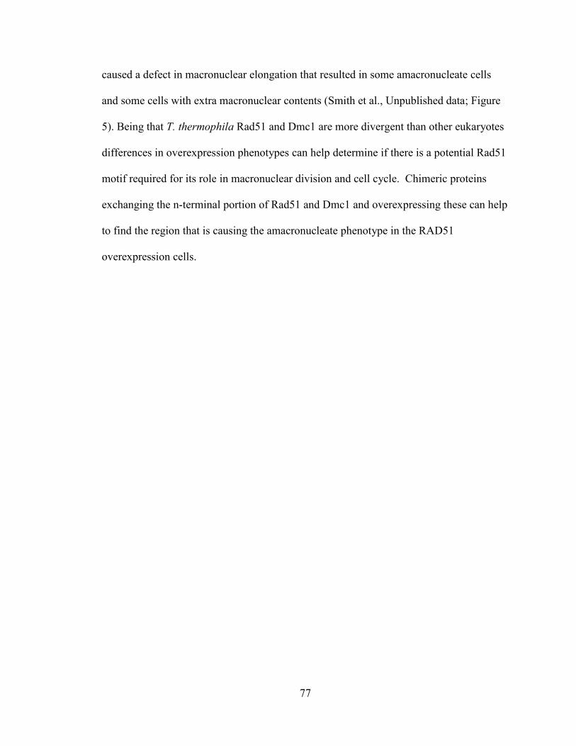
77
caused a defect in macronuclear elongation that resulted in some amacronucleate cells
and some cells with extra macronuclear contents (Smith et al., Unpublished data; Figure
5). Being that T. thermophila Rad51 and Dmc1 are more divergent than other eukaryotes
differences in overexpression phenotypes can help determine if there is a potential Rad51
motif required for its role in macronuclear division and cell cycle. Chimeric proteins
exchanging the n-terminal portion of Rad51 and Dmc1 and overexpressing these can help
to find the region that is causing the amacronucleate phenotype in the RAD51
overexpression cells.

78
REFERENCES
Altschul, S.F., Wootton, J.C., Gertz, E.M., Agarwala, R., Morgulis, A., Schaffer, A.A.,
and Yu, Y. (2005). Protein databases searches using compositionally adjusted
substitution matrices FEBS J. 272:5101-5109.
Aerssens, J., Armstrong, M., Gilissen, R., and Cohen, N. (2001). The human genome: an
introduction. Oncologist 6(1):100-9.
Bishop, D.K., Park, D., Xu, L., and Kleckner, N., (1992). DMC1: a meiosis specific yeast
homolog of E. coli RecA required for recombination, synaptonemal complex
formation, and cell cycle progression. Cell 69, 439–456.
Bishop, D.K. (1994). RecA homologs Dmc1 and Rad51 interact to form multiple nuclear
complexes prior to meiotic chromosome synapsis. Cell 79, 1081–1092.
Brown, M.S., and Bishop, D.K. (2015). DNA strand exchange and RecA homologs in
meiosis. CSH Perspect. Biol. 7, p. 016659.
Buchhop, S., Gibson, M.K., Wang, X.W., Wagner, P., Sturzbecher, H.W., and Harris,
C.C. (1997). Interaction of p53 with the human Rad51 protein. Nuc. Acids Res.
25:3868–3874.
Bugreev, D.V., Huang, F., Mazina, O.M., Pezza, R.J., Voloshin, O.N., Camerini-Otero,
R.D., and Mazin, A.V. (2014). HOP2-MND1 modulates RAD51 binding to
nucleotides and DNA. Nat. Commun. 5:4198–4207.
Campbell, C., and Romero, D. P. (1998). Identification and characterization of the
RAD51 gene from the ciliate Tetrahymena thermophila. Nuc. Acids Res. 26(13),
3165–3172.
Chang, H.Y., Liao, C.-Y., Su, G.-C., Lin, S.W., Wang, H.W., and Chi, P. (2015).
Functional Relationship of ATP Hydrolysis, Presynaptic Filament Stability, and
Homologous DNA Pairing Activity of the Human Meiotic Recombinase
DMC1. J. Biol. Chem. 290(32), 19863–19873.
Chen, Y.K., Leng, C.H., Olivares, H., Lee, M.H., Chang, Y.C., Kung, W.M., Ti, S.C., Lo,
Y.H., Wang, A.H., and Chang, C.S. (2004). Heterodimeric complexes of Hop2
and Mnd1 function with Dmc1 to promote meiotic homolog juxtaposition and
strand assimilation. Proc. Natl Acad. Sci. U.S.A.101:10572–10577.
Chi, P., San Filippo, J., Sehorn, M. G., Petukhova, G. V., and Sung, P. (2007). Bipartite
stimulatory action of the Hop2–Mnd1 complex on the Rad51 recombinase. Genes
Dev. 21(14), 1747–1757.

79
Cloud, V., Chan, Y.L., Grubb, J., Budke, B., and Bishop, D.K. (2012). Rad51 is an
accessory factor for Dmc1-mediated joint molecule formation during meiosis.
Science 337(6099):1222–1225.
Choudhury, A., Zhao, H., Jalali, F., Al Rashid, S., Ran, J., Supiot, S., Kiltie, A.E., and
Bristow, RG. (2009). Targeting homologous recombination using imatinib results
in enhanced tumor cell chemo-sensitivity and radiosensitivity. Mol. Cancer Ther.
8:203–213.
Davis, A. J., Lee, K.J., and Chen, D. J. (2013). The N-terminal Region of the DNA-
dependent Protein Kinase Catalytic Subunit Is Required for Its DNA Double-
stranded Break-mediated Activation. J. Biol. Chem. 288(10), 7037–7046.
Dresser, M.E., Ewing, D.J., Conrad, M.N., Dominquez, A.M., Barstead, R., Jiang, H.,
and Kodadek, T. (1997). Dmc1 functions in a Saccharomyces cerevisiae meiotic
pathway that is largely independent of the Rad51 pathway. Genetics 147, 533–
544.
Eisen, J. A., Coyne, R. S., Wu, M., Wu, D., Thiagarajan, M., Wortman, J. R., . . . and
Orias, E. (2006). Macronuclear Genome Sequence of the Ciliate Tetrahymena
thermophila, a Model Eukaryote. PLoS Biology 4(9), e286.
Featherstone, C., and Jackson, S. P. (1999). DNA double-strand break repair. Current
Biology. 9(20), R759-R761.
Gasior, S.L., Wong, A.K., Kora, Y., Shinohara A., and Bishop D.K. (1998). Rad52
associates with RPA and functions with Rad55 and Rad57 to assemble meiotic
recombination complexes. Genes Dev. 12(14):2208–2221.
Hakem, R. (2008). DNA-damage repair; the good, the bad, and the ugly. EMBO J. 27(4),
589–605.
Hannay, J.A., Liu, J., Zhu, Q.S., Bolshakov, S.V., Li, L., Pisters, P.W., Lazar, A.J., Yu
D., Pollock, R.E., and Lev, D. (2007). Rad51 overexpression contributes to
chemo-resistance in human soft tissue sarcoma cells: a role for p53/activator
protein 2 transcriptional regulation. Mol. Cancer Ther. 6:1650–1660.
Helleday, T., Eshtad, S., and Nik-Zainal, S. (2014). Mechanisms underlying mutational
signatures in human cancers. Nat. Rev. Genet. 15(9), 585–598.
Henry, J.M., Camahort, R., Rice, D.A., Florens, L., Swanson, S.K., Washburn, M.P., and
Gerton, J.L. (2006). Mnd1/Hop2 facilitates Dmc1-dependent interhomolog
crossover formation in meiosis of budding yeast. Mol. Cell. Biol. 26:2913–2923.
Howard-Till, R. A., Lukaszewicz, A., and Loidl, J. (2011). The Recombinases Rad51 and
Dmc1 Play Distinct Roles in DNA Break Repair and Recombination Partner
Choice in the Meiosis of Tetrahymena. PLoS Genetics 7(3), e1001359.

80
Jackson, S. P. (2002). Sensing and repairing DNA double-strand
breaks. Carcinogenesis 23:687-696.
Jasin, M. (2002). Homologous repair of DNA damage and tumorigenesis: The BRCA
connection. Oncogene 21, 8981-8993.
Khanna, K.K., and Jackson, S. P. (2001). DNA double-strand breaks: signaling, repair
and the cancer connection. Nat. Genet. 27:247-254.
Kim, P. M., Allen, C., Wagener, B. M., Shen, Z., and Nickoloff, J. A. (2001).
Overexpression of human RAD51 and RAD52 reduces double-strand break-
induced homologous recombination in mammalian cells. Nuc. Acids Res. 29(21),
4352–4360.
Koonin, E.V., Tatusov, R.L., and Rudd, K.E. (1995). Sequence similarity analysis of
Escherichia coli proteins: functional and evolutionary implications. Proc. Natl.
Acad. Sci. U.S.A. 92, 11921–11925, 5–74.
Koonin, E.V. (1993). A superfamily of ATPases with diverse functions containing either
classical or deviant ATP-binding motif. J. Mol. Biol. 229, 11639.
Koonin, E.V. (1993b). A common set of conserved motifs in a vast variety of putative
nucleic acid-dependent ATPases including MCM proteins involved in the
initiation of eukaryotic DNA replication. Nuc. Acids Res. 21, 2541 – 2547.
Lee, J.Y., Terakawa, T., Qi, Z., Steinfeld, J.B., Redding, S., Kwon, Y., Gaines, W.A.,
Zhao, W., Sung, P., and Greene, E.C. (2015). Base triplet stepping by the
Rad51/RecA family of recombinases. Science 349, pp. 977-981.
Leipe, D.D., Wolf, Y.I., Koonin, E.V., and Aravind, L. (2002). Classification and
evolution of P-loop GTPases and related ATPases. J. Mol. Biol. 317, 41 – 72.
Leu, J.Y., Chua, P.R., and Roeder, G.S. (1998). The meiosis-specific Hop2 protein of S.
cerevisiae ensures synapsis between homologous chromosomes. Cell 94:375–386.
Lieber, M. R. (2010). The Mechanism of Double-Strand DNA Break Repair by the Non
homologous DNA End Joining Pathway. Annu. Rev. Biochem. 79, 181–211.
Liu, N. (2002). XRCC2 Is Required for the Formation of Rad51 Foci Induced by Ionizing
Radiation and DNA Cross-Linking Agent Mitomycin C. J. Biomed. Biotech. 2(2),
106–113.
Li, X., and Heyer, W.D. (2008). Homologous recombination in DNA repair and DNA
damage tolerance. Cell Res. 18(1), 99–113.
Li, X., and Heyer W.D. (2009). RAD54 controls access to the invading 30-OH end after
RAD51-mediatedDNA strand invasion in homologous recombination in
Saccharomyces cerevisiae. Nuc. Acids Res. 37(2):638–646.

81
Li, Y., Wang, W.Y., Xiao, J.H., Xu, F., Liao, D.Y., Xie, L., Wang, J., and Luo, F. (2017).
Overexpression of Rad51 Predicts Poor Prognosis in Colorectal Cancer: Our
Experience with 54 Patients. PLoS ONE 12(1): e0167868.
Loidl, J. and Scherthan, H. (2004). Organization and pairing of meiotic chromosomes in
the ciliate Tetrahymena thermophila. J. Cell Sci. 117, 5791–5801.
Maréchal, A., and Zou, L. (2013). DNA Damage Sensing by the ATM and ATR
Kinases. Cold Spring Harbor Perspectives in Biology 5(9), a012716.
Marsh, T. C., Cole, E. S., Stuart, K. R., Campbell, C., and Romero, D. P. (2000). RAD51
is required for propagation of the germinal nucleus in Tetrahymena
thermophila. Genetics 154(4), 1587–1596.
Marsh, T.C., Cole, E.S., and Romero, D.P. (2001). The Transition from Conjugal
Development to the First Vegetative Cell Division Is Dependent on RAD51
Expression in the Ciliate Tetrahymena thermophile. Genetics 157,1591-1598.
Masson, J. Y., and West, S. C. (2001). The Rad51 and Dmc1 recombinases: a non-
identical twin relationship. Trends Biochem. Sci. 26, 131-136.
Mizuta, R., LaSalle, J.M., Cheng, H.L., Shinohara, A., Ogawa, H., Copeland, N., Jenkins,
N.A., Lalande, M., and Alt. F.W. (1997). RAB22 and RAB163/mouse BRCA2:
Proteins that specifically interact with the RAD51 protein Proc. Natl. Acad. Sci.
USA 94: 6927-6932.
Mochizuki, K., Novatchkova, M., and Loidl, J. (2008). DNA double-strand breaks, but
not crossovers, are required for the reorganization of meiotic nuclei
in Tetrahymena. J. Cell Sci. 121(Pt 13), 2148–2158.
Neale, M.J., and Keeney, S. (2006). Clarifying the mechanics of DNA strand exchange in
meiotic recombination. Nature 442:153–158.
Ogawa, T., Yu, X., Shinohara, A., and Egelman, E.H. (1993). Similarity of the yeast
RAD51 filament to the bacterial RecA filament. Science 259:1896–1899.
Okorokov, A.L., Chaban, Y.L., Bugreev, D.V., Hodgkinson, J., Mazin, A.V., and Orlova,
E.V. (2010). Structure of the hDmc1-ssDNA Filament Reveals the Principles of
Its Architecture. PLoS ONE 5(1), e8586.
Orias E. (2012). Tetrahymena thermophila Genetics: Concepts and Applications Meth.
Cell Biol. 109, 301-325.
Orias E. (2000).Toward sequencing the Tetrahymena genome: Exploiting the gift of
nuclear dimorphism. J. Eukaryot. Microbiol. 47:328–333.

82
Peng, M., Bakker, J.L., Dicioccio, R.A., Gille, J.J., Zhao, H., Odunsi, K., Sucheston, L.,
Jaafar, L., Mivechi, N.F., Waisfisz, Q. and Ko, L. (2013). Inactivating mutations
in GT198 in familial and early-onset breast and ovarian cancers. Genes Cancer.
4:15–25.
Peterson, D.S., Gao, Y., Asokan, K., and Gaertig, J. (2002). The circumsporozoite protein
of Plasmodium falciparum is expressed and localized to the cell surface in the
free-living ciliate Tetrahymena thermophila. Mol. Biochem. Parasitol. 122:119–
126.
Petukhova, G.V., Romanienko, P.J., and Camerini-Otero, R.D. (2003). The Hop2 protein
has a direct role in promoting interhomolog interactions during mouse
meiosis. Dev. Cell 5:927–936.
Pezza, R.J., Petukhova, G.V., Ghirlando, R., and Camerini-Otero, R.D. (2006). Molecular
activities of meiosis-specific proteins. Hop2, Mnd1, and the Hop2–Mnd1
complex. J. Biol. Chem. 281:18426–18434.
Pittman, D.L., Cobb, J., Schimenti, K.J., Wilson, L.A., Cooper, D.M., Brignull, E.,
Handel, M.A., and Schimenti, J.C. (1998). Meiotic prophase arrest with failure of
chromosome synapsis in mice deficient for Dmc1, a germline-specific RecA
homolog. Mol. Cell 1:697–705.
Ploquin, M., Petukhova, G.V., Morneau, D., Dery, U., Bransi, A., Stasiak, A., Camerini-
Otero, R.D., and Masson, J.Y. (2007). Stimulation of fission yeast and mouse
Hop2-Mnd1 of the Dmc1 and Rad51 recombinases. Nuc. Acids Res. 35:2719–
2733.
Qiao, G.B., Wu, Y.L., Yang, X.N., Zhong, W.Z., Xie, D., Guan, X.Y., Fischer, D.,
Kolberg, H.C., Kruger, S., and Stuerzbecher, H.W. (2005). High-level expression
of Rad51 is an independent prognostic marker of survival in non-small-cell lung
cancer patients. Brit. J. Cancer 93(1), 137–143.
Ramsden, D.A., and Gellert, M. (1998). Ku protein stimulates DNA end joining by
mammalian DNA ligases: a direct role for Ku in repair of DNA double-strand
breaks. EMBO J., 17, 609-614.
Ray, C. (1956). Meiosis and nuclear behavior in Tetrahymena pyriformis. J.
Protozool. 3, 88–96.
Reymer, A., Frykholm, K., Morimatsu, K., Takahashi M., and Nordén, B. (2009).
Structure of human Rad51 protein filament from molecular modeling and site-
specific linear dichroism spectroscopy. Proc. Natl. Acad. Sci. U.S.A.
106 (32) 13248-13253.

83
Ristic, D., Modesti, M., Heijden, T.V.D., Noort, J., Dekker, C., Kanaar, R., and Wyman,
C. (2005). Human Rad51 filaments on double- and single-stranded DNA:
correlating regular and irregular forms with recombination function. Nuc. Acids
Res. 33 (10): 3292-3302.
Rockmill, B., Sym, M., Scherthan, H., and Roeder, G.S. (1995). Roles for two RecA
homologs in promoting meiotic chromosome synapsis. Genes Dev. 9:2684–2695.
Rolfsmeier, M., Laughery, M.F., and Haseltine, C.A. (2010). Repair of DNA Double-
Strand Breaks following UV Damage in Three Sulfolobus solfataricus Strains. J.
Bacteriol. 192 (19): 4954-4962.
Rothkamm, K., Krüger, I., Thompson, L. H., and Löbrich, M. (2003). Pathways of DNA
Double-Strand Break Repair during the Mammalian Cell Cycle. Mol. Cell.
Biol. 23(16), 5706–5715.
Schwacha, A. and Kleckner, N. (1997). Interhomolog bias during meiotic recombination:
meiotic functions promote a highly differentiated interhomolog only pathway.
Cell 90, 1123–1135
Sebesta, M., Burkovics, P., Haracska, L., Krejci, L. (2011). Reconstitution of DNA repair
synthesis in vitro and the role of polymerase and helicase activities. DNA Repair
(Amst) 10(6):567–576.
Sebesta, M., and Krejci, L. (2016). Mechanism of Homologous Recombination (PDF
Download Available) in DNA replication, recombination, and repair: molecular
mechanisms and pathology. Tokyo: Springer. Retrieved Jan, 2017.
Seong, C., Sehorn, M.G., Plate, I., Shi, I., Song, B., Chi, P., Mortensen, U., Sung, P., and
Krejci, L. (2008). Molecular anatomy of the recombination mediator function of
Saccharomyces cerevisiae Rad52. J. Biol. Chem. 283(18):12166–12174.
Shang, Y., Li, B., and Gorovsky, M.A. (2002). Tetrahymena thermophila contains a
conventional gamma-tubulin that is differentially required for the maintenance of
different microtubule-organizing centers. J. Cell Biol.158:1195–1206.
Sharan, S.K., Morimatsu, M., Albrecht, U., Lim, D.S., Regel, E., Dinh, C., Sands, A.,
Eichele, G., Hasty, P., and Bradley, A. (1997). Embryonic lethality and radiation
hypersensitivity mediated by Rad51 in mice lacking Brca2. Nature, 386: 804-810.
Shinohara, A., Ogawa, H., and Ogawa, T. (1992). Rad51 protein involved in repair and
recombination in S. cerevisiae is a RecA-like protein. Cell 69, 457–470.
Shinohara, A., Gasior, S., Ogawa, T., Kleckner, N. and Bishop, D. K.
(1997). Saccharomyces cerevisiae recA homologues RAD51and DMC1 have
both distinct and overlapping roles in meiotic recombination. Genes to Cells, 2:
615–629.

84
Slupianek, A., Schmutte, C., Tombline, G., Nieborowska-Skorska, M., Hoser, G.,
Nowicki, M.O., Pierce, A.J., Fishel, R., and Skorski, T. (2001). BCR/ABL
regulates mammalian RecA homologs, resulting in drug resistance. Mol. Cell.
8:795–806.
Smith, J.J., Cole, E.S., and Romero, D.P. (2004). Transcriptional control
of RAD51 expression in the ciliate Tetrahymena thermophila. Nuc. Acids
Res. 32(14), 4313–4321.
Sung, P. (1997). Yeast Rad55 and Rad57 proteins form a heterodimer that functions with
replication protein A to promote DNA strand exchange by Rad51 recombinase.
Genes Dev. 11(9):1111–1121.
Stover, N.A., Krieger, C.J., Binkley, G., Dong, Q., Fisk, D.G., Nash, R., Sethuraman, A.,
Weng, S., and Cherry J.M. (2006). Tetrahymena Genome Database (TGD): a new
genomic resource for Tetrahymena thermophile research. Nuc. Acids Res. 34
(Database issue): D500-3.
Tanaka, K., Kagawa, W., Kinebuchi, T., Kurumizaka, H., and Miyagawa, K. (2002).
Human Rad54B is a double-stranded DNA-dependent ATPase and has
biochemical properties different from its structural homolog in yeast,
Tid1/Rdh54. Nuc. Acids Res. 30(6), 1346–1353.
Tarsounas, M., Davies, A. A., and West, S. C. (2004). RAD51 localization and activation
following DNA damage. Philosophical Transactions of the Royal Society B:
Biological Sciences, 359(1441), 87–93.
Waters, R. (2006). Maintaining genome integrity. EMBO Reports, 7(4), 377–381.
Wolfe, J., Hunter, B. and Adair, W.S. (1976). A cytological study of micronuclear
elongation during conjugation in Tetrahymena. Chromosoma 55, 289–308.
Xiong, J., Lu, X., Lu, Y., Zeng, H., Yuan, D., Feng, L., Chang, Y., Josephine, B.,
Gorovsky, M., Fu, C., and Miao, W. (2011). Tetrahymena Gene Expression
Database (TGED): A resource of microarray data and co-expression analyses for
Tetrahymena. Science China Life Sciences 54(1): 65-67.
Yoshida, K., Kondoh, G., Matsuda, Y., Habu, T., Nishimune, Y., and Morita, T. (1998)
The mouse recA-like gene DMC1 is required for homologous chromosome
synapsis during meiosis. Mol. Cell 1, 707–718.
Yu, X., Jacobs, S. A., West, S. C., Ogawa, T., and Egelman, E. H. (2001). Domain
structure and dynamics in the helical filaments formed by RecA and Rad51 on
DNA. Proc. Natl. Acad. Sci. U.S.A. 98(15), 8419–8424.
85
Zangen, D., Kaufman, Y., Zeligson, S., Perlberg, S., Fridman, H., Kanaan, M.,
Abdulhadi-Atwan, A., Abu Libdeh, A., Gussow, A., Kisslov, I., Carmel, L.,
Renbaum, P., and Levy-Lahad, E. (2011). XX Ovarian Dysgenesis Is Caused by a
PSMC3IP/HOP2 Mutation that Abolishes Coactivation of Estrogen-Driven
Transcription. Amer. J. Hum. Genet. 89(4), 572–579.
Zhao, W., Saro, D., Hammel, M., Kwon, Y., Xu, Y., Rambo, R.P., and Sung, P. (2014).
Mechanistic insights into the role of Hop2–Mnd1 in meiotic homologous DNA
pairing. Nuc. Acids Res. 42(2), 906–917.
Zhao, W., and Sung, P. (2015). Significance of ligand interactions involving Hop2-Mnd1
and the RAD51 and DMC1 recombinases in homologous DNA repair and XX
ovarian dysgenesis. Nuc. Acids Res. 43(8), 4055–4066.

86
APPENDICES
Appendix A: Tetrahymena Rad51 and Dmc1 Epitope Tags Constructs.
SnapGene program was used to insert RAD51 and DMC1 from the pENTR
cloning vector into GTW vectors containing GFP, RFP, HA, and FLAG tags. Tagged
constructs of Rad51 and Dmc1 are shown in (Figure s A3-A10). Figure s A1 and A2 are
pENTR maps used as entry clone containing the RAD51 and DMC1 genes. Each
construct was confirmed using restriction enzymes that had sites within and out of the
genes. Banding patterns resulted from gel electrophoresis were confirmed and compared
with the stimulated gel. The plasmids were purified and transformed into Tetrahymena to
use for further experimentation. Each construct was digested with unique restriction
enzymes mentioned in the Figure legends.

87
Figure A1. pENTR-RAD51 construct map. The Figure shows the features of pENTR-
RAD51 displaying the following: restriction sites, the T7 promoter (purple), Kanamycin
resistance gene (light green), RAD51 gene (gray), attL1 and attL2 sites (green) allow
recombinational cloning of RAD51 in the entry construct with a Gateway® destination
vector. SnapGene was used to predict fragment sizes (3,485 bp, 390 bp) digested with
EcoRI.

88
Figure A2. pENTR-DMC1 plasmid map. The Figure shows the features of pENTR-
DMC1 displaying the following: restriction sites, the T7 promoter (purple), Kanamycin
resistance gene (light green), DMC1 gene (gray), attL1 and attL2 sites (green) allow
recombinational cloning of DMC1 in the entry construct with a Gateway® destination
vector. SnapGene was used to predict fragment sizes (2,892 bp, 784 bp, 566 bp) digested
with NsiI.

89
Figure A3. pBM2HA-RAD51 plasmid map. Map of 2HA-RAD51 shows the basic
following features: RAD51 gene (gray), the 2HA tag (pink), the MTT promoter (orange),
Ampicillin resistance gene (light green), and BTU1-5' NTS and 3' NTS (blue). SnapGene
predicted fragments sizes (6,169 bp, 1,352 bp) which was digested using PstI and BamHI
restriction enzymes.

90
Figure A4. pBM2HA-DMC1 plasmid map. Map of 2HA-DMC1 shows the basic
following features: DMC1 gene (gray), the 2HA tag (pink), the MTT promoter (orange),
Ampicillin resistance gene (light green), and BTU1-5' NTS and 3' NTS (blue). SnapGene
predicted fragment sizes (6,351 bp, 1,524 bp) which was digested using BamHI
restriction enzyme.
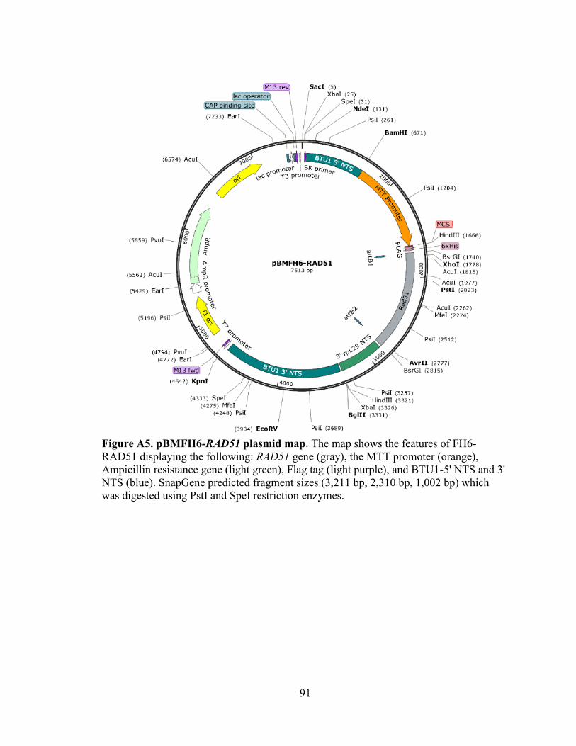
91
Figure A5. pBMFH6-RAD51 plasmid map. The map shows the features of FH6-
RAD51 displaying the following: RAD51 gene (gray), the MTT promoter (orange),
Ampicillin resistance gene (light green), Flag tag (light purple), and BTU1-5' NTS and 3'
NTS (blue). SnapGene predicted fragment sizes (3,211 bp, 2,310 bp, 1,002 bp) which
was digested using PstI and SpeI restriction enzymes.

92
Figure A6. pBMFH6-DMC1 plasmid map. The map shows the features of FH6-DMC1
displaying the following: DMC1 gene (gray), the MTT promoter (orange), Ampicillin
resistance gene (light green), Flag tag (light purple), and BTU1-5' NTS and 3' NTS
(blue). SnapGene predicted fragment sizes (3,211 bp, 2,575 bp, 2,080 bp) which was
digested using SpeI restriction enzymes.

93
Figure A7. pBMGFP-DMC1 plasmid map. The map shows the features of GFP-DMC1
displaying the following: restriction enzymes, DMC1 gene (gray), the MTT promoter
(orange), Ampicillin resistance gene (light green), GFP fluorescent tag (green), BTU1-5'
NTS and 3' NTS (blue), and attB1 and attB2 sites for recombination cloning (blue).
SnapGene predicted fragment sizes (7,436 bp, 901 bp, 178 bp) which was digested using
HindIII restriction enzymes.

94
Figure A8. pBMGFP-RAD51 plasmid map. The map shows the features of GFP-
RAD51 displaying the following: restriction enzymes, RAD51 gene (gray), the MTT
promoter (orange), Ampicillin resistance gene (light green), GFP fluorescent tag (green),
BTU1-5' NTS and 3' NTS (blue), and attB1 and attB2 sites for recombination cloning
(blue). SnapGene predicted fragment sizes (6,435 bp, 2,013 bp) which was digested using
PstI and BamHI restriction enzymes.

95
Figure A9. pBMRFP-RAD51 plasmid map. The map shows the features of RFP-
RAD51 displaying the following: restriction enzymes, RAD51 gene (gray), the MTT
promoter (orange), Ampicillin resistance gene (light green), RFP fluorescent tag (red),
BTU1-5' NTS and 3' NTS (blue), and attB1 and attB2 sites for recombination cloning
(blue). SnapGene predicted fragment sizes (6,440 bp, 2,034 bp) which was digested using
PstI and BamHI restriction enzymes.

96
Figure A10. pBMRFP-DMC1 plasmid map. The map shows the features of RFP-
DMC1 displaying the following: restriction enzymes, DMC1 gene (gray), the MTT
promoter (orange), Ampicillin resistance gene (light green), RFP fluorescent tag (red),
BTU1-5' NTS and 3' NTS (blue), and attB1 and attB2 sites for recombination cloning
(blue). SnapGene predicted fragment sizes (7,249 bp, 1,292 bp) which was digested using
SphI restriction enzyme.

97
Appendix B: T-COFFEE Alignment and T. thermophila Hop2-Mnd1 Proteins.
Protein sequences were obtained for T. thermophila Rad51, Dmc1, Hop2, and
Mnd1 using TGD website (www.ciliate.org). T. thermophila proteins were compared
with homologs of various other organisms. A TCOFFEE alignment was obtained to show
the best alignment, and if there are any conserved domains between different sequences.
The Walker A and Walker B motifs, found in Rad51 and Dmc1, are conserved motifs
among all species and RecA homolog shown in (Figure s B1 and B2)
Hop2 and Mnd1 protein sequences were obtained and two paralogs of each
protein were identified in T. thermophila (Table B1). The two T. thermophila Hop2
(HOP2 and HOPP2) and Mnd1 (MND1 and MNDP1) paralogs were chosen as the best
hit and had low E-values compared to their homologs in Saccharomyes cerevisiae (Table
B1). Microarray data for the two HOP2 paralogs show that HOP2 is absent of expression
except during conjugation whereas HOPP2 has some increased expression during
starvation as well as during conjugation and is expressed even during vegetative growth
unlike HOP2 (Figure B3). Microarray data for the two MND1 paralogs shows that
MND1 has a low expression with some expression during conjugation but is minimal
whereas MNDP1 has a higher expression even during vegetative growth and increases
during conjugation similar to RAD51 and DMC1 (Figure B3).

98

99

100

101
Figure B1. T-COFFEE Alignment of DMC1. The protein sequence of T. thermophila
and other species were entered into TCOFFEE alignment generator. Red shows the best
alignment; yellow shows regions with average similarities between the sequences, then
blue shows where there are no similarities between aligned sequences.

102

103

104

105
Figure B2. T-COFFEE Alignment of RAD51. The protein sequence of T. thermophila
Rad51 and other species were entered into TCOFFEE alignment generator. Red shows
the best alignment; yellow shows regions with average similarities between the
sequences, then blue shows where there are no similarities between aligned sequences.

106
Table B1. T. thermophila Hop2-Mnd1 homologs.
T. thermophila
Gene Name
Gene Identification
(TTHERM_#)
S. cerevisiae
BLAST E-value
Gene Description
HOP2
TTHERM_00794620
5e-05
HOP2 has a role in
chiasmata and meiotic
bivalent formation.
HOPP2 TTHERM_01190440 1e-10 Ubiquitously
expressed paralog of
the meiotically
expressed gene HOP2
(TTHERM_00794620
). Essential for
vegetative growth.
MND1 TTHERM_00300659
(Formerly
TTHERM_00300660
before annotation
correction)
8e-12 The Mnd1 protein
forms a complex with
Hop2 to promote
homologous
chromosome pairing
and meiotic DSB
repair. Mnd1 requires
Hop2 to localize to
chromosomes. It is a
meiotic version.
MNDP1 TTHERM_00382290
8e-14 The Mnd1 protein
forms a complex with
Hop2 to promote
homologous
chromosome pairing
and meiotic DSB
repair. Mnd1 requires
Hop2 to localize to
chromosomes. It’s a
ubiquitously
expressed version
copy