Embed Size (px)
Citation preview
doi:10.22336/rjo.2018.31
Romanian Journal of Ophthalmology, Volume 62, Issue 3, July-September 2018. pp:203-211
GENERAL ARTICLE
203 Romanian Society of Ophthalmology
© 2018
Investigation of importance of the structural parameters of the eyeball and of the technical parameters of cataract surgery on
corneal endothelial changes Nanu Razvan Vladimir*, Ungureanu Emil**, Istrate Sinziana Luminita**, Vrapciu Alexandra**, Cozubas Roxana***, Carstocea Laura**** , Voinea Liliana Mary**, Ciuluvica Radu***** *“Sf Ioan” Emergency Hospital, Bucharest, Romania **Ophthalmology Department, University Emergency Hospital; “Carol Davila” University of Medicine and Pharmacy, Bucharest, Romania ***”Grigore Alexandrescu” Emergency Hospital for Children, Bucharest, Romania **** PhD Student, “Carol Davila” University of Medicine and Pharmacy, Bucharest, Romania *****Anatomy Department, “Carol Davila” University of Medicine and Pharmacy, Bucharest, Romania
Correspondence to: Cozubas Roxana, MD, ”Grigore Alexandrescu” Emergency Hospital for Children, Bucharest, Dorobanti Policlinic, 58 Calea Dorobantilor, District 1, Code 010575, Bucharest, Romania, Mobile phone: +40727 727 006, E-mail: [email protected]
Accepted: August 19th, 2018
Abstract The aim of the here presented study was to look into the importance of the structural parameters of the eyeball, in relationship with the technical parameters of cataract surgery regarding the corneal endothelial changes made by it. Material and method: The paper refers at a prospective study in which we included eighty-eight consecutive eyes from eighty-six different patients having age-related cataract and a visual acuity of a values less than 0.8 considering best possible correction with it. The patients didn’t have other obvious causes for the decreased visual acuity. The patients included in the present study were admitted at the Ophthalmology Department from Bucharest University Emergency Hospital between the month of April 2015 and February 2016 (ten months). Results: When we compared lots A, B with C, in regarding to the decrease of EDC, the results were relatively very similar. We had only one comparison for which we obtained a statistical significance, and that was for cataracts classified as group IV of hardness; here, between the first and the third lot, at seven days postoperatively we obtain p = 0.0472812. Conclusions: The conclusion for the present research was that in regarding cataract phaco-emulsification surgery we obtained a statistical significance when it comes to the destruction of epithelial cells. The results were observed, giving the depth of the anterior chamber, in cataracts classified in subgroup IV of hardness, only between patients who had a small depth of chamber comparing with those who had a large depth of the anterior chamber of the eye. When it comes to patients who had severe cataracts and small ACD, we need to attract more attention when the surgeon performs the maneuver and to keep an eye on the use of the adhesives which has viscoelastic in order to obtain additional corneal protection. Keywords: cataract surgery, phacoemulsification, endothelial cell density, viscoelastic

Romanian Journal of Ophthalmology 2018; 62(3): 203-211
204
Romanian Society of Ophthalmology © 2018
Abbreviations ACD = anterior chamber depth; ECD = endothelial cell density, EPT = effective time of phacoemulsification.
The purpose of the present study
The purpose of the here presented study was to search the importance of anatomical, structural and constitutional parameters of the eyeball, when it comes to technical parameters in relationship with cataract surgery at the level of corneal endothelial variations and changes. This goal was brought into light by the fact that now there is no consensus in the present literature, on the need for differential approach of the cases, if we take into consideration and according to these parameters; in present, literature is insufficiently standardized and systematized [1-5].
Even today, cataract surgery remains the most accepted common surgical procedure [6] even in countries with advanced economies. Of course, in the last years, the procedure benefited from significant progresses, in regarding the technology but also in surgical technique, leading to superior outcomes, a rapid and increased postoperative recovery after the surgery, and a significant decrease concerning the complication rates.
Material and method
The here presented study was a prospective type that included 88 consecutive eyes from 86 different patients who had age-related cataracts and having less than 0.8 visual acuity with the best possible correction, without other obvious causes of decreased visual acuity. The patients were admitted to the Ophthalmology Department of Bucharest University Emergency Hospital between April 2015 and February 2016. Inclusion criteria for the patients admitted in this study:
patients who had over 18 years of age, to whom we have explained the need in regarding the intervention, if there were possible variants at the time and patients who were able to sign an informed consent at the time of enrolling;
patients who experienced age-related cataract, grades 2-4, with ACD above 1.5 mm;
patients with medically controlled open-angle primitive glaucoma;
intraocular pressure below 21 mm Hg without treatment;
in regarding of the distribution of patients with gender, we didn’t observe the proportion, we enrolled consecutive patients in the here presented study;
our patients were race type Caucasians, but this wasn’t a criteria that we followed for inclusion, but a regular distribution of the population in Romania.
Exclusion criteria
patients who had other types of cataract than the one observed and studied here - age-related cataract;
we had patients who had Fuchs corneal dystrophy, we took them out from here, but they were admitted in a sub-study to be presented later;
patients who had some sort of history of ocular inflammatory disease;
patients with ECD running below 1800 cells/ mm2;
when it comes to intraocular pressure, the patients who had it greater than 21 mmHg at the time of presentation were excluded from the present study;
patients with anterior-eye surgery; patients who turned out to be with
complicated cataract surgery – those whom we thought that it was necessary to implant capsular tension rings into the bag of the lens;
patients who didn’t follow the rules regarding the ulterior visits were also eliminated from the study.
Objectives of the present study
We had a main objective in regarding the study which was to spot a possible relationships between the structural and anatomical

Romanian Journal of Ophthalmology 2018; 62(3): 203-211
205
Romanian Society of Ophthalmology © 2018
parameters of the eyeball (ACD, crystalline hardness, crystalline lens, ECD prior to the surgery, pachymetry), technical parameters regarding the cataract surgery (ETP) and post-operative structural modification at various intervals of ECD respectively.
Secondary endpoints were to determine a relationship which can appear between the above-mentioned structural and anatomical parameters and the recovery of visual acuity in the postoperative period.
Initial examination
The initial examination was conducted using anamnesis, a wide general exam, a visual acuity measurement, examination of the anterior pole and the fundus, biometry, pachymetry, intraocular pressure determined by aplanotonometry, keratometry, a specular microscopy.
Visual acuity was taken with a Snellen optotype.
Aplanotonometry was performed in order to obtain intraocular pressure, with the Goldman aplanotonometer. Measurements were made using topical anesthesia with 4% oxy-buprocaine hydro-chloride. All intraocular pressure measurements were made by the same medical staff in order to ensure good reproducibility when it comes to different patients as well as when it comes to different measurements made for the same patient. The results were rounded, and we noted the nearest full intraocular pressure (we didn’t use fractional values).
Biometry was achieved with a biometer Ocu Scan (Alcon), by immersion, using topical anesthesia made with 4% oxy-buprocaine hydro-chloride. We took ten valid measurements for each of the patient.
Keratometry was made with the Topcon autorefractometer.
Pachymetry was performed with the Omac Scan (Alcon) – a biometric probe - it was also checked using a specular microscope Topcon. We took ten valid measurements for each patient and we monitored the central thickness of the cornea.
Specular microscopy was investigated with a specular microscope from Topcon. We took three central measurements for each of the patient. We marked the center of cells for 50 cells in the endothelial cornea in each case. In
this measurement, we monitored the ECD, the percentage of the cells and their distribution as a size. The endothelial cell loss was counted using the formula: (initial endothelial cells - postoperative endothelial cells)/ (initial endothelial cells x 100).
Surgical technique
Phacoemulsification was achieved in all cases in the hand of the same surgeon. Mydriasis was made by alternative administration of Tropicamide in concentration of 1% and also Phenylephrine 10%. Anesthesia was a topical one, made by administrating of 4% oxy-buprocaine hydro-chloride and a gel 4% tetracaine. We performed two corneal contra-incisions in the clear corneas at hours ten and two using a pre-calibrated diamond blade of 1.2. For each lot, half of the interventions were done by coaxial technique and half by bimanual technique.
For the coaxial technique, the main incision was in the clear cornea, triplanar, with diamond blade pre-incision, then biplanar with a 2.2 mm knife. Cohesive viscoelastic was injected into eyeball (the anterior chamber). A continuous circular capsulorhexis with a cystotome and a Duckwort & Kent capsulorhexis clip type Inamura was performed. The hydro-dissection and the hydro-delineation were performed using a 26 G cannula. The phacoemulsification was made using the technique named phaco chop. The technique of the irrigation/ aspiration was executed bimanually. After insertion of the pseudophakia into the bag, viscoelastic aspiration and wound hydro suture were performed.
In the bimanual technique, phacoemulsification was achieved using a phaco-tip without a sleeve and the irrigation chopper placed on the two 1.2 mm paracentesis. Capsulorhexis was made with a Kershner One-Pinch capsulorhexis clip. Otherwise, the technique of the surgery was similar. At the end of the surgery, we inserted viscoelastic into the bag and also into the eye (anterior chamber). We chose one of the contra-incisions in order to minimize the astigmatism and we widened it to 1.8 mm in order to introduce the foldable lens in the bag.
Postoperative treatment was conducted with drops of indometacin and a fixed

Romanian Journal of Ophthalmology 2018; 62(3): 203-211
206
Romanian Society of Ophthalmology © 2018
combination of betamethasone and chloramphenicol qid for 7 days, then three times per day for another 3 weeks. We called back the patients at one day, one week, 4 weeks, 3 months, in order to check parameters: pachymetry, ECD, visual acuity. Distribution on lots
In total, we observed 88 eyes with age-related cataract (the nuclear density was between 2 and 4); patients met the inclusion conditions in the study. Subsequently, two of the patients didn’t show up at the moment indicated for the postoperative controls; therefore, they were excluded from the present study.
We divided the patients into 3 lots depending on the ACD. Lot A presented patients with preoperative ACD between 1.5 mm and 2.3 mm. Lot B showed patients with the ACD between 2.3 mm and 3.5 mm. Lot C showed patients with a preoperative ACD of more than 3.5 mm.
Lot A was composed of patients with ACD between 1.5 mm and 2.3 mm. 19 patients were included here in this group (21.59% of the patients). Of these, 7 patients experienced grade II cataract (36.84%), 9 patients grade III (47.36%) and 3 of them had grade IV (15.78%).
Lot B was accomplished from patients with ACD between 2.3 mm and 3.5 mm. 51 patients (57.95%) were part of this group. Of these, 16 patients had grade II cataract (31.37%), 24 patients grade III (47.05%), and 11 patients grade IV (21.56%).
Lot C was made up of patients with an ACD of more than 3.5 mm. 18 patients (20.45%) were
included in this group. Of these, 7 patients experienced grade II cataract (44.44%), 9 patients grade III (47.36%) and two patients grade IV (10.52%).
Results
The table and figures below show the distribution of patients by age group and gender distribution.
There were 39 male patients (44.31%) and 49 female patients (55.68%). When it comes to age distribution, we observed that the patients were predominantly in the age group from 60 to 69 years of age (30 patients, 34.09%) and in the age group 70-79 years (36 patients, 40.90) and less in the 80-89 age groups (14 patients, 15.9%) and 90 years or over (8 patients, 9.09%) (Fig. 1).
Following controls, for lot A, the following
data was recorded - Table 1.
Table 1. Patients in group A Patients with * were operated by bimanual technique
Patient Sex Cataract grade ECD initial ECD at 7 days
ECD at 1 month
ECD at 3 months
EPT
B.I. * F II 2207 2184 2189 2192 2.6
A.M. M III 2307 2154 2148 2199 4.1
G.V. M III 2512 2488 2507 2520 3.3
Z.A.* F IV 1909 1756 1824 1819 7.3
R.O.* F III 2088 1980 2020 2036 5.4
A.P. F II 2166 2145 2157 2155 1.6
M.B.* M III 1987 1912 1923 1945 2.8
L.P. M II 2735 2722 2710 2715 1.8
Fig. 1 Distribution of patients by age and gender

Romanian Journal of Ophthalmology 2018; 62(3): 203-211
207
Romanian Society of Ophthalmology © 2018
M.A.* F III 2267 2146 2156 2175 4.2
U.R. M III 2187 2073 2162 2178 3.4
V.M.* F IV 2275 1966 2124 2155 5.9
M.N. F II 2307 2085 2172 2248 3.7
P.P. F III 2569 2443 2453 2449 3.7
R.O. M II 2005 1988 1945 2026 1.4
S.T.* F III 2400 2210 2315 2324 4.2
T.F. F II 2333 2245 2259 2281 2.3
R.B. M IV 2154 1987 2009 2076 3.3
C.B.* F II 2763 2667 2641 2689 1.5
V.C.* M III 2343 2189 2233 2251 2.7
Preoperative ECD analysis revealed an average of 2290.21 cel/ mm2 +/ - 112.26 (95% confidence) (Fig. 2).
At 7 days, the ECD distribution was
2175.7895 ± 165.9038 (99% confidence) - a 6.99% loss of ECD, at 1 month it was 2207.7368 ± 155.4301 (99% confidence) and at 3 months it was 2285.9474 ± 179.8151.
We measured the mean pachymetry preoperatively at the level of 544.26 ± 23.15 microns. At 7 days it was 561 ± 36.62 microns, at 1 month it was 551 ± 28.16 microns and at 3 months it was 548 ± 26.75 microns.
The mean time of phacoemulsification was 3.4316 ± 0.7491 (99% confidence). For cataracts
in the IV grade subgroup, the mean time was 5.5 ± 1.8392 (99% confidence). For those in the IIIrd subgroup, the mean time was 3.7556 ± 0.6431 (95% confidence), and for the subgroup II of hardness, the average time was 2.1286 ± 0.7593 (95% confidence). We weren’t able to find statistically significant differences between surgery performed by coaxial and bimanual techniques.
The results for lot B are detailed in Table 2.
Variation of ECD in group A
0
500
1000
1500
2000
2500
3000
Initial ECD ECD at 7 days ECD at 1 month ECD at 3 months ECD Examination
No
of
en
do
the
lia
l c
ell
s
B.I. A.M. G.V. Z.A. R.O. A.P. M.B. L.P. M.A. U.R. V.M. M.N. P.P. R.O. S.T. T.F. R.B. C.B. V.C.
Fig. 2 Variation of endothelial cells in group A
Romanian Journal of Ophthalmology 2018; 62(3): 203-211
208
Romanian Society of Ophthalmology © 2018
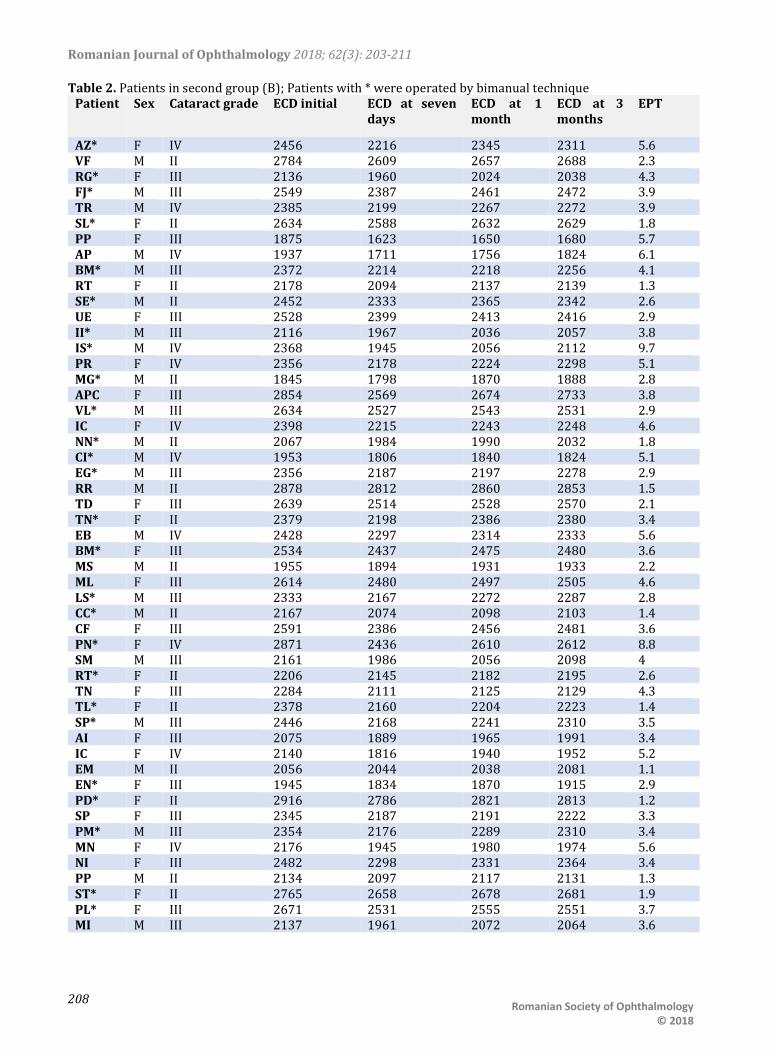
Table 2. Patients in second group (B); Patients with * were operated by bimanual technique Patient Sex Cataract grade ECD initial ECD at seven
days ECD at 1 month
ECD at 3 months
EPT
AZ* F IV 2456 2216 2345 2311 5.6 VF M II 2784 2609 2657 2688 2.3 RG* F III 2136 1960 2024 2038 4.3 FJ* M III 2549 2387 2461 2472 3.9 TR M IV 2385 2199 2267 2272 3.9 SL* F II 2634 2588 2632 2629 1.8 PP F III 1875 1623 1650 1680 5.7 AP M IV 1937 1711 1756 1824 6.1 BM* M III 2372 2214 2218 2256 4.1 RT F II 2178 2094 2137 2139 1.3 SE* M II 2452 2333 2365 2342 2.6 UE F III 2528 2399 2413 2416 2.9 II* M III 2116 1967 2036 2057 3.8 IS* M IV 2368 1945 2056 2112 9.7 PR F IV 2356 2178 2224 2298 5.1 MG* M II 1845 1798 1870 1888 2.8 APC F III 2854 2569 2674 2733 3.8 VL* M III 2634 2527 2543 2531 2.9 IC F IV 2398 2215 2243 2248 4.6 NN* M II 2067 1984 1990 2032 1.8 CI* M IV 1953 1806 1840 1824 5.1 EG* M III 2356 2187 2197 2278 2.9 RR M II 2878 2812 2860 2853 1.5 TD F III 2639 2514 2528 2570 2.1 TN* F II 2379 2198 2386 2380 3.4 EB M IV 2428 2297 2314 2333 5.6 BM* F III 2534 2437 2475 2480 3.6 MS M II 1955 1894 1931 1933 2.2 ML F III 2614 2480 2497 2505 4.6 LS* M III 2333 2167 2272 2287 2.8 CC* M II 2167 2074 2098 2103 1.4 CF F III 2591 2386 2456 2481 3.6 PN* F IV 2871 2436 2610 2612 8.8 SM M III 2161 1986 2056 2098 4 RT* F II 2206 2145 2182 2195 2.6 TN F III 2284 2111 2125 2129 4.3 TL* F II 2378 2160 2204 2223 1.4 SP* M III 2446 2168 2241 2310 3.5 AI F III 2075 1889 1965 1991 3.4 IC F IV 2140 1816 1940 1952 5.2 EM M II 2056 2044 2038 2081 1.1 EN* F III 1945 1834 1870 1915 2.9 PD* F II 2916 2786 2821 2813 1.2 SP F III 2345 2187 2191 2222 3.3 PM* M III 2354 2176 2289 2310 3.4 MN F IV 2176 1945 1980 1974 5.6 NI F III 2482 2298 2331 2364 3.4 PP M II 2134 2097 2117 2131 1.3 ST* F II 2765 2658 2678 2681 1.9 PL* F III 2671 2531 2555 2551 3.7 MI M III 2137 1961 2072 2064 3.6

Romanian Journal of Ophthalmology 2018; 62(3): 203-211
209
Romanian Society of Ophthalmology © 2018
At 7 days, the ECD distribution was 2196 ± 104.3111 (99% confidence) - a 6.87% loss of ECD, at 1 month it was 2248.6275 ± 104.1418
(99% confidence) and at 3 months it was 2266.8431 ± 102.0706 (99%) – Table 3.
Table 3. ECD distribution at 7 days versus preoperative Data Summary A B Total n 51 51 102
Σx 120293 111996 232289
Σx2 287618165 249806276 537424441
SS 3884716.980 3863060 8422680.990
mean 2358.6863 2196 2277.3431
Results Meana-Meanb t df P One-tailed <.0001
162.6863 +13.66 50 Two-tailed <.0001
Observed Confidence Intervals
0.95 0.99
Meana 2358.6863 ± 78.4523
± 104.6031
Meanb 2196 ± 78.2333
± 104.3111
Meana-Meanb
[Assuming equal sample variances]
162.6863 ± 23.9363
± 31.9191
Meana-Meanb
[Assuming unequal sample variances
--- ± ---
± ---
Correlated Samples
The mean pachymetry was preoperatively at 551.26 ± 28.75 microns. At 7 days it was 560.18 ± 34.26 microns, at 1 month it was 553.31 ± 29.38 microns and at 3 months it was 552.45 ± 28.89 microns.
The mean time of phacoemulsification was 3.5765 ± 0.6609 (99% confidence). For cataracts in the IVth subgroup, the mean time was 6.0556 ± 2.1625 (99% confidence). For those in the IIIrd
subgroup, the mean time was 3.7556 ± 0.6431 (95% confidence), and for the subgroup of hardness II the average time was 2.1286 ± 0.7593 (95% confidence). We weren’t able to find a statistically significant difference between the technique of surgery with coaxial and bimanual.
The results that we obtain in lot C are here presented in Table 4.
Table 4. Patients in lot C Patients with * were operated by bimanual technique
patient sex Cataract grade
ECD initial ECD at 7 days
ECD at 1 month
ECD at 3 months
EPT
RG F III 2356 2116 2245 2311 5.6 PP* M II 2784 2711 2723 2756 1.3 RG* F III 2136 2086 2099 2118 3.3 LO M III 2349 2245 2276 2318 3.7 TR* M IV 2358 2199 2267 2298 4.9 LS* F II 2653 2588 2632 2629 1.8 PP F III 1975 1788 1845 1877 4.7 CA M II 1937 1888 1823 1903 2.1 MN* M III 2132 2014 2086 2145 4.1 TR F II 2095 2066 2088 2084 1.3

Romanian Journal of Ophthalmology 2018; 62(3): 203-211
210
Romanian Society of Ophthalmology © 2018
EU M II 2452 2333 2365 2342 2.6 BD* F III 2258 2119 2183 2193 2.9 MI* M III 2116 1967 2036 2057 3.8 GH M IV 2368 1945 2056 2112 7.7 CS* F II 2356 2178 2224 2298 5.1 CF* M II 1845 1798 1870 1888 2.8 MG F III 2854 2569 2674 2733 3.8 PI M III 2634 2527 2543 2531 2.9
At 7 days, the ECD distribution was
2174.2778 ± 188.3079 (99% confidence) - a 6.05% loss of EDC, at 1 month 2224.1667 ± 188.4944 (99% confidence) and at 3 months
2255.1667 ± 142.0706 (99%) as we presented it in Table 5.
Table 5. ECD distribution at 7 days versus preoperative
Data Summary A B Total n 18 18 36
Σx 41658 39137 80795
Σx2 97801086 86384925 184186011
SS 1390588 1290215.61 2857343.63
mean 2314.3333 2174.2778 2244.3056
Results Meana-Meanb t df P One-tailed <.0001
140.0556 +6.01 17 Two-tailed <.0001
Observed Confidence Intervals
0.95 0.99
Meana 2314.3333 ± 142.2398
± 195.4954
Meanb 2174.2778 ± 137.0102
± 188.3079
Meana-Meanb
[Assuming equal sample variances]
140.0556 ± 49.1424
± 67.5416
Meana-Meanb
[Assuming unequal sample variances
--- ± ---
± ---
Correlated Samples The mean pachymetry was preoperatively
at 551.26 ± 28.75 microns. At 7 days, it was 560.18 ± 34.26 microns, at 1 month it was 553.31 ± 29.38 microns and at 3 months it was 552.45 ± 28.89 microns.
The mean time of phacoemulsification was 3.5778 ± 1.113 (99% confidence). For cataracts in the IVth subgroup, the mean time was 6.3 ± 17.794 (99% confidence). For those in the subgroup III, the average time was 3.8667 ± 0.6657 (95% confidence), and for the subgroup II of hardness, the average time was 2.4286 ± 1.2164 (95% confidence). There were no statistically significant differences between
patients operated by coaxial and bimanual techniques.
Lot comparison
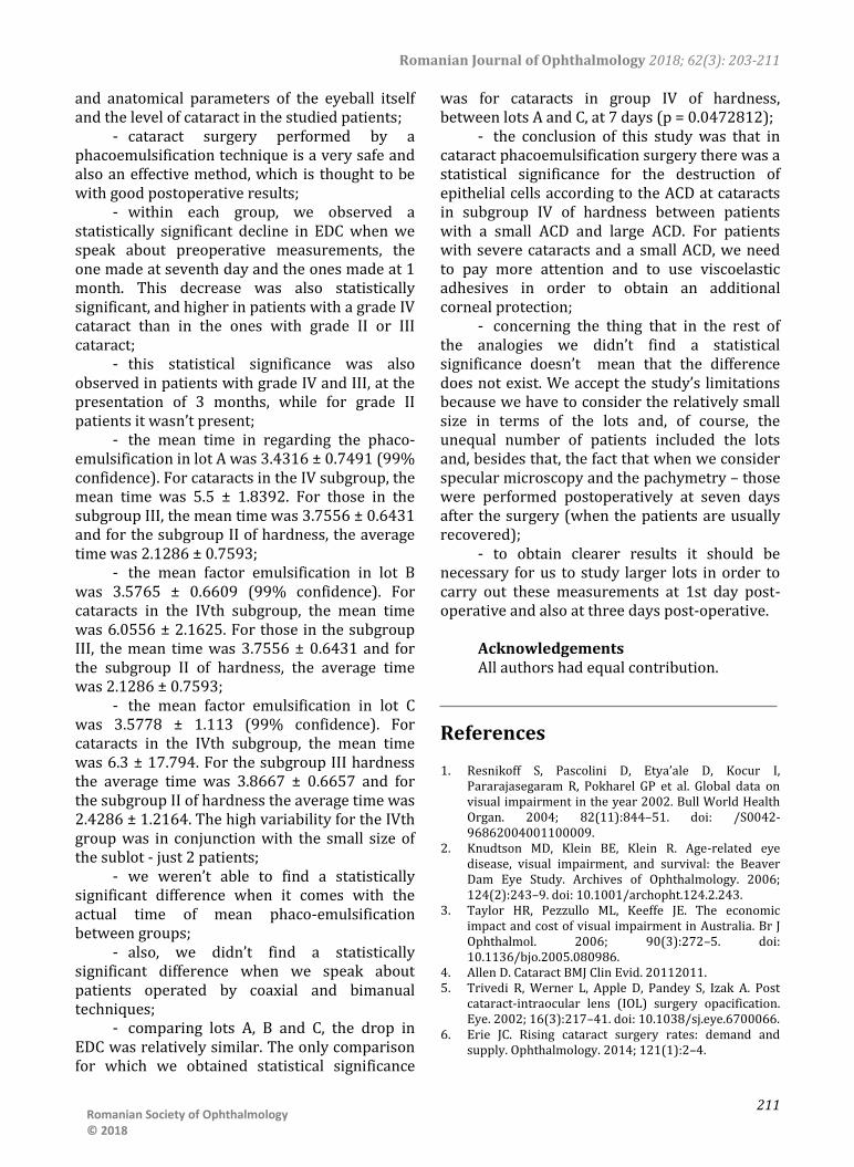
By comparing lots A, B and C, the drop in EDC was relatively similar. We obtain a single statistical significance for a comparison regarding cataracts in the fourth lot of hardness, between lots A and C, at seven days after the presentation (p = 0.0472812).
Conclusions
We observed a very big distributional variability when we speak about the structural
Romanian Journal of Ophthalmology 2018; 62(3): 203-211
211
Romanian Society of Ophthalmology © 2018
and anatomical parameters of the eyeball itself and the level of cataract in the studied patients;
- cataract surgery performed by a phacoemulsification technique is a very safe and also an effective method, which is thought to be with good postoperative results;
- within each group, we observed a statistically significant decline in EDC when we speak about preoperative measurements, the one made at seventh day and the ones made at 1 month. This decrease was also statistically significant, and higher in patients with a grade IV cataract than in the ones with grade II or III cataract;
- this statistical significance was also observed in patients with grade IV and III, at the presentation of 3 months, while for grade II patients it wasn’t present;
- the mean time in regarding the phaco-emulsification in lot A was 3.4316 ± 0.7491 (99% confidence). For cataracts in the IV subgroup, the mean time was 5.5 ± 1.8392. For those in the subgroup III, the mean time was 3.7556 ± 0.6431 and for the subgroup II of hardness, the average time was 2.1286 ± 0.7593;
- the mean factor emulsification in lot B was 3.5765 ± 0.6609 (99% confidence). For cataracts in the IVth subgroup, the mean time was 6.0556 ± 2.1625. For those in the subgroup III, the mean time was 3.7556 ± 0.6431 and for the subgroup II of hardness, the average time was 2.1286 ± 0.7593;
- the mean factor emulsification in lot C was 3.5778 ± 1.113 (99% confidence). For cataracts in the IVth subgroup, the mean time was 6.3 ± 17.794. For the subgroup III hardness the average time was 3.8667 ± 0.6657 and for the subgroup II of hardness the average time was 2.4286 ± 1.2164. The high variability for the IVth group was in conjunction with the small size of the sublot - just 2 patients;
- we weren’t able to find a statistically significant difference when it comes with the actual time of mean phaco-emulsification between groups;
- also, we didn’t find a statistically significant difference when we speak about patients operated by coaxial and bimanual techniques;
- comparing lots A, B and C, the drop in EDC was relatively similar. The only comparison for which we obtained statistical significance
was for cataracts in group IV of hardness, between lots A and C, at 7 days (p = 0.0472812);
- the conclusion of this study was that in cataract phacoemulsification surgery there was a statistical significance for the destruction of epithelial cells according to the ACD at cataracts in subgroup IV of hardness between patients with a small ACD and large ACD. For patients with severe cataracts and a small ACD, we need to pay more attention and to use viscoelastic adhesives in order to obtain an additional corneal protection;
- concerning the thing that in the rest of the analogies we didn’t find a statistical significance doesn’t mean that the difference does not exist. We accept the study’s limitations because we have to consider the relatively small size in terms of the lots and, of course, the unequal number of patients included the lots and, besides that, the fact that when we consider specular microscopy and the pachymetry – those were performed postoperatively at seven days after the surgery (when the patients are usually recovered);
- to obtain clearer results it should be necessary for us to study larger lots in order to carry out these measurements at 1st day post-operative and also at three days post-operative.
Acknowledgements All authors had equal contribution.
References
1. Resnikoff S, Pascolini D, Etya’ale D, Kocur I, Pararajasegaram R, Pokharel GP et al. Global data on visual impairment in the year 2002. Bull World Health Organ. 2004; 82(11):844–51. doi: /S0042-96862004001100009.
2. Knudtson MD, Klein BE, Klein R. Age-related eye disease, visual impairment, and survival: the Beaver Dam Eye Study. Archives of Ophthalmology. 2006; 124(2):243–9. doi: 10.1001/archopht.124.2.243.
3. Taylor HR, Pezzullo ML, Keeffe JE. The economic impact and cost of visual impairment in Australia. Br J Ophthalmol. 2006; 90(3):272–5. doi: 10.1136/bjo.2005.080986.
4. Allen D. Cataract BMJ Clin Evid. 20112011. 5. Trivedi R, Werner L, Apple D, Pandey S, Izak A. Post
cataract-intraocular lens (IOL) surgery opacification. Eye. 2002; 16(3):217–41. doi: 10.1038/sj.eye.6700066.
6. Erie JC. Rising cataract surgery rates: demand and supply. Ophthalmology. 2014; 121(1):2–4.