Embed Size (px)
Citation preview
Application Example
Experimental conditionsThe ceramic substrates were prepared by uniaxial pressing and electrophoretic deposition. The ceramics were sintered under various conditions, which resulted in different grain size in sintered substrates. The surface of the ceramic sub-strates was grinded and polished.The osteoblastic cell cultures MG63 were cultivated in-vitro on the zirconia and alumina surfaces. After the cultivation, the adhered cells were fixed with 3% glutaraldehyde in 0,1 M phosphate buffer for one hour. The cells were rinsed three times with 0.1 M phosphate buffer, dehydrated through a graded ethanol series (50%, 60%, 70%, 80%, 90%, 96%, 100%) and critical point dried with liquid CO2.
In the field of current implantology, the conventional usage of titanium alloys is being replaced by ceramic materials. Bioceramics are made by sintering of the ceramic powders (e. g. zirconia or alumina powders) and they are characterized by excellent hardness and tribological properties. Zirconia ceramics are becoming prevalent among biomaterials used in dental implantology. Biocompatible materials such as zirconium oxide (ZrO2), and aluminium oxide (Al2O3) enable the production and growth of healthy tissues. The usage of bioceramics in dental implantology supplements standard titanium implants thanks to their higher tastefulness, “one piece” implantation and suitability for patients allergic to thetitanium. SEM is an important tool for investigation of biocompatibility of selected materials. The aim of this study was to investigate osteoblastic spreading in contact with various oxide ceramics. The spreading of the osteoblastic cells MG63 on the zirconia and alumina surfaces was observed using a MIRA3 FEG SEM in the low vacuum mode in order to evaluate the biocompatibility of these ceramic materials.
Investigation of cell spreading on bioceramic materials
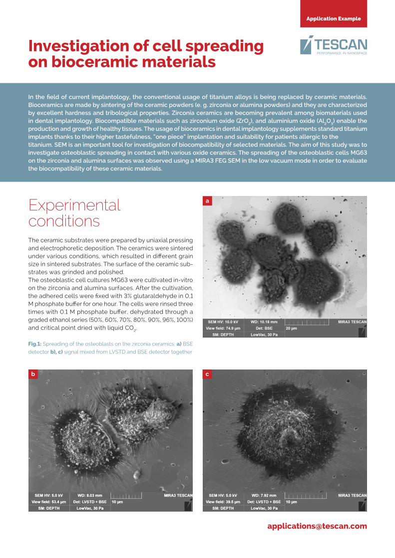
Fig.1: Spreading of the osteoblasts on the zirconia ceramics: a) BSE
detector b), c) signal mixed from LVSTD and BSE detector together
a
b c

TESCAN ORSAY HOLDING, a.s.
Libušina tř. 21, 623 00 Brno - Kohoutovice / Czech Republic
(phone) +420 530 353 411 / (email) [email protected] / [email protected] www.tescan.com
Application Example Investigation of cell spreading on bioceramic materials
ConclusionThe results of these experiments showed that the bio-logical activity depends on the grain size and chemical composition of the substrates. The ZrO2 ceramics indicated better bioactivity than Al2O3. An overview of alumina ceramics shows that os-teo-blastic cells do not grow in a large extent and spread less than the cells mentioned above. Zirconium oxide (ZrO2) does not show any cytotoxic effect, it provides a good interaction with osteoblasts, and makes the cells capable of elaborating the extracellular matrix. All these facts are evident from this study and confirms the suitability of the zirconia ceramics in the field of implantology.
A high resolution field emission scanning electron microscope MIRA3 FEG SEM was used to study the growth of osteoblasts on the ceramic surface. The samples were observed without any metal coating in the UniVac mode. The chamber pressure was set to 30 Pa in order to reduce charging artifacts. The im-ages were obtained using a combination of a LVSTD detector (Low Vacuum Secondary TESCAN Detector; Everhart-Thorn-ley type) and a BSE detectors. The images obtained by the BSE detector enhanced the material structure of the ceramic surface. (figs. 1a, 2b, 2c). The LVSTD detector is a detector of secondary electrons designed for a low vacuum mode, thus visualizes the topograhic features. By mixing the signals from the LVSTD and BSE detectors using a SEM detector & mixer, we were able to obtain the desired information. (figs. 1b, 1c, 2a). Due to the high topograpghy dimensions of the cells, a dedicated DEPTH mode was used to completely visualize all details with high depth of focus (figs. 1a, 1b, 1c, 2a).
� References[1] Matoušek, A., Kukletová, M., Cihlář, J. Influence of Nanograin Size
ZrO2 and Al2O3 Ceramics on Biological Re-sponse of Cells. Key Engi-
neering Materials [online]. 2013, roč. 25, vol. 587, s. 132-137.
[2] Hisbergues, M., at al: Established Facts and Perspectives for a
Biomaterial in Dental Implantology. Journal of Biomedical Material
Research Part B: Applied Biomaterials, 2008, vol. 88B, pp. 519 – 529.
� AcknowledgementThe samples were provided by Aleš Matoušek, Ph.D., a Junior Research-
er of Advanced Ceramic Materials at the CEITEC - Central European
Institute of Technology, Brno University of Technology, the Czech Re-
public. The work was supported by the project CZ.1.05/1.1.00/02.0068
financed from European Regional Development Fund.
a
b
c
Fig 2: Spreading of the osteoblasts on Al2O3 surface; a) signal mixed from
LVSTD and BSE detectors together, b), c) signal from a BSE detector
TE
SC
AN
re
serv
es
the
rig
ht to
cha
nge
the
do
cum
ent
with
ou
t no
tice
. 20
15.10
.9