Embed Size (px)
Citation preview

Aus der Augenklinik des Universitätsklinikums Hamburg-Eppendorf
Direktor: Prof. Dr. med G. Richard
Investigation about T lymphocyte activation by human
corneal cell types
Dissertation
Zur Erlangung des Grades eines Doktors der Medizin dem Fachbereich Medizin der
Universität Hamburg vorgelegt von
Fan Wang
Aus Changchun, Jilin, P. R. China
Hamburg 2003
Angenommen vom Fachbereich Medizin
der Universität Hamburg am: 2. 12. 2003
Veröffentlicht mit Genehmigung des Fachbereichs
Medizin der Universität Hamburg
Prüfungsausschuss, der/die Vorsitzende: Prof. Dr. K. Engelmann
Prüfungsausschuss: 2. Gutachter/in: Prof. Dr. D. V. Domarus
Prüfungsausschuss: 3. Gutachter/in: Prof. Dr. F. Nolte

Contents
Abbreviations……………………………………………………………………...... 1
1. Aim of the study…………………………………………………………………...... 2
2. Introduction……………………………………………………………………......... 3
2.1 The human eye and the cornea………………………………………………........... 3
2.2 Corneal disease and corneal transplantation……………………………..……......... 4
2.3 Complications after transplantation- graft rejection…………………………........... 8
2.4 Components of the immune system related to graft rejection…………………….... 9
2.4.1 Role of CTL in corneal graft rejection………………………………….........10
2.4.2 Role of CD4+ immune mechanisms of corneal graft rejection……………….11
2.4.3 T cell allorecognition………………………………………………….……...13
2.4.4 Mechanism of T cells activation……………………………………………...14
2.4.5 Regulation of HLA class II and CD40 expression by ?- interferon………......17
2.5 Effector mechanisms in allograft rejection…………………………………………18
3. Material and Methods…………………………………………………………...... 20
3.1 Cell culture……………………………………………………………………….... 20
3.1.1 Isolation of Peripheral Blood Mononuclear Cells (PBMC)……………….... 20
3.1.2 Human Corneal Epithelial cells …………………………………………….. 20
3.1.3 Primary human corneal endothelial cells………………………….……........ 21
3.1.4 SV40 transfected human corneal endothelial cells………….………….........21
3.1.5 Human retinal pigment epithelial cell isolation and cultivation…………….. 22
3.2 Preparation of chamber slides……………………………………………………... 23
3.3 FACS Scan Analysis……………………………………………………………..... 23 3.3.1 Cell Preparation for FACS……………….………………………………….. 23 3.3.2 FACS analysis procedure………………………….………………………....24
3.4 Immunohistochemical staining procedure……………………………………….....25
3.4.1 Corneal samples…………..………………………………………………..... 25
3.4.2 Cultured cells ……………………………..……………………………….... 27
3.4.3 Immunohistochemical staining procedure………..………………………......27
4. Results……………………………………………………………………………….28
4.1 Human corneal and RPE cells ……………………………………………………..28
4.1.1 Expression of HLA-DP, DQ, DR………………………...……..….…….......28

4.1.2 Expression of CD40 ……………………………..………………………...... 31
4.2 Cocultured cells…………………………………………….…………………....... 33
4.2.1 FACS analysis…………………………..…………………………………... 33
4.2.2 HLA-DP, DQ, DR expression. ………………………………..…………......39
4.2.3 CD80, 86, 154 expression………...……………………………………....... ..42
4.2.4 CD80, 86, 154 expression on corneal epithelial cells…………………....... ...47
4.2.5 Corneal transplantation rejection section staining…………………...…….... 51
5. Discussion…………………………………………………………………………...57
5.1 Expression of HLA-DP, DQ, DR and T cell activation …………………………...58
5.1.1 Human corneal epithelial cells……………………..……………………....... 58
5.1.2 Human corneal endothelial cells (HCEC)……………………………….…...60
5.1.3 Immortalized human corneal endothelial cells…………………………….... 60
5.1.4 Human retinal pigment epithelial cells (RPE)………………………………. 61
5.1.5 Transplantation rejection cornea section……………………………………..62
5.2 CD40/CD154 pathway……………………………………………………………. .63
5.3 CD28 and CD80/CD86 costimulatory signal …………………………………….. 65
5.4 T lymphocytes…………………………………………………………………….. 66
6. Summary………………………………………………………………………....... 68
Zusammenfassung……...…………………………….………………………….... 69
7. Reference……….……………………………………………………..………….... 71
Ackowledgements……...…………………………….………………………......... 84
Curriculum Vitae…………………….……………………………………………. 85
Lebenslauf ..……………………………………………………………………….. 86
Eidesstattliche Versicherung.....…………………………………………………...87

Abbreviations
1
Abbreviations
APC antigen presenting cell
CD4+ T cells helper T cells
CD8+ T cells cytotoxic T cells
CTL cytotoxic T-lymphocyte
DTH delayed-type hypersensitivity
EDTA ethylenediamine tetraacetic acid
FACS fluorescence activated cell sorter
FCS fetal calf serum
HCEC human corneal endothelial cells
HLA human leucocyte antigen
IL-2 interleukin 2
KO knock out
PBMCs peripheral blood mononuclear cells
PBS phosphate buffered saline
PHA phytahematoagglutinin
SV40 Simian Virus 40
SV40 transfected HCEC Simian Virus 40 human corneal endothelial cells
MHC major histocompatibility complex
MST median survival time
?-IFN ? interferon
RPE retinal pigment epithelial
Th T helper
TCR T cell receptor

Aim of the study
2
1. Aim of the study
Keratoplasty is a surgical procedure in which part or all of the cornea is replaced by
healthy corneal tissue from a donor. In spite of keratoplasty is the most successful
transplantation, cornea graft rejection is still the most frequent complication after
corneal grafting and often leads to irreversible transplant failure. It has been concluded
that corneal graft rejection, like other forms of organ transplantation, is a T cell
mediated immune process. The development of an effective immune response required
T cell activation by a first antigen as well as by a second costimulatory signal. The first
signal is delivered when the T cell receptor (TCR) binds to the allogeneic peptide/HLA
complex of the antigen presenting cells (APC), and the CD4 (in helper T cells) or CD8
(in cytotoxic T cells) co-receptors bind to a constant part of HLA (MHC) class-II or
HLA (MHC) class-I molecules respectively. CD28-B7 (CD80/CD86) interactions
provides possible “second signals” which are necessary for optimal T cell activation and
IL-2 production. Other T cell surface molecules, such as CD40 ligand (CD154) also
contribute to signal 2. Following signals 1 and 2, the T cell is fully activated so that the
genes encoding lymphokines and lymphokine receptors are transcribed and translated.
In the Cornea Bank of the University Eye Hospital Hamburg-Eppendorf, methods
for isolation and cultivation of human corneal epithelial cells and endothelial cells are
established. These cultured cells should be examined for HLA expression. Additionally
a method for isolation and cultivation of peripheral blood cells has to be established to
investigate, whether co-culture of corneal cells and peripheral blood cells will lead to T
cell activation. Furthermore, the expression of molecules with the potential to serve as
second stimulator for T cell activation should be analyzed in the co-cultured cell
system.
From the histology department, slides from corneas of patients who had cornea
graft failures are available. This material offers the possibility to prove the relevance of
the results obtained by the cell culture experiments. Presence of proteins involved in the
T cell activation process in vitro will be analyzed by immunohistochemical staining of
the corneas.

Introduction
3
2. Introduction
2.1 The human eye and the cornea
The famous aphorism “the eye is the window of spirit”, not only emphasizes the
importance of the eye, but also render prominent the presence of a window-pane, the
cornea. The cornea, the conjuctiva, and the intervening transition area, known as the
limbus, comprise the tissues of the ocular surface. The cornea is a tissue highly
specialized to refract and transmit light. From histology aspect, the cornea consist of
epithelium, stroma and endothelium. Although this avascular tissue seems simple in
composition, it is extraordinarily regular and precisely arranged. All three layers have
an uniform and consistent arrangement throughout the tissue in order to precisely bend
and transmit light through to the lens, thence to the retina. (Fig. 2.1). The stroma is the
middle connective tissue layer that is approximately 500 µm thick and comprises about
90 percent of the cornea. It is arranged in three clearly defined layers of extracellular
matrix. These include, bordering the epithelium, the thin 8- to 10-µm Bowman´s layer;
the middle lamellar stroma, and adjacent to the endothelium, the 8- to 12-µm
Descemet´s membrane, the thickened basement membrane secreted by the corneal
endothelium. The corneal endothelium is a single layered, low cuboidal endothelium. It
plays a major role in maintaining stroma hydration through Na-K activated adenosine
triphosphatase (ATPase) present in the basalateral membranes of the cells. Unlike the
epithelium, the human endothelium is not a self-renewing cell layer. The number of
endothelial cells decreases with age. As cells decrease in number, they become thinner
and attenuated (Fig. 2.1) (Smolin and Thoft 1994).
Fig. 2.1 Section through the central cornea. a=epithelium, b=Bowman´s layer, c=lamellar stroma, d=Descemet´s membrane, e=endothelium. [Smolin and Thoft (1994) The cornea-Scientific Foundations and Clinical Practice. Boston]

Introduction
4
2.2 Corneal disease and corneal transplantation
There are many causes preventing normal morphology and function of the cornea,
such as clouding of the cornea and abnormal corneal contour. For example: eye injuries
that leave a dense white scar on the cornea, these injuries may include penetrating
wounds from a sharp object, burns, or chemical contamination of the eye. Cataract or
other eye surgeries can prompt corneal clouding. Corneal infection can also lead to
scarring, the infection may be bacterial, viral, or fungal in origin. Various herpes viruses
are known to cause such scarring. Keratoconus is the most typical disease for abnormal
morphology. It may scar the center of the cornea or distort vision so severely that
glasses or contact lenses are of little help. Corneal dystrophies may cause clouding, and
some inherited diseases of the cornea results in abnormal function of the cornea. To
solve these problems and retrieve a healthy cornea, the unique technology is corneal
transplantation (keratoplasty). The history of corneal transplantation reaches back over 150 years. The first
documented report of a successful penetrating keratoplasty in a human subject was
performed by Zirm in 1906. As we enter the millennium, corneal transplantation
remains the oldest, most common, and arguably, the most successful form of tissue
transplantation (Niederkorn 2001). Neither HLA typing nor systemic
immunosuppression (except in the case of high-risk individuals who have either
received a previous corneal transplant or who have prevascularized graft beds) is
performed routinely, it is remarkable that typical 2-year survival rates for initial grafts
onto avascular graft beds are in excess of 90% (The Collaborative Corneal
Transplantation Studies Research Group 1992). Keratoplasty is a surgical procedure in
which part or all of the cornea is replaced by healthy corneal tissue from a donor. It can
also be said it is a surgery to replace the clear window on the front of the eye (the
cornea). Corneal transplant procedures may restore vision to otherwise blind eyes in
some cases. It is divided into two forms: lamellar and penetrating keratoplasty. Lamellar
keratoplasty: it is defined as removal and replacement of less than the total thickness of
the cornea. As a rule, lamellar grafts tend to be relatively large (> 8 mm in diameter),
and they replace tissue removed by deep stroma dissection. The host Descemet´s
membrane and the endothelium are left intact and serve as the base onto which the
donor tissue is laid. It is suitable for those corneal conditions in which the pathologic

Introduction
5
changes are limited to anterior stromal and surface irregularities but in which the
endothelium is healthy (Smolin and Thoft 1994). Most of the keratoplasty were
mentioned to be penetrating keratoplasty. Tissue strengthening is the primary goal
(tectonic) and increased visual acuity is the immediate aim. In the western world, the
most frequent indication is keratoconus (followed eye Fuch´s endothelial dystrophies or
secondary endothelial decompensation in the beginning of the 80th). Pseudophakic
bullous keratopathy may account for about 17% of all corneal transplant procedures in
the beginning of the south. Less frequent indications include corneal ulceration, corneal
scars, herpes simplex and Varicella zoster viral infections leading to scarring, or
congenital opacities of the cornea (Table 2.1).

Introduction
6
author/country number of patients
time 1. priority 2. priority 3. priority
Dandona/India 1997
1964 scarring 28,1 %
re-keratoplasty 17,1 %
ulcerating keratitis 12,2 %
Sharif KW/England 1993
3555 1971–1990 re-keratoplasty 40,8 %
keratoconus 17 %
herpetic keratitis 5 %
Frucht-Pery J/Israel 1997
1018 1961–1990 Keratoconus 21,8 %
re-keratoplasty 11,1 %
herpetic keratitis 9,3 %
Flowers CW, California/USA
1104 1989–1993 bullous keratopathy 24,8 %
re-keratoplasty 21,3 %
scarring / herpetic keratitis 11,1 %
Haamann, P/Denmark 1994
180 1984– 993 bullous keratopathy 28,3 %
keratitis 13,9 % Fuchs dystrophy 13,9 %
De Cock R/Israel, 1994
416 198 –1992 keratoconus keratitis/scarring herpetic keratitis
Legeais, JM/Paris, France 2001
3736 1980–1999 keratoconus 28,8 %
herpetic keratitis 10,9 %
re-keratoplasty 9,9 %
Australian Corneal Graft Registry, 1993
3608 1985–1991 keratoconus 31 %
bullous keratopathy 31 %
re-keratoplasty 14 %
Dobbins KR, Middle West USA, 2000
4217 1982–1996 bullous keratopathy 31,5 %
Fuchs dystrophy 23,2 %
Keratoconus 11,4 %
Chen WL, Taiwan, 2001
770 1987–1999 scarring 27,9 % re-keratoplasty 21,0 %
ulcerating keratitis 17,9 %
Hovding G, Norway, 1999
keratoconus
Maeno A, Canada, 2000
6222 1964–1997 re-keratoplasty keratoconus bullous keratopathy
Graupner, M, Erlangen, Germany, 2000
207 1997–1999 keratoconus 44,9 %
Fuchs dystrophy 25,1 %
bullous keratopathy 16,9 %
Cursiefen C, Erlangen, Germany, 1998
2557 1992–1996 keratoconus 20,9 %
scarring/keratitis 20,4 %
bullous keratopathy 17 %
Lois N, Pennsylvania, USA, 1997
2186 1989–1995 bullous keratopathy 26,0 %
re-keratoplasty 17,8 %
Fuchs dystrophy 15,7 %
Chan CM, Singapore, 1997
327 1991–1995 bullous keratopathy 26,3 %
re-keratoplasty 11,9 %
corneal dystrophy 10,4 %
Table 2.1 Prevalence of diagnoses leading to perforating keratoplasty in dependence upon region and time. [Engelmann K, Fell A, Fankhauser II F (2002) Indikationsstellung zur Keratoplastik bei Keratokonus. Z Prakt Augenheilkd]

Introduction
7
A
B
C
Fig 2.2 Cornea ulceration, herpes simplex, and damage or scar from disease or trauma lead to cornea opaque (A). Excision of recipient tissue: Pupils are usually miosed pre-op to avoid injuring the lens and causing cataract. The recipient cornea is then removed using a corneal trephine, Trephination is done with either the manual, motorized, or vacuum trephine. The donor button is ideally 0.5 mm larger than the planned recipient opening (In case if the donor button is trephined from the endothelial side). No over size, if both corneas are rephined from the epithelial side (B). The donor cornea is gently sewn into recipient bed, four cardinal interrupted sutures are applied at 12, 3, 6, 9 o´clock respectively. Interrupted or running sutures are then performed for final fixation (C). Taken from (www.eyemdlink.com/EyeProcedure.asp?EyeProcedureID=65).

Introduction
8
2.3 Complications after transplantation- graft rejection
In spite of keratoplasty is the most successful transplantation, cornea graft rejection
is still the most frequent complication after corneal grafting and often leads to
irreversible transplant failure. It is defined as a specific process in which a graft, having
been clear for at least several weeks (usually longer), suddenly succumbs to graft edema
in conjunction with inflammatory signs. This process is immunologically mediated,
which represents the end stage of the immunologic reaction that is no longer reversible.
Many inferences about the immunobiology of corneal graft rejection have been based
on clinical observations; however, confirmation of these hypotheses requires
prospective studies under controlled settings. The prudent use of animal models has
fostered analytic studies on the immunobiology of corneal allografts without the
complicating and confounding effects of topical steroids that are typically used on most
keratoplasty patients. Although animal models of penetrating keratoplasty have been in
use for almost a half-century, until recently, progress in understanding the immune
mechanisms of corneal graft rejection has been slow. Nevertheless, the widespread use
of rodent models of orthotopic corneal transplantation has shed new light on the
pathogenesis of corneal graft rejection. These findings indicate that prevention and
therapy of allograft rejection is still the most challenging field of today´s keratoplasty. It
is well known that T cells are critical to graft rejection. An immunohistochemical study
by Williams et al. (1989), has shown that half the leucocytes detectable in corneal grafts
are T lymphocytes, two-thirds bore MHC class II markers and one-fifth carried myeloid
cell markers. Rejection of organ and tissue allografts occurs because the mammalian genome
contains several polymorphic loci that encode widely expressed tissue antigens. Persons
who do not express a given allele at any of these loci recognize the protein encoded by
that allele as foreign and mount a vigorous immune response that leads to graft
rejection. The most important genes are clustered within the major histocompatibility
complex (MHC), which in humans is known as the HLA complex. For many reasons, it
is likely that the human leukocyte antigen (HLA) alloantigens are involved in corneal
graft rejection. They are known to be the main targets for the immunological reactions
leading to rejection of transplanted solid organs. There are two main classes of HLA
molecule: HLA class I and class II. The function of the classical HLA class-I molecules,

Introduction
9
which include HLA-A, B, C, is to present peptides to CD8+ T cells (cytotoxic cells),
whereas the function of HLA class- II molecules, HLA-DR, DQ and DP, is to present
peptides to CD4+ T cells (helper T cells) (Berry 1999). HLA class I antigens are
expressed by the corneal epithelium, keratocytes and endothelium. HLA class II
molecules are found on dendritic cells (Langerhans cells) in the superficial epithelium
layers, and their expression is induced by inflammation due to infection, rejection and
even the transplantation procedure itself (Whitsett and Stulting 1984; Fujikawa et al.
1982; Young et al. 1985; Tressler 1984). Their normal function is to bind fragments of
antigenic peptides derived from invading microorganisms and present them to cells,
which recognize them through their antigen receptors. This recognition step initiates T-
cell-mediated immune responses.
2. 4 Components of the immune system related to graft rejection
Despite of almost 50 years of research using on keratoplasty, the precise
immunologic mechanisms of corneal graft rejection remain a mystery. The seminal
studies of Maumenee (1951) provided the first evidence that the immune system might
contribute to corneal graft failure. In the late 1960s and mid-1970s, Khodadoust and
Silverstein (1969, 1976) performed a series of studies using a rabbit model of
keratoplasty demonstrating that corneal graft rejection was a cell-mediated
phenomenon, could be adoptively transferred to naive hosts, all three layers of the
corneal allograft were independently susceptible to immune rejection and rejection
could be induced by adoptive transfer with immune lymphocytes. These findings led
many investigators to conclude that corneal graft rejection, like other forms of organ
transplantation, was a T cell mediated immune process. Two fundamental T cell-
mediated effector mechanisms have been implicated in organ graft rejection: piecemeal
necrosis of corneal cells by CD8+ CTL (cytotoxic T lymphocyte) and DTH (delayed-
type hypersensitivity) mediated by CD4+ T cells. Although many cells can participate in the process of cornea transplant rejection,
only T lymphocytes appear to be absolutely required (Niederkorn 2001). Lymphocytes
arise from stem cells in the bone marrow that divide to give rise to an expanding
population of uncommitted lymphocyte precursors. In mammals, which have no bursa
of Fabricius, lymphocyte precursors do not seem to require a special extramedullary
environment to continue their development into B lymphocytes. They are believed to

Introduction
10
complete their differentiation in the bone marrow. In both mammals and birds, other
lymphocyte precursors are carried in the blood from the bone marrow to the thymus,
where soluble factors produced by stromal cells induce their differentiation into T
lymphocytes, which are capable of binding antigen and causing lysis of foreign cells.
There are several kinds of T lymphocytes, but the principal categories are cytotoxic T-
lymphocytes, which are the effector cells of cell-mediated immune responses, and
helper T-lymphocytes, which participate in both humoral and cell-mediated responses
(Leffell 1997). 2.4.1 Role of CTL in corneal graft rejection
The development of the rat model of penetrating keratoplasty by Williams and
Coster in 1985, and the mouse model by She and coworkers in 1990, created new tools
for examining the role of CTLs in the rejection of orthotopic corneal allografts. Studies
in both the rat and mouse models of corneal transplantation demonstrated a correlation
between the appearance of donor-specific CTLs and corneal graft rejection (Van der
Veen et al. 1998; Minamoto et al. 1994; Ksander et al. 1996; Pleyer et al. 1995).
Moreover, the role of lymphocytes in corneal graft rejection was supported by the
appearance of the so-called "epithelial rejection line," which was characterized by a
discrete zone of dead and dying epithelial cells surrounded by leukocytes, in front of
which were apparently normal donor epithelium and behind which was a thin layer of
dead donor epithelial cells (Khodadoust and Silverstein 1969). Classic CTL express the
CD8 surface marker and use cytolytic proteins, called perforins, that perforate the cell
membranes of target cells. Such piecemeal necrosis is consistent with the pattern of
rejection one might observe if corneal graft rejection were mediated by CTLs, that is,
CTL-mediated killing is contact dependent and occurs in a piecemeal fashion, and led
many investigators to conclude that corneal graft rejection is mediated by CTL.
Therefore, if corneal graft rejection were solely mediated by CTL, one would predict
that corneal allograft rejection would be impaired or prevented in mice deficient in
either of these molecules (Niederkorn 2001). However, studies using in vivo depletion
of CD8+ T cells with monoclonal antibody failed to significantly enhance corneal graft
survival in mice and thus cast doubt on the importance of conventional CTLs in corneal
graft rejection. (He et al 1991) Recently, Hegde and Niederkorn (2000) took advantage
of two different gene knockout mice to evaluate the role of perforin- and CD8+ T-cell–

Introduction
11
dependent mechanisms in the rejection of orthotopic corneal allografts, they found that
corneal graft rejection occurs unabated in both CD8 knockout (KO) mice and in
perforin KO mice, both of which are incapable of developing allospecific CTL
responses, and concluded that CTLs do not play a role in the rejection of MHC and
minor H mismatched corneal grafts. The ability of both perforin- as well as CD8+ T-
cell–deficient hosts to reject donor corneas as effectively as wild-type controls indicated
that conventional CTLs are not essential for corneal allograft rejection.
2.4.2 Role of CD4+ immune mechanisms of corneal graft rejection
In some previous studies, a close correlation between the development of DTH
(delayed-type hypersensitivity) to donor histocompatibility antigens and corneal
allograft rejection has been emphasized (Joo 1995; Sonoda and Streilein 1993).
Classical DTH responses are mediated by a subset of T cells that express the CD4
surface determinant. Using rats and mice as experimental model, depletion of CD4+ T
cells in vivo with monoclonal antibody leads to a sharp reduction in corneal graft
rejection. Moreover, it is unsuccessful for CD4 KO mice either to develop DTH
responses to donor histocompatibility antigens or to reject their orthotopic corneal
allografts (Niederkorn 2001).
Suppression of CD4+ T cell dependent process seems to be able to improve corneal
allograft survival if DTH were necessary for corneal graft rejection. The unique immune
privilege of the anterior chamber was used to indicate this hypothesis. Investigation on
rats, mice, and primates have displayed that antigens introduced into the anterior
chamber of the eye generate a deviant immune response culminating in the antigen-
specific downregulation of DTH. Anterior chamber inoculation of cells bearing donor
histocompatibility antigens can significantly suppress the host´s DTH response to donor
histocompatibility antigens, and remarkablely increase the corneal allograft survival in
both rat and mouse models of penetrating keratoplasty (Niederkorn and Mellon 1996).
According to different functions and the cytokines, CD4+ T cells can be further
divided into two classes: Th1 and Th2 cells. Th1 cells produce interleukin-2 (IL-2) and
the proinflammator cytokine interferon-? (IFN-?). DTH is a classic Th1-mediated
lesion, which is characterized by the presence of a mononuclear infiltrate and a
conspicuous absence of granulocytes. Otherwise, Th2 cells can produce a unique
spectrum of cytokines, which includes IL-4, IL-5 and IL-10. Current dogma confirms

Introduction
12
that DTH is a Th1 immune process mediated by CD4+ T cells, which produce the
proinflammatory cytokine IFN-?. Studies in both humans and experimental animals
have found that rejected corneal allografts contain mononuclear infiltrates, including
CD4+T cells. Moreover, the Th1 cytokine (IFN-?) as one of the important cytokines can
be expressed by the infiltrating inflammatory cells in rejecting rat coneal allografts.
Yamada et al. (1999) studies with mice genetically deficient in expression of CD4
or CD8 molecules were performd to determine which T cells are responsible for
rejection of orthotopic corneal allografts in mice. Corneas grafted to CD8KO mice were
rejected with an incidence and tempo indistinguishable from that in wild-type control
animals. By contrast, MHC only, and minor-H-only incompatible corneal grafts
survived indefinitely in eyes of CD4KO mice. Approximately 50% of corneal grafts that
confronted CD4KO recipients with both MHC and minor H alloantigens experienced
delayed rejection, whereas similar grafts in wild-type recipient were rejected acutely.
The authors concluded that CD8+ T cells played little or no role in corneal graft
rejection and that MHC is exclusively mediated by CD4+ T cells.
Similar study was performed by Tanaka et al in 2000 who showed that the time and
tempo of corneal xenograft rejection is acute rejection of orthotopic corneal allografts.
Acute rejection is mediated almost identical in normal mice compared with mice in
which the µ chain of immunoglobulin had been disrupted. In the study by Tanaka et al.,
mice with a disrupted ß-2 microglobulin (ß2µ) gene also rejected guinea pig corneal
grafts in an acute manner similar to normal mice. This finding strongly suggests that the
acute phase of cell-mediated xenograft rejection in mice is neither mediated by CD8+
cytotoxic T cells nor by NK T cells (both of which are depleted in ß2µ knockout mice).
Alternatively, Tanaka et al. showed that mice deficient in CD4+ T cells no longer reject
guinea pig corneal grafts acutely (MST, median survival time) 27 days, implying that
mice reject corneal xenografts acutely, using xenoreactive CD4+ T cells (Tanaka et al.
2000).
Mice with genetic or induced T–cell deficiency cannot reject grafts because they
lack the cellular mechanisms to recognize an antigen as foreign. Duquesnoy et al.
(1991) propagated and characterized lymphocytes from transplant biopsies. The
technique of growing T-cells line from rejected allografts using recombinant
interleukin-2 (IL-2) has enabled recognition of the cells involved in allograft rejection

Introduction
13
(Wackenheim-Urlacher et al. 1995). Rejected corneas were invaded by a mixture of
activated CD4+ and CD8+ T-cells, with one population being predominant. The
development of an effective immune response required T cell activation by antigen and
a second costimulatory signal (Janeway and Bottomly 1994). Immune responses may be
divided into three phases: recognition, activation and effector mechanisms. The same is
the case for alloimmune responses.
2.4.3 T cell allorecognition
T cells are able to recognize foreign (non-self) peptides in complexes with self-
HLA (MHC) molecules. The peptide/HLA complex is recognized by the T cells by its
specific TCR (T cell receptor). During the development from bone marrow stem cells,
the maturing T cell population is able to generate a very large number of TCR variants,
which are able to recognize an enormous variety of peptide/HLA complexes. During
maturation in the thymus, however, only those T cells that are able to recognize
peptides presented by self-HLA molecules are positively selected. T cells that generate
TCRs that are only able to recognize peptides presented by foreign-HLA molecules are
useless to the body and, are not selected; they die of neglect.
HLA molecules are general peptide receptors that do not distinguish between
peptides derived from foreign or self-proteins. They therefore bind all peptides that, in
form and charge, fit into their cleft. Positively selected T cells are therefore able to
recognize self and non-self (foreign) peptides alike. T cells that are able to recognize
self-peptides bound to self-HLA molecules are potentially autoreactive and this may be
dangerous. They are therefore deleted during maturation in the thymus (negative
selection) or are otherwise inactivated or silenced in the periphery.
Recipient T cells are able to perceive allogeneic donor cells and tissue in two
different ways:
Indirect allorecognition: T cells may recognize foreign peptides derived from
proteins of the transplant and presented by the self-HLA molecules of recipient antigen-
presenting cells (APC).
Direct allorecognition: T cells directly recognize foreign (non-self) HLA molecules
or (usually foreign) peptide/HLA complexes that are present on the transplanted cells.

Introduction
14
Fig. 2.3 An overview of indirect and direct allorecognition. CD4+ T cells (T) involved in indirect recognition are able to recognize material from transplanted (Tx) cells (cross-hatched) after the transplanted cells -or fragments from them-are taken up, processed and displayed by a host professional antigen-presenting cells (PAPC). B cells (B) may perceive foreign molecules directly on the transplanted cell. CD4+ T cells and CD8+ T cells involved in direct allorecognition recognize the peptide/HLA complexes on transplanted cells. [Berry (1999) Transplantation pathology: a guide for practicing pathologists . Springer, 1999] 2.4.4 Mechanism of T cells activation
The two-signal model of T-cell activation is still valid after 30 years. (Bernard et al.
2002) It was originally proposed by (Bretscher and Cohn 1970) in an attempt to account
for self-tolerance in the periphery. The model in its present form assumes that T cells
require two distinct but synergistive signals to be activated by antigen-presenting cells
(APC). The first signal is delivered when the T cell receptor (TCR) binds with sufficient
avidity to the allogeneic peptide/HLA complex of the antigen presenting cells (APC),
and the CD4 (in helper T cells) or CD8 (in cytotoxic T cells) co- receptors bind to a
constant part of HLA (MHC) class-II or HLA (MHC) class-I molecules respectively.
An activation signal 1 is transduced into the T cell, it is responsible for the specificity of
the immune response. Signal 1 is necessary, but not sufficient to activate T cells. Other
signals: collectively called signal 2, or costimulatory, are not antigen-specific, but are
also necessary for activation. Many T-cell molecules may serve as receptors for
costimulatory signals; the CD28 molecule is the best characterized of these molecules.
CD28 has two known ligands, B7-1 (CD80) and B7-2 (CD86), both of which are
expressed primarily on activated antigen-presenting cells. Other T cell surface
molecules, such as CD40 ligand (CD154), CD2, LFA1 and ICAM 1, contribute to
signal 2. Upon antigen presentation, the receptor-ligand pairs CD28-B7 (CD80/CD86)
and CD40-CD154 are essential for the initiation and amplification of T-cell-dependent

Introduction
15
immune responses. CD28 B7 (CD80/CD86) interactions provide “second signals”
necessary for optimal T cell activation and IL-2 production, whereas CD40-CD154
signals co-stimulate B-cell, macrophage, endothelial cell and T-cell activation (Larsen
1996). Following signals 1 and 2, the T cell is fully activated so that the genes encoding
lymphokines and lymphokine receptors are transcribed and translated.
Fig. 2.4 CD4+ T cells, which bind only the peptide/human leucocyte antigen (HLA) complex and not other accessory molecules, will get only signal-1, will ignore the foreign peptide/HLA or will be made anergic (a). CD4+ T cells which, by their T cell receptors (TCR), recognize the peptide/HLA complex and with their CD28 molecule bind to the CD80 and/or CD86 molecules, get both signal-1 and signal-2. This will lead to a positive (+) signal for synthesis of interleukin-2 (IL-2), which binds to the IL-2 receptor. Consequently, the T cell will be fully activated and will proliferate. PAPC, professional antigen-presenting cells (b). [Berry (1999) Transplantation pathology : a guide for practicing pathologists. Springer, 1999]

Introduction
16
Optimal and sustained T cell responses require costimulatory signals delivered
through one or more receptors on the surface of T cells (Janeway and Bottomly 1994).
T cell costimulaiton by B7 molecules plays an important role in the regulation of
alloimmune responses. The CD28-B7 pathway is critical to T-cell costimulatory
activation in alloimmune responses. There is increasing evidence that ongoing T-cell
recognition of alloantigen and activation are key mediators of chronic allograft rejection
(Womer et al. 2000). B7 blockade by CTLA4 immunoglobulin (Ig), which binds B7-1
and B7-2, prevents acute allograft rejection and induces donor-specific tolerance in
several experimental transplant models (Pearson et al, 1996, 1997). In some models, B7
blockade also prevents the development of chronic rejection (Sayegh et al. 1997; Kim et
al. 2001). Several studies have been undertaken to address the mechanisms of tolerance
by blocking CD28 mediated costimualtion. Depending upon the model examined,
anergy, deletion, and suppression have all been implicated (Bluestone 1995; Sayegh et
al. 1995; Tran et al. 1997; Judge et al. 1996). Blockade of CTLA-4 or B7-1 significantly
accelerated graft rejection. In contrast, B7-2 blockade significantly prolonged allograft
survival, and unexpectedly, reversed the acceleration of graft rejection caused by
CTLA-4 blockade (Yamada et al. 2001). Blocking the interaction of the CD28
costimulatory receptor with its ligands, CD80 and CD86, inhibits in vivo immune
responses, such as allograft rejection, and in some instances induces tolerance (Thomas
et al. 1999). When Anti-CD80 and anti-CD86 monoclonal antibodies were administered
after orthotopic corneal allograft in mice, it showed prolonging corneal allograft
survival (Kagaya et al. 2002). The ability of CD28 to mediate signaling of T cells has
been extensively demonstrated in a number of experimental systems, but evidence has
also been reported for signaling of B cells through CD80/86 (Hirokawa et al. 1996;
Jeannin et al. 1997; Kasprowicz et al. 2000; Suvas et al. 2002). Thus, a role for
CD80/86 signaling in costimulus-dependent activation of B cells is plausible but has not
been directly assessed (Joanne et al. 2003).
It is well established that the CD154: CD40 pathway is important in the generation
of cell-mediated immunity. Blocking CD154: CD40 led to enhanced tolerance in
chemically induced diabetic mice (Parker et al. 1995). In experimental transplantation,
blockade of CD40-CD40 ligand interactions has proved effective in permitting long
term graft survival, disruption of the CD154:CD40 pathway conferred increased
Introduction
17
acceptance of cardiac allografts (Larsen et al. 1996; Hancock et al. 1996; Niimi et al.
1998; Shepherd and Kerkvliet 1999), treatment with humanized monoclonal antibody
against CD154 prevents acute renal allograft rejection in nonhuman primates (Kirk et al.
1999), and suggest that the generation of allograft immunity is dependent on the
interaction of CD154 with CD40. Likewise, Van ED et al. (1995), showed that T cells
activated in the absence of CD40 were unable to help normal B cells to undergo Ig class
switching or germinal center formation. Recently, blockade of CD40: CD154 pathway
has been approved for clinical evaluation, Jones et al. (2000) demonstrated that the
administration of an anti-CD154 mAb prevent CD4+ T cell mediated rejection, but
CD8+ T cells remained fully functional. Qian and Dana (2002) further confirmed that
the use of systemic anti-CD154 therapy in the mouse model of corneal transplantation
promote corneal allograft survival (Qian et al. 2001).
2.4.5 Regulation of HLA class II and CD40 expression by ?-interferon
For many reasons, it is likely that the human leukocyte antigen (HLA) alloantigens
are involved in corneal graft rejection. They are known to be the main targets for the
immunological reactions leading to rejection of transplanted organs (Bertelmann et al.
2002). The HLA class II antigens, are the products of the immune response genes and
play a major regulatory role in interaction between immunocytes. Originally, the class II
antigens were thought to be expressed only on the surface of the cells of the immune
system. However, in many cell types that do not normally express the HLA class II
antigens, it has been shown that during cellular immune responses such as those found
in autoimmune diseases or allograft rejection, these cells express the class II antigens at
inflammatory sites (Hall et al. 1984; Most et al. 1986; Volc-Platzen et al. 1984; Chan et
al. 1986). In addition, the expression of class II antigens can be induced by interferon
gamma (?-interferon) in vitro.
In the cornea, it has been shown that class II antigen expression appears to be
limited normally to Langerhans cells and endothelial cells lining limbal blood vessels.
However, HLA antigen are expressed on corneal cells after allograft rejection. ?-
interferon can induce the expression of HLA-DR antigen on epithelial cells, stromal
fibroblasts, and endothelial cells in vitro. Young et al. 1985; Dreizen et al. 1988;
Donnely et al. 1988; EL-Asrar et al. (1989) induced HLA-DR expression on human
cornea epithelium, using excised scleral margin pieces. And Iwata et al. (1992)

Introduction
18
successfully induced HLA class II antigen expression on human cornea cells in culture
by ?-interferon.
The same method used on RPE cells, Gabrielian et al. (1994), Osusky et al. (1997)
confirmed retinal pigment epithelial cells induced to express HLA-DR by incubation
with interferon gamma, suggested that MHC class II expressing RPE cells could
contribute to immune and inflammatory activity in the eye by presenting antigens to T
lymphocytes. Willermain et al. (2000) demonstrated ?-interferon activated human
retinal pigment epithelial cells expressed CD40, but not CD80 or CD86, and they did
not stimulate allogeneic resting T cells and downregulated phytohemagglutinin (PHA)-
activated allogeneic T cells via a cell-to-cell contact dependent mechanism, but inhibit
T-cell proliferation, partly through induction of apoptosis.
2.5 Effector mechanisms in allograft rejection
When alloreactive CD4+ T cells are activated on recognition of foreign peptide
HLA class-II complexes, they proliferate and, at the same time, secrete a set of
lymphokines. CD4+ T cells may differentiate into T helper 1 (Th1) cells or T helper-2
(Th2) The alloreactive T cell response seems to be mainly a Th1 response, with
secretion of IL-2 and interferon gamma (γ−IFN). IL-2 helps sustain proliferation of
CD4+ T cells that may differentiate into T helper 1 (Th1) cells or T helper-2 (Th2) and
CD8+ T cells that, at the same time, have recognized foreign peptide/HLA class-I
complexes. If NK cells fail to recognize their self-HLA molecules on the grafted cells,
they are no longer inhibited and may kill the target cells. ?-IFN both enhances the killer
potential of CD8+T and NK cells (nature killer cells) and, meanwhile, activates
monocytes/macrophages. Help from alloreactive CD4+ Th2 cells is required for
activation, proliferation and differentiation of alloreactive HLA-specific B cells into
Plasma cells that -depending on the HLA disparity between donor and recipient -secrete
antibodies specific for donor HLA class-I or class-II molecules.
In concert, alloreactive T cells, NK cells and monocytes attack and destroy the
foreign cells, both by direct cell-to-cell contact killing and by means of the cytokines
secreted by these effector cells. Direct cell-to-cell contact T cell killing is mediated
through two different pathways. One is the expression of Fas L by activated T cells,
which binds to Fas (CD95) on the target cell and, thereby, initiates apoptosis of the
target cells. Alternatively, CD8+ T or NK cells may kill target cells through the release

Introduction
19
of granules containing perforin and granzyme B. Perforin will, at high concentrations,
lead to necrotic cell death by forming lytic pores in the cell membrane, or it may, at
sublytic concentrations, enhance the transport of granzyme B into the cell where it
initiates an apoptotic program similar to that mediated by Fas. During acute rejection,
CD4+ T cells, CD8+T cells, NK cells, B cells and monocytes /macrophages are all
present among graft-infiltrating cells, and cytotoxic effector and delayed-type
hypersensitivity are the main mechanisms responsible for the destruction of the graft.
If HLA antibodies are produced after transplantation, they may -in concert with T
and NK cells -contribute to rejection by activating complement or by enhancing the
activity of monocytes and NK cells, via enhanced Fc receptor binding to target cells. By
binding directly to HLA molecules of the endothelial cells of the transplant and
activating complement, preformed HLA class-II specific antibodies do not usually cause
hyperacute rejection, probably because endothelial cells express low levels of HLA
class-II molecules (Berry 1999).

Material and Methods
20
3. Material and Methods
3.1 Cell culture
3.1.1 Isolation of Peripheral Blood Mononuclear Cells (PBMC)
Peripheral blood mononuclear cells (PBMC) were separated from fresh blood of
healthy volunteer (The blood bank, University Hospital in Hamburg-Eppendorf (UKE),
Germany) by HISTOPAQUE-1077 (SIGMA DIAGNOSTICS; INC; USA) density-
gradient. Approximately 15 ml blood was collected from normal human volunteer
donors in accordance with the ethical standards of the Hamburg-Eppendorf University
Hospital Institutional Review Board on human experimentation. 10 ml HISTOPAQUE-
1077 was added into centrifuge tube, and 10ml fresh blood was laid onto the
HISTOPAQUE-1077. The gradient was centrifuged at 400 g for 30 minutes. After
centrifugation, the opaque interface was transfered into a clean conical centrifuge tube,
and 10 ml Isotonic Phosphate Buffered Saline Solution was added. The cell suspension
was centrifuged at 250 g for 10 minutes. This washing step was repeated twice. Cell
pellet was resuspended in RPMI 1640 medium with 2mM L-glutamine, 1mM Hepes
solution (GIBCO TM Invitrogen, Karlsruhe, Germany), supplemented with 10% FCS
(Fetal Calf Serum, GIBCO TM Invitrogen, Karlsruhe, Germany) and 100 unit/ml
penicillin-streptomycin (GIBCO TM Invitrogen, Karlsruhe, Germany). A cell count and
trypan blue exclusion viability assay was performed. A new blood donor was used for
each repetitive experiment.
3.1.2 Human Corneal Epithelial cells
Human limbal epithelial cells were isolated from donor corneas unsuitable for
keratoplasty or from the remaining scleral rims of donor corneas after trephination.
Donor corneas/scleral rims were put into 24-well culture dishes with 0.5 ml culture
medium (Pellegrini et al. 1997). Epithelial cells of the limbal area were scrapped by
means of a hockey knife. The corneas/ scleral rims were removed and the cells were
cultured at 37°C and 5% CO2. Isolated limbal epithelial cells were incubated with
epithelium medium, consisting of Ham´s F12 and Dulbecco´s modified Eagle´s medium
(DMEM; 1:1, GIBCO TM Invitrogen, Karlsruhe, Germany), containing 10ng/ml mouse
epidermal growth factor (Seromed Biochrome, Berlin), 5ug/ml bovine insulin (Sigma),
0.1ug/ml cholera toxin (Sigma), 5mg/l transferrin (Sigma), 0.18mmol/l adenine
(Sigma), 0.4mg/l hydrocortison (Sigma), 2nmol/l 3, 3, 5 triiodin-thyronine (Sigma),
Material and Methods
21
4mmol/l glutamine (Biochrom KG, Berlin), 40 µg/mL gentamicin (GIBCO TM
Invitrogen, Karlsruhe, Germany), amphotericin B (Biochrom KG, Berlin) and 10% fetal
calf serum (FCS, GIBCO TM Invitrogen, Karlsruhe, Germany), in 24-well culture
cluster (Costar, Coring Incorporated, U.S.A). The medium was changed twice a week.
3.1.3 Primary human corneal endothelial cells
Human donor corneas were prepared for organ culture as described previously
(Böhnke, 1991). The corneas used in these experiments were considered unsuitable for
corneal transplantation purpose due to low endothelial cell density.
Corneas were removed from postmortem human eyes. The donor age ranged from
30 to 70 years. Corneas were excised and stored in MEM medium supplemented with
10% FCS at 37°C up to 3 months. Human corneas were cultured in MEM supplemented
with 2% FCS, 6% dextran, and penicillin (100 E/ml)/streptomycin (100 µg/ml)
(Seromed Biochrom, Germany) 24 hours before denuded of their endothelium.
The corneas were transferred to a plastic dish with the epithelium turned
downwards. The endothelium was covered with a few drops of collagenase IV
(5mg/ml). Corneas were incubated at 37°C for 90 minutes. For isolation of HCEC the
endothelium was rinsed with 20 ml of IF (a 1:1 mixture of Iscove´s Dulbecco´s
modified Eagle´s medium and Ham´s F12) using a sterile syringe combined with a thin
needle (No.14, 0.65×30mm). Isolated cells were separated by centrifugation at 900 g for
10 minutes, resuspended in HCEC growth medium as described (Engelmann and Friedl
1989) and seeded in one well of a 24-well plate. All culture dished used for HCEC were
coated using a mixture of laminin (GIBCO TM Invitrogen, Karlsruhe, Germany) and
chondroitin sulfate (0.5%, Sigma) in culture medium. The cells were maintained at
37°C in a humidified atmosphere containing 5% CO2. For passaging the cells, medium
was removed from the confluent primary cultures, cells were rinsed once with PBS and
further treated with an 0.25% trypsin and 0.5% ethylenediamine tetraacetic acid (EDTA,
Sigma) solution for approximately 5 minutes.
3.1.4 SV40 transfected human corneal endothelial cells
Most of the experiment were performed using the SV40 transformed HCEC-cell
line, which was established in the laboratory as described elsewhere (Bednarz et al.
2000). The immortalized cells were cultured in medium-F99 [a 1:1 mixture of Ham´s
F12 (GIBCO TM Invitrogen, Karlsruhe, Germany) and M199 (GIBCO TM Invitrogen,

Material and Methods
22
Karlsruhe, Germany)] supplemented with 5% fetal calf serum. They were cultivated in
25 cm2 costar flasks (Corning Incorporated, U. S. A) and maintained at 37°C in a
humidified atmosphere containing 5% CO2.
3.1.5 Human retinal pigment epithelial cell isolation and cultivation
Media and supplements were purchased as powders and dissolved with ultrapure
water (Millipore Milli Quf plus filtration unit) according to the manufacturer´s
instructions. Collagenase type I and IV (Sigma, Germany) were dissolved in basal
medium F99, a 1:2 mixture of Ham´s F12 and medium 199 (GIBCO TM Invitrogen,
Karlsruhe, Germany). All media were supplemented with 50 µg gentamycin/ml
(GIBCO TM Invitrogen, Karlsruhe, Germany) and 2.5 µg amphotericin B /ml (Seromed
Biochrom, Germany). For coating of culture dishes a 0.1% (w/v) gelatin (Merck,
Germany) solution was used.
Human donor eyes were obtained from the Institutes of Pathology and Forensic
Medicine of the University Hospital in Hamburg Eppendorf (UKE). Twelve donors
were selected. Donor age ranged from 24 to 65 years (45 ±19.2 years), and postmortem
times ranged from 12 to 41 h (23.0±8.2h). The cornea was excised from each eye ball.
Then 3~5mm of the sclera, iris, lens and vitreous body, retina were removed by
mechanical method. The remaining chorioid and attaching RPE (retinal pigment
epithelium) were detached from sclera.The prepared choroid was washed in phosphate
buffered saline (PBS) and incubated in 2 ml collagenase IA+IV (1+1)-mixture
(concentration: 0.5 mg/ml; each type 0.25 mg/ml, Sigma) for 1-16 hours at 37°C and
5% CO2. For coating, 0.5 ml of a 0.1% (w/v) gelatin (Merck, Germany) solution was
added to every well of a 24-culture plate. The plate was incubated for 30 minutes. After
removing the gelatin solution, the culture wells were washed with PBS. The cell
suspension was separated from the choroid after incubation with a collagenase IA+IV
(1+1)-mixture for 16 hours by centrifugation at 100 g for 5 minutes at room
temperature. The cell pellet was resuspended in RPE-medium, and the cell suspension
was distributed into the coated 24 well dish. Incubation was performed for 24 hours at
37°C and 5% CO2 humidified environment. Then the medium of the culture wells was
aspirated and the cells were washed with PBS. Finally 1 ml of RPE-medium was
supplied to each culture well. The medium was changed every 2-3 days. RPE cells were
incubated at 37°C and 5% CO2 humidified incubator.

Material and Methods
23
3.2 Preparation of chamber slides
Cultured human corneal epithelial cells, human corneal endothelial cells (HCEC),
immortalized human corneal endothelial cells (SV40 transfected HCEC) and human
retinal pigment epithelial cells (RPE), were seeded on 25 cm2 culture flasks, as
described before. They were treated with 0.25% trypsin and 0.5% ethylenediamine
tetraacetic acid (EDTA, Sigma) to single cell suspension. Cells were counted by means
of a Coulter Counter (COULTER Electronics GmbH, Krefeld). 2×10⁴ cells were seeded
into the wells of a chamber slides (Nalge Nunc International). Parts of the cells were
supplied with γ-IFN (1000 U/ml). After three days, the cells became confluent. One part
of the cells cultured on chamber slides was washed twice in PBS. The cells were fixed
with 70% ethanol in 50 mmol glycin buffer or 10% formalin. They were used for
indirect immunohistochemical staining by mouse anti-human monoclonal antibody of
HLA-DP, DQ, DR, CD40, CD80, CD86, CD154. The cells of the remaining chamber
slides were washed twice in PBS and fresh purified PBMCs were directly added (3×10⁵
cells /well). All cocultures were maintained in RPMI 1640 medium supplemented with
10% FCS, unless otherwise noted and incubated in 5% CO2 at 37°C for two days.
PBMCs cell suspension was poured out, and the cells attaching on the chamber slides
were fixed with 70% ethanol in 50 mmol glycin buffer or 10% formalin. The fixed cells
were used for indirect immunohistochemical staining by mouse anti-human monoclonal
antibody against HLA-DP, DQ, DR, CD40, CD80, CD86 and CD154.
3.3 FACS Scan Analysis
3.3.1 Cell Preparation for FACS
Cell culture and preparation method were described above. After treatment with
0.25% trypsin and 0.5% ethylenediamine tetraacetic acid (EDTA, Sigma), the cells were
counted and diluted to a concentration of 2×10⁴/ml. 1 ml cell suspension of human
corneal epithelial cells, human corneal endothelial cells, SV40 transfected human
corneal endothelial cells and retinal pigment epithelial cells were delivered onto 12-well
culture well. 1000 U γ-IFN was supplied into one well, was not supplied anything in
another well. The cells were incubated in humidified 37°C, 5% CO2 atmosphere.
Medium was changed twice a week. Three days later, the cells became confluent. All
the cells were then washed with PBS twice and fresh purified PBMCs were directly
added (1×10⁶) into all culture wells, including the cells stimulated with or without γ-

Material and Methods
24
IFN. All cocultures were maintained in RPMI 1640 supplemented with 10% FCS
medium, unless otherwise noted and incubated in humidified 5% CO2, 37°C
atmosphere. After cocultured for two days, the cocultured cell suspension was harvested
for FACS analysis. FACS scan analysis was used on lymphocytes harvested after
sufficient propagation. Two-color analysis was performed by means of FACS analyzer
(Becton-Dickinson FACS-Calibur).
3.3.2 FACS analysis procedure
Following coculture for 2 days, the PBMCs were carefully removed via pipette from
coculture well, avoiding touch sticking cells on the bottom. It was not possible to get all
the PBMCs out of the coculture due to adhesion between the two types of cells. A
recovery percentage was estimated to be not less than 75% (number of PBMCs
recovered from coculture in comparison the number of PBMCs recovered from single
culture). The cells were washed with PBS, then anti-CD3FITC (PharMingen
International) and anti-CD69PE (PharMingen International), or isotype control antibody
were added, followed by incubation at 4°C for 20 minutes. Then 1 ml phosphate
buffered saline (PBS) was added to each sample, following centrifugation at 100 g for
10 minutes and removal of supernatants. Pellets were resuspended in 500 µl PBS, and
propidium iodide (Sigma Chemical Co) was added to the samples 10 minutes before FC
analysis, at a final concentration of 1 mg/ml, and analyzed within 4 hours. The FC
instrument (FACScalibur, Becton Dickinson) was set using Auto comp software
(Becton Dickinson) in conjunction with Calibrites (Becton Dickinson), or by an in-
house instrument setting used for analyzing triple immunofluorescence stained samples.
For each sample, 5000-10000 cells were acquired and saved as list mode data using
CellQ uest software (Becton Dickinson). Scatter regions for gating of lymphocytes and
for monocytes based on their morphologic features (forward scatter for size) and
staining by propidium iodide were initially optimized and thereafter used for all FC
analysis (Fig. 4.3). The lymphocyte region was set to exclude very large lymphoblasts
as these cells frequently were non-viable (shown by PI uptake) with increased
autofluorescence. A few activated lymphoblasts were contained in the monocyte gate
(see Fig. 4.4).
The expression of MHC class II (HLA-DP, DQ, DR), CD3, CD69 molecules on the
cells was quantified by flow cytometry analysis with fluorescein isothiocyanate (FITC)

Material and Methods
25
-or phycoerythrin (PE) -conjugated specific mouse monoclonal antibodies and control
isotypes (all from Becton-Dickinson, Mountain View, CA). Cells were prepared
according to standard prodedures. Briefly, 500,000 cells were washed and incubated for
20 minutes at 4°C in phosphate-buffered saline containing 0.1% NaN3, 1% bovine
serum albumin, and 10% human serum, to inhibit subsequent nonspecific labeling after
antibody binding to fragment crystalline receptors (FcR). The cells were then incubated
with saturating amounts (1µg/106 cells) of FITC-or PE-conjugated antibodies directed
against specific surface antigens, for 30 minutes at 4°C, in the dark. Cells were then
washed and resuspended in staining buffer before being analyzed using a FACScan flow
cytometer and the Cellquest software (Becton-Dickinson).
3.4 Immunohistochemical staining procedure
3.4.1 Corneal samples
A total of 15 diseased corneas from patients undergoing penetrating keratoplasty
were obatained at the Department of Ophthalmology, University Hospital in Hamburg
Eppendorf (UKE), Germany. In the cases, 9 corneas were diagnosed as shown (Table
3.1). Age arranged from 54 to 82 years, average age: 68.6±9.6 years, male 3, female 6.
Other 5 patients performed as control samples included keratoconus corneas. Age
arranged from 22 to 33 years, average age: 27.5± 4.5 years, male 2, female 3. All the
Human corneas were obtained by penetrating keratoplasty and sent to our Ophthalmic
Histology Laboratory, fixed in 10% formalin, embedded in paraffin block, and cut into
3 µm sections.

Material and Methods
26
Sex Date of
birth
The first diagnosis Previous
KP time
The second
diagnosis
Current
KP time
Female 04.04.1935 Cornea guttata by endothelial dystrophy
2000 Transplant decompensation
2000
Female 05.05.1924 Cornea decompensation after cataract surgery
1992 Transplant decompensation
2002
Female 28.10.1933 Sencondary endothelium atrophy
2001 Transplant decompensation, transplant ulcus
2002
Female 16.12.1942 Measles keratitis 1970 Transplant decompensation
2002
Female 08.09.1924 Chemical burn 1982 Transplant decompensation, vascularisation
1988
Female 21.05.1921 Cornea decompensation after phacoemulsification and posterior chamber lens implantation
2000 Transplant decompensation
2001
Male 05.07.1948 Corneal lattice dystrophy
1985 Transplant decompensation
2001
Male 03.04.1920 Transplant decompensation after keratoplasty after cataract surgery and posterior chamber lens implantation
1999 Transplant decompensation
2002
Male 28.02.1937 Chemical burn 1999 Transplant decompensation
2002
Table 3.1 Patients suffered from corneal transplant rejection. KP: keratoplasty

Material and Methods
27
3.4.2 Cultured cells
Cultured human corneal epithelial cells, human corneal endothelial cells (HCEC),
immortalized human corneal endothelial cells (SV40 transfected HCEC) and human
retinal pigment epithelial cells (RPE), which had been cocultivated with PBMCs were
fixed by 70% ethanol in 50 mmol glycin buffer or 10% formalin on chamber slides.
Indirect immunohistochemical staining procedure was as described below by
monoclonal antibody of HLA-DP, DQ, DR (DAKO, Denmark); CD40 (DPC Biermann);
CD154 (PharMingen International); CD80 (Ancell Corporation, U. S. A); CD86
(DAKO, Denmark).
3.4.3 Immunohistochemical staining procedure
After deparaffinization and rehydration, corneal sections were incubated with
Protein Block Serum-Free (DAKO, Denmark) 10 minutes. Then were incubated
respectively with 1:100 diluted mouse primary CD4 monoclonal antibody (Dianova) 60
minutes; CD8, HLA-DP, DQ, DR (DAKO, Denmark); CD40 (DPC Biermann); CD154
(PharMingen International) respectively 10 minutes. Followed by sequential 10 minutes
incubations with biotinylated link antibody, slides were rinsed for five minutes with
wash solution three times. Alkaline phosphatase- labelled streptavidin (DAKO,
Denmark) was supplied and incubated for 10 minutes, slides were rinsed as before.
Staining was completed after incubation with substrate-chromogen solution (DAKO,
Denmark) 10 minutes. After the slides were rinsed, counterstaining was accomplished
by hematoxylin. The sections were dipped into ammonia water, then specimens were
mounted in an aqueous-based medium (AQUATEX, MERCK, Germany). Sections
were photographed with a digital microscope with camera (Olympus U-LH 100 HG,
Olympus Optical Co. Ltd. Japan)
Results
28
4. Results
In this chapter, the results are presented in three parts. The first part is concerned
with expression of cell surface molecules on human corneal epithelial cells, human
corneal endothelial cells (HCEC), SV40 transfected human corneal endothelial cells
(SV40 transfected HCEC) and human retinal pigment epithelial cells (RPE), including
HLA-DP, -DQ, -DR, and the CD40 molecule. The second part is involved in the
immunological characterization of cocultured cells: human corneal epithelial cells,
HCEC, SV40 transfected HCEC and RPE cocultured with peripheral blood
mononuclear cells (PBMCs). The third part deals with immunohistochemical analyses
of tissues sections from corneal transplantation rejections.
4.1 Human corneal and RPE cells
4.1.1 Expression of HLA-DP, DQ, DR
Cultured human corneal epithelial cells, human corneal endothelial cells (HCEC),
immortalized human corneal endothelial cells (SV40 transfected HCEC) and human
retinal pigment epithelial cells (RPE), were cultivated as described in the material and
method section. Part of the cultures were supplemented with γ-IFN (1000 U/ml). After
three days, the cells became confluent, and immunohistochemical staining using mouse
anti-human monoclonal antibody was performed. Cultured human corneal epithelial
cells, human corneal endothelial cells (HCEC), immortalized human corneal endothelial
cells (SV40 transfected HCEC) and human retinal pigment epithelial cells (RPE), were
analyzed respectively, regarding to expression of HLA antigens. There was no staining
on HCEC and SV40 transfected HCEC. Human retinal pigment epithelial cells (RPE)
showed a low staining (Fig 4.1). Only less than 5% of the cells seemed to exhibit HLA
expression. In contrast, staining was very frequent for cultures of human corneal
epithelial cells (more than 50% of the cells displaying HLA antigens ). Additionally,
HLA expression was analyzed after γ-IFN treatment. HLA class II antigen expression
was clearly responsive to γ-IFN, all the cell types showed upregulation of HLA class II
antigen expression. After γ-IFN stimulation, among these cells, nearly 100% positive
staining were shown by RPE and human corneal epithelial cells, more than 95% for
human corneal endothelial cells (HCEC), but only less than 25% for immortalized
human corneal endothelial cells (SV40 transfected HCEC). In most of these samples,

Results
29
positive staining could be observed either on the cell surface or in cytoplasm. The
results are summarized in Table 4.1.
Human corneal
epithelial cells
HCEC SV40 transfected
HCEC
Human RPE
-γ-IFN +++ - - +
+γ-IFN ++++ +++ + ++++
Table 4.1: Immunohistochemical detection of HLA-DP, DQ, DR (human leukocyte antigen) expression on cultured corneal cell types and retinal cells. Cells were treated or not with γ-IFN (γ-interferon) 1000 U/ml. HCEC: human corneal endothelial cells; RPE: retinal pigment epithelial cells; SV40 transfected HCEC: immortalized human corneal endothelial cells . Relative staining intensity was indicated by: - (negative), + (weak, <25%), ++ (intermediate, 25~50% ), +++ (strong, 50~75% ), ++++ (very strong, 75~100%).

Human corneal epithelial cells HCEC SV40 transfected HCEC Human RPE
Human corneal epithelial HCEC+γ-IFN SV40 transfected HCEC Human RPE+γ-IFN Cells+γ-IFN +γ-IFN Fig. 4.1: Expression of HLA class II molecule on cultured human corneal and RPE cells. Human corneal epithelial cells, Human corneal endothelial cells (HCEC), SV40 transfected human corneal endothelial cells (SV40 transfected HCEC) and human retinal pigment epithelial cells (RPE) were treated without and with ?-IFN for three days. Following fixation with 70% ethanol in 50 mmol glycin buffer, cells were examined for expression of HLA -DP, DQ, DR by indirect staining with mouse anti-human HLA class II monoclonal antibody. Anti-mouse biotinylated secondary antibody and streptavidin conjugated alkaline phosphatase were used. The binding antibody were visualized by a red precipitate at the antigen site. All cell types showed upregulation of HLA expression after ?-IFN treatment. Magnification 100x.

Results
31
4.1.2 Expression of CD40
The cultivation of human corneal epithelial cells, human corneal endothelial cells
(HCEC), immortalized human corneal endothelial cells (SV40 transfected HCEC) and
human retinal pigment epithelial cells (RPE) were performed as the material and
methods part. Some cultured cells were supplemented with γ-IFN 1000 U/ml. Three
days later, the confluent cells were used for immunoperoxidase staining with mouse
anti-human monoclonal antibody. Cultured human corneal epithelial cells, human
corneal endothelial cells (HCEC), immortalized human corneal endothelial cells (SV40
transfected HCEC) and human retinal pigment epithelial cells (RPE), were separately
analyzed, considering for the expression of CD40. The SV40 transfected HCEC and
RPE cells were negative for CD40 expression. Approximately 95% human corneal
endothelial cells (HCEC) indicated extremely high positive staining. No doubt, human
corneal epithelial cells remained the highest positive results, almost 100%. Furthermore,
CD40 antigen expression was determined after γ-IFN treatment, all kinds of the cells
indicated upregulation of CD40 expression. After γ-IFN stimulation, CD40 expression
was enhanced to probably 5% positive staining on human retinal pigment epithelial
cells (RPE) and immortalized human corneal endothelial cells (SV40 transfected
HCEC). Particularly, about 100% positive staining were observed on human corneal
epithelial cells and human corneal endothelial cells (HCEC). CD40 antigen was
detected not only on the cell membrane, but also in the cytoplasm. So the positive cell
showed more dense staining than without γ-IFN induction. The results concerning of
CD40 expression are summarized in Table 4.2.
Human corneal
epithelial cells
HCEC SV40
transfected
HCEC
Human RPE
- γ-IFN ++++ + - -
+γ-IFN ++++ ++++ + +
Table 4.2: Immunohistochemical determination of CD40 expression on cultured human corneal and retinal cells. Cells were treated or not with γ-IFN (γ-interferon) 1000 U/ml; HCEC: human corneal endothelial cells; RPE: retinal pigment epithelial; SV40 transfected HCEC: immortalized human corneal endothelial cells. Staining was rated as - (negative), + (weak, <25%), ++ (intermediate, 25~50% ), +++ (strong, 50~75%), ++++ (very strong, 75~100%).

Human corneal epithelial cells HCEC SV40 transfected HCEC Human RPE
Human corneal epithelial HCEC+γ-IFN SV40 transfected HCEC Human RPE+γ-IFN Cells+γ-IFN +γ-IFN Fig. 4.2: Expression of CD40 molecule on cultured human corneal and RPE cells. Human corneal epithelial cells, human corneal endothelial cells (HCEC), SV40 transfected human corneal endothelial cells (SV40 transfected HCEC) and human retinal pigment epithelial cells (RPE) were cultivated and stained as described in Fig. 3.1. Expression of CD40 occurred in the corneal cell types. In human corneal epithelial and endothelial cells expression was upregulated by ?-IFN treatment, whereas the culture of transfected HCEC no upregulation was seen. Human RPE did not show CD40 expression. Only after ?-IFN treatment, some stained cells appeared within the culture. Magnification 100x.

Results
33
4.2 Cocultured cells
4.2.1 FACS analysis
Human corneal epithelial cells, human corneal endothelial cells (HCEC),
immortalized human corneal endothelial cells (SV40 transfected HCEC) and human
retinal pigment epithelial cells (RPE), were cultivated as described in the material and
method section. One part of the cells was treated with γ-IFN (1000 U/ml) for three days.
After cultures became confluent on the culture cluster bottom, purified fresh PBMCs
(peripheral blood mononuclear cells) and anti-CD28 monoclonal antibody (at a
concentration of 1µg/ml) were simultaneously provided into all the culture wells, and
cultures were continued without γ-IFN treatment. The negative control sample was
performed only with PBMCs. As positive control cells, the PBMCs were cocultivated
with immobilized anti-CD3 monoclonal antibody (4µg/ml) and anti-CD28 monocloanl
antibody (1µg/ml). All the cells were cocultivated with PBMC for two days, then the
suspended cells were carefully harvested from the cocultures for FACS analysis
(fluorescence activated cell sorter) with anti-CD3FITC, anti-CD69PE monoclonal
antibodies to quantify the percentage of activated T lymphocytes (Figs. 4.3-4.6) LR
(lower right side) means CD3 positive but CD69 negative T lymphocytes; UR (upper
right side) shows both CD3 and CD69 positive cells, representing activated T
lymphocytes (Fig 4.3).
As shown in Table 4.3, after coculture with different cells, the highest level of
response was obtained with human corneal epithelia cells pre-stimulated with ?-IFN,
which induced CD69 expression (Fig 4. 5). In contrast, human retinal pigment epithelial
cells (RPE) seemed to have little or no effect on T lymphocyte activation. It was
interesting that, there was no distinct difference of T lymphocyte activation between the
cells treated with ?-IFN and without ?-IFN. Human corneal endothelial cells (HCEC)
and immortalized human corneal endothelial cells (SV40 transfected HCEC), also could
stimulate T lymphocyte to upregulate CD69 on the cell surface, but not as strongly as
human corneal epithelial cells.

Results
34
both CD3 FITC, CD69 PE
( percentage of activated T cells)
Human corneal epithelial cells+?-IFN
24%
Human corneal epithelial cells
27%
Human corneal endothelial cells (HCEC)
+?-IFN
10%
Human corneal endothelial cells (HCEC) 11%
Immortalized Human corneal endothelial
cells (SV40 transfected HCEC) +?-IFN
18%
Immortalized Human corneal endothelial
cells (SV40 transfected HCEC)
18%
Human retinal pigment epithelial cells (RPE)
+?-IFN
7%
Human retinal pigment epithelial cells (RPE) 5%
Untreated PBMCs (negative control) 1%
PBMCs +a CD3+a CD28 (positive control) *1: 36%, *2: 71%
Table 4.3 FACS (fluorescence activated cell sorter) analysis . Human corneal epithelial cells, HCEC, SV40 transfected HCEC, RPE were cultivated. One part of the cells was treated with γ-IFN (1000 U/ml) for three days. Fresh purified PBMCs (peripheral blood mononuclear cells) and anti-CD28 monoclonal antibody (1µg/ml) were simultaneously provided into all the culture well, and cultured continued without γ-IFN. The negative control sample contained only PBMCs. As positive control cells, PBMCs were cultivated with immobilized anti-CD3 (4µg/ml) and anti-CD28 (1µg/ml) monocloanl antibodies. After cocultivation for two days, cells in suspension were harvested for FACS analysis with mouse anti-human CD3FITC, CD69PE monoclonal antibodies to quantify the percentage of activated T lymphocytes. Percentages of activated T lymphocytes were calculated from the data shown in Figs. 4.3-4.6 and represent the CD3FITC and CD69PE positive population as the percentage of total CD3FITC positive cells. *1 activate T cells in lymphocyte gate Fig. 4.3, *2 activated T cells in monocyte gate Fig. 4.4

Fig. 4.3. PBMCs gated on lymphocytes. PBMCs were purified by Histopaque 1077 from healthy blood donor. A dot plots comparing forward scatter: FSC (cell size) and Propidium Iodide (PI)*. B dot plots comparing CD3 FITC and CD69 PE monoclonal antibodies. PBMCs in untreated were cultured for two days without stimulatory antibodies as negative control. The number in upper right side (percentage of total gated cells) represents both CD3 FITC and CD69 PE positive cells (activated T lymphocytes). The number in lower right side represents CD3 FITC positive and CD69 PE negative cells (nonactivated T lymphocytes). Cells stimulated with anti CD3 and CD28 monoclonal antibodies were cultured with immobilized anti-CD3 (4 µg/ml) and anti-CD28 (1 µg/ml) for two days as positive control. Soluble cells were harvested and costained for cell surface expression of the T cell receptor (anti-CD3 FITC) and the early activation antigen (anti-CD69 PE). Cells were then analyzed for cell size and granularity and fluorescence with an automated flow cytometer (BD FACS-Calibur). * Box indicates the cells gated for fluorescence analysis.
R4
R4
1%
20%
71%
33%
untreated
antiCD3+antiCD28
B
FSC CD3
CD69
dead cells
lymphocytes
monocytes
A

Fig. 4.4. PBMCs gated on monocytes. PBMCs were purified and cultivated as described in Fig. 4.3.
R5
R5 8%
3%
12%
5%
monocytes
B
A

Fig. 4.5. PBMCs gated on lymphocytes cocultivated with RPE (retinal pigment epithelial cells) and human corneal epithelial cells. PBMCs were purified as before. RPE and human corneal epithelial cells were pretreated with γ-IFN (B) and without γ-IFN (C) for three days. Fresh purified PBMCs were cocultivated with RPE and human corneal epithelial cells. Two days later, Soluble cells were harvested and costained for cell surface expression of the T cell receptor (anti-CD3 FITC) and the early activation antigen (anti-CD69 PE). Cells were then analyzed for cell size and granularity and fluorescence with an automated flow cytometer (BD FACS-Calibur). The number in upper right side (percentage of total gated cells) represents both CD3 FITC and CD69 PE positive cells (activated T lymphocytes). The number in lower right side represents CD3 FITC positive and CD69 PE negative cells (nonactivated T lymphocytes).
R4
R4
+γ-IFN
Human corneal epithelial cells
-γ-IFN
RPE
5% 4%
69% 70%
16% 19%
50% 51%
monocytes and RPE cells
lymphocytes
dead cells
monocytes and corneal epithelial cells
B A C

Fig. 4.6. PBMCs gated on lymphocytes cocultivated with human corneal endothelial cells (HCEC) and SV40 transfected human corneal endothelial cells (SV40 transfected HCEC). Cells were cocultured and analysized as Fig. 4.5.
R4
R4
A B C
7% 66%
8% 66%
12% 56%
13% 58%
HCEC
+γ-IFN -γ-IFN
monocytes and HCEC
monocytes and SV40 transfected HCEC
dead cells
lymphocytes
SV40 transfected HCEC

Results
39
4.2.2 HLA-DP, DQ, DR expression
Cultured human corneal epithelial cells, human retinal pigment epithelial cells
(RPE), human corneal endothelial cells (HCEC) and immortalized human corneal
endothelial cells (SV40 transfected HCEC) were presented as described in the material
and method section. Portion of the cells were induced with γ-IFN 1000 U/ml for three
days. All kinds of the cells attached and became confluent on the culture well bottom;
purified fresh PBMCs (peripheral blood mononuclear cells) and anti-CD28 monoclonal
antibody at a concentration of 1µg/ml were simultaneously supplied into all the culture
wells both of γ-IFN treatment and without treatment. All the cells cocultivated with
PBMC for two days. Then the cocultivated cell suspension was harvested for FACS
analysis (fluorescence activated cell sorter). The cells remained on the culture dish
including cultured human corneal epithelial cells, human retinal pigment epithelial cells
(RPE), human corneal endothelial cells (HCEC) and immortalized human corneal
endothelial cells (SV40 transfected HCEC) and PBMCs were fixed by 70% glycin
ethanol. Performed for indirect immunohistochemical staining by mouse anti-human
HLA-DP, DQ, DR monoclonal antibody. The HLA class II antigen expression on
human retinal pigment epithelial cells (RPE), human corneal epithelial cells, human
corneal endothelial cells (HCEC) and immortalized human corneal endothelial cells
(SV40 transfected HCEC) cocultivated with PBMC were determined as qualitative
analysis. Not only 4 kinds of confluent cells expressed HLA-DP, DQ, DR antigen
induced by γ-IFN, was consistent with the results shown before, moreover, the
lymphocytes attached on the confluent cell surface have been activated, showed HLA-
DP, DQ, DR positive. From the size aspect, lymphocytes were more enlarged than
unstained lymphocytes. Furthermore, about the lymphocytes amounts including
attached on the confluent cell surface and positive expression of HLA-DP, DQ, DR, the
human epithelial cell induced with γ-IFN still indicated the best capability for
lymphocytes activation. Then the human retinal pigment epithelial cells (RPE) and
human corneal endothelial cells (HCEC). Fewer HLA positive staining cells were
observed on immortalized human corneal endothelial cells (SV40 transfected HCEC). It
seemed like there was a little enhanced HLA-DP, DQ, DR expression on 4 kinds of
cells with γ-IFN induction than without γ-IFN. HLA- DP, DQ, DR antigen was also
expressed on the B cells and most monocytes in peripheral blood; so the PBMCs

Results
40
purified from the same donor were examined. The smear slides were handled as control
sample. Nevertheless, there were only some HLA-DP, DQ, DR positive cells on the
PBMCs smear slides. The staining effect was not very strong, and the positive cell
number was compared with 4 kinds cocultivated cells. (Table 4.4 and Fig. 4.7)
Epithelial cells
HCEC
SV40
transfected
HCEC
RPE
- γ-IFN
Epi +++
PBMC +++
HCEC -
PBMC +++
SV40 transfected
HCEC -
PBMC +
RPE +
PBMC+++
+γ-IFN
Epi ++++
PBMC ++++
HCEC ++++
PBMC +++
SV40 transfected
HCEC -
PBMC +
RPE ++++
PBMC +++
Table 4.4 : Expression of HLA-DP, DQ, DR on corneal cells or retinal cells cocultivated with PBMCs (peripheral blood mononuclear cells). Human corneal cells and retinal cells were pre-stimulated or not with γ-IFN (γ-interferon) 1000 U/ml for three days. Freshly purified PBMCs and anti-CD28 monoclonal antibody at a concentration of 1µg/ml were cocultivated with all four cell types. After two days, soluble cells were collected for FACS analysis. Attaching cells were fixed with 70% ethanol in 50 mmol glycin buffer. Cells determined for expression of HLA -DP, DQ, DR by indirect staining with mouse anti-human HLA -DP, DQ, DR monoclonal antibody.HCEC : human corneal endothelial cells; RPE: retinal pigment epithelial; SV40 transfected HCEC: immortalized human corneal endothelial cells. Staining was rated as no staining: - (negative), + (weak, <25%), ++ (intermediate, 25~50%), +++ (strong, 50~75%), ++++ (very strong, 75~100%).

Human corneal epithelial cells HCEC+PBMCs SV40 transfected HCEC Human RPE+PBMCs +PBMCs +PBMCs
Human corneal epithelial HCEC+γ-IFN+PBMCs SV40 transfected HCEC Human RPE+γ-IFN+PBMCs Cells+γ-IFN+PBMCs +γ-IFN+PBMCs Fig. 4.7: Expression of HLA class II on human corneal and RPE cells cocultured with PBMCs. Human corneal epithelial cells, human corneal endothelial cells (HCEC), SV40 transfected human corneal endothelial cells (SV40 transfected HCEC) and human retinal pigment epithelial cells (RPE) were pretreated or not with ?-IFN for three days. Freshly purified PBMCs were cocultivated with all four types cell. After two days, soluble cells were collected for FACS analysis. Attaching cells were fixed with 70% ethanol in 50 mmol glycin buffer. Cells were determined for expression of HLA -DP, DQ, DR. Primary mouse anti-human HLA class II monoclonal antibody, anti-mouse biotinylated secondary antibody and streptavidin conjugated alkaline phosphatase were used in the indirect staining procedure. The binding antibody was visualized as red precipitate at the antigen site after addition of substrate-chromogen solution. Cells were counterstained with hematoxylin. ?-IFN induced upregulation of HLA expression on all kinds of the cell, including activated T cells. The activated T cells are smaller than the adherent growing corneal and retinal cells. They are seen as small round, red colored dots in these pictures. Magnification 100x.

Results
42
4.2.3 CD80, 86, 154 expression
The procedure of cell preparation was the same as cocultured corneal cells and
retinal cells with PBMCs. Meanwhile, after the cells utilized for FACS analysis were
transferred; The remaining cells on the culture well bottom including cultured human
corneal epithelial cells, human corneal endothelial cells (HCEC), immortalized human
corneal endothelial cells (SV40 transfected HCEC), human retinal pigment epithelial
cells (RPE), and PBMCs were fixed by 10% formalin. Then the indirect
immunohistochemical staining were respectively accomplished with mouse anti-human
CD80, CD86, CD154 monoclonal antibody. None of the cells exhibited CD154
expression, even after induction by γ-IFN. The CD80, CD86 antigens were not
expressed on RPE cells with and without γ-IFN. And also no positive lymphocytes were
observed. Concerning human corneal epithelial cells, both CD80 and CD86 were
expressed very strongly, containing also parts of positive stained lymphocytes.
Interestingly, probably not more than 50% CD80 positive cells were presented in
cultures of HCEC and less than 5% positive on lymphocytes. But about 5% CD86
positive staining only could be seen on HCEC pre-stimulated with γ-IFN. The HCEC
without γ-IFN stimulation and lymphocytes were all negative for CD86. Except for
about 95% CD80 positive expression and nearly 50% positive lymphocytes were
indicated on SV40 transfected HCEC with and without γ-IFN treatment, no CD86
positive staining was shown. (Table 4.5 and Fig. 4.8-10)
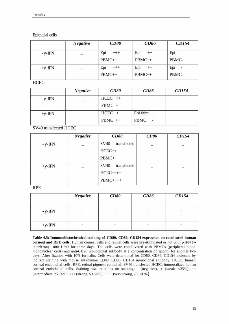
Results
43
Epithelial cells
Negative CD80 CD86 CD154
- γ-IFN _ Epi +++
PBMC++
Epi ++
PBMC++
Epi -
PBMC-
+γ-IFN _ Epi +++
PBMC++
Epi ++
PBMC++
Epi -
PBMC-
HCEC
Negative CD80 CD86 CD154
- γ-IFN _ HCEC ++
PBMC +
_ _
+γ-IFN _ HCEC +
PBMC ++
Epi faint +
PBMC -
_
SV40 transfected HCEC Negative CD80 CD86 CD154
- γ-IFN _ SV40 transfected
HCEC++
PBMC++
_ _
+γ-IFN _ SV40 transfected
HCEC++++
PBMC++++
_ _
RPE
Negative CD80 CD86 CD154
- γ-IFN - - - -
+γ-IFN - - - -
Table 4.5: Immunohistochemical staining of CD80, CD86, CD154 expression on cocultured human corneal and RPE cells . Human corneal cells and retinal cells were pre-stimulated or not with γ-IFN (γ-interferon) 1000 U/ml for three days. The cells were cocultivated with PBMCs (peripheral blood mononuclear cells) and anti-CD28 monoclonal antibody at a concentration of 1µg/ml for another two days. After fixation with 10% formalin. Cells were determined for CD80, CD86, CD154 molecule by indirect staining with mouse anti-human CD80, CD86, CD154 monoclonal antibody. HCEC: human corneal endothelial cells; RPE: retinal pigment epithelial; SV40 transfected HCEC: immortalized human corneal endothelial cells. Staining was rated as no staining: - (negative), + (weak, <25%), ++ (intermediate, 25~50%), +++ (strong, 50~75%), ++++ (very strong, 75~100%).

Human corneal epithelial cells HCEC SV40 transfected HCEC Human RPE
Human corneal epithelial HCEC+γ-IFN SV40 transfected HCEC+γ-IFN Human RPE+γ-IFN Cells+γ-IFN Fig. 4.8: CD80 expression on human corneal and RPE cells cocultured with PBMCs. Human corneal epithelial cells, human corneal endothelial cells (HCEC), SV40 transfected human corneal endothelial cells (SV40 transfected HCEC) and human retinal pigment epithelial cells (RPE) were respectively stimulated or not with ?-IFN for three days. The cells were cocultivated with PBMCs for another two days. After fixation with 10% formalin, cells were analyzed for CD80 expression by indirect staining with mouse anti-human CD80 monoclonal antibody. The staining prodedure was performed as Fig. 4.1. Human corneal epithelial cells, HCEC, SV40 transfected HCEC and cocultured PBMCs were found to be positive. There was no distinct difference between cells with or without ?-IFN treatment. RPE were negative. Magnification 100x.

Human corneal epithelial cells HCEC SV40 transfected HCEC Human RPE
Human corneal epithelial HCEC+γ-IFN SV40 transfected HCEC+γ-IFN Human RPE+γ-IFN Cells+γ-IFN Fig. 4.9: CD86 expression on human corneal and RPE cells cocultured with PBMCs. Human corneal epithelial cells, human corneal endothelial cells (HCEC), SV40 transfected human corneal endothelial cells (SV40 transfected HCEC) and human retinal pigment epithelial cells (RPE) were cocultivated and stained as in Fig. 4.8. CD86 molecule was detected with mouse anti-human CD86 monoclonal antibody. Human corneal epithelial cells with and without ?-IFN treatment were found to be positive. HCEC pre-stimulated with ?-IFN were seen faint staining. The other cells did not exhibit any staining. Magnification 100x.

Human corneal epithelial cells HCEC SV40 transfected HCEC Human RPE
Human corneal epithelial HCEC+γ-IFN SV40 transfected HCEC+γ-IFN Human RPE+γ-IFN Cells+γ-IFN Fig. 4.10: CD154 expression on human corneal and RPE cells cocultured with PBMCs. Human corneal epithelial cells, human corneal endothelial cells (HCEC), SV40 transfected human corneal endothelial cells (SV40 transfected HCEC) and human retinal pigment epithelial cells (RPE) were cocultivated and stained as in Fig. 4.8. CD154 molecule was analyzed with mouse anti-human CD154 monoclonal antibody. All the cells were negative for CD154 expression. Magnification 100x.

Results
47
4.2.4 CD80, 86, 154 expression on corneal epithelial cells
The method of cultivated human corneal epithelial cells were recorded as described
in the material and method part. Half of the cells were treated with γ-IFN 1000 U/ml for
three days, another part without any treatment. Then all of the cells directly fixed by
10% formalin, following immunohistochemical staining of CD80, CD86, CD154
antigen expression. More than 95% CD80 positive cells were presented in cultures of
human corneal epithelial cells either with or without γ-IFN induction. There was no
CD86 expression on the human corneal epithelial cells. Compared with CD154 negative
expression without γ-IFN treatment, approximately 95% corneal epithelial cells with γ-
IFN stimulation showed faint positive staining (Table 4.5 and Fig. 4.11). To investigate
further expression of human corneal epithelial cells, PBMCs were respectively
cocultivated with human corneal epithelial cells without any treatment for two days,
four days, six days. Then the immunohistochemical staining of CD80, CD86, CD154
expression were determined by the attached human corneal epithelial cells. The
cocultivated cell suspension was also harvested and smear slides were performed. The
same antigen measurement of lymphocytes. It was obviously that from the second day
of coculture, human corneal epithelial cells and lymphocytes became strong positive for
CD80 expression. Following coculture process, human corneal epithelial cells and
lymphocytes increased CD80 expression until the sixth day. From smear slides aspect,
whether the lymphocytes number or the positive quantity, there was the best results on
the sixth day. Since the second day, CD86 antigen expression exhibited on the human
corneal epithelial cells and lymphocytes (more than 50%), but there was no distinct
difference following the change of culture period about antigen expression. It was the
same phenomena of CD86 positive staining with smear slides. Concerning CD154,
there was no faint positive staining until the sixth day. All the results were summarized
on the Table 4.6, Fig. 4.12.

Results
48
Negative CD80 CD86 CD154
- γ-IFN _ +++ _ _
+γ-IFN _ +++ _ Faint +
Table 4.6: Immunohistochemical detection of CD80, CD86, CD154 expression on human corneal epithelial cells without γ-IFN (γ-interferon). Cultured human corneal epithelial cells were directly fixed with 10% formalin. Immunohistochemical detection of CD80, CD86, CD154 was performed by indirect staining with mouse anti-human CD80, CD86, CD154 monoclonal antibody. HCEC: human corneal endothelial cells; RPE: retinal pigment epithelial; SV40 transfected HCEC: immortalized human corneal endothelial cells. Staining was rated as no staining: - (negative), + (weak, <25%), ++ (intermediate, 25~50%), +++ (strong, 50~75%), ++++ (very strong, 75~100%).
2 days 4 days 6 days
CD80 epi +++; PBMC+++ epi ++++; PBMC++++ epi ++++; PBMC++++
CD86 epi ++; PBMC++ epi ++; PBMC++ epi ++; PBMC++
CD154 epi - ; PBMC - epi - ; PBMC - epi ++++; PBMC++
Table 4.7: Immunohistochemical detection of CD80, CD86, CD154 expression on human corneal epithelial cells cocultivated with PBMCs (peripheral blood mononuclear cells) respectively for two days, four days and six days. Human corneal epithelial cells were cocultured with freshly purified PBMCs for two days, four days and six days. Immunohistochemical detection of CD80, CD86, CD154 was performed by indirect staining with mouse anti-human CD80, CD86, CD154 monoclonal antibody. HCEC : human corneal endothelial cells; RPE: retinal pigment epithelial; SV40 transfected HCEC: immortalized human corneal endothelial cells. Staining was rated as no staining: - (negative), + (weak, <25%), ++ (intermediate, 25~50%), +++ (strong, 50~75%), ++++ (very strong, 75~100%).

CD80:
without coculture coculture 2 days coculture 4 days coculture 6 days CD86:
without coculture coculture 2 days coculture 4 days coculture 6 days CD154:
without coculture coculture 2 days coculture 4 days coculture 6 days Fig. 4.11: CD80, CD86 and CD154 expression on human corneal epithelial cells and human corneal epithelial cells cocultured with PBMCs. Part of cultured human corneal epithelial cells were directly fixed with 10% formalin. PBMCs were respectively cocultivated with another part of the cells for two days, four days and six days. Immunohistochemical detection of CD80, CD86, CD154 was performed as Fig. 4.1. CD80 was expressed in all cultures. Expression of CD86 and CD154 apeared only after cocultured with PBMCs was seen from day two. Expression of CD154 appeared only after day six. Magnification 100x.

CD80:
2 days 4 days 6 days CD86:
2 days 4 days 6 days CD154:
2 days 4 days 6 days Fig. 4.12: CD80, CD86 and CD154 expression on PBMCs cocultivated with human corneal epithelial cells. PBMCs were respectively cocultivated with human corneal epithelial cells for two days, four days and six days. The cocultivated cells were harvested and smear slides were performed. Cells were fixed and stained as before using antibodies against CD80, CD86, CD154. The cells in the supernatant showed strong positive reaction with CD80 during culture; CD86 was expressed, but no change was observed during culture time; CD154 was not expressed until day six. Magnification 200x.

Results
51
4.2.5 Corneal transplantation rejection section staining
The corneal transplantation rejection grafts and keratoconus grafts were used for
paraffin section. Method in detail was described in material and method part. Then 9
rejection corneas and 5 keratoconus sections were analyzed by immunohistochemical
staining using monoclonal antibody for CD4, CD8, CD40, CD154, HLA-DP, DQ, DR.
All five control corneal buttons (keratoconus) were negative for the set of antibody
markers studied except for CD8 antibody. No inflammatory cells were seen and no
HLA class II molecules were detected on the surface of epithelium, stroma or
endothelium of keratoconus sections. On the opposite, the rejected, failed grafts showed
strong staining for HLA-DP, DQ, DR (Fig. 4.15). The percentage of HLA class II
molecules positivity was found particularly on cells in the basal layers of the corneal
epithelium and even on endothelial cells. Also in the stroma, the positive cells were
sited on the proliferated neovascular endothelial cells, some positive cells were around
vessels, the others were diffusely distributed with the stroma. Especially, nearby the
graft margin, the conjunctiva tissue were proliferated and were combined with invaded
neovascular, HLA-DP, DQ, DR were exhibited strong positive staining. Throughout 9
from patients shown transplantation rejected corneas, there were three cornea sections
indicated faint positive staining on the superficial layer of corneal epithelium with anti-
CD40 antibody (Fig. 4.16). Regarding to the expression of CD4 on the rejection corneas,
there were only two positive results of nine cornea sections (Fig. 4.13). Faintly stained
scaled cells were more nearby neovasculars in the stroma. No staining was found
including the epithelium and endothelium. Concerning the CD154 expression, no
positive results were determined in epithelium, stroma and endothelium (Fig. 4.17).
CD8 antigen expression was positive throughout the whole epithelium. It was also
positive on the endothelium for all of the control samples. But there was no staining in
the stroma. About the rejection corneas, CD8 antigen were also shown on the entire
epithelium and endothelium. Not only that, there were proliferated neovasculars mixed
in the stroma, CD8 positive cells were also observed around the vessels. CD8 was also
showed on keratoconus grafts, the results could not be compared as control samples
(Fig. 4.14).

A1 A2 A3 A4
B1 B2 B3 B4 Fig. 4.13: CD4 expression in corneas derived from patients with keratoconus or graft rejection. Slides of paraffin embedded corneas from patients with keratoconus (A1–A4) or graft rejection (B1–B4) were deparaffinized and rehydrated. The specimens were incubated with mouse anti-human CD4 monoclonal antibody. After incubation with biotinylated link antibody and alkaline phosphatase-labelled streptavidin, staining was completed by adding substrate –chromogen solution, resulting in a colored precipitate at the antigen site. 1: epithelial site; 2: stroma; 3: endothelial site; 4: corneal graft edge. There were no CD4 expressing cells found in corneas derived from patients with keratoconus. In corneas obtained from patients suffering graft rejection, faint CD4 expression was found only near the graft edge (B4). Magnification 200x.

A1 A2 A3 A4
A1 A2 A3 A4
Fig. 4.14: CD8 expression in corneas obtained from patients with keratoconus or graft rejection. Immunohistochemical staining procedure of corneal slides from patients with keratoconus (A1–A4) or graft rejection (B1–B4) were performed as described in Fig. 4.13 using mouse anti-human CD8 monoclonal antibody. 1: epithelial site; 2: stroma; 3: endothelial site; 4: corneal graft edge. CD8 expression was shown on epithelium (A1), endothelium (A3) and epithelium of graft edge of corneas (A4) from patients with keratoconus. CD8 expressing cells were also found on epithelium (B1), cells around newly formed vessels in the stroma (B2), endothelium (B3). Strong stained cells were also presented on the graft edge (B4) of corneas from patients suffering rejection. Magnification 200x.

A1 A2 A3 A4
B1 B2 B3 B4 Fig. 4.15: HLA-DP, DQ, DR expression in corneas obtained from patients with keratoconus or graft rejection. Corneas from patients with keratoconus (A1–A4) or graft rejection (B1–B4) were performed immunohistochemical staining procedure as described in Fig. 4.13 using mouse anti-human HLA -DP, DQ, DR monoclonal antibody. 1: epithelial site; 2: stroma; 3: endothelial site; 4: corneal graft edge. Lessly HLA expression was found on superficial stroma (A2) and stroma of graft edge (A4) of corneas from patients with keratoconus. On the opposite, stronger staining was seen on most of the basal epithelium (B1); diffused staining in stromal (B2); endothelium (B3) and intensive staining on graft edge (B4) of the corneas from patients suffering rejection. Magnification 200x.

A1 A2 A3 A4
B1 B2 B3 B4 Fig. 4.16: CD40 expression in corneas obtained from patients with keratoconus or graft rejection. Corneas from patients with keratoconus (A1–A4) or graft rejection (B1–B4) were performed immunohistochemical staining procedure as described in Fig. 4.13 using mouse anti-human CD40 monoclonal antibody. 1: epithelial site; 2: stroma; 3: endothelial site; 4: corneal graft edge. There was no CD40 expression on cells of corneas from patients with keratoconus. Only faint CD40 staining was shown on superficial epithelium (B1) and epithelium nearby graft edge (B4) of the corneas from patients suffering rejection. Magnification 200x.

A1 A2 A3 A4
B1 B2 B3 B4 Fig. 4.17: CD154 expression in corneas obtained from patients with keratoconus or graft rejection. Corneas from patients with keratoconus (A1–A4) or graft rejection (B1–B4) were performed immunohistochemical staining procedure as described in Fig. 4.13 using mouse anti-human CD40 monoclonal antibody. 1: epithelial site; 2: stroma; 3: endothelial site; 4: corneal graft edge. CD154 expression was not seen on cells of corneas from patients with keratoconus and graft rejection. Magnification 200x.

Discussion
57
5. Discussion
Keratoplasty represents the most common and successful transplantation of solid
tissue in humans. Transplants are usually performed to improve vision and increase the
quality of life in patients with damaged or diseased corneas. Corneal transplantation is
therefore of significant clinical, biological, and economic relevance. In uncomplicated
cases, when the first allografts are performed in avascular beds, the two year survival
rate is over 90%. However, approximately 30% of the patients undergoing routine
penetrating keratoplasty have a rejection episode, and around 10% of grafts fail because
of allograft rejection. Immunological rejection is a leading cause of corneal graft failure.
The “high risk” recipient is directly correlated with the severity of corneal
vascularisation (Kuffová L et al. 1999). For many reasons, it is likely that the human
leukocyte antigen (HLA) alloantigens are involved in corneal graft rejection. They are
known to be the main targets for the immunological reactions leading to rejection of
transplanted solid organs. It has been concluded that corneal graft rejection was a T
lymphocyte mediated immune process (Bertelmann et al. 2002). In this course, T
lymphocyte immune responses may be divided into three phases: recognition, activation
and effecter mechanisms. T cells are able to recognize foreign peptides in complexes
with self-HLA molecules. To activate T cells, two signals are necessary. Signal 1 is
delivered through T cell receptor that binds to the allogeneic peptide/HLA complex of
the antigen presenting cells (APC), and the CD4 or CD8 coreceptor binds to a constant
part of HLA class-II or HLA class-I molecule respectively. Signal 2 or costimulatory
molecules include CD28 and its ligands B7-1 (CD80) and B7-2 (CD86), both are
expressed primarily on activated antigen-presenting cells. Other T cell surface
molecules, such as CD40 ligand (CD154) contribute to signal 2. Following signal 1 and
2, the T cell is fully activated. This study was performed to determine HLA class II
expression, and expression of the molecules participating in the process of T cell
activation in human corneal epithelial cells, human corneal endothelial cells (HCEC),
and immortalized human corneal endothelial cells (SV40 transfected HCEC).
Knowledge of possible reaction ways for immunological processes may be helpful
in preventions corneal graft rejection. Furthermore, retinal pigment epithelial cells
(RPE) were included in this study, because transplantation of RPE cells, seems to be a
promising therapy in cases of RPE degeneration. Until now, no study was performed to
Discussion
58
analyze possible RPE rejection processes. Expression of HLA- antigens was found in all
four cell types although this expression required ?-IFN (?- interferon) induction in case
of human corneal endothelial cells. Therefore, the presence of HLA-antigens on these
cells may contribute to T -cell activation. The results concerning this activation will be
discussed for each cell type separately.
5.1 Expression of HLA-DP, DQ, DR and T cell activation
5.1.1 Human corneal epithelial cells
About HLA-DP, DQ, DR antigen expression on human corneal epithelial cells,
(Iwata et al. 1992) reported there was no class II antigen expressed on human corneal
epithelial cells without ?-IFN treatment, but the expression of all these MHC class II
antigens were induced on human corneal epithelial cells in culture by ?-IFN. Using the
cultivation technique described in this study, HLA-DP, DQ, DR antigens were
expressed on human corneal epithelial cells without any stimulation; approximately
more than 50% of the cells showed positive staining. Nevertheless, HLA class II
expression was still enhanced by ?-IFN treatment, leading to nearly 100% staining on
human corneal epithelial cells.
Studies by FACS analysis and immunohistochemical staining demonstrated that
human corneal epithelial cells activated human T lymphocytes to express CD69 (Fig.
3.5) and HLA-DR (Fig.3.7). Human corneal epithelial cells induced a higher level of T
cell activation than the other cell types, and there was no distinct difference between
cells induced or not with ?-IFN. Due to strong HLA classII expression, human corneal
epithelial cells can serve as antigen presenting cells, and the T cell receptor can bind to
allogeneic peptide/HLA complex. The CD4 coreceptor can bind to a constant part of
HLA class II or CD8 can bind to HLA class I molecules respectively. Thereby a first
activation signal was transduced into the T cell. The CD28 molecule served as
costimulatory signal. CD28 has two known ligands, B7-1 (CD80) and B7-2 (CD86),
both of which are expressed primarily on activated antigen-presenting cells. The
immunohistochemical staining results showed that CD80 molecules were also expressed
on human corneal epithelial cells, so CD28 and B7-1 (CD80)/B7-2 (CD86) could have
contributed to signal 2. Following signal 1 and signal 2, the T cells were fully activated.
In some experiments, human corneal epithelial cells without ?-IFN treatment
stimulated allogeneic lymphocytes, but always less than ?-IFN treated human corneal

Discussion
59
epithelial cells did. In previous study (Iwata et al. 1994), human corneal epithelial cells
were regarded as a stimulator, and mixed with responder PBMCs in microtiter plates.
Furthermore, one µCi of 3H-thymidine was added to the cultures. 18 hours later, the
cultures were harvested on glass fiber filters, 3H-thymidine incorporation was measured
by a liquid scintillation counter. This method has the disadvantage that also radioactivy
is registered that derived from incorporation into the epithelial cells. Compared with the
experimental method, in my study, cultured human corneal epithelial cells attached on
the bottom of culture dish. Then the PBMCs cell suspension was supplied into human
epithelial cell culture well. After 48 hours, most of the lymphocytes still remained in the
cell suspension, few lymphocytes firmly covered on the human epithelial cells. The cell
suspension was carefully transferred for FACS analysis. The purpose of my cultivation
method was to ensure that only the pure lymphocyte suspension was used to detect the
lymphocyte proliferation condition. Because the human epithelial cells still attached on
the culture cluster after the lymphocytes were delivered. So the risk that human corneal
epithelial cells were mixed into lymphocytes were minimize.
Therefore the method used in this study minimizes the risk of overdetermination of
T cell activation but it leads to a certain underestimation. This phenomenon is described
in the following. The cocultured PBMCs in the experiment process should be
emphasized. The human epithelial cells and PBMCs were cocultivated on the chamber
slides. Afterward, the cocultured cells were determined by HLA-DP, DQ, DR
monoclonal antibody. The activated lymphocytes could be distinguished by the
antibody. The results showed that not only the epithelial cells were positive with HLA
class II. Furthermore, correspondingly the lymphocytes attached on the slides became
more larger than nonactived lymphocytes, more important is that most of the attaching
lymphocytes were positive for HLA class II. Hereby, in case of the most activated
lymphocytes were attached on the coculture well, the cell amounts used for FACS
analysis were decreased. It means, actually, the effect of lymphocyte activation by
human epithelial cells has been underestimated in FACS analyses, due to cell adhesion.
In fact, the lymphocytes activation impression was more larger than determined. A
larger number of adherent lymphocytes were observed in cocultures with human corneal
epithelial cells than in cocultures with other cells (Fig. 4.7), consistent with a strong
activation - and proliferation - of lymphocytes by these cells.

Discussion
60
5.1.2 Human corneal endothelial cells (HCEC)
In contrast to corneal epithelial cells, cultured cornal endothelial cells exhibited
HLA expression only after ?-IFN induction. The result was similar with former results
from EL-Asrar et al. (1989). The difference concerns the material and method which
had been used by these authors. They excised 3 mm corneal scleral margins, and
divided them into small parts. The pieces of cornea were directly placed into culture
medium and incubated with recombinant Escherichia coli-derived interferon-gamma in
a dose of 5 mg/l and 50 mg/l. Four days later, the corneal pieces were used to prepare
frozen section for immunoperoxidase staining of the corneal cell layers. Our laboratory
has established HCEC cell cultures, and we have used these cultures to evaluate the
profiles of HCEC separated from the original corneal tissue. It could be found that in
my study, that investigation of monolayer cell cultures are a simple method which is
more directly and more convenient to observe the behaviour of HCEC.
T cell activation in cocultures was determined by FACS analysis. The percentage of
activated lymphocytes was lower in HCEC cocultures than in SV40 transfected HCEC
and human corneal epithelial cells but higher than in RPE colcultures (Fig. 4.5). This is
consistent with the finding that there was no abundant expression of costimulatory
molecules, such as CD80, CD86, on HCEC (Figs. 4.8 and 4.9). Evidently, the activation
signal was not strongly delivered to lymphocytes.
5.1.3 Immortalized human corneal endothelial cells
HLA class II antigen expression by immortalized human corneal endothelial cells
(SV40 transfected HCEC) was analyzed for the first time. It was found that the cells
expressed HLA-DP, DQ, DR, only after treatment with ?-IFN. Therefore there was no
difference between transfected and nontransfected HCEC concerning HLA-DP, DQ,
DR expression. Nevertheless only less than 25% SV40 transfected HCEC exhibited
HLA expression. It was appealing that if the cells were in subconfluent condition, the
HLA-DP, DQ, DR positive staining was observed distinctly. On the opposite, after the
SV40 transfected HCEC ultimately became confluent, the HLA class II positive
staining was difficult to be found or only occasionally be detected. We could explain
that because confluence might be a turning point from the proliferation stage to the
differentiation stage. But it seemed there was no significant difference of HLA-DP, DQ,

Discussion
61
DR antigens induction from subconfluent and confluent cultures of human corneal
epithelial cells, HCEC and RPE cells.
5.1.4 Human retinal pigment epithelial cells (RPE)
In our laboratory, a method has been established for HLA class II antigen induction
with ?-IFN on RPE cells (Valtink et al. 1999). Though HLA class II antigens were
never examined by immunohistochemical staining method. Like corneal epithelial cells,
RPE cells exhibited strong HLA-DP, DQ, DR antigen induction with ?-IFN. From
FACS analysis results, RPE cells showed low ability of T lymphocytes activation. In a
former report about RPE cells (Jorgensen et al. 1998), Fas ligand (FasL) expression was
detected by flow cytometry and immunohistochemistry staining method. The results
indicated that RPE cells expressed FasL and induced TCR-independent apoptosis in
activated human T cells through Fas- FasL interaction. Retinal pigment epithelial cells
may constitute an immunologic functional barrier against potentially harmful T cells.
Few years later, Kaetel et al. (2002) continued this research and found that human
retinal pigment cells inhibit the proliferation of activated T cells by a cell contact-
dependent mechanism. The inhibition of the T cells´ proliferation correlates with a
decreased expression of IL2R-ß and – ? chains. The T cells regain their ability to
proliferate and increase their IL2R-ß and – ? chain expression within 24 hour after
removal from the coculture. It was concluded that the cultured human RPE cells inhibit
the proliferation of activated T cells by a process that does not involve apoptosis. It
depended on cell contact but the involved surface molecules were not revealed. The
proliferation inhibition correlated with a modulation of the T cells´ expression of IL2R
(interleukin-2 receptor), and was reversible. In the above mentioned investigation, the
human RPE cells were respectively pre-stimulated with IL-2 (interleukin-2) or PHA
(phytahematoagglutinin), and then the RPE cells were cocultivated with lymphocytes.
But in our study, there was an important difference to this. That was because we directly
used human RPE cells for cocultivation with lymphocytes without any stimulator factor
of activation. Nevertheless the obtained results were similar with former research.
Human RPE cells could no t activate resting T lymphocytes. About the question whether
RPE cells can induce lymphocytes´ apoptosis and by which mechanism, more
investigations are attempted for clarification of the intracellular pathways through
which inhibition of lymphocytes is achieved.

Discussion
62
5.1.5 Transplantation rejection cornea section
To investigate HLA-DP, DQ, DR antigens expression in cornea transplants which
were removed after graft rejection. Ten rejection cornea grafts were examined by
immunoperoxidase staining method. The results demonstrated that HLA class II
antigens were expressed in all corneal sections. Extensive HLA class II expression was
observed on corneal epithelium, stroma, and even on endothelium. Prominent
expression on epithelium was seen in the basal layers. In the stroma, some positive cells
diffused, some were distributed around vascular, and some HLA positive cells were
expressed on the proliferated neovascular endothelial cells. Especially, it has known that
corneal transplantation rejection was frequently happened on neovasclar invaded
corneal graft. So nearby the rejection graft margin, strong positive HLA class II antigen
expression was seen in the proliferated and thickened conjunctiva tissue. It is well
known that Langerhans cells, which expressed HLA class II antigens, prominently
existing in limbal and peripheral cornea, can be potent APCs (antigen presenting cells).
The FACS analysis results of using cultured human corneal epithelial cells treated with
or without ?-IFN support this opinion. Regarding to T lymphocytes activation, there
was almost no difference from pre-stimulation with ?-IFN or without ?-IFN. It is
possible that ?-IFN is produced locally by the interaction of corneal limbus or peripheral
epithelial cells especially Langerhans cells in donor tissue with recipient lymphocytes.
Then subsequent production of ?-IFN stimulates HLA class II antigens expression on
human corneal epithelial cells, which may initiate corneal graft rejection. On the other
hand, HLA-DP, DQ, DR expression on cocultivated corneal cells pre-treated with or
without ?-IFN and the lymphocytes attaching on these cells were analyzed, showed that
HLA-DP, DQ, DR expression occurred on both cell types. The staining results using
cocultivated cells supported the previous outcome of independent corneal cells with
HLA class II expression. It seemed there was no or little ?-IFN secretion among human
corneal epithelial cells and lymphocytes. It could also be hypothesized that the coculture
period of two days was not long enough. The grafts described as chronic graft rejections
in clinical observation have been accepted much medicine therapy, therefore, the
inflammatory and immunology reactions have been occurred repeatedly during a long
time. So it can be also hypothesized that enough ?-IFN secretion requires a longer

Discussion
63
cocultivation period between corneal epithelial cells and lymphocytes. This purpose
need to be confirmed in the next step study.
5.2 CD40/CD154 pathway
CD40, is a 50-kDa integral membrane glycoprotein. It is found on a variety of
antigen-presenting and mesenchymal cells, including B lymphocytes, dendritic cells,
macrophages, monocytes, microglia, endothelial cells, and epithelial cells (Grewal and
Flavell 1996; Bourcier et al. 2000; Brignole et al. 2000). CD154, also known as CD40
ligand (CD40L), CD40 ligand (CD154), is a 39-kDa type II membrane glycoprotein and
member of the tumor necrosis factor (TNF) superfamily. It is preferentially expressed
on activated CD4+ cells and mast cells, contribute to signal 2 of T cell activation. Over
the past several years, the central role of CD40-CD154 interaction in mediating T-cell–
mediated immune responses has been firmly established (Grewal and Flavell 1996).
CD154-dependent activation of T cells occurs through signaling of CD40 at the level of
antigen-presenting cells (APCs), which enhances requisite costimulatory pathways,
including expression of B7.1/B7.2 (CD80/CD86) (Yang and Wilson 1996).
Accordingly, it has been shown that blockade of the CD40-CD154 pathway is sufficient
to induce tolerance to Th1-mediated contact hypersensitivity (Qian et al. 2001).
In the cornea, it is thought that HLA class II usually is expressed on limbal and
peripheral cornea epithelium, and not in the epithelial cells of the central cornea. This is
similar to CD40 expression, as demonstrated in the sections. Therefore, there was the
possibility that CD40 and HLA class II probably have some similar character, and
might be expressed on the same epithelial population. To test this possibility, the same
experimental procedure was done with CD40 as HLA class II. But the results showed a
little difference with HLA class II. Without ?-IFN treatment, RPE and SV40 transfected
HCEC were negative for CD40. About 95% HCEC indicated CD40 positive staining.
Human corneal epithelial cells still remained nearly 100% positive for CD40
expression. After ?-IFN induction, RPE and SV40 transfected HCEC were enhanced to
almost 5% positive expression, HCEC were achieved almost 100% CD40 positive
staining. The human corneal epithelial cells, the quantity of CD40 antigen expression
increased. The cells were obtained not only in the cell membrane, but also in the
cytoplasm-in conclusion- the positive cells showed more dense staining than without γ-
IFN induction. No doubt, human corneal epithelial cells were the most sensitive cells

Discussion
64
for CD40 among these cultured cells. Foregoing research in normal cornea, Iwata et al.
(2002) concluded that the limbal epithelial cells were predominantly positive for CD40.
Positive staining was also found to a lesser extent on the cells in the basal layer of
peripheral corneal epithelium. Epithelial cells of the central cornea showed no
immunoreactivity for CD40 molecule in the sections. But human corneal epithelial cells
and HCEC were all negative for CD154 molecule, CD154 antigens could not be
determined on RPE and SV40 transfected HCEC. Only after longer culture periods,
expression of CD154 became obvious on human corneal epithelial cells.
Comparing with the conclusion, CD40 expression on the rejection cornea
epithelium was determined in our study, but not CD154. The rejection corneal grafts
were obtained from the second keratoplasty had taken place, a control normal cornea
from a keratoconus patient was obtained. It was impossible to examine the corneal
epithelial cells of the limbal and peripheral region, because these structures were not
explanted. It could be found that three corneal sections of ten rejected grafts showed
faint CD40 positive staining on the superficial layer of corneal epithelium. Especially
CD40 positive cells were nearby the graft margin. It has well known that, cells
expressing CD40 are mainly hematopoietic cells, antigen presenting cells, such as B
cells, monocytes-macrophages, and dendritic cells (Iwata et al. 2002). It is preferential
expression of HLA-DP, DQ, DR and CD40 on human corneal epithelial cells that
implicate the immunological and the proliferative potential function.
In this study, the human corneal epithelial cells were isolated from limbus area of
corneal tissue. As has been demonstrated, proinflammatory cytokines such as ?-IFN
induced marked HLA class II and CD40 expression on cultured human corneal
epithelial cells. Recently, increasing evidence stressed the critical role of CD40/CD40L
(CD154) interactions in the regulation of immune responses, especially mediated by T
lymphocytes. The ligation of CD40 on the surface of professional APCs (dendritic cells,
B cells, macrophages) by CD40L expressed on activated T cells promotes further
upregulation of costimulatory molecules (B7.1, B7.2, CD40) expression on these APCs
and high IL-12 secretion, which increase their antigen-presentation and costimulatory
capacity (Grewal and Flavell 1996; Schoenberger et al. 1998). The CD40:CD154
pathway, initially described as having a role in B-cell activation, has been recognized as
a key pathway for T-cell activation as well (Durie et al. 1994). Stimulation of CD40

Discussion
65
provides important signals for antibody production by B cells and strongly induces B7
expression on all antigen-presenting cells. In this manner, the CD40:CD154 system may
have an important role in T-cell costimulation. Activation of antigen-presenting cells
through CD40 also induces the expression of adhesion molecules and inflammatory
cytokines that participate in T cell activation. There is the possibility that the CD40-
CD40L interaction in corneal epithelium at the limbus may trigger production of
proinflammatory cytokines such as ?-IFN and TNF-α released by human corneal
epithelial cells.
5.3 CD28 and CD80/CD86 costimulatory signal
It was also demonstrated in this study that CD80, CD86 molecules could be
expressed on human corneal epithelial cells after cocultivated with PBMCs. CD80
antigen was expressed on HCEC and PBMC coculture system. CD80, CD86 could not
be determined on RPE and SV40 transfected HCEC. Among these cells, human corneal
epithelial cells still were the most sensitive for CD80, CD86 expression.
Two lines of evidence highlight the role of the B7:CD28 pathway of T-cell
costimulation in transplant rejection. First, drugs that block the interactions between B7
and CD28 molecules can induce long- lasting nonresponsiveness of T cells to
alloantigens in vitro. Second, B7 molecules are induced on vascular endothelium within
24 hours after the transplantation of vascularized organs (Hancock et al. 1996). This
means that T cells can receive costimulatory signals throughout the transplanted organ
itself. Anti-CD80 and anti-CD86 monoclonal antibodies (mAbs) were administered
after orthotopic corneal allograft transplantation (Kagaya et al. 2002). The combined
use of anti-CD80 and anti-CD86 mAbs was effective in prolonging corneal allograft
survival. Few CD80 (+) or CD86 (+) cells were observed in the cornea treated with anti-
CD80/CD86 mAbs. These results demonstrated a significant role of CD80 and CD86
costimulatory molecules in corneal allograft rejection.
Considering the expression of HLA class II, CD40, CD80, CD86 on human corneal
epithelial cells and FACS analysis results, it could be concluded that human corneal
epithelial cells can participate in corneal immune responses as potential antigen
presenting cells, including immunological reaction of corneal graft rejection. It is well
known that, in humans, rejection of lamellar corneal grafts are rare compared to
rejections of penetrating grafts (Khodaoust and Silverstein 1972). This observation

Discussion
66
suggested that human corneal endothelial cells play a key role in alloimmunization of
corneal graft recipients. It has also been demonstrated that expression of CD40, CD80
on HCEC, and HLA class II and CD86 molecular could be induced by ?-IFN.
5.4 T lymphocytes
From ten rejection corneal sections, only in two samples CD4+ T cells were found
nearby the neovascular and graft margin. In contrast, the epithelium from the
keratoconus as well as rejection corneas were all positive using of the anti CD8
antibody. It has been reported that cytotoxic T cells (CD8+) play no essential role in
rejection of orthotopic corneal allografts in mice (Yamada et al. 2001). Another
research (Boisgerault et al. 2001) concluded that only CD4+ T cells activated via
indirect allorecognition have the ability to reject allogeneic corneal grafts. Although
alloreactive CD8+ T cells are activated via the direct pathway, they are not fully
competent and can not contribute to the rejection unless they receive an additional
signal provided by professional APCs in the periphery. Then the effect of CD8+ T cells
in corneal transplantation rejection should be attempted continuously. About ?-IFN,
anti-human interferon-gamma F(ab´)2 (Fabs) antibodies were administered in patients
who experienced corneal transplant rejection as eyedrops by Skurkovich et al. (2002).
The results showed that the transplant grafts became fully transparent, eye inflammation
disappeared. Visual acuity also increased. It means ?-IFN is significant
proinflammatory cytokine in corneal transplantation.
All the phenomenon indicated, human corneal epithelial cells dominantly
participate in the corneal transplantation rejection process. Then the corneal
transplantation rejection process initiation can be supposed. The physiological
activation of T lymphocytes involves at least two signals. Foreign peptides derived from
proteins are presented to CD4+ T cells (helper T cells) by HLA-DR, DQ and DP of
human corneal epithelial cells. When a TCR binds to the allogeneic peptide/HLA class
II complex, and the CD4 (in helper T cells) co-receptors bind to a constant part of HLA
class-II, the first activation signal is delivered into the CD4+ T cell. Signal 2 results
from the binding of constitutively expressed CD28 molecules on T cells to CD80 or
CD86 molecules on human corneal epithelial cells. Other T cell surface molecules, such
as CD40 ligand (CD154), CD2, LFA1 and ICAM 1, contribute to signal 2. Following
signals 1 and 2, CD4+ T cell is fully activated so that the genes encoding lymphokines

Discussion
67
and lymphokine receptors are transcribed and translated. When alloreactive CD4+ T
cells are activated on recognition of foreign peptide HLA class-II complexes, they
proliferate and, at the same time, secrete a set of lymphokines. The alloreactive T cell
response seems to be mainly a Th1 (T helper 1) response, with secretion of IL-2 and
interferon gamma (γ-IFN). IL-2 helps to sustain proliferation of CD4+. Help from
alloreactive CD4+ Th2 cells is required for activation, proliferation and differentiation
of alloreactive HLA-specific B cells into plasma cells that -depending on the HLA
disparity between donor and recipient -secrete antibodies specific for donor HLA class-I
or class-II molecules. γ-IFN continuously stimulate corneal cells, including epithelial,
stroma, endothelial and vascular endothelial cells to express antigen and surface
costimulatory molecular, such as CD80, CD86, CD40. These molecules contribute and
induce an alloimmune response in the recipient graft.
The current studies have demonstrated the activation process of T lymphocytes and
have analyzed constitutively expression of HLA class II antigen, the costimulatory
molecular which participate T lymphocyte activation, such as CD80, CD86, CD40 on
human corneal epithe lial cells, HCEC, SV40 transfected HCEC, RPE cultured cells.
Furthermore, the upregualted expression of these cells treated with γ-IFN were also
determined. It is concluded that human corneal epithelial cells are important potential
antigen presenting cells. The corneal transplantation rejection is initiated by corneal
epithelial cell contact with T lymphocytes. The presence of HLA class II can be
induced on human corneal endothelial cells during its immune destruction. And this
observation implicated that induced HLA class II alloantigen expression on corneal
endothelium may be a contributing factor in the rejection of corneal allografts.
Summary
68
6. Summary
The main reason for cornea graft failure after keratoplasty is related to graft
rejection. Comparable to the situation after solid organ transplantation, activation of T
cells may be involved in this rejection process. The aim of the present study was to
investigate the possibility of T cell activation by corneal cells. T cell activation requires
two distinct signals from antigen presenting cells.
In a first step the expression of HLA antigens was studied in cultured human corneal
endothelial and epithelial cells. Human retinal pigment epithelial cells served as control.
This examination revealed that HLA expression occurred in corneal epithelial and also
endothelial cells. Therefore these cells may serve as antigen presenting cells. In a
second step the human corneal cells were co-cultured with peripheral blood cells.
Activation of T lymphocytes purified from peripheral blood cells was analysed by
immunohistochemical staining and FACS analysis. The co-culture of corneal cells and
peripheral blood cells led to expression of CD69, a marker for activated T cells, in part
of the peripheral blood cells population. Furthermore expression of CD80 and CD86
was found on human corneal epithelial cells. Human corneal endothelial cells expressed
only CD80 but not CD86. CD80 and CD86 may serve as the second signal for T cell
activation. Another molecule that can serve as the required second signal for T cell
activation is CD154-a ligand of CD40. This molecule could be detected on human
corneal epithelial cells but only after prolonged co-culture time of six days.
Additionally to the cell culture experiments, human corneal grafts were investigated
that were exchanged after transplant failure. These investigations revealed CD4 positive
T cells participated in the corneal graft rejection. Furthermore, CD40 positive cells were
determined in the superficial epithelium.
In summary this study has shown that human corneal epithelial cells seem to
participate in the corneal transplantation rejection process, as potential antigen
presenting cells. The corneal transplant rejection may be initiated by corneal epithelial
cell contact with T lymphocytes. The current studies have demonstrated the activation
process of T lymphocytes. The costimulatory molecule which participate in T
lymphocyte activation, such as CD80, CD86, CD40, CD154 was also exhibited on
human corneal epithelial cells, HCEC. The results were consistent with former report
that cytotoxic T cells (CD8+) play no essential role in rejection of orthotopic corneal
allografts in mice.

Zusammenfassung
69
Zusammenfassung
Der Hauptgrund für ein Transplantatversagen nach Keratoplastik ist eine
Transplantatabstoßung. In Anlehnung an die Situation nach Transplantation
solider Organ könnte die Aktivierung von T-Zellen an diesem Abstoßungsprozess
beteiligt sein. Ziel dieser Arbeit war es, die Möglichkeit der T-Zell Aktivierung
durch korneale Zellen zu untersuchen. Für die Aktivierung von T-Zellen durch
Antigen-Präsentierende-Zellen sind zwei unabhängige Signale notwendig.
Im ersten Teil der Arbeit wurde die Expression von HLA-Antigenen in
kultivierten kornealen Endothel- und Epithelzellen untersucht. Humane retinale
Pigmentepithelzellen dienten zur Kontrolle. Diese Untersuchungen haben gezeigt,
dass sowohl in kornealen Endothel- als auch Epithelzellen HLA-Antigen
exprimiert werden. Daher können diese Zellen als Antigen-Präsentierende-Zellen
fungieren. Im zweiten Teil der Arbeit wurden die kornealen Zellen mit einer T-
Zell enthaltenen Fraktion humaner peripherer Blutzellen cokultiviert. Die
Aktivierung der T-Zellen wurde sowohl immunhistochemisch als auch durch
FACS-Analyse untersucht. Die Cokultivierung führte zur Expression von CD69,
einem Marker für aktivierte T-Zellen. Außerdem wurde Expression von CD80
und CD86 in kornealen Epithelzellen nachgewiesen. Korneale Endothelzellen
exprimierten nach Cokultivierung CD80, nicht aber CD86. CD80 und CD86
können als zweites Signal für die Aktivierung von T-Zellen fungieren. Ein
weiteres Molekül, das als zweites Signal für die T-Zell Aktivierung dienen kann
ist CD154; ein Ligand für CD40. Dieses Molekül konnte in kornealen
Epithelzellen nachgewiesen werden, jedoch erst nach einer Cokultivierungszeit
von mindestens sechs Tagen.
Zusätzlich zu den Untersuchungen an Zellkulturen wurden humane korneale
Transplantate untersucht, die nach Abstoßungsreaktionen ausgetauscht worden
waren. Diese Untersuchungen zeigten das Auftreten von CD4 positiven T-Zellen
bei Abstoßungsraktionen. Ferner wurden CD40 positive Zellen im kornealen
Epithel nachgewiesen.
Zusammenfassend hat diese Untersuchung gezeigt, dass das korneale Epithel am
Prozess der Transplantatabstoßung beteiligt zu sein scheint und zwar in Form von
Antigen-Präsentierenden-Zellen. Die Transplantatabstoßung könnte demnach
durch einen Kontakt von kornealen Epithelzellen und T-Zellen ausgelöst werden.

Zusammenfassung
70
Moleküle, die als notwendiges zweites Signal für die T-Zell Aktivierung dienen
können wie CD80, CD86, CD40 und CD154 werden ebenfalls von kornealen
Epithelzellen exprimiert.

References
71
7. References
Ayliffe W, Alam Y, Bell EB, McLeod D, Hutchinson IV (1992) Prolongation of rat
corneal graft survival by treatment with anti-CD4 monoclonal antibody. Br J
Ophthalmol 76:602-606
Bednarz J, Teifel M, Friedl P, Engelmann K (2000) Immortalization of human corneal
endothelial cells using electroporation protocol optimized for human corneal endothelial
and human retinal pigment epithelial cells. Acta Ophthalmol Scand 78:130-136
Bernard A, Lamy L, Alberti I (2002) The two-signal model of T-cell activation after 30
years. Transplantation 73:31-35
Bertelmann E, Jaroszewski, Pleyer U (2002) Corneal allograft rejection: current
understanding. Ophthalmologica 216:2-12
Berry CL (1999) Transplantation pathology: a guide for practicing pathologists.
Springer, Berlin
Bluestone JA (1995) New perspectives of CD28-B7 mediated T cell costimulation.
Immunity 2:555-559
Boisgerault F, Liu Y, Anosova N, Ehrlich E, Dana MR, Benichou G (2001) Role of
CD4+ and CD8+ T cells in allorecognition: lessons from corneal transplantation. J
Immunol 167:1891-1899
Borriello F, Sethna MP, Boyd SD, Schweitzer AN, Tivol EA, Jacoby D, Strom TB,
Simpson EM, Freeman GJ, Sharpe AH (1997) B7-1 and B7-2 have overlapping, critical
roles in immunoglobulin class switching and germinal center formation. Immunity
6:303-313

References
72
Bourcier T, De Saint-Jean M, Brignole F, Goguel A, Baudouin C (2000) Expression of
CD40 and CD40 ligand in the human conjunctival epithelium. Invest Ophthalmol Vis
Sci 41:120-126
Bretscher PA, Cohn M (1970) A theory of self and not-self discrimination. Science
169:1042-1049
Brignole F, Pisella PJ, Goldschild M, De Saint JM, Goguel A, Baudouin C (2000) Flow
cytometric analysis of inflammatory markers in conjunctival epithelial cells of patients
with dry eyes. Invest Ophthalmol Vis Sci 41:1356-1363
Chan CC, Detric B, Nussenblatt RB, Palestine AG, Fujikawa LS, Hooks JJ (1986)
HLA-DR antigen on retinal pigment epithelial cells from patients with uveitis. Arch
Ophthalmol 104:725-729
Dong VM, Yuan XL, Coito AJ, Waaga AM, Sayegh MH, Chandraker A (2002)
Mechanisms of targeting CD28 by a signaling monoclonal antibody in acute and
chronic allograft rejection. Transplantation 73:1310-1317
Donnely JJ, Chan LC, Xi MS, Rockey JH (1988) Effect of human corneal fibroblasts on
lymphocyte proliferation in vitro. Exp Eye Res 47:61-70
Dreizen NG, Whitsett CF, Atulting RD (1988) Modulation of HLA antigen expression
on corneal epithelial and stromal cells. Invest Ophthalmol Vis Sci 29:933-939
Duquesnoy RJ, Trager JDK, Zeevi A (1991) Propagation and characterization of
lymphocytes from transplant biopsies. Immunology 10:455-459
Durie FH, Foy TM, Masters SR, Laman JD, Noelle RJ. (1994) The role of CD40 in the
regulation of humoral and cell-mediated immunity. Immunol Today 15:406-411

References
73
EL-Asrar AMA, Oord JJVD, Billiau A, Desmet V, Emarah MH, Missotten L (1989)
Recombinant interferon-gamma induces HLA-DR expression on human corneal
epithelial and endothelial cells in vitro: a preliminary report. Br J Ophthalmol 73:587-
590
Engelmann K, Friedl P (1989) Optimization of culture conditions for human corneal
endothelial cells. In Vitro Cell Cev Biol 25:1065-1072
Ferguson SE, Han S, Kelsoe G, Thompson CB (1996) CD28 is required for germinal
center formation. J Immunol 156:4576-4581
Fujikawa LS, Colvin RB, Bann AK (1982) Expression of HLA-A/B/C and DR locus
antigens on epithelial, stromal and endothelial cells of the human cornea. Cornea 1:213-
218
Gabrielian K, Osusky R, Sippy BD, Ryan SJ, Hinton DR (1994) Effect of TGFß on
interferon ?-induced HLA-DR expression in human retinal pigment epithelial cells.
Invest Ophthalmol Vis Sci 35:4253-4259
Grewal IS, Flavell RA (1996) A central role of CD40 ligand in the regulation of CD4+
T cell responses. Immunol Today 17:410-414
Hall BM, Bishop GA, Duggin GG, Horvath JS, Philips J, Tiller DJ (1984) Increased
expression of HLA DR antigen on renal tubular cells in renal transplants: Relevance to
the rejection response. Lancet 2:247-251
Hancock WW, Sayegh MH, Zheng XG, Peach R, Linsley PS, Turka LA (1996)
Costimulatory function and expression of CD40 ligand, CD80, and CD86 in
vascularized murine cardiac allograft rejection. Proc Natl Acad Sci 93:13967-13972
Hegde S, Niederkorn JY (2000) The role of cytotoxic T lymphocytes in corneal graft
rejection. Invest Ophthalmol Vis Sci 41:3341-3347
References
74
He YG, Ross J, Niederkorn JY (1991) Promotion of murine orthotopic corneal allograft
survival by systermic administration of anti-CD4 monoclonal antibody. Invest
Ophthalmol Vis Sci 32:2723-2728
Hirokawa M, Kuroki J, Kitabayashi A, Miura AB (1996) Transmemebrane signaling
through CD80 (B7-1) induces growth arrest and cell spreading of human B lymphocytes
accompanied by protein tyrosine phosphorylation. Immunol Lett 50:95-98
Iwata M, Kiritoshi A, Roat MI, Yahihashi A, Thoft RA (1992) Regulation of HLA class
II antigen expression on cultured corneal epithelium by interferon-gamma. Invest
Ophthalmol Vis Sci 33:2714-2721
Iwata M, Yagihashi A, Toat MI, Zeevi A, Iwaki Y, Thoft RA (1994) Human leukocyate
antigen –class II- positive human corneal epithelial cells activate allogeneic T cells.
Invest Ophthalmol Vis Sci 35:3991-4000
Iwata M, Soya K, Sawa M, Sakimoto T, Hwang DG (2002) CD40 expression in normal
human cornea and regulation of CD40 in cultured human corneal epithelial and stromal
cells. Invest Ophthalmol Vis Sci 43:348-357
Janeway CA Jr, Bottomly K (1994) Signals and signs for lymphocyte responses. Cell
76:275-285
Jeannin P, Delneste Y, Lecoanet-Henchoz S, Gauchat JF, Ellis J, Bonnefoy JY (1997)
CD86 (B7-2) on human B cells. A functional role in proliferation and selective
differentiation into IgE-and IgG4-producing cells. J Biol Chem 272:15613-15619
Jones ND, Maurik AV, Hara M, Spriewald BM, Witzke O, Morris PJ, Wood KJ (2000)
CD40-CD40 ligand-independent activation of CD8+ T cells can trigger allograft
rejection. J Immunol 165:1111-1118

References
75
Joo C-K, Pepose JS, Stuart M (1995) T-cell mediated responses in a murine model of
orthotopic corneal transplantation. Invest Ophthalmol Vis Sci 36:1530-1540
Jorgensen A, Wiencke AK, Ia Cour M, Kaestel CG, Madsen HO, Hamann S, Lui GM,
Scherfig E, Prause JU, Svejgaard A, Odum N, Nissen MH, Ropke C (1998) Human
retinal pigment epithelial cell-induced apoptosis in activated T cells. Invest Ophthalmol
Vis Sci 39:1590-1599
Judge TA, Tang A, Spain LM, Gratiot-Deans J, Sayegh MH, Turka LA (1996) The in
vivo mechanism of action of CTLA4Ig. J Immunol 156:2294-2299
Judge TA, Wu Z, Zheng XG, Sharpe AH, Sayegh MH, Turka LA (1999) The role of
CD80, CD86, and CTLA4 in alloimune responses and the induction of long-term
allograft survival. J Immunol 162:1947-1951
June CH, Bluestoen JA, Nadler LM, Thompson CB (1994) The B7 and CD28 receptor
families. Immunol Today 15:321-331
Kaetel CG, Jorgensen A, Nielsen M, Eriksen KW, Odum N, Nissen MH, Ropke C
(2002) Human aretinal pigment epithelial cells inhibit proliferation and IL2R expression
of activated T cells. Exp Eye Res 74:627-637
Kagaya F, Hori J, Kamiya K, Kaji Y, Oshika T, Amano S, Yamagami S, Tsuru T,
Tanaka S, Matsuda H, Yagita H, Okumura K (2002) Inhibition of murine corneal
allograft rejection by treatment with antibodies to CD80 and CD86. Exp Eye Res
74:131-139
Kasprowicz DJ, Kohm AP, Berton MT, Chruscinski AJ, Sharpe A, Sanders VM (2000)
Stimulation of the B cell receptor, CD86(B7-2), and the ß2-adrenergic receptor
intrinsically modulates the level of IgG1 and IgE produced per B cell. J Immunol
165:680-690
References
76
Kearney ER, Walunas TL, Karr RW, Morton PA, Loh DY, Bluestone JA, Jenkins MK
(1995) Antigen-dependent clonal expansion of a trace population of antigen-specific
CD4+ T cells in vivo is dependent on CD28 costimulation and ingibited by CTLA-4. J
Immunol 155:1032-1036
Khodadoust AA, Silverstein AM (1969) Transplantation and rejection of individual cell
layers of the cornea. Invest Ophthalmol 8:180-195
Khodaoust AA, Silverstein AM (1972) Studies on the nature of the privilege enjoyed by
corneal allografts. Invest Ophthalmol Vis Sci 11:137-148
Khodadoust AA, Silverstein AM (1976) Induction of corneal graft rejection by passive
cell transfer. Invest Ophthalmol 15:89-95
Kim KS, Denton MD, Chandraker A, et al (2001) CD28-B7-mediated T cell
costimulation in chronic cardiac allograft rejection: differential role of B7-1 in initiation
versus progression of graft arteriosclerosis. Am J pathol 158:977-986
Kirk AD, Burkly LC, Batty DS, Baumgartner RE, Berning JD, Buchanan K, Fechner
JH, Germond RL, Kampen RL, Patterson NB, Swanson SJ, Tadaki DK, Tenhoor CN,
White L, Knechtle SJ, Harlan DM (1999) Treatment with humanized monoclonal
antibody against CD154 prevents acute renal allograft rejection in nonhuman primates.
Nature Medicine 5:686-693
Krensky AM, Weiss A, Crabtree G, Davis MM, Parham P (1990) T lymphocyte-antigen
interactions in transplantation rejection. N Engl J Med 322:510-517
Ksander BR, Sano Y, Streilein JW (1996) Role of donor-specific cytotoxic T cells in
rejection of corneal allografts in normal and high-risk eyes. Transpl Immunol. 4:49-52
Kuffová L, Holán V, Lumsden L, Forrester JV, Filipec M (1999) Cell subpopulations in
failed human corneal grafts. Br J Ophthalmol 83:1364-1369

References
77
Larkin DFP, Alexander RA, Cree IA (1997) Infiltration inflammatory cell phenotypes
and apoptosis in rejected human corneal allografts. Eye 11:68-74
Larkin DFP, Calder VL, Lightman SL (1997) Identification and characterization of cells
infiltrating the graft and aqueous humor in rat corneal allograft rejection. Clin Exp
Immunol 107:381-391
Larsen CP, Alexander DZ, Hollenbaugh D, Elwood ET, Ritchie SC, Aruffo A, Hendrix
R, Pearson TC (1996) CD40-gp39 interactions play a critical role during allograft
rejection: suppression of allograft rejection by blockade of the CD40-gp39 pathway.
Transplantation 61:4-9
Larsen CP, Elwood ET, Alexander DZ, Ritchie SC, Hendrix R, Tucker-Burden C, Cho
HR, Aruffo A, Hollenbaugh D, Linsley PS, Winn KJ, Pearson TC (1996) Long-term
acceptance of skin and cardiac allografts after blocking CD40 and CD28 pathways.
Nature 381:434-438
Leffell MS (1997) Handbook of human immunolgy. CRC Press, 1997
Linsley PS, Ledbetter JA (1993) The role of the CD28 receptor during T cell responses
to antigen. Annu Rev Immunol 11:191-212
Lumsden JM, Williams JA, Hodes RJ (2003) Differential Requirements for Expression
of CD80/86 and CD40 on B Cells for T-Dependent Antibody Responses In Vivo. J
Immunol 170:781-787
Maumenee AE (1951) The influence of donor-recipient sensitization on corneal grafts.
Am J Ophthalmol 34:142-152

References
78
Minamoto A, Sakata H, Tsumura K, Yamamoto M, Kusunoki S (1994) Induction of
cytotoxic T lymphocytes from splenocytes after orthotopic penetrating keratoplasty in
the rat Ophthalmologica 208:105-109
Most JM, Knapp W, Wick G (1986) Class II antigens in Hashimoto Thyroiditis: I.
Synthesis and expression of HLA-DR and HLA-DQ by thyroid epithelial cells. Clin
Immunol Immunopathol 41:165-174
Niederkorn JY (2001) Mechanisms of corneal graft rejection: the sixth annual Thygeson
Lecture, presented at the Ocular Microbiology and Immunology Group meeting. Cornea
20:675-679
Niederkorn JY, Mellon J (1996) Anterior chamber-associated immune deviation
promotes corneal graft survival. Invest Ophthalmol Vis Sci 37:2700-2707
Niimi M, Pearson TC, Larsen CP, Alexander DZ, Hollenbaugh D, Aruffo A, Linsley
PS, Thomas E, Campbell K, Fanslow WC, Geha RS, Morris PJ, Wood KJ (1998) The
role of the CD40 pathway in alloantigen-induced hyporesponsiveness in vivo. J
Immunol 161:5331-5337
Osusky R, Dorio RJ, Arora YK, Ryan SJ, Walker SM (1997) MHC class II positive
retinal pigment epithelial (RPE) cells can function as antigen-presenting cells for
microbial superantigen. Ocul Immunol Inflamm 5:43-50
Parker DC, Greiner DL, Phillips NE, Appel MC, Steele AW, Durie FH, Noelle RJ,
Mordes JP, Rossini AA (1995) Survival of mouse pancreatic islet allografts in recipients
treated with allogeneic small lymphocytes and antibody to CD40 ligand. Proc Natl Acad
Sci 92:9560-9564
Pearson TC, Alexander DZ, Hendrix R, et al (1996) CTLA4-Ig plus bone marrow
induces long-term allograft survival and donor specific unresponsiveness in the murine
model. Evidence for hematopoietic chimerism. Transplantation 61:997-1004

References
79
Pearson TC, Routenberg KI, Abrams J, Linsley PS, Larsen CP (1997) Prolongation of
renal allo-graft survival with CTLA in the Rhesus macaque. American Society of
Transplant Physicians (Abstract)
Pellegrini G, Traverso CE, Franzi AT, Zingirian M, Cancedda R, De Luca M (1997)
Long-term restoration of damaged corneal surfaces with autologous cultivated corneal
epithelium. Lancet 349:990-993
Pleyer U, Milani JK, Dukes A, Chou J, Lutz S, Ruckert D, Thiel HJ, Mondino BJ
(1995) Effect of topically applied anti-CD4 monoclonal antibodies on orthotopic
corneal allografts in a rat model. Invest Ophthalmol Vis Sci. 36:52-61
Qian Y, Dana MR (2002) Effect of locally administered anti-CD154 (CD40 ligand)
monoclonal antibody on survival of allogeneic corneal transplants. Cornea 21:592-597
Qian Y, Boisgerault F, Benichou G, Dana MR (2001) Blockade of CD40-CD154
costimulatory pathway promotes survival of allogeneic corneal transplants. Invest
Ophthalmol Vis Sci 42:987-994
Reiser H, Stadecker MJ (1996) Costimulatory B7 molecules in the pathogenesis of
infectious and autoimmune diseases. N Engl J Med 335:1369-1377
Sayegh MH, Zheng XG, Magee C, Hancock W, Turka L (1997) Donor antigen is
necessary for the prevention of chronic rejection in CTLA4Ig-treated murine cardiac
allografts. Transplantation 64:1646-1650
Sayegh MH, Akalin E, Hancock WW, Russell ME, Carpentr CB, Turka LA (1995)
CD28-B7 blockade after alloantigenic challenge in vivo inhibits Th1 cytokines but
spares Th2. J Exp Med 181:1869-1874

References
80
Schoenberger SP, Toes RE, vander der Voort EI, Offringa R, Melief CJ (1998) T-cell
help for cytotoxic T lymphocytes is mediated by CD40-CD40L interactions. Nature
393:480-483
Shahinian A, Pfeffer K, Lee KP, Kundig TM, Kishihara K, Wakeham A, Kawai K,
Ohashi PS, Thompson CB, Mak TW (1993) Differential T cell costimulatory
requirements in CD28-deficient mice. Science 261:609-612
Shepherd DM, Kerkvliet NI (1999) Disruption of CD154:CD40 blocks generation of
allograft immunity without affecting APC activation. J Immunol 163:2470-2477
She S-C, Steahly LP, Moticka EJ (1990) Intracameral injection of allogeneic
lymphocytes enhances corneal graft survival. Invest Ophthalmol Vis Sci 31:1950-1956
Skurkovich S, Kasparov A, Nabut N, Skurkovich B (2002) Treatment of corneal
transplant rejection in humans with anti-interferon-gamma antibodies. Am J
Ophthalmol. 133:829-830
. Smolin G and Thoft RA (1994) The cornea.-Scientific Foundations and Clinical
Practice. Boston
Sonoda Y, Sano Y, Ksander B, Streilein JW (1995) Characterization of cell-mediated
immune responses elicited by orthotopic corneal allografts in mice. Invest Ophthalmol
Vis Sci 36:427-434
Sonoda Y, Streilein JW (1993) Impaired cell-mediated immunity in mice bearing
healthy orthotopic corneal allografts. J Immunl 150:1727-1734
Suvas S, Singh V, Sahdev S, Vohra H, Agrewala JN (2002) Distinct role of CD80 and
CD86 in the regulation of the activation of B cell and B cell lymphoma. J Biol Chem
277:7766-7775

References
81
Tanaka K, Sonoda K, Streilein JW (2001) Acute rejection of orthotopic corneal
xenografts in mice depends on CD4 (+) T cells and self-antigen –presenting cells. Invest
Ophthalmol Vis Sci 42:2878-2884
Tanaka K, Yamada J, Streilein JW (2000) Xenoreactive CD4+ T cells and acute
rejection of orthotopic guinea pig cornea in mice. Invest Ophthalmol Vis Sci 41:1827-
1832
The Collaborative Corneal Transplantation Studies Research Group (1992) The
collaborative corneal transplantation studies (CCTS): effectiveness of histocompatibility
matching in high-risk corneal transplantation. Arch Ophthalmol 110:1392-1403
Tran HM, Nickerson PW, Restifo AC, Ivis-Woodward, MA, Patel A, Allen RD, Strom
TB, O´Connell PJ (1997) Distinct mechanisms for the induction and maintenance of
allograft tolerance with CTLA4-Fc treatment. J Immunol 159:2232-2239
Tressler PA, Foulks GN, Sanfilippo F (1984) The expression of HLA antigens on
human corneal tissue. Am J Ophthalmol 98:763-772
Valtink M, Engelmann K, Strauß O, Krüger R, Löliger C, Ventura AS, Richard G
(1999) Physiological features of primary cultures and subcultures of human retinal
pigment epithelial cells before and after cryopreservation for cell transplantation.
Graefe´s Arch Clin Exp Ophthalmol 237:1001-1006
Van ED, Kikutani H, Gray D (1995) CD40 ligand-transduced co-stimulation of T cells
in the development of helper function. Nature 378:620-623
Van der Veen G, Broersma L, Van Rooijen N, Van Rij G, Van der Gaag R (1998)
Cytotoxic T lymphocytes and antibodies after orthotropic penetrating keratoplasty in
rats treated with dichloromethylene diphosphonate encapsulated liposomes. Curr Eye
Res 17:1018-1026

References
82
Volc-Platzen B, Madjic O, Knapp W, Wolff K, Hinterberger W, Lechner K, Stingle G
(1984) Evidence of HLA-DR biosynthesis by human keratinocytes in disease. J Exp
Med 159:1784-1789
Wackenheim-Urlacher A, Kantelip B, Falkenrodt A et al (1995) T-cell repertoire of
normal, rejected, and pathological corneas: phenotype and function. Cornea 14:450-456
Whitsett CF, Stulting RD (1984) The distribution of HLA antigens on human corneal
tissue. Invest Ophthalmol Vis Sci 25:519-524
Willermain F, Caspers-Velu L, Baudson N, Dubois C, Hamdane M, Willems F, Velu T,
Bruyns C (2000) Role and expression of CD40 on human retinal pigment epithelial
cells. . Invest Ophthalmol Vis Sci 41:3485-3491
Williams KA, Coster DJ (1985) Penetrating corneal transplantation in the inbred rat.
Invest Ophthalmol Vis Sci 26:23-30
Williams KA, Standfield SD, Wing SJ et al (1992) Patterns of corneal graft rejection in
the rabbit and reversal of rejection with monoclonal antibodies. Transplantation 54:38-
43
Williams KA, White MA, Ash JK et al (1989) Leukocytes in the graft bed are
associated with corneal graft failure: analysis by immunohistology and actuarial graft
survival. Ophthalmology 96:38-44
Womer KL, Vella JP, Sayegh MH (2000) Chronic allograft dysfunction: mechanisms
and new approaches to therapy. Semin Nephrol 20:126-147
Yamada A, Kishimoto K, Dong VM, Sho M, Salama AD, Anosova AG, Benichou G,
Mandelbrot DA, Sharpe AH, Turka LA, Auchincloss H, Sayegh MH (2001) CD28-
independent costimulation of T cells in alloimmune responses. J Immunol 167:140-146

References
83
Yamada J, Ksander BR, Streilein JW (2001) Cytotoxic T cells play no essential role in
acute rejection of orthotopic corneal allografts in mice. Investigative Ophthalmology
Visual Science 42:386-392
Yamada J, Kurimoto I, Streilein JW (1999) Role of CD4+ T cells in immunobiology of
orthotopic corneal transplants in mice. Invest Ophthalmol Vis Sci 40:2614-2621
Yamagami S, Tsuru T, Ohkawa T, Endo H, Isobe M (1999) Suppression of allograft
rejection with anti-aß T cell receptor antibody in rat corneal transplantation.
Transplantation 67:600-604
Yang Y, Wilson JM (1996) CD40 ligand-dependent T cell activation: requirement of
B7-CD28 signaling through CD40. Science 273:1862-1867
Young E, Stark WJ, Prendergast RA (1985) Immunology of corneal allograft rejection:
HLA-DR antigens on human corneal cells. Invest Ophthalmol Vis Sci 26:571-574
Zhai Y, Shen XD, Gao F, Coito AJ, Wasowska BA, Salama A, Schmitt I, Busuttil RW,
Sayegh MH, Kupiec-Weglinski JW (2002) The CD154-CD40 T cell costimulation
pathway is required for host sensitization of CD8+ T cells by skin grafts via direct
antigen presentation. J Immunol 169:1270-1276

84
Acknowledgements
I greatly appreciate the encouragement and hospitality of Prof. Dr. Katrin
Engelmann and Dr. Jürgen Bednarz giving me the opportunity to work in Cornea Bank,
University Eye Hospital Hamburg-Eppendorf, Germany.
I am sincerely grateful to Prof. Dr. Friedrich Koch-Nolte for his kind help and
guidance during my thesis work.
I would like to express my gratitude also to Fenja Braasch for her support in some
experiments.
I thank very much Monika Valtink, Sibylle Altenähr, Asbed Aintablian, Vinodh
Kakassery, Amin Farah for their valuable suggestions during my work.
At last, I like to thank Dr. Xiao-Bo Zhou and Tahera Ahmad for their help.

85
CURRICULUM VITAE
Name: Fan Wang
Sex: Female
Date of Birth: 14, 01, 1971
Place of Birth: Changchun, Jilin Province, P. R. China
Marital Status: Married
Education:
1977-1982 Secondary Experimental Primary School, Changchun, Jilin,
P. R. China
1982-1988 Experimental High School, Changchun, Jilin, P. R. China
1988-1994 Department of Medicine, Norman Bethune University of Medical
Sciences, Changchun, Jilin, P. R. China
(graduated with Bachelor of Medicine)
1995-1998 Department of Ophthalmology, China-Japan Union Hospital, Norman
Bethune University of Medical Sciences, Changchun, Jilin, P. R. China
(graduated with Master of Medicine)
Experience:
1994-1999 Ophthalmologist
Department of Ophthalmology, China-Japan Union Hospital, Norman
Bethune University of Medical Sciences, Changchun, Jilin, P. R. China
1999-2000 Visiting Scholar
Department of Ophthalmology, Kitasato University Hospital,
Kanagawa, Japan
2000-2001 Ophthalmologist
Department of Ophthalmology, China-Japan Union Hospital, Norman
Bethune University of Medical Sciences, Changchun, Jilin, P. R. China
Since 2002 Doctoral student
Cornea Bank, University Eye Hospital Hamburg-Eppendorf, Germany

86
Lebenslauf
Name: Fan Wang
Geschlecht: weiblich
Geburtsdatum: 14. 01. 1971
Geburtsort: Changchun, Jilin Province, P. R. China
Familienstand: verheiratet
Ausbildung:
1977-1982 Grundschule (Secondary Experimental Primary School),
Changchun, Jilin, P. R. China
1982-1988 Gymnasium (Experimental High School), Changchun, Jilin, P. R.
China
1988-1994 Abteilung für Inne re Medizin, Norman Bethune University of
Medical Sciences, Changchun, Jilin, P. R. China
(Abschluß: Bachelor of Medicine)
1995-1998 Augenklinik, China-Japan Union Hospital, Norman Bethune
University of Medical Sciences, Changchun, Jilin, P. R. China
(Abschluß: Master of Medicine)
Beruflicher Werdegang:
1994-1999 Tätigkeit als Augenärztin
Augenklinik, China-Japan Union Hospital, Norman Bethune
University of Medical Sciences, Changchun, Jilin, P. R. China
1999-2000 Ausbildungsstipendium
Augenklinik, Kitasato University Hospital, Kanagawa, Japan
2000-2001 Tätigeit als Augenärztin
Augenklinik, China-Japan Union Hospital, Norman Bethune
University of Medical Sciences, Changchun, Jilin, P. R. China
seit 2002 Promotionsstipendium
Hornhautbank, Universitäts-Augenklinik Hamburg-Eppendorf,
Deutschland

87
EIDESSTATTLICHE VERSICHERUNG
Ich versichere ausdrücklich, dass ich die Arbeit selbständig und ohne fremde Hilfe
verfasst, andere als die von mir angegebenen Quellen und Hilfsmittel nicht benutzt und
die aus den benutzten Werken wörtlich oder inhaltlich entnommenen Stellen einzeln
nach Ausgabe (Auflage und Jahr des Erscheinens), Band und Seite des benutzten
Werkes kenntlich gemacht habe.
Ferner versichere ich, dass ich die Dissertation bisher nicht einem Fachvertreter an
einer anderen Hochschule zur Überprüfung vorgelegt oder mich anderweitig um
Zulassung zur Promotion beworben habe.