Embed Size (px)
Citation preview

LEC301: Dissertation with work placement
Investigating the impact of ultrasonic algal control on Daphnia in a freshwater ecosystem
Eleanor Hedge
This dissertation is submitted in partial fulfilment of the requirements of the BSc degree in Environmental Biology of Lancaster University
Supervisor: Dr Ian Dodd
Submission date: 18/01/2013
Word Count: 7605
Copy

1
Declaration
I declare that, except where explicitly stated, this work is entirely my own. I have not submitted it in substantially the same form towards the award of a degree or other qualification. It has not been written or composed by any other person, and all sources have been appropriately references or acknowledged.
i. Signed: __________________________________________ Date: __________________
ii. Print name: ______________________________________________________________

2
Abstract
Developing environmental methods to control algal growth in lakes is necessary as current
technologies are expensive and have environmental consequences. Ultrasound has been developed
as a control measure; it is known to kill and inhibit growth of algae through acoustic activation and
induction of programmed cell death (PCD). There is currently limited research into the effects
ultrasound may have on non-target aquatic organisms, an issue this study begins to address through
a field study and a laboratory experiment.
A man-made lake with sectioning walls at Forest Hills golf club, Lancaster was utilised as a field site;
two bays were treated with mid-frequency wavelength (˜580kHz) ultrasound and the other two
were controls. Weekly measurements were taken over 2.5 months of numbers of Daphnia present,
numbers of individual organisms and numbers of species. No detrimental effect was observed in
ultrasound treated bays with regards to any of the three variables.
The laboratory experiment comprised two Daphnia cultures grown in RT (Daphnia growth) medium,
one of which was subjected to ultrasound. For five days daily measurements were recorded of
Daphnia numbers at various distances from the ultrasound source. This study indicated that
ultrasound did not increase mortality in Daphnia nor did the presence of an ultrasound device
influence the dispersion of Daphnia within the tank.
This study found mid-frequency ultrasound wavelengths (˜580kHz) to have no damaging effect on
Daphnia.
The greatest benefit of ultrasonic algal control would be if it could be developed to be applied to
bodies of water that are used as a drinking source, these are significantly larger than the 2.4km2 lake
investigated and so parameters such as ultrasound frequency may need to be increased. If ultrasonic
algal control technologies develop to be commercially viable, the issues of non-target organism
damage will need to be addressed in greater detail.
Introduction and Background
Owing to the combination of natural aquatic process, such as circulation, flow, upwelling and
subsequent relaxation, with human activities, such as intensive farming and mass industry,
unnaturally large quantities of reduced nitrogen and phosphorus are frequently leached into water
systems (Dai et al, 2012; & Sellner et al, 2003). The increase in nutrients provides the ideal habitat
for algal and cyanobacterial reproduction and growth resulting in algal blooms, which, directly cause
3
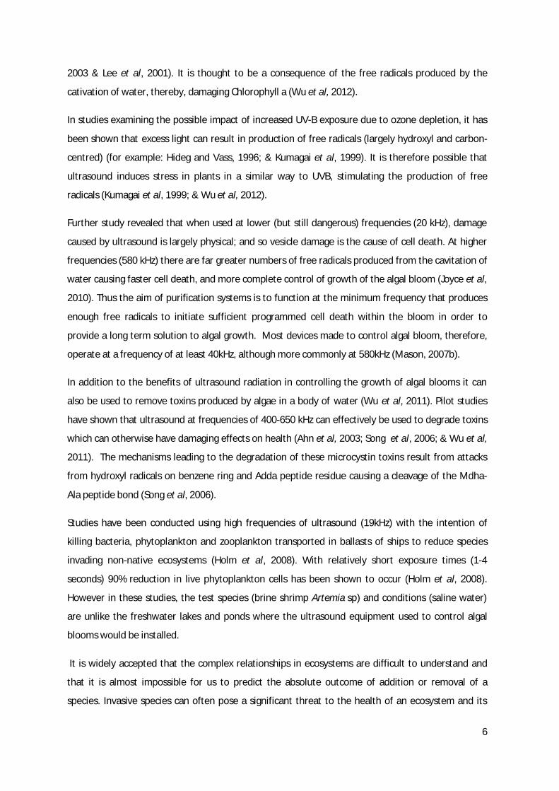
several biological problems. If a body of water is a drinking source, expensive methods of
purification, such as chlorination, are required to allow it to be free from toxins, and therefore, safe
to drink and if the water serves as a recreational site there is a loss in terms of profit or enjoyment
(Ahn et al, 2003; Dai et al, 2012; Eberhart et al, 2012; & Himberg et al, 1989). In addition to the
unpleasant attributes of algal blooms namely it is unsightly and often produces a bad taste or odour
(Ahn et al, 2003), cyanobacteria can also produce harmful toxins such as neurotoxins and
hepatotoxins (Ahn et al, 2003 & Dai et al, 2012). For example, the major bloom forming Microcystis
aeruginosa is a cyanobacterial species commonly found in eutrophic bodies of water, such as lakes,
ponds and reservoirs, and produces microcystins, potent hepatotoxins (Yoshida et al, 2008). Above
threshold levels, both microcystins (above 10000 cells ml-1) and associated hepatotoxins can be
poisonous to humans, causing liver failure, and also animals both domestic and wild: effects have
been most frequently monitored in cattle, dogs, pigs and waterfowl (Beasley et al 1989; Jochimsen
et al, 1998; Tango et al, 2004; & Wu et al, 2012).
Current methods of controlling algal blooms can be categorised into engineering, chemical and
biological methods (Wang et al, 2011). Engineering methods are not permanent and include
dredging the sludge or mechanical removal of the algae (Wang et al, 2011). Chemical methods, such
as copper algaecides, are harmful to the surrounding environment due to the adverse effect they
have on non-target freshwater organisms e.g. Daphnia, algaecides are also an expensive option for
developing countries (Saro et al, 2012; Wang et al, 2011 & Wu et al, 2011). There is a safer biological
option; planting macrophytes (e.g. Myriophyllum verticillatum) to intercept blooms, absorb the
leached nutrients and excrete polyphenols with negative allelopathic effects resulting in inhibited
algal growth, however the survival rate of these macrophytes is relatively low and so this is not a
reliable method of control (Chang et al¸2012; &Wang et al, 2011).
An alternative option is becoming more available, and has been shown to be effective if applied
correctly at irregular intervals daily throughout the year (Wu et al, 2011). Ultrasound radiation
provides a reliable way of inhibiting algal growth and killing cells without the secondary pollution
effects of chemical methods (Wu et al, 2011).
Successful ultrasonic algal control devices operate at mid frequencies (see table 1) (Wu et al, 2011).
Short exposures of high frequency ultrasound can to be used safety in medicine e.g. during an
ultrasound examination of a developing embryo (Tang et al, 2004). However, the higher frequency,
acoustic, waves of ultrasound have the ability to cause greater damage to cells; at frequencies of 580
kHz, maximum intensity exposure has been found to reduce algal mass by almost 50% in only 30
minutes (Joyce et al, 2010).

4
Table 1: The frequency of ultrasound wavelengths used to control algal blooms in context with low and high frequency uses. (Chandrapala et al, 2012, Joyce et al, 2010 & Wu et al, 2011)
Ultrasound frequency Intensity Use
20-100kHz Low E.g. Food processing
580 kHz Medium E.g. Control algal growth
<20MHz High E.g. Medical imaging
There are thought to be two main ways in which ultrasound radiation can lead to algal cell death:
disruption of the gas vesicles and also production of free radicals. Gas vesicles are vital to the
function of planktonic microorganisms; they provide buoyancy which allows them to migrate
vertically through the water column (Walsby, 1994). Buoyancy is necessary as it allows planktonic
microorganisms, including cyanobacteria, to obtain light energy from the sun that penetrates into
the top of the water column, which is necessary for photosynthesis (Addy & Green, 1996).
Disruption of the vesicles would disrupt buoyancy and therefore restrict the light available for
photosynthesis and will lead to a reduction in function and even cell death (Addy & Green, 1996; and
Wu et al, 2011). Free radicals (messenger molecules) initiate programmed cell death and disruption
of photosynthetic activities (Tang et al, 2004; & Ahn et al, 2003).
Gas vacuoles in algae are comprised of groups of microscopic vesicles, individually with a diameter
of approximately 75 millimicrons and a length ranging from 0.2 – 1.0 microns (Bowen & Jensen,
1965). Gas vesicles are found in several planktonic species of algae (Sharma et al, 2010). These
vesicles provide buoyancy which allows migration of cyanobacterial species e.g. Microcystis
aeruginosa, through the varying depths of the water column (Sharma et al, 2010). This migration
allows the algae to obtain the essential nutrients and light necessary for survival and the disruption
of such systems often proves fatal (Sharma et al, 2010). When applied to algae, the high frequency
sound waves causes collapse of the gas vesicles within the cells, and corresponding collapse of the
vacuole, through a process known as cavitation (Bowen & Jensen, 1965; & Zhang et al, 2009).
Acoustic cavitation is the rapid collapse of bubbles in water triggered by the ultrasound, this results
in high temperatures (>5000K) and pressures (>100 MPa), known as hot-spots, within the cells
(Zhang et al, 2009). There is a school of thought that this vesicle collapse is the primary cause of algal
cell death in response to ultrasound (Tang et al, 2004; Ahn et al, 2003; & Nakano et al, 2001). When
the vesicles are damaged, buoyancy is lost and the algal cells can no longer remain at the top of the
water column; they become ‘sedimented’ (Ahn et al, 2003). As light often fails to penetrate the
water column to the sediment, photosynthesis cannot occur and so these species of vesicle

5
containing algae are susceptible to damage from ultrasound (Ahn et al, 2003). However, this is not a
one-off method of control. Damaged gas vesicles have been seen to reform, restoring full function in
a relatively short time period (24 hours) (Lee et al, 2001), and so the repeated application of
ultrasonic is vital to its long term success in algal bloom control i.e. frequent exposure. Further
research is necessary to determine the biological mechanisms that allow this apparent vesicle
reformation.
The action of messenger molecules formed from the sonolysis of water, e.g. OH radicals, has also
been noted (Mason, 2007a). The production of these messenger molecules is thought to be due to
the presence of gas (oxygen) (Hart and Henglein, 1985). In experiments involving the irradiation of
solutions with ultrasound, solutions in the absence of oxygen failed to produce free radicals; the H
atoms forms instead H2 and the majority of OH forms hydrogen peroxide (Hart and Henglein, 1985).
However, with oxygen present there is formation of HO2 and OH radicals and O atoms (Hart and
Henglein, 1985). With such free radical production is it easy to see how other, non-target, organisms
may be jointly affected.
‘Sonoxide’ is a water purification system developed by Ashland in 2002 (Mason, 2007b). The system
combines an air supply with high frequency ultrasound exposure; this combination harms previously
healthy cyanobacterial cells through the production of free radicals (Mason, 2007a; & Mason,
2007b). The cells are then triggered into ‘Programmed Cell Death’ and in doing so produce, as of yet
unidentified, “signalling protein molecules” which are transmitted to the other cells in the algal
bloom (Mason, 2007b). Programmed cell death is then triggered by these signalling protein
molecules in cells in the biofilm of the water, including algae (Mason, 2007b).
Ultrasound can also be utilised to control non-vesicle containing algae as long as it is a filamentous
species such as the Spirogyra genus (Purcell, 2009). Although there have been far fewer studies
conducted into these species of algae and the effect ultrasound has upon them, it has been shown
that when silica is present in the cells, cavitating gas bubbles directly compromise the structure of
the filament, leading to damage to the joints and cell lysis (Purcell, 2009).
Although there is limited understanding at present of the specific mechanisms that disrupt
photosynthesis, studies have shown that, in addition to the collapse of gas vesicles, inhibition of
photosynthetic systems of blue-green algal blooms is also apparent; studies have shown that
ultrasound can reduce photosynthetic activity by 40.5% (Lee et al, 2001; Zhang et al, 2006a; & Wu et
al, 2012). Ultrasound-induced free radical production also directly inhibits photosynthesis (Ahn et al,
6
2003 & Lee et al, 2001). It is thought to be a consequence of the free radicals produced by the
cativation of water, thereby, damaging Chlorophyll a (Wu et al, 2012).
In studies examining the possible impact of increased UV-B exposure due to ozone depletion, it has
been shown that excess light can result in production of free radicals (largely hydroxyl and carbon-
centred) (for example: Hideg and Vass, 1996; & Kumagai et al, 1999). It is therefore possible that
ultrasound induces stress in plants in a similar way to UVB, stimulating the production of free
radicals (Kumagai et al, 1999; & Wu et al, 2012).
Further study revealed that when used at lower (but still dangerous) frequencies (20 kHz), damage
caused by ultrasound is largely physical; and so vesicle damage is the cause of cell death. At higher
frequencies (580 kHz) there are far greater numbers of free radicals produced from the cavitation of
water causing faster cell death, and more complete control of growth of the algal bloom (Joyce et al,
2010). Thus the aim of purification systems is to function at the minimum frequency that produces
enough free radicals to initiate sufficient programmed cell death within the bloom in order to
provide a long term solution to algal growth. Most devices made to control algal bloom, therefore,
operate at a frequency of at least 40kHz, although more commonly at 580kHz (Mason, 2007b).
In addition to the benefits of ultrasound radiation in controlling the growth of algal blooms it can
also be used to remove toxins produced by algae in a body of water (Wu et al, 2011). Pilot studies
have shown that ultrasound at frequencies of 400-650 kHz can effectively be used to degrade toxins
which can otherwise have damaging effects on health (Ahn et al, 2003; Song et al, 2006; & Wu et al,
2011). The mechanisms leading to the degradation of these microcystin toxins result from attacks
from hydroxyl radicals on benzene ring and Adda peptide residue causing a cleavage of the Mdha-
Ala peptide bond (Song et al, 2006).
Studies have been conducted using high frequencies of ultrasound (19kHz) with the intention of
killing bacteria, phytoplankton and zooplankton transported in ballasts of ships to reduce species
invading non-native ecosystems (Holm et al, 2008). With relatively short exposure times (1-4
seconds) 90% reduction in live phytoplankton cells has been shown to occur (Holm et al, 2008).
However in these studies, the test species (brine shrimp Artemia sp) and conditions (saline water)
are unlike the freshwater lakes and ponds where the ultrasound equipment used to control algal
blooms would be installed.
It is widely accepted that the complex relationships in ecosystems are difficult to understand and
that it is almost impossible for us to predict the absolute outcome of addition or removal of a
species. Invasive species can often pose a significant threat to the health of an ecosystem and its

7
ability to function (Nehring & Kolthoff, 2011). An example of such a species is Ludwigia grandiflora,
the water primrose, which originates in South America is currently threatening aquatic ecosystems
of Germany after being introduced in 2004 (Nehring & Kolthoff, 2011). Initially only a few individuals
were noted in Germany, however since 2009 rapid growth and spread of the plant have led to it
being placed on the German Black List for fears that the new species will greatly harm the existing
native ecosystems (Nehring & Kolthoff, 2011). If ultrasound is found to harm a component of the
aquatic ecosystem it will be difficult to predict the possible consequences.
In a UK freshwater ecosystem there are likely to be several different species of organisms,
depending on trophic status; the poorer the quality of the lake the lower the levels of available
nutrients and so species adapted to these conditions flourish where others cannot survive (Reynolds,
1998). In addition to the plant life within a freshwater ecosystem, there are likely to by several types
of zooplankton; species of: Daphnia, Cyclops, Rotifer, for example and also macroinvertebrates e.g.
species of Crustacean, Oligochaeta, Nematoda, amongst others (Reynolds, 1998).
The aim of this investigation is to establish whether the use of ultrasound as a method of algal
bloom control has any detrimental effects on an individual species (Daphnia) within an aquatic, lake
ecosystem.
Materials and method
Field Experiment
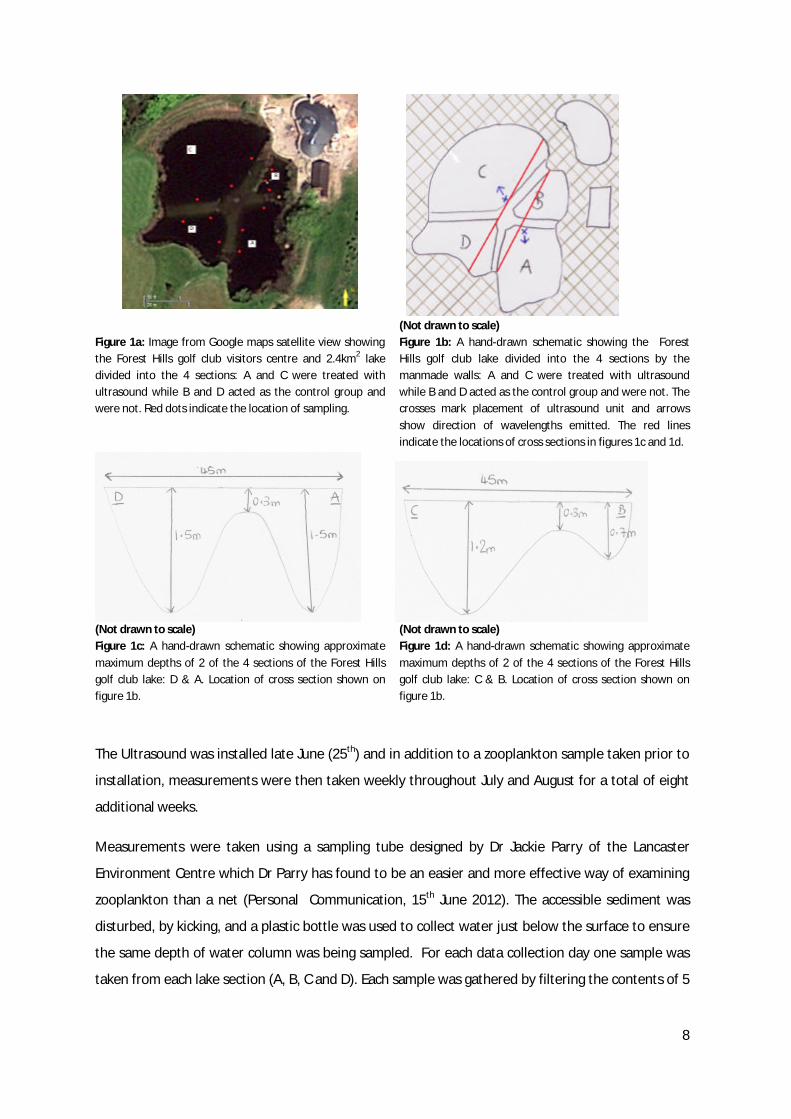
This phase of study was undertaken at Forest Hills golf club, Hazelrigg, Lancaster. An established
man-made lake, with approximate area 2.4km2, is situated there and in addition to the access to an
electricity supply that the ultrasound demands, the lake has the unusual benefit of being partitioned
into four sections. These barriers provide walls allowing a pathway for access to the fountain
situated in the lake’s centre, however the main benefit for the purpose of this study was the
separation allowing for two sections of the lake to be subjected to ultrasound (A&B) while the other
two may as the control (C&D) (see figures 1a, b, c & d). Ultrasound can penetrate solids with
relatively small thicknesses, which explains its use for medical imaging (Chan & Perlas, 2011)
however, with careful placement of the ultrasound unit, wavelengths into control areas can be
eliminated. One ultrasound unit was placed per treatment section on edge of a perimeter wall (see
figure 1b), the ultrasound unit faced into the section and was angled down slightly to avoid
wavelengths passing over the wall into a control section. Some assumptions need to be made at this
stage, for more information, see “Assumptions and limitations”.
8
Figure 1a: Image from Google maps satellite view showing the Forest Hills golf club visitors centre and 2.4km2 lake divided into the 4 sections: A and C were treated with ultrasound while B and D acted as the control group and were not. Red dots indicate the location of sampling.
(Not drawn to scale) Figure 1b: A hand-drawn schematic showing the Forest Hills golf club lake divided into the 4 sections by the manmade walls: A and C were treated with ultrasound while B and D acted as the control group and were not. The crosses mark placement of ultrasound unit and arrows show direction of wavelengths emitted. The red lines indicate the locations of cross sections in figures 1c and 1d.
(Not drawn to scale) Figure 1c: A hand-drawn schematic showing approximate maximum depths of 2 of the 4 sections of the Forest Hills golf club lake: D & A. Location of cross section shown on figure 1b.
(Not drawn to scale) Figure 1d: A hand-drawn schematic showing approximate maximum depths of 2 of the 4 sections of the Forest Hills golf club lake: C & B. Location of cross section shown on figure 1b.
The Ultrasound was installed late June (25th) and in addition to a zooplankton sample taken prior to
installation, measurements were then taken weekly throughout July and August for a total of eight
additional weeks.
Measurements were taken using a sampling tube designed by Dr Jackie Parry of the Lancaster
Environment Centre which Dr Parry has found to be an easier and more effective way of examining
zooplankton than a net (Personal Communication, 15th June 2012). The accessible sediment was
disturbed, by kicking, and a plastic bottle was used to collect water just below the surface to ensure
the same depth of water column was being sampled. For each data collection day one sample was
taken from each lake section (A, B, C and D). Each sample was gathered by filtering the contents of 5

9
x 500ml bottles through the net at the three positions illustrated in figure 1a (therefore a total of
7.5L of lake water was filtered to obtain one sample).Once collected in the tube, specimens were
carefully placed in containers filled with 70% ethanol and 30% pure water. The nets were examined
for any caught individual organisms and these were carefully removed with tweezers and added to
the sample container, finally the net flushed through with several sprays of pure water. The samples
where then transported back to the laboratory in the containers of ethanol and pure water and
identified (to species level where possible) using images and descriptions from a number of
freshwater identification guide books (Macan, 1959; Olsen et al, 2001; & Greenhalgh & Ovenden,
2007). Before identification, each sample was randomly assigned (using a random number
generator) a number and this replaced the label identifying lake section on the sampling tube. This
task was carried out by an individual who had no role in the species identification, this removed bias
from the identification process as the origin of the sample (treatment or control) was unknown.
As small freshwater zooplankton are shown to be sensitive to temperature; their growth can be
limited due to temperature extremes (Vidal, 1980; & Napper, 2009), the water temperature of each
bay was recorded at every sampling location on each visit.
Laboratory experiment
Two tanks, previously constructed for another experiment involving the company, were provided by
Sustainable Soil and Water Ltd in order to conduct the laboratory experiment. The first tank (the
control) was a simple 0.38m x 0.25m x 0.69m design, the second (the treatment) was the same
dimensions but with an additional cylindrical protuberance which houses the ultrasound unit, both
are made from a thick polymer (see figures 2a & 2b).
Figure 2a: The plastic 0.38m x 0.25m x 0.69m tank used as the control in the laboratory experiment. (Provided by Sustainable Soil & Water Ltd)
Figure 2b: The plastic 0.38m x 0.25m x 0.69m tank with ultrasound attachment, used as the treatment in the laboratory experiment. (Provided by Sustainable Soil & Water Ltd)

10
The in-built ultrasound unit is a “Pool Tec 10” obtained from hughes-sonic-systems.com, the device
operates at 110-240V and emits ultrasound at a frequency range of between 45-60Hz. Although this
is lower than the frequencies used in lakes, the structure of the tanks reflects wavelengths more due
to the normal incidence with which the wave comes into contact with the walls (90°) (Fellah et al,
2003), therefore in order to replicate the conditions of the lake as closely as possible a lower
frequency is used. The Daphnia were kept in RT media which was created following the procedure
described by Tollrian (1993) (see table 2). This choice was influenced by the PhD research of Piers
Napper (2009) which selected RT medium as a next best substitute to spring water, which would
have been too expensive to purchase for two large tanks which require at least 65 L each in order to
fully submerge the ultrasound unit.
Table 2: Elements (mg l-1) in RT medium adapted from Tollrian 1993. Two elements were excluded from the stock solution (Li (LiCl) and Se (Na2SeO3.5H2O)) due to lack of supply. As they were only trace elements and their benefit to daphnia could not be identified their absence was not thought to be significant. *TES = C6H15NO6S (N-Tris [hydroxymethyl] –methyl – 2 – aminoethane – sulphonic acid; Sigma T-1375) 8.5ml HCL (1N) per tank was used to lower the pH to 7.9 and a conductivity was found to be >220 µS and so no additional Ca(OH)2 was required. Trace elements (stock solution) EDTA (disodium salt) 500.0 B (H3BO3) 572.0 Fe (FeCl3) 322.46 Mn (MnCl2.4H2O) 72.0 K (KBr) 7.5 Mo (Na2MoO4.2H2O) 12.5 Cu (CuCl2.H2O) 6.5 Co (CoCl2.6H20) 20.0 I (KI) 0.6 10ml stock solution 1-1 medium Main elements TES* 85 CaCl2.2H2O 39 NaNO3 50 MgSO4.7H20 20 Na2SiO3.5H2O 10 KCl 10 CaCO3 13 Ca(OH)2 30
The Daphnia were fed on a supply of dried blue green algae (Spirulina) every 3 days, found to be an
acceptable substitute to live algae if unavailable (Napper, 2009). They were kept by a window within
their preferred temperature range (15-20°C) (Napper, 2009) and with equal access to sunlight.

11
The number of Daphnia present was measured daily, with sampling occurring at the same time each
day. 360ml samples were collected from the same depth in the water column daily (in the morning)
and were taken at 5 fixed distances from the ultrasound unit (1= closest to unit, 5= furthest away).
The samples were then examined under a microscope and numbers of Daphnia were recorded, once
counted Daphnia were returned to their original tank.
Statistical analysis
The field measurements contained data nested within categories; the data can be grouped into
treatment and control but then sub-divided into the lake sections A, B, C and D. The data were
analysed for differences in weekly measurement of Daphnia number, number of species and number
of individuals. This analysis was carried out using a two-level nested-design hierarchical ANOVA
(analysis of variance) in the statistics software package SPSS. The fixed factor for the ANOVA was the
week of the observation and the nested factors were the treatment type (treated vs non-treated)
followed by the lake section (A and C or B and D).
Further analysis involved calculating coefficient of variance (%) in SPSS, as the scales of the variables
(number of Daphnia, number of species and number of individuals) were different, calculation of the
coefficients of variance allowed for comparison of variation of the data between the three.
The water temperatures for each lake section were averaged and linear regression performed in
SPSS to test for a relationship between each of the three variables and the temperature of the
water. This was a precautionary measure to ensure that the slight, natural fluctuation in water
temperature was not sufficient to cause increased mortality to the zooplankton, in particular
Daphnia.
The laboratory data were analysed in two ways: firstly the number of Daphnia were calculated and
secondly the data was tested to determine if there was a relationship between number of Daphnia
and distance from the ultrasound unit.
To test the number an independent t-test was performed (also known as a Levene’s test), in
addition, data were plotted of number against time and a linear regression fitted to further
determine if ultrasound effected reproduction or mortality.
The relationship of Daphnia number and distance from ultrasound was tested by performing a
further linear regression.
12
Assumptions and limitations
Due to uncontrollable nature of some aspects of a field study there are a few limitations of the
experiment and assumptions that must be made.
Though the lake is split into four sections these sections are not of equal surface area and in
addition they are not of equal depth. From observation, sections A and D are deeper than A and B
with due to lack of access it is not possible to assess the volume of each section (see figure 1c). It is
also difficult to judge where the perimeter boundaries for each section lie are there are large
numbers of reeds present surrounding the lake. Due to this limitation, this study assumes that
volume of the bays is not a factor in the effect of ultrasound or on the number or composition of
species present.
Each bay varied in terms on plant composition and density. This potentially could impact results as
certain organisms may prefer the habitat of one bay over other, regardless of ultrasound. Again, due
to access, there is no data in this study on plant composition in bays and as such is a limitation. For
the duration of this study it will be assumed that the lake habitat is uniform enough not to cause a
significant organism preference for bay that will impact the effects of ultrasound.
While the bays have barriers that prevent large-scale transfer of water, organism and, in the case of
this study, ultrasound exchange they are not fully independent. Due to the placement of ultrasound
units it is very unlikely that ultrasound wavelengths were reaching the control sections of the lake.
However, it is undeniable that water and organisms would be able to move between bays. As this
study was investigating the presence of organisms and species compositions within the bays and not
mortality, this is not a major limiting factor. Even if the zooplankton did not appreciate the
ultrasound and had a behavioural reaction to it which involved moving from one bay to another, this
would be shown in the results as ultrasound bays would have lower number. However, during the
study we will assume that each bay is independent in terms of ultrasound treatment.
Results
Field experiment
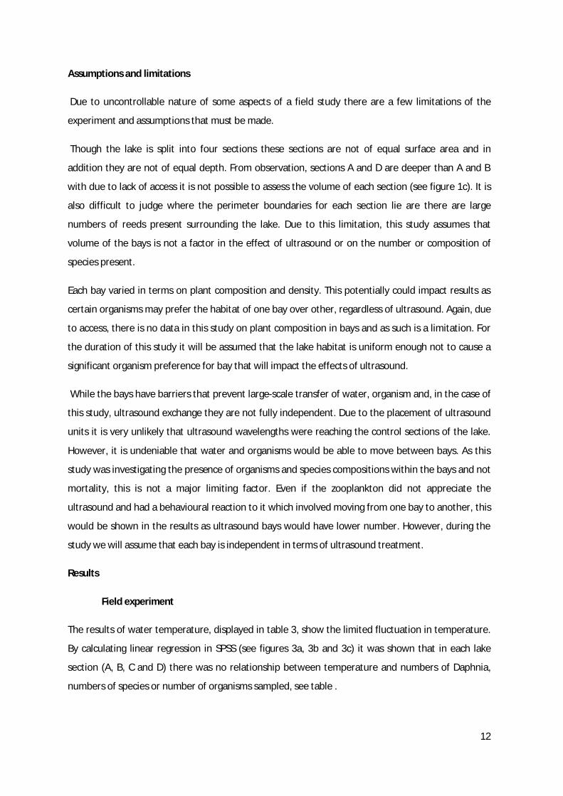
The results of water temperature, displayed in table 3, show the limited fluctuation in temperature.
By calculating linear regression in SPSS (see figures 3a, 3b and 3c) it was shown that in each lake
section (A, B, C and D) there was no relationship between temperature and numbers of Daphnia,
numbers of species or number of organisms sampled, see table .
13
Table 3: The weekly average water temperature (°C) for each lake section. The water temperatures were measured at the same water collection points as shown in figure 1a. Calculations were performed in SPSS.
Week water temperature recorded Average recorded water temperature (°C)
1 2 3 4 5 6 7 8 9
A 15.5 15.4 15.4 15.4 14.5 15.6 15.2 14.3 15.9 B 15.6 14.4 15.7 14.2 14.3 14.1 14.6 14.8 14.6 C 14.4 14.7 15.3 15.3 15.3 15.8 15.0 14.4 15.3 D 15.3 15.2 14.7 15.2 14.4 15.3 15.5 14.4 14.8
Figure 3a: Regression lines fitted to data from each lake section (A, B, C, D) to test the relationship between the number of Daphnia observed and the average water temperature of each lake section 0C. Linear regression in SPSS found no relationship between temperature and Daphnia numbers for any of the lake sections (see table 4).
Figure 3b: Regression lines fitted to data from each lake section (A, B, C, D) to test the relationship between the number of species observed and the average water temperature of each lake section 0C. Linear regression in SPSS found no relationship between temperature and species numbers for any of the lake sections (see table 4).
14
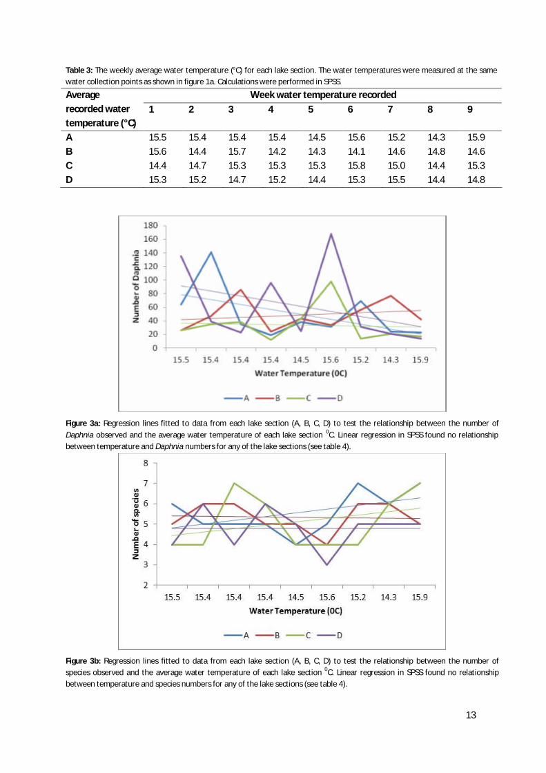
Figure 3b: Regression lines fitted to data from each lake section (A, B, C, D) to test the relationship between the number of individual organisms observed and the average water temperature of each lake section 0C. Linear regression in SPSS found no relationship between temperature and the number of individual organisms for any of the lake sections (see table 4). Table 4: Results from linear regressions performed in SPSS to test the relationships between each of the three variables (number of Daphnia, number of individuals and number of species) and the average weekly water temperature (0C) in each lake section (A, B, C and D). All adjusted R2 values are low, the highest only 0.385, showing poor fits of the data points to the trend line. All p-values were greater than 0.05 proving no observed significance between water temperature and each variable.
Section tested Variable tested for relationship with water temperature
Adjusted R2
P-value Significant? (<0.05)
Number of Daphnia 0.127 0.313 No A Number of individuals 0.124 0.343 No
Number of species 0.037 0.346 No Number of Daphnia 0.077 0.238 No Number of individuals 0.212 0.119 No
B
Number of species 0.036 0.292 No C Number of Daphnia 0.214 0.118 No Number of individuals 0.385 0.044 No Number of species 0.125 0.748 No D Number of Daphnia 0.237 0.104 No Number of individuals 0.180 0.141 No Number of species 0.136 0.845 No
The numbers of Daphnia present in both control and ultrasound-treated sections of the lake
fluctuated greatly (figure 4a). In the control sections (B and D) the highest average recorded number
was in week 6 with a mean of 101 Daphnia detected in each section per 7.5L sample and the lowest
in week 9 where a mean of only 29.5 Daphnia were recorded. In ultrasound treated sections the
largest average number of Daphnia found per 7.5L sample per week was during week 2 where a
mean of 87.5 Daphnia were collected and the lowest was week 4 where the mean was only 19
15
Daphnia per section. This fluctuation in results can be shown in figure 4a, where whilst the numbers
decrease from week 1 to week 9, due to the data in the interim weeks there is no clear trend in the
data. The lack of obvious trend is shown by performing a hierarchical (nested) ANOVA (results in
table 5) where the p-value calculated when comparing number of Daphnia between treated and
non-treated groups (p=0.875) illustrates the lack of significant relationship; ultrasound had no effect
on Daphnia numbers in this field study.
The number of individual organisms present in control sections of the lake and ultrasound treated
sections of the lake again varied greatly (figure 4b). In the control sections (B and D) the highest
average recorded number of individuals was in week 1 with a mean of 112 individual organisms
detected in each section and the lowest in week 5 where a mean of only 50.5 individual organisms
were collected. In ultrasound treated sections the largest average number of individual organisms
found per week was during week 2 where a mean of 95 individual organisms were collected and the
lowest was week 9 where the mean was only 32 individual organisms per section. This fluctuation in
results can be shown in figure 4b, where whilst the numbers decrease from week 1 to week 9 there
is again no clear trend in the data. The lack of obvious trend was again shown by performing a
hierarchical (nested) ANOVA (results in table 5) where the p-value calculated when comparing
number of individual organisms between treated and non-treated groups was shown to be 0.229
therefore there is no significance and it can be said ultrasound had no effect on individual organisms
numbers in this study.
The number of species present in control sections of the lake and ultrasound treated sections of the
lake varied once more (figure 4c). In the control sections (B and D) the highest average recorded
number of species was in weeks 2,7 and 9 where a mean of 6 species were detected in each section
and the lowest in weeks 6 and 8 where a mean of only 3.5 species were collected. In ultrasound
treated sections the largest average number of species present per week was during weeks 3, 8 and
9 where a mean of 6 species were collected and the lowest was week 5 where the mean number of
species per section was found to be 4. These smaller fluctuations in results can be seen in figure 4c,
where whilst the numbers decrease from week 1 to week 9 there is again no clear trend in the data.
The lack of obvious trend was again shown by performing a hierarchical (nested) ANOVA (results in
table 5) where the p-value calculated when comparing number of species between treated and non-
treated groups was shown to be 0.288 therefore there is no significance and it can be said
ultrasound had no effect on the number of species present in this study, a full list of species found in
this study can be seen in appendix I & II.

16
Figure 4a: Line graph illustrating how number of Daphnia fluctuated over the 9 week study, both in control (blue) and ultrasound treated (red) sections of the lake. Each data point represents the mean number of Daphnia observed per visit in a 7.5L sample from either a control or treatment bay as labelled. A hierarchical ANOVA shows no significant trend in the data collected (see table 3). Error bars represent +1SE of the mean. Calculations were performed in SPSS.
Figure 4b: Line graph illustrating how numbers of individual organismsfluctuated over the 9 week study, both in control (blue) and ultrasound treated (red) sections of the lake. Each data point represents the mean number of individual organisms (see appendix I & II for full species list) observed per visit in a 7.5L sample from either a control or treatment bay as labelled. A hierarchical ANOVA shows no significant trend in the data collected (see table 3). Error bars represent +1SE of the mean. Calculations were performed in SPSS.
Figure 4a: Line graph illustrating how numbers of different species present in samples fluctuated over the 9 week study, both in control (blue) and ultrasound treated (red) sections of the lake. Each data point represents the mean number of species (see appendix I & II for full species list) observed per visit in a 7.5L sample from either a control or treatment bay as labelled. A hierarchical ANOVA shows no significant trend in the data collected (see table 3). Error bars represent +1SE of the mean. Calculations were performed in SPSS.
Table 5: Results from the hierarchical ANOVA showing no significance (all p values>0.05) between treatment (ultrasound) and control groups for number of Daphnia, number of individuals and number of species observed over a 9 week field study. Calculations were performed in SPSS. P value Significant (<0.05)? Number of Daphnia 0.875 No Number of individual organisms 0.229 No Number of species 0.288 No

17
Due to the large differences in data values and therefore mean averages of the data sets displayed in
figures 4a, 4b and 4c coefficient of variance percentages (table 6) were calculated in excel in order to
provide a comparison between the three. There was higher variability in the data sets containing
sample numbers of Daphnia than both number of individuals and number of species data sets for
control and treatment lake sections. The data sets containing number of individual organisms found
contained more variability in both control and ultrasound treated bays than number of species
sampled.
Table 6: The coefficient of variance (%) for the three test variables (number of species, number of individuals and number of Daphnia) in both control and treatment (ultrasound) groups. Percentage of variance is highest for the number of Daphnia observed and lowest for number of species. Calculations were performed in SPSS.
Coefficient of variance (%) Number of Species Number of individuals Number of Daphnia Control 20 30 42 Ultrasound 15 41 54
Laboratory experiment
In the control tank the number of Daphnia did not significantly alter throughout the course of the
week (see figure 5a). The linear regression in SPSS calculated an adjusted R2 of 0.101 (see table 7);
this shows that the data points correspond poorly to the line of best fit. This lack of trend is
confirmed with the P-value of 0.485 which shows no significant relationship between the number of
Daphnia in the tank and the duration of the 5 day experiment. Similar results were found in the
control tank as can be seen in figure 5b. The adjusted R2 value of 0.095 shows the data points fit
even less well to the trend line than the data provided through observations of the Daphnia number
in the control tank. The linear regression provides a P-value of 0.478 (table 7) which again shows no
significant relationship.
The lack in differences of number of Daphnia in each tank was confirmed with an independent t-test
comparing the daily numbers of Daphnia observed. The t-test, performed in SPSS, calculated the
mean daily Daphnia number to be for the 1.72 control and 1.40 in the treated tank (see table 8). The
P-value calculated was 0.321 and so there is no significant difference between the Daphnia numbers
in both tanks.

18
Figure 5a: A scatter plot displaying the relationship between the total numbers of Daphnia observed in the 1800ml of water sampled from the control tank (no ultrasound) daily over the course of the experiment. Each data point represents the total number of Daphnia sampled per day. A linear regression was fitted to the data series in SPSS and a significance of fit to the data points of 0.174 was calculated, indicating poor fit. Calculations were performed in SPSS.
Figure 5b: A scatter plot displaying the relationship between the total numbers of Daphnia observed in the 1800ml of water sampled from the experimental tank (with ultrasonic device present) daily over the course of the experiment. Each data point represents the total number of Daphnia sampled per day. A linear regression was fitted to the data series in SPSS and a significance of fit to the data points of 0.179 was calculated, indicating an almost equally poor fit of data to a trend line as in the control tank. Calculations were performed in SPSS.
Table 7: Linear regression data analysing relationships between the number of Daphnia over time and the placement of Daphnia in relation to the placement of the ultrasound emitting unit in both a controlled environment (no ultrasound) and a test environment (with ultrasonic device present). As all p-values were >0.05 none are classed as a significant. Calculations were performed in SPSS.
Tank tested Variables tested Adjusted R2 P-value Significant? (<0.05) Number of Daphnia and Day
0.101 0.485 No Control
Average daily Daphnia number and distance from ultrasound unit
0.169 0.562 No
Number of Daphnia and Day
0.095 0.478 No Ultrasound
Average daily Daphnia number and distance from ultrasound unit
0.0258 0.699 No
Table 8: T-test data analysing the difference in means between Daphnia numbers in a control (no ultrasound) and a test environment (with ultrasonic device present). As the p-value is >0.05 there is no significant difference between the environments. Calculations were performed in SPSS.
Comparison Mean average number of Daphnia per section per day
P-value Significant? (<0.05)
Control
1.72
Ultrasound tank 1.40
0.321
No
19
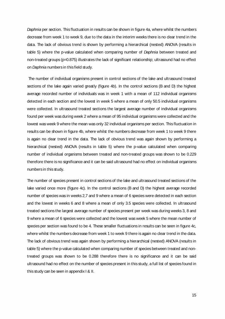
The placement of the Daphnia within the tank was also investigated. In the control tank there was
no ultrasound unit present and so the measurements of distance from ultrasound (1-5) mirror the
distances from the unit in the test tank. In both cases; control and test, adjusted R2 values were low:
0.169 and 0.0258 respectively, indicating a lack of trend in the data sets (see table 7). Linear
regression provided P-Values of 0.562 for the control data and 0.699 for the test data; therefore
there is also no significant relationship between number of Daphnia and placement in the either
tank.
Figure 6a: A scatter plot showing the relationship between the average numbers of Daphnia observed in the 360ml of water sampled at each distance from the ultrasound unit in the control tank (no ultrasound). Each data point represents the average number of Daphnia observed over the duration of the experiment. A linear regression was fitted to the data series in SPSS and a significance of fit to the data points of 0.123 was calculated indicating poor fit. Calculations were performed in SPSS.
Figure 6b: A scatter plot showing the relationship between the average numbers of Daphnia observed in the 360ml of water sampled at each distance from the ultrasound unit in the experimental tank (with ultrasonic device present). Each data point represents the average number of Daphnia observed over the duration of the experiment. A linear regression was fitted to the data series in SPSS and a significance of fit to the data points of 0.057 was calculated indicating an even poorer fit than in figure 6a. Calculations were performed in SPSS.
Discussion
The analysis of the average water temperatures throughout the course of the experiment reveals
that despite the natural fluctuations in temperature, there was no relationship between water
temperature and any of the three variables (Daphnia number, number of individuals or number of
species). It was necessary to measure temperature as is it possible a sudden change in temperature
could have had a detrimental effect on zooplankton numbers and provided a false relationship
between ultrasound and mortality, however this has not been the case. In addition to the lack of
relationship between temperature and Daphnia in the study, this field experiment shows that
commercial ultrasound use also does not appear to have a detrimental effect on the number of
species within a freshwater ecosystem nor the number of these organisms. This indicates that the
mid-frequency ultrasound wavelengths emitted do not reduce reproduction, increase mortality rates
or negatively alter the environment in a way that decreases its suitability for zooplankton. The
20
laboratory experiment supports the above findings, by indicating that commercial ultrasound may
not have a detrimental impact on Daphnia in this setting. In addition they show that Daphnia do not
appear to migrate away from the source of the ultrasound. However, due to Daphnia being sensitive
to laboratory conditions and the lack of repetition within the study this cannot be said with
confidence. There were no signs however that ultrasound caused immediate mortality in Daphnia
however as at least some specimens survived for the full 5 days being subjected to ultrasound
wavelengths.
Field experiments are vital in testing ecological consequences of a non-natural influence as they
involve the full ecosystem which is impossible to accurately recreate in a more sterilised
environment (for example: Tipping et al, 1999 and Smith et al, 1999) however for a more controlled
setting laboratory experiments provide data which is easier to interpret as there is a more obvious
relationship between cause and effect (For example: Hartgers et al, 1998) and so conducting both
often allows the deepest possible understanding (for example: Kestrup & Ricciardi, 2009). With this
study the field experiment found ultrasound to have no short-term effect on the aquatic ecosystem
of the freshwater lake and this was supported by the investigation conducted in the laboratory.
Although zooplankton are known as sensitive organisms, they are frequently used to assess good
water quality (Gannon and Stemberger, 1978; & Lal et al, 1984). It may be possible that they are
more resilient than algae to ultrasound stress. The sensitivity of Daphnia to certain abiotic stresses
largely relates to their moulting cycle which is closely related to their reproductive cycle (see figure
6); if Daphnia are exposed to a pollutant at a particularly sensitive stage, this can be detrimental to
the success of reproduction (Lal et al, 1984). This study shows that Daphnia are do not appear to be
sensitive to mid-frequency ultrasound unlike other stresses such as copper, zinc and insecticides
which are known to have detrimental effects on Daphnia (Hoang and Klaine 2007; & Hayasaka et al,
2012) however it does not say why this might be.

21
Figure 7: Diagram depicting the various morphological stages of Daphnia. The red dots represent the various moulting stages throughout the lifecycle which are undergone prior to maturity. During these moulting phases, Daphnia are particularly sensitive. (Adapted from: Ebert, 2005)
Daphnia, like other zooplankton, have the ability to swim because of their morphology; their 2nd
antennae are used to propel themselves through the water (Ringelberg, 1999). This allows them to
migrate through the water column allowing them to access nutrients, light, to locate suitable mates
and, if conditions are unfavourable, to sink to the floor of the water system to lay their “resting
eggs” (Destasio et al, 1995; & Ringelberg, 1999). Gas vesicles provide buoyancy which are needed by
non-motile organisms to stay at the top of the water column (Walsby, 1994). However, as non-
photosynthetic organsisms Daohnia are not required to remain solely at the top of the water column
and due to their propulsion ability Daphnia do not require a gas vesicle (Walsby, 1994). As so much
of the research isolated the disruption of the gas vesicle by acoustic cavitation (Tang et al, 2004; Ahn
et al, 2003; & Nakano et al, 2001), and the zooplankton lack these air pockets this may explain the
lack of effect ultrasound has on them. However, other studies suggested that the production of free
radicals was the cause of algal reduction in non-vacuole containing algae (Mason, 2007a), the
production of these messenger molecules could explain why high-frequency ultrasound has been
proven to be fatal to zooplankton in ballasts of ships (Holm et al, 2008) but does not seem to explain
the lack of fatality in the field portion of this study.

22
As ultrasound has been shown to be detrimental to some species of zooplankton (Artemia sp.,
Ceriodaphnia dubia, Brachionus plicatilis, Brachionus calyciflorus, and Philodina sp.) found in the
ballast of ships (Holm et al, 2008) it is logical to conclude that under some conditions zooplankton
will be affected by ultrasound. Even if zooplankton are not susceptible to endogenous free radicals
being produced in algae as a result of ultrasonic wavelengths (which hasn’t been investigated
specifically in this study) and are more resilient to lower frequencies of ultrasound due to their lack
of gas vesicles it is possible that at some, currently unknown, higher frequency of ultrasound
wavelength, zooplankton will become sensitive to its effects.
Due, again, to their ability to move (Ringelberg, 1999), it could simply be that the zooplankton are
eliciting a behavioural response that is allowing them to escape the harmful wavelengths of
ultrasound. Laboratory study indicated that this may not be the case as there didn’t seem to be any
relationship between numbers of Daphnia and location within the tank. This is perhaps not
surprising as it is difficult to comprehend that this response would already exist as ultrasound is a
new stress for zooplankton and behavioural responses are either taught through mimicry, innate or
learned over a period of trial and error (Zentall, 2006), it is however possible that zooplankton may
evolve to partake in a behavioural response in the future.
This research reveals that ultrasonic control appears to be a method of algal control that is safe to
the Daphnia, and potentially other zooplankton, present in aquatic ecosystems such as the one
studied. By using ultrasound as an alternative to previous control methods such as mechanical,
chemical or biological means it is possible that the negative attributes associated with reliability and
environmental damage can be avoided (Wu et al, 2011). Ultrasound units are smaller and easier to
install and operate than mechanical machinery and technologies currently available as algal bloom
control methods and therefore more accessible (Wang et al, 2011). This could be particularly
important to small businesses or homeowners who wish to control algal blooms or if there is an
unsightly algal bloom in a domestic, relatively small water system. And while chemical methods of
control such as the copper algaecides mentioned previously have been known to harm non-target
organisms (Saro et al, 2012; Wang et al, 2011; & Wu et al, 2011), this study has shown that Daphnia
are not harmed when subjected to wavelengths of ultrasound and so suggests that ultrasonic algal
control could be an ecosystem-friendly option. Easily the most environmentally friendly option are
methods of biological control (Chang et al, 2012; &Wang et al, 2011), however due to their low rate
of reliability this is not considered a feasible control method of large-scale algal control (Chang et al,
2012). While ultrasonic control requires a power supply in order to operate, making it less
environmentally friendly than biological control, it is certainly more dependable (Zhang et al, 2006b).
23
Therefore using ultrasonic control instead of previously established methods of algal control will be
easier and more readily available, have fewer environmental consequences and be a more reliable
option.
Further study considerations
The field experiment conducted for this study examined the effect of ultrasound over a 9 week
period due to time restrictions involved. A more thorough investigation may compare the effect of
ultrasound treatment to control over a much longer time period as it common place in aquatic
ecosystem monitoring studies. It is possible that while the wavelengths of ultrasound are not directly
harming the zooplankton, perhaps due to their lack of gas vesicles, the loss of vesicle-containing
organisms such as aquatic cyanobacteria and planktonic bacteria (Walsby et al, 1992), could pose a
long term threat to the ecosystem. Either by the reduction of a species of unknown importance in
the ecosystem or by the build-up of a substance released during acoustic cavitation, there are
potentially unknown consequences that a nine week study would not pick up on.
Algal growth causes aesthetic problems in smaller bodies of water such as ornamental lakes and
ponds (Ahn et al, 2003). Larger concerns arise when toxins contaminate drinking water (Ahn et al,
2003; & Dai et al, 2012) or when the presence of algae causes disruption of leisure activities such as
when the Great North Swim had to be cancelled in 2010 (BBC, 2010). Contamination of larger bodies
of water can therefore have large, detrimental economic consequences, which are becoming
increasingly harmful to local societies due to the struggling world economy (Ahn et al, 2003; & BBC,
2010). Larger-scale studies need to be conducted firstly to determine the feasibility of the use of
ultrasonic algae control for such big volumes of water and also to determine the practicalities of it
e.g. how many units would be required, would power be available, would frequencies need to
increase etc. The current expense of carrying out such investigations could prove to be beneficial to
future generations if it provides a less expensive algal growth control method.
Previous study has shown that free radical production can lead to algal cell death when exposed to
ultrasound (Mason, 2007a), however, the specific free radicals thought to be triggering programmed
cell death are yet to be fully understood (Ahn et al, 2003; & Broekman et al, 2010). It is thought the
difficulty in identifying these substances is due to their ability to be active at low quantities for very
short periods of time, and therefore linking the presence of trace molecules to cell mortality is
incredibly difficult (Broekman et al, 2010). While this study did not find a negative effect of
ultrasound on zooplankton, it cannot be concluded that this indicates a resistance to these
messenger molecules; only if free radicals are tested for, found to be present and still no detrimental
24
effects are observed could this conclusion be drawn and this will be incredibly difficult to achieve
with today’s technologies (Broekman et al, 2010).
Daphnia are known to be a sensitive zooplankton (Gannon and Stemberger, 1978; & Lal et al, 1984)
however it is also know that other organisms, such as Ceriodaphnia, can be more sensitive to
different stresses for example insecticides (Hayasaka et al, 2012). It may therefore be of interest to
conduct further laboratory trials with freshwater species of zooplankton with a lower stress
threshold than Daphnia to ensure that they do not suffer with the use of ultrasonic algal control.
Previous study involving saltwater ecosystems have shown that ultrasound at high frequencies can
be used to remove non-native zooplankton species (Holm et al, 2008). It would be logical to
conclude, therefore, that there is a limit to how much ultrasound zooplankton can receive before
levels become harmful. As ultrasonic control has the potential to become a widely used technology,
it would be sensible to conduct further study to determine the safe frequencies of ultrasound in
various different conditions.
Conclusion
This study aimed to determine if new methods of ultrasonic algal control would cause detrimental
effects on freshwater ecosystems, as some predecessor methods are prone to do. Studies in both
the field and in the laboratory indicated that there are no such negative consequences of ultrasound
use for Daphnia and ultrasound could develop into the best current method of algal control in terms
of the lack of known environmental damage caused to non-target organisms.
Ultrasound still has its flaws: it cannot be a completely “green” technology, as it has been referred
to in some literature (for example: Hutchinson, 2008) as it requires a power supply, it isn’t a
permanent, one-off solution (Wu et al, 2011) and there is no present way to determine the amount
of money that large scale algal control by ultrasound may cost, of if it is even possible. However,
previous studies show it is more reliable on a smaller scale than truly green biological methods of
control and this study indicates that ultrasound may not have the same damaging effect on non-
target organisms seen in other studies that used chemical methods of control (Wang et al, 2011; &
Wu et al, 2011)
This study is a starting point for this area of research. It shows how on a small scale there is no
apparent impact of ultrasound on Daphnia, and therefore may not be one for freshwater
ecosystems as a whole but there are still many more points to address. Further research must be
performed to develop the technology from its current application in small ponds, lakes and pools to
operate at a more industrial scale. In order to control algal growth and prevent toxins in large bodies
25
of drinking water, particularly in developing countries where chemicals are currently often used out
of necessity, larger, more powerful ultrasound devices will doubtless be needed. It is imperative that
such advances in the equipment involved do not come at a cost to the non-target plants and
organisms living as part of freshwater ecosystems and so studies such as this must be conducted at
each stage of production.
Ultrasound technologies are presently used to great effect in the UK for algal control largely for
aesthetic reasons, but their real social and environmental benefit will be further afield in
economically less developed countries where they could replace currently used chemical
alternatives. The potential is vast but these technologies are still at an early stage of development.
Acknowledgements
I would firstly like to thank Simon Brockholes for the concept of this project, without his provision of
equipment and interest in the field I would not have been given an opportunity to partake any active
study into this area of environmental biology.
Secondly, my biggest thank-you is to Dr Ian Dodd, my dissertation supervisor, who did not fail to
provide answers to my questions and never ceased to allow me to intrude upon his valuable office
hours.
Thanks to the staff and owners of Forest Hills golf club, Lancaster, for allowing me to wade around in
their lake all summer.
I am very grateful to both Dr Jackie Parry and Shane Rothwell from Lancaster’s environment centre
for their advice, guidance and assistance both out in the field and during the laboratory experiment.
As always, I must acknowledge my dad, Richard Hedge, for putting up with me and my incessant
worrying and for being an excellent personal taxi service. And in particular I would like to mention
Stephanie Cotton, largely for her help in the laboratory and moral support but also for thinking I am
much better at statistics than I actually am, it’s a great boost to my ego.
References
Addy, K. & Green, L. (May,1996) Phospjorus and Lake Aging. Natural resources facts. Retrieved from: http://www.uri.edu/ce/wq/ww/Publications/Phosphorus.pdf
Ahn, C., Park, M., Joung, S., Kim, H., Jang, K. & Oh, H. (2003) Growth inhibition of cyanobacteria by ultrasonic radiation: laboratory and enclosure studies. Environmental Science and Technology, 37, 3031 – 3037.
26
BBC (2010, September 3). Cumbria Great North Swim cancelled over safety fears. Retrieved from: http://www.bbc.co.uk/news/uk-england-cumbria-11175208
Beasley, V. R., Cook, W. O., Dahlem, A. M., Hooser, S. B., Lovell, R. A. & Valentine, W. M. (1989) Algae intoxication in livestock and waterfowl. The veterinary clinics of North America. Food animal practice, 5, 345-361.
Bowen, C. C. & Jensen, T. E. (1965) Blue-green algae: fine structure of the gas vacuoles. Science, 147, 1460 – 1462.
Broekman, S., Pohlmann, O., Beardwood, E. S. & Cordemans de Meulenaer, E. (2010) Ultrasonic treatment for microbiological control of water systems. Ultrasonics Sonochemistry, 17, 6, 1041-1048.
Chan, V. & Perlas, A. (2011) Basics of ultrasound imaging. Retrieved from: http://www.google.co.uk/url?sa=t&rct=j&q=&esrc=s&source=web&cd=4&sqi=2&ved=0CEUQFjAD&url=http%3A%2F%2Fwww.springer.com%2Fcda%2Fcontent%2Fdocument%2Fcda_downloaddocument%2F9781441916792-c1.pdf%3FSGWID%3D0-0-45-1061051-p174073264&ei=PGT1UMLmAeLM0AW4kYD4AQ&usg=AFQjCNGa1YfXRBrZWGIF8LPwdwg93PBMJQ&bvm=bv.41018144,d.d2k
Chang, X., Eigemann, F. & Hilt, S. (2012) Do macrophytes support harmful cyanobacteria? Interactions with a green alga reverse the inhibiting effects of macrophytes allelochemicals on Microcystis aeruginosa. Harmful Algae, 19, 76-84.
Dai, G. Z, Shang, J. L. & Qui, B.S. (2012) Ammonia may play an important role in succession of cyanobacterial blooms and the distribution of common algal species in shallow freshwater lakes. Global change biology, 18, 1571 – 1581.
Destasio, B. T., Rudstam, L. G., Haning, A., Soranno, P. & Allen, Y. C. (1995) An in-situ test of the effects of food quality on Daphnia population growth. Hydrobiologia, 307, 1-3, 221-230.
Eberhart, B. T. L., Bill, B. D. & Trainer, V. L. (2012) Remote sampling of harmful algal blooms: A case study on the Washington State coast. Harmful Algae, 19, 39-45.
Ebert, D (2005) Ecology, Epidemiology, and Evolution of Parasitism in Daphnia [Internet]. Available from: http://www.ncbi.nlm.nih.gov/entrez/query.fcgi?db=Books
Fellah, Z. E. A., Depollier, C., Berger, S., Lauriks, W., Trompette, P. & Chapelon, J. Y. (2003) Determination of transport parameters in air-saturated porous materials via reflected ultrasonic waves. Journal of the Acoustical Society of America, 114, 5, 2561-2596.
Gannon, J. E. & Stemberger, R. S. (1978) Zooplankton as indicators of Water Quality. Transactions of the American Microscopical Society, 97, 1, 16tor-35.
Greenhalgh, M. & Ovenden, D. (2007) Freshwater life. London:Collins.
Hart, E. J. & Henglein, A. (1985) Free radical and free atom reactions in the sonolysis of aqueous iodide and formate solutions. J. Phys. Chem, 89, 4342-4347.

27
Hartgers, E. M., Aalderink, G. H., Van den Brink, P. J., Gylstra, R., Wiegman, J. W. F. & Brock, T. C. M. (1998) Ecotoxicological threshold levels of a mixture of herbicides (atrazine, diuron and metolachlor) in freshwater microcosms. Aquatic Ecology, 32, 135-152.
Hayasaka, D., Korenaga, T., Suzuki, K, Sanchez-Bayo, F. & Goka, K. (2012) Differences in susceptibility of five cladoceran species to two systemic insectivides, imidacloprid and fipronil. Exotoxicology, 21, 2, 421-427.
Hideg, E. & Vass, I. (1996) UV-B induced free radical production in plant leaves and isolated thylakoid membranes. Plant science, 115, 2, 251-260.
Himberg, K., Keijola, A. M, Hiisvirta, L., Pyysala, H. & Sivonen, K (1989) The effects of water-treatment processes on the removal of hepatotoxin from microcystis and oscillatoria cyanobacteria cyanobacteria – a laboratory study. Water research, 23, 8, 979-984.
Hoang, T. C. and Klaine, S. J. (2007) Influence of organism age on metal toxicity to Daphnia magna. Environmental toxicology and chemistry, 26, 6, 1198-1204.
Holm, E. R., Stamper, D. M., Brizzolara, R. A., Barnes, L., Deamer, N. & Burkholder, J. M. (2008) Sonication of bacteria, phytoplankton and zooplankton: Application to treatment of ballast water. Marine Pollution Bulletin, 56, 1201-1208.
Hutchinson, G. (2008, April) Sound water practises, ultrasonic technology controls algae and biofilm. Retrieved from: http://www.spartanwatertreatment.com/articles/ultra-sound-algae-biofilm-control.pdf
Jochimsen, E. M., Carmichael, W. W., An, J., Cardo, D. M., Cookson, S. T., Holmes, C. E. M., Antunes, M.B., de Melo Filho, D. A., Lyra, T. M., Barreto, V. S. T., Azevedo, S. M. F. O. & Jarvis, W. R. (1998) Liver failure and death after exposure to microcyctins at a hemodialysis center in brazil. The new England journal of medicine, 338, 973-878.
Joyce, E. M., Wu, X. & Mason, T. J. (2010) Effect of ultrasonic frequency and power on algae suspensions. Journal of Environmental Science and Health Part A, 45, 863-866.
Kestrup, A. M. & Ricciardi, A. (2009) Environmental heterogeneity limits the local dominance of an invasive freshwater crustacean. Biological Invasions, 11, 9, 2095-2105.
Kumagai, J., Katoh, H., Miyazaki, T., Hidema, J. & Kumagai, T. (1999) Differences in the sensitivity to UVB radiation of two cultivars of rice (Oryza sativa L.) based on observation of long-lived radicals. Journal of radiation research, 40, 4, 303-310.
Lal, H., Misra, V., Viswanathan, P. N. & Murti, C. R. K. (1984) The water flea (Daphnia-manga) as a sensitive indicator for the assessment of toxicity of synthetic detergents. Ecotoxicology and environmental safety, 8, 5, 447-450.
Lee, T. J., Nakano, K. & Matsumara, M. (2001) Ultrasonic irradiation for blue-green algae bloom control. Environmental Technology, 22, 383-390.
Macan, T. T. (1959) A Guide to Freshwater Invertebrate Animals. London: Longman.
Mason, T. J. (2007a) Developments in ultrasound – Non-medical. Progress in biophysics and molecular biology, 93, 116-175.

28
Mason, T. J. (2007b) Sonochemistry and the environment – providing a “green” link between chemistry, physics and engineering. Ultrasonics Sonochemistry, 14, 476-483.
Nakano, K., Lee, T. J. & Matsumura, M. (2001) In situ algal bloom control by the integration of ultrasonic radiation and jet circulation to flushing. Environmental Science and Technology, 35, 4941 – 4946.
Napper. P. C. M. (2009) Ecological functional genomics: the genetic basis of phenotypic plasticity in Daphnia pulex. Unpublished PhD Thesis, Lancaster University, Lancaster.
Nehring, S. & Kolthoff, D. (2011) The invasice water primrose Ludwigia grandiflora (Michaux) Greuter & Burdet (Spermatophyta: Onagraceae) in Germany: First record and ecological risk assessment. Aquatic invasions, 6, 83-89.
Olsen, L., Sunesen, J. & Pedersen, B. V. (2001). Small Freshwater Creatures. New York: Oxford University Press.
Purcell, D. (2009) Control of algal growth in reservoirs with ultrasound. Unpublished Phd Thesis, Cranfield University, Bedfordshire.
Reynolds, C. S. (1998) What factors influence the species composition of phytoplankton in lakes of different trophic status? Hydrobiologia, 369, 11-26.
Ringelberg, J. (1999) The photo behaviour of Daphnia spp. as a model to explain diel vertical migration in zooplankton. Biological reviews of the Cambridge philosophical society, 74, 4, 397-423.
Rothhaupt, K. O. (2000) Plankton population dynamics: food web interactions and abiotic constraints. Freshwater Biology, 45, 105-109.
Saro, L., Lopes, I., Martins, N. & Riberio, R. (2012) Testing hypotheses on the resistance to metals by Daphnia longispina: Differential acclimation, endpoint association, and fitness costs. Environmental toxicology and chemistry, 31, 4, 909-915.
Sellner, K. G., Doucette, G. J. & Kirkpatrick, G. J. (2003) Harmful algal blooms: causes, impacts and detection. J Ind Microbiol Biotechnol, 30, 383-406.
Sharma, N. K., Choudhary, K. K., Bajpai, R. & Rai, A. K. (2010) Freshwater cyanobacterial (blue-green algae) blooms: causes, consequences and control. Impact, monitoring and management of potential environmental pollution, 72-95.
Smith, G. R., Retting, J. E., Mittelbach, G. G., Valiulis, J. L. & Schaack, S. R. (1999) The effects of fish on assemblages of amphibians in ponds: A field experiment. Freshwater Biology, 41, 4, 829-837.
Song, W., De La Cruz, A. A., Rein, K. & O’Shea, K. E. (2006) Ultrasonically degreadtion of Microsystin-LR and –RR: Identification of Products, effect of pH, formation and destruction of peroxides. Environemntal Science technology, 40, 12, 3941-3946.
29
Tang, J. W., Wu, Q. Y., Hao, H. W., Chen, Y. & Wu, M. (2004) Effect of 1.7 MHz ultrasound on a gas-vaculoate cyanobacterium and a gas-vacuole negative cyanobacterium. Colloids and Surfaces B: Biointerface. 36, 115-121.
Tango, P., Butler, W. & Wazniak, C. (2004) Assessment of harmful algae bloom species in the Maryland Costal Bays. Maryland’s Coastal Bays: Ecosystem Health Assessment, 11-27.
Tipping, E., Woof, C., Rigg, E., Harrison, A. F., Ineson, P., Taylor, K., Benham, D., Poskitt, J., Rowland, A. P., Bol, R. & Harkness, D. D. (1999) Climatic influences on the leaching of dissolved organic matter from upland UK moorland soils, investigated by a field manipulation experiment. Environmental International, 25, 1, 83-95.
Tollrian, R. (1993) Neckteeth formation in Daphnia pulex as an example of continuous phenotypic plasticity: morphological effects of Chaoborus kairomene concentration and their quantification. Journal of Plankton Research, 15, 11, 1309-1318.
Vidal, J. (1980) Physioecology of zooplankton. I. Effects of phytoplankton, concentration, temperature, and body size on the growth rate of Calanis pacificus and Pseudocalanus sp. Marine Biology, 56, 2, 111-134.
Wang, Z., Dunhai, L., Qin, H. & Li, Y. (2011) An integrated method for removal of harmful cyanobacterial blooms in eutrophic lakes. Environmental Pollution, 160, 34-41.
Walsby, A. E. (1994) Gas Vesicles. Microbiological reviews, 58, 1, 94-144.
Wu, X., Joyce, E. M. & Mason, T.J. (2011) The effects of ultrasound on cyanobacteria. Harmful Algae, 10, 738-743.
Wu, X., Joyce, E. M. & Mason, T.J. (2012) Evaluation of the mechanisms of the effect of ultrasound on Microcystic aeruginosa at different untrasonic frequencies. Water research, 46, 2851-2858.
Yoshida, M., Yoshida, T., Kashima, A., Taskashim, Y., Hosoda, N., Nagasaki, K. & Hiroishi, S. (2008) Ecological dynamics of the toxic bloom-formong cyanobacterium Microcyctis aeruginosa and its cyanophages in freshwater. Applied and environmental microbiology, 74, 3269 – 3273.
Zentall, T. R. (2006) Imitation: definitions, evidence and mechanisms. Animal cognition, 9, 335-353.
Zhang, G., Zhang, P., Liu, H. & Wang, B. (2006a) Ultrasonic damages on cyanobacterial photosynthesis. Ultrasound Sonochemistry, 13, 501-505.
Zhang, G. M., Wang, B, Zhang, P. Y., Wang, L & Wang, H (2006b) Removal of algae by sonication-coagulation. Journal of environmental science and health part A-Toxic hazardous substances and environmental engineering, 41, 7, 1379-1390.
Zhang, G., Zhang, P. & Fan, M. (2009) Ultrasound-enhanced coagulation for Microcystis aeruginosa removal. Ultrasonics Sonochemistry, 16, 334-338.

30
Appendices
Appendix I: Raw data showing the sum species and corresponding number each week for lake sections B and D (the control sections). 1 2 3 4 5 6 7 8 9 Capnia bifrons 1 3 Cyclops sp. 28 19 6 21 8 3 9 11 Cyclops strenuus 23 6 15 3 3 2 9 Daphnia 161 86 109 120 68 202 87 98 59 Diaptomus sp. 4 Fly larvae 2 2 Gammarus spp. 33 12 4 6 6 3 3 9 4 Hydropsyche sp. 1 Hydrozetes lacustris 9 5 1 Hydrozetes lacustris 2 3 1 3 7 10 Orthocladius sp. 2 5 22 12 Tanytarsus sp. 2 4 Thaumalea testacea 1
Appendix II: Raw data showing the sum species and corresponding number each week for lake sections A and C (the ultrasound treated sections). 1 2 3 4 5 6 7 8 9 Caenis horaria 1 1 Capnia bifrons 2 6 1 Cyclops sp. 5 5 12 17 11 6 7 7 Cyclops strenuus 26 7 2 10 5 7 12 7 6 Daphnia 86 175 73 38 81 129 83 45 37 Diaptomus sp 25 Gammarous spp. 17 4 2 5 7 4 4 1 6 Hydropsyche sp. 1 1 Hydrozetes lacustris 2 4 4 3 5 6 3 Nemurella picteti 25 Orthocladius sp. 1 2 12 2 3 Scarodytes halensis 7