Embed Size (px)
Citation preview
1
Investigating microplastic ingestion by zooplankton
Submitted by Craig John Dedman, to the University of Exeter as a thesis for the degree of Masters by Research in Biosciences, November 2014. This thesis is available for Library use on the understanding that it is copyright material and that no quotation from the thesis may be published without proper acknowledgement. I certify that all material in this thesis which is not my own work has been identified and that no material has previously been submitted and approved for the award of a degree by this or any other University. (Signature) ………………………………………………………………………………

2
3
Investigating microplastic ingestion by zooplankton
Abstract
Microplastic pollution is a ubiquitous threat in the marine environment. The
ingestion of microscopic debris (<5 mm) by marine zooplankton is a
phenomenon of high ecological concern. This thesis presents new information
regarding the ingestion of microplastics by marine zooplankton. The calanoid
copepod, Centropages typicus, abundant in North Atlantic and Mediterranean
coastal waters was found to ingest Polyamide-6 Nylon powder (μ = 30 μm),
Polyethylene microbeads (μ = 20 μm) and artificial rope fibres (μ = 14.76 μm)
that had been labelled with RADGLO fluorescent powder (475 nm) in the
presence and absence of natural prey. Feeding experiments were conducted
upon four zooplankton species; Acartia tonsa, Porcellanid larvae, Calanus
helgolandicus and Oithona similis. Exposure to microplastic particles resulted in
an energy deficit in all species with the exception of O. similis when feeding
upon a natural assemblage of algae for 24 hours, though this was only
statistically significant for A. tonsa exposed to a mixture of 10 μm and 20 μm
Polystyrene spheres (100 particles mL-1). Zooplankton displayed altered feeding
behaviour. High-speed video analysis allowed for the mechanisms of
microplastic detection, capture and subsequent rejection or ingestion to be
observed. It was found that long-range chemodetection is unlikely to occur;
rather cells are detected upon contact with setae. Individuals appear to reject
microplastic particles in response to the physical properties of microplastic
particles. These studies provide fundamental information on the ingestion and
biological effects of microplastic debris upon zooplankton, knowledge of which
is important given the key role that zooplankton play in the transfer of energy to
higher trophic levels and, thus, ecosystem function. These findings provide
pathways for further research and highlight the influence that feeding strategy
and prey selectivity may have in determining the negative effects associated
with microplastic uptake.
4
Acknowledgements
Particular thanks are given to Professor Tamara Galloway and Dr Matthew Cole
for their continued support and guidance throughout the entirety of this research
project. Gratitude is also shown to Elaine Fileman and Dr Pennie Lindeque for
their support during research carried out at Plymouth Marine Laboratory. I
would like to give thanks to Dr Thomas Kiørboe for his hospitality and
opportunity to use facilities at Denmark Technical University, and to Dr Rodrigo
Gonçalves for his expertise and assistance with high-speed filming. The use of
facilities and materials at all centres of research carried out during this project is
greatly appreciated, as well as, the advice and support of research technician
staff, especially Darren Rowe of the University of Exeter.
5
Contents
1 Chapter 1 – What are microplastics; their effects and occurrence in the
marine environment? 11
1.1 Microplastics and their occurrence in the marine environment 13
1.2 The effects of microplastics upon marine biota 16
1.3 Microplastics and zooplankton 19
1.4 Aims 20
2 Chapter 2 - Microplastic ingestion by zooplankton 23
2.1 Bioavailability of microplastics 26
2.2 The effects of microplastic ingestion by zooplankton 27
2.3 Investigating the ingestion of different plastic types by Centropages typicus 29
2.3.1 Methods 30
2.3.1.1 Preparing plastics for exposure 30
2.3.1.2 Copepod sampling 33
2.3.1.3 Natural seawater 34
2.3.1.4 Experimental set-up 34
2.3.1.5 Assessing the ingestion of different microplastic types by C. typicus 34
2.3.2 Results 35
2.3.2.1 Ingestion of microplastics 35
2.3.2.2 Investigating the ingestion of different microplastic types in the absence of
natural prey 37
2.3.2.3 Investigating the ingestion of different microplastic types in the presence of
natural prey 37
2.3.2.4 Adherence of microplastics 38
2.3.3 Discussion 39
3 Chapter 3 – The effects of microplastic exposure upon marine copepods with
varying feeding strategies 47
3.1 Feeding strategies in the zooplankton 49
3.2 Prey detection by Feeding-current and Ambush feeding zooplankton 49
3.3 The effects of microplastic exposure upon feeding in the zooplankton 52
3.4 Investigating the effects of microplastic exposure upon feeding in the zooplankton
53 53
3.4.1 Methods 55
3.4.1.1 Zooplankton for experimentation 55
6
3.4.1.2 Natural algae 56
3.4.1.3 Microplastic Suspensions 56
3.4.1.4 General experimental set-up 56
3.4.1.5 FlowCAM Analysis 58
3.4.1.6 Calculation of Ingestion rates and Carbon uptake 58
3.4.1.7 Statistical Analysis 58
3.4.1.8 The effects of a mixture of 10/20 μm polystyrene microspheres on the
feeding behaviour of the copepod, Acartia tonsa, and Porcellanid larvae 58
3.4.1.9 The effects of microplastic exposure upon the feeding rate of a feeding-
current strategist and an ambush feeder under Phaeocystis bloom
conditions. 59
3.4.2 Results 61
3.4.2.1 A. tonsa exposure to 100 MP mL-1 10/20 µm mixed suspension with natural
assemblage seawater 61
3.4.2.2 Porcellanid larvae exposure to 100 MP mL-1 10/20 µm mixed suspension
with natural assemblage seawater 63
3.4.2.3 Ingestion of 20 μm Polystyrene spheres by C. helgolandicus and O. similis
65 65
3.4.2.4 The effects of microplastic exposure upon feeding behaviour in the
presence of a Phaeocystis bloom 66
3.4.3 Discussion 70
3.4.3.1 The effects of microplastic exposure upon the feeding behaviour of
zooplankton 70
3.4.3.2 Drivers of altered feeding in the presence of microplastics 74
3.4.3.3 The effects of Phaeocystis upon grazing studies 76
3.4.3.4 Limitations of research 77
3.4.3.5 Conclusions 77
4 Chapter 4 - Assessing the detection and subsequent acceptance/rejection of
microplastic particles by marine copepods 81
4.1 Prey selection in copepods 83
4.2 The utilisation of high-speed filming in microplastic research 86
4.3 Using high-speed filming to assess the ability of Temora longicornis to
accept/reject microplastic particles 87
4.3.1 Methods 87
4.3.1.1 High-speed video analysis of microplastic ingestion by T. longicornis 87
4.3.1.1.1 Experimental set-up 87
4.3.1.1.2 Microplastic suspensions 87
7
4.3.1.1.3 High-speed filming 87
4.3.1.1.4 Analysis of High-speed videos 88
4.3.1.2 Investigating the ingestion of microplastic particles by the marine copepod,
T. longicornis 88
4.3.1.2.1 Experimental set-up 88
4.3.1.2.2 Microplastic suspensions 88
4.3.1.2.3 Imaging of microplastic ingestion 88
4.3.2 Results 89
4.3.2.1 High-speed filming of copepod feeding behaviour when exposed to
microplastic particles 89
4.3.2.2 Imaging of T. longicornis exposed to Yellow Fluorescent Polystyrene
Spheres 93
4.3.3 Discussion 95
5 Chapter 5 – Microplastics; small problem? Or major issue? 103
6 References 111
7 Appendices 119
7.1 Appendix 1 119
7.2 Appendix 2 130
8
List of Figures and Tables
Figure 1 Concentration of plastic debris in oceanic surface waters across the globe
(Cózar, et al., 2014)
Table 2.1 Microplastics used for investigation
Figure 2.1 Location of Station L4
Figure 2.2 Ingestion of microplastics by C. typicus
Figure 2.3 Example images of ingestion and adherence of RADGLO labelled
microplastic particles in the absence of natural prey by C. typicus.
Figure 2.4 Ingestion and adherence of RADGLO labelled microplastic particles in the
presence of natural prey by C. typicus.
Figure 2.5 Percentage of individuals displaying adherence of microplastic particles.
Table 3.1 Summary of Experimental Set-Up
Figure 3.1 Ingestion rate of algal prey by A. tonsa and associated carbon uptake when
exposed to 10/20 μm Polystyrene spheres 100 MP mL-1.
Figure 3.2 Ingestion rate of individual prey types by A. tonsa when exposed to
10/20 μm Polystyrene spheres 100 MP mL-1.
Figure 3.3 Ingestion rate of algal prey by Porcellanid larvae and associated carbon
uptake when exposed to 10/20 μm Polystyrene spheres 100 MP mL-1.
Figure 3.4 Ingestion rate of individual prey types by Porcellanid larvae when exposed
to 10/20 μm Polystyrene spheres 100 MP mL-1.
Figure 3.5 Examples of microplastic ingestion by Calanus helgolandicus when exposed
to 20μm Yellow Fluorescent Polystyrene spheres
Figure 3.6 Ingestion rate of algal prey by C. helgolandicus and associated carbon
uptake when exposed to 20 μm Polystyrene spheres 75 MP mL-1.
Figure 3.7 Ingestion rate of individual prey types by C. helgolandicus when exposed to
20 μm Polystyrene spheres 75 MP mL-1.
Figure 3.8 Ingestion rate of algal prey by O. similis and associated carbon uptake when
exposed to 20 μm Polystyrene spheres 75 MP mL-1.
Figure 3.9 Ingestion rate of individual prey types by O. similis when exposed to 20 μm
Polystyrene spheres 75 MP mL-1.
9
Figure 4.1 Bar chart summarising video analysis of acceptance/rejection behaviour of
T. longicornis when exposed to a range of microplastics particles.
Figure 4.2 Freeze-frame images taken from high-speed video recording of microplastic
particle (30 μm) rejection by an adult T. longicornis (vertical view).
Figure 4.3 Freeze-frame images taken from high-speed video recording of microplastic
particle (30 μm) rejection by an adult T. longicornis (lateral view).
Figure 4.4 Freeze-frame images taken from high-speed video recording of microplastic
particle (30 μm) rejection by a T. longicornis nauplius (lateral view).
Figure 4.5 Ingestion of Yellow Fluorescent Polystyrene Spheres by T. longicornis
Figure 4.6 Adherence of 20 μm Yellow Fluorescent Polystyrene Spheres to
T. longicornis.
Figure 7.1.1 Investigating the ingestion of various microplastic types in the absence of
natural prey
Table 7.1.2 Summary of image data for the ingestion of various microplastic types in
the absence of natural prey.
Figure 7.1.2 Investigating the ingestion of various microplastic types in the presence of
natural prey
Table 7.1.2 Summary of image data for the ingestion of various microplastic types in
the presence of natural prey.
Table 7.2.1 Summary data of High-speed video recordings of Temora longicornis
exposed to a range of microplastic particles.
Figure 7.2.1 Exposure to 10 μm Yellow Fluorescent Polystyrene spheres
Table 7.2.2 Summary data of imaging of T. longicornis exposed to 10 μm Yellow
Fluorescent Polystyrene spheres
Figure 7.2.2 Exposure to 20 μm Yellow Fluorescent Polystyrene spheres
Table 7.2.3 Summary data of imaging of T. longicornis exposed to 20 μm Yellow
Fluorescent Polystyrene spheres

10
11
Chapter 1 – Introduction
What are microplastics? Their effects and occurrence
in the marine environment

12
13
1.1 Microplastics and their occurrence in the marine environment
The release of anthropogenic waste into the marine environment is becoming
an increasingly prominent concern, highlighted by the EU Marine Strategy
Framework Directive (EC, 2008) as a key area of research in the fields of
ecotoxicology and conservation. Although a relatively recent concern, past
research carried out during the 1992-93 ‘British Steel Round the World Yacht
Race’ estimated that 6 billion kilograms of waste was dumped into the sea each
year (Tait & Dipper, 1998). Plastics are recognised as the most common type of
marine debris, constituting 60-80% of all marine waste and 90% of all floating
particles (Gordon, 2006). There is a considerable global demand for plastic
products, the annual production of plastics in 2012 stood at an estimated 288
million tonnes, representing a 2.8% increase upon the previous year
(PlasticsEurope, 2013). It is believed that 10% of plastics manufactured are
likely to end up in the marine environment (Thompson, 2006).
Of particular concern is the occurrence of small particles of plastic, termed
“microplastics”, in the ocean. Microplastics are small plastic fragments, varying
in shape and size, less than 5 mm in diameter (Arthur, et al., 2009), which enter
the marine environment in one of two ways. Microplastics that are manufactured
to be of microscopic size, such as those used in air blasting or “microbeads” or
“microexfoliates” in cosmetic products (Fendall & Sewell, 2009), are referred to
as primary microplastics (Cole, et al., 2011). Secondary microplastics refer to
microplastics that are produced by the degradation and breakdown of larger
plastic debris (Cole, et al., 2011) by photo-degradation, oxidation and chemical
abrasion (Andrady, 2003; Browne, et al., 2007). Such microplastics can enter
the environment directly via run-off, or indirectly as a result of activities including
fishing and shipping (Andrady, 2011). Degradation of plastics is defined as a
chemical change that considerably decreases the average molecular weight of
the polymer (Andrady, 2011), leading to the eventual break up of plastic
material as it becomes brittle enough to fall apart. Typically there are four types
of degradation that can occur within the marine environment, each categorised
by the agency causing the degradation (Andrady, 2011);
Biodegradation, the action of living organisms, primarily microbes;
Photodegradation, the action of light;
14
Thermooxidative degradation, the action of slow oxidative breakdown at
moderate temperatures;
Hydrolysis, the breakdown of polymers following reactions with water.
The primary mechanism of degradation occurring within the oceans is
photodegradation, with the UV-B radiation in sunlight initiating the breakdown of
polymers such as low-density polyethylene, high-density polyethylene,
polypropylene and polyamides (Andrady, 2011). This initial breakdown then
allows the action of thermooxidative degradation to occur, when oxygen is
available, further degrading plastic material (Andrady, 2011). It is thought that if
biodegradation does occur, however, it is several orders of magnitude slower
than photodegradation (Andrady, 2011). Lobelle and Cunliffe (2011), however,
found that over a 3 weeks investigation into the effects of microbial action upon
microplastics no plastic-degrading microorganisms were present, suggesting
that biodegradation may not be widespread. It was identified, though, that
biofilms rapidly formed upon microscopic debris (Lobelle & Cunliffe, 2011). As a
result the physiochemical properties of the plastic are altered, making particles
more neutrally buoyant and changing their position within the water column
(Lobelle & Cunliffe, 2011). Hydrolysis in seawater, like biodegradation, is not
considered a significant mechanism of degradation of plastic (Andrady, 2011).
Microplastics have been accumulating in the world’s oceans for over four
decades (Thompson, et al., 2004; 2005) and are likely to continue to be of
concern for future generations as certain polymers can take over 500 years to
decompose (Gorman, 1993; UNESCO, 1994). Studies have attempted to gain
an insight into the distribution of plastic debris across the global ocean. Cózar et
al. (2014) provide a summary of findings on the abundance and distribution of
plastic debris upon the sea surface (Figure 1). In this study, data from regional
surveys, published reports and collected via the Malaspina 2010
circumnavigation was synthesised and used to produce a world map of floating
plastic debris distribution (Cózar, et al., 2014).
15
The highest densities of plastic litter occur in the convergence zones of the five
subtropical gyres (see Figure 1), the most notable of which is the North Pacific
central gyre, referred to as the “Great Pacific Garbage Patch”, first observed by
the oceanographer Charles Moore. Plastics tend to collect in oceanic gyres due
to the fact they are readily carried by ocean currents, and where these currents
converge, plastics are deposited and collect in high densities (Maximenko, et
al., 2012). The plastic abundance of coastal areas was lower than that of the
open ocean. Other studies also indicate the lower abundance of plastic debris in
coastal areas; studies upon the microplastic concentration of the North Western
Mediterranean Sea found the highest concentrations of >0.36 particles/m2 with
particles ranging in the size of 0.3 mm-5 mm in shelf areas (Collignon, et al.,
2012). Such concentrations are relatively low compared to those collected from
the open ocean as displayed in Figure 1. Data collected during in this study was
carried out relatively close to the coast, 90% of the 40 stations examined had
plastics present (Collignon, et al., 2012). However, Cózar et al (2014)
concluded that the occurrence of plastic pollution in the ocean was less than
Figure 1 Adapted from Cózar et al. (2014): Concentration of plastic debris in oceanic surface waters
across the globe. The legend (top right) represents mass concentrations of plastics at each sample
point. Average concentrations of 442 survey sites are provided (taken from 1127 net surface
trawls). Zones where microplastics are predicted to accumulate are represented by grey shading,
with dark grey indicating inner zones of accumulation, and light grey indicating outer zones of
accumulation. White areas are predicted as areas unlikely to experience accumulation of plastic
debris (Cózar, et al., 2014).
This image has been removed by the author of this thesis for copyright reasons.
16
expected and there appears to be an issue of missing plastic particularly in the
size range below 200 μm. This issue may result from the fact that sampling
particles <100 μm presents many technical difficulties (Hidalgo-Ruz, et al.,
2012; Cole, 2014). Resolving the fate of missing plastic is of great importance
and it is put forward by Cózar et al. (2014) that there are four possible sinks for
floating plastic pollution; shore deposition, thought unlikely in the open ocean;
nano-fragmentation by photodegradation and ocean forces; biofouling,
increasing the density of plastic particles causing them to sink; and ingestion by
zooplankton and other marine biota (Cózar, et al., 2014). In examining the
plastic content of the stomachs of mesopelagic fish, Davidson and Asch (2011)
reported that plastic occurrence was on the same order of magnitude as those
found on the ocean surface (Cózar, et al., 2014). Therefore, although
uncertainties still remain in the abundance and distribution of microplastics in
the global ocean exist. The evidence available at this time suggests that
microscopic plastic debris is a widespread and prolific contaminant, which is
likely to increase in the future given the rate at which plastics are produced,
their resistance to degradation and disposable nature (Thompson, et al., 2004).
1.2 The effects of microplastics upon marine biota
The effect of larger items of plastic debris upon marine biota has been widely
studied (Laist, 1987; Derraik, 2002), with 267 species recorded to be affected
by plastic pollution worldwide (Moore, 2008). For example, there is clear
evidence for the obvious physical harm of plastics on marine organisms through
direct contact. Due to the rigidity and complex structures of plastics many
marine species can become entangled in plastic materials. The 2008 ICC
Report, Ocean Conservancy, found that 443 animals and birds were found
entangled in plastic debris within their study area. This is a conservational
concern as it is often protected species such as turtles and dolphins that are
affected. Damage to coral reef has also been recorded, as plastic due to its rigid
structure comes into contact with coral, causing physical damage. A study
carried out upon the Florida Keys reef shows significant damage by fishing
equipment on the reef (Chiappone, et al., 2005). Damage to coral reefs is an
environmental concern, due to the large biodiversity that relies on coral
systems.
17
Less studied is the effect of smaller microplastic particles, discussed above,
upon marine biota. Studies have found interactions between such particles and
a range of marine organisms, the significance of which is highlighted by the fact
that in such investigations microplastics were present in 94% of sampled
seabirds (Lozano & Mouat, 2009) and 35% of sampled plankton-feeding fish
(Boerger, et al., 2010), suggesting that interactions between the contaminant
and animals is likely.
Due to their small size, microplastics are considered bioavailable to a large
range of marine organisms (Cole, et al., 2011) and ingestion of microplastic
particles has been recorded in a number of species, including; zooplankton
(Cole et al, 2013), fish (Boerger, et al., 2010; Davison & Asch, 2011), seabirds
(van Franeker, et al., 2011), decapod crustaceans (Murray & Cowie, 2011),
mussels (Browne, et al., 2008) and amphipods, lugworms and barnacles
(Thompson, et al., 2004). However, it is considered that it is those species at
lower trophic levels that are most susceptible to microplastic ingestion (Wright,
et al., 2013). Many of these species display limited selectivity of food particles
and feed upon any particles that are of an appropriate size (Moore, 2008). The
ingestion of microplastics has the potential to cause a number of adverse
effects upon biota, as observed in laboratory studies. Setälä et al. (2014)
studied zooplankton and found that ingested particles could either pass through
the gut, or block and accumulate in the digestive tract of organisms, thus
mechanically disturbing feeding and digestion. Differing gut retention times of
microplastics have been found during laboratory studies. Experiments carried
out by Cole et al. (2013) upon ingestion of microplastic by zooplankton saw gut
retention times for polystyrene particles (7.3-30.6 μm in diameter) of up to 7
days, although the majority of particles were passed through the gut in a matter
of hours (Cole, et al., 2013). Watts et al. (2014), examined gut retention of
polystyrene particles by the shore crab, Carcinus maenus. Here, particles of 10
μm were retained in the gut for up to 14 days. In other cases microplastic
particles were translocated from the gut to other internal organs of the
organism. For example, Browne et al. (2008), found that microplastics could
transfer from the gut of blue mussels (Mytilus edulis) into the circulatory system,
where they remained for up to 48 days, although not having a significant
biological effect upon the individual (Browne, et al., 2008). Increased gut
18
retention times, and persistence of microplastics within animal tissues, is a
concern due to the fact that it may give a false sense of satiation and hence
reduce feeding rates in organisms (Gregory, 2009), and increase the likelihood
of chemically induced problems occurring (Setälä, et al., 2014), due to the
leaching of additives such as plasticisers into organisms, many of which are
considered to be toxic.
Following ingestion and retention of microplastic particles, the potential for
trophic level transfer of microplastics occurs as a result of predation. This
process has been demonstrated in several laboratory studies (Farrell & Nelson,
2013; Setälä, et al., 2014; Watts, et al., 2014). Farrell and Nelson (2013),
exposed the mussel species, Mytilus edulis, to 0.5 μm fluorescent polystyrene
spheres, and subsequently fed them to crabs. Tissue samples of the crabs was
then analysed over 21 days, where spheres were detected in the stomach,
hepatopancreas, ovary and gills (Farrell & Nelson, 2013). Evidence for
translocation to the haemolymph was also present (Farrell & Nelson, 2013),
however, spheres of a very small size were used (<3 μm), and it is not known
whether translocation would occur with larger particles. The number of spheres
was highest in the haemolymph after 24 hours. After 21 days of analysis the
haemolymph was almost clear of spheres (Farrell & Nelson, 2013). This study
clearly showed the trophic transfer of very small microplastic particles between
two benthic species. Trophic transfer of microplastics has also been
demonstrated in planktonic species; Setälä et al. (2014) exposed a range of
planktonic species including mysid shrimp, copepods and polychaete larvae to
10 μm fluorescent polystyrene spheres. Ingestion was recorded in all taxa
examined. Zooplankton that had ingested the spheres were then offered as
prey to mysid shrimp and analysis via microscopy of the mysid intestine showed
the presence of zooplankton prey and polystyrene spheres after 3 hours of
feeding (Setälä, et al., 2014).
Other than ingestion, other means by which microplastic can interact with, and
cause effects upon marine biota have been identified. Watts et al. (2014)
examined the routes of entry of 8-10 μm fluorescent polystyrene spheres into
the shore crab, Carcinus maenus. It was shown in this study that microplastics
could be taken up by the crab via inspiration across the gills, as well as through
ingesting previously exposed food, in this case the mussel, Mytilus edulis
19
(Watts, et al., 2014). The spheres showed retention times of up to 14 days in
the gut following ingestion, and up to 21 days following inspiration (Watts, et al.,
2014), once again displaying the issue of microplastic retention in organisms.
The findings showed that benthic species such as crabs could be at risk of
plastic exposure via inspiration, and also displays another example of trophic
level transfer of microplastic particles, a phenomenon which is likely common in
the marine environment. Additional research is required to confirm this in higher
trophic levels.
The transfer of adhered pollutants to wildlife via microplastic particles is a
process of great interest, and ecological concern. Microplastics readily adsorb
waterborne pollutants such as persistent organic pollutants (POPs). Due to the
large surface area-to-volume ratio of microplastics, it is thought that if ingested,
marine biota may be exposed to leached additives, which could interfere with
important biological processes, including; reproduction, development and
carcinogenesis (Barnes, et al., 2009). Studies have suggested that small plastic
particles exposed to organic pollutants have the potential to pass on these
substances to organisms, potentially resulting in a toxic effect. Seabird chicks
were fed a diet of fish or fish and resin pellets both containing polychlorinated
biphenyls (PCBs), which are highly persistent and hydrophobic contaminants
widely found in the marine environment. After 42 days, preen gland oil was
extracted from the birds and analysed. Results showed that in both groups,
PCB concentrations increased (Teuten, et al., 2009). However, to determine the
uptake of PCBs from the resin pellets alone, a different form of PCB was added
in a higher concentration to the resin pellet. Following a repeat experiment,
chicks eating the fish alone showed no change in PCB uptake, whereas the
chicks being fed the resin pellets had significantly increased levels of the PCB
present (Teuten, et al., 2009). The significance of this study was to prove that
by ingesting microplastics it was very likely that marine organisms would be
subjected to the effects of the chemicals present in the plastic, highlighting the
potential for such chemicals to bioaccumulate through the food chain.
1.3 Microplastics and zooplankton
The zooplankton consists of a group of free-floating heterotrophic animals which
inhabit the world’s aquatic environments. By living in this manner zooplankton,
particularly the holoplankton which spend their entire life cycle as plankton, are
20
exposed to any contaminant present within the water column. The density of
plastic debris in the sea appears high in the sea surface (Cózar, et al., 2014),
where zooplankton are also potentially in their highest abundances. In over 60%
of 6136 surface plankton net tows, carried out upon the surface of the western
North Atlantic Ocean and Caribbean Sea from 1986 to 2008, plastic particles of
microscopic size were identified (Law, et al., 2010). It can be assumed then that
it is feasible that interactions between zooplankton and marine debris such as
microplastics are likely to occur. The effects of such interactions are of great
interest and high importance to examine, given that zooplankton provide a key
transfer of energy to higher trophic levels, and if contaminated may pass
accumulative pollutants to higher trophic levels. There are, therefore, great
economic and human health interests in the ingestion of microplastic particles
by zooplankton, as it has the potential to affect the quality and safety of
commercial fishery products, and the health of the natural ecosystem.
1.4 Aims
This thesis aims to explore the ingestion of microplastics of different types by a
range of zooplankton species.
In Chapter 2, we aim to investigate whether different microplastic types, for
example polystyrene spheres and nylon fibres both common in the marine
environment, will be ingested by zooplankton at different rates.
It is hypothesised, here, that the feeding strategy demonstrated by a particular
zooplankton species, will influence the extent to which ingestion of microplastics
affects feeding behaviour. This hypothesis will be investigated in Chapter 3.
This is examined using different zooplankton species exposed to a mixture of
natural prey and microplastic particles.
In Chapter 4, the current research suggesting that zooplankton can accept or
reject microplastic particles prior to their ingestion, will be tested. This
hypothesis is investigated by utilising high-speed video recording. Such
experimentation also allows for a more detailed analysis of how zooplankton
handle microplastic particles.
It is anticipated that this comprehensive study will provide fundamental scientific
knowledge to allow for further advancements in the field of microplastic
21
ingestion by zooplankton, a phenomenon which has crucial implications in
terms of energy transfer for the marine ecosystem as a whole.

22
23
Chapter 2
Microplastic ingestion by zooplankton

24
25
The aims of this thesis, outlined in Chapter 1, focus upon the effects that
microscopic plastic debris present in the marine environment may have upon
zooplankton. However, ingestion of plastic particles by zooplankton, in particular
copepods, has been recorded in laboratory studies since the early 1970’s. This
earlier work mainly aimed to observe feeding mechanisms and prey selection in
study species, where polystyrene particles were often used to represent prey of
differing size or nutritional value. Examples of such work can be found in papers
by Frost et al., and Donaghay and Small, (Frost, 1972, 1977; Donaghay &
Small, 1979).
In recent years, following increased awareness of microscopic debris occurring
within the marine environment, studies have been carried out to assess the
potential for microplastic particles to enter and pass through the food chain.
Subsequently, effects of such ingestion upon the individual had been assessed.
In experiments carried out upon a range of zooplankton by Cole et al. (2013),
ingestion of microplastics (2-30 μm diameter) was found in 14 out of 16 taxa. In
Cole’s study it was demonstrated that exposure to 7.3 μm polystyrene beads
significantly reduced the feeding rate of algae in the copepod Centropages
typicus. Ingestion of microplastics was not seen in species that display raptoral
predation (Kiørboe, 2011), which feed actively by grasping mobile prey and did
not proceed to capture the immobile microplastic particles (Cole, et al., 2013).
Microplastics were found in Cole’s study to adhere to external appendages with
the potential to reduce the fitness of the organism by impacting upon prey
detection, feeding, mating and predator avoidance (Cole, et al., 2013).
Microplastic ingestion by zooplankton has also been recorded by Setälä et al.
(2014) in a range of taxa including; mysid shrimp, copepods, polychaete larvae
and rotifers. In this study 10 μm fluorescent polystyrene microspheres were
actively ingested by animals, a similar size range as studied by Cole et al.
(2013). Ingestion of smaller particles (0.05-6 μm) has also been recorded in the
copepod species Tigriopes japonicas (Lee, et al., 2013) suggesting that
microplastic particles in a wide size range (~0.05-30 μm) are bioavailable to
zooplankton species.
26
2.1 Bioavailability of microplastics
As described in Chapter 1, microplastics commonly occur in the upper water
column (Law, et al., 2010), where zooplankton are also in their highest
abundances. Indeed plastic debris has been recorded in a number of surface
plankton samples across the globe, for example 59% of 203 samples carried
out in waters between the Japan and Bering Seas (Day, et al., 1989) and 62%
of 247 samples from Cape Cod to the Caribbean (Colton, et al., 1974)
contained plastic material. Since microplastics are predicted to be carried along
ocean currents in a similar manner to zooplankton, interactions are likely.
The small size of microplastics means that they are biologically available to a
wide range of marine organisms (Cole, et al., 2011), particularly those
occupying lower trophic levels (Wright, et al., 2013) and under experimental
conditions this has been proven. It has been shown that as plastic fragments
into smaller particles, the chances of ingestion by marine biota may increase.
For example particles of 3 μm were more readily ingested by bivalve molluscs
than 9 μm particles of polystyrene (Browne, et al., 2007). In addition to size,
several other factors have been identified as influential in determining the
bioavailabilty of microplastic particles within marine environments. Wright et al.
(2013) addresses each of these factors individually, and here I will summarise
the key findings from this work.
Size is the first and most obvious factor that will determine a particle’s
bioavailability, as a small size of an item increases its availability to a number of
organisms across a larger range of trophic levels (Wright, et al., 2013). It is
thought that many lower trophic level species display limited selectivity between
potential food particles and subsequently capture anything within an appropriate
size range (Moore, 2008). As a result if a particle falls within a set size boundary
it will be ingested. The small size of microplastics may also cause passive
ingestion through normal feeding by higher trophic level organisms (Wright, et
al., 2013). Such passive ingestion is thought to occur in a number of cetacean
species, for example the fin whale (Fossi, et al., 2012).
The second factor identified as playing a part in determining a microplastic
particle’s bioavailability is density. Density of particles will determine the position
of a particle within the water column (Wright, et al., 2013) and therefore
27
determine which species are likely to encounter such items. Due to the similarity
between the density of microplastic particles and algae, microplastics have the
potential to be prey alternatives for planktivores and as a result may be
captured and ingested in a similar manner (Brillant & MacDonald, 2000).
Abundance of a particular microplastic type will also play a role in the
determination of bioavailability of a microplastic particle. With increased
abundance of particles the likelihood of encounters between biota and
microplastic will be increased, therefore increasing the potential for ingestion
(Wright, et al., 2013).
The final characteristic of microplastic particles addressed by Wright et al.
(2013) is colour. It is thought that colour may increase the likelihood of ingestion
if the microplastic resembles natural prey (Wright, et al., 2013).This would rely
on the organism’s ability to detect and recognise colour. Some commercially
important fish and their larvae displaying visual predation may ingest
microplastics in this manner, as they resemble their natural zooplankton prey
(Wright, et al., 2013). Studies have found that the most common particles
reported in monitoring studies are transparent, making up 49% of sampled
particles, followed by white (25.5%), blue (16.9%) and black/grey (5.2%) (Shaw
& Day, 1994).
Although it is likely that the four factors described above are the main
determinants of a microplastic particle’s bioavailability in the marine
environment, there are other factors that must be considered including; shape
and surface characteristics, surface charge and degree of degradation or
biofouling.
2.2 The effects of microplastic ingestion by zooplankton
Previous studies have concluded that exposure to microplastics can
significantly impact upon the health and fitness of copepods (Cole, 2014).
Following ingestion of microplastic the initial effects upon the organism are likely
to occur in the digestive tract, or gut, of the animal. In ingestion studies
examining the uptake of polystyrene spheres by copepods, particles had the
potential to be retained in the gut for up to 7 days (Cole, et al., 2013). However,
in the same study generally microplastics were egested in a number of hours, at
a rate similar to that of natural prey (Cole, et al., 2013). Thus egestion may
28
provide a potential source of secondary uptake via coprophagy of faecal pellets
by other zooplankton or marine species. The retention of microplastics has the
potential to cause physical harm to the individual. Clumping or knotting with
other plastic particles or algal filaments was reported in the decapod
crustacean, Nephrops norvegicus (Murray & Cowie, 2011). Such gut retention
and blockages may negatively affect the manner in which zooplankton species
ingest and subsequently digest food, and potentially may increase the likelihood
of chemical effects being endured by the individual.
Exposures using 7.3 μm polystyrene spheres carried out by Cole et al. (2013)
revealed that the presence of microplastics could reduce the ingestion of algal
cells. This was a dose-response relationship and a concentration of 4000
microplastics (MP) mL-1 significantly reduced algal ingestion (Cole, et al., 2013).
Such a reduction in feeding of natural prey has the potential to have negative
consequences for the individual by limiting energy uptake (Cole, et al., 2013).
This problem may be exacerbated in species that have low lipid reserves.
Whose limited energy uptake is likely to lead to increased mortality, and
decreased fecundity and growth (Ayukai, 1987). Additional studies investigating
the effects of microplastic upon copepod feeding were carried out upon the well-
studied calanoid copepod, Calanus helgolandicus (Cole, 2014), a common
species found throughout Europe and the North East Atlantic (Bonnet, et al.,
2005). Here, using 20 μm polystyrene spheres 75 MP mL-1, the ingestion rate of
the copepod was significantly reduced; with a decreased carbon uptake and a
shift in ingested algal cell size (Cole, 2014).
However, such negative effects of microplastic exposure were not identified
during a study carried out upon the marine larvae of the sea urchin, Tripneustes
gratilla (Kaposi, et al., 2014). Here, the larvae were found to ingest polyethylene
microspheres in a dose-related relationship, however, no significant effect upon
survival was identified (Kaposi, et al., 2014). Kaposi et al. (2014) argued that
environmentally relevant concentrations of microplastics appeared to have little
effect upon the planktonic larvae stage of this species. Ingestion rates were
decreased following biofouling of the microplastic particles (Kaposi, et al.,
2014). Such biofouling increased the size of particles and caused aggregates to
form, thus, reducing the attractiveness of particles to larvae. This may account
for the reduction in uptake displayed in this study (Kaposi, et al., 2014).
29
Lee et al. (2013) examined the effects of exposure on the copepod Tigriopes
japonicas. Here, animals were exposed to a range of polystyrene spheres,
sized 0.05 μm, 0.5 μm and 6 μm (Lee, et al., 2013). All sizes of spheres were
ingested and no mortality was suffered by adult copepods (Lee, et al., 2013).
However, nauplii of the species suffered increased mortality when exposed to
0.05 μm and 6 μm particles in the F0 generation, and 0.5 μm spheres resulted in
significant reductions in survival in the F1 generation (Lee, et al., 2013). Such
findings suggest that negative impacts upon juvenile copepod stages may result
following exposure to microplastics such as polystyrene beads (Lee, et al.,
2013). The lack of negative effects occurring to adult copepods in this study
may result from the fact that study plastics were <6 μm in size and may not
have been detected significantly, or if ingested may have been egested at a
higher rate than larger particle sizes.
Imaging of individuals following exposure to microplastics reveals the
occurrence of adherence of particles to the feeding appendages and swimming
legs of copepods (Cole, et al., 2013). The adherence of particles is likely to
adversely affect an individual due to the role external appendages play in
swimming and feeding behaviour (Cole, et al., 2013).
2.3 Investigating the ingestion of different plastic types by Centropages
typicus
It is clear that microplastic ingestion under laboratory conditions is widespread
across a number of species (Cole, et al., 2013; Lee, et al., 2013; Kaposi, et al.,
2014; Setälä, et al., 2014). However, to date, it is mainly commercially produced
polystyrene spheres, uniform in size and shape that have been offered to study
organisms in ingestion studies. Such particles are very useful in
experimentation due to their ease of use and effectiveness in quantifying uptake
through measurements carried out by multisizer equipment. Polystyrene
particles, therefore, can act as an effective way of quantifying uptake of
microplastics under different scenarios, and present a relatively labour-free
process to examine ingestion by a particular species. However, particles used
commonly in research do not effectively match those microplastic particles that
wild zooplankton are likely to come into contact with within the marine
environment.
30
A large range of plastic types are regularly deposited into the marine
environment, the most common of which include; polyethylene, polystyrene and
polypropylene. Studies carried out by Sadri and Thompson (2014) upon the
Tamar Estuary, UK, found the listed plastics to constitute 40%, 25% and 19%,
respectively, of the total particles identified (Sadri & Thompson, 2014). Such
plastics degrade differently under marine conditions, and therefore will produce
microplastics on different scales, and in differing forms, such as; fibres,
spherical and non-uniform shapes. Such microplastics, as described in Chapter
1 enter the marine environment via two routes; primary microplastics are
produced to be of small size for their initial use and enter the ocean via run-off
(Andrady, 2011), whereas secondary plastics are larger pieces of debris that
have degraded to a microscopic size following degradation within the ocean
(Andrady, 2011). It is likely, therefore, that zooplankton species will react
differently to the various microplastic types that they are likely to come into
contact with, hence, making some particles more likely to be ingested than
others.
Investigating the ingestion of various microplastic types in a zooplankton
species, which can effectively represent the likely behaviour of wild
zooplankton, therefore is a high priority. Centropages typicus is a copepod
species which is well-studied, displaying behaviour that is shared with other
copepod species. The study outlined below aimed to investigate whether C.
typicus ingested a range of microplastic types, representing the key plastics
likely to be bioavailable to organisms within the marine environment; primary
microplastics, secondary microplastics and fibrous microplastics.
2.3.1 Methods
2.3.1.1 Preparing plastics for exposure
In order to examine the ingestion of different microplastic types by zooplankton,
a number of test plastics had to be identified. In order to attain a comprehensive
overview of microplastic ingestion, it was decided to study a primary
microplastic, secondary microplastic and microfibre, all thought of as common in
the natural environment. The plastics selected for investigation are outlined in
Table 2.1.
31
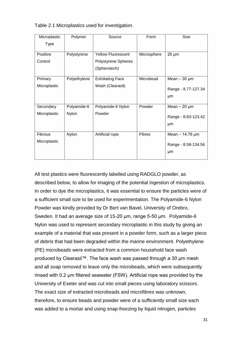
Table 2.1 Microplastics used for investigation.
Microplastic
Type
Polymer Source Form Size
Positive
Control
Polystyrene Yellow Fluorescent
Polystyrene Spheres
(Spherotech)
Microsphere 20 μm
Primary
Microplastic
Polyethylene Exfoliating Face
Wash (Clearasil)
Microbead Mean – 30 μm
Range - 8.77-127.34
μm
Secondary
Microplastic
Polyamide-6
Nylon
Polyamide-6 Nylon
Powder
Powder Mean – 20 μm
Range - 8.83-123.42
μm
Fibrous
Microplastic
Nylon Artificial rope Fibres Mean – 14.76 μm
Range - 8.58-134.56
μm
All test plastics were fluorescently labelled using RADGLO powder, as
described below, to allow for imaging of the potential ingestion of microplastics.
In order to dye the microplastics, it was essential to ensure the particles were of
a sufficient small size to be used for experimentation. The Polyamide-6 Nylon
Powder was kindly provided by Dr Bert van Bavel, University of Orebro,
Sweden. It had an average size of 15-20 μm, range 5-50 μm. Polyamide-6
Nylon was used to represent secondary microplastic in this study by giving an
example of a material that was present in a powder form, such as a larger piece
of debris that had been degraded within the marine environment. Polyethylene
(PE) microbeads were extracted from a common household face wash
produced by Clearasil™. The face wash was passed through a 30 μm mesh
and all soap removed to leave only the microbeads, which were subsequently
rinsed with 0.2 μm filtered seawater (FSW). Artificial rope was provided by the
University of Exeter and was cut into small pieces using laboratory scissors.
The exact size of extracted microbeads and microfibres was unknown,
therefore, to ensure beads and powder were of a sufficiently small size each
was added to a mortar and using snap-freezing by liquid nitrogen, particles
32
were ground to decrease their size. The subsequent particles were then
weighed and using the protocol set-out by (Lindegarth & Jonsson, 1991)
particles were dyed with RADGLO Radiant Color™ powder. RADGLO was
added to the ground particles in the ratio of 20:1 (dry weight) and mixed in air
thoroughly. The RADGLO powder and particle mixture was then transferred to a
fume hood where sufficient acetone (approximately 50 mL) was added to cover
the entire mixture and left to evaporate overnight to complete the dying process.
The resultant mass was then re-homogenised using a pestle and mortar and
passed through a 65 μm mesh to remove any larger particles. The labelled
particles were then suspended in 0.2 μm FSW and retained in a foil covered
bottle to prevent any degradation to fluorescence from exposure to light. In
order to ensure microfibre particles remained well mixed and to prevent
aggregation, 50 μL Tween 20 (0.001% v/v), known not to have a toxic effect
(Lindegarth & Jonsson, 1991) was added to the microfibre suspension.
Stock cultures were then diluted using 0.2 μm FSW and passed through a 10
μm mesh to size fraction particles and remove any unattached RADGLO
powder that may alter results. To ensure stock cultures were well mixed,
suspensions were sonicated for 2 minutes. The subsequent stock was passed
though FlowCAM to measure the concentration of plastics present within the
stock as well as the average size and size range (see Table 2.1), in order to
prepare microplastics for study. The volumes of microplastic stocks required to
be added to seawater was calculated using the equation of V1 x C1 = V2 x V2, to
produce a test concentration of 100 MP mL-1.
33
2.3.1.2 Copepod sampling
Copepods were collected from Station L4 (50° 15.00' N, 4° 13.02' W) (see
Figure 2.1), 12 km South of Plymouth, by the Plymouth Quest Research Vessel,
operated by Plymouth Marine Laboratory (PML). A 200 μm net was used to
sample zooplankton via vertical tows and samples were transferred to bottles
and transported within insulated boxes to PML. Once returned to the laboratory
zooplankton were assessed in terms of abundance using a WILD M5-48084
optical microscope, and the calanoid copepod C. typicus was chosen as the
subject of this study due to its abundance and owing to the fact that C. typicus
has been recorded in previous studies as ingesting microplastic (Cole, et al.,
2013). Adult female C. typicus were picked out and transferred to an aerated 5L
beaker filled with 0.2 μm FSW and conditioned without food overnight at
ambient sea temperature (~17°C).
Station L4
50° 15.00' N, 4° 13.02' W
Figure 2.1. Station L4 (50° 15.00' N, 4° 13.02' W), indicated by the red point. Located 12 km
South of Plymouth. Image adapted from Google Maps™.
This image has been removed by the author of this thesis for copyright reasons.
34
2.3.1.3 Natural seawater
In order to replicate natural conditions, natural seawater containing a natural
assemblage of phytoplankton was used to examine uptake of microplastics in
the presence of natural prey. Seawater was collected alongside zooplankton
samples at Station L4 by the Plymouth Quest Research Vessel at a depth of
10m. Samples were then returned to PML and stored at ambient sea
temperature (~17°C). Seawater was passed through a 100 μm mesh before
experimental set-up to ensure removal of any microzooplankton which may
have altered findings of the investigation.
2.3.1.4 Experimental set-up
Individual females of C. typicus were added to 35 mL glass bottles using stork-
billed forceps. Bottles had been treated with either 0.2 μm FSW or natural
seawater, spiked with the corresponding microplastic type to produce a test
concentration of 100 MP mL-1. A negative control was also studied, where no
microplastic was added to seawater samples. Five replicates of all treatments;
negative control, positive control, primary microplastic, secondary microplastic
and fibrous microplastic; were set-up and fixed to a plankton wheel, to ensure
suspensions remained well mixed, rotating at <5 rpm. Therefore, for each
plastic type, five individual females of C. typicus were exposed to microplastic
particles in the presence and absence of algal prey. Exposures were carried out
for 24 hours in darkness at ambient sea temperature (~17°C) in order to best
replicate natural conditions and prevent growth of algal cells. Replicates were
carried out to ensure reliability of data and provide grounds for statistical
analysis.
2.3.1.5 Assessing the ingestion of microplastic types by C. typicus
Following exposure samples were passed into a 50 μm mesh and washed into
a sample tube using 0.2 μm filtered FSW. Formalin (4%) was then added to the
sample tube to preserve specimens for imaging.
Individuals were placed on slides and imaged using a TBM1000 microscope
with Prior V31LD4 fluorescent emission attachment and QIClick™ camera. A
fluorescence wavelength of 475 nm was used and camera settings set to
optimise the view of fluorescence, such that fluorescence appeared as blue on
35
images. All individuals were imaged, and all examples of ingestion and
adherence of microplastic particles were recorded.
2.3.2 Results
2.3.2.1 Ingestion of microplastics
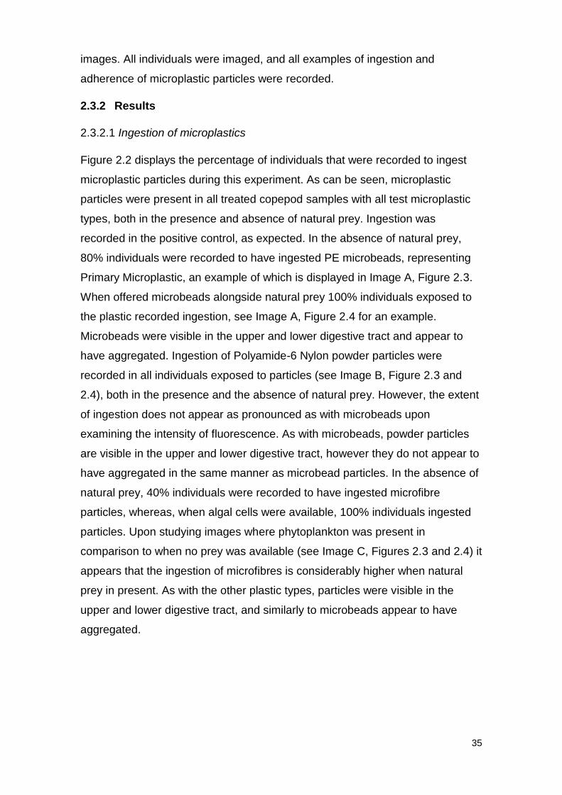
Figure 2.2 displays the percentage of individuals that were recorded to ingest
microplastic particles during this experiment. As can be seen, microplastic
particles were present in all treated copepod samples with all test microplastic
types, both in the presence and absence of natural prey. Ingestion was
recorded in the positive control, as expected. In the absence of natural prey,
80% individuals were recorded to have ingested PE microbeads, representing
Primary Microplastic, an example of which is displayed in Image A, Figure 2.3.
When offered microbeads alongside natural prey 100% individuals exposed to
the plastic recorded ingestion, see Image A, Figure 2.4 for an example.
Microbeads were visible in the upper and lower digestive tract and appear to
have aggregated. Ingestion of Polyamide-6 Nylon powder particles were
recorded in all individuals exposed to particles (see Image B, Figure 2.3 and
2.4), both in the presence and the absence of natural prey. However, the extent
of ingestion does not appear as pronounced as with microbeads upon
examining the intensity of fluorescence. As with microbeads, powder particles
are visible in the upper and lower digestive tract, however they do not appear to
have aggregated in the same manner as microbead particles. In the absence of
natural prey, 40% individuals were recorded to have ingested microfibre
particles, whereas, when algal cells were available, 100% individuals ingested
particles. Upon studying images where phytoplankton was present in
comparison to when no prey was available (see Image C, Figures 2.3 and 2.4) it
appears that the ingestion of microfibres is considerably higher when natural
prey in present. As with the other plastic types, particles were visible in the
upper and lower digestive tract, and similarly to microbeads appear to have
aggregated.
36
Figure 2.1 Ingestion of microplastics by C. typicus. Pie charts displaying the percentage of
individuals recorded as ingesting microplastic particles when exposed to different plastic types in
the concentration of 100 MP mL-1. N = 5 for all treatments, except for D - Secondary Microplastic
with prey (n = 4). 100% ingestion indicates microplastic particles present in all 5 individuals
exposed to each treatment.
37
2.3.2.2 Investigating the ingestion of different microplastic types in the absence
of natural prey
2.3.2.3 Investigating the ingestion of different microplastic types in the presence
of natural prey
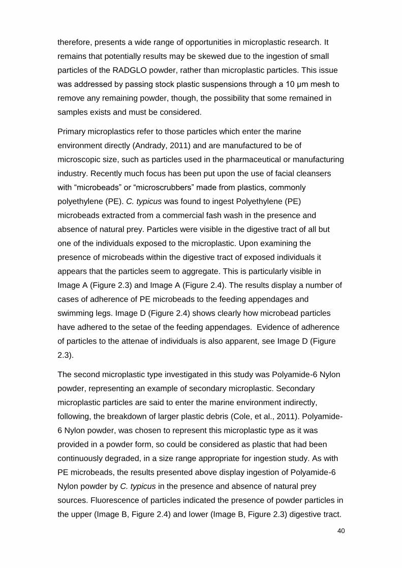
A B C
D E F
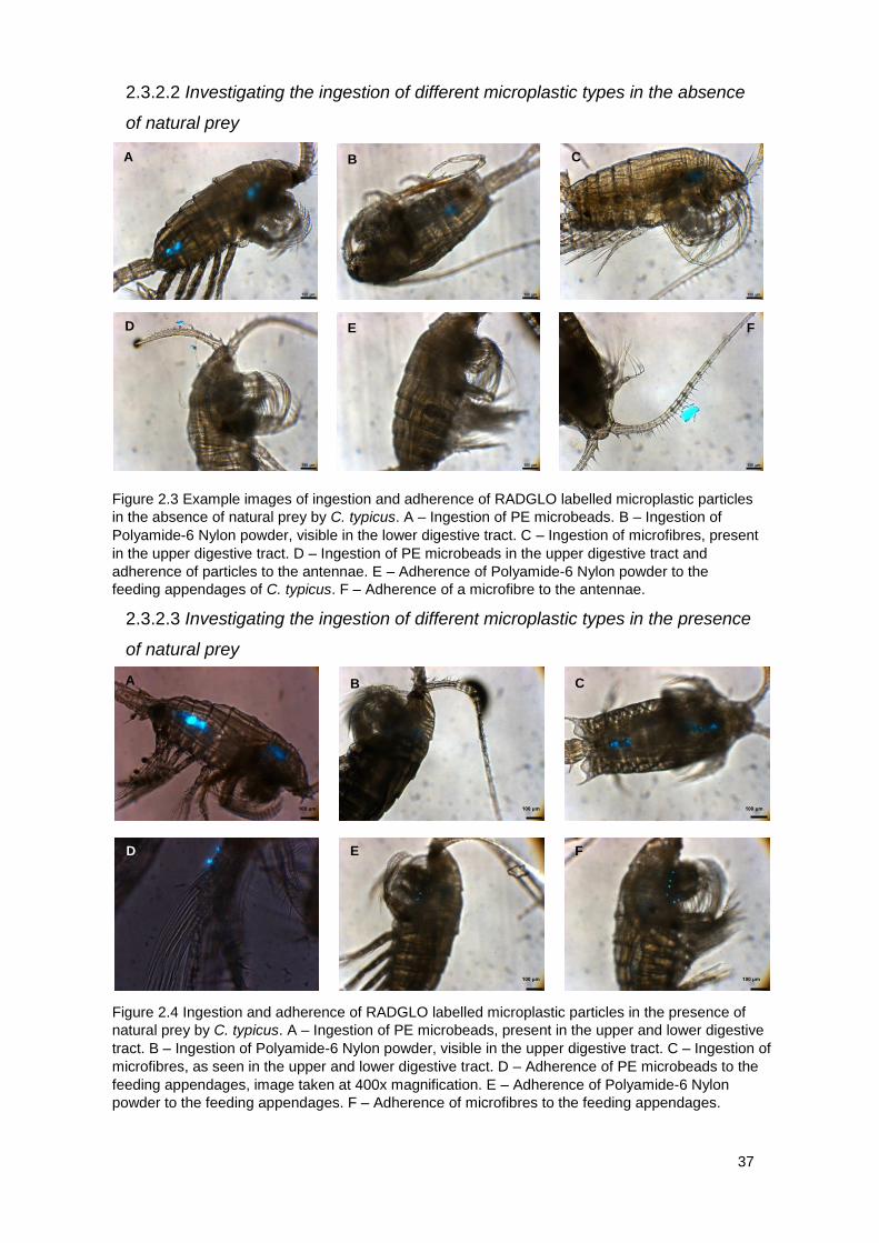
Figure 2.4 Ingestion and adherence of RADGLO labelled microplastic particles in the presence of
natural prey by C. typicus. A – Ingestion of PE microbeads, present in the upper and lower digestive
tract. B – Ingestion of Polyamide-6 Nylon powder, visible in the upper digestive tract. C – Ingestion of
microfibres, as seen in the upper and lower digestive tract. D – Adherence of PE microbeads to the
feeding appendages, image taken at 400x magnification. E – Adherence of Polyamide-6 Nylon
powder to the feeding appendages. F – Adherence of microfibres to the feeding appendages.
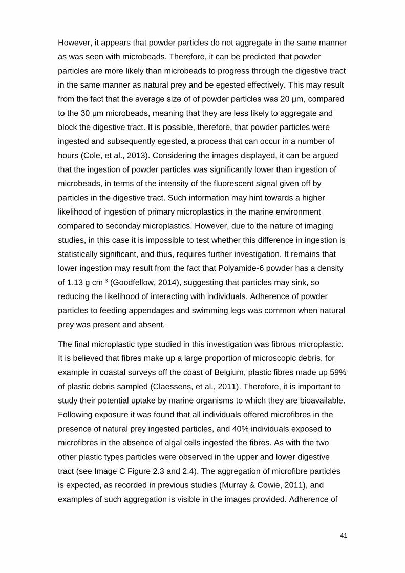
A B C
D E F
100 μm 100 μm 100 μm
100 μm 100 μm
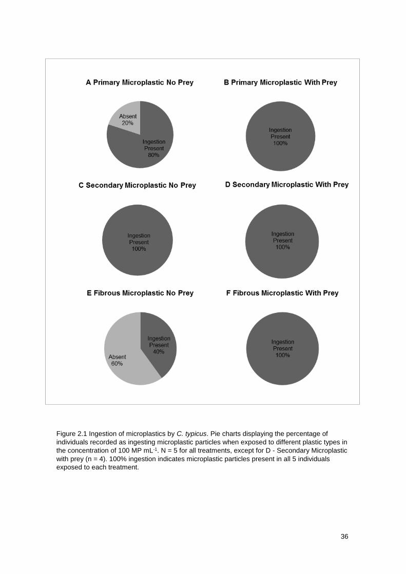
Figure 2.3 Example images of ingestion and adherence of RADGLO labelled microplastic particles
in the absence of natural prey by C. typicus. A – Ingestion of PE microbeads. B – Ingestion of
Polyamide-6 Nylon powder, visible in the lower digestive tract. C – Ingestion of microfibres, present
in the upper digestive tract. D – Ingestion of PE microbeads in the upper digestive tract and
adherence of particles to the antennae. E – Adherence of Polyamide-6 Nylon powder to the
feeding appendages of C. typicus. F – Adherence of a microfibre to the antennae.
38
2.3.2.4 Adherence of microplastics
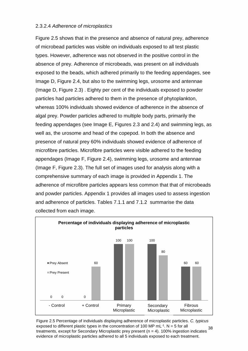
Figure 2.5 shows that in the presence and absence of natural prey, adherence
of microbead particles was visible on individuals exposed to all test plastic
types. However, adherence was not observed in the positive control in the
absence of prey. Adherence of microbeads, was present on all individuals
exposed to the beads, which adhered primarily to the feeding appendages, see
Image D, Figure 2.4, but also to the swimming legs, urosome and antennae
(Image D, Figure 2.3) . Eighty per cent of the individuals exposed to powder
particles had particles adhered to them in the presence of phytoplankton,
whereas 100% individuals showed evidence of adherence in the absence of
algal prey. Powder particles adhered to multiple body parts, primarily the
feeding appendages (see Image E, Figures 2.3 and 2.4) and swimming legs, as
well as, the urosome and head of the copepod. In both the absence and
presence of natural prey 60% individuals showed evidence of adherence of
microfibre particles. Microfibre particles were visible adhered to the feeding
appendages (Image F, Figure 2.4), swimming legs, urosome and antennae
(Image F, Figure 2.3). The full set of images used for analysis along with a
comprehensive summary of each image is provided in Appendix 1. The
adherence of microfibre particles appears less common that that of microbeads
and powder particles. Appendix 1 provides all images used to assess ingestion
and adherence of particles. Tables 7.1.1 and 7.1.2 summarise the data
collected from each image.
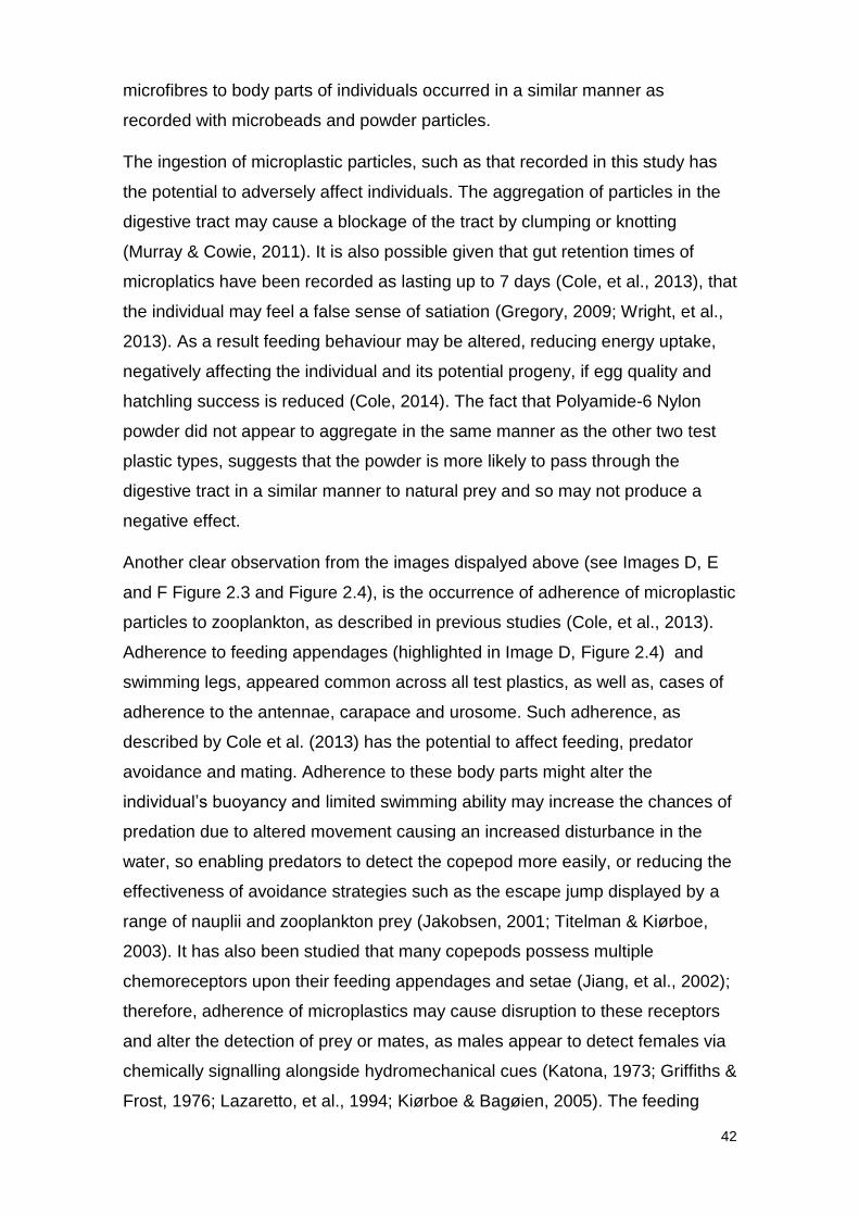
Figure 2.5 Percentage of individuals displaying adherence of microplastic particles. C. typicus
exposed to different plastic types in the concentration of 100 MP mL-1. N = 5 for all
treatments, except for Secondary Microplastic prey present (n = 4). 100% ingestion indicates
evidence of microplastic particles adhered to all 5 individuals exposed to each treatment.
0 0
100 100
60
0
60
100
80
60
- Control + Control PrimaryMicroplastic
SecondaryMicrolastic
FibrousMicroplastic
Percentage of individuals displaying adherence of microplastic particles
Prey Absent
Prey Present
Secondary Microplastic
39
2.3.3 Discussion
The results presented here, provide the first evidence that a common marine
copepod can readily ingest a range of microplastic types, including Polyethylene
microbeads, Polyamide-6 Nylon powder and artificial rope microfibres. Such
information has the potential to gain further insight into the entry of microplastic
into the food chain, and provides methods which may be utilised in future
research.
In previous laboratory studies the ingestion of microplastic particles by
zooplankton has been recorded (Cole, et al., 2013; Kaposi, et al., 2014; Lee, et
al., 2013), although in most cases it is polystyrene or polyethylene
microspheres, uniform in shape and size that have been used in this research.
In order to produce environmentally relevant results it is important to consider
that microplastic particles present within the marine environment are unlikely to
be of uniform shape or size. With this in mind, this study was designed to
examine the uptake of the different forms of microplastic likely to be found in the
marine environment. Although in order to confirm microplastic ingestion as seen
in previous studies, fluorescently labelled polystyrene microspheres were
utilised as a positive control. It is important to examine microplastic ingestion in
the presence of natural prey sources, which would be available to organisms
alongside any microscopic marine debris in the environment. To address this
the study was replicated using seawater collected from the same site as study
organisms, and in the absence of prey.
The RADGLO dyeing methodology presented here is considered effective in
microplastic research. RADGLO was implemented by researchers initially as a
manner to examine hydrodynamics and settling behaviour of bivalve larvae
(Lindegarth & Jonsson, 1991). In these studies larvae were encouraged to
ingest labelled polyvinyl chloride (PVC) particles to allow for visualisation of
individuals (Lindegarth & Jonsson, 1991). RADGLO adhered to PVC due to
electrostatic forces (Lindegarth & Jonsson, 1991) and it was predicted that the
dye would be likely to bind to a number of plastic types. The protocol outlined by
Lindegarth and Jonsson (1991) was utilised to dye plastics and altered to meet
the demands of the study presented here. All plastics were dyed effectively, and
as can be seen in Figures 2.3 and 2.4, particles were clearly visible under
fluorescence. The novel method of using RADGLO to dye plastic particles,
40
therefore, presents a wide range of opportunities in microplastic research. It
remains that potentially results may be skewed due to the ingestion of small
particles of the RADGLO powder, rather than microplastic particles. This issue
was addressed by passing stock plastic suspensions through a 10 μm mesh to
remove any remaining powder, though, the possibility that some remained in
samples exists and must be considered.
Primary microplastics refer to those particles which enter the marine
environment directly (Andrady, 2011) and are manufactured to be of
microscopic size, such as particles used in the pharmaceutical or manufacturing
industry. Recently much focus has been put upon the use of facial cleansers
with “microbeads” or “microscrubbers” made from plastics, commonly
polyethylene (PE). C. typicus was found to ingest Polyethylene (PE)
microbeads extracted from a commercial fash wash in the presence and
absence of natural prey. Particles were visible in the digestive tract of all but
one of the individuals exposed to the microplastic. Upon examining the
presence of microbeads within the digestive tract of exposed individuals it
appears that the particles seem to aggregate. This is particularly visible in
Image A (Figure 2.3) and Image A (Figure 2.4). The results display a number of
cases of adherence of PE microbeads to the feeding appendages and
swimming legs. Image D (Figure 2.4) shows clearly how microbead particles
have adhered to the setae of the feeding appendages. Evidence of adherence
of particles to the attenae of individuals is also apparent, see Image D (Figure
2.3).
The second microplastic type investigated in this study was Polyamide-6 Nylon
powder, representing an example of secondary microplastic. Secondary
microplastic particles are said to enter the marine environment indirectly,
following, the breakdown of larger plastic debris (Cole, et al., 2011). Polyamide-
6 Nylon powder, was chosen to represent this microplastic type as it was
provided in a powder form, so could be considered as plastic that had been
continuously degraded, in a size range appropriate for ingestion study. As with
PE microbeads, the results presented above display ingestion of Polyamide-6
Nylon powder by C. typicus in the presence and absence of natural prey
sources. Fluorescence of particles indicated the presence of powder particles in
the upper (Image B, Figure 2.4) and lower (Image B, Figure 2.3) digestive tract.
41
However, it appears that powder particles do not aggregate in the same manner
as was seen with microbeads. Therefore, it can be predicted that powder
particles are more likely than microbeads to progress through the digestive tract
in the same manner as natural prey and be egested effectively. This may result
from the fact that the average size of of powder particles was 20 μm, compared
to the 30 μm microbeads, meaning that they are less likely to aggregate and
block the digestive tract. It is possible, therefore, that powder particles were
ingested and subsequently egested, a process that can occur in a number of
hours (Cole, et al., 2013). Considering the images displayed, it can be argued
that the ingestion of powder particles was significantly lower than ingestion of
microbeads, in terms of the intensity of the fluorescent signal given off by
particles in the digestive tract. Such information may hint towards a higher
likelihood of ingestion of primary microplastics in the marine environment
compared to seconday microplastics. However, due to the nature of imaging
studies, in this case it is impossible to test whether this difference in ingestion is
statistically significant, and thus, requires further investigation. It remains that
lower ingestion may result from the fact that Polyamide-6 powder has a density
of 1.13 g cm-3 (Goodfellow, 2014), suggesting that particles may sink, so
reducing the likelihood of interacting with individuals. Adherence of powder
particles to feeding appendages and swimming legs was common when natural
prey was present and absent.
The final microplastic type studied in this investigation was fibrous microplastic.
It is believed that fibres make up a large proportion of microscopic debris, for
example in coastal surveys off the coast of Belgium, plastic fibres made up 59%
of plastic debris sampled (Claessens, et al., 2011). Therefore, it is important to
study their potential uptake by marine organisms to which they are bioavailable.
Following exposure it was found that all individuals offered microfibres in the
presence of natural prey ingested particles, and 40% individuals exposed to
microfibres in the absence of algal cells ingested the fibres. As with the two
other plastic types particles were observed in the upper and lower digestive
tract (see Image C Figure 2.3 and 2.4). The aggregation of microfibre particles
is expected, as recorded in previous studies (Murray & Cowie, 2011), and
examples of such aggregation is visible in the images provided. Adherence of
42
microfibres to body parts of individuals occurred in a similar manner as
recorded with microbeads and powder particles.
The ingestion of microplastic particles, such as that recorded in this study has
the potential to adversely affect individuals. The aggregation of particles in the
digestive tract may cause a blockage of the tract by clumping or knotting
(Murray & Cowie, 2011). It is also possible given that gut retention times of
microplatics have been recorded as lasting up to 7 days (Cole, et al., 2013), that
the individual may feel a false sense of satiation (Gregory, 2009; Wright, et al.,
2013). As a result feeding behaviour may be altered, reducing energy uptake,
negatively affecting the individual and its potential progeny, if egg quality and
hatchling success is reduced (Cole, 2014). The fact that Polyamide-6 Nylon
powder did not appear to aggregate in the same manner as the other two test
plastic types, suggests that the powder is more likely to pass through the
digestive tract in a similar manner to natural prey and so may not produce a
negative effect.
Another clear observation from the images dispalyed above (see Images D, E
and F Figure 2.3 and Figure 2.4), is the occurrence of adherence of microplastic
particles to zooplankton, as described in previous studies (Cole, et al., 2013).
Adherence to feeding appendages (highlighted in Image D, Figure 2.4) and
swimming legs, appeared common across all test plastics, as well as, cases of
adherence to the antennae, carapace and urosome. Such adherence, as
described by Cole et al. (2013) has the potential to affect feeding, predator
avoidance and mating. Adherence to these body parts might alter the
individual’s buoyancy and limited swimming ability may increase the chances of
predation due to altered movement causing an increased disturbance in the
water, so enabling predators to detect the copepod more easily, or reducing the
effectiveness of avoidance strategies such as the escape jump displayed by a
range of nauplii and zooplankton prey (Jakobsen, 2001; Titelman & Kiørboe,
2003). It has also been studied that many copepods possess multiple
chemoreceptors upon their feeding appendages and setae (Jiang, et al., 2002);
therefore, adherence of microplastics may cause disruption to these receptors
and alter the detection of prey or mates, as males appear to detect females via
chemically signalling alongside hydromechanical cues (Katona, 1973; Griffiths &
Frost, 1976; Lazaretto, et al., 1994; Kiørboe & Bagøien, 2005). The feeding
43
appendages and antennae also utilise mechanoreceptors, which similarly may
be disrupted by adherence of foreign particles. Given, the importance of the
antennae in predator and prey detection (Strickler & Bal, 1973; Fleminger,
1975; Strickler, 1975; Viitasalo, et al., 1998) and positioning within the water
column it can be predicted that adherence of particles to the antennae is likely
to interefere with these processes, and as such reduce the fitness of the
organism. It would be highly interesting to examine the length of time that
particles remain attached to individuals, or investigate the effect upon swimming
and feeding behaviour.
Although the results appear to provide clear evidence that ingestion of a
number of microplastic types occurs in the copepod species, C. typicus, a
number of limitations to this study exist and must be addressed before
presenting conclusions of this investigation. Firstly, due to the difficult nature of
sampling microplastic debris (Hidalgo-Ruz, et al., 2012; Cole, et al., 2014) the
concentration of microplastics sized <100 μm within the natural environment is
currently unknown, and requires addressing. With that in mind it can be argued
that the concentrations used in this experiment do not represent those that are
found in the natural environment. However, as this experiment was proposed
initially as an exploration into the ingestion of various plastic types it can be
considered that the concentration used is not of high importance. It can be
argued, though, that differences observed between the uptake of plastics in the
presence and absence of natural prey may be due to the fact that the relatively
lower number of potential prey particles per mililitre of seawater present in
samples where no prey was offered to copepods meant that feeding rate was
reduced to conserve energy (Lam & Frost, 1976), as a result less microplastic
was ingested. Similarly patterns of adherence between the two treatments may
also have been affected in this way.
Another factor that may have affected results was the aggregation of particles
within suspensions. Any aggregation producing too large particles to be
ingested by C. typicus would affect results, therefore, there is a possibilty that
ingestion was reduced in some cases due to aggregation. However, it can be
argued that similar aggregation may occur in the natural environment. Observed
in a few cases was the occurrence of large fibres which were unlabelled and
adhered to individuals (see Image F1b, Figure 7.1.1; Image NP4a, Figure 7.1.2,
44
Appendix 1). The presence of such fibres may have altered results or led to the
adherence of other particles, not caused by the behaviour of the copepod.
Further research is required to address the limited evidence that exists on the
relative abundances of different microplastic types within the natural
environment. Such data would be highly beneficial and allow research to be
directed at polymers which are abundant, but new methods must be formulated
to overcome the difficulties arising from sampling debris <100 μm (Hidalgo-Ruz,
et al., 2012; Cole, et al., 2014).
Regardless of the limitations outlined above, the results provided here display
evidence of ingestion of a range of plastic types by a common copepod species.
The recorded effects of microplastic ingestion examined by previous research
were addressed earlier in this chapter. Although a number of different negative
effects are presented, all centre around the problem of reduced energy reserves
resulting from an increased sense of satiation as described by Gregory (2009),
decreased feeding or altered feeding behaviour (Cole, 2014). The decrease in
energy reserves caused by microplastic ingestion is a ecological concern
considering the importance of zooplankton in transfering energy to higher
trophic levels. A reduction in energy available to species that feed upon
zooplankton, including a number of commercially important fish and their larvae,
may negatively affect the stability of populations within environments where
microplastic polltuion is present. Such an occurrence would be a concern for
those associated with the fisheries industry and conservationists alike. By
utilising methods such as those presented in this study we may be able to
identify species most at risk from ingesting commonly found microplastic
particles, and assess the potential of such particles being passed through the
food chain to higher trophic levels.

45

46
47
Chapter 3
The effects of microplastic exposure upon marine copepods
with varying feeding strategies

48
49
3.1 Feeding strategies in the zooplankton
The zooplankton represent a large and taxonomically diverse group of species,
which display a range of physical forms and life strategies in order to live
successfully within the world’s ocean currents and water systems. Such areas
represent a “viscous and nutritionally dilute world” and as a result in order to
survive, a volume of water 106 times their own body size must be covered daily
by zooplankton (Kjellerup & Kiørboe, 2012).Therefore, it is of great importance
that the feeding strategy adopted by a species is efficient. The ingestion of
microplastics further reduces the nutritional value of feeding and increases the
requirement for effective prey capture, which may also be inhibited by the
presence of microplastic within the environment. Generally zooplankton display
one of three major feeding strategies (Kjellerup & Kiørboe, 2012); they generate
a feeding current and capture prey within this current; they are ambush feeders
and capture prey that pass within a capture radius; or they cruise through water
and capture encountered prey (Kjellerup & Kiørboe, 2012). All of these feeding
types have different methods by which they detect and subsequently capture
prey, therefore, it is likely that those species displaying different feeding
strategies to one another are likely to interact with and be affected by
microplastic exposure in different manners.
3.2 Prey detection by Feeding-current and Ambush feeding zooplankton
It could be hypothesised that those copepod species which feed by generating
a feeding current are more likely to ingest microplastics as they draw particles
towards feeding appendages and may ingest particles in a passive manner
whilst filtering. Ambush predators on the other hand, which tend to feed on
motile prey (Saiz & Kiørboe, 1995), theoretically, should not detect immobile
microplastic particles and as a result significant ingestion is not predicted in
these species. However, experimental studies have shown microplastic
ingestion in both feeding-current strategists and ambush feeders (Cole, et al.,
2013). The manners in which ambush feeders and feeding-current strategists
detect prey will be discussed below.
It has been studied and appears widely accepted that ambush feeders detect
prey using hydromechanical cues (Yen, et al., 1992). Prey is detected due to
changes in the hydrodynamics of ambient water caused by a particle, which as
50
a result causes bending of the setae and allows the individual to identify the
location of the prey particle (Yen, et al., 1992). Such is the case in the copepod
Oithona similis where it is proposed that motile prey is perceived remotely by
hydrodynamic disturbances generated in ambient water by a prey particle
(Kiørboe & Visser, 1999) detected by numerous long mechanoreceptory setae
on the antennules (Svensen & Kiørboe, 2000). The presence of
mechanosensory setae upon antenna appears common in copepods which are
blind and rely on such appendages to perceive motile prey (Strickler, 1975).
This mechanism of prey detection, however, limits ambush-feeding zooplankton
to preying upon motile prey as non-motile cells would fail to produce a
disturbance in ambient water and would as a result not be detected. Ambush
feeders have been found to feed upon faecal pellets falling from above; hence,
producing a hydromechanical signal, such behaviour is displayed by Oithona
similis (Gonzáles & Smetacek, 1984).
In laboratory studies upon the mechanisms by which prey is captured, the
movement of feeding appendages is described as similar between feeding-
current generating and ambush feeding zooplankton (Koehl & Strickler, 1981).
It has been studied using high-speed filming techniques that feeding-current
strategists propel water past themselves by flapping their appendages and then
actively capture small parcels of water that contain food particles by opening
and closing their second maxillae (Koehl & Strickler, 1981). The ambush
feeding copepod O. similis captures prey by detecting the location of the cell by
detection of hydromechanical cues by the antennules or telson and propels
itself towards the cell placing its mouthparts at the location where the prey cell
was discovered (Svensen & Kiørboe, 2000). In this case the prey remains
almost stationary and unaffected by the motion of the copepod until the feeding
basket is opened and prey directed towards the mouth (Kiørboe, et al., 2009).
Such evidence counters previous ideas that attack jumps, which are used by
some ambush feeders, should be theoretically ineffective as prey cells should
be pushed away by the forward jumping attacker due to the thick viscous
boundary surrounding the attacking zooplankton (Bruno, et al., 2012 ). Hence it
has more recently been revealed that like feeding-current generating
individuals, ambush feeders direct prey towards the mouth by motions of the
51
feeding appendages, rather than approaching prey cells by direct attack jumps
(Bruno, et al., 2012 ).
Feeding-current strategists are believed to detect prey using biochemical cues
and have the potential to select prey based upon their biochemical composition
(Tiselius, et al., 2013). Tiselius et al. (2013) provide a model of detection which
can generally be applied to feeding-current producing zooplankton; it is
proposed that the sphere of chemicals surrounding a potential prey cell
elongates when the prey cell is situated within the feeding current. As a result
the leading edge of this chemical signal reaches the individual before the prey
cell itself, giving the opportunity for the zooplankton to accept or reject the prey
cell based upon its chemical signal and alter its feeding-current away or toward
the capture area (Tiselius, et al., 2013). This mechanism of chemical reception
is also represented by Kjellerup and Kiørboe (2012), where it is suggested that
the chemical signal arrives at the zooplankton approximately 0.5 seconds
before the prey cell itself allowing a feeding-current strategist time to accept or
reject prey. Ambush feeders, on the other hand, do not have this ability and as
a result must feed upon particles purely on the extent to which they disturb the
hydrodynamics of ambient water. Two processes of chemoreception occur in
feeding-current feeding zooplankton; first long-range chemoreception governs
the generation of the feeding-current and particle capture, subsequently short-
range chemoreception at the mouth induces particle ingestion (Koehl &
Strickler, 1981).
It can be argued, therefore, that the key difference between prey detection in
ambush and feeding-current strategists, is that ambush feeders cannot detect
prey through biochemical means, so limiting selectivity. Feeding-current
strategists, on the other hand, have the potential to select particles based on a
biochemical signal. Secondly, due to the mechanistic manner in which ambush
feeders detect prey, it is only motile prey which are likely to be detected,
whereas, feeding-current strategists that possess both mechanistic and
biochemical detection are able to detect motile and non-motile prey, so
increasing the number of prey sources available to them.
52
3.3 The effects of microplastic exposure upon feeding in the zooplankton
Studies carried out by Cole et al. (2013) and Setälä et al. (2014), alongside
results presented in this thesis (Chapter 2), have provided evidence that
ingestion of microplastic particles is widespread among a range of species and
polymer types under laboratory conditions. One major concern of this is that
microplastic ingestion may disrupt normal feeding behaviour, and thus,
negatively affect the fitness of the individual.
Further work carried out by Cole (2014) focussed upon the effects of
microplastics upon the fecundity and feeding of the calanoid copepod, Calanus
helgolandicus. Here it was found that exposure to <75 polystyrene MP mL-1
negatively affected the feeding rate and carbon uptake in the study species, an
11% reduction in algal uptake and 40% reduction in carbon biomass ingested
was observed (Cole, 2014).
In the work carried out by Cole (2014) it was also discovered that the common
calanoid species C. helgolandicus displayed a switching behaviour in terms of
prey selection when exposed to microplastics. It was observed that individuals
feeding upon T. weissflogii alone ingested all size ranges of the species (11.6–
17.0 µm), and those exposed to 20 μm polystyrene beads only ingested algal
cells in the size range of 11.6–14.8 µm (Cole, 2014).
However, it is not only copepods which have been found to show altered
feeding activity when exposed to small plastic particles. The blue mussel,
Mytilus edulis, showed a decrease in filtering activity when exposed to
polystyrene nanoplastics (30 nm); however, the concentration of plastic still
decreased in treatments (Wegner, et al., 2012). Despite the smaller size of the
nanoplastics, it could be suggested that exposure to polystyrene microplastics
may also produce an adverse effect. Other stress effects were also seen such
as the production of pseudofaeces, a mechanism known to be a cleaning
mechanism, preventing the gills being blocked by particulate matter (Jørgensen,
1981), and as a rejection mechanism for inedible particles (Wegner, et al.,
2012). Similarly polystyrene microplastic spheres have been recorded to adhere
to feeding appendages of copepods (Cole, et al., 2013), as mentioned in
Chapter 2. Such adherence may adversely affect feeding and swimming
53
behaviour (Cole, et al., 2013) and thus, reduce the effectiveness of natural
feeding, potentially creating an energy deficit for affected individuals.
Reduced feeding activity as a result of microplastic exposure was also recorded
in the marine worm, Arenicola marina (Wright, et al., 2013). This reduction in
feeding was attributed to suppressed feeding activity or adverse effects caused
by the unplasticised polyvinylchloride (UVPC) microplastics used in the study.
The lack of a protein coat on the particles was thought to decrease the
adherence of particles to the worm’s feeding appendages, so reducing feeding
efficiency (Wright, et al., 2013). Once again the reduction in feeding shown
produced a decrease in energy levels recorded in the worms, therefore,
reducing their fitness. Due to the fact that Arenicola is a keystone species within
its natural environment (Wright, et al., 2013), this energy deficit is likely to have
knock-on effects throughout the ecosystem.
It appears then that microplastic exposure causes alterations in normal feeding
behaviour, reducing the energy uptake of individuals, and thus, creating an
energy deficit in the trophic level. Due to the fact that that the species that tend
to feed on prey in the size range of microplastics, generally occupy lower levels
in the food web, it is particularly important to examine the effects that
microplastics are having upon feeding to best inform decision makers of the
negative effects of microplastic debris within the marine environment.
3.4 Investigating the effects of microplastic exposure upon feeding in the
zooplankton
To further examine the effects that microplastic particles are having upon the
feeding behaviour of zooplankton, it was decided to carry out two grazing
experiments, examining two zooplankton species, each displaying a different
feeding strategy and role within the ecosystem.
The first study was designed to examine the effects of microplastic exposure
upon a holoplankton species, Acartia tonsa, and a meroplankton species,
Porcellanid larvae. A. tonsa is a copepod species common throughout coastal
areas and neritic temperate seas (Saiz & Kiørboe, 1995) which displays two
54
feeding strategies (Jonsson & Tiselius, 1990). When feeding upon non-motile
phytoplankton prey, A. tonsa generates a feeding-current, and feeds upon cells
captured within this current (Saiz & Kiørboe, 1995). On the other hand, when
feeding upon motile prey such as ciliates, the species adopts ambush feeding
(Jonsson & Tiselius, 1990). Prey is perceived via mechanical detection through
disturbances in the water column caused by the moving prey, and subsequently
using an attack jump motion the cell is captured (Saiz & Kiørboe, 1995). A.
tonsa can therefore, be described as representative of a number of copepod
species, feeding in two of the three common feeding modes displayed by
copepods. As a result, the species is a highly interesting subject of research
into effects of contaminants upon feeding behaviour. Generally it is believed
that A. tonsa adopts the feeding strategy that is most likely to increase energy
intake, so altering feeding behaviour due the relative abundance of motile and
non-motile prey in the water column (Kiorboe, et al., 1996). It could be predicted
then that in a closed system, such as those used in experimental studies, the
addition of microplastic particles, representing non-motile prey, may lead to the
adoption of the feeding-current strategy by A. tonsa due to the increase in non-
motile cells available. For this reason, along with the evidence displayed by
Cole et al. (2013) displaying ingestion of 7.3-30.6 μm polystyrene spheres by
another member of the Acartia genus, Acartia clausii, as well as the potential for
adherence of microplastics to feeding appendages, which appears common
across a range of taxa (see Chapter 2). It can be assumed likely that
microplastic exposure will affect feeding behaviour and associated carbon
uptake in A. tonsa.
To date, microplastic research has focussed upon copepod species spending
their entire life history in the plankton and less research into potential effects
upon meroplankton species exists. It is therefore, of great interest to examine
the effects of microplastic exposure upon larvae whose development is likely to
be affected by changes in energy uptake and nutrient availability. Porcellanid
larvae feed upon algal prey, detecting prey as it comes into contact with the
individual (Gonor & Gonor, 1973), therefore relying on chance for encounter
(Mooler, 1978; Meyers & Hagood, 1984; Mootz & Epifanio, 1974; Kurmaly, et
al., 1989, 1990; Stickney & Perkins, 1981; Barros & Valenti, 2003). However, in
order to avoid predation if a cell comes into contact with the individual’s
55
posterior or the top or sides of the carapace the larva reacts quickly and moves
away from the cell immediately (Gonor & Gonor, 1973). On the other hand, cells
that come into contact with the setae of the larva are captured and passed to
the mouthparts (Gonor & Gonor, 1973). Individuals are thought to be sensitive
to disturbances within the water column, however, if contact is not made with
the potential prey item attempts to capture the cell typically fail (Gonor & Gonor,
1973). By feeding in this manner Porcellanid larvae are unable to distinguish
between natural prey and microplastic particles until cells are passed to the
mouth. It can be predicted then that individuals are likely to interact with
microplastic particles if they are present within the marine environment. Studies
carried out by Cole et al. (2013) provide evidence for the ingestion of 30.6 μm
polystyrene microspheres by Porcellanid larvae. Therefore, it can be suggested
based on the effects recorded in copepods (Cole, 2014) and other invertebrates
such as marine worms (Wright, et al., 2013) that effects upon energy uptake are
likely to occur in decapod larvae when exposed to microplastic particles.
The second study was designed to investigate whether the effects upon feeding
were influenced by feeding type. It was hypothesised that the effects upon
feeding should be less pronounced in an ambush feeding species, Oithona
similis, compared to a feeding-current strategist, Calanus helgolandicus, owing
to the fact that ambush feeding species are not expected to detect non-mobile
prey such as microplastic particles. Feeding-current strategists on the other
hand filter particles into a capture radius, thus, are likely to capture microplastic
particles where concentration of microplastics are high. With this in mind, it is
hypothesised that feeding behaviour and subsequent carbon ingestion will be
unaffected when the ambush predator, O. similis, is exposed to microplastics.
However, C. helgolandicus, a species that feeds using a feeding-current is
expected to suffer an energy deficit following exposure to microplastic particles.
3.4.1 Methods
3.4.1.1 Zooplankton for experimentation
Zooplankton samples for experimentation were collected from Station L4 (see
Figure 2.1, Page 33) by the Plymouth Quest Research Vessel operated by PML
via a medium (200 μm) and fine (63 μm) surface trawl of plankton nets.
Samples were then transferred to the laboratory at PML for species
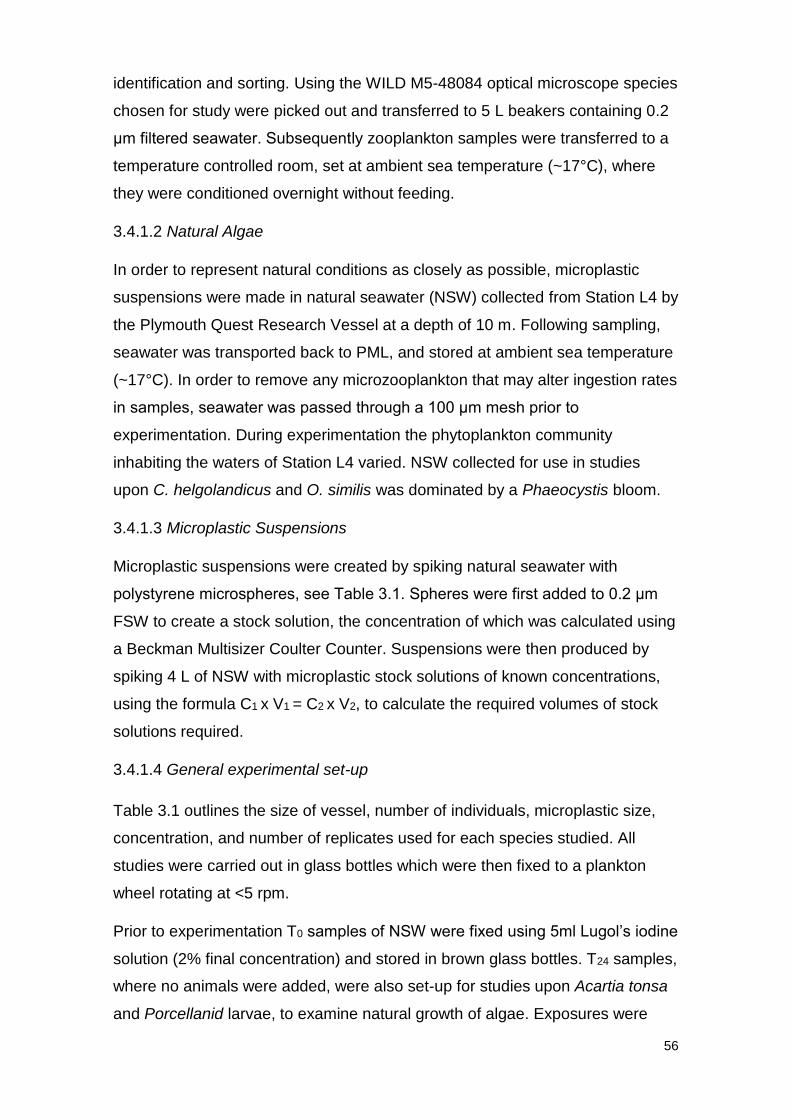
56
identification and sorting. Using the WILD M5-48084 optical microscope species
chosen for study were picked out and transferred to 5 L beakers containing 0.2
μm filtered seawater. Subsequently zooplankton samples were transferred to a
temperature controlled room, set at ambient sea temperature (~17°C), where
they were conditioned overnight without feeding.
3.4.1.2 Natural Algae
In order to represent natural conditions as closely as possible, microplastic
suspensions were made in natural seawater (NSW) collected from Station L4 by
the Plymouth Quest Research Vessel at a depth of 10 m. Following sampling,
seawater was transported back to PML, and stored at ambient sea temperature
(~17°C). In order to remove any microzooplankton that may alter ingestion rates
in samples, seawater was passed through a 100 μm mesh prior to
experimentation. During experimentation the phytoplankton community
inhabiting the waters of Station L4 varied. NSW collected for use in studies
upon C. helgolandicus and O. similis was dominated by a Phaeocystis bloom.
3.4.1.3 Microplastic Suspensions
Microplastic suspensions were created by spiking natural seawater with
polystyrene microspheres, see Table 3.1. Spheres were first added to 0.2 μm
FSW to create a stock solution, the concentration of which was calculated using
a Beckman Multisizer Coulter Counter. Suspensions were then produced by
spiking 4 L of NSW with microplastic stock solutions of known concentrations,
using the formula C1 x V1 = C2 x V2, to calculate the required volumes of stock
solutions required.
3.4.1.4 General experimental set-up
Table 3.1 outlines the size of vessel, number of individuals, microplastic size,
concentration, and number of replicates used for each species studied. All
studies were carried out in glass bottles which were then fixed to a plankton
wheel rotating at <5 rpm.
Prior to experimentation T0 samples of NSW were fixed using 5ml Lugol’s iodine
solution (2% final concentration) and stored in brown glass bottles. T24 samples,
where no animals were added, were also set-up for studies upon Acartia tonsa
and Porcellanid larvae, to examine natural growth of algae. Exposures were
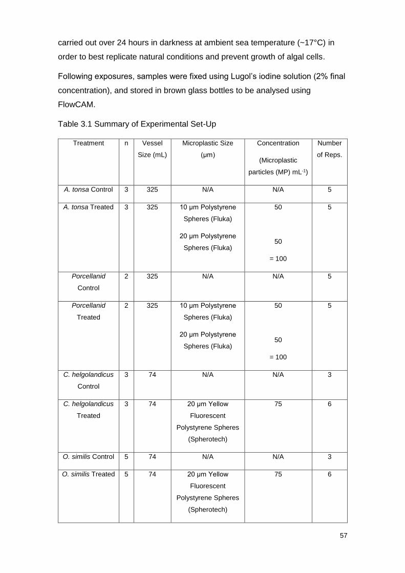
57
carried out over 24 hours in darkness at ambient sea temperature (~17°C) in
order to best replicate natural conditions and prevent growth of algal cells.
Following exposures, samples were fixed using Lugol’s iodine solution (2% final
concentration), and stored in brown glass bottles to be analysed using
FlowCAM.
Table 3.1 Summary of Experimental Set-Up
Treatment n Vessel
Size (mL)
Microplastic Size
(μm)
Concentration
(Microplastic
particles (MP) mL-1)
Number
of Reps.
A. tonsa Control 3 325 N/A N/A 5
A. tonsa Treated 3 325 10 μm Polystyrene
Spheres (Fluka)
20 μm Polystyrene
Spheres (Fluka)
50
50
= 100
5
Porcellanid
Control
2 325 N/A N/A 5
Porcellanid
Treated
2 325 10 μm Polystyrene
Spheres (Fluka)
20 μm Polystyrene
Spheres (Fluka)
50
50
= 100
5
C. helgolandicus
Control
3 74 N/A N/A 3
C. helgolandicus
Treated
3 74 20 μm Yellow
Fluorescent
Polystyrene Spheres
(Spherotech)
75 6
O. similis Control 5 74 N/A N/A 3
O. similis Treated 5 74 20 μm Yellow
Fluorescent
Polystyrene Spheres
(Spherotech)
75 6
58
3.4.1.5 FlowCAM analysis
FlowCAM enables the user to sort phytoplankton samples into specific
categories of interest; for this study we examined the uptake of diatoms,
dinoflagellates, pennates, ciliates, unidentified <10 μm cells and unidentified 10-
20 μm cells. Samples were passed through FlowCAM in autoimage mode with a
particles used per image (PPI) of approximately 1.10. All samples were viewed
using a 10x Flow Cell. Following sampling through FlowCAM and classification
into prey categories, summary information including cells mL-1, average size
(μm) and biovolume (μm3) were exported for further analysis.
3.4.1.6 Calculation of Ingestion rates and Carbon uptake
Using the equation of Frost (1972) the ingestion rate and subsequent carbon
uptake of each algal type and total algae was calculated for each species.
3.4.1.7 Statistical Analysis
Using Microsoft Excel, Student’s T-tests were used to assess whether any
significant differences were apparent between the ingestion rates and carbon
uptake of each prey type, and in total, between the control and treated groups.
Results were classed as significantly different to one another when the p-value
was <0.05. Any cases of negative ingestion rate or carbon uptake were omitted
from statistical analysis.
3.4.1.8 The effects of a mixture of 10/20 μm polystyrene microspheres on the
feeding behaviour of the copepod, Acartia tonsa, and Porcellanid larvae
Following identification, Porcellanid larvae of a similar size, and adult female A.
tonsa were transferred to 5 L beakers containing 0.2 μm FSW, and conditioned
overnight at ambient sea temperature (~17°C).
Based on previous research it was decided to use a concentration of 100 MP
mL-1 for the study. This was made up of 50 10 μm polystyrene microspheres
(Fluka) mL-1 and 50 20 μm polystyrene microspheres (Fluka) mL-1, in order to
represent natural conditions where microplastics are unlikely to all be of the
same size
Microplastic solutions were added to 325 mL bottles, with 5 replicates for control
(no microplastic) and treated (with microplastic) groups for each species. Four
59
T0 samples of NSW were fixed using 5 mL Lugol’s iodine solution (2% final
concentration). Four 325 mL samples of NSW were also bottled in order to
record algal growth without grazing. Two Porcellanid larvae and 3 A. tonsa
individuals were added to their respective bottles and fixed to a plankton wheel
set at <5 rpm. Exposures were carried out for 24 hours at ambient sea
temperature (~17°C) in darkness. Following exposure samples were filtered
through a 100 μm mesh to remove zooplankton and fixed in Lugol’s iodine
solution (2% final concentration).
T0 and T24 samples without grazing were run through the FlowCAM at a rate of
1 mL min-1 with a total sample volume of 45 mL, recording all cells in the size
range of 8-100 μm. T24 samples with grazing, both control and treated groups,
were concentrated from 90 mL to 30 mL and run through the FlowCAM at 0.625
mL min-1. A total sample volume of 30 mL was analysed with a concentration
factor of 3, recording cells in the size range of 8-100 μm.
3.4.1.9 The effects of microplastic exposure upon the feeding rate of a feeding-
current strategist and an ambush feeder under Phaeocystis bloom conditions.
Adult female C. helgolandicus and Oithona similis were picked out and
conditioned overnight at ambient sea temperature (17°C). The following day, 3
individuals of C. helgolandicus and 5 individuals of O. similis per replicate were
added to 74 mL glass bottles treated with NSW. The community of
phytoplankton inhabiting NSW collected at the time of experimentation was
dominated by a Phaeocystis bloom, a species argued to have multiple effects
upon zooplankton feeding ecology (Turner, et al., 2002), therefore, of interest in
studies upon feeding behaviour. Control treatments had no microplastics added,
other bottles were spiked with 20 μm Yellow Fluorescent Polystyrene spheres
(Spherotech) at a concentration of 75 MP mL-1. This concentration of
microplastics was chosen following work carried out by Cole (2014) who found
effects upon feeding following exposure to 20 μm beads displayed by C.
helgolandicus at this concentration. Controls were repeated 3 times, and
treatment groups were repeated 6 times. Exposures were run for 24 hours fixed
to a plankton wheel rotating at <5 rpm at ambient sea temperature (17°C).
60
Due to a technical error T24 samples with no animals added were not recorded,
therefore, algal growth was assumed to be 0, due to the fact that experiments
were carried out in darkness.
Following exposure, copepod specimens were filtered out of samples using a
100 μm mesh and preserved in 5% ethanol solution and transported to the
imaging unit at PML. Individuals were then placed on slides and imaged using
the Olympus IMT-2 microscope with Canon camera attachment. A fluorescence
wavelength of 475 nm was used to excite microplastics and camera settings set
to optimise the view of fluorescence. All individuals were imaged, and all
examples of ingestion and adherence of microplastic particles were recorded.
Samples were fixed using Lugol’s iodine solution (2% final concentration) and
passed through FlowCAM as described above.
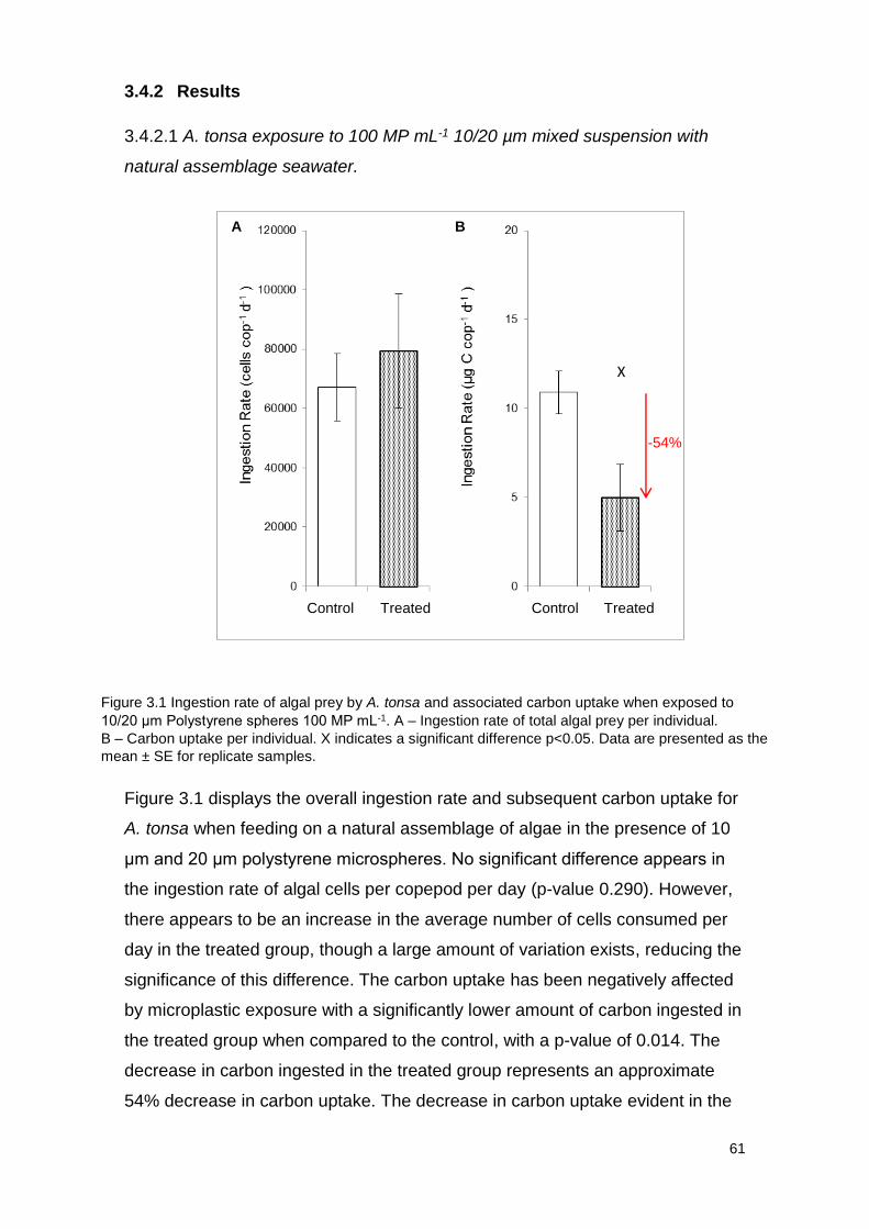
61
3.4.2 Results
3.4.2.1 A. tonsa exposure to 100 MP mL-1 10/20 µm mixed suspension with
natural assemblage seawater.
Figure 3.1 displays the overall ingestion rate and subsequent carbon uptake for
A. tonsa when feeding on a natural assemblage of algae in the presence of 10
μm and 20 μm polystyrene microspheres. No significant difference appears in
the ingestion rate of algal cells per copepod per day (p-value 0.290). However,
there appears to be an increase in the average number of cells consumed per
day in the treated group, though a large amount of variation exists, reducing the
significance of this difference. The carbon uptake has been negatively affected
by microplastic exposure with a significantly lower amount of carbon ingested in
the treated group when compared to the control, with a p-value of 0.014. The
decrease in carbon ingested in the treated group represents an approximate
54% decrease in carbon uptake. The decrease in carbon uptake evident in the
X
X
Figure 3.1 Ingestion rate of algal prey by A. tonsa and associated carbon uptake when exposed to
10/20 μm Polystyrene spheres 100 MP mL-1. A – Ingestion rate of total algal prey per individual.
B – Carbon uptake per individual. X indicates a significant difference p<0.05. Data are presented as the
mean ± SE for replicate samples.
*
Control Control Treated Treated
-54%
Control Treated
A B
X
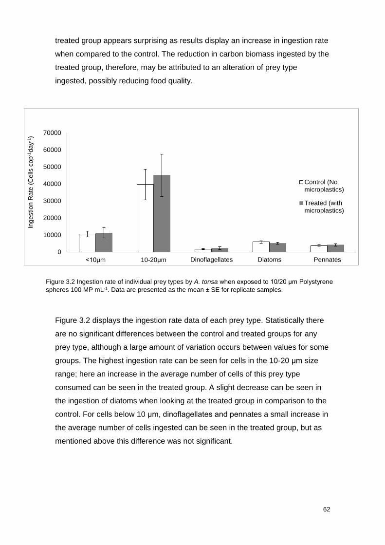
62
treated group appears surprising as results display an increase in ingestion rate
when compared to the control. The reduction in carbon biomass ingested by the
treated group, therefore, may be attributed to an alteration of prey type
ingested, possibly reducing food quality.
Figure 3.2 displays the ingestion rate data of each prey type. Statistically there
are no significant differences between the control and treated groups for any
prey type, although a large amount of variation occurs between values for some
groups. The highest ingestion rate can be seen for cells in the 10-20 μm size
range; here an increase in the average number of cells of this prey type
consumed can be seen in the treated group. A slight decrease can be seen in
the ingestion of diatoms when looking at the treated group in comparison to the
control. For cells below 10 μm, dinoflagellates and pennates a small increase in
the average number of cells ingested can be seen in the treated group, but as
mentioned above this difference was not significant.
0
10000
20000
30000
40000
50000
60000
70000
<10μm 10-20μm Dinoflagellates Diatoms Pennates
Control (Nomicroplastics)
Treated (withmicroplastics)
Ingestion R
ate
(C
ells
cop
-1day
-1)
Figure 3.2 Ingestion rate of individual prey types by A. tonsa when exposed to 10/20 μm Polystyrene
spheres 100 MP mL-1. Data are presented as the mean ± SE for replicate samples.
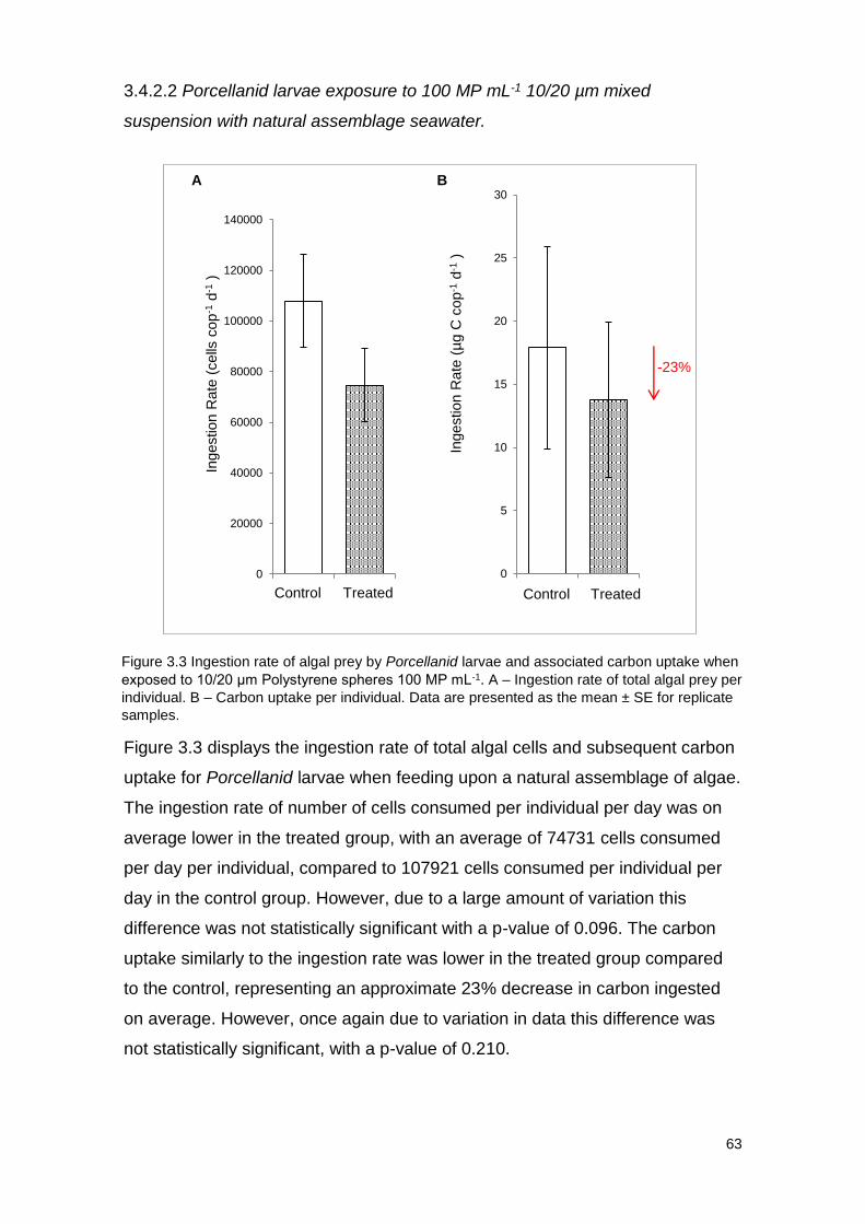
63
3.4.2.2 Porcellanid larvae exposure to 100 MP mL-1 10/20 µm mixed
suspension with natural assemblage seawater.
Figure 3.3 displays the ingestion rate of total algal cells and subsequent carbon
uptake for Porcellanid larvae when feeding upon a natural assemblage of algae.
The ingestion rate of number of cells consumed per individual per day was on
average lower in the treated group, with an average of 74731 cells consumed
per day per individual, compared to 107921 cells consumed per individual per
day in the control group. However, due to a large amount of variation this
difference was not statistically significant with a p-value of 0.096. The carbon
uptake similarly to the ingestion rate was lower in the treated group compared
to the control, representing an approximate 23% decrease in carbon ingested
on average. However, once again due to variation in data this difference was
not statistically significant, with a p-value of 0.210.
Control Control Treated Treated
0
5
10
15
20
25
30
1 2
Ingestion R
ate
(µg C
cop
-1d
-1)
0
20000
40000
60000
80000
100000
120000
140000
1 2
Ingestion R
ate
(cells
cop
-1d
-1)
-23%
Control Treated Control Treated
A B
Figure 3.3 Ingestion rate of algal prey by Porcellanid larvae and associated carbon uptake when
exposed to 10/20 μm Polystyrene spheres 100 MP mL-1. A – Ingestion rate of total algal prey per
individual. B – Carbon uptake per individual. Data are presented as the mean ± SE for replicate
samples.
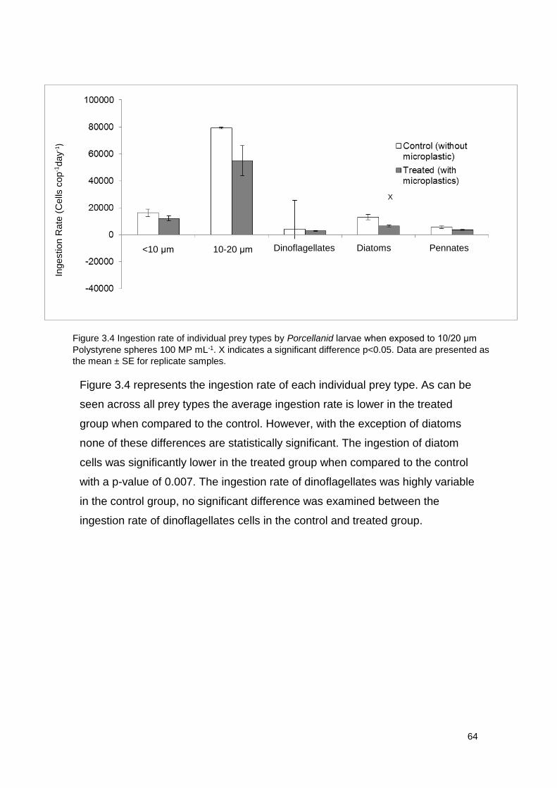
64
Figure 3.4 represents the ingestion rate of each individual prey type. As can be
seen across all prey types the average ingestion rate is lower in the treated
group when compared to the control. However, with the exception of diatoms
none of these differences are statistically significant. The ingestion of diatom
cells was significantly lower in the treated group when compared to the control
with a p-value of 0.007. The ingestion rate of dinoflagellates was highly variable
in the control group, no significant difference was examined between the
ingestion rate of dinoflagellates cells in the control and treated group.
*
Prey type
Ingestion R
ate
(C
ells
cop
-1day
- 1)
<10 μm 10-20 μm Dinoflagellates Diatoms Pennates
X
Figure 3.4 Ingestion rate of individual prey types by Porcellanid larvae when exposed to 10/20 μm
Polystyrene spheres 100 MP mL-1. X indicates a significant difference p<0.05. Data are presented as
the mean ± SE for replicate samples.
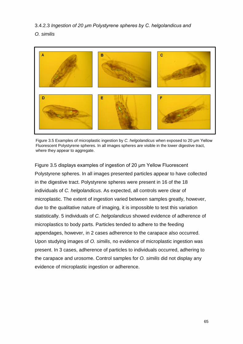
65
3.4.2.3 Ingestion of 20 μm Polystyrene spheres by C. helgolandicus and
O. similis
Figure 3.5 displays examples of ingestion of 20 μm Yellow Fluorescent
Polystyrene spheres. In all images presented particles appear to have collected
in the digestive tract. Polystyrene spheres were present in 16 of the 18
individuals of C. helgolandicus. As expected, all controls were clear of
microplastic. The extent of ingestion varied between samples greatly, however,
due to the qualitative nature of imaging, it is impossible to test this variation
statistically. 5 individuals of C. helgolandicus showed evidence of adherence of
microplastics to body parts. Particles tended to adhere to the feeding
appendages, however, in 2 cases adherence to the carapace also occurred.
Upon studying images of O. similis, no evidence of microplastic ingestion was
present. In 3 cases, adherence of particles to individuals occurred, adhering to
the carapace and urosome. Control samples for O. similis did not display any
evidence of microplastic ingestion or adherence.
Figure 3.5 Examples of microplastic ingestion by C. helgolandicus when exposed to 20 μm Yellow
Fluorescent Polystyrene spheres. In all images spheres are visible in the lower digestive tract,
where they appear to aggregate.
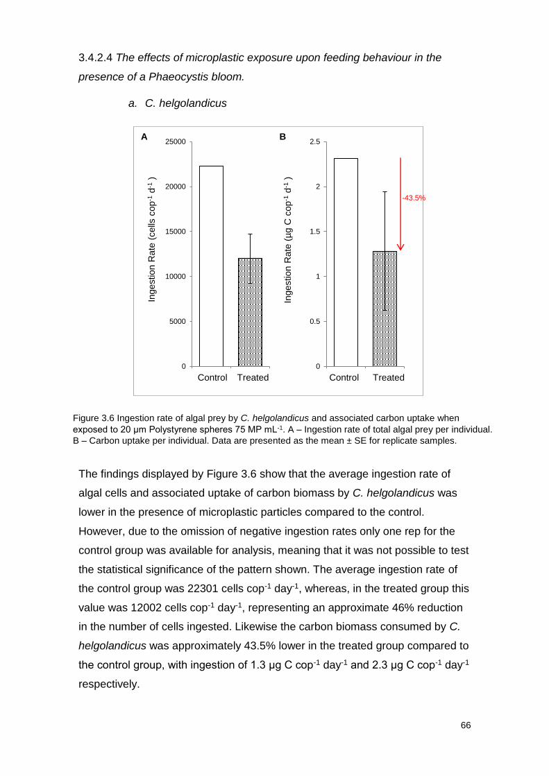
66
3.4.2.4 The effects of microplastic exposure upon feeding behaviour in the
presence of a Phaeocystis bloom.
a. C. helgolandicus
The findings displayed by Figure 3.6 show that the average ingestion rate of
algal cells and associated uptake of carbon biomass by C. helgolandicus was
lower in the presence of microplastic particles compared to the control.
However, due to the omission of negative ingestion rates only one rep for the
control group was available for analysis, meaning that it was not possible to test
the statistical significance of the pattern shown. The average ingestion rate of
the control group was 22301 cells cop-1 day-1, whereas, in the treated group this
value was 12002 cells cop-1 day-1, representing an approximate 46% reduction
in the number of cells ingested. Likewise the carbon biomass consumed by C.
helgolandicus was approximately 43.5% lower in the treated group compared to
the control group, with ingestion of 1.3 μg C cop-1 day-1 and 2.3 μg C cop-1 day-1
respectively.
0
5000
10000
15000
20000
25000
1 2
Ingestion R
ate
(cells
cop
-1d
-1)
0
0.5
1
1.5
2
2.5
1 2
Ingestion R
ate
(µg C
cop
-1d
-1)
Control Control Treated Treated
A B
Figure 3.6 Ingestion rate of algal prey by C. helgolandicus and associated carbon uptake when
exposed to 20 μm Polystyrene spheres 75 MP mL-1. A – Ingestion rate of total algal prey per individual.
B – Carbon uptake per individual. Data are presented as the mean ± SE for replicate samples.
-43.5%
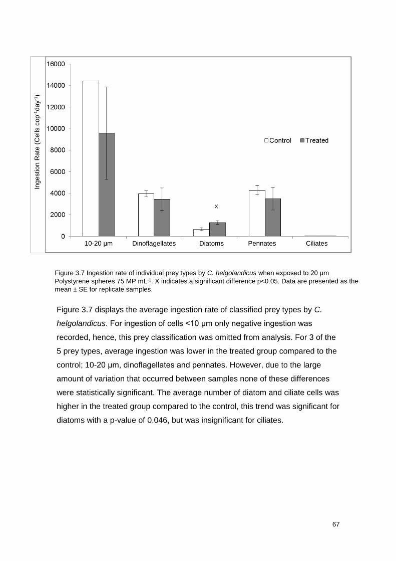
67
Figure 3.7 displays the average ingestion rate of classified prey types by C.
helgolandicus. For ingestion of cells <10 μm only negative ingestion was
recorded, hence, this prey classification was omitted from analysis. For 3 of the
5 prey types, average ingestion was lower in the treated group compared to the
control; 10-20 μm, dinoflagellates and pennates. However, due to the large
amount of variation that occurred between samples none of these differences
were statistically significant. The average number of diatom and ciliate cells was
higher in the treated group compared to the control, this trend was significant for
diatoms with a p-value of 0.046, but was insignificant for ciliates.
Figure 3.7 Ingestion rate of individual prey types by C. helgolandicus when exposed to 20 μm
Polystyrene spheres 75 MP mL-1. X indicates a significant difference p<0.05. Data are presented as the
mean ± SE for replicate samples.
10-20 μm Dinoflagellates Diatoms Pennates Ciliates
X
Ingestion R
ate
(C
ells
cop
-1day
- 1)
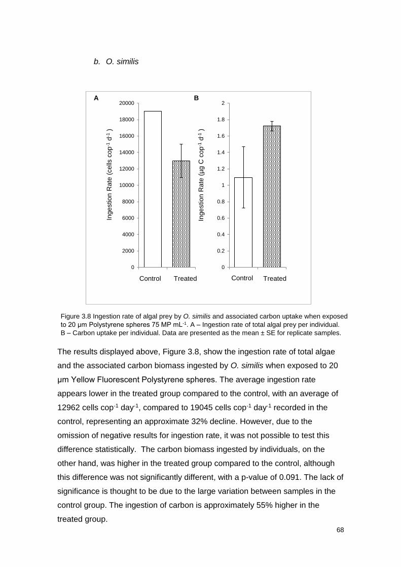
68
b. O. similis
The results displayed above, Figure 3.8, show the ingestion rate of total algae
and the associated carbon biomass ingested by O. similis when exposed to 20
μm Yellow Fluorescent Polystyrene spheres. The average ingestion rate
appears lower in the treated group compared to the control, with an average of
12962 cells cop-1 day-1, compared to 19045 cells cop-1 day-1 recorded in the
control, representing an approximate 32% decline. However, due to the
omission of negative results for ingestion rate, it was not possible to test this
difference statistically. The carbon biomass ingested by individuals, on the
other hand, was higher in the treated group compared to the control, although
this difference was not significantly different, with a p-value of 0.091. The lack of
significance is thought to be due to the large variation between samples in the
control group. The ingestion of carbon is approximately 55% higher in the
treated group.
0
2000
4000
6000
8000
10000
12000
14000
16000
18000
20000
1 2
Ingestion R
ate
(cells
cop
-1d
-1)
0
0.2
0.4
0.6
0.8
1
1.2
1.4
1.6
1.8
2
1 2
Ingestion R
ate
(µg C
cop
-1d
-1)
Control Treated Control Treated
A B
Figure 3.8 Ingestion rate of algal prey by O. similis and associated carbon uptake when exposed
to 20 μm Polystyrene spheres 75 MP mL-1. A – Ingestion rate of total algal prey per individual.
B – Carbon uptake per individual. Data are presented as the mean ± SE for replicate samples.
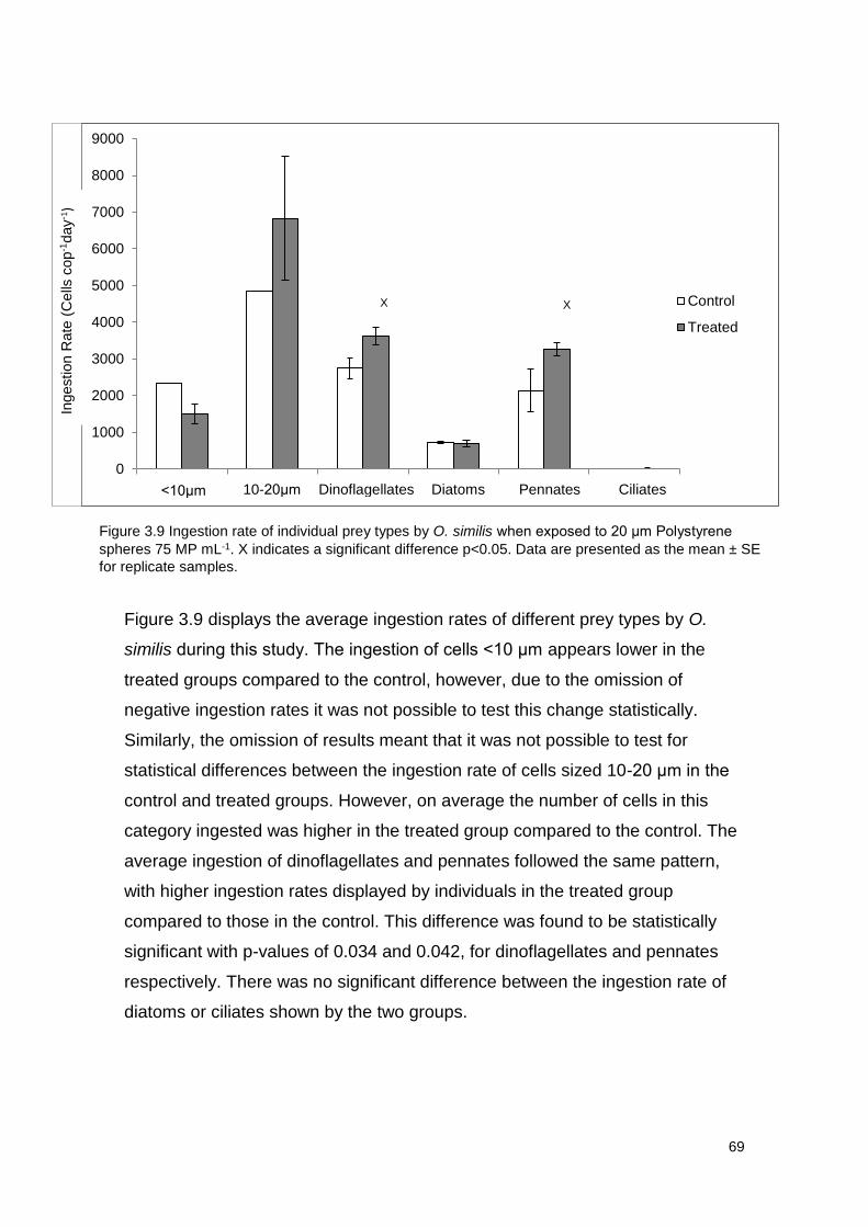
69
Figure 3.9 displays the average ingestion rates of different prey types by O.
similis during this study. The ingestion of cells <10 μm appears lower in the
treated groups compared to the control, however, due to the omission of
negative ingestion rates it was not possible to test this change statistically.
Similarly, the omission of results meant that it was not possible to test for
statistical differences between the ingestion rate of cells sized 10-20 μm in the
control and treated groups. However, on average the number of cells in this
category ingested was higher in the treated group compared to the control. The
average ingestion of dinoflagellates and pennates followed the same pattern,
with higher ingestion rates displayed by individuals in the treated group
compared to those in the control. This difference was found to be statistically
significant with p-values of 0.034 and 0.042, for dinoflagellates and pennates
respectively. There was no significant difference between the ingestion rate of
diatoms or ciliates shown by the two groups.
0
1000
2000
3000
4000
5000
6000
7000
8000
9000
1 2 3 4 5 6
Control
Treated
10-20μm <10μm Dinoflagellates Diatoms Pennates Ciliates
X X
Figure 3.9 Ingestion rate of individual prey types by O. similis when exposed to 20 μm Polystyrene
spheres 75 MP mL-1. X indicates a significant difference p<0.05. Data are presented as the mean ± SE
for replicate samples.
Ingestion R
ate
(C
ells
cop
-1day
- 1)
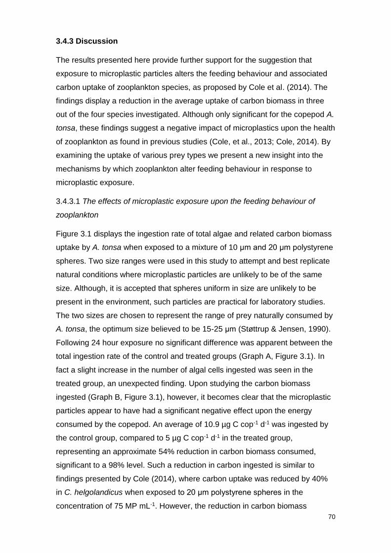
70
3.4.3 Discussion
The results presented here provide further support for the suggestion that
exposure to microplastic particles alters the feeding behaviour and associated
carbon uptake of zooplankton species, as proposed by Cole et al. (2014). The
findings display a reduction in the average uptake of carbon biomass in three
out of the four species investigated. Although only significant for the copepod A.
tonsa, these findings suggest a negative impact of microplastics upon the health
of zooplankton as found in previous studies (Cole, et al., 2013; Cole, 2014). By
examining the uptake of various prey types we present a new insight into the
mechanisms by which zooplankton alter feeding behaviour in response to
microplastic exposure.
3.4.3.1 The effects of microplastic exposure upon the feeding behaviour of
zooplankton
Figure 3.1 displays the ingestion rate of total algae and related carbon biomass
uptake by A. tonsa when exposed to a mixture of 10 μm and 20 μm polystyrene
spheres. Two size ranges were used in this study to attempt and best replicate
natural conditions where microplastic particles are unlikely to be of the same
size. Although, it is accepted that spheres uniform in size are unlikely to be
present in the environment, such particles are practical for laboratory studies.
The two sizes are chosen to represent the range of prey naturally consumed by
A. tonsa, the optimum size believed to be 15-25 μm (Støttrup & Jensen, 1990).
Following 24 hour exposure no significant difference was apparent between the
total ingestion rate of the control and treated groups (Graph A, Figure 3.1). In
fact a slight increase in the number of algal cells ingested was seen in the
treated group, an unexpected finding. Upon studying the carbon biomass
ingested (Graph B, Figure 3.1), however, it becomes clear that the microplastic
particles appear to have had a significant negative effect upon the energy
consumed by the copepod. An average of 10.9 µg C cop-1 d-1 was ingested by
the control group, compared to 5 µg C cop-1 d-1 in the treated group,
representing an approximate 54% reduction in carbon biomass consumed,
significant to a 98% level. Such a reduction in carbon ingested is similar to
findings presented by Cole (2014), where carbon uptake was reduced by 40%
in C. helgolandicus when exposed to 20 μm polystyrene spheres in the
concentration of 75 MP mL-1. However, the reduction in carbon biomass
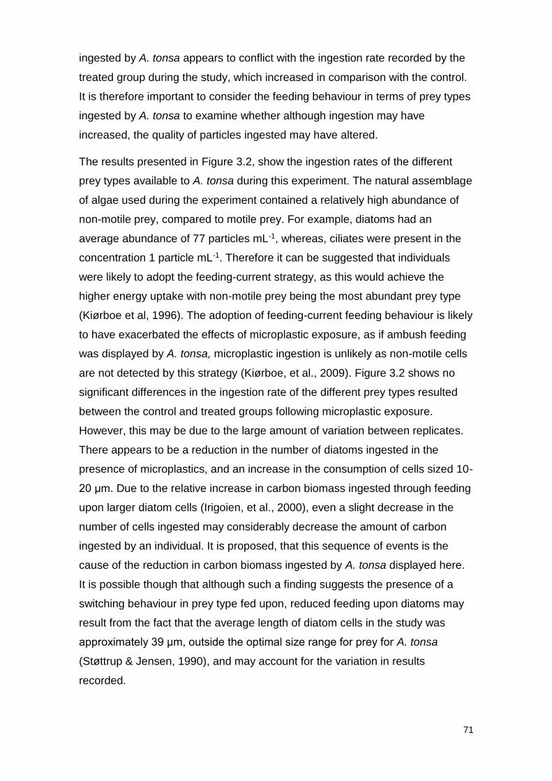
71
ingested by A. tonsa appears to conflict with the ingestion rate recorded by the
treated group during the study, which increased in comparison with the control.
It is therefore important to consider the feeding behaviour in terms of prey types
ingested by A. tonsa to examine whether although ingestion may have
increased, the quality of particles ingested may have altered.
The results presented in Figure 3.2, show the ingestion rates of the different
prey types available to A. tonsa during this experiment. The natural assemblage
of algae used during the experiment contained a relatively high abundance of
non-motile prey, compared to motile prey. For example, diatoms had an
average abundance of 77 particles mL-1, whereas, ciliates were present in the
concentration 1 particle mL-1. Therefore it can be suggested that individuals
were likely to adopt the feeding-current strategy, as this would achieve the
higher energy uptake with non-motile prey being the most abundant prey type
(Kiørboe et al, 1996). The adoption of feeding-current feeding behaviour is likely
to have exacerbated the effects of microplastic exposure, as if ambush feeding
was displayed by A. tonsa, microplastic ingestion is unlikely as non-motile cells
are not detected by this strategy (Kiørboe, et al., 2009). Figure 3.2 shows no
significant differences in the ingestion rate of the different prey types resulted
between the control and treated groups following microplastic exposure.
However, this may be due to the large amount of variation between replicates.
There appears to be a reduction in the number of diatoms ingested in the
presence of microplastics, and an increase in the consumption of cells sized 10-
20 μm. Due to the relative increase in carbon biomass ingested through feeding
upon larger diatom cells (Irigoien, et al., 2000), even a slight decrease in the
number of cells ingested may considerably decrease the amount of carbon
ingested by an individual. It is proposed, that this sequence of events is the
cause of the reduction in carbon biomass ingested by A. tonsa displayed here.
It is possible though that although such a finding suggests the presence of a
switching behaviour in prey type fed upon, reduced feeding upon diatoms may
result from the fact that the average length of diatom cells in the study was
approximately 39 μm, outside the optimal size range for prey for A. tonsa
(Støttrup & Jensen, 1990), and may account for the variation in results
recorded.
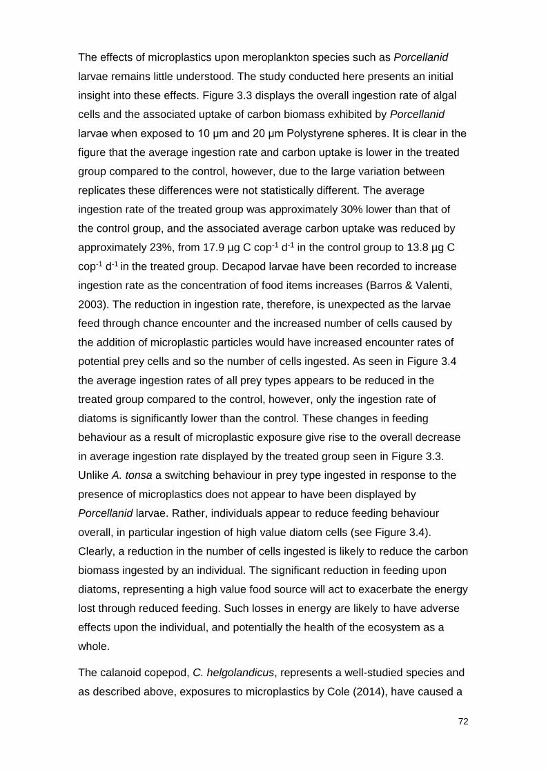
72
The effects of microplastics upon meroplankton species such as Porcellanid
larvae remains little understood. The study conducted here presents an initial
insight into these effects. Figure 3.3 displays the overall ingestion rate of algal
cells and the associated uptake of carbon biomass exhibited by Porcellanid
larvae when exposed to 10 μm and 20 μm Polystyrene spheres. It is clear in the
figure that the average ingestion rate and carbon uptake is lower in the treated
group compared to the control, however, due to the large variation between
replicates these differences were not statistically different. The average
ingestion rate of the treated group was approximately 30% lower than that of
the control group, and the associated average carbon uptake was reduced by
approximately 23%, from 17.9 µg C cop-1 d-1 in the control group to 13.8 µg C
cop-1 d-1 in the treated group. Decapod larvae have been recorded to increase
ingestion rate as the concentration of food items increases (Barros & Valenti,
2003). The reduction in ingestion rate, therefore, is unexpected as the larvae
feed through chance encounter and the increased number of cells caused by
the addition of microplastic particles would have increased encounter rates of
potential prey cells and so the number of cells ingested. As seen in Figure 3.4
the average ingestion rates of all prey types appears to be reduced in the
treated group compared to the control, however, only the ingestion rate of
diatoms is significantly lower than the control. These changes in feeding
behaviour as a result of microplastic exposure give rise to the overall decrease
in average ingestion rate displayed by the treated group seen in Figure 3.3.
Unlike A. tonsa a switching behaviour in prey type ingested in response to the
presence of microplastics does not appear to have been displayed by
Porcellanid larvae. Rather, individuals appear to reduce feeding behaviour
overall, in particular ingestion of high value diatom cells (see Figure 3.4).
Clearly, a reduction in the number of cells ingested is likely to reduce the carbon
biomass ingested by an individual. The significant reduction in feeding upon
diatoms, representing a high value food source will act to exacerbate the energy
lost through reduced feeding. Such losses in energy are likely to have adverse
effects upon the individual, and potentially the health of the ecosystem as a
whole.
The calanoid copepod, C. helgolandicus, represents a well-studied species and
as described above, exposures to microplastics by Cole (2014), have caused a
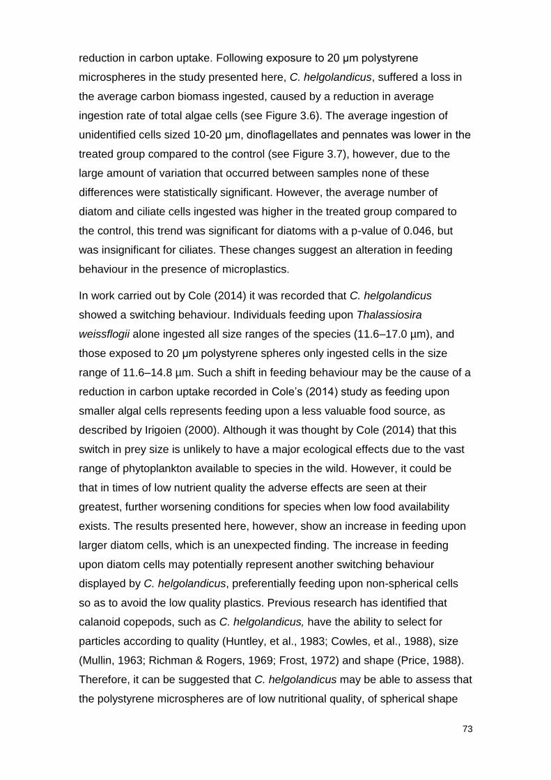
73
reduction in carbon uptake. Following exposure to 20 μm polystyrene
microspheres in the study presented here, C. helgolandicus, suffered a loss in
the average carbon biomass ingested, caused by a reduction in average
ingestion rate of total algae cells (see Figure 3.6). The average ingestion of
unidentified cells sized 10-20 μm, dinoflagellates and pennates was lower in the
treated group compared to the control (see Figure 3.7), however, due to the
large amount of variation that occurred between samples none of these
differences were statistically significant. However, the average number of
diatom and ciliate cells ingested was higher in the treated group compared to
the control, this trend was significant for diatoms with a p-value of 0.046, but
was insignificant for ciliates. These changes suggest an alteration in feeding
behaviour in the presence of microplastics.
In work carried out by Cole (2014) it was recorded that C. helgolandicus
showed a switching behaviour. Individuals feeding upon Thalassiosira
weissflogii alone ingested all size ranges of the species (11.6–17.0 µm), and
those exposed to 20 μm polystyrene spheres only ingested cells in the size
range of 11.6–14.8 µm. Such a shift in feeding behaviour may be the cause of a
reduction in carbon uptake recorded in Cole’s (2014) study as feeding upon
smaller algal cells represents feeding upon a less valuable food source, as
described by Irigoien (2000). Although it was thought by Cole (2014) that this
switch in prey size is unlikely to have a major ecological effects due to the vast
range of phytoplankton available to species in the wild. However, it could be
that in times of low nutrient quality the adverse effects are seen at their
greatest, further worsening conditions for species when low food availability
exists. The results presented here, however, show an increase in feeding upon
larger diatom cells, which is an unexpected finding. The increase in feeding
upon diatom cells may potentially represent another switching behaviour
displayed by C. helgolandicus, preferentially feeding upon non-spherical cells
so as to avoid the low quality plastics. Previous research has identified that
calanoid copepods, such as C. helgolandicus, have the ability to select for
particles according to quality (Huntley, et al., 1983; Cowles, et al., 1988), size
(Mullin, 1963; Richman & Rogers, 1969; Frost, 1972) and shape (Price, 1988).
Therefore, it can be suggested that C. helgolandicus may be able to assess that
the polystyrene microspheres are of low nutritional quality, of spherical shape
74
and in a particular size range and alter its feeding behaviour so as to only
accept non-spherical or smaller prey.
The ambush feeder, O. similis, unlike the three other species examined here,
did not appear to suffer from the negative effects that resulted from microplastic
exposure as recorded in previous studies (Cole, 2014). Upon studying Figure
3.8 it is clear that no significant difference occurred between the ingestion rate
of total cells and associated carbon biomass uptake of control and treated
groups. The results support the prediction that little or no effect will be suffered
by an ambush feeder following exposure to microplastic. By feeding in such a
manner, non-motile particles such as microplastics are unlikely to be detected
since they do not create a disturbance within the water column (Kiørboe, et al.,
2009). This being said, Figure 3.9, displaying the ingestion rates of specified
prey types suggests that alterations in feeding behaviour have been caused by
the presence of microplastic particles. The ingestion of non-spherical prey,
diatoms and pennates, was significantly higher in the treated group compared
to the control. Such a finding is unexpected as O. similis feeds as an ambush
predator and by feeding in this manner should not detect non-motile prey such
as diatom and pennate cells. The findings presented in this study, therefore,
suggest O. similis may possess the ability to switch feeding strategies in a
similar manner to A. tonsa.
3.4.3.2 Drivers of altered feeding in the presence of microplastics
There are a number of factors which may play a role in the alteration of feeding
activity of zooplankton, as a result of microplastic exposure. The first of which
may be due to a false sense of satiation which may act to reduce feeding rates
in organisms. Such processes could be attributed to the recorded extended gut
retention times seen in microplastic exposure studies, including those carried
out upon zooplankton (Cole, et al., 2013), and other organisms such as mussels
(Browne, et al., 2008). Increased gut retention times also increases the length
of time for the chemical constituents of plastics to leach into organisms and
cause an adverse effect (Setälä, et al., 2014). The decreases in fitness that
arise from such stresses may result in reduced feeding activity or an alteration
in feeding activity, as the organism may not be able to feed efficiently. Another
negative effect of the extended retention times of microplastics is the possibility
75
of particles to clump and knot (Murray & Cowie, 2011), a process seen in
decapod crustaceans, which again is likely to alter feeding in some way.
The ability of copepods to select particles in terms of their food quality has been
examined and will be discussed in detail in Chapter 4. It is argued that the
copepod species studied possess the ability to actively accept or reject particles
based on their quality (Donaghay & Small, 1979). However, in essence quality
selection is in some way size selection (Irigoien, et al., 2000), as with increased
size, particles offer a larger amount of biovolume to be ingested so increasing
nutritional value. Therefore, if we assume that a copepod species is able to
detect that a microplastic particle of a certain size is of low nutritional value and
they are in high density so that the encounter rate of such particles is high, the
copepod may actively reject particles in the size range of the microplastic
particles. This may be the mechanism behind the switching behaviour seen in
this study and in studies such as those carried out by Cole (2014) where an
exposure to uniformly sized polystyrene spheres was used. However, it is
unlikely that such behaviour would be observed in the wild as it is not likely that
microplastics are of a uniform size, shape and structure.
The lack of a switching behaviour shown by Porcellanid larvae suggest that,
unlike copepods, decapod larvae do not possess the same selective behaviour
as displayed by copepods during feeding. As a result it can be assumed that
different mechanisms are driving the reduction in feeding by Porcellanid larvae
when microplastic particles are present. Cole et al. (2013) recorded that
Porcellanid larvae are capable of ingesting microplastic particles (Cole, et al.,
2013). Therefore, reduced feeding is more likely caused by individuals feeling a
false sense of satiation, as described by (Gregory, 2009; Wright, et al., 2013),
(Murray & Cowie, 2011), (Cole, et al., 2013).
Similarly to copepods, Porcellanid larvae possess appendages used in feeding
and swimming; For example the endopodites are used during feeding and
require frequent cleaning (Gonor & Gonor, 1973) by the individual to ensure
feeding efficiency. Microplastics have been recorded to adhere to feeding
appendages (Cole, et al., 2013) as described in Chapter 2. Therefore, if
particles adhere to the feeding appendages of Porcellanid larvae as has been
studied with copepods, it may disrupt normal functioning of the endopodites,
76
thus, reducing feeding efficiency and the number of cells ingested. The
generation of a feeding-current by C. helgolandicus and A. tonsa during feeding
is likely to increase the likelihood of adherence of microplastic particles to
feeding appendages, due to the continual movement of appendages in order to
generate the current. Such adherence is likely to negatively affect feeding by
reducing the ability to catch prey cells.
3.4.3.3 The effects of Phaeocystis upon grazing studies
During the preparation of studies involving C. helgolandicus and O. similis a
Phaeocystis bloom occurred in the Western Channel. As a result, the natural
assemblage of algae used during the investigation was dominated by
Phaeocystis, a species argued to have multiple effects upon zooplankton
feeding ecology (Turner, et al., 2002). Phaeocystis sp. is common in
phytoplankton communities and its range spreads across worldwide waters in
coastal and open ocean systems (Kashkin, 1963). The species is problematic
as it possesses a rapid growth rate allowing it to become dominant over a few
days (Bautista, et al., 1992). The life cycle of Phaeocystis alternates between a
solitary stage of motile flagellated cells sized 3.5 μm which group to form non-
motile colonies of up to 10 mm in diameter (Gieskes & Kraay, 1975; Verity, et
al., 1988). The alga is believed to have negative effects upon the fitness of
zooplankton due to the fact that it is a member of the Prymnesiophyceae family,
recognised as producing toxic compounds (Bautista, et al., 1992). As well as
this, the concentration of polyunsaturated acids and vitamin C is low in
Phaeocystis (Claustre, et al., 1990), meaning that it may not represent a
nutritionally valuable food source for zooplankton (Bautista, et al., 1992).
However, research conducted by other groups suggests that Phaeocystis may
not be as problematic as first predicted. C. helgolandicus fed Phaeocystis
received adequate energy for egg production and survivorship and hatchling
success was high, suggesting that Phaeocystis does not have a toxic effect
upon C. helgolandicus (Turner, et al., 2002). The presence of Phaeocystis then
may not have altered results presented in this study for C. helgolandicus.
Although it was conceded by Turner et al. (2002) that the Mediterranean strain
of Phaeocystis used in their study may not induce as toxic effects as those that
inhabit the North Sea and Polar regions known to produce toxicants such as
acrylic acid (Turner, et al., 2002). Therefore, it can be argued that the results of
77
this study may have been significantly affected by the presence of Phaeocystis
in samples reducing the fitness of individuals and altering feeding behaviour.
Hence, its presence may be a factor in the occurrence of negative ingestion
rates which were omitted from analysis. As a result it would be highly beneficial
to repeat the experiment at a time where Phaeocystis is absent from the
phytoplankton community to remove any possibility that its presence altered
results.
3.4.3.4 Limitations of research
As well as the possible negative effects of Phaeocystis, a number of other
limitations may have altered the results studies presented here. The first and
most apparent limitation is the extent of omitted data following the production of
negative ingestion rates in a number of samples. Secondly, the use of 3 C.
helgolandicus and 5 O. similis per 74 mL may have overstocked bottles and
resulted in stress on copepods and insufficient prey availability, thus, altering
behaviour. Fluorescent microspheres were not used in studies upon A. tonsa
and Porcellanid larvae, hence imaging to assess ingestion of particles was not
possible. By imaging individuals further insights into the drivers of altered
feeding behaviour could have been made. Overall it is felt that grazing studies
such as those carried out here can be effective in analysing the effects of
microplastics and other contaminants upon the ingestion rates and associated
carbon biomass uptake by zooplankton species. The use of FlowCAM to
categorise algae into specific prey types provides an interesting and insightful
manner in which to examine results, and allows for additional consideration of
the mechanisms that may be driving any patterns observed in altered uptake of
cells and carbon.
3.4.3.5 Conclusions
Although limitations of this study are apparent and not all results are supported
by statistical significance, the discussion above allows for additional
consideration of the role of plankton type and feeding type in determining the
effect that the presence of microplastics has upon the feeding behaviour of
zooplankton. It is felt that by repeating studies in the absence of Phaeocystis,
and using a larger vessel volume for studies upon C. helgolandicus and O.
similis that results will be more reliable allowing for statistical testing and
78
confirmation of findings. Results suggest that species displaying ambush
feeding strategies feeding upon motile prey are less affected by the negative
effects associated with microplastic exposure compared to those species that
feed by generation of a feeding current. Meroplankton species appear
negatively affected by microplastic exposure in a similar manner to
holoplankton, however, additional research is required to confirm findings.
Microplastics seem to alter the feeding behaviour of zooplankton in terms of
size and type of prey ingested, such changes have the potential to have
ecosystem effects where microplastic pollution is high.
Overall the results presented in this study provide additional support for the
growing amount of evidence that exposure to microplastic particles causes an
energy deficit to marine organisms (Wright, et al., 2013; Cole, 2014). Such a
response is highly concerning due to the vital role played by zooplankton in
transferring energy to higher trophic levels in the ecosystem.

79

80
81
Chapter 4
Assessing the detection and subsequent acceptance/rejection
of microplastic particles by marine copepods

82
83
The ability of copepods to accept or reject potential food particles has been a
subject of investigation for a number of years. The opportunity now exists to
record subjects in high detail using high-speed video recording equipment,
allowing the researcher to clearly examine the physical characteristics of
interactions between prey and individuals. During this chapter the ability of
copepods to select particular prey will be reviewed, before introducing an
exploratory study investigating microplastic ingestion by Temora longicornis. A
study carried out in order to examine the species’ ability to reject microplastic
particles.
4.1 Prey selection in copepods
Zooplankton represents the first trophic level in marine food webs, playing a
fundamental role in making primary production available to higher trophic levels
(Tiselius, et al., 2013). It is, therefore, of great importance to the ecosystem as a
whole that zooplankton species such as copepods are able to detect and feed
upon prey effectively, and select prey of high nutritional value. The manner by
which zooplankton typically detect and capture prey is described in Chapter 2,
however, it has been found that a number of zooplankton species possess the
ability to discriminate between particles of different quality (Donaghay & Small,
1979). Copepods, particularly calanoid species of the genus Calanus, have
been studied intensively, analysing the feeding behaviour they display in terms
of selecting prey. For example it has been noted that Calanus species possess
the ability to reject or actively select prey cells that they consider suitable for
ingestion (Leising, et al., 2005). The abilities of studied copepod species to
select prey and distinguish one prey species from another will be evaluated, in
order to identify whether such behaviours are likely to affect the ingestion of
microplastics in copepod species.
Many studies address the issue of size of prey particles, examining whether
there is any preference displayed by groups of individuals when offered prey of
differing sizes. Frost (1972) found that there was a greater uptake of larger cells
over smaller particle as a higher feeding efficiency is apparent when feeding
upon larger cells. In Frost’s experiments Calanus pacificus fed on both large
and small particles, but handled the larger particles with much greater
effectiveness (Frost, 1972). This study mirrored views by Mullin (1963) and
Richman and Rogers (1969) who also noted that copepods seemed to prefer
84
larger cells over smaller ones. Other studies also note that small particles are
not effectively captured; the lack of feeding on pelagophytes and cyanobacteria
by Calanus helgolandicus in feeding experiments carried out by Irigoien (2000)
was believed to be due to size selection and the reduced effectiveness of
capturing smaller prey items (Paffenhöfer, 1988; Kleppel, et al., 1998)
The ability of calanoid copepods to discriminate between particles of different
food quality has also been addressed in a number of studies (Huntley, et al.,
1983; Cowles, et al., 1988). However, it could be argued that size selection
described above is in essence quality selection (Irigoien, et al., 2000). The
reason for this is that a larger particle represents an important increase in the
volume of cytoplasm ingested (Irigoien, et al., 2000), therefore increasing the
nutritional value of the cell. As a result a zooplankton species may be at a
greater advantage ingesting fewer large cells retaining energy, rather than
actively searching and feeding upon a large number of small cells. Though, it
has been found that copepods are better able to handle large cells compared to
small cells when feeding and tend to be inefficient at feeding on particles
smaller than 5-10 µm (Nival & Nival, 1976; Berggreen, et al., 1988).
This suggests that rather than selecting for prey because of its size or nutritional
value, copepods simply feed on prey cells most efficiently captured. This being
said, a number of zooplankton species have been found to discriminate
between particles of different quality (Donaghay & Small, 1979). Some calanoid
copepods have been studied to distinguish between valuable (phytoplankton
cells) and non-valuable particles (polystyrene beads) either by rejecting un-
natural prey (Donaghay & Small, 1979) or by only ingesting them in low
quantities (Frost, 1972; Fernandez, 1979; Huntley, et al., 1983). However, other
calanoids such as C. helgolandicus ingest large quantities of low value particles
when natural prey is also offered (Paffenhöfer, 1972; Paffenhöfer & Sant, 1985).
However, this behaviour may be deliberate action by the species to diversify the
diet, a behaviour seen in other studies where there is higher selectivity for other
prey types, when one prey species is dominant (Irigoien, et al., 2000). This
behaviour may ensure that the species obtains all the essential elements and
nutrients it requires for full fitness.
The action of actively selecting for variability in the diet appears common place
across many studied calanoid species. For example, in grazing experiments
85
conducted by Irigoien et al. (2000) upon C. helgolandicus it was found that the
species displayed great variability in prey selection. It was suggested that C.
helgolandicus may vary its diet by actively selecting prey items other than the
most abundant available (Irigoien, et al., 2000). It appears clear, then, that
significant differences exists in the abilities of distinct species in selecting for
prey.
Prey selection due to shape and taste has also been recorded in some species
(Price, 1988; Atkinson, 1995). The taste of particular toxic dinoflagellates, for
example, caused rejection by copepods (Huntley, et al., 1986). More recent
studies have also displayed an ability of some zooplankton to actively reject
cells that could cause toxic damage to them (Leising, et al., 2005). It was
suggested by Huntley (1986) that copepods may use chemical signals in order
to identify and actively reject unsuitable or toxic prey items. This theory was
echoed by studies carried out by Leising et al. (2005) upon the calanoid
copepod, Calanus pacificus, where it was noted that this species may have
“considerable abilities” to reject or actively select particles considered most
suitable for ingestion. Toxic particles may be avoided due to the selection
pressure that exists if the prey item has the potential to adversely affect
reproduction (Leising, et al., 2005).
The selection mechanisms used by zooplankton have a vital role in the
dynamics of the marine ecosystem as a whole. The reasons for this is that by
feeding upon prey items of different nutritional quality, or toxic value, the
resulting growth and development of the copepod may be altered (Kleppel,
1993; Kleppel & Burkart, 1995; Ban, et al., 1997; Hygum, et al., 2000; Irigoien,
et al., 2002; Paffenhöfer, 2002; Ianora, et al., 2004; Leising, et al., 2005),
affecting both the individual and its future progeny. Therefore, the selectivity of
prey items made by a particular species has the potential to affect the energy
transfer to higher trophic levels. This could be a concern as many zooplankton
species, such as C. helgolandicus, are key prey species for many species of
commercially important fish larvae (Cole, 2014). Microplastics therefore, if
selected for due to size parameters or lack of chemical cues, may negatively
affect energy reserves and growth in zooplankton as they represent little or no
nutritional value. As recorded above (Chapter 3) and in previous studies (Cole,
2014), the ingestion of microplastic particles appears to affect zooplankton
86
health and potentially the health of commercial fish larvae stocks in the local
environment.
4.2 The utilisation of high-speed filming in microplastic research
High-speed filming techniques offer a great opportunity to study an interaction
for which the exact mechanisms remain unknown and hard to examine. Such
equipment has been used effectively by research groups studying the
mechanisms by which zooplankton feed and move through the water column,
particularly by the Kiørboe group at the Denmark Technical University (see
Kiørboe, 2011). Through grazing studies and implementing fluorescent
microscopy it has been shown that a range of zooplankton species do ingest
microplastics (Cole, et al., 2013) and the effects of such ingestion include
reductions in fitness and fecundity (Cole, 2014), as well as, changes in feeding
behaviour, as described in Chapter 3. However, due to the small size of
microplastics and copepods themselves, the exact mechanisms behind the
interactions between individuals and particles and subsequent behaviours are
little understood.
High-speed filming allows the researcher to study the way that a microplastic
particle is handled by an individual and allows for the investigation into whether
such copepod species are able to actively reject microplastic particles. As well
as this, by introducing natural prey, there is the opportunity to examine size or
food quality selectivity behaviour in more detail. Such research sheds light on
the abilities of copepods to cope with microplastics within their environment,
once more allowing for environmental effects to be inferred.
With this in mind a study was designed to best display the range of behaviours
of copepod species when exposed to microplastics in detail. Subsequent
Imaging of individuals exposed to fluorescent microplastic particles would then
provide clear evidence for ingestion of microplastic particles, which may not be
caught on film, as well as any evidence of adherence of particles to individuals.
The research would then allow us to further understand copepod’s abilities to
accept/reject prey particles based upon their nutritional value, chemical
composition, shape and structure.
87
4.3 Using high-speed filming to assess the ability of Temora longicornis to
accept/reject microplastic particles
4.3.1 Methods
4.3.1.1 High-speed video analysis of microplastic ingestion by T. longicornis
4.3.1.1.1 Experimental set-up
Over 20 individuals of T. longicornis cultivated under laboratory conditions at
Denmark Technical University (DTU) of varying life stages were added to a 50
mL filming tank and filming took place for approximately one hour per treatment.
A new filming tank and set of copepods were utilised for each treatment.
4.3.1.1.2 Microplastic suspensions
In order to examine the effects of a range of microplastic treatments on the
feeding behaviour of T. longicornis, six microplastic suspensions were set-up to
be added to filming tanks for filming; 200 μL 10 μm Yellow Fluorescent
Polystyrene spheres (Spherotech) were added directly to the 50 mL filming
tank; 200 μL 20 μm Yellow Fluorescent Polystyrene spheres (Spherotech)
were added directly to the 50 mL filming tank; 25 μL 30 μm polystyrene beads
(Fluka) added directly to the 50 mL filming tank; 25 μL 30 μm polystyrene beads
(Fluka) added directly to the 50 mL filming tank alongside Rhodomonas salina
prey; 25 μL 30 μm polystyrene beads (Fluka) stored in 1 mL effluent from R.
salina culture overnight to attempt to give particles a biological signal and added
directly to the 50 mL filming tank; And finally, 25 μL 30 μm polystyrene beads
(Fluka) were suspended in 1 mL seawater taken from copepod culture vessels
overnight, to encourage biofilm formation, and added directly to the 50 mL
filming tank.
4.3.1.1.3 High-speed filming
Interactions between individuals and microplastics were filmed using a Phantom
v210 high speed camera, at 1000 frames per second. After an interaction was
viewed on screen, video was recorded for 5 seconds previous to the interaction
in order to capture the entirety of the interaction. Infra-red lighting was used
during filming so that behaviour was not altered by the presence of natural light.
88
4.3.1.1.4 Analysis of High-speed videos
Following recording, videos were placed into three classifications; capture and
ingestion; capture and rejection; and miss. The life stage of the individual was
recorded, along with the number of spheres interacted with and a summary of
actions observed in the recording. Freeze-frame images were produced from
recordings to summarise the key actions of the interaction between copepod
and microplastic particle.
4.3.1.2 Ingestion of microplastic particles by the marine copepod, T. longicornis
4.3.1.2.1 Experimental set-up
Over 20 individuals of T. longicornis cultivated under laboratory conditions at
DTU of varying life stages were added to a 50 mL culture bottle and exposed to
high densities of 10 μm and 20 μm Yellow Fluorescent Polystyrene spheres
(Spherotech) for 2-3 hours.
4.3.1.2.2 Microplastic suspensions
Microplastic suspensions of 10 μm and 20 μm Yellow Fluorescent Polystyrene
spheres were produced as described above (see Section 4.3.1.1.2).
4.3.1.2.3 Imaging of microplastic ingestion
Following exposure, specimens were preserved in 5% ethanol solution and
transported back to the imaging unit at Exeter University’s Aquatic Resource
Centre. Individuals were then placed on slides and imaged using the TBM1000
microscope with Prior V31LD4 fluorescent emission attachment and QIClick™
camera. A fluorescence wavelength of 475 nm was used and camera settings
set to optimise the view of fluorescence. All individuals were imaged, and all
examples of ingestion and adherence of microplastic particles were recorded.
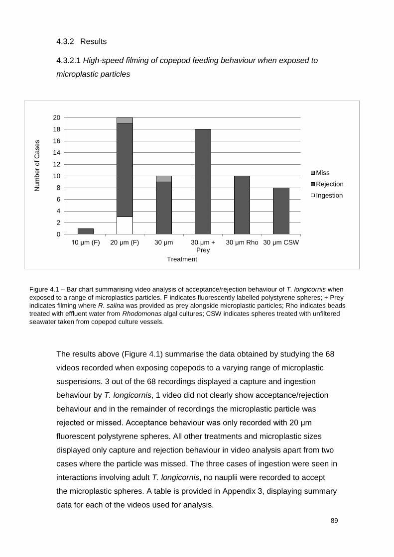
89
4.3.2 Results
4.3.2.1 High-speed filming of copepod feeding behaviour when exposed to
microplastic particles
The results above (Figure 4.1) summarise the data obtained by studying the 68
videos recorded when exposing copepods to a varying range of microplastic
suspensions. 3 out of the 68 recordings displayed a capture and ingestion
behaviour by T. longicornis, 1 video did not clearly show acceptance/rejection
behaviour and in the remainder of recordings the microplastic particle was
rejected or missed. Acceptance behaviour was only recorded with 20 μm
fluorescent polystyrene spheres. All other treatments and microplastic sizes
displayed only capture and rejection behaviour in video analysis apart from two
cases where the particle was missed. The three cases of ingestion were seen in
interactions involving adult T. longicornis, no nauplii were recorded to accept
the microplastic spheres. A table is provided in Appendix 3, displaying summary
data for each of the videos used for analysis.
0
2
4
6
8
10
12
14
16
18
20
10 μm (F) 20 μm (F) 30 μm 30 μm + Prey
30 μm Rho 30 μm CSW
Miss
Rejection
Ingestion
Figure 4.1 – Bar chart summarising video analysis of acceptance/rejection behaviour of T. longicornis when
exposed to a range of microplastics particles. F indicates fluorescently labelled polystyrene spheres; + Prey
indicates filming where R. salina was provided as prey alongside microplastic particles; Rho indicates beads
treated with effluent water from Rhodomonas algal cultures; CSW indicates spheres treated with unfiltered
seawater taken from copepod culture vessels.
Treatment
Num
ber
of C
ases
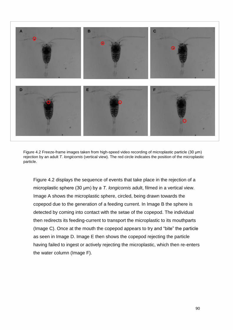
90
Figure 4.2 displays the sequence of events that take place in the rejection of a
microplastic sphere (30 μm) by a T. longicornis adult, filmed in a vertical view.
Image A shows the microplastic sphere, circled, being drawn towards the
copepod due to the generation of a feeding current. In Image B the sphere is
detected by coming into contact with the setae of the copepod. The individual
then redirects its feeding-current to transport the microplastic to its mouthparts
(Image C). Once at the mouth the copepod appears to try and “bite” the particle
as seen in Image D. Image E then shows the copepod rejecting the particle
having failed to ingest or actively rejecting the microplastic, which then re-enters
the water column (Image F).
A B C
D E F
Figure 4.2 Freeze-frame images taken from high-speed video recording of microplastic particle (30 μm)
rejection by an adult T. longicornis (vertical view). The red circle indicates the position of the microplastic
particle.
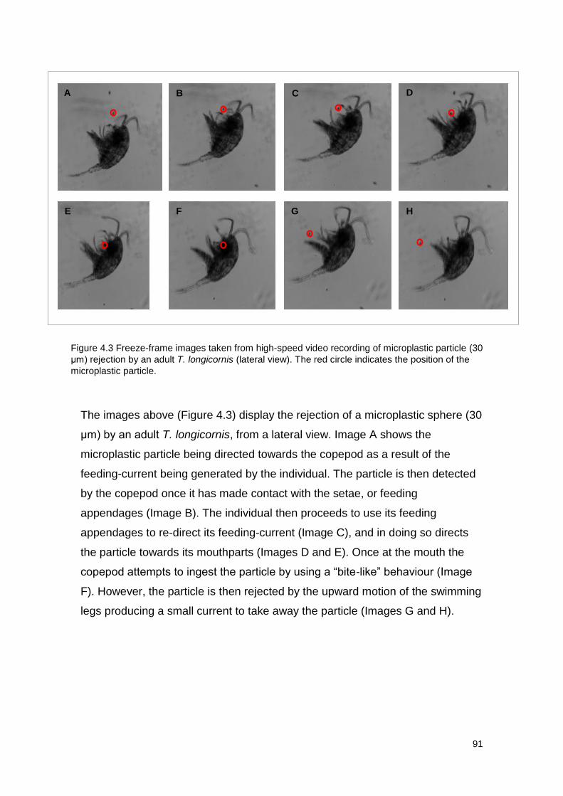
91
The images above (Figure 4.3) display the rejection of a microplastic sphere (30
μm) by an adult T. longicornis, from a lateral view. Image A shows the
microplastic particle being directed towards the copepod as a result of the
feeding-current being generated by the individual. The particle is then detected
by the copepod once it has made contact with the setae, or feeding
appendages (Image B). The individual then proceeds to use its feeding
appendages to re-direct its feeding-current (Image C), and in doing so directs
the particle towards its mouthparts (Images D and E). Once at the mouth the
copepod attempts to ingest the particle by using a “bite-like” behaviour (Image
F). However, the particle is then rejected by the upward motion of the swimming
legs producing a small current to take away the particle (Images G and H).
A B C D
E F G H
Figure 4.3 Freeze-frame images taken from high-speed video recording of microplastic particle (30
μm) rejection by an adult T. longicornis (lateral view). The red circle indicates the position of the
microplastic particle.
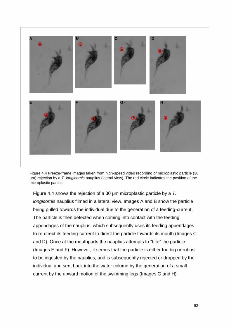
92
Figure 4.4 shows the rejection of a 30 μm microplastic particle by a T.
longicornis nauplius filmed in a lateral view. Images A and B show the particle
being pulled towards the individual due to the generation of a feeding-current.
The particle is then detected when coming into contact with the feeding
appendages of the nauplius, which subsequently uses its feeding appendages
to re-direct its feeding-current to direct the particle towards its mouth (Images C
and D). Once at the mouthparts the nauplius attempts to “bite” the particle
(Images E and F). However, it seems that the particle is either too big or robust
to be ingested by the nauplius, and is subsequently rejected or dropped by the
individual and sent back into the water column by the generation of a small
current by the upward motion of the swimming legs (Images G and H).
B A C D
E F G H
Figure 4.4 Freeze-frame images taken from high-speed video recording of microplastic particle (30
μm) rejection by a T. longicornis nauplius (lateral view). The red circle indicates the position of the
microplastic particle.
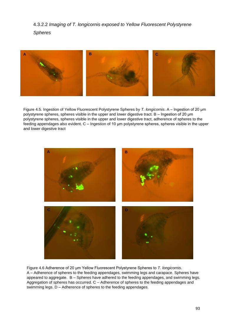
93
4.3.2.2 Imaging of T. longicornis exposed to Yellow Fluorescent Polystyrene
Spheres
A B C
Figure 4.5. Ingestion of Yellow Fluorescent Polystyrene Spheres by T. longicornis. A – Ingestion of 20 μm
polystyrene spheres, spheres visible in the upper and lower digestive tract. B – Ingestion of 20 μm
polystyrene spheres, spheres visible in the upper and lower digestive tract, adherence of spheres to the
feeding appendages also evident. C – Ingestion of 10 μm polystyrene spheres, spheres visible in the upper
and lower digestive tract
A B
C D
Figure 4.6 Adherence of 20 μm Yellow Fluorescent Polystyrene Spheres to T. longicornis.
A – Adherence of spheres to the feeding appendages, swimming legs and carapace. Spheres have
appeared to aggregate. B – Spheres have adhered to the feeding appendages, and swimming legs.
Aggregation of spheres has occurred. C – Adherence of spheres to the feeding appendages and
swimming legs. D – Adherence of spheres to the feeding appendages.
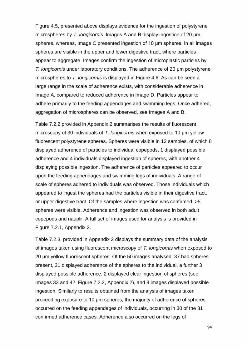
94
Figure 4.5, presented above displays evidence for the ingestion of polystyrene
microspheres by T. longicornis. Images A and B display ingestion of 20 μm,
spheres, whereas, Image C presented ingestion of 10 μm spheres. In all images
spheres are visible in the upper and lower digestive tract, where particles
appear to aggregate. Images confirm the ingestion of microplastic particles by
T. longicornis under laboratory conditions. The adherence of 20 μm polystyrene
microspheres to T. longicornis is displayed in Figure 4.6. As can be seen a
large range in the scale of adherence exists, with considerable adherence in
Image A, compared to reduced adherence in Image D. Particles appear to
adhere primarily to the feeding appendages and swimming legs. Once adhered,
aggregation of microspheres can be observed, see Images A and B.
Table 7.2.2 provided in Appendix 2 summarises the results of fluorescent
microscopy of 30 individuals of T. longicornis when exposed to 10 μm yellow
fluorescent polystyrene spheres. Spheres were visible in 12 samples, of which 8
displayed adherence of particles to individual copepods, 1 displayed possible
adherence and 4 individuals displayed ingestion of spheres, with another 4
displaying possible ingestion. The adherence of particles appeared to occur
upon the feeding appendages and swimming legs of individuals. A range of
scale of spheres adhered to individuals was observed. Those individuals which
appeared to ingest the spheres had the particles visible in their digestive tract,
or upper digestive tract. Of the samples where ingestion was confirmed, >5
spheres were visible. Adherence and ingestion was observed in both adult
copepods and nauplii. A full set of images used for analysis is provided in
Figure 7.2.1, Appendix 2.
Table 7.2.3, provided in Appendix 2 displays the summary data of the analysis
of images taken using fluorescent microscopy of T. longicornis when exposed to
20 μm yellow fluorescent spheres. Of the 50 images analysed, 37 had spheres
present, 31 displayed adherence of the spheres to the individual, a further 3
displayed possible adherence, 2 displayed clear ingestion of spheres (see
Images 33 and 42 Figure 7.2.2, Appendix 2), and 8 images displayed possible
ingestion. Similarly to results obtained from the analysis of images taken
proceeding exposure to 10 μm spheres, the majority of adherence of spheres
occurred on the feeding appendages of individuals, occurring in 30 of the 31
confirmed adherence cases. Adherence also occurred on the legs of
95
individuals, head and in one case the urosome (see image 20, Figure 7.2.2,
Appendix 2). A vast range of the scale of spheres that adhered to individuals
was seen once again, for example a large scale of adherence seen in Image 2
(Figure 7.2.2, Appendix 2), and a low range of adherence as seen in Image 46,
(Figure 7.2.2, Appendix 2). Ingested spheres appeared to be present in the
digestive tract or upper digestive tract. Like exposures to 10 μm spheres,
adherence and ingestion was seen in both adult copepods and nauplii alike. A
full set of images used for analysis is provided in Figure 7.2.2, Appendix 2.
4.3.3 Discussion
Active selection is described as “a behavioural response with active dietary
choice”, where an individual rejects or accepts a prey cell based upon its
physical or chemical properties (Kiorboe, et al., 1996). Passive selection, on the
other hand, implies a more simple interaction where differences in encounter
rates and handling or capture efficiencies, determine the ingestion of particular
prey (Schultz & Kiørboe, 2009). It would appear by studying high-speed video
evidence, that the copepod Temora longicornis and its nauplii in general
possess the ability to reject microplastic particles in the size range of 10-30 μm
once captured. However, the mechanism behind this rejection is one which is
difficult to analyse. Much discussion is required to assess whether this rejection
is an action carried out by the individual in response to the microplastic
particle’s physical and chemical properties, or whether the rejection is more of a
passive mechanism due to the mechanics of microplastic ingestion.
Fluorescent imaging showed that for 10 μm and 20 μm microplastic exposures
both adult T. longicornis and nauplii displayed evidence for ingestion of
microplastic spheres in accordance with previous studies (Cole et al, 2013).
Therefore, contradictions occur between the findings of video analysis and
imaging, raising questions over the mechanisms behind interactions between
copepods and microplastics, and the effectiveness of high-speed filming for
analysing such behaviour, all of which will be explored below.
T. longicornis can be described as a feeding-current strategist (Jakobsen, et al.,
2005). Feeding by the generation of a feeding-current which draws particles out
of the water column towards the individual, where they are subsequently
detected and directed to the mouthparts and ingested (Kjellerup & Kiørboe,
96
2012). It is believed that feeding-current strategists are capable of both
chemical and mechanical detection, detecting prey due to chemical cues
(Tiselius et al, 2013) and by disturbances within the water column. The theory of
chemical detection is based on the belief that a prey cell is surrounded by a
chemical signal which elongates when under the influence of the feeding-
current produced by a copepod, therefore, sending a chemical signal to the
individual before the prey cell arrives itself (Tiselius et al, 2013). This allows the
individual the opportunity to alter its feeding current to either direct the prey cell
towards its mouthparts or back into the water column.
However, there is a growing argument among those studying the mechanisms
behind prey detection by zooplankton, arguing that such long-range chemical
detection is unlikely to occur and instead only mechanical detection is used to
detect prey cells (Gonçalves, personal communication). In this case a prey cell
is directed by the feeding-current of a copepod, which is produced at all times
during feeding behaviour. Eventually the prey cell comes into contact with the
setae, or feeding appendages, of the individual which then uses its feeding
appendages to re-direct the cell towards the mouth and subsequently ingests
the particle. Short-range chemical detection, or assessment, of potential prey
cells once caught is thought likely to occur (Koehl & Strickler, 1981), with
research suggesting that copepods possess the ability to taste (Price, 1988)
and use chemoreceptors located on the setae (Friedman & Strickler, 1975) to
assess the biochemical composition of the captured particle. Copepods are also
believed to be able to display size and quality selection of prey particles (Frost,
1972; Donaghay & Small, 1979)
The videos recorded during this study support the views above, that long-range
chemoreception is not likely to occur and it seems that detection of potential
prey occurs after the contact of cell to the feeding appendages of the individual.
This was observed in all cases where interactions occurred between copepods
and particles. As well as this, microplastic particles produced for research are
unlikely to have a significant biofilm on their surface that could produce a
biochemical signal, and therefore if long-range chemoreception was apparent
they should not be detected. However, as evident in recordings, feeding-
currents are still produced in their presence. In all of the 68 videos analysed,
detection of the particle occurred after coming into contact with the feeding
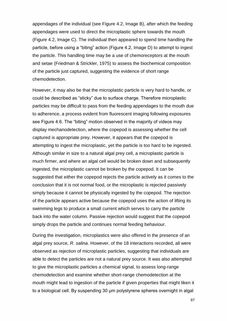
97
appendages of the individual (see Figure 4.2, Image B), after which the feeding
appendages were used to direct the microplastic sphere towards the mouth
(Figure 4.2, Image C). The individual then appeared to spend time handling the
particle, before using a “biting” action (Figure 4.2, Image D) to attempt to ingest
the particle. This handling time may be a use of chemoreceptors at the mouth
and setae (Friedman & Strickler, 1975) to assess the biochemical composition
of the particle just captured, suggesting the evidence of short range
chemodetection.
However, it may also be that the microplastic particle is very hard to handle, or
could be described as “sticky” due to surface charge. Therefore microplastic
particles may be difficult to pass from the feeding appendages to the mouth due
to adherence, a process evident from fluorescent imaging following exposures
see Figure 4.6. The “biting” motion observed in the majority of videos may
display mechanodetection, where the copepod is assessing whether the cell
captured is appropriate prey. However, it appears that the copepod is
attempting to ingest the microplastic, yet the particle is too hard to be ingested.
Although similar in size to a natural algal prey cell, a microplastic particle is
much firmer, and where an algal cell would be broken down and subsequently
ingested, the microplastic cannot be broken by the copepod. It can be
suggested that either the copepod rejects the particle actively as it comes to the
conclusion that it is not normal food, or the microplastic is rejected passively
simply because it cannot be physically ingested by the copepod. The rejection
of the particle appears active because the copepod uses the action of lifting its
swimming legs to produce a small current which serves to carry the particle
back into the water column. Passive rejection would suggest that the copepod
simply drops the particle and continues normal feeding behaviour.
During the investigation, microplastics were also offered in the presence of an
algal prey source, R. salina. However, of the 18 interactions recorded, all were
observed as rejection of microplastic particles, suggesting that individuals are
able to detect the particles are not a natural prey source. It was also attempted
to give the microplastic particles a chemical signal, to assess long-range
chemodetection and examine whether short-range chemodetection at the
mouth might lead to ingestion of the particle if given properties that might liken it
to a biological cell. By suspending 30 μm polystyrene spheres overnight in algal
98
culture effluent and seawater taken from copepod culture vessels, it was
believed that biofilm formation would occur (Zettler, et al., 2013). However,
these particles were treated as described above and rejected in the same
manner. Suggesting that chemoreception is unlikely to be the determining factor
in rejection of microplastic particles. Rather, it appears mechanistic factors are
driving the behaviour observed in videos. However, it must be considered that
for the majority of treatments 30 μm spheres were used for this study, and it
may be that this size was too large for the copepod to ingest, especially for
nauplii. Though ingestion of 30.6 μm spheres by T. longicornis has been
recorded in previous studies (Cole, et al., 2013). Therefore it would be of
interest to observe the same study with smaller particles, and any findings
presented here must be treated with caution and in an exploratory nature. It
seems, then, from analysing video data alone that the copepod species, T.
longicornis, possesses the ability to reject microplastic particles when offered as
prey, and given a chemical signal. However, by utilising fluorescent polystyrene
spheres to represent microplastic particles, the opportunity arises to examine
whether any ingestion of particles which was not recorded on film exists. By
doing this, the issue that only rejections are recorded due to the relative ease at
which rejections are seen on screen when recorded, compared to ingestion, is
negated. Imaging resulting from fluorescent microscopy allows for additional
information regarding the interaction of copepods and microplastic particles to
be recorded.
When we study the images collected from fluorescent microscopy for 10 μm
(Figure 7.2.1, Appendix 2) and 20 μm (Figure 7.2.2, Appendix 2) polystyrene
spheres it is clear that there is a large number of copepods encountering
microplastics within a closed system. The majority of images where spheres are
present display adherence of microplastics to the copepod, particularly to the
feeding appendages (see Figure 4.6), such adherence has also been recorded
by Cole et al. (2013). The adherence of microplastics to the feeding
appendages of individuals, has been discussed in detail in Chapter 2, and has
the potential to significantly reduce the fitness of the organism as feeding
appendages play vital roles in feeding, swimming and mating behaviour (Cole et
al, 2013). Adherence may act to reduce motility of feeding appendages,
affecting the production of the feeding-current, or may act to disrupt the
99
detection of particles and subsequent capture. Although the majority of images
display adherence of particles, a number of cases of ingestion or possible
ingestion of microplastic spheres were apparent. Providing evidence that
ingestion of microplastics by T. longicornis does occur, as found by (Cole, et al.,
2013), recording ingestion of particles sized 7.3-30.6 μm. The ingestion of
microplastics is potentially problematic, as discussed in previous chapters.
However, the number of cases of ingestion versus rejection raises the question;
why were only 3 out of 68 video recordings examples of ingestion? And, are
individuals of T. longicornis rejecting more microplastic particles than they are
accepting? Such questions could only be answered by combining a grazing
experiment with high-speed filming. This study would be difficult to set-up due to
the labour intensive nature of high-speed filming and the length of grazing
experiments, typically run for 24 hours. However, despite the difficulties in set-
up, such a study would be greatly beneficial in microplastic ingestion research,
and thus, should be considered in the future.
High-speed filming represents a novel and highly interesting manner of studying
the interaction between individuals and particles within a laboratory
environment. However, there are uncertainties in results where interactions
recorded on film, such as those presented in this study, may repeatedly show
the same individual interacting with particles rather than a set number of
recordings of each individual placed into the filming chamber. This may result in
findings that are not representative of the entire group, rather are individual
responses. For example, of the 68 recordings used for analysis in the study
presented here, it is not possible to state how many videos were of each
individual copepod. Such a limitation could be addressed by only filming one
individual at a time, however, such a set-up is not likely to produce many
recorded interactions and is likely to be very time consuming. If a technique
could be developed to identify individual members of the group so that
recordings could be analysed in terms of each individual’s response it would be
highly beneficial. However at this time such a method does not exist, and it is
felt that the probability of the same individual being filmed repeatedly is unlikely.
As such in analysing recordings we assume that interactions are representative
of the group, however, we treat the evidence with caution regarding the
100
limitations discussed and experimental nature of research, thus, it is used
alongside consideration of findings from other studies.
Through consideration of the findings outlined in this study, and in reference to
the mechanisms by which prey is detected and subsequently captured by
different feeding strategies it is proposed here, that, feeding strategy is likely to
influence microplastic uptake by zooplankton. As described in Chapter 3,
feeding-current producing zooplankton perceive prey chemically and such
possess the ability to select prey based upon the biochemical composition of
the particle (Tiselius et al, 2013). This mechanism in theory should reduce the
likeliness of a feeding-current feeding individual ingesting a microplastic particle
which does not carry a biological signature. However, it has been studied that
microplastics in the marine environment can develop biofilms, hence giving the
particle an altered biochemical composition which might be perceived by the
predating zooplankton. This pattern has been observed under laboratory
conditions where clean particles used in experiments were less likely to be
ingested than older particles that have developed biofilms (Lobelle et al, 2011).
However, results of this study suggest that mechanistic rejection is the main
driver behind rejection of microplastic particles by T. longicornis, and that long-
range chemodetection appears unlikely to occur. However, short-range
chemodetection may be apparent in zooplankton displaying both ambush
feeding and feeding-current feeding, allowing for rejection of a non-prey item
following capture.
High-speed filming has been identified as a novel and highly interesting method
by which to examine interactions between microplastic particles and
zooplankton, and the opportunities for future research are endless. Filming
allows multiple endpoints to be collected, such as; real handling time of
particles, ingestion rate, the effects of adherence, length of adherence of
particles and effects upon locomotion. It is the belief of the researcher here that
focus should be put upon the differences in interactions between copepods
displaying the two major feeding strategies; ambush feeding and feeding-
current strategists; in order to assess the differences in microplastic uptake and
detection as proposed above. Assessing any differences in uptake of aged
particles more representative of those found in the natural environment,
compared to “clean” particles typically used in the laboratory would be highly
101
beneficial. By combining high-speed filming and the use of fluorescent particles
and microscopy it is thought that a comprehensive study into the ingestion of
microplastic particles and the specific mechanisms behind such ingestion can
be achieved.

102
103
Chapter 5
Microplastics; small problem? Or major issue?

104
105
The work presented in this thesis provides further evidence for the ingestion of
microplastics by a range of zooplankton taxa under laboratory studies as
recorded by Cole et al. (2013) and Setälä et al. (2014). In addition, a new
insight into the effects that microplastic exposure has upon the feeding
behaviour of zooplankton is presented. Through the utilisation of a novel
technique in the field of microplastic research, high-speed filming, the ability of
copepod’s to reject microplastic particles is assessed, and a new way in which
interactions between microscopic plastic debris and zooplankton can be
investigated is introduced.
To date, it is primarily only polystyrene microspheres that have been utilised in
order to assess microplastic ingestion by zooplankton. Such particles have high
effectiveness in laboratory study, however, being of uniform shape and size do
not accurately represent those particles likely to be present in the environment.
The research presented in Chapter 2 aimed to investigate the ingestion of three
plastic types, considered abundant in the marine environment; primary
microplastics, secondary microplastics and fibrous microplastics. As a result of
this study ingestion of all plastic types was recorded in the calanoid copepod,
Centropages typicus, suggesting that ingestion of microplastic particles within
the environment is likely. As well as supporting previous findings regarding
microplastic ingestion, the study outlined in Chapter 2 presents a novel and
effective way in which to fluorescently label plastics for use in ingestion studies
(see Section 2.3.1.1, Chapter 2). The RADGLO labelling protocol opens a wide
range of future research opportunities, allowing for the study of ingestion of
common plastic contaminants or those collected from environmental sampling,
so as to further increase our knowledge in this area. Although in study used to
examine individual effects, such particles labelled with RADGLO could be used
to examine tropic level transfer of microplastic particles collected from the
environment. Studies such as those carried out by Setälä et al. (2014) could be
adapted so as to assess whether particles are passed onto a higher trophic
following feeding of previously exposed zooplankton to their predators. A study
carried out in this manner would allow the consideration of whether
microplastics found in the environment, varying in shape and form are likely to
be passed through the food chain.
106
The second major research topic investigated within this thesis, is the effects
that microplastic exposure has upon feeding behaviour of zooplankton. It has
been recorded that exposure to microplastic particles has caused reduced
feeding activity in marine worms (Wright, et al., 2013) and the copepod Calanus
helgolandicus (Cole, 2014), resulting in an energy deficit. Due to the importance
of such species, which occupy lower trophic levels in the marine environment, in
terms of energy transfer, such reductions in energy uptake are likely to have
knock-on effects for the entire ecosystem. By examining a range of copepod
species, displaying a range of feeding behaviours, as well as one holoplankton
species, Porcellanid larvae, this thesis provides a new insight into the effects
that microplastic exposure is likely to have upon different zooplankton species.
Carbon ingestion was reduced in three of the four species studies, although
only one reduction was statistically significant. Such findings support previous
findings by Cole (2014). It was only those copepod species displaying feeding-
current feeding behaviour, C. helgolandicus and Acartia tonsa, that suffered a
reduction in carbon uptake following microplastic exposure. Oithona similis, an
ambush predator did not appear to suffer any effect on carbon uptake in the
presence of microplastic particles, suggesting that feeding-current strategists
are more at risk from the negative effects of microplastic exposure. A decrease
in carbon uptake was also evident in Porcellanid larvae.
During this study FlowCAM was utilised to examine the uptake of various prey
types by each of the four zooplankton species, in order to investigate the
mechanism of reductions in carbon uptake, or any changes in ingestion of
particular prey. Such switching behaviour was recorded by Cole (2014) where
C. helgolandicus showed a shift in prey size ingested, away from cells in the
size range of the microplastics added to suspensions used in investigation.
Similarly the findings presented in Chapter 3 of this thesis display a range of
switching behaviours displayed by all of the copepod species studied. As well
as altering ingestion of prey in distinct size ranges, C. helgolandicus and O.
similis appeared to increase ingestion of non-spherical prey. This suggests that
copepods may be able to detect spherical microplastics and decrease ingestion
on spherical items. However, O. similis being an ambush feeder, theoretically
should not be able to detect non-motile prey (Kiørboe, et al., 2009), therefore,
this study also suggests that O. similis, like A. tonsa, may be able to display
107
feeding-current and ambush feeding strategies depending on the relative
concentrations of motile, non-motile or high-quality prey. Porcellanid larvae
were not recorded to display switching behaviour, instead reduced feeding of all
prey types. This suggests that reduced feeding may be the result of satiation
(Gregory, 2009), blocking of the digestive tract (Browne, et al., 2008), or the
presence of a selective behaviour to reduce feeding rate due to the presence of
non-valuable microplastic particles.
The energy deficit suffered by individuals as recorded in studies presented in
this thesis and previous research (Wright, et al., 2013; Cole, 2014) is a concern.
As discussed in Chapter 3, a reduction in energy ingested by an individual not
only is likely to cause a reduction in the fitness of the individual but has the
potential to affect its progeny and the ecosystem as a whole. As studied by Cole
(2014) an energy deficit can lower egg quality and hatchling success, therefore
potenitally reducing the population of a species within an ecosytem over a long
timescale. As well as this, due to trophic transfer, the reduction of energy at a
low level in the food chain is likely to considerably lower the energy available to
higher trophic levels. The decreased carbon ingested by larval stages, such as
Porcellanid larvae, is likely to reduce survivorship and development into adults.
All effects are a cause of concern for fisheries and conservationists alike, as the
reductions of energy available to organisms within the ecosystem are likely to
reduce the health and quality of populations and commercial stocks. As a result
it would be of high interest to examine the altered energy uptake by higher
trophic level organisms after feeding on zooplankton prey that had been offered
a diet of natural algal prey and algae mixed with microplastic particles, such as
those suspensions used in the study presented in Chapter 3. Such research
would clearly demonstrate the potential ecosystem level effects of microplastic
exposure, and potentially allow us to model any alterations in energy transfer
through trophic levels. In terms of meroplankton, such as the Porcellanid larvae
examined in this thesis, long term studies examining the development of larvae
into adults when exposed to microplastic particles would enable us to
understand whether microplastics are affecting larval development in any
manner.
Chapter 4 of this thesis aimed to investigate the ability of copepod species to
reject microplastic particles. By utlising high-speed filming techniques, typically
108
used to study locomotive mechanisms in zooplankton, it was possible to
observe the manner in which a copepod detected, captured and handled a
microplastic particle. It was found that Temora longicornis appeared to be able
to reject microplastic particles, however, this rejection was observed to be likely
due to mechanistic properties where the microplastic particle was too large or
rigid to be ingested by the individual. Through this research it was also possible
to assess the presence of chemodetection by the species. It appeared that
long-range chemodetection, believed common in feeding-current feeding
copepods (Koehl & Strickler, 1981) did not exist, rather particles were detected
after coming into contact with feeding appendages. The use of high-speed
filming introduces a novel technique to be implemented in microplastic research
providing many opportunities for further research, including the examination of
various plastic types, as well as examing the effects of adherence of particles in
producing feeding-currents. However, one limitation of high-speed filming is that
it is possible to miss ingestion of particles, therefore, by utilising fluorescent
particles it is possible to assess ingestion following exposure.
Overall it appears that microplastic ingestion is likely to occur in zooplankton
species where concentrations of microplastics are high in the marine
environment. The effects of microplastic exposure seem to be negative,
especially in species that feed by generation of a feeding current. Zooplankton
appear to alter feeding behaviour to avoid microplastic particles, such switching
behaviour in turn affects the carbon ingested by the individual. It is of high
importance that research efforts are placed on discovering the concentration of
microplastic debris present in the environment sized <100 μm, this will allow for
more environmentally relevant studies to be produced and more ecosystem
related effects to be considered.
In terms of this thesis a range of further research opportunities exist. Firstly the
RADGLO fluorescent labelling protocol presented in Chapter 2 allows a wide
range of plastic types to be examined in laboratory investigations, reducing the
limitation that polystyrene microspheres do not accurately represent particles in
the environment. Secondly, the effects of reduced carbon uptake require further
investigation. It would be highly benficial to repeat studies outlined in Chapter 3
to gain more insights into alterations in feeding behaviour. The examination into
effects on other holoplankton species would also be interesting, as well as
109
examining the development of meroplankton larvae under long-term exposures
to microplastic particles. Investigating the loss of energy to higher trophic levels
would also be a key area of research, feeding species occupying higher trophic
levels with zooplankton exposed to microplastics, in order to assess the net loss
of energy transferred. Finally, high-speed filming techniques provide an
opportunity to gain a clearer understanding of how individuals interact with
microplastic particles.

110
111
References
Andrady, A., 2003. Plastics and the environment. In: A. L. Andrady, ed. Publisher: John
Wiley and Sons.
Andrady, A., 2011. Microplastics in the marine environment. Marine Pollution Bulletin,
62, pp. 1596–1605.
Arthur, C., Baker, J. & Bamford, H., 2009. Proceedings of the International Research
Workshop on the Occurrence, Effects and Fate of Microplastic Marine Debris. s.l.,
NOAA Technical, Memorandum NOS-OR&R30.
Atkinson, A., 1995. Omnivory and feeding selectivity in five copepod species during
spring in the Bellingshausen Sea, Antarctica. ICES Journal of Marine Science, 52, pp.
385-396.
Ayukai, T., 1987. Discriminate feeding of the calanoid copepod Acartia clausi in
mixtures of phytoplankton and inert particles. Marine Biology, 94, pp. 579-587.
Ban, S. et al., 1997. The paradox of diatom-copepod interactions. Marine Ecology
Progress Series, 157, pp. 287-293.
Barnes, D., Galgani, F., Thompson, R. & Barlaz, M., 2009. Accumulation and
fragmentation of plastic debris in global environments.. Philosophical Transactions of
the Royal Society B: Biological Sciences, 364, pp. 1985-1998.
Barros, H. & Valenti, W., 2003. Ingestion rates of Artemia nauplii for different larval
stages of Macrobrachium rosenbergii. Aquaculture, 217, pp. 223-233.
Bautista, B., Harris, R., Tranter, P. & Harbour, D., 1992. In situ copepod feeding and
grazing rates during a spring bloom dominated by Phaeocystis sp. in the English
Channel. Journal of Plankton Research, 14, pp. 691-703.
Boerger, C., Lattin, G., Moore, S. & Moore, C., 2010. Plastic ingestion by planktivorous
fishes in the North Pacific Central Gyre. Marine Pollution Bulletin, 60, pp. 2275–2278.
Bonnet, D. et al., 2005. An overview of Calanus helgolandicus ecology in European
waters. Progress in Oceanography, 65, pp. 1-53.
Brillant, M. & MacDonald, B., 2000. Postingestive selection in the sea scallop
Placopecten magellanicus (Gmelin): the role of particle size and density. Jounal of
Experimental Marine Biology and Ecology, 253, pp. 211-227.
Browne, M. et al., 2008. Ingested microscopic plastic translocates to the circulatory
system of the mussel, Mytilus edulis (L.). Environmental Science & Technology, 42, pp.
5026-5031.
Browne, M., Galloway, T. & Thompson, R., 2007. Microplastic an emerging
contaminant of potential concern? Integrated Environmental Assessment and
Management, 3, pp. 559-561.
Bruno, E., Marc, C., Borg, A. & Kiørboe, T., 2012 . Prey Detection and Prey Capture in
Copepod Nauplii. PLOSone, 7, pp. e47906.
Chiappone, M., Dienes, H., Swanson, D. W. & Miller, S., 2005. Impacts of lost fishing
gear on coral reef sessile invertebrates in the Florida Keys National Marine Sanctuary.
Biological Conservation, 121, pp. 221-230.
Claessens, M. et al., 2011. Occurrence and distribution of microplastics in marine
sediments along the Belgian coast. Marine Pollution Bulletin, 62, pp. 2199-2204.
112
Claustre, H. et al., 1990. A biochemical investigation of a Phaeocystis sp. bloom in the
Irish Sea. Journal of the Marine Biological Association of the UK, 70, pp. 197-207.
Cole, M., 2014. The impacts of microplastics on zooplankton. [Online]
Available at: http://ethos.bl.uk/OrderDetails.do?uin=uk.bl.ethos.615590
[Accessed 16 9 2014].
Cole, M. et al., 2013. Microplastic Ingestion by Zooplankton. Environmental Science &
Technology, 47, pp. 6646-6655.
Cole, M., Lindeque, P., Halsband, C. & Galloway, T. S., 2011. Microplastics as
contaminants in the marine environment: A review. Marine Pollution Bulletin, 62, pp.
2588–2597.
Collignon, A. et al., 2012. Neustonic microplastic and zooplankton in the North Western
Mediterranean Sea. Marine Pollution Bulletin, 64, pp. 861-864.
Colton, J., Knapp, F. & Burns, B., 1974. Plastic particles in surface waters of the
Northwestern Atlantic. Science, 185, pp. 491-497.
Cowles, T., Olson, R. & Chisholm, S., 1988. Food selection by copepods:
discrimination on the basis of food quality. Marine Biology, 100, pp. 41-49.
Cózar, A. et al., 2014. Plastic debris in the open ocean. PNAS, 111, pp. 10239–10244.
Davison, P. & Asch, R., 2011. Plastic ingestion by mesopelagic fishes in the North
Pacific Subtropical Gyre. Marine Ecology Progress Series, 432, pp. 173–180.
Day, R., Shaw, D. & Ignell, S., 1989. The quantitative distribution and characteristics of
neuston plastic in the North Pacific Ocean. 1985–1988. Honolulu, Hawaii, NOAA.
Derraik, J. G., 2002. The pollution of the marine environment by plastic debris: a
review. Marine Pollution Bulletin, 44, pp. 842-852.
Donaghay, P. & Small, L., 1979. Food selection capabilities of the estuarine copepod
Acartia clausi. Marine Biology, 52, pp. 137-146.
EC, 2008. EC Directive 2008/56/EC of the European Parliament and the Council of 17
June 2008 establishing a framework for community action in the field of marine
environmental policy (Marine Strategy Framework Directive). Official Journal of the
European Union, 19(L 164).
Farrell, P. & Nelson, K., 2013. Trophic level transfer of microplastic: Mytilus edulis (L.)
to Carcinus maenas (L.). Environmental Pollution, 177, pp. 1-3.
Fendall, L. & Sewell, M., 2009. Contributing to marine pollution by washing your face:
Microplastics in facial cleansers. Marine Pollution Bulletin, 58, pp. 1225–1228.
Fernandez, F., 1979. Particle selection in the nauplius of Calanus pacificus.1979.
Journal of Plankton Research, 1, pp. 313-327.
Fleminger, A., 1975. Pattern, number, variability and taxonomlc significance of
integumental organs (sensilla and glandular pores) in the genus Eucalanus (Copepoda
Calanoida). Fish Bull US, 71, pp. 965-1010.
Fossi, M. et al., 2012. Are baleen whales exposed to the threat of microplastics? A
case study of the Mediterranean fin whale (Balaenoptera physalus). Marine Pollution
Bulletin, 64, pp. 2374-2379.
113
Friedman, M. & Strickler, J., 1975. Chemoreceptors and feeding in calanoid copepods
(Arthropoda:Crustacea). Proceedings of the National Academy of Sciences of the
United States of America, 72, pp. 4185-4188.
Frost, B., 1977. Feeding behavior of Calanus pacificus in mixtures of food particles.
Limnology and Oceanography, 22, pp. 472-492.
Frost, B., 1972. Effects of size and concentration of food particles on the feeding
behavior of the marine planktonic copepod Calanus pacificus. Limnology and
Oeanography, 17, pp. 805-815.
Gieskes, W. & Kraay, G., 1975. The phytoplankton spring bloom in Dutch coastal
waters of the North Sea. Netherlands Journal of Sea Research, 9, pp. 166-196..
Gonor, S. & Gonor, J., 1973. Feeding, cleaning, and swimming behavior in larval
stages of porcellanid crabs (Crustacea: Anomura). Fishery bulletin, 71, pp. 225-234.
Gonzáles, H. & Smetacek, V., 1984. The possible role of the cyclopoid copepod
Oithona in retarding vertical flux of zooplankton faecal material.. Marine Ecology
Progress Series, 113, pp. 233-246.
Goodfellow, 2014. Polyamide - Nylon 6. [Online]
Available at: http://www.goodfellow.com/E/Polyamide-Nylon-6.html
[Accessed 6 10 2014].
Gordon, M., 2006. Eliminating Land-based Discharges of Marine Debris In: California:
a Plan of Action from the Plastic Debris Project, Sacramento, CA: California State
Water Resources Control Board.
Gorman, M., 1993. Environmental Hazards––Marine Pollution. Santa Barbara: ABC-
CLIO Inc.
Gregory, M., 2009. Environmental implications of plastic debris in marine settings e
entanglement, ingestion, smothering, hangers-on, hitch-hiking and alien invasions..
Philosophical Transactions of the Royal Society of London B: Biological Sciences, 364,
pp. 2013-2025.
Griffiths, A. M. & Frost, B. W., 1976. Chemical communication in the marine planktonic
copepods Calanus pacificus and Pseudocalanus sp. Crustaceana, 30, pp. 1-9.
Hidalgo-Ruz, V., Gutow, L., Thompson, R. C. & Thiel, M., 2012. Microplastics in the
Marine Environment: A Review of the Methods Used for Identification and
Quantification. Environmental Science & Technology, 46, pp. 3060-3075.
Huntley, M., Sykes, P., Rohan, S. & Marin, V., 1986. Chemically-mediated rejection of
dinoflagellate prey by the copepods Calanuspacificus and Paracalanusparvus:
mechanism, occurrence and significance. Marine Ecology Progress Series, 28, pp.
105-120.
Huntley, M., Barthel, K. & Star, J., 1983. Particle rejection by Calanus pacificus:
discrimination between similarly sized particles. Marine Biology, 74, pp. 151-160.
Hygum, B., Rey, C., Hansen, B. & Tande, K., 2000. Importance of food quantity to
structural growth rate and neutral lipid reserves accumulated in Calanus finmarchicus.
Marine Biology, 136, pp. 1057-1073.
Ianora, A. et al., 2004. Aldehyde suppression of copepod recruitment in blooms of a
ubiquitous planktonic diatom. Nature, 429, pp. 403–407.
114
Irigoien, X. et al., 2002. Copepod hatching success in marine ecosystems with high
diatom concentrations. Nature, 419, pp. 387–389.
Irigoien, X. et al., 2000. Feeding selectivity and egg production of Calanus
helgolandicus in the English Channel. Limnology and Oceanography, 45, pp. 44-54.
Jakobsen, H., Halvorsen, E., Hansen, B. W. & Visser, A., 2005. Effects of prey motility
and concentration on feeding in Acartia tonsa and Temora longicornis: the importance
of feeding modes. Journal of Plankton Research, 27, pp. 775-785.
Jakobsen, H., 2001. Escape response of planktonic protists to fluid mechanical signals.
Marine Ecology Progress Series, 214, pp. 67-78.
Jiang, H., Osborn, T. R. & Meneveau, C., 2002. Chemoreception and the deformation
of the active space in freely swimming copepods: a numerical study. Journal of
Plankton Research, 24, pp. 495-510.
Jonsson, P. & Tiselius, P., 1990. Feeding behaviour, prey detection and capture
efficiency of the copepod Acartia tonsa feeding on planktonic ciliates. Marine Ecology
Progress Series, 60, pp. 35-44.
Jørgensen, C., 1981. Feeding and cleaning mechanisms in the suspension feeding
bivalve Mytilus edulis. Marine Biology, 65, pp. 159-163.
Kaposi, K., Mos, B., Kelaher, B. & Dworjanyn, S., 2014. Ingestion of microplastic has
limited impact on a marine larva. Environmental Science & Technology, 48, pp. 1638-
45.
Kashkin, N., 1963. Materials on the ecology of Phaeocystis pouchetii (Hariot)
Lagerheim, 1893 (Chrysophyceae). I. Habitat and specification of biogeographical
characteristics. Okeanologiya, 3, pp. 697-705.
Katona, S., 1973. Evidence for sex pheromones in planktonic copepods. Limnology
and Oceanography, 18, pp. 574-583.
Kiørboe, T., 2011. How zooplankton feed: Mechanisms, traits and trade-offs. Biological
Reviews, 86, pp. 311−339.
Kiørboe, T. et al., 2009. Mechanisms and feasibility of prey capture in ambush feeding
zooplankton. Proceedings of the National Academy of Sciences of the United States of
America, 106, pp. 12394–12399.
Kiørboe, T. & Bagøien, E., 2005. Motility patterns and mate encounter rates in
planktonic copepods. Limnology and Oceanography, 50, pp. 1999-2007.
Kiorboe, T., Saiz, E. & Viitasalo, M., 1996. Prey switching behaviour in the planktonic
copepod Acartia tonsa., Marine Ecology Progress Series, 143, pp. 65-75.
Kiørboe, T. & Visser, A., 1999. Predator and prey perception in copepods due to
hydromechanical signals. Marine Ecology Progress Series, 179, pp. 81-95.
Kjellerup, S. & Kiørboe, T., 2012. Prey detection in a cruising copepod. Biology Letters,
8, pp. 438-441.
Kleppel, G. S., Burkart, C. & Houchin, L., 1998. Nutrition and the regulation of egg
production in the calanoid copepod Acartia tonsa. Limnology and Oceanography, 43,
pp. 1000-1007.
Kleppel, G., 1993. On the diets of calanoid copepods. Marine Ecology Progress Series,
99, pp. 183-195.
115
Kleppel, G. & Burkart, C., 1995. Egg production and the nutritional environment of
Acartia tonsa: the role of food quality in copepod nutrition. ICES Journal of Marine
Science, 52, pp. 297-304.
Koehl, M. A. R. & Strickler, J. R., 1981. Copepod feeding currents: food capture at low
Reynolds number. Limnology and Oceanography, 26, pp. 1026-1073.
Kurmaly, K., Jones, D. & Yule, A., 1990. Acceptability and digestion of diets fed to
larval zoeas of Homarus gammarus and the role of dietary conditioning behaviour.
Marine Biology, 106, pp. 181-190.
Kurmaly, K., Jones, D., Yule, A. & East, J., 1989. Comparative analyses of the growth
and survival of Penaeus monodon (Fabricius) larvae, from protozoea 1 to postlarvae 1,
on live feeds, artificial diets and on combination of both. Aquaculture, 81, pp. 27-45.
Laist, D. W., 1987. Overview of the Biological Effects of Lost and Discarded Plastic
Debris in the Marine Environment. Marine Pollution Bulletin, 18, pp. 319-326.
Lam, R. & Frost, B., 1976. Model of copepod filtering response to changes in size and
concentration of food. Limnology and Oceanography, 21, pp. 490-500.
Law, K. L. et al., 2010. Plastic Accumulation in the North Atlantic Subtropical Gyre.
Science, 329, pp. 1185-1188.
Lazaretto, I., Franco, F. & Battaglia, B., 1994. Reproductive behaviour in the
harpacticoid copepod Tigris fulvus. Hydrobiologia, 293, pp. 229–234.
Lee, K.-W., Shim, W., Kwon, O. & Kang, J.-H., 2013. Size-dependent effects of micro
polystyrene particles in the marine copepod Tigriopus japonicus. Environmental
Science & Technology, 47, pp. 11278-11283.
Leising, A. et al., 2005. Copepod grazing during spring blooms: Does Calanus pacificus
avoid harmful diatoms? Progress in Oceanography, 67, pp. 384-405.
Leising, A. et al., 2005. Copepod grazing during spring blooms: Can Pseudocalanus
newmani induce trophic cascades? Progress in Oceanography, 67, pp. 406-421.
Lindegarth, M. & Jonsson, P., 1991. Fluorescent microparticles: A new way of
visualizing sedimentation and larval settlement. Limnology and Oceanography, 36, pp.
1476-1480.
Lobelle, D. & Cunliffe, M., 2011. Early microbial biofilm formation on marine plastic
debris. Marine Pollution Bulletin, 62, pp. 197-200.
Lozano, R. & Mouat, J., 2009. Marine Litter in the North-East Atlantic Region:
Assessment and Priorities for Response. s.l.:KIMO International.
Maximenko, N., Hafner, J. & Niiler, P., 2012. Pathways of marine debris derived from
trajectories of Lagrangian drifters. Marine Pollution Bulletin, 65, pp. 51-62.
Meyers, S. & Hagood, R., 1984. Flake diets and larval crustacean culture. Progressive
Fish Culturist, 46, pp. 225-229.
Mooler, T., 1978. Feeding behaviour of larvae and postlarvae of Macrobrachium
rosenbergii (De Man) (Crustacea Palaemonidae). Journal of Experimental Marine
Biology Ecology, 35, pp. 251-258.
Moore, C., 2008. Synthetic polymers in the marine environment: a rapidly increasing,
long-term threat.. Environmental Research, 108, pp. 131-139.
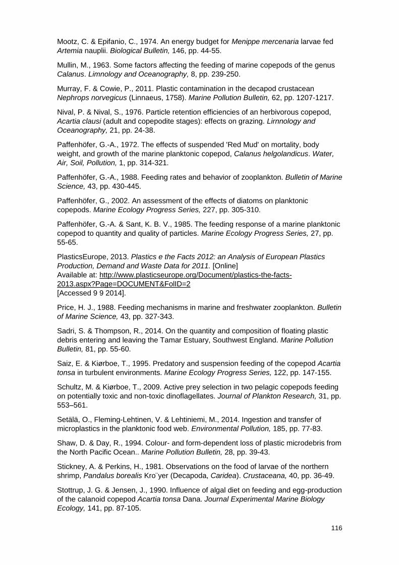
116
Mootz, C. & Epifanio, C., 1974. An energy budget for Menippe mercenaria larvae fed
Artemia nauplii. Biological Bulletin, 146, pp. 44-55.
Mullin, M., 1963. Some factors affecting the feeding of marine copepods of the genus
Calanus. Limnology and Oceanography, 8, pp. 239-250.
Murray, F. & Cowie, P., 2011. Plastic contamination in the decapod crustacean
Nephrops norvegicus (Linnaeus, 1758). Marine Pollution Bulletin, 62, pp. 1207-1217.
Nival, P. & Nival, S., 1976. Particle retention efficiencies of an herbivorous copepod,
Acartia clausi (adult and copepodite stages): effects on grazing. Lirnnology and
Oceanography, 21, pp. 24-38.
Paffenhöfer, G.-A., 1972. The effects of suspended 'Red Mud' on mortality, body
weight, and growth of the marine planktonic copepod, Calanus helgolandicus. Water,
Air, Soil, Pollution, 1, pp. 314-321.
Paffenhöfer, G.-A., 1988. Feeding rates and behavior of zooplankton. Bulletin of Marine
Science, 43, pp. 430-445.
Paffenhöfer, G., 2002. An assessment of the effects of diatoms on planktonic
copepods. Marine Ecology Progress Series, 227, pp. 305-310.
Paffenhöfer, G.-A. & Sant, K. B. V., 1985. The feeding response of a marine planktonic
copepod to quantity and quality of particles. Marine Ecology Progress Series, 27, pp.
55-65.
PlasticsEurope, 2013. Plastics e the Facts 2012: an Analysis of European Plastics
Production, Demand and Waste Data for 2011. [Online]
Available at: http://www.plasticseurope.org/Document/plastics-the-facts-
2013.aspx?Page=DOCUMENT&FolID=2
[Accessed 9 9 2014].
Price, H. J., 1988. Feeding mechanisms in marine and freshwater zooplankton. Bulletin
of Marine Science, 43, pp. 327-343.
Sadri, S. & Thompson, R., 2014. On the quantity and composition of floating plastic
debris entering and leaving the Tamar Estuary, Southwest England. Marine Pollution
Bulletin, 81, pp. 55-60.
Saiz, E. & Kiørboe, T., 1995. Predatory and suspension feeding of the copepod Acartia
tonsa in turbulent environments. Marine Ecology Progress Series, 122, pp. 147-155.
Schultz, M. & Kiørboe, T., 2009. Active prey selection in two pelagic copepods feeding
on potentially toxic and non-toxic dinoflagellates. Journal of Plankton Research, 31, pp.
553–561.
Setälä, O., Fleming-Lehtinen, V. & Lehtiniemi, M., 2014. Ingestion and transfer of
microplastics in the planktonic food web. Environmental Pollution, 185, pp. 77-83.
Shaw, D. & Day, R., 1994. Colour- and form-dependent loss of plastic microdebris from
the North Pacific Ocean.. Marine Pollution Bulletin, 28, pp. 39-43.
Stickney, A. & Perkins, H., 1981. Observations on the food of larvae of the northern
shrimp, Pandalus borealis Kro¨yer (Decapoda, Caridea). Crustaceana, 40, pp. 36-49.
Stottrup, J. G. & Jensen, J., 1990. Influence of algal diet on feeding and egg-production
of the calanoid copepod Acartia tonsa Dana. Journal Experimental Marine Biology
Ecology, 141, pp. 87-105.
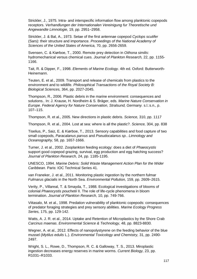
117
Strickler, J., 1975. Intra- and interspecific information flow among planktonic copepods
receptors. Verhandlungen der Internationalen Vereinigung fur Theoretische und
Angewandte Limnologie, 19, pp. 2951–2958.
Strickler, J. & Bal, A., 1973. Setae of the first antennae copepod Cyclops scutifer
(Sars): their structure and importance. Proceedings of the National Academy of
Sciences of the United States of America, 70, pp. 2656-2659.
Svensen, C. & Kiørboe, T., 2000. Remote prey detection in Oithona similis:
hydromechanical versus chemical cues. Journal of Plankton Research, 22, pp. 1155-
1166.
Tait, R. & Dipper, F., 1998. Elements of Marine Ecology. 4th ed. Oxford: Butterworth-
Heinemann.
Teuten, E. et al., 2009. Transport and release of chemicals from plastics to the
environment and to wildlife. Philosophical Transactions of the Royal Society B:
Biological Sciences, 364, pp. 2027-2045.
Thompson, R., 2006. Plastic debris in the marine environment: consequences and
solutions.. In: J. Krause, H. Nordheim & S. Bräger, eds. Marine Nature Conservation in
Europe. Federal Agency for Nature Conservation, Stralsund, Germany. s.l.:s.n., p.
107–115.
Thompson, R. et al., 2005. New directions in plastic debris. Science, 310, pp. 1117
Thompson, R. et al., 2004. Lost at sea: where is all the plastic?. Science, 304, pp. 838
Tiselius, P., Saiz, E. & Kiørboe, T., 2013. Sensory capabilities and food capture of two
small copepods, Paracalanus parvus and Pseudocalanus sp.. Limnology and
Oceanography, 58, pp. 1657-1666.
Turner, J. et al., 2002. Zooplankton feeding ecology: does a diet of Phaeocystis
support good copepod grazing, survival, egg production and egg hatching success?
Journal of Plankton Research, 24, pp. 1185-1195.
UNESCO, 1994. Marine Debris: Solid Waste Management Action Plan for the Wider
Caribbean. Paris: IOC Technical Series 41.
van Franeker, J. et al., 2011. Monitoring plastic ingestion by the northern fulmar
Fulmarus glacialis in the North Sea. Environmental Pollution, 159, pp. 2609–2615.
Verity, P., Villareal, T. & Smayda, T., 1988. Ecological investigations of blooms of
colonial Phaeocystis pouchetii II. The role of life-cycle phenomena in bloom
termination. Journal of Plankton Research, 10, pp. 749-766.
Viitasalo, M. et al., 1998. Predation vulnerability of planktonic copepods: consequences
of predator foraging strategies and prey sensory abilities. Marine Ecology Progress
Series, 175, pp. 129-142.
Watts, A. J. R. et al., 2014. Uptake and Retention of Microplastics by the Shore Crab
Carcinus maenas. Environmental Science & Technology, 48, pp. 8823-8830.
Wegner, A. et al., 2012. Effects of nanopolystyrene on the feeding behavior of the blue
mussel (Mytilus edulis L.). Environmental Toxicology and Chemistry, 31, pp. 2490-
2497.
Wright, S. L., Rowe, D., Thompson, R. C. & Galloway, T. S., 2013. Miroplastic
ingestion decreases energy reserves in marine worms. Current Biology, 23, pp.
R1031–R1033.
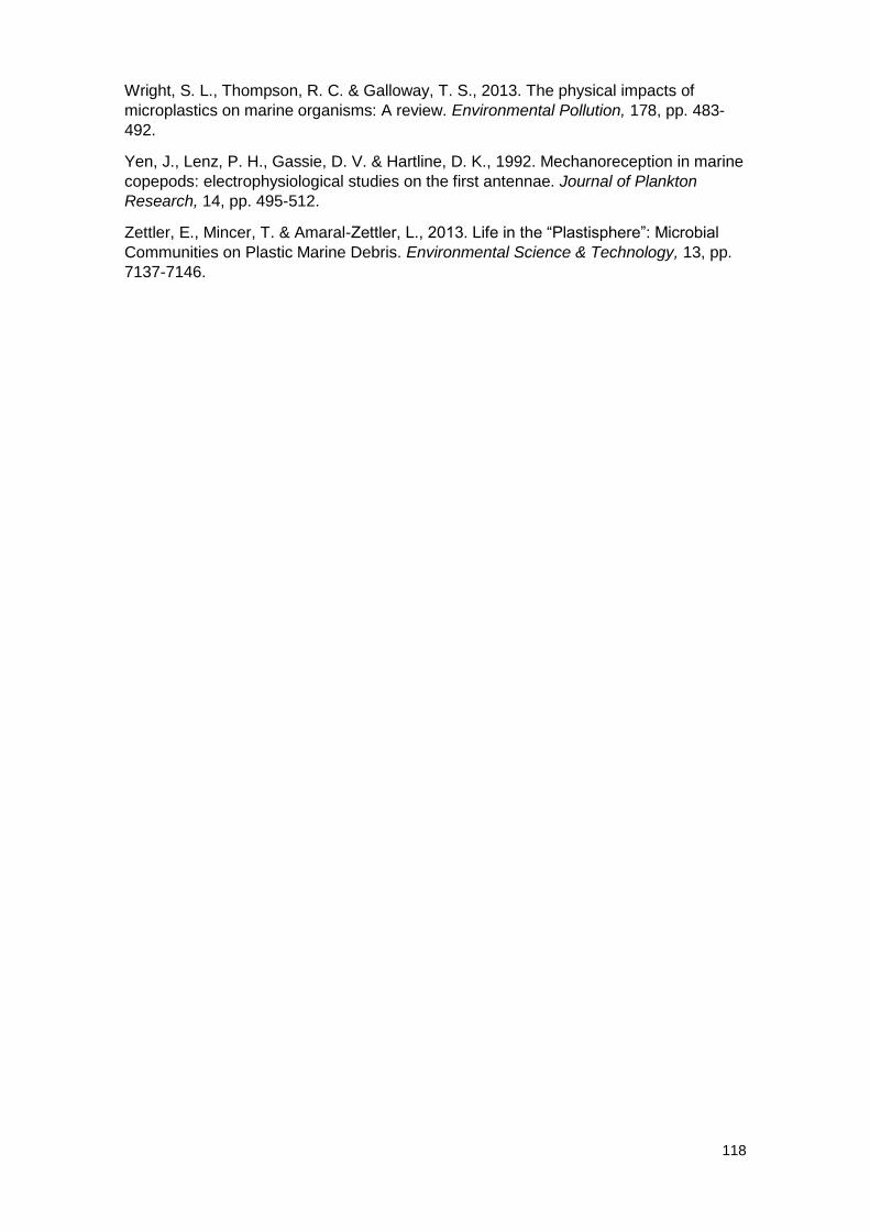
118
Wright, S. L., Thompson, R. C. & Galloway, T. S., 2013. The physical impacts of
microplastics on marine organisms: A review. Environmental Pollution, 178, pp. 483-
492.
Yen, J., Lenz, P. H., Gassie, D. V. & Hartline, D. K., 1992. Mechanoreception in marine
copepods: electrophysiological studies on the first antennae. Journal of Plankton
Research, 14, pp. 495-512.
Zettler, E., Mincer, T. & Amaral-Zettler, L., 2013. Life in the “Plastisphere”: Microbial
Communities on Plastic Marine Debris. Environmental Science & Technology, 13, pp.
7137-7146.
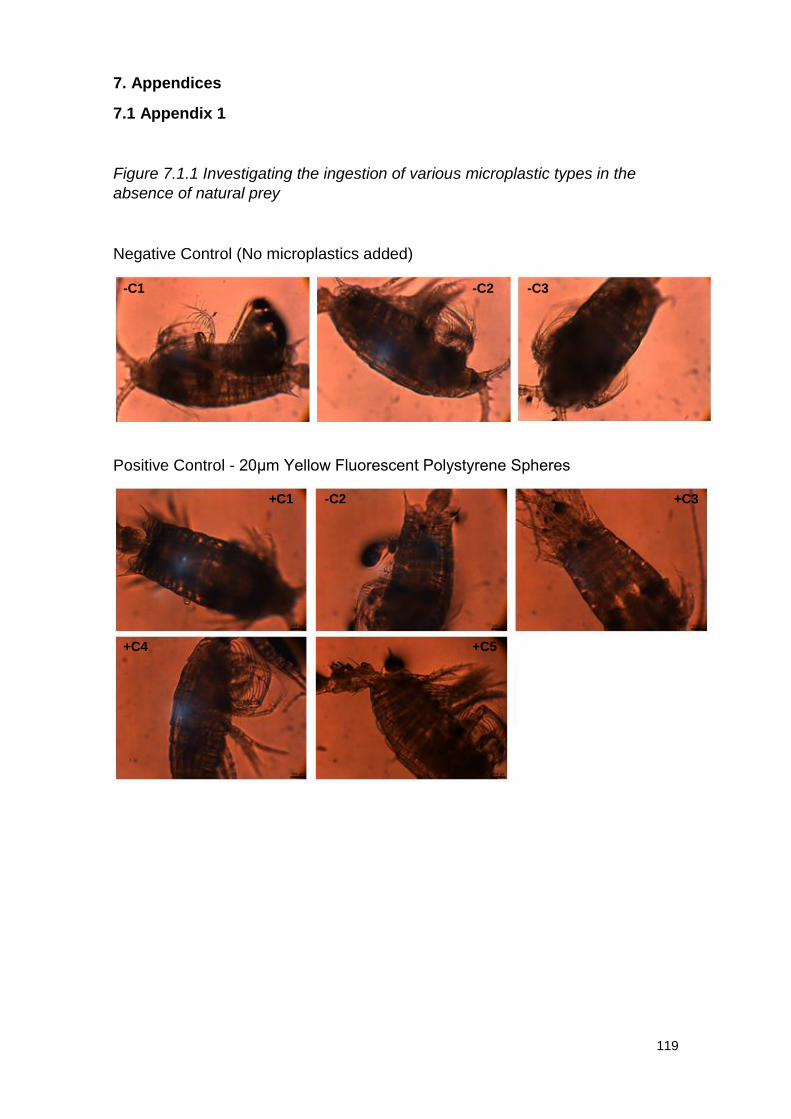
119
7. Appendices
7.1 Appendix 1
Figure 7.1.1 Investigating the ingestion of various microplastic types in the
absence of natural prey
Negative Control (No microplastics added)
Positive Control - 20μm Yellow Fluorescent Polystyrene Spheres
-C1 -C2 -C3
+C1 -C2 +C3
+C4 +C5
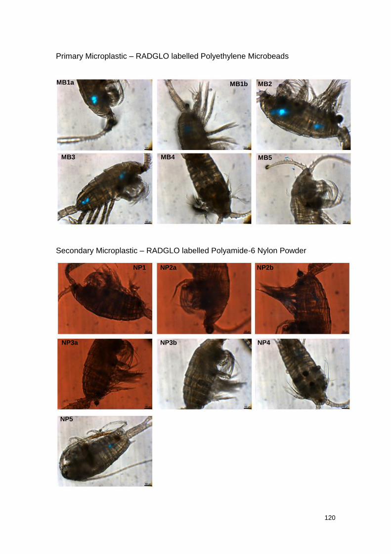
120
Primary Microplastic – RADGLO labelled Polyethylene Microbeads
Secondary Microplastic – RADGLO labelled Polyamide-6 Nylon Powder
-C1 -C1 -C1
NP1 NP2a NP2b
NP3a NP3b NP4
NP5
-C1 MB1b MB2
MB3 MB4 MB5
MB1a
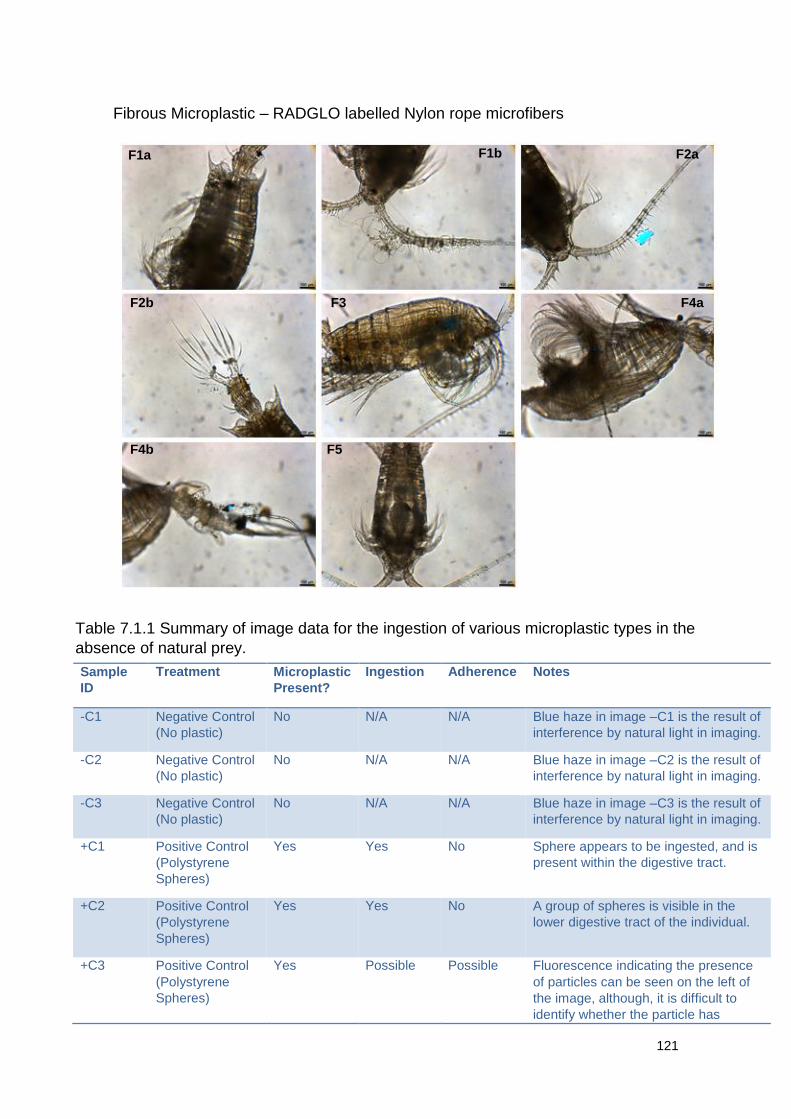
121
Fibrous Microplastic – RADGLO labelled Nylon rope microfibers
Sample
ID
Treatment Microplastic
Present?
Ingestion Adherence Notes
-C1 Negative Control
(No plastic)
No N/A N/A Blue haze in image –C1 is the result of
interference by natural light in imaging.
-C2 Negative Control
(No plastic)
No N/A N/A Blue haze in image –C2 is the result of
interference by natural light in imaging.
-C3 Negative Control
(No plastic)
No N/A N/A Blue haze in image –C3 is the result of
interference by natural light in imaging.
+C1 Positive Control
(Polystyrene
Spheres)
Yes Yes No Sphere appears to be ingested, and is
present within the digestive tract.
+C2 Positive Control
(Polystyrene
Spheres)
Yes Yes No A group of spheres is visible in the
lower digestive tract of the individual.
+C3 Positive Control
(Polystyrene
Spheres)
Yes Possible Possible Fluorescence indicating the presence
of particles can be seen on the left of
the image, although, it is difficult to
identify whether the particle has
F1a F1b F2a
F2b F3 F4a
F4b F5
Table 7.1.1 Summary of image data for the ingestion of various microplastic types in the
absence of natural prey.
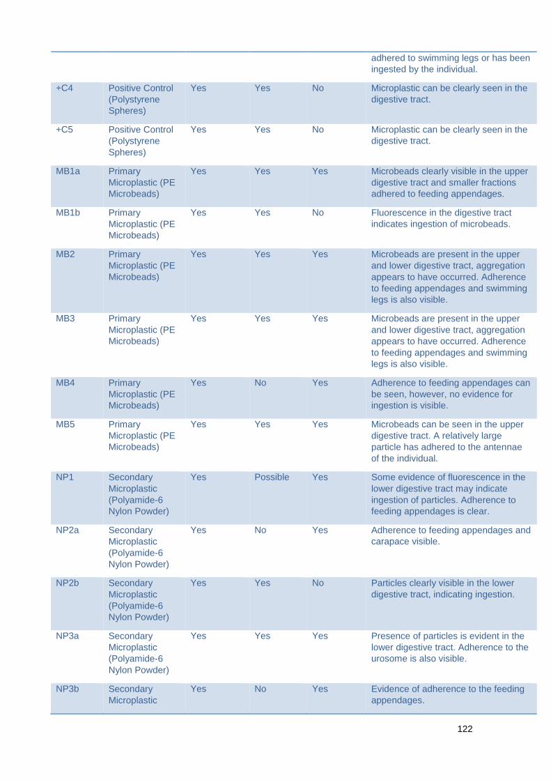
122
adhered to swimming legs or has been
ingested by the individual.
+C4 Positive Control
(Polystyrene
Spheres)
Yes Yes No Microplastic can be clearly seen in the
digestive tract.
+C5 Positive Control
(Polystyrene
Spheres)
Yes Yes No Microplastic can be clearly seen in the
digestive tract.
MB1a Primary
Microplastic (PE
Microbeads)
Yes Yes Yes Microbeads clearly visible in the upper
digestive tract and smaller fractions
adhered to feeding appendages.
MB1b Primary
Microplastic (PE
Microbeads)
Yes Yes No Fluorescence in the digestive tract
indicates ingestion of microbeads.
MB2 Primary
Microplastic (PE
Microbeads)
Yes Yes Yes Microbeads are present in the upper
and lower digestive tract, aggregation
appears to have occurred. Adherence
to feeding appendages and swimming
legs is also visible.
MB3 Primary
Microplastic (PE
Microbeads)
Yes Yes Yes Microbeads are present in the upper
and lower digestive tract, aggregation
appears to have occurred. Adherence
to feeding appendages and swimming
legs is also visible.
MB4 Primary
Microplastic (PE
Microbeads)
Yes No Yes Adherence to feeding appendages can
be seen, however, no evidence for
ingestion is visible.
MB5 Primary
Microplastic (PE
Microbeads)
Yes Yes Yes Microbeads can be seen in the upper
digestive tract. A relatively large
particle has adhered to the antennae
of the individual.
NP1 Secondary
Microplastic
(Polyamide-6
Nylon Powder)
Yes Possible Yes Some evidence of fluorescence in the
lower digestive tract may indicate
ingestion of particles. Adherence to
feeding appendages is clear.
NP2a Secondary
Microplastic
(Polyamide-6
Nylon Powder)
Yes No Yes Adherence to feeding appendages and
carapace visible.
NP2b Secondary
Microplastic
(Polyamide-6
Nylon Powder)
Yes Yes No Particles clearly visible in the lower
digestive tract, indicating ingestion.
NP3a Secondary
Microplastic
(Polyamide-6
Nylon Powder)
Yes Yes Yes Presence of particles is evident in the
lower digestive tract. Adherence to the
urosome is also visible.
NP3b Secondary
Microplastic
Yes No Yes Evidence of adherence to the feeding
appendages.
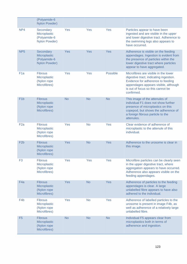
123
(Polyamide-6
Nylon Powder)
NP4 Secondary
Microplastic
(Polyamide-6
Nylon Powder)
Yes Yes Yes Particles appear to have been
ingested and are visible in the upper
and lower digestive tract. Adherence to
the swimming legs also appears to
have occurred.
NP5 Secondary
Microplastic
(Polyamide-6
Nylon Powder)
Yes Yes Yes Adherence is visible on the feeding
appendages. Ingestion is evident from
the presence of particles within the
lower digestive tract where particles
appear to have aggregated.
F1a Fibrous
Microplastic
(Nylon rope
Microfibres)
Yes Yes Possible Microfibres are visible in the lower
digestive tract, indicating ingestion.
Evidence for adherence to feeding
appendages appears visible, although
is out of focus so this cannot be
confirmed.
F1b Fibrous
Microplastic
(Nylon rope
Microfibres)
No No No This image of the attenules of
individual F1 does not show further
presence of microplastics on this
copepod, but shows the adherence of
a foreign fibrous particle to the
attenules.
F2a Fibrous
Microplastic
(Nylon rope
Microfibres)
Yes No Yes Clear evidence of adherence of
microplastic to the attenule of this
individual.
F2b Fibrous
Microplastic
(Nylon rope
Microfibres)
Yes No Yes Adherence to the urosome is clear in
this image.
F3 Fibrous
Microplastic
(Nylon rope
Microfibres)
Yes Yes Yes Microfibre particles can be clearly seen
in the upper digestive tract, where
aggregation appears to have occurred.
Adherence also appears visible on the
feeding appendages.
F4a Fibrous
Microplastic
(Nylon rope
Microfibres)
Yes No Yes Adherence of particles to the feeding
appendages is clear. A large
unlabelled fibre appears to have also
adhered to the individual.
F4b Fibrous
Microplastic
(Nylon rope
Microfibres)
Yes No Yes Adherence of labelled particles to the
urosome is present in image F4b, as
well as adherence of a relatively large
unlabelled fibre.
F5 Fibrous
Microplastic
(Nylon rope
Microfibres)
No No No Individual F5 appears clear from
microplastics both in terms of
adherence and ingestion.
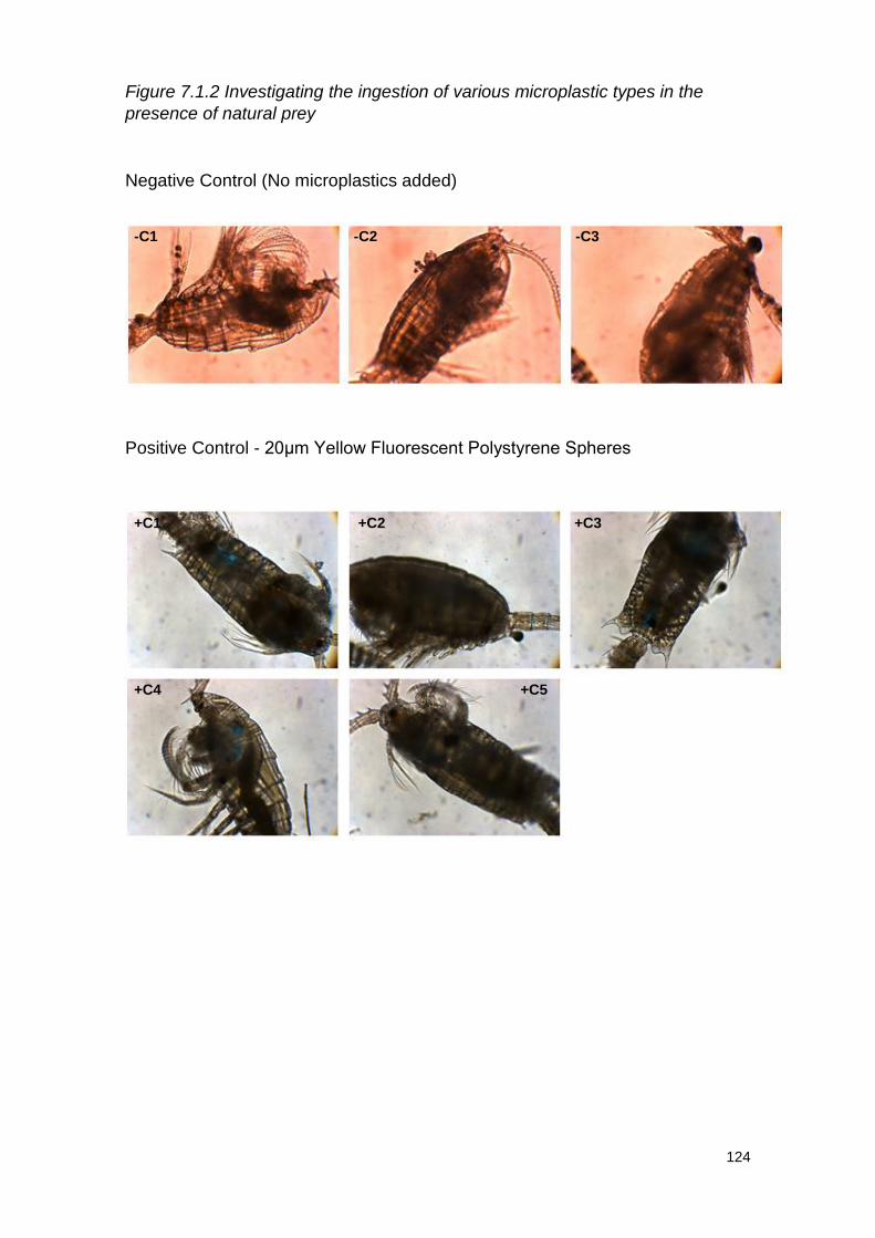
124
Figure 7.1.2 Investigating the ingestion of various microplastic types in the
presence of natural prey
Negative Control (No microplastics added)
Positive Control - 20μm Yellow Fluorescent Polystyrene Spheres
-C1 -C2 -C3
+C1 +C2 +C3
+C4 +C5
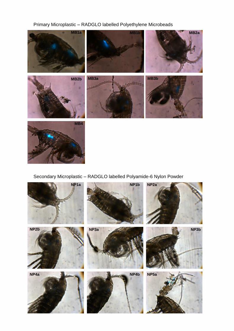
125
Primary Microplastic – RADGLO labelled Polyethylene Microbeads
Secondary Microplastic – RADGLO labelled Polyamide-6 Nylon Powder
MB1a MB1b MB2a
MB2b MB3a MB3b
MB4
NP1a NP1b NP2a
NP2b NP3a NP3b
NP4a NP4b NP5a
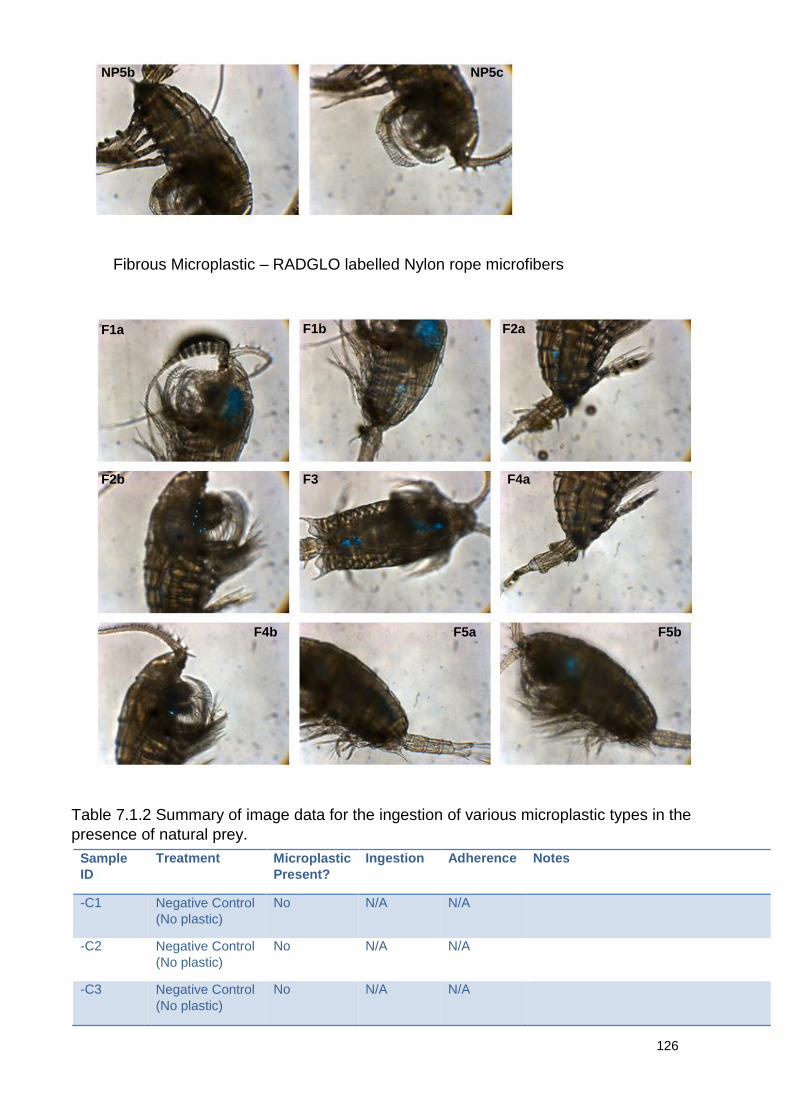
126
Fibrous Microplastic – RADGLO labelled Nylon rope microfibers
Sample
ID
Treatment Microplastic
Present?
Ingestion Adherence Notes
-C1 Negative Control
(No plastic)
No N/A N/A
-C2 Negative Control
(No plastic)
No N/A N/A
-C3 Negative Control
(No plastic)
No N/A N/A
NP5b NP5c
F1a F1b F2a
F2b F3 F4a
F4b F5a F5b
Table 7.1.2 Summary of image data for the ingestion of various microplastic types in the
presence of natural prey.
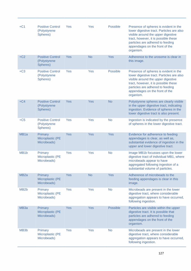
127
+C1
Positive Control
(Polystyrene
Spheres)
Yes
Yes
Possible
Presence of spheres is evident in the
lower digestive tract. Particles are also
visible around the upper digestive
tract, however, it is possible these
particles are adhered to feeding
appendages on the front of the
organism.
+C2 Positive Control
(Polystyrene
Spheres)
Yes No Yes Adherence to the urosome is clear in
this image.
+C3 Positive Control
(Polystyrene
Spheres)
Yes Yes Possible Presence of spheres is evident in the
lower digestive tract. Particles are also
visible around the upper digestive
tract, however, it is possible these
particles are adhered to feeding
appendages on the front of the
organism.
+C4 Positive Control
(Polystyrene
Spheres)
Yes Yes No Polystyrene spheres are clearly visible
in the upper digestive tract, indicating
ingestion. Evidence of spheres in the
lower digestive tract is also present.
+C5 Positive Control
(Polystyrene
Spheres)
Yes Yes No Ingestion is indicated by the presence
of spheres in the lower digestive tract.
MB1a Primary
Microplastic (PE
Microbeads)
Yes Yes Yes Evidence for adherence to feeding
appendages is clear, as well as,
substantial evidence of ingestion in the
upper and lower digestive tract.
MB1b Primary
Microplastic (PE
Microbeads)
Yes Yes No Image MB1b focusses upon the lower
digestive tract of individual MB1, where
microbeads appear to have
aggregated following ingestion of a
substantial volume of particles.
MB2a Primary
Microplastic (PE
Microbeads)
Yes No Yes Adherence of microbeads to the
feeding appendages is clear in this
image.
MB2b Primary
Microplastic (PE
Microbeads)
Yes Yes No Microbeads are present in the lower
digestive tract, where considerable
aggregation appears to have occurred,
following ingestion.
MB3a Primary
Microplastic (PE
Microbeads)
Yes Yes Possible Particles are visible within the upper
digestive tract. It is possible that
particles are adhered to feeding
appendages on the front of the
organism.
MB3b Primary
Microplastic (PE
Microbeads)
Yes Yes No Microbeads are present in the lower
digestive tract, where considerable
aggregation appears to have occurred,
following ingestion.
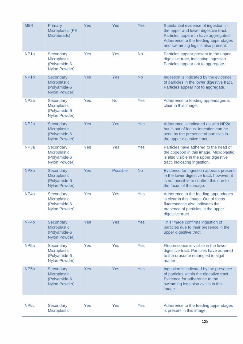
128
MB4 Primary
Microplastic (PE
Microbeads)
Yes Yes Yes Substantial evidence of ingestion in
the upper and lower digestive tract.
Particles appear to have aggregated.
Adherence to the feeding appendages
and swimming legs is also present.
NP1a Secondary
Microplastic
(Polyamide-6
Nylon Powder)
Yes Yes No Particles appear present in the upper
digestive tract, indicating ingestion.
Particles appear not to aggregate.
NP1b Secondary
Microplastic
(Polyamide-6
Nylon Powder)
Yes Yes No Ingestion is indicated by the evidence
of particles in the lower digestive tract.
Particles appear not to aggregate.
NP2a Secondary
Microplastic
(Polyamide-6
Nylon Powder)
Yes No Yes Adherence to feeding appendages is
clear in this image.
NP2b Secondary
Microplastic
(Polyamide-6
Nylon Powder)
Yes Yes Yes Adherence is indicated as with NP2a,
but is out of focus. Ingestion can be
seen by the presence of particles in
the upper digestive tract.
NP3a Secondary
Microplastic
(Polyamide-6
Nylon Powder)
Yes Yes Yes Particles have adhered to the head of
the copepod in this image. Microplastic
is also visible in the upper digestive
tract, indicating ingestion.
NP3b Secondary
Microplastic
(Polyamide-6
Nylon Powder)
Yes Possible No Evidence for ingestion appears present
in the lower digestive tract, however, it
is not possible to confirm this due to
the focus of the image.
NP4a Secondary
Microplastic
(Polyamide-6
Nylon Powder)
Yes Yes Yes Adherence to the feeding appendages
is clear in this image. Out of focus
fluorescence also indicates the
presence of particles in the upper
digestive tract.
NP4b Secondary
Microplastic
(Polyamide-6
Nylon Powder)
Yes Yes Yes This image confirms ingestion of
particles due to their presence in the
upper digestive tract.
NP5a Secondary
Microplastic
(Polyamide-6
Nylon Powder)
Yes Yes Yes Fluorescence is visible in the lower
digestive tract. Particles have adhered
to the urosome entangled in algal
matter.
NP5b Secondary
Microplastic
(Polyamide-6
Nylon Powder)
Yes Yes Yes Ingestion is indicated by the presence
of particles within the digestive tract.
Evidence for adherence to the
swimming legs also exists in this
image.
NP5c
Secondary
Microplastic
Yes
Yes
Yes
Adherence to the feeding appendages
is present in this image.
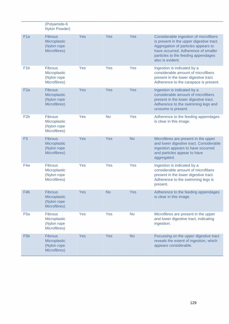
129
(Polyamide-6
Nylon Powder)
F1a Fibrous
Microplastic
(Nylon rope
Microfibres)
Yes Yes Yes Considerable ingestion of microfibers
is present in the upper digestive tract.
Aggregation of particles appears to
have occurred. Adherence of smaller
particles to the feeding appendages
also is evident.
F1b Fibrous
Microplastic
(Nylon rope
Microfibres)
Yes Yes Yes Ingestion is indicated by a
considerable amount of microfibers
present in the lower digestive tract.
Adherence to the carapace is present.
F2a Fibrous
Microplastic
(Nylon rope
Microfibres)
Yes Yes Yes Ingestion is indicated by a
considerable amount of microfibers
present in the lower digestive tract.
Adherence to the swimming legs and
urosome is present.
F2b Fibrous
Microplastic
(Nylon rope
Microfibres)
Yes No Yes Adherence to the feeding appendages
is clear in this image.
F3 Fibrous
Microplastic
(Nylon rope
Microfibres)
Yes Yes No Microfibres are present in the upper
and lower digestive tract. Considerable
ingestion appears to have occurred
and particles appear to have
aggregated.
F4a Fibrous
Microplastic
(Nylon rope
Microfibres)
Yes Yes Yes Ingestion is indicated by a
considerable amount of microfibers
present in the lower digestive tract.
Adherence to the swimming legs is
present.
F4b Fibrous
Microplastic
(Nylon rope
Microfibres)
Yes No Yes Adherence to the feeding appendages
is clear in this image.
F5a Fibrous
Microplastic
(Nylon rope
Microfibres)
Yes Yes No Microfibres are present in the upper
and lower digestive tract, indicating
ingestion.
F5b Fibrous
Microplastic
(Nylon rope
Microfibres)
Yes Yes No Focussing on the upper digestive tract
reveals the extent of ingestion, which
appears considerable.
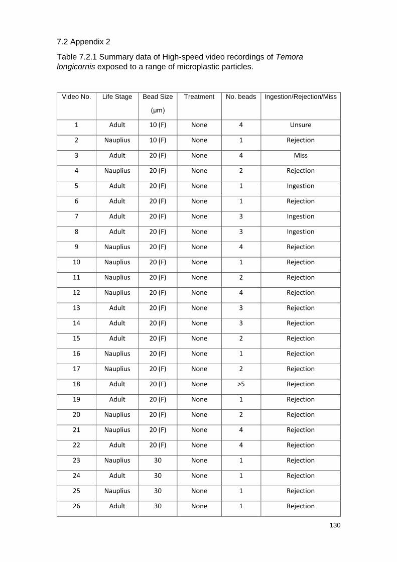
130
7.2 Appendix 2
Table 7.2.1 Summary data of High-speed video recordings of Temora
longicornis exposed to a range of microplastic particles.
Video No. Life Stage Bead Size
(μm)
Treatment No. beads Ingestion/Rejection/Miss
1 Adult 10 (F) None 4 Unsure
2 Nauplius 10 (F) None 1 Rejection
3 Adult 20 (F) None 4 Miss
4 Nauplius 20 (F) None 2 Rejection
5 Adult 20 (F) None 1 Ingestion
6 Adult 20 (F) None 1 Rejection
7 Adult 20 (F) None 3 Ingestion
8 Adult 20 (F) None 3 Ingestion
9 Nauplius 20 (F) None 4 Rejection
10 Nauplius 20 (F) None 1 Rejection
11 Nauplius 20 (F) None 2 Rejection
12 Nauplius 20 (F) None 4 Rejection
13 Adult 20 (F) None 3 Rejection
14 Adult 20 (F) None 3 Rejection
15 Adult 20 (F) None 2 Rejection
16 Nauplius 20 (F) None 1 Rejection
17 Nauplius 20 (F) None 2 Rejection
18 Adult 20 (F) None >5 Rejection
19 Adult 20 (F) None 1 Rejection
20 Nauplius 20 (F) None 2 Rejection
21 Nauplius 20 (F) None 4 Rejection
22 Adult 20 (F) None 4 Rejection
23 Nauplius 30 None 1 Rejection
24 Adult 30 None 1 Rejection
25 Nauplius 30 None 1 Rejection
26 Adult 30 None 1 Rejection
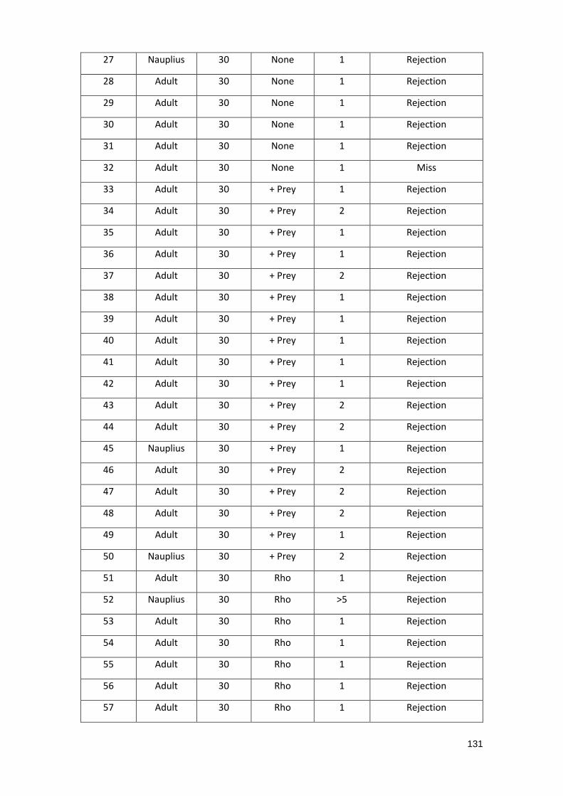
131
27 Nauplius 30 None 1 Rejection
28 Adult 30 None 1 Rejection
29 Adult 30 None 1 Rejection
30 Adult 30 None 1 Rejection
31 Adult 30 None 1 Rejection
32 Adult 30 None 1 Miss
33 Adult 30 + Prey 1 Rejection
34 Adult 30 + Prey 2 Rejection
35 Adult 30 + Prey 1 Rejection
36 Adult 30 + Prey 1 Rejection
37 Adult 30 + Prey 2 Rejection
38 Adult 30 + Prey 1 Rejection
39 Adult 30 + Prey 1 Rejection
40 Adult 30 + Prey 1 Rejection
41 Adult 30 + Prey 1 Rejection
42 Adult 30 + Prey 1 Rejection
43 Adult 30 + Prey 2 Rejection
44 Adult 30 + Prey 2 Rejection
45 Nauplius 30 + Prey 1 Rejection
46 Adult 30 + Prey 2 Rejection
47 Adult 30 + Prey 2 Rejection
48 Adult 30 + Prey 2 Rejection
49 Adult 30 + Prey 1 Rejection
50 Nauplius 30 + Prey 2 Rejection
51 Adult 30 Rho 1 Rejection
52 Nauplius 30 Rho >5 Rejection
53 Adult 30 Rho 1 Rejection
54 Adult 30 Rho 1 Rejection
55 Adult 30 Rho 1 Rejection
56 Adult 30 Rho 1 Rejection
57 Adult 30 Rho 1 Rejection
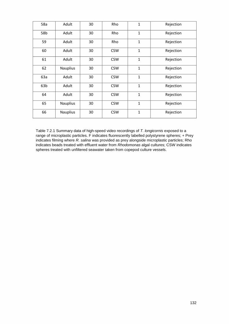
132
58a Adult 30 Rho 1 Rejection
58b Adult 30 Rho 1 Rejection
59 Adult 30 Rho 1 Rejection
60 Adult 30 CSW 1 Rejection
61 Adult 30 CSW 1 Rejection
62 Nauplius 30 CSW 1 Rejection
63a Adult 30 CSW 1 Rejection
63b Adult 30 CSW 1 Rejection
64 Adult 30 CSW 1 Rejection
65 Nauplius 30 CSW 1 Rejection
66 Nauplius 30 CSW 1 Rejection
Table 7.2.1 Summary data of high-speed video recordings of T. longicornis exposed to a
range of microplastic particles. F indicates fluorescently labelled polystyrene spheres; + Prey
indicates filming where R. salina was provided as prey alongside microplastic particles; Rho
indicates beads treated with effluent water from Rhodomonas algal cultures; CSW indicates
spheres treated with unfiltered seawater taken from copepod culture vessels.
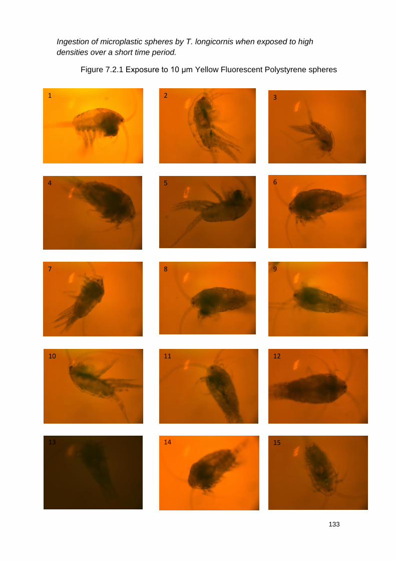
133
Ingestion of microplastic spheres by T. longicornis when exposed to high
densities over a short time period.
Figure 7.2.1 Exposure to 10 μm Yellow Fluorescent Polystyrene spheres
1 2 3
4 5 6
7 8 9
10 11 12
13 14 15
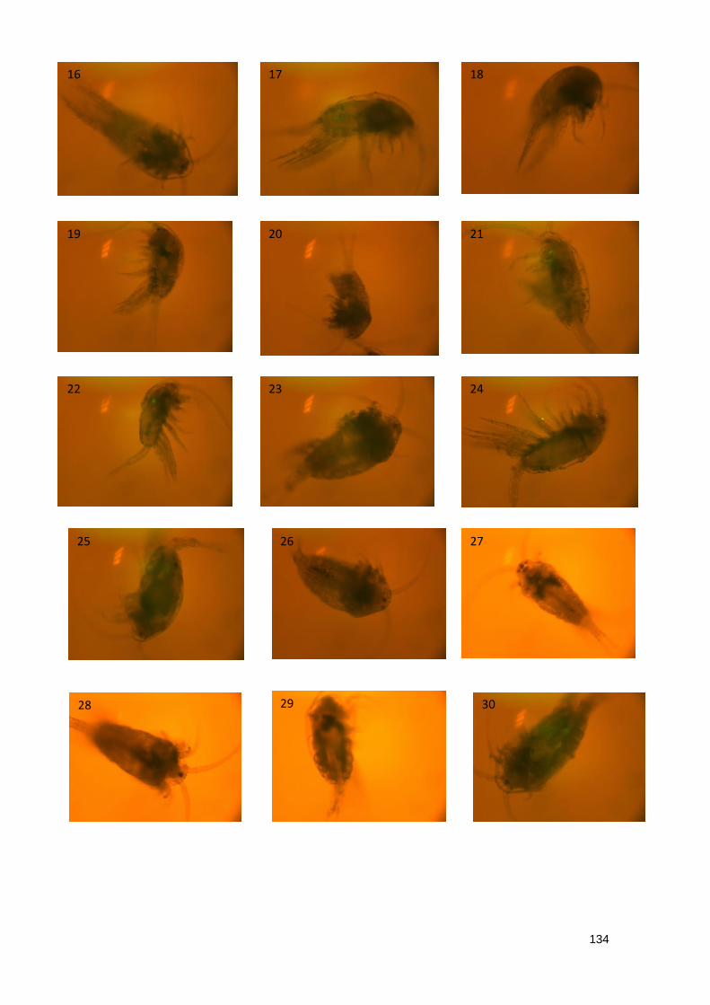
134
16 17 18
19 20 21
22 23 24
25 26 27
28 29 30

135
Table 7.2.2 Summary data of imaging of T. longicornis exposed to 10 μm
Yellow Fluorescent Polystyrene spheres
Ap
pro
x.
nu
mbe
r o
f
sp
he
res
ing
este
d
1
Sp
he
re
Lo
catio
n
Upper
Dig
estive
Tra
ct
Ing
estion
no
no
no
no
no
no
no
no
no
no
possib
le
no
no
no
no
Sca
le o
f
Ad
he
rence
(1-5
)
1 2 2
Ad
he
red t
o
Fe
edin
g
Appendages
Fe
edin
g
Appendages
Fe
edin
g
Appendages
Ad
he
rence
no
no
no
no
no
no
no
no
no
no
yes
no
yes
no
yes
Sp
he
res
Pre
se
nt
no
no
no
no
no
no
no
no
no
no
yes
no
yes
no
yes
Sp
he
re
Siz
e (
μm
)
10
10
10
10
10
10
10
10
10
10
10
10
10
10
10
Life
Sta
ge
Adult
Adult
Naupliu
s
Adult
Adult
Adult
Adult
Adult
Adult
Adult
Adult
Adult
Adult
Adult
Adult
Ind
ivid
ua
l
1
2
3
4
5
6
7
8
9
10
11
12
13
14
15
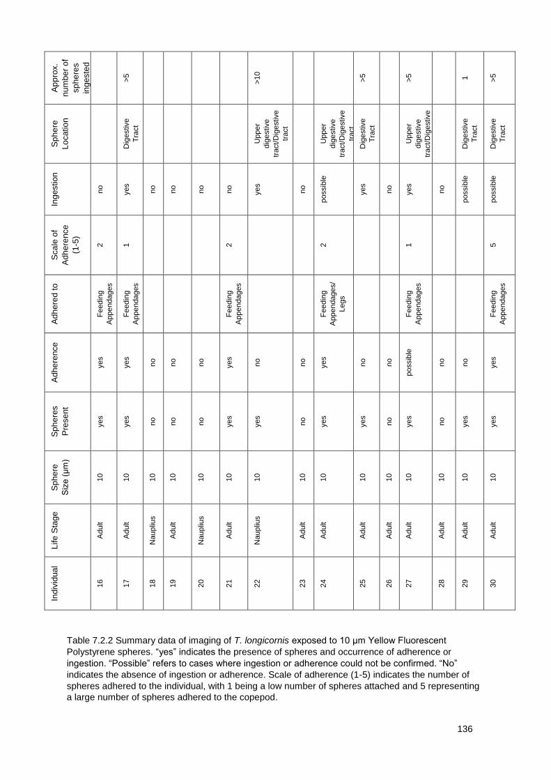
136
Ap
pro
x.
nu
mbe
r o
f
sp
he
res
ing
este
d
>5
>10
>5
>5
1
>5
Sp
he
re
Lo
catio
n
Dig
estive
Tra
ct
Upper
dig
estive
tract/D
igestive
tract
Upper
dig
estive
tract/D
igestive
tract
Dig
estive
Tra
ct
Upper
dig
estive
tract/D
igestive
tract
Dig
estive
Tra
ct
Dig
estive
Tra
ct
Ing
estion
no
yes
no
no
no
no
yes
no
possib
le
yes
no
yes
no
possib
le
possib
le
Sca
le o
f
Ad
he
rence
(1-5
)
2
1 2 2 1 5
Ad
he
red t
o
Fe
edin
g
Appendages
Fe
edin
g
Appendages
Fe
edin
g
Appendages
Fe
edin
g
Appendages/
Legs
Fe
edin
g
Appendages
Fe
edin
g
Appendages
Ad
he
rence
yes
yes
no
no
no
yes
no
no
yes
no
no
possib
le
no
no
yes
Sp
he
res
Pre
se
nt
yes
yes
no
no
no
yes
yes
no
yes
yes
no
yes
no
yes
yes
Sp
he
re
Siz
e (
μm
)
10
10
10
10
10
10
10
10
10
10
10
10
10
10
10
Life
Sta
ge
Adult
Adult
Naupliu
s
Adult
Naupliu
s
Adult
Naupliu
s
Adult
Adult
Adult
Adult
Adult
Adult
Adult
Adult
Ind
ivid
ua
l
16
17
18
19
20
21
22
23
24
25
26
27
28
29
30
Table 7.2.2 Summary data of imaging of T. longicornis exposed to 10 μm Yellow Fluorescent
Polystyrene spheres. “yes” indicates the presence of spheres and occurrence of adherence or
ingestion. “Possible” refers to cases where ingestion or adherence could not be confirmed. “No”
indicates the absence of ingestion or adherence. Scale of adherence (1-5) indicates the number of
spheres adhered to the individual, with 1 being a low number of spheres attached and 5 representing
a large number of spheres adhered to the copepod.
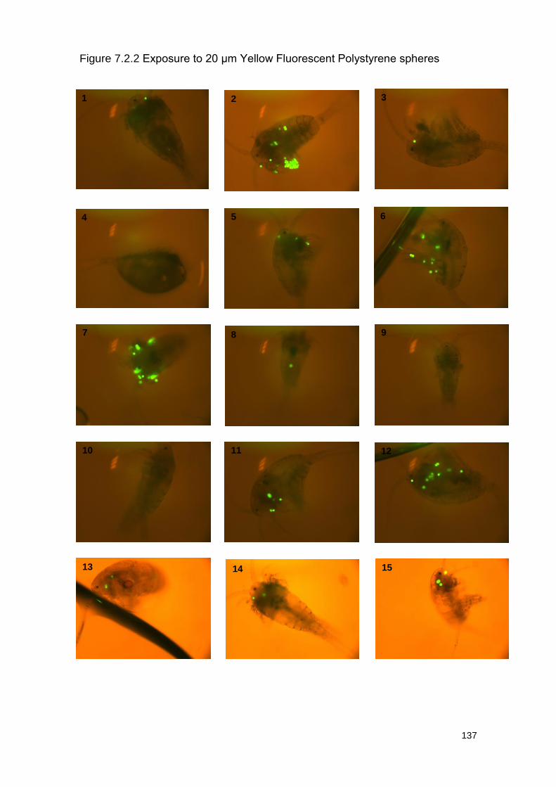
137
Figure 7.2.2 Exposure to 20 μm Yellow Fluorescent Polystyrene spheres
1 2 3
4 5 6
7 8 9
10 11 12
13 14 15
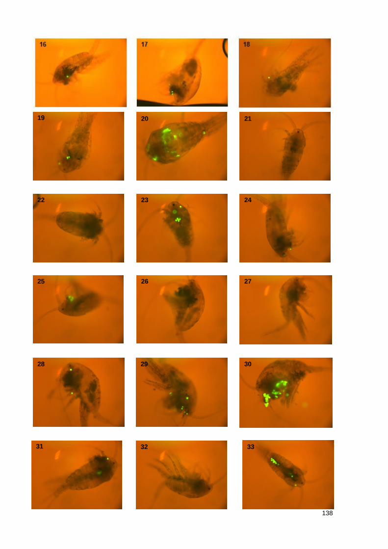
138
19 20 21
22 23 24
25 26 27
28 29 30
31 32 33
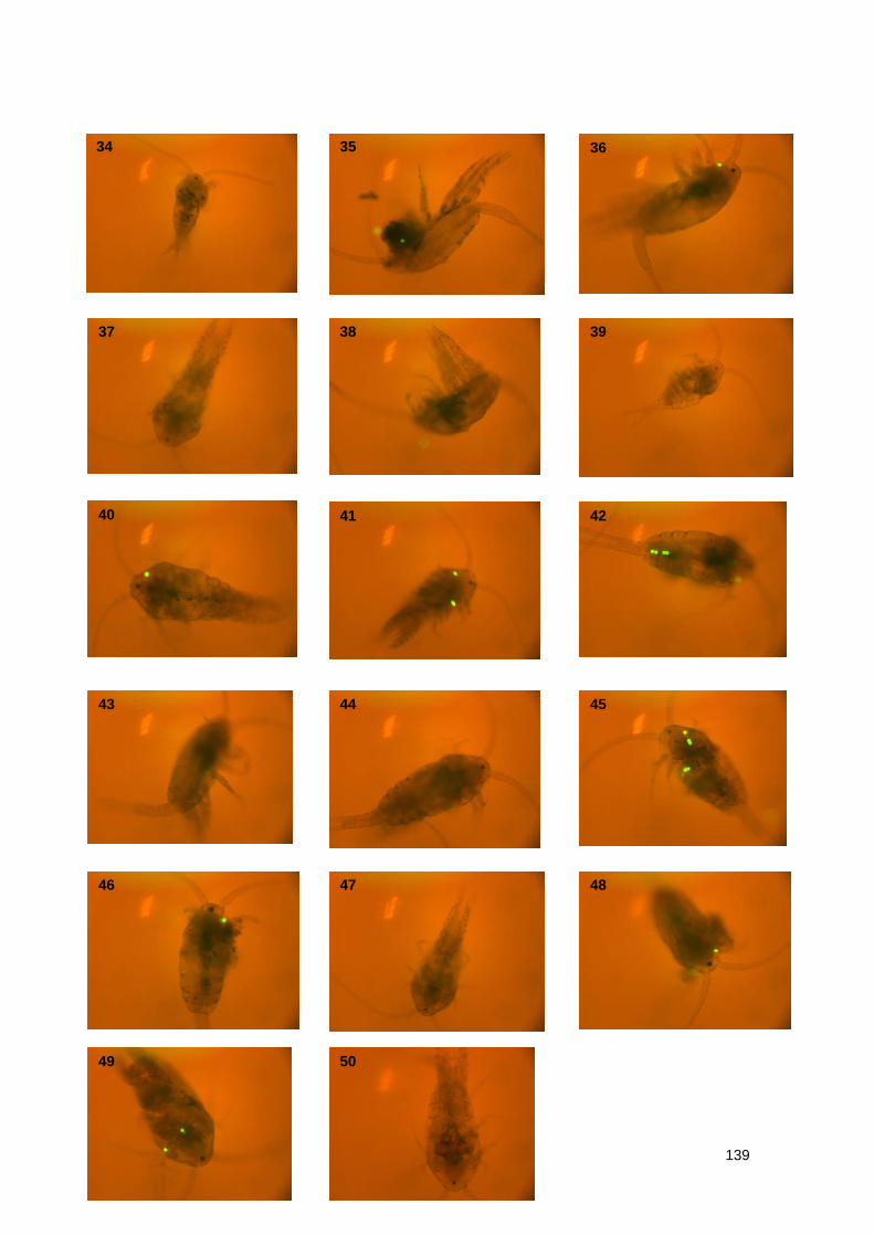
139
34 35 36
37 38 39
40 41 42
43 44 45
46 47 48
49 50
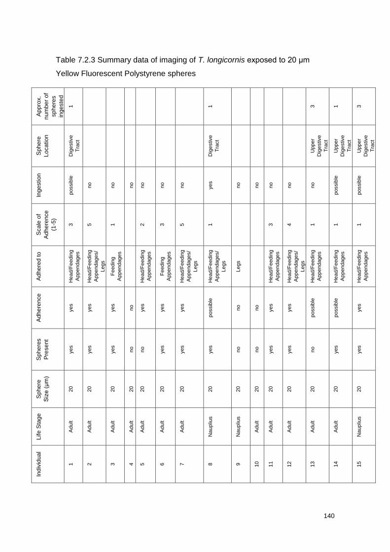
140
Table 7.2.3 Summary data of imaging of T. longicornis exposed to 20 μm
Yellow Fluorescent Polystyrene spheres
Ap
pro
x.
nu
mbe
r o
f
sp
he
res
ing
este
d
1
1 3
1
3
Sp
he
re
Lo
catio
n
Dig
estive
Tra
ct
Dig
estive
Tra
ct
Upper
Dig
estive
Tra
ct
Upper
Dig
estive
Tra
ct
Upper
Dig
estive
Tra
ct
Ing
estion
possib
le
no
no
no
no
no
no
yes
no
no
no
no
no
possib
le
possib
le
Sca
le o
f
Ad
he
rence
(1-5
)
3
5
1 2
3
5
1 3
4
1
1
1
Ad
he
red t
o
Head/F
eedin
g
Appendages
Head/F
eedin
g
Appendages/
Legs
Fe
edin
g
Appendages
Head/F
eedin
g
Appendages
Fe
edin
g
Appendages
Head/F
eedin
g
Appendages/
Legs
Head/F
eedin
g
Appendages/
Legs
Legs
Head/F
eedin
g
Appendages
Head/F
eedin
g
Appendages/
Legs
Head/F
eedin
g
Appendages
Head/F
eedin
g
Appendages
Head/F
eedin
g
Appendages
Ad
he
rence
yes
yes
yes
no
yes
yes
yes
possib
le
no
no
yes
yes
possib
le
possib
le
yes
Sp
he
res
Pre
se
nt
yes
yes
yes
no
no
yes
yes
yes
no
no
yes
yes
no
yes
yes
Sp
he
re
Siz
e (
μm
)
20
20
20
20
20
20
20
20
20
20
20
20
20
20
20
Life
Sta
ge
Adult
Adult
Adult
Adult
Adult
Adult
Adult
Naupliu
s
Naupliu
s
Adult
Adult
Adult
Adult
Adult
Naupliu
s
Ind
ivid
ua
l
1
2
3
4
5
6
7
8
9
10
11
12
13
14
15

141
A
pp
rox.
nu
mbe
r o
f
sp
he
res
ing
este
d
5
Sp
he
re
Lo
catio
n
Upper
dig
estive tra
ct
Ing
estion
no
no
no
no
no
no
no
possib
le
no
no
no
no
no
no
no
Sca
le o
f
Ad
he
rence
(1-5
)
1
1
1
2
5 1
2
1
4
1 1
2
5
Ad
he
red t
o
Fe
edin
g
Appendages
Fe
edin
g
Appendages
Fe
edin
g
Appendages
Fe
edin
g
Appendages
Head/F
eedin
g
appendages/L
egs/
Uro
som
e
Head
Head/F
eedin
g
Appendages
Head/F
eedin
g
Appendages
Fe
edin
g
Appendages
Fe
edin
g
Appendages
Fe
edin
g
Appendages
Head/F
eedin
g
Appendages
Fe
edin
g
Appendages/L
egs
Ad
he
rence
yes
yes
yes
yes
yes
no
yes
yes
yes
yes
yes
no
yes
yes
yes
Sp
he
res
Pre
se
nt
yes
yes
yes
yes
yes
no
yes
yes
yes
yes
yes
no
yes
yes
yes
Sp
he
re
Siz
e (
μm
)
20
20
20
20
20
20
20
20
20
20
20
20
20
20
20
Life
Sta
ge
Naupliu
s
Adult
Adult
Adult
Adult
Adult
Adult
Adult
Adult
Adult
Adult
Naupliu
s
Adult
Adult
Adult
Ind
ivid
ua
l
16
17
18
19
20
21
22
23
24
25
26
27
28
29
30
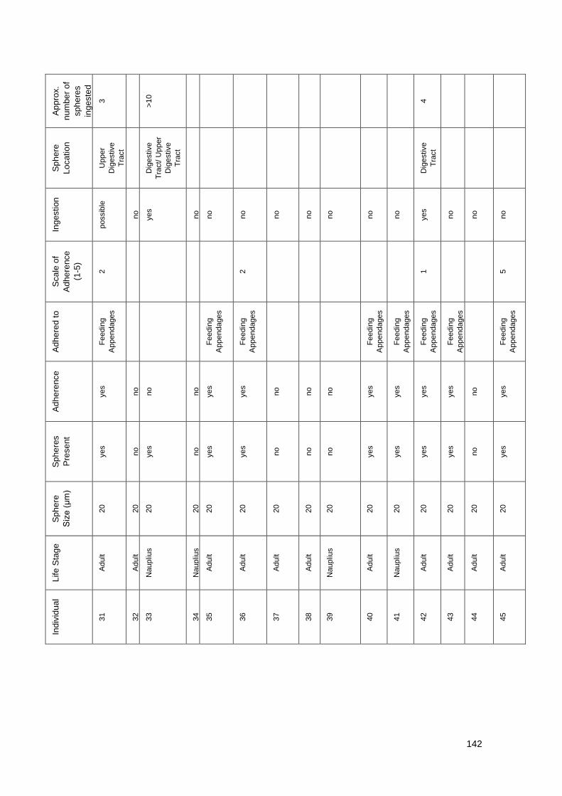
142
A
pp
rox.
nu
mbe
r o
f
sp
he
res
ing
este
d
3
>10
4
Sp
he
re
Lo
catio
n
Upper
Dig
estive
Tra
ct
Dig
estive
Tra
ct/ U
pper
Dig
estive
Tra
ct
Dig
estive
Tra
ct
Ing
estion
possib
le
no
yes
no
no
no
no
no
no
no
no
yes
no
no
no
Sca
le o
f
Ad
he
rence
(1-5
)
2 2 1 5
Ad
he
red t
o
Fe
edin
g
Appendages
Fe
edin
g
Appendages
Fe
edin
g
Appendages
Fe
edin
g
Appendages
Fe
edin
g
Appendages
Fe
edin
g
Appendages
Fe
edin
g
Appendages
Fe
edin
g
Appendages
Ad
he
rence
yes
no
no
no
yes
yes
no
no
no
yes
yes
yes
yes
no
yes
Sp
he
res
Pre
se
nt
yes
no
yes
no
yes
yes
no
no
no
yes
yes
yes
yes
no
yes
Sp
he
re
Siz
e (
μm
)
20
20
20
20
20
20
20
20
20
20
20
20
20
20
20
Life
Sta
ge
Adult
Adult
Naupliu
s
Naupliu
s
Adult
Adult
Adult
Adult
Naupliu
s
Adult
Naupliu
s
Adult
Adult
Adult
Adult
Ind
ivid
ua
l
31
32
33
34
35
36
37
38
39
40
41
42
43
44
45
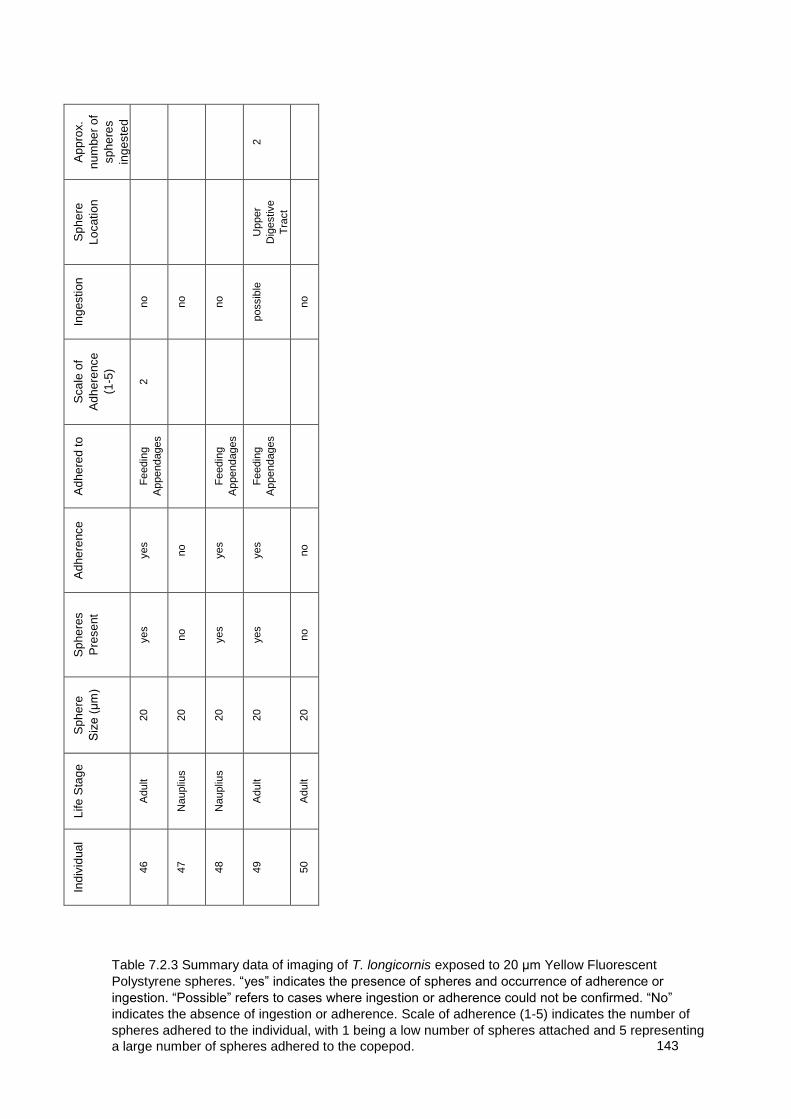
143
A
pp
rox.
nu
mbe
r o
f
sp
he
res
ing
este
d
2
Sp
he
re
Lo
catio
n
Upper
Dig
estive
Tra
ct
Ing
estion
no
no
no
possib
le
no
Sca
le o
f
Ad
he
rence
(1-5
)
2
Ad
he
red t
o
Fe
edin
g
Appendages
Fe
edin
g
Appendages
Fe
edin
g
Appendages
Ad
he
rence
yes
no
yes
yes
no
Sp
he
res
Pre
se
nt
yes
no
yes
yes
no
Sp
he
re
Siz
e (
μm
)
20
20
20
20
20
Life
Sta
ge
Adult
Naupliu
s
Naupliu
s
Adult
Adult
Ind
ivid
ua
l
46
47
48
49
50
Table 7.2.3 Summary data of imaging of T. longicornis exposed to 20 μm Yellow Fluorescent
Polystyrene spheres. “yes” indicates the presence of spheres and occurrence of adherence or
ingestion. “Possible” refers to cases where ingestion or adherence could not be confirmed. “No”
indicates the absence of ingestion or adherence. Scale of adherence (1-5) indicates the number of
spheres adhered to the individual, with 1 being a low number of spheres attached and 5 representing
a large number of spheres adhered to the copepod.

144