Embed Size (px)
Citation preview
22
Investigating Host Induced Meiosis in a Fungal Plant Pathogen
B. J. Saville, M. E. Donaldson and C. E. Doyle Trent University,
Canada
1. Introduction
Fungal spores lend the smut and rust plant diseases their names. Smut fungi produce massive numbers of dark, dust like, thick walled teliospores and the name, smut, is derived from the older definition meaning dark smudge from soot, smoke or dirt. The rust fungi produce diseases characterised by the production of pustules erupting from the plant surface. They contain urediniospores which are often orange or rusty in colour. Spores are essential for fungal survival, providing a means of dispersal and often a structure to protect the fungus; they are also integral to fungal meiosis. Smut and rust fungi are biotrophs, meaning they derive their nutrients from living plant hosts. This interaction is very intimate, involving fungal penetration of the plant cell walls but not the plasma membranes (e.g. Snetselaar & Mims, 1992; Voegele & Mendgen, 2003). As such, most smut and rust fungi have only evolved to infect (and become meiotically competent within) one or a limited number of host species.
The economic impact of these pathogens is well illustrated by considering two crops significantly damaged by them: corn and wheat. According to Capitol Commodity Services (2011), corn remains the largest valued crop in the United States, totalling $67 billion in 2010. World-wide, corn crops were estimated at $163 billion in 2010 (U.S. Grains Council, 2010). The comparable numbers for wheat were $13 billion, and $140 billion, respectively (Capitol Commodity Services, 2011; U.S. Department of Agriculture, 2011). Although mitigated by varieties with partial resistance, the maize crop loss resulting from common smut of corn, caused by Ustilago maydis, is 2% annually, equivalent to ~$1 billion (Allen et al., 2011; Martinez-Espinoza et al., 2002). Wheat crop losses due to wheat leaf rust Puccinia triticina Eriks, which is the most common and widely distributed wheat rust, results in trace to 10% crop losses in many countries around the world. In the US, from 2000 to 2004, the loss was $350 million/year, and it can be $100 million/year in Canada. The production in China is more than twice that of the US and commonly suffers 10-30% crop loss per year (Huerta-Espino et al., 2011). There is also an extreme threat from emerging races of stripe rust of wheat (Puccinia striiformis f. sp. tritici) and wheat stem rust (Puccinia graminis f. sp. tritici). The emerging stem rust races are referred to collectively as UG99 after their location of origin (Uganda), and year of detection (1999). These races are virulent on the vast majority of wheat varieties cultivated around the world. It is predicted that if resistant varieties are not developed and utilized that the UG99 epidemic in Africa will become global (Singh et al., 2011).
www.intechopen.com
Meiosis - Molecular Mechanisms and Cytogenetic Diversity 412
The impact of smut and rust fungi is limited by deploying resistant crop varieties; however, the fungi overcome the resistance leading to cycles in which varieties with new resistances are released and fungi with new virulence genotypes arise. While new virulence alleles ultimately result from mutation, genotypic diversity is created through recombination. Some populations of leaf rust have a genetic structure consistent with an asexual dikaryotic population “within which stepwise mutation at avirulence or virulence loci regularly occurs” (Ordoñez & Kolmer, 2009). In contrast, greatly increased genetic diversity and epidemics of stem rust have been linked to sexual reproduction (Burdon & Reolfs, 1985; Jin, 2011) and eradicating the alternate host for stem rust, common barberry and other Berberis spp. on which sexual reproduction occurs, has provided substantial benefit in controlling wheat stem rust (Roelfs, 1982) and, inadvertently, stripe rust of wheat (Jin, 2011). Further, the corn smut pathogen U. maydis exists in predominantly out-crossing populations (Barnes et al., 2004). This suggests a key role for sexual reproduction in the emergence and maintenance of virulence genotypes.
The rust fungi are obligate biotrophs and cannot be cultured outside their hosts. The wheat rusts, as typified by stem rust, have five spore stages and require two completely unrelated hosts (Schumann & D’Arcy, 2009). The primary host is wheat and the alternative host is barberry. This complex and interesting life cycle will not be discussed in detail here except to note that, in the stem rust life cycle, meiosis likely initiates in planta followed by teliospore maturation (see paragraph below on rust teliospore microscopy). The diploid teliospores are produced late in the season on the primary host, wheat. They germinate and complete meiosis yielding basidiospores that infect the alternate host. In contrast to the rust fungi, the model fungal biotrophic pathogen U. maydis (Banuett, 1995; Brefort et al., 2009) is readily cultured in the laboratory on defined media and its sexual cycle can be completed within 28 days following injection of compatible haploid cells into seedlings of the host Zea mays (corn). U. maydis is amenable to genetic analysis and molecular manipulation, including homologous gene replacement, and several vectors are available for gene expression analysis. An annotated version of the genome sequence of U. maydis was released in 2007 (Kämper et al., 2006) and the annotation continues to be improved (e.g. Donaldson & Saville, 2008; Doyle et al., 2011; Ho et al., 2007; Kronstad, 2008; Morrison et al., in preparation). This allows molecular manipulation of U. maydis outside the host, followed by molecular analysis in the host.
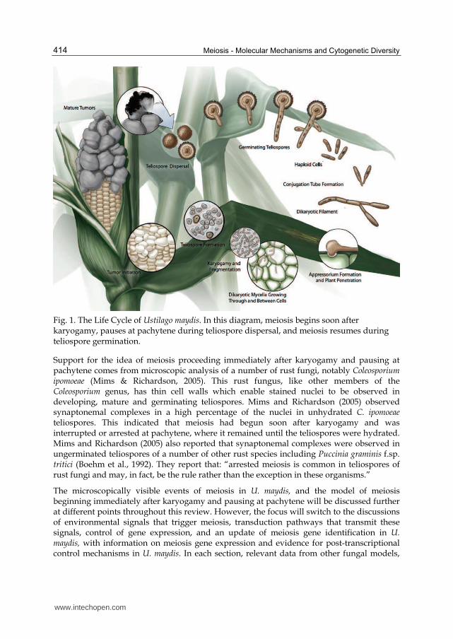
The U. maydis life cycle (Figure 1) begins with teliospore germination and the completion of meiosis to create haploid basidiospores, which divide by budding. Compatible non-pathogenic haploids fuse to form the pathogenic filamentous dikaryon, which proliferates, branches, and penetrates the plant via the formation of specialised cells called appressoria. It grows within and between plant cells eliciting the formation of a tumour. Banuett and Herskowitz (1996) describe a series of developmental events that U. maydis undergoes in the tumour leading to teliospore formation. These events occur within the enlarged host cells and include: 1) the formation of hyphal branches at close intervals, 2) the production of a mucilaginous matrix in which the hyphae are embedded and the hyphal tips become lobed, 3) hyphae fragmentation, 4) rounding of fragmented hyphae and 5) the deposition of a pigmented thick cell wall. The pigmented teliospores enter a dormant state, the tumours disintegrate, and the teliospores are dispersed, continuing the cycle.
www.intechopen.com
Investigating Host Induced Meiosis in a Fungal Plant Pathogen 413
An overview of how meiosis proceeds in U. maydis was presented by Donaldson and Saville (2008). Since the early stages of meiosis occur in planta and meiosis is temporally linked to the formation of thick walled dormant teliospores, direct microscopic observation of meiotic events has not been possible. Therefore, it is informative to review how meiosis precedes in the related homobasidiomycete, Coprinopsis cinerea. This fungus can be induced to form mushrooms (fruiting bodies) in culture and, in these fruiting bodies, meiosis proceeds in a synchronous manner with over 60% of the approximately 10 million basidia in a given cap at the same stage (Pukkila et al., 1984). Kües, (2000) reviewed meiosis in C. cinerea and noted that chromatid duplication in premeiotic S phase is followed by karyogamy, and the cytological events of prophase I precede with the condensation and alignment of chromosomes (leptotene), synapsis (zygotene), and recombination nodule appearance (pachytene). This process, from post karyogamy to pachytene, is completed in six hours (Celerin et al., 2000). It is followed by desynapsis (diplotene) and the transition to metaphase (diakinesis). The second meiotic division occurs fairly rapidly following interphase, with prophase II through telophase II being completed in ~1 hour. The second division occurs in the same plane as the first, across the longitudinal axis of the basidium. Then, chromatid separation is followed by the four nuclei migrating toward the basidium tip where basidiospores form and the nuclei migrate into them then complete a round of mitosis. This overview of basidiomycete meiosis provides a framework for U. maydis investigations.
U. maydis, like other smut fungi, does not form a fruiting body. Instead, when teliospores germinate, basidia are formed in which meiosis is completed. So while a C. cinerea fruiting body has millions of basidia undergoing meiosis, in U. maydis, millions of teliospores are dispersed and each produces a basidium. What we know of the cytological events of meiosis in U. maydis is that when hyphae are enveloped in the mucilaginous matrix during teliospore formation, they contain a single nucleus, indicating karyogamy has occurred (Banuett & Herskowitz, 1996). If U. maydis meiosis follows the pathway of C. cinerea, then premeiotic S phase and the duplication of chromatids would have been completed before karyogamy occurred. The next meiotic event we are aware of in U. maydis is the germination of the teliospore when the nucleus is in late prophase I (O’Donnell & McLaughlin, 1984). Between karyogamy and germination the teliospore is dormant with extremely limited metabolic activity. This indicates that major meiotic events cannot be occurring; this leaves the possibility that, either there is a pause after karyogamy and meiosis continues with teliospore germination, or that prophase I and recombination events begin immediately after karyogamy and pause, perhaps at the pachytene checkpoint, when the teliospore becomes dormant. Following germination, the metaphase I spindles align with the longitudinal axis of the metabasidium, and a transverse septum forms, indicating the completion of telophase I and leading to the formation of two cells (O’Donnell & McLaughlin, 1984). This is rapidly followed by meiosis II, in which the nucleus in each cell migrates to a central location, divides and septa are formed, resulting in three haploid nuclei, each in a basidium cell. The fourth nucleus migrates to the base of the teliospore (Ramberg & McLaughlin, 1980). Basidospores form by budding, each basidium cell nucleus migrates into the respective basidiospore and divides, then one nucleus remains and the other migrates back into the basidium cell (Banuett, 1995). Basidiospores continue to divide by budding.
www.intechopen.com
Meiosis - Molecular Mechanisms and Cytogenetic Diversity 414
Fig. 1. The Life Cycle of Ustilago maydis. In this diagram, meiosis begins soon after karyogamy, pauses at pachytene during teliospore dispersal, and meiosis resumes during teliospore germination.
Support for the idea of meiosis proceeding immediately after karyogamy and pausing at pachytene comes from microscopic analysis of a number of rust fungi, notably Coleosporium ipomoeae (Mims & Richardson, 2005). This rust fungus, like other members of the Coleosporium genus, has thin cell walls which enable stained nuclei to be observed in developing, mature and germinating teliospores. Mims and Richardson (2005) observed synaptonemal complexes in a high percentage of the nuclei in unhydrated C. ipomoeae teliospores. This indicated that meiosis had begun soon after karyogamy and was interrupted or arrested at pachytene, where it remained until the teliospores were hydrated. Mims and Richardson (2005) also reported that synaptonemal complexes were observed in ungerminated teliospores of a number of other rust species including Puccinia graminis f.sp. tritici (Boehm et al., 1992). They report that: “arrested meiosis is common in teliospores of rust fungi and may, in fact, be the rule rather than the exception in these organisms.”
The microscopically visible events of meiosis in U. maydis, and the model of meiosis beginning immediately after karyogamy and pausing at pachytene will be discussed further at different points throughout this review. However, the focus will switch to the discussions of environmental signals that trigger meiosis, transduction pathways that transmit these signals, control of gene expression, and an update of meiosis gene identification in U. maydis, with information on meiosis gene expression and evidence for post-transcriptional control mechanisms in U. maydis. In each section, relevant data from other fungal models,
www.intechopen.com
Investigating Host Induced Meiosis in a Fungal Plant Pathogen 415
notably Saccharomyces cerevisiae, Schizosaccharomyces pombe and C. cinerea, will be reviewed for comparison and insight.
2. Signals triggering meiosis initiation in fungi: Genetic and environmental signals
The switch from mitotic division to meiosis involves a dramatic shift in cellular processes; in fact, this can be considered the most traumatic change a cell can undergo. Therefore, it is essential that entry into meiosis is tightly controlled, preventing the inappropriate execution of the meiotic program. The signals that trigger meiosis vary extensively between organisms, possibly due to a need for organisms to respond to the unique environmental niches in which they reside (Pawlowski et al., 2007). While the signals that trigger meiosis initiation are different, the timing is conserved from yeast to mice, occurring prior to the premeiotic S phase (Pawlowski et al., 2007). While this may seem obvious, the complex developmental processes that precede and accompany meiosis have obscured the timing until recently (Pawlowski et al., 2007). In this section, the nature and timing of signals leading to the initiation of meiosis in the model laboratory fungi S. cerevisiae, S. pombe and C. cinerea are reviewed and used as a reference in presenting hypotheses regarding meiotic initiation signals for the model plant pathogen U. maydis.
2.1 Saccharomyces cerevisiae
Meiosis in the ascomycete fungus S. cerevisiae has been extensively studied. For the purposes of this discussion, we will consider meiosis initiation as the first of three stages; the others being DNA replication, recombination, and the meiotic divisions leading to haploid products (Honigberg, 2003). This separation is interesting because it indicates an order of events different than that of C. cinerea.
In S. cerevisiae, meiosis initiation is triggered by environmental and genetic signals working in concert. To be receptive to the environmental signals, S. cerevisiae cells must be diploid and possess both MATa and MAT┙ mating type alleles (reviewed in Piekarska et al., 2010). MATa and MAT┙ encode components of the transcriptional repressor a1/┙2, (Mitchell, 1994; Piekarska et al., 2010). The environmental signal involves three nutritional shifts: 1) the absence of an essential nutrient, 2) the presence of a non-fermentable carbon source, and 3) the absence of glucose (Honigberg, 2003). The essential nutrient typically eliminated in laboratory studies is nitrogen and, while there may also be a direct requirement for nitrogen sensing, limiting carbon, phosphates or sulphates can also provide the required signal to trigger meiosis initiation (Honigberg, 2003; Mitchell, 1994). The CO2 produced through respiration, stimulated by the presence of a non-fermentable carbon source, results in the alkalization of the media, which may be a component of the 2nd shift (Honigberg, 2003). While respiration is a required signal throughout meiosis, the non-fermentable carbon source is only required prior to meiosis I (Honigberg, 2003; Piekarska et al., 2010). Finally, the presence of glucose can override the other signals and repress meiosis in S. cerevisiae, (Honigberg, 2003; Mitchell, 1994; Piekarska et al., 2010).
While the signal transduction pathways will be discussed later, other key aspects of meiosis initiation in S. cerevisiae are the timing of entry and the link between genetic and nutritional signals. A S. cerevisiae diploid cell commits to mitosis before DNA replication in the S phase.
www.intechopen.com
Meiosis - Molecular Mechanisms and Cytogenetic Diversity 416
It has been proposed that the commitment involves the separation of the spindle pole bodies (SPBs) during the cell cycle. In budding yeast cells where the SPBs are still together, the cells may arrest and enter the meiotic cycle, whereas after the SPBs have separated, the cell can no longer enter the meiotic cell cycle, and must complete mitosis (Simchen, 2009). Starvation for an essential nutrient results in the arrest of the cell in G1, when the SPBs are together (Honigberg, 2003; Piekarska et al., 2010), this specific arrest allows a switch to meiosis. The nutritional and genetic signals also converge to initiate meiosis through the transcriptional regulation of two main inducers of meiosis, Ime1 (initiator of meiosis), which is a transcription factor that stimulates the expression of many early meiosis genes and Ime2, a serine/threonine protein kinase. The expression of Ime1 is controlled by the a1/┙2 repressor and by nutritional signals (Honigberg, 2003; Mitchell, 1994). Multiple nutritional signals converge on Ime2 as well. The full expression of both of these genes is essential to the initiation and continuation of meiosis in S. cerevisiae (Honigberg, 2003; Mitchell, 1994).
2.2 Schizosaccharomyces pombe
Similar to S. cerevisiae, the initiation of meiosis in S. pombe requires diploid cells and nutrient starvation. However, S. pombe also requires pheromone signalling, whereas the S. cerevisiae pheromones are turned off after mating. In S. pombe, mating type is determined by the mat1 locus. Each mating type allele codes two proteins, mat1-P codes mat1-Pc and mat1-Pi, while
mat1-M codes mat1-Mc and mat1-Mi. mat1-Mc and mat1-Pc are essential for mating and meiosis, as they control pheromone and receptor production (Harigaya & Yamamoto, 2007; Yamamoto, 1996a). When compatible haploid cells are nitrogen starved, pheromone signalling is induced, which initiates cellular fusion and the formation of the diploids (Nielsen, 1993). These diploids grow mitotically, under rich nutrient conditions, but under nutrient starvation conditions they arrest in G1 and proceed to meiosis. As in S. cerevisiae,
meiosis can only occur if the cells arrest in G1; beyond this point they are committed to mitosis (Harigaya & Yamamoto, 2007). However, meiosis in S. pombe will not proceed without the pheromone signal. Diploid cells lacking one pheromone receptor can still undergo meiosis but those lacking both cannot (Yamamoto, 1996a, 1996b).
The linkage to environmental signals in S. pombe comes through Ste11, a transcription factor expressed under nutrient starvation (Yamamoto, 1996a, 1996b). Ste11 controls the expression of mat1-Mc and mat1-Pc along with other mating and meiosis genes. Ste11 plays a similar role in S. pombe to Ime1 in S. cerevisiae, as both transcription factors respond to environmental signals and lead to meiotic initiation. However, despite their functional similarities, these two proteins are not structurally similar (Burns et al., 2010a). Mat1-Mc and Mat1-Pi together stimulate the expression of Mei3, an inhibitor of Pat1, a serine/threonine protein kinase that itself inhibits meiotic initiation (Harigaya & Yamamoto, 2007; Willer et al., 1995). All of these signals converge on Mei2, an RNA binding protein which is essential for entry into, and continuation of, meiosis in fission yeast (Harigaya & Yamamoto, 2007).
2.3 Coprinopsis cinerea
C. cinerea is a filamentous, basidiomycete fungus that can be induced to form fruiting bodies (mushrooms) in the laboratory. As noted above, it is a model for the study of meiosis because the millions of basidia, the cells in which meiosis occurs, in a single cap develop
www.intechopen.com
Investigating Host Induced Meiosis in a Fungal Plant Pathogen 417
synchronously. Initiation of meiosis in C. cinerea depends on light cues, not the nutritional cues used by S. cerevisiae and S. pombe. It has been hypothesized that linking fruiting body formation and meiosis to light/dark cycles provides a selective advantage because the fruiting bodies would be produced when animals are grazing, and C. cinerea depends upon animal ingestion for dispersal (Lu, 2000). This hypothesis is consistent with the concept of fungi responding to niche specific signals to initiate meiosis.
C. cinerea is well tuned to changes in lighting. Not only is light essential for karyogamy and the initiation of meiosis (Lu, 2000), but increasing the intensity of light speeds up the process, with less time being required to reach karyogamy under more intense light. It is proposed that the number of photons received is important in stimulating the progression of the cell into the premeiotic S phase and karyogamy (Lu, 2000). This timing is the same as the other fungi; therefore, although the signals are very different, the timing of commitment to meiosis is conserved, with the signal that initiates meiosis coming before the premeiotic S phase.
As may be expected, based on the different environmental triggers for initiation, C. cinerea has no orthologs to either Ime1 or Ste11, the master meiotic regulators from S. cerevisiae and S. pombe, respectively. However, studies have shown that during meiosis, successive waves of transcription occur in C. cinerea, much like waves noted in both yeasts (Burns et al., 2010b). Hence, it may not be unreasonable to assume that there is a heretofore unidentified transcription factor that responds, directly or indirectly, to light signals and initiates this transcriptional program, making it similar in function, if not structure, to the regulators in budding and fission yeast.
2.4 Ustilago maydis
U. maydis is the model biotrophic basidiomycete plant pathogen (Banuett, 1995; Brefort et al., 2009). Like S. cerevisiae and S. pombe, there is a genetic and an environmental requirement for the initiation and completion of meiosis. U. maydis has two mating type loci, the
multiallelic b locus and the diallelic a locus. The b locus codes a pair of homeodomain proteins that act as transcription factors when a heterodimeric protein consisting of polypeptides from different alleles is formed. The a locus codes for the pheromone and pheromone receptors. Alleles at each of these loci must be different in order for haploid cells to mate and for the maintenance of filamentous growth (reviewed in Banuett, 1995, 2002, 2010); however, only b locus heterozygosity is required for completion of meiosis (Banuett & Herskowitz, 1989). The environmental input required for meiosis is growth within the plant, and Banuett and Herskowitz (1996) suggested a peptide produced by the plant may stimulate karyogamy. In light of the earlier discussions here regarding a requirement for a signal to initiate premeiotic S phase and karyogamy, the suggestion by Banuett and Herskowitz (1996) could easily be extrapolated to suggest that the plant peptide stimulates premeiotic S phase and subsequent karyogamy in U. maydis. As reviewed by Banuett (2002), Kahmann and Kämper (2004), and Klosterman et al. (2007), the influence of the mating type loci is modulated by nutrition, pH, temperature, oxygen tension and plant signals, so it is possible that other factors influence meiosis in U. maydis.
To provide context for the possibility that nutritional conditions act as a signal influencing meiosis, Horst et al. (2010a) determined that, upon infection, U. maydis creates a strong nitrogen and carbon sink around the site of infection, and this stimulates the productivity of the remaining source leaves, allowing import of nutrients to the developing tumour tissue.
www.intechopen.com
Meiosis - Molecular Mechanisms and Cytogenetic Diversity 418
It is believed that the imported sucrose is used for building the tumour and for feeding U. maydis, and that the nitrogen may be fuelling both host defense protein synthesis and fungal growth (Horst et al., 2010b). This indicates that nutrition availability is an important aspect of the plant-pathogen interaction and that there is a competition for nutrients between the fungus and the plant. As an important part of its ecological niche, it is conceivable that changes in nutrient availability influence the progression of meiosis in U. maydis.
Genes that are involved in regulating the transition to meiosis have not yet been identified in U. maydis. Bioinformatic comparisons have determined that U. maydis does not possess an ortholog to Ime1, the master regulator of meiosis in S. cerevisiae (Donaldson & Saville, 2008). However, an ortholog to Ste11, the key transcription factor in S. pombe, is present in U.
maydis. This putative ortholog is known as Prf1 in U. maydis and its function has been previously characterized. Interestingly, it is a transcription factor that is involved in the sexual development of U. maydis in response to environmental signals, much like Ste11. Prf1
is involved in regulating a and b mating type gene expression, resulting in high levels of pheromones and receptors during mating, and controlling b gene expression during pathogenesis (Hartmann et al., 1999). Four different environmental signals affect Prf1: the carbon source, pheromones, the b heterodimer and the cAMP pathway. These signals act to control Prf1 transcriptionally and post-transcriptionally (Hartmann et al., 1999). This is similar to the function of Ste11 in S. pombe, which controls pheromone gene expression in response to environmental signals, allowing for conjugation, the initiation of the sexual cycle and the commencement of meiosis. It is feasible that Prf1 is also involved in initiating meiosis in U. maydis, possibly by stimulating the expression of a gene that controls further meiotic gene expression.
3. Signal transduction pathways and meiotic progression
The requirement for genetic and environmental signals to stimulate the entrance into meiosis implies there must be a way to transduce the environmental signals and integrate them with the genetic status of the cells. In this section we provide an overview of the signal transduction in S. cerevisiae and S. pombe and then, with this background, we link what is known about pathogenic signal transduction in U. maydis to its potential role in meiosis. Since research on these organisms has historically emphasized different levels of the signal transduction pathways, the focus in each section varies. In S. cerevisiae, major regulators are known and the focus has been on transcriptional control, and as such the overview will focus on signalling as it influences transcription. In S. Pombe, the major regulator is also known, but the emphasis has not been as strongly focused on transcription so this section is somewhat more pathway oriented. In U. maydis, the master regulators are not known so the knowledge of signal transduction in pathogenesis is reviewed and a model is presented for how this may stimulate the initiation of meiosis.
3.1 Saccharomyces cerevisiae
The initiation and continuation of meiosis are linked to environmental cues through multiple signal transduction pathways in Saccharomyces cerevisiae. The master controller of meiosis is the gene Ime1. The influences on this gene primarily result in changes in its transcription, which is controlled by the genetic and environmental signals. Ime1 has an
www.intechopen.com
Investigating Host Induced Meiosis in a Fungal Plant Pathogen 419
unusually large promoter region of 2,100bp, which is divided into 4 different Upstream Controlling Sequences, UCS1-4. These UCSs respond to different signals. Nutritional signals affect UCS1 and 2, with UCS2 promoting the transcription of Ime1 and UCS1 inhibiting it. Cell-type signals affect UCS3 and 4, repressing the expression of Ime1 in MAT-insufficient cells (Sagee et al., 1998). This section will focus on how these different signals are relayed to influence the transcription of this master controller.
3.1.1 Genetic control
The cell-type signals that control Ime1 are transmitted through a repressor, Rme1, and an activator, Ime4. In haploid S. cerevisiae, the RME1 protein inhibits meiosis by repressing the expression of Ime1. It does so by binding to the Rme1 Repressor Element (RRE), within UCS4 of the Ime1 promoter (Covitz & Mitchell, 1993; Sagee et al., 1998). In diploid MATa/MAT┙ cells, the proteins a1, a MATa product, and ┙2, a MAT┙ product, form the a1/┙2 heterodimer. This heterodimer binds upstream of Rme1 and directly represses its transcription (Covitz et al., 1991). Repression of Rme1 results in the de-repression of Ime1, as RME1 is no longer available to bind to the RRE, and in this way Rme1 transmits the cell-type signal directly to Ime1. IME4 expression is necessary for the full expression of Ime1 (Shah & Clancy, 1992). The a1/┙2 heterodimer regulates Ime1 expression by repressing the transcription of Ime4 antisense and allowing the transcription of Ime4 sense transcript (discussed further in section 6.4). The MATa/MAT┙ cell-type signal also regulates Ime1 expression through the UCS3 repressor region, however the protein involved has not been identified (Sagee et al., 1998). The status of the diploid cell is determined by the mating type loci, and the outlined transcriptional control pathways ensure that Ime1 is only expressed, and meiosis can only proceed, in diploid MATa/MAT┙ cells.
3.1.2 Nutritional control
The nutritional signals that control meiotic initiation in S. cerevisiae are transmitted through a signalling network composed of the RAS, cAMP and TOR pathways, all of which regulate the expression of transcription factor Ime1 and kinase Ime2 (reviewed in Piekarska et al., 2010).
While nitrogen limitation is often described as a requirement for meiosis in S. cerevisiae, starvation of any essential nutrient can stimulate meiosis. In each case, nutrient starvation may not have a direct effect; rather, it may act indirectly since nutrient limitation results in G1 arrest, and G1 arrest is required for meiosis initiation (Honigberg & Purnapatre, 2003). The response to nitrogen starvation is mediated, in part, by the TOR pathway. This pathway controls the expression of metabolism genes and, as such, its role in meiosis is also proposed to be indirect, because Tor2 causes changes in metabolism that result in G1 arrest (Honigberg & Purnapatre, 2003). G1 arrest is essential to meiosis initiation, as we have discussed. CLN3 is a G1 cyclin that is part of the mitotic G1 to S phase transition. In nitrogen deprived cells, CLN3 is strongly down-regulated (Gallego et al., 1997). G1 cyclins, like CLN1, 2 and 3 down-regulate Ime1 in cells grown in nutrient rich medium. This, in turn, represses the initiation of meiosis until cells are starved (Colomina et al., 1999). Starvation triggers a reduction in G1 cyclin levels resulting in the cells arresting at G1 (Colomina et al., 1999; Gallego et al., 1997). Lowered cyclin levels allow IME1 to be transferred to the nucleus, where it initiates meiosis by stimulating transcription of early meiotic genes (Zaman et al.,
www.intechopen.com
Meiosis - Molecular Mechanisms and Cytogenetic Diversity 420
2008). Apart from the indirect effects of nitrogen limitation, Ime1 transcription may be directly influenced by nitrogen limitation, since deletion of the UCS1 upstream controlling sequence allows meiosis in the presence of nitrogen (Kassir et al., 2003). This nitrogen signal is transmitted through Cdc25, a positive regulator of the cAMP/PKA and MAPK pathways. The pathway involved in the transduction of Cdc25’s effect on Ime1 is currently unknown (Kassir et al., 2003).
Carbon source is another essential element in the regulation of Ime1 activity, both in repressing its function under non-favourable conditions and in activating its function under favourable conditions. Carbon source signals act on UCS2, the only controlling region that possess upstream activating sequences (UAS). In the presence of glucose, Ime1 transcription is repressed at UCS2, preventing Ime1 expression. UCS1 is also a target for repressing Ime1
when glucose is present (Kassir et al., 2003). However, UCS2’s promoter activity is stimulated in the presence of a non-fermentable carbon source (Sagee et al., 1998; Kassir et al., 2003). So when glucose is absent, Ime1 expression is stimulated, but it is opposed by the constitutive repressor elements of USC2 and UCS1, unless nitrogen is also limited, which results in a high level of Ime1 expression, inducing meiosis (Govin & Berger, 2009; Kassir et al., 2003).
The cAMP/PKA pathway is known to transmit the glucose signal to Ime1 in many ways. Glucose is sensed by the G coupled receptor, GPR1, which activates GPA2, a component of a transmembrane heterotrimeric G protein that activates PKA through adenylyl cyclase (reviewed in Honigberg & Purnapatre, 2003). Adenylyl cyclase activity increases the level of cAMP in the cell, and increased cAMP leads to repression of Ime1 (Kassir et al., 2003). Repression is mediated through transcription factor MSN2 which stimulates Ime1
transcription, but with increased cAMP levels it is not transmitted to the nucleus, preventing Ime1 activation (Kassir et al., 2003). SOK2 is another DNA binding protein that mediates the response of Ime1 to glucose through the cAMP/PKA pathway. SOK2 functions as a repressor by associating with MSN2. When glucose is not present, SOK2 is converted to an activator (Shenhar & Kassir, 2001). As a further control, Sok2 expression is dependent on glucose, when cells are growing in a non-fermentable carbon source, SOK2 levels drop dramatically, alleviating its repression of Ime1 (Shenhar & Kassir, 2001). RIM15 is a serine/threonine protein kinase that is inactivated by PKA phosphorylation when cells are growing in glucose rich media and is increased in acetate media. RIM15 promotes the disassembly of the Ume6 repressor complex, contributing to the activation of Ime1 (Zaman et al., 2008). Intracellular acidification of yeast cells also plays into the cAMP pathway. Lowered pH inside the cell stimulates Ras2, which stimulates cAMP synthesis (Thevelein & De Winde, 1999). Outside of the cAMP/PKA pathway, glucose sensing also affects Ime1 through the Snf1 signal transduction pathway. Glucose inhibits the SNF1 protein kinase, which is necessary for full expression of Ime1. However, this is not the only use for SNF1; it also plays a role in Ime2 regulation and spore formation (Honigberg & Lee, 1998).
It is clear that the regulation of Ime1 integrates multiple factors to control meiotic initiation in response to environmental cues. However, Ime1 is not the only target of nutritional regulation; Ime2 is a meiosis specific protein kinase that is the second major regulator of meiosis in S. cerevisiae. It affects multiple stages of meiotic progression, and its transcription and activity are controlled by nitrogen and carbon source signals. Ime2 activity is inhibited
www.intechopen.com
Investigating Host Induced Meiosis in a Fungal Plant Pathogen 421
by glucose through GPA2, the ┙ subunit of the heterotrimeric G-protein (a component of the cAMP/PKA pathway). When active GPA2 interacts with the C terminus of IME2, this interaction represses the activity of IME2, which in turn inhibits entry into meiosis (Donzeau & Bandlow, 1999). Glucose also modifies the protein stability of IME2 through the glucose sensors SNF3 and RGT2 (Rubin-Bejerano et al., 1996). UME6 binds to the Ime2 promoter, repressing transcription in the presence of glucose and nitrogen. During vegetative growth, Ime2 expression is repressed, like many early meiosis genes, by the UME6-SIN3-RPD3 complex. Under meiotic conditions, UME6 disassociates from SIN3 and RPD3, forming a complex with IME1, which activates the transcription of Ime2 (Honigberg & Purnapatre, 2003; Purnapatre et al., 2005). The stabilization of this UME6-IME1 complex requires starvation for both nitrogen and glucose. The stabilization is mediated through phosphorylation by RIM11, a glycogen synthase kinase (Chung et al., 2001; Purnapatre et al., 2005) and RIM15, a protein kinase. The expression of RIM15 is repressed when glucose is present in the media and the activity of RIM15 and RIM11 are repressed through the cAMP/PKA pathway, which destabilizes the UME6-IME1 complex (Honigberg & Purnapatre, 2003; Piekarska et al., 2010; Xiao & Mitchell, 2000). Finally, media alkalization effects the expression of Ime2 through the activation of the UME3-UME5 complex, which has been shown to be required for the full expression of Ime2 (Cooper & Strich, 2002; Honigberg & Purnapatre, 2003). Thus it is clear that carbon and nitrogen nutritional signals converge on both Ime1 and Ime2 in order to control the initiation of meiosis.
3.2 Schizosaccharomyces pombe
In S. pombe, the master controller of meiosis is Ste11, a transcription factor that stimulates both mating and meiosis. It triggers the expression of both mating type loci and Mei2, another key meiosis control gene (Sugimoto et al., 1991). Regulation of both Ste11 and Mei2 integrates cell type and environmental signals that lead to initiation of meiosis in S. pombe. This section will focus on how these signals are conveyed to the regulators of meiosis through signal transduction pathways, and how these pathways are required for the initiation of meiosis.
3.2.1 Genetic control
A requirement for meiosis in S. pombe is that cells are diploid and contain mating type loci mat1-P which codes mat1-Pc and mat1-Pi, as well as mat1-M, which codes mat1-Mc and mat1-Mi. The genes mat1-Mc and mat1-Pc stimulate pheromone signalling and are essential for both mating and meiosis (Willer et al., 1995). The expression of these two genes requires STE11 (Yamamoto, 1996a) and Ste11 is only expressed under nutrient starvation conditions (see next prargraph). The pheromones produced bind to their respective receptors (Yamamoto, 1996b). A G protein ┙ subunit, GPA1, is coupled to the pheromone receptors, transmitting the signal downstream (Obara et al., 1991). This activates a MAP kinase cascade including: MAPKKK BYR2, MAPKK BYR1 and MAPK SPK1 (reviewed in Yamamoto, 1996b). Signals received at SPK1 are transmitted to stimulate expression of mat1-Pi and mat1-Mi. These gene products then allow for the initiation of meiosis by stimulating the expression of Mei3 (Willer et al., 1995; Yamamoto, 1996b). Another GTP binding protein, RAS1, helps to regulate the MAPK cascade through activating BYR2. RAS1 binds to BYR2 and controls its translocation to the plasma
www.intechopen.com
Meiosis - Molecular Mechanisms and Cytogenetic Diversity 422
membrane (Bauman et al., 1998). Interestingly, a Ras homolog in S. cerevisiae, Ras1, is also involved in meiotic initiation, but by repressing it through the cAMP/PKA pathway (Honigberg & Purnapatre, 2003). This is another example of how similar signals are utilized in different ways by divergent organisms. The requirement for starvation to stimulate Ste11, which stimulates expression at the mating type loci, provides a link between environmental signals and genetic status of the cells.
3.2.2 Nutritional control
Sexual development in S. pombe requires nutrient starvation; with nitrogen starvation, in particular, playing an essential role. Starvation initiates mating, which is typically immediately followed by meiosis. The nutritional signals are linked to meiosis through the cAMP/PKA pathway. In S. pombe, similar to S. cerevisiae, increased levels of intracellular cAMP inhibit meiosis progress, while lower levels lead to its initiation (reviewed in Yamamoto, 1996a). When cells are growing in nutrient rich media, cAMP levels are high, but when they are transferred to nitrogen-free media, the cAMP levels decrease by approximately 50% before meiosis occurs. The cAMP then increases to a level greater than or equal to those in nutrient rich media during sporulation (the last stage of meiosis). When the cAMP level is artificially elevated in the cells, it results in sterility (Mochizuki & Yamamoto, 1992). Carbon starvation also results in a decrease in intracellular cAMP levels and can contribute to the initiation of meiosis (Isshiki et al., 1992). The cAMP levels in S.
pombe are controlled by GPA2, the ortholog of GPA2 in Saccharomyces cerevisiae, a heterotrimeric G protein which controls the activity of adenylate cyclase. Gpa2 null mutants had low levels of intracellular cAMP and were able to mate and sporulate, even in rich media (Honigberg & Purnapatre, 2003; Isshiki et al., 1992). GPA2 is necessary for the cell to be able to increase cAMP levels upon glucose stimulation, indicating that it is directly involved in sensing carbon starvation. The ability of the GPA2 mutant to sporulate, even on nitrogen rich media, may also indicate its involvement in nitrogen sensing (Isshiki et al., 1992). Changes in cAMP levels alter the activity of Protein Kinase A (PKA). PKA controls the expression of the major meiosis control gene Ste11 through its impact on RST2 (Kunitomo et al., 2000). RST2 binds to an upstream cis-element, inducing Ste11 expression. Phosphorylation of RST2 by PKA suppresses its ability to induce transcription of Ste11. PKA activity also controls the nuclear localization of RST2, where high levels of PKA result in RST2 being mostly located in the cytoplasm, while low levels result in it being found in the nucleus (Higuchi et al., 2002). When nutritional starvation results in the decrease in cAMP, and thus PKA activity, this results in the activation of RST2, which in turn stimulates Ste11 expression, leading to meiotic gene expression. In addition to control over Ste11, the cAMP signalling pathway also acts to control Mei2, another crucial regulator of meiosis initiation. As with Ste11, increased cAMP levels inhibit the expression of Mei2 (Y. Watanabe et al., 1988). MEI3 inactivates PAT1, which inhibits both MEI2 and STE11 through phosphorylation. When MEI3 is expressed, its inactivation of PAT1 results in the accumulation of unphosphorylated and active Mei2. The active MEI2 stimulates the continuation of meiosis. In fact, the expression of Mei3 can bypass both nutritional and genetic requirements and result in ectopic meiosis (Peng et al., 2003). MEI3, is a substrate of PKA; however, decreased phosphorylation does not affect MEI3’s ability in inactivate PAT1 (Peng et al., 2003). Regardless of this remaining uncertainty, this information clearly
www.intechopen.com
Investigating Host Induced Meiosis in a Fungal Plant Pathogen 423
indicates that the cAMP pathway transmits the nutritional starvation signal and influences the initiation of meiosis on multiple levels in S. pombe.
The cAMP/PKA pathway is not the only signal transduction pathway for nutritional sensing. The TOR pathway transmits signals involved in nitrogen source availability. Genes induced by TOR2, a component of the TORC1 complex, include those induced by nitrogen starvation. Tor2 is a negative regulator of meiosis, with Tor2 inhibition increasing meiosis (Matsuo et al., 2007). TOR2 forms a complex with, and inhibits the function of both Ste11 and Mei2, leading to the repression of meiosis (Álvarez & Moreno, 2006). The TOR pathway interacts with the PKA pathway; both are used as a means to drive cell growth and inhibit sporulation. They also work together to regulate Ste11 expression and localization within the cell. STE11 is located throughout the cell, but under meiosis conditions, it builds up in the nucleus. PKA appears to have a controlling role in nuclear localization, when PKA is absent, STE11 localizes to the nucleus, even in the presence of TOR2; the absence of TOR2 also results in nuclear localization of STE11 (Valbuena & Moreno, 2010). Cells with constitutively active Tor2 are impaired in mating, but they regain functional mating when PKA is deactivated, suggesting that PKA is a more potent regulator. When PKA is at a high level in the cell, RST2 represses Ste11 transcription, and mating and meiosis are inhibited (Valbuena & Moreno, 2010). This indicates that the cAMP/PKA pathway interacts with the TOR pathway to control expression and localization of STE11. This, in turn, controls the initiation of meiosis.
There is one additional pathway that transmits nutrient starvation signals to Ste11, the stress response pathway (SRP). Stress includes starvation, the typical trigger for meiosis initiation in S. pombe. The SRP includes the MAPKK WIS1 and MAPK STY1 that play a role in meiosis initiation and stress response in the cell (Kato et al., 1996; Shiozaki & Russell, 1996; Wilkinson et al., 1996). STY1 phosphorylates and modifies the activity of the transcription factor, ATF1 (Shiozaki & Russell, 1996; Wilkinson et al., 1996). ATF1 is necessary for the expression of Ste11 during nutrient starvation (Takeda et al., 1995). Therefore, nutrient starvation signals are also transmitted through the stress response pathway to control Ste11
expression and meiosis in S. pombe. This is notably different from what occurs in S. cerevisiae, where the closest homolog to STY1 is HOG1, which in budding yeast responds only to osmotic stress, not stress in general (Wilkinson et al., 1996).
3.3 Ustilago maydis
In U. maydis research, the focus has been on signals leading to pathogenesis. A look at the life cycle of this fungus (Figure 1) illustrates how closely pathogenesis is tied to the events of sexual reproduction. There are differences given U. maydis is a basidiomycete, for example, when compatible haploid cells fuse they form a filamentous dikaryon and not a diploid. This dikaryon is the pathogenic form and persists for some time before karyogamy is stimulated and meiosis ensues. Recall this is also the situation in the model basidiomycete mushroom, C. cinerea. However, like the yeasts, the proteins coded at the U. maydis mating type loci interact with the output of signal transduction pathways to influence continued development toward meiosis. In order to integrate signals from the mating type loci with environmental signals it is reasonable to expect that, in U. maydis, these signals converge on a given gene or gene(s). These genes have not yet been identified. In the following
www.intechopen.com
Meiosis - Molecular Mechanisms and Cytogenetic Diversity 424
discussion, the signal transduction pathways, as they are currently understood, will be outlined. There are interesting similarities to signalling pathways in the yeasts and this enables hypotheses to be generated regarding the signalling leading to meiosis in U. maydis.
3.3.1 Host/environmental control
Mating, morphogenesis and pathogenicity depend on the cAMP/PKA and MAPK pathways in U. maydis. While each pathway transmits signals independently, there is crosstalk between them. The cAMP pathway, as it has been elucidated thus far, begins with a heterotrimeric G protein for which the ┙ subunit and the ┚ subunit have been identified. The ┙ submit is GPA3, the only one of four ┙ subunits coded by U. maydis that influences the cAMP pathway. GPA3 mutants are sterile and unable to respond to pheromone signalling, thus unable to mate (Regenfelder et al., 1997). GPA3 associates with the ┚ subunit BPP1 and together they convey the signal to adenylate cyclase, UAC1, the next component of the pathway (Muller et al., 2004). Adenylate cyclase produces cAMP, which activates protein kinase A (PKA) by causing its regulatory subunit, UBC1, to dissociate from the catalytic subunit, ADR1 (Feldbrugge et al., 2004; Gold et al., 1997). cAMP signalling in U. maydis has several roles. Its influences: 1) alter the expression of a and b mating type genes in response to pheromone signalling (Kaffarnik et al., 2003), 2) direct the switch from budding to filamentous growth (Lee et al., 2003), and 3) control pathogenic development (Gold et al., 1997). The influence on filamentous growth is linked to cAMP levels and thus PKA activity. Lower levels of cAMP or altered PKA activity, such as is the case with a defective ADR1 subunit, results in constitutive filamentous growth, while high cAMP/PKA levels result in a budding phenotype (Lee et al., 2003). The influence of the cAMP pathway on pathogenic development was determined through mutation of Ubc1 (the PKA regulatory subunit), which resulted in high PKA activity. U. maydis strains with these mutations were able to colonize the plant, but were unable to form tumours or teliospores. Uac1 mutants, with low PKA activity, are non-pathogenic (Gold et al., 1997). This suggests that tight control of PKA is required for proper progression of pathogenesis, with low PKA being required for filamentous growth, followed by increased PKA activity needed for infection of the plant, and then lowered PKA once again for tumour and teliospore formation (Gold et al., 1997). Consistent with the requirement for tight control, U. maydis strains carrying a constitutively active Gpa3 can infect corn, leading to tumour formation but not teliospore development (Krüger et al., 2000). It was suggested that the difference between the Uac1 and the Gpa3 mutant phenotype is due to different levels of PKA activity in the two mutants, with the Gpa3 mutant likely representing a less active version with a less defective pathogenic cycle (Krüger et al., 2000). Thus, carefully regulated levels of cAMP appear to be required throughout sexual development, and pathogenesis.
In addition to cAMP signalling, mating and pathogenesis are also regulated by a MAPK signalling cascade. The pathway consists of MAPKKK Ubc4/Kpp4, MAPKK Ubc5/Fuz7 and MAPK Ubc3/Kpp2 and it may respond to signals transmitted through Ras2, a U. maydis homolog of the S. pombe Ras1 (Muller et al., 2003). Evidence supports the pheromone signal being transmitted through a single MAPK pathway, which is also similar to S. pombe (Muller et al., 2003). In a parallel pathway to pheromone response, the MAPK cascade is necessary for appressorium formation and function, as well as filamentous growth in the plant. While Kpp2 is required for appressorium formation, a second MAPK, Kpp6, is
www.intechopen.com
Investigating Host Induced Meiosis in a Fungal Plant Pathogen 425
involved in plant penetration (Muller et al., 2003). Unlike S. pombe, no known G-protein ┙ subunit plays a role in the U. maydis MAPK cascade; however, a plant signal likely influences this pathway through some means, since the maintenance of U. maydis filamentous growth requires the host plant. One possibility is through the link with another pathway. This is suggested because disruption of the MAPK pathway resulted in repression of the constitutive filamentous growth phenotype that is caused by Adr1 mutation (Muller et al., 2003), recall ADR1 is the catalytic subunit of PKA which is activated by cAMP.
Exploration of the links between the cAMP/PKA and MAPK pathways revealed crosstalk through proteins that are putative orthologs to two major meiotic regulators discussed above. The first is CRK1, which is an IME2 related protein kinase. Ime2 is a key meiotic regulator and target of environmental signals in S. cerevisiae. The second is Prf1, a putative ortholog of Ste11, a key regulator of meiosis in S. pombe. Crk1 is a target of environmental stimuli in U. maydis: Crk1 mutants are impaired in their response to environmental signals, and Crk1 is highly expressed when cells are grown in nutrition stress conditions (Garrido & Pérez-Martín, 2003). Crk1 also plays a role in mating and pathogenesis since Crk1 mutants are unable to mate on plates and have attenuated pathogenesis producing few tumours and no observed black teliospores (Garrido et al., 2004). Crk1 is also involved in cell morphogenesis. When Crk1 is overexpressed, it causes filamentous growth. When Crk1 is inactivated, it suppresses the constitutive filamentous growth that results from Adr1 and Gpa3 mutants. This indicates that it acts downstream of these cAMP pathway genes, however high levels of Crk1 cannot repress the budding phenotype of a Ubc1 mutant, indicating it cannot override all cAMP mediated responses (Garrido & Pérez-Martín, 2003). The expression of Crk1 is regulated by both the cAMP and MAPK pathways, which have antagonistic effects on its transcription. Crk1 is transcriptionally repressed by the cAMP pathway, with high PKA levels resulting in a low level of Crk1 expression and vice versa (Garrido & Pérez-Martín, 2003). The MAPK pathway, conversely, positively regulates Crk1 expression, with Kpp2 (a MAPK) mutants resulting in much lower levels of Crk1 in the cell (Garrido & Pérez-Martín, 2003). KPP2 also interacts physically with CRK1, and is required for the role of CRK1 in cell morphogenesis (Garrido et al., 2004). In addition, Fuz7 (a MAPKK) is required for activation of Crk1. FUZ7 phosphorylates CRK1, activating it (Garrido et al., 2004). Thus Crk1 is clearly involved in the integration of the cAMP and MAPK signalling pathways. However, many of the phenotypes of Crk1 mutants appear to result from an effect on Prf1, since Crk1 controls the transcription of Prf1.
PRF1 is an HMG protein that controls the expression of mating type genes, which regulate mating, pathogenesis and cell morphology. PRF1 binds to the pheromone response element, or PRE, upstream in the a and b loci, stimulating their expression (Hartmann et al., 1996). Therefore, a Prf1 mutant strain is unable to mate because it is unable to produce or respond to pheromones. The receptor and pheromone are coded by the a mating type locus. Through its control of the b mating–type locus Prf1 influences filamentous growth and pathogenesis. A solopathogenic Prf1 mutant is unable to cause tumours when it infects the plant (Hartmann et al., 1996). It is possible that Prf1 acts as a mediator for response to plant signals during pathogenic growth. PRF1 is controlled through the cAMP/PKA and MAPK pathways, facilitating control by pheromones and environmental signals. PRF1 has phosphorylation sites for both MAPK and PKA, and mutations in either of these sites impede mating. This indicates that both MAPK and PKA phosphorylation
www.intechopen.com
Meiosis - Molecular Mechanisms and Cytogenetic Diversity 426
are required for proper function of PRF1 in sexual development (Kaffarnik et al., 2003). Interestingly, these phosphorylation sites also determine which genes are activated by PRF1, with MAPK phosphorylation being necessary for b gene expression, but not for a, while PKA phosphorylation is required for both (Kaffarnik et al., 2003). The MAPK pathway may also have a role in inducing the transcription of Prf1, as a constitutively active Fuz7 can increase Prf1 levels (Kaffarnik et al., 2003). Beyond these post-translational controls, Prf1 has a cis-regulatory element in its promoter that is termed the UAS, an upstream activator sequence. The Prf1 UAS appears to regulate transcription of Prf1 in response to cAMP and carbon source signals. Glucose or sucrose stimulates Prf1 transcription via the UAS (Hartmann et al., 1999). High cAMP levels repress Prf1 transcription though the UAS. It is important to note that this is a separate mechanism from the post-transcriptional activation of Prf1 by PKA, and this seemingly contradictory activity of the cAMP pathway results in increase in a gene expression at moderate cAMP levels, but repression through transcriptional control at higher levels (Hartmann et al., 1999). This emphasizes the fine scale control imparted by cAMP levels. The cAMP pathway could also mediate the carbon source signal, or it could be mediated by a separate pathway; this is not yet elucidated (Hartmann et al., 1999).
The link between PRF1 and CRK1 is that CRK1 is required for transcriptional activation of Prf1 through the UAS (Garrido et al., 2004). This provides another avenue for Prf1 control by MAPK and cAMP. Kaffarnik et al. (2003) theorized that the cAMP and MAPK paths may be required to control mating because mating typically occurs on the plant, and if sensing the plant results in increased cAMP levels, this would be sufficient to increase a gene expression, increasing pheromone expression and making mate detection easier, then pheromone signalling would feed back into the cAMP and MAPK pathways. The MAPK pathway would then initiate conjugation tube formation and mating (Muller et al., 2003), and the cAMP and MAPK pathways would increase the transcription and the activity of Prf1, triggering b gene expression, and pathogenesis. Thus it is clear that the integration between the two signalling pathways provides a mechanism whereby a plant signal received before penetration could lead to the subsequent events of pathogenesis; however, what triggers meiosis?
The discovery that a decrease in cAMP level is required for the completion of teliospore development suggests that the fungus must lower cAMP levels during pathogenesis to allow teliospores to form. This could be in response to a signal received from the plant. Interestingly, as we discussed above, a decrease in cAMP/PKA levels is necessary to stimulate meiosis in both S. cerevisiae and S. pombe. Since, in U. maydis, meiosis initiation begins around the time of teliospore formation, it is compelling to link the arrest of teliospore development, resulting from elevated cAMP levels, to meiosis. This mutation-stimulated arrest occurs sometime between when the hyphae form lobed tips, and when they fragment and begin rounding and swelling (Krüger et al., 2000). Interestingly, this is very shortly after the time that karyogamy occurs during normal pathogenic development, recall karyogamy occurs before hyphal fragmentation, but after the cells are imbedded in the mucilaginous matrix (Banuett & Herskowitz, 1996). These findings can be integrated in a model where the U. maydis dikaryon infects the plant and grows within and between cells, stimulating the initiation of tumour formation, and then it receives a signal from the plant which leads to the fungal cells entering premeiotic S phase and karyogamy, concomitant
www.intechopen.com
Investigating Host Induced Meiosis in a Fungal Plant Pathogen 427
with the reduction of cAMP, allowing teliospore formation to proceed. The signals for karyogamy and teliospore formation must at least be interrelated, as these two processes need to proceed simultaneously to avoid crucial disruptions in both developmental pathways. As meiosis proceeds, the teliospore develops such that when it enters a dormant state, meiosis arrests at pachytene. This could be the result of reaching the end of a developmental cascade initiated by the plant signals, or a response to another plant signal.
4. Control of meiotic gene expression
The signals received from the environment are transduced through the pathways noted above and result in cascades of transcription that guide meiosis. These waves of transcription have been well studied in S. cerevisiae and S. pombe. In this section, knowledge of these transcriptional cascades is reviewed and compared. The existing data regarding transcription during meiosis in U. maydis is then presented and compared to that of the yeasts.
4.1 Saccharomyces cerevisiae
In S. cerevisiae, the stages of meiosis have been defined by the waves of genes expressed in a transcriptional cascade. Typically these genes are classified as early, middle and late, depending upon their time of expression during meiosis. Some researchers have found it necessary to further subdivide expression, and, as such, genes may be referred to as belonging to an intermediate expression time; for example, mid-late genes are expressed before late genes, but after the typical middle gene expression (Chu et al., 1998; Mitchell, 1994). In this section we provide an overview of the transcriptional waves, with information on the control of transcription and the relationship of expression to meiotic progression.
4.1.1 Initiation of meiosis
The master regulator of meiosis in S. cerevisiae is IME1, a transcription factor that initiates the transcriptional cascade. It is the point of integration of environmental signals and directly controls the expression of early meiosis genes. Under meiotic conditions, IME1 interacts with UME6. UME6 was first identified as a repressor of meiotic genes under vegetative growth conditions. It binds at the upstream repression sequence 1 (URS1) found in target genes (Mitchell, 1994). However, during meiotic growth, UME6 forms a complex with IME1, and this complex activates early meiotic genes, often through the URS1 (Chu et al., 1998; Mitchell, 1994; Rubin-Bejerano et al., 1996). URS1 is a weak upstream activator sequence, and as such, the signal to initiate transcription is often augmented by binding of activator ABF1 at a distinct recognition sequence (Vershon & Pierce, 2000). Based on their expression patterns, early genes have been subdivided into three groups: early (I) induction, early (II) induction and early-middle induction (Chu et al., 1998). These early genes are involved in controlling DNA replication and the events of prophase I: chromosome pairing, homologous recombination and spindle pole body movements (Chu et al., 1998; reviewed in Piekarska et al., 2010). Interestingly, though the early-middle phase genes grouped with other early genes, most lack the URS1, indicating that they are unlikely to be controlled by IME1/UME6. Instead, about half of these genes possess the MSE (middle sporulation element) indicating expression is controlled by NDT80 (Chu et al., 1998). Ime2 is a key early
www.intechopen.com
Meiosis - Molecular Mechanisms and Cytogenetic Diversity 428
gene whose transcription is initiated by IME1. IME2 acts through a second pathway that does not directly involve IME1. So there is an IME1 dependant pathway that does not involve IME2 and an IME2 dependant pathway (Mitchell et al., 1990; Mitchell, 1994). IME2 is a cdk-like protein kinase, which plays a key role in transitions in meiosis. It acts to amplify transcription of meiosis genes including itself, activates NDT80 to trigger middle meiosis gene expression and stabilizes Clb cyclins through its inhibition of the APC/C (anaphase promoting complex/cyclosome), which in turn controls chromosome segregation (Marston & Amon, 2004; reviewed in Piekarska et al., 2010). IME2 also targets SIC1, resulting in its degradation, triggering the initiation of the S phase and premeiotic DNA replication (Piekarska et al., 2010). Additionally, IME2 phosphorylates IME1, which then signals it for destruction by the proteasome (Guttmann-Raviv et al., 2002). In this way, IME1 initiates meiosis and IME2 reinforces this and enables it to proceed to the next stage of meiosis, middle gene expression.
4.1.2 Commitment and continuation
Fully active NDT80 is necessary for the full expression of middle meiosis genes and thus for the continuation of meiosis in S. cerevisiae. These middle meiosis genes are required for meiotic divisions, and include genes such as B-type cyclins and those involved in spore morphogenesis (Chu & Herskowitz, 1998). NDT80 binds to the conserved MSE element, found upstream of 70% of middle meiosis genes (Chu et al., 1998; Chu & Herskowitz, 1998). NDT80 competes for some of the MSEs with another transcription factor, SUM1, which acts as a repressor of middle meiosis genes during vegetative growth and early meiosis. NDT80 and SUM1 bind to overlapping, yet different, sequences within the MSE, resulting in MSEs that function as Sum1 repressors, Ndt80 activators, or both simultaneously (Pierce et al., 2003). A combination of upstream elements also controls the expression of Ndt80. While Ndt80 is a middle meiosis gene, it is expressed slightly before the rest of the middle meiosis genes. This expression pattern, termed pre-middle, results from two URS1s and two MSEs, located upstream of Ndt80 (Pak & Segall, 2002a). During vegetative growth, expression of Ndt80 is repressed by both UME6 and SUM1 acting on URS1 and MSE respectively. After the initiation of meiosis, the UME6 repressor complex is replaced with UME6-IME1, but expression is still repressed by the MSE (Pak and Segall, 2002a). IME2 and CDK1 phosphorylate SUM1, leading to its release from the MSE and relieving its repression of Ndt80 (Ahmed et al., 2009; Pak & Segall, 2002a; Shin et al., 2010). This allows for low level Ndt80 expression, stimulated by IME1 at URS1. The expressed NDT80 then binds to the MSE in its own promoter region, stimulating expression, leading to full middle gene expression and progression into the first meiotic divisions. However, both Sum1 and Ndt80 expression are also controlled by the pachytene checkpoint, which can prevent the expression of middle meiotic genes. Middle gene expression is essential for the cell to exit from the pachytene checkpoint and enter into meiotic divisions. The pachytene checkpoint, or meiotic recombination checkpoint, is part of a surveillance system in eukaryotic cells that arrests the cell cycle in response to defects. To ensure the integrity of the events of meiosis, this checkpoint prevents the cell from exiting the pachytene stage of prophase I and entering into meiotic divisions before the completion of recombination (Roeder & Bailis, 2000). Cells arrested at pachytene are not yet committed to meiosis, meaning they can revert to mitotic growth if conditions are adjusted. This is the last point at which the cell can return to mitotic growth, as after the transition to the first meiotic divisions, the cell is committed to meiosis
www.intechopen.com
Investigating Host Induced Meiosis in a Fungal Plant Pathogen 429
(Shuster & Byers, 1989). This makes the pachytene checkpoint the “point of no return” for the cell, allowing one last chance for the cell to arrest and abort meiotic progression, and revert to mitotic growth. In Dmc1 mutants, DSB repair is impaired, in Zip1 mutants SC formation is impaired and in Hop2 mutants, synapsis is defective. Each of these mutants trigger pachytene arrest (Roeder & Bailis, 2000). Checkpoint arrest is mediated through proteins that monitor synapsis and recombination and exert their effects on downstream targets of checkpoint regulation. The checkpoint targets and stabilizes SWE1, a kinase that inactivates CDC28, preventing exit from the pachytene, and SUM1, which represses the expression of Ndt80 (Pak & Segall, 2002b; Roeder & Bailis, 2000). The checkpoint machinery also directly inhibits the activity of NDT80 by inhibiting its phosphorylation (Hepworth et al., 1998; Pak & Segall, 2002b; Tung et al., 2000). The CDC28/Clb complex allows cell cycle progression past the pachytene checkpoint and into meiotic divisions (Tung et al., 2000). Fully active NDT80 then allows for the expression of middle meiosis genes, leading to meiotic divisions and spore formation and full commitment to meiosis.
The final waves of meiotic gene expression are mid-late and late genes. These genes are involved in spore wall formation and spore maturation, but the transcription factors that initiate their expression are not currently known (Chu et al., 1998; Vershon & Pierce, 2000). In the mid-late genes, 36% have at least one MSE located upstream, indicating that these may be regulated by NDT80, SUM1 or both. Their delay in expression is theorized to be due to other negative regulatory elements (NREs) present in the promoter region that delay expression until the mid-late phase. The factor that acts on these regulatory elements is not known; however, there is evidence that it requires a co-repressor complex of SSN6 and TUP1 (Chu et al., 1998; Vershon & Pierce, 2000). The late genes do not contain either of the previously identified regulatory elements and the control of their expression is not yet understood (Vershon & Pierce, 2000). What is known, however, is that it requires two separate pathways, one involving SPS1 and SMK1, part of a MAPK cascade, and one involving SWM1, a middle meiosis gene that is part of the anaphase promoting complex (Piekarska et al., 2010; Vershon & Pierce, 2000). Smk1 transcription is regulated through the APC, which links the completion of meiosis to spore formation and maturation, as controlled by the late genes, and through the RAS/cAMP pathway; indicating that nutritional control is still having an effect on spore formation, even after the full commitment to meiosis is made (reviewed in Piekarska et al., 2010). There is still much to learn about the control of meiotic gene expression in Saccharomyces cerevisiae, but is it clear that tightly controlled waves of transcription, coupled with a key meiotic checkpoint, ensure that each stage of this transcriptional cascade proceeds only when the cell is prepared to proceed.
4.2 Schizosaccharomyces pombe
In Schizosaccharomyces pombe, meiosis is controlled by the key transcription factor Ste11 and the RNA binding protein, MEI2. As in S. cerevisiae, many genes are differentially expressed once meiosis is initiated. Mata et al. (2002), proposed four temporal classes; starvation/pheromone induced genes, early genes, middle genes, and late genes. While this progression is similar to S. cerevisiae, the control of meiosis in S. cerevisiae and S. pombe are highly divergent. There are few conserved genes among these species and the regulatory machinery differs. A transcription analysis of S. pombe with comparison to the
www.intechopen.com
Meiosis - Molecular Mechanisms and Cytogenetic Diversity 430
core meiotic transcriptome from two strains of S. cerevisiae identified 75 shared genes (Mata et al., 2002). This compares to hundreds of genes with meiosis specific expression in each species (Mata et al., 2002). As such, one would expect differences in the transcriptional control of meiosis between S. cerevisiae and S. pombe. Here we provide an overview of how transcription triggered by Ste11 initiates meiosis, how Mei2 then controls meiotic progression, and how the transcription factors control different waves of transcription during meiosis in S. pombe.
4.2.1 Initiation of meiosis
The first genes induced during S. pombe meiosis are those that act in response to starvation, including nitrogen transporters, metabolism and mating type regulators. This is followed by the expression of genes involved in pheromone signalling and entry into meiosis, including Ste11 and Mei2. STE11 is a transcription factor that is essential to the initiation of meiosis. It controls the transcription of several key genes, including Mei2 and the mating type genes mat1-Pc and mat1-Mc, required for the initiation and continuation of meiosis (Mata et al., 2002). STE11 is an HMG-box protein, responsible for the expression of nitrogen responsive genes during starvation conditions. Ectopic expression of Ste11 in vegetative growth conditions triggers mating and meiosis, while Ste11 disruptions result in sterility (Sugimoto et al., 1991). This indicates an essential role for Ste11 in S. pombe meiosis. STE11 binds DNA at TR (T-rich) boxes, present in varying copy numbers upstream of target genes including matP, matM , Mei2 and Ste11 itself (Sugimoto et al., 1991). The mating type genes are required for pheromone signalling which stimulates Ste11 activity (Harigaya & Yamamoto, 2007). STE11 also binds to the TR box upstream of its gene, stimulating its own expression. This positive feedback loop reinforces the cell’s commitment to meiosis (Kunitomo et al., 2000). Ste11 activity is inhibited by CDK phosphorylation and since STE11 is highly unstable, the protein rapidly disappears if it is not able to stimulate its own expression (Kjærulff et al., 2007). This provides a means to tightly control expression of Ste11. CDK activity is low in the beginning of G1, and then increases through S and into G2. When CDK activity increases, STE11 is phosphorylated and degraded, this restricts STE11 function to G1 (Kjærulff et al., 2007). This is similar to the regulation in S. cerevisiae of IME1 by G1 cyclins in response to nutrient signals, which trigger arrest at the G1 phase (Colomina et al., 1999). This may indicate that CDK phosphorylation of a transcription factor plays a role in restricting meiosis initiation to the G1 phase in many organisms.
The stimulation of Mei2, mat1-Pc and mat1-Mc expression by STE11 leads to another level of meiotic control. Mating–type loci gene expression leads to pheromone production which stimulates Mei3 expression. MEI3 inactivates Pat1, which functions to prevent the expression of both Ste11 and Mei2 during vegetative growth and in haploid cells (Yamamoto, 1996a). In this way, STE11 is responsible for the expression of both itself and Mei2 in two different ways; directly through stimulation at the TR box, and indirectly through the pheromone response pathway which leads to the expression of Mei3. MEI2, expression leads to the induction of meiosis (Yamamoto, 1996a). Early genes are expressed after the initiation of meiosis, they are involved in S phase, chromosome pairing and recombination. Many of the genes expressed at this time contain an upstream element, the MluI box, which suggests they are controlled by the CDC10, RES2, REP1 transcription complex (Mata et al., 2002).
www.intechopen.com
Investigating Host Induced Meiosis in a Fungal Plant Pathogen 431
4.2.2 Commitment and continuation
Mei2 is critical for the mitotic-meiotic switch in fission yeast and for the commitment of the cell to meiosis (Y. Watanabe et al., 1988). Like S. cerevisiae, S. pombe makes the critical decision to enter meiosis before the premeiotic S phase (Marston & Amon, 2004). However, unlike budding yeast, once Mei2 is active, S. pombe is fully committed to meiosis, and the cell cannot be induced to revert to mitosis (Y. Watanabe et al., 1988). It should be noted that Mei2 is also required for meiosis I (reviewed in Yamamoto, 1996b). MEI2 contains three RNA recognition motifs and this RNA binding capability is essential for its function in stimulating meiotic initiation and continuation through meiosis I. The RNA that interacts with MEI2 to promote premeiotic DNA synthesis is currently unknown; however, it has been found that MEI2 must interact with meiRNA, the non-functional RNA product of Sme2, to successfully promote entry into meiosis I (Watanabe & Yamamoto, 1994, cited in Yamamoto, 1996b). meiRNA is required for the import of Mei2 into the nucleus before meiosis I (Yamashita et al., 1998). Mei2 is located in the cytoplasm of the cell during vegetative growth, but it condenses into a single spot within the nucleus during meiotic prophase. These dots can be identified in the nucleus even before premeiotic DNA synthesis and then they fade away after the first meiotic division (Yamashita et al., 1998). The formation of this dot requires MEI2 and meiRNA association, un-associated MEI2 and meiRNA remain in the cytoplasm. Once in the nucleus, MEI2 forms the dot and promotes meiosis I. meiRNA is then no longer required for the function of MEI2 in promoting meiosis I; however, MEI2 binding to other RNAs is crucial (Yamashita et al., 1998). Once in the nucleus, MEI2 promotes meiosis I by modifying the availability of meiosis specific mRNA transcripts. In vegetatively growing cells, meiosis specific mRNAs are selectively eliminated by the MMI1 RNA binding protein, which interacts with an RNA element termed the DSR (determinant of selective removal) (Harigaya et al., 2006). During meiosis, MMI1 changes its localization within the nucleus, from several spots to a single dot, which overlaps the MEI2 dot. It is believed that MEI2 sequesters MMI1, preventing it from eliminating meiosis specific genes, resulting in their stable expression (Harigaya et al., 2006). One of these stabilized genes is Mei4, a transcription factor involved in controlling middle meiosis genes and necessary for meiosis I (Harigaya et al., 2006; Yamamoto, 2010).
Mei4 is a meiosis specific transcription factor that binds to an element upstream of its target genes termed FLEX-D, which activates their transcription. It is part of the cascade that controls meiosis in S. pombe and Mei4 mutants arrest in prophase I (Horie et al., 1998). Mei4 is key to the expression of genes during middle meiosis in S. pombe, the genes involved in meiotic divisions, as well as its own expression. Mei4 is autoregulated, and it possesses two FLEX-like sequences in its 5’ upstream region, so low levels of Mei4 expression result in greater transcription (Abe & Shimoda, 2000). Middle genes include cell cycle regulators like Cdc25, kinases, components of the SPB and other genes required for progression through the cell cycle. Also represented were genes involved in cell morphogenesis, membrane trafficking, and possibly spore formation (Mata et al., 2002). Interestingly, two of the genes controlled by Mei4, Mde3 and Pit1, are homologs to S. cerevisiae Ime2. These genes are involved in sporulation and asci formation, but they do not seem to delay meiotic progression (Abe & Shimoda, 2000). This makes sense, based on their different timing of expression, as Ime2’s role in early and middle meiosis requires it to be expressed in early meiosis, not middle meiosis like Mde3 and Pit1. The upstream regions of more than half of
www.intechopen.com
Meiosis - Molecular Mechanisms and Cytogenetic Diversity 432
these genes have elements similar to the Mei4 binding motif and 90% of known Mei4 target genes are found to be up-regulated in the middle phase of meiosis, (Mata et al., 2002). In this way, Mei4 demonstrates some functional similarities to the S. cerevisiae Ndt80, which is also expressed during middle meiosis, where it regulates its own expression, promotes the expression of other middle meiosis genes and is essential for progression to the first meiotic divisions. Therefore, although they are not related proteins, NDT80 and MEI4 seem to fulfil functionally equivalent roles, indicating that this post-initiation/premeiotic division stage is a conserved component of meiotic completion.
Late meiosis genes in S. pombe are involved in spore formation and they are expressed after the meiotic divisions. These include stress response, cell cycle regulation and cell wall formation genes (Mata et al., 2002). Many late genes have a binding site for Atf transcription factors and over half of late genes are regulated by ATF21 and ATF31. These transcription factors are expressed during the middle phase of meiosis, and induce late gene expression (Mata et al., 2002). The conservation of transcriptional waves in S. pombe and S. cerevisiae as well as C. cinerea (Burns et al., 2010b) suggests this may be a wide spread mechanism to ensure that the orderly progression of meiosis.
4.3 Ustilago maydis
The Ustilago maydis meiotic transcriptional program has not been elucidated; however, initial data in this area is available in the form of transcript profiling during periods of sexual development and bioinformatic analysis comparing U. maydis genes to known meiotic genes in other organisms. Here we will review and update the results of these past analyses, reflect on what they suggest regarding transcriptional control of meiosis in U. maydis and propose future experiments.
Zahiri et al. (2005) used cDNA microarray hybridization experiments to investigate changes in gene expression during teliospore germination. They selected two time points for investigation: 4 hrs and 11 hrs post induction of germination. Transcript levels at these time points were compared to those in the dormant teliospore. To provide context, recall that U. maydis teliospores germinate at late prophase I (O'Donnell & McLaughlin, 1984). Therefore, by the time teliospores germinate, the early stages of meiosis are completed and the stage is set for completion of meiosis I. Early biochemical experiments showed that, during teliospore germination, total RNA increased steadily; however, protein synthesis did not proceed at a measurable level until approximately 6 hrs after inducing germination. From 6hrs onward, protein synthesis increased linearly with time (Tripathi & Gottlieb, 1974). Zahiri et al. (2005) identified genes whose transcript levels decreased upon germination and proposed that these transcripts were stored in the dormant spore and degraded as germination proceeded, possibly following translation. These transcripts were proposed to code proteins required early in the germination process, or for reinitiating meiosis before visible signs of germination were evident. Genes involved in early meiosis would be expected to be captured in this study, and Zahiri et al. (2005) identified those involved in recombination, DNA repair, transcription, translation, protein turnover and assembly, stress response, and metabolism. The interpretation of Zahiri et al. (2005) was that the array of genes found was consistent with change from a state of dormancy to one of physiological activity, and with the events of early meiosis, notably DNA recombination and repair. Upon re-examination, two of the genes in this category, Rad51 and Brh2, in addition to their role in
www.intechopen.com
Investigating Host Induced Meiosis in a Fungal Plant Pathogen 433
meiosis, are required for teliospore germination. Null mutants of Rad51 and Brh2 are unable to produce basidia (Kojic et al 2002). Transcript presence for these genes in the dormant teliospore, therefore, may or may not support the occurrence of the early stages of meiosis in the early stages of teliospore germination. Interestingly, Rad51 transcript level decreases as germination proceeds, while the Brh2 transcript level increases. Brh2 expression is consistent with it having an ongoing role in the events of germination and/or meiosis. This data also showed evidence of waves of transcription. Zahiri et al. (2005) noted that genes upregulated at the later stage of germination and meiosis included: DNA repair, protein turnover, cell wall synthesis and metabolism. Cell wall synthesis genes are involved in basidium formation, which would fit with genes typically expected to be expressed during late meiosis. The increased expression of genes involved in protein turnover is believed to indicate the physiological transition and we now see that Brh2, a gene thought to be only involved in DNA repair, may have other roles during teliospore germination. Zahiri et al. (2005) proposed that the dormant teliospore is in a premeiotic state and that it begins meiosis immediately upon induction of germination. However, Donaldson and Saville (2008) proposed that DNA replication occurred before karyogamy, while the cell is still in planta. This means that the initial steps of meiosis up to prophase I would occur before the teliospore enters dormancy, with the cell arresting at the pachytene checkpoint (Donaldson & Saville, 2008). This would be consistent with the observation that as the basidium forms, the cell is already in late prophase I, clearing the pachytene checkpoint and beginning meiotic divisions only a short time after induction of germination, and before any new protein synthesis has been detected in the teliospore (Donaldson & Saville, 2008). The reinterpretation of the Zahiri et al. (2005) data, with the knowledge of alternate roles for some genes otherwise considered to be meiosis genes (Banuett, 2010), indicates that the Donaldson and Saville (2008) interpretation is more likely correct.
Donaldson and Saville (2008) performed comparative genomic analysis between U. maydis, and S. cerevisiae, S. pombe, and N. crassa. They identified 164 potential U. maydis orthologs to meiosis genes found in other fungi, of which 66 genes overlapped with the core meiotic genes conserved between S. cerevisiae and S. pombe (Mata et al., 2002). Potential orthologs were identified to several key meiotic genes, including: Ime2, Ndt80, Ume6, and Ste11.
Notably absent, however, were U. maydis orthologs to Mei4, Ime1, Atf21 and Atf31 (Donaldson & Saville, 2008). Of the orthologs that were identified, the Ime2 and Ste11 orthologs, Crk1 and Prf1, respectively, have been well characterized in U. maydis. Although both Crk1 and Prf1 are involved in mating and pathogenic development of U. maydis
(discussed earlier in this chapter), a direct link to meiotic initiation has not been shown for either gene. This could indicate that these two proteins perform a different role in U. maydis; influencing mating, but not directly influencing meiosis; or that the role in meiosis could simply be obscured by the fact that Crk1 and Prf1 mutants prevent pathogenesis, arresting development before meiosis occurs. Distinguishing between these possibilities will require further investigation.
Ndt80 in U. maydis (hereinafter referred to as UmNdt80) is highly divergent from its S.
cerevisiae ortholog. It was identified based on similarity with a N. crassa gene that had a low level of similarity to S. cerevisiae Ndt80 (Donaldson & Saville, 2008; Borkovich et al., 2004). U.
maydis strains in which UmNdt80 is deleted are capable of mating, pathogenesis, tumour formation and teliospore production. However, UmNdt80 mutant teliospores are tan
www.intechopen.com
Meiosis - Molecular Mechanisms and Cytogenetic Diversity 434
coloured, in contrast to the dark brown teliospores formed by a wild-type U. maydis
infection. These UmNdt80 mutant teliospores are meiotically deficient, with up to 95% germinating as diploids, suggesting that they failed to complete meiosis (Doyle & Saville, unpublished). These initial analyses of UmNdt80, a potential meiotic control gene, suggest that it plays an essential role in teliospore formation and meiotic completion. UmNdt80 appears to play a role after mating and initiation of pathogenesis is complete, affecting the later events of meiosis and spore formation. This is similar to the role of NDT80 in S.
cerevisiae, which is interesting when one considers that NDT80 and UmNDT80 are highly divergent proteins, with only the NDT80-PhoG active site showing similarity. From this, we infer that UmNdt80 functions as a transcription factor involved in meiotic progression and teliospore formation, but its expression is not regulated like S. cerevisiae Ndt80. U. maydis is a fungal pathogen that achieves meiotic competence and forms teliospores only in planta, it is clear that the environmental signals leading to meiosis and teliospore formation are different than those involved with meiosis in S. cerevisiae. Consistent with this, U. maydis lacks the main transcription factor (Ime1) that stimulates the expression of Ndt80 in S.
cerevisiae and the U. maydis Ime2 ortholog, Crk1 (Donaldson & Saville, 2008) does not have the direct link to meiosis exhibited by the S. cerevisiae, Ime2.
While the bioinformatic and functional analyses of transcription factors in U. maydis have revealed some interesting possibilities concerning meiotic control, further wet lab experiments are required. The focus of these experiments will be to understand the progression of gene expression during U. maydis meiosis and the role of UmNDT80 in this process. We will investigate the functions of U. maydis orthologs for S. cerevisiae genes involved in meiosis, including Spo11, and Rim11. We are also investigating the upstream control of UmNdt80, with a goal of working back to find transcription factors involved in the initiation of meiosis and teliospore formation. These, and other studies underway, will provide the tools to identify the environmental (plant) signals required for meiotic initiation.
5. Meiosis gene presence and function in fungi
In this section we provide an overview on how meiosis genes can be identified in organisms with a sequenced genome. We then provide a comparative analysis of the presence and absence of “core meiosis genes” in select pathogenic and non-pathogenic fungi and close with a transcriptional analysis of predicted U. maydis meiosis genes. The transcriptional analysis section contains a brief discussion of the possible post transcriptional events that control meiosis gene expression in U. maydis.
5.1 Identification of meiosis genes in fungi
The use of DNA microarrays facilitated the identification of a large number of genes involved in meiosis and sporulation in S. cerevisiae and S. pombe. Chu et al. (1998) identified over 1000 S. cerevisiae genes with differential expression (induced or repressed) during meiosis, representing nearly 16% of the budding yeast transcriptome. Genes were grouped based on their temporal expression into 7 distinct clusters; fine-tuning the previously identified early, middle, mid-late, and late meiotic- and sporulation-specific gene clusters (Mitchell, 1994). Primig et al. (2000) identified strain-dependent meiosis-specific genes by
www.intechopen.com
Investigating Host Induced Meiosis in a Fungal Plant Pathogen 435
comparing the meiotic transcriptome of two yeast strains (SK1 and W303), which differ in their rates of sporulation. A core set of 900 genes with strain-independent meiotic expression was observed and the inventory of meiosis- and sporulation-specific genes in budding yeast was expanded with the identification of 650 previously unreported meiosis-specific genes. In both studies, the functions of uncharacterized genes were inferred as meiotic- or sporulation-specific, based on their temporal expression (Chu et al., 1998; Primig et al., 2000). Schlecht and Primig (2003) further defined a set of 75 core meiotic- and sporulation-specific genes for S. cerevisiae. Mata et al. (2002) identified approximately 1000 S. pombe genes with a fourfold increase in expression during meiotic growth conditions compared to vegetative growth conditions. Mata et al. (2002) compared the meiosis-specific fission yeast genes to the core strain-independent meiosis-specific genes (Primig et al., 2000) and identified 75 genes upregulated during meiosis in both budding and fission yeast. This core group of shared genes between the two diverged yeast species contained genes related to the process of meiosis and sporulation (Rec8, Dmc1, and Hop2 for example) but lacked genes related to the timing of their expression; indicating the control of meiosis evolved after budding and fission yeast diverged from a common ancestor (Mata et al., 2002). Mata and Bahler (2003) expanded their analysis of S. pombe and observed that organism-specific genes (orphans) were over-represented during meiosis and sporulation, particularly in meiotic prophase. Mata and Bahler (2003) noted that meiotic structural proteins are poorly conserved, contrasting the identification of core recombination proteins among eukaryotes (Villeneuve & Hillers, 2001).
Information gained from the DNA microarray studies in budding yeast and fission yeast was used to identify meiotic orthologs in the filamentous ascomycete Neurospora crassa and the basidiomycete pathogen U. maydis (Borkovich et al., 2004; Donaldson & Saville, 2008). Putative orthologs for the majority (~77%) of core meiotic genes shared between S. cerevisiae
and S. pombe were identified in U. maydis. The inability to detect all of the core meiotic genes indicates that either meiosis genes have diverged beyond sequence recognition using blastp-analysis in U. maydis relative to the yeasts, or there are basidiomycete- or U. maydis-specific meiosis genes that replace the functions of identified yeast genes (Donaldson & Saville, 2008).
5.2 The core eukaryotic meiosis-specific machinery
Much attention has been given to the identification of a conserved set of core meiotic proteins across eukaryotes. Villeneuve and Hillers (2001) identified meiotic recombination proteins shared between animals, plants and fungi; presumably stemming from a common ancestor. Key components of the conserved meiotic recombination machinery include: SPO11, RAD50/MRE11, DMC1, RAD51, MSH4/MSH5, and MLH1. While analyzing the Giardia lamblia genome, Ramesh et al. (2005) augmented the list of core meiotic proteins identified by Villeneuve and Hillers (2001) to include HOP1, HOP2, MND1, RAD52, MSH2, MSH6, MLH2, MLH3, and PMS1. While identifying meiotic homologs in Trichomonas
vaginalis, Malik et al. (2007) added RAD1, MER3, SMC1-5, RAD18, RAD21, REC8, PDS5, and SCC3 to the list of core meiotic proteins conserved across a wide range of plants, animals and fungi. Of the 29 proteins conserved across eukaryotes, only SPO11, HOP1, HOP2, MND1, DMC1, MSH4, MSH5, MER3, and REC8 are reported to be functional solely during meiosis (Malik et al., 2007). Interestingly, when looking at the phylogenetic distribution of
www.intechopen.com
Meiosis - Molecular Mechanisms and Cytogenetic Diversity 436
the 29 core meiotic proteins across the fungi studied by Malik et al. (2007), one can observe that the loss of key meiosis proteins is restricted to the 9 proteins only functional during meiosis (with the exception of MLH2 and MLH3 in some fungi).
Table 1 shows the presence and absence of these 9 proteins across the model yeasts S.
cerevisiae and S. pombe, the model basidiomycete mushroom, C. cinereus, and fungal plant pathogens, including: Gibberella zeae (wheat head blight fungus), Magnaporthe grisea (rice blast fungus), Sporisorium reilianum (maize head smut), and U. maydis (common smut of corn). We will focus our discussion on the loss of HOP2, MND1, DMC1 and HOP1.
The absence of an ortholog to DMC1 is coincident with a loss of MND1, and HOP2 (reviewed in Neale & Keeney, 2006). G. zeae, M. grisea, S. reilianum, and U. maydis lack clear orthologs to DMC1, MND1, and HOP2 (Table 1). Loss of DMC1, MND1, and HOP2 is not unique to the fungal plant pathogens as it has been reported in Drosophila melanogaster,
Caenorhabditis elegans, and N. crassa (Malik et al., 2007). Recently in a comparison of eight Candida genomes, these genes were absent in C. guilliermondii and C. lusitaniae (Butler et al., 2009). In an excellent review of the homologous recombination system in U. maydis,
Holloman et al. (2008) concluded BRH2, RAD51, and REC2, a paralog to RAD51, efficiently mediate homologous pairing during meiosis in a RAD51-dependent manner (in the absence of DMC1, MND1, and HOP2). Additionally, given its capacity to promote DMC1-like DNA strand exchange, it was speculated that REC2 evolved as a substitute for DMC1 in U. maydis
(Holloman et al., 2008). While the U. maydis REC2 is a weak ortholog to S. cerevisiae REC57 (e-value = 4-e4; reported in Holloman et al., 2008), there is a clear ortholog in S. reilianum (e-value = 0.0). Therefore, it would not be surprising for REC2 to compensate for the loss of DMC1 in S. reilianum as well.
A recognizable ortholog to HOP1 is absent in G. zeae, M. grisea, S. reilianum, and U. maydis (Table 1). This loss is not unique to the fungal plant pathogens, as D. melanogaster, N. crassa,
C. guilliermondii and C. lusitaniae lack clear HOP1 orthologs (Butler et al., 2009; Malik et al., 2007). HOP1 is a structural component of the synaptonemal complex (SC), a tripartite structure which holds homologous chromosomes together during meiosis (reviewed in Loidl, 2006). The absence of clear orthologs to other synaptonemal complex proteins: ZIP1, ZIP2, ZIP3, and RED1, coupled with the inability to observe SCs microscopically (Fletcher, 1981), has brought into question whether SC formation occurs in U. maydis (Donaldson & Saville, 2008; Holloman et al., 2008). It should be noted that, with the exception of HOP1, SC proteins show great sequence divergence between organisms (Loidl, 2006). Additionally, SC formation may occur in U. maydis during teliospore development (in planta), or prior to teliospore germination when the thick cell wall interferes with SC visualization (Fletcher, 1981). Therefore, SC formation in U. maydis may occur. Conversely, SC formation may not occur in U. maydis, as is the case in Aspergillus nidulans, and S. pombe (Loidl, 2006). In S.
pombe, linear elements (LinEs) are formed in the absence of SCs. It has been suggested that the low chromosome number in S. pombe enables an abridged version of the SC machinery to efficiently pair and recombine chromosomes during meiosis (Loidl, 2006). In this mechanism, an ortholog to S. cerevisiae RED1 (S. pombe REC10) is essential for the formation of LinEs, while HOP1, and MEK1 are active in their formation, but not required (Loidl, 2006). U. maydis contains an ortholog only to MEK1 (Donaldson & Saville, 2008), suggesting that it is unable to form LinEs.
www.intechopen.com
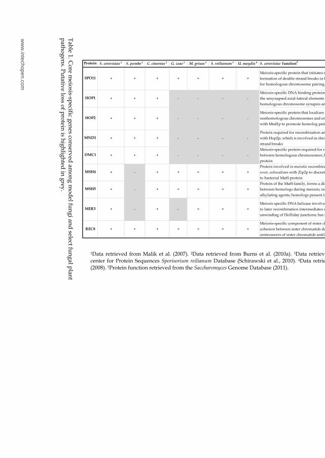
Inve
stig
atin
g H
ost In
du
ce
d M
eio
sis
in a
Fu
ng
al P
lan
t Pa
tho
ge
n
437
Table 1. C
ore meiosis-sp
ecific genes conserved am
ong mod
el fungi and
select fungal p
lant p
athogens. Pu
tative loss of protein is highlighted
in grey.
Protein S. cerevisiae 1 S. pombe 1 C. cinereus 2 G. zeae 1 M. grisea 1 S. reilianum 3 U. maydis 4 S. cerevisiae function5
SPO11 + + + + + + +
Meiosis-specific protein that initiates meiotic recombination by catalyzing the
formation of double-strand breaks in DNA via a transesterification reaction; required
for homologous chromosome pairing and synaptonemal complex formation
HOP1 + + + - - - -
Meiosis-specific DNA binding protein that displays Red1p dependent localization to
the unsynapsed axial-lateral elements of the synaptonemal complex; required for
homologous chromosome synapsis and chiasma formation
HOP2 + + + - - - -
Meiosis-specific protein that localizes to chromosomes, preventing synapsis between
nonhomologous chromosomes and ensuring synapsis between homologs; complexes
with Mnd1p to promote homolog pairing and meiotic double-strand break repair
MND1 + + + - - - -
Protein required for recombination and meiotic nuclear division; forms a complex
with Hop2p, which is involved in chromosome pairing and repair of meiotic double-
strand breaks
DMC1 + + + - - - -
Meiosis-specific protein required for repair of double-strand breaks and pairing
between homologous chromosomes; homolog of Rad51p and the bacterial RecA
protein
MSH4 + - + + + + +
Protein involved in meiotic recombination, required for normal levels of crossing
over, colocalizes with Zip2p to discrete foci on meiotic chromosomes, has homology
to bacterial MutS protein
MSH5 + - + + + + +
Protein of the MutS family, forms a dimer with Msh4p that facilitates crossovers
between homologs during meiosis; msh5-Y823H mutation confers tolerance to DNA
alkylating agents; homologs present in C. elegans and humans
MER3 + - + - + + +
Meiosis specific DNA helicase involved in the conversion of double-stranded breaks
to later recombination intermediates and in crossover control; catalyzes the
unwinding of Holliday junctions; has ssDNA and dsDNA stimulated ATPase activity
REC8 + + + + + + +
Meiosis-specific component of sister chromatid cohesion complex; maintains
cohesion between sister chromatids during meiosis I; maintains cohesion between
centromeres of sister chromatids until meiosis II; homolog of S. pombe Rec8p
1Data retrieved from Malik et al. (2007). 2Data retrieved from Burns et al. (2010a). 3Data retrieved from the Munich Information center for Protein Sequences Sporisorium reilianum Database (Schirawski et al., 2010). 4Data retrieved from Donaldson and Saville (2008). 5Protein function retrieved from the Saccharomyces Genome Database (2011).
ww
w.intechopen.com
Meiosis - Molecular Mechanisms and Cytogenetic Diversity 438
5.3 Core meiosis-specific gene expression in U. maydis
In order to gain insight into the timing of meiosis in planta, we investigated the expression of four core meiosis-specific genes in U. maydis (Table 1). We included a fifth gene, Mre11, a key component in the MRE11-RAD50-XRS2 (MRX) complex, which processes double strand break ends prior to homologous recombination (Holloman et al., 2008). Schlecht and Primig (2003) identified Mre11 as being meiosis and sporulation specific in S. cerevisiae. Using a combination of statistical analysis and PCR, Ho et al. (2007) identified a conserved hypothetical gene (Um01426) to be highly expressed in the dormant teliospore, compared to haploid cells grown in rich media. This gene was included in our analysis to estimate the timing of teliospore maturation in the in planta time course expression analysis. Glyceraldehyde 3-phosphate dehydrogenase (UmGapd) is constitutively expressed in U. maydis and its expression was used to detect the presence of U. maydis cells in planta.
Golden Bantam seedlings were infected with compatible U. maydis haploids (FB1, FB2) and leaf samples were taken 2, 4, 6, 8, 10, and 14 days post infection (dpi). RNA was isolated from these six leaf time-points, as well as dikaryotic and forced diploid mycelia grown filamentously, teliospores isolated from mature tumours on infected ears of corn, and individual compatible haploids. Equal amounts of RNA were used as template for reverse transcriptase reactions primed with oligo-d(T)16. The resulting cDNA was diluted and equal amounts were used as template for PCR. When possible, primers were designed to flank introns (UmSpo11, Um01426, and UmGapd), to clarify the difference in sizes of the amplified products between PCRs with cDNA as template and genomic DNA as template (Figure 2a). The results presented as “leaf” represent a combination of plant and fungal RNA isolated from infected leaves while the U. maydis cell type results are RT-PCR results from pure fungal RNA. Therefore, the results are displayed as two separate panels (leaf samples, or U. maydis cell-types; Figure 2a). Given that the amount of RNA going into first strand synthesis was equivalent in each panel, the resulting RT-PCR product viewed on the ethidium-bromide stained agarose gel may be interpreted as representing the relative expression for each transcript. Comparison between panels is not valid. We estimated the relative expression of the meiosis-specific transcripts for each gene individually, for the leaf and U. maydis cell-types (Figure 2b).
Banuett and Herskowitz (1996) provide an excellent framework for a comparison between the initiation of meiosis and homologous recombination in relation to hyphal development and teliospore formation in planta. It should be cautioned that differences in the timing of U. maydis development may arise due to the type of maize variety infected, and plant growth conditions (Banuett & Herskowitz, 1996). The total RNA isolated from leaf samples is expected to contain RNA from U. maydis cells at different stages of development since U. maydis development is asynchronous in planta. Additionally, changes in transcript levels does not necessarily imply changes in the respective protein levels, especially since meiosis genes are known to be post-transcriptionally regulated (Burns et al., 2010b). We will attempt to determine the timing of meiosis initiation and homologous recombination relative to the in planta development of U. maydis. Given that we used a different maize variety and infected corn seedlings 2 days later than Banuett and Herskowitz (1996), we might expect inconsistencies between the molecular and morphological studies. Nonetheless, it is interesting that the expression of Um01426, a gene highly expressed in mature teliospores (Ho et al., 2007) is upregulated 6-8 dpi, in line with the time that teliospore formation is initiated, and its expression peaks 14 dpi when mature teliospores are visible (Figure 2a,b; Banuett & Herskowitz, 1996).
www.intechopen.com
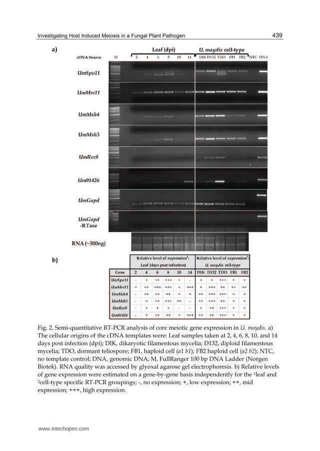
Investigating Host Induced Meiosis in a Fungal Plant Pathogen 439
Fig. 2. Semi-quantitative RT-PCR analysis of core meiotic gene expression in U. maydis. a) The cellular origins of the cDNA templates were: Leaf samples taken at 2, 4, 6, 8, 10, and 14 days post infection (dpi); DIK, dikaryotic filamentous mycelia; D132, diploid filamentous mycelia; TDO, dormant teliospore; FB1, haploid cell (a1 b1); FB2 haploid cell (a2 b2); NTC, no template control; DNA, genomic DNA; M, FullRanger 100 bp DNA Ladder (Norgen Biotek). RNA quality was accessed by glyoxal agarose gel electrophoresis. b) Relative levels of gene expression were estimated on a gene-by-gene basis independently for the 1leaf and 2cell-type specific RT-PCR groupings; -, no expression; +, low expression; ++, mid expression; +++, high expression.
www.intechopen.com
Meiosis - Molecular Mechanisms and Cytogenetic Diversity 440
Spo11 expression has been used to signal the transition between non-meiotic and meiotic cells in S. cerevisiae and S. pombe (Burns et al., 2010b). We detected unspliced UmSpo11 at 2 dpi where filaments have been observed on the leaf epidermal surface (Banuett & Herskowitz, 1996). A discussion on the implications of alternatively spliced UmSpo11 will follow; however, if the spliced variant detected at 4 dpi (Figure 2a) yields a protein that is responsible for double strand break formation at a time where dikaryotic filaments penetrate through the stomata and branch within the plant cells, then the dikaryotic filament may be prepped to initiate the transition from mitotic to meiotic cells directly after karyogamy occurs (9-10 dpi; Banuett & Herskowitz, 1996). Premeiotic DNA replication in C. cinerea occurs prior to karyogamy (reviewed in Burns et al., 2010b). In U. maydis, the timing of premeiotic DNA replication, relative to karyogamy is unknown. The expression of many genes involved in homologous recombination (UmMre11, UmMsh4, UmMsh5, and UmRec8) prior to the time-points suggested for hyphal fragmentation and karyogamy is intriguing. We may have detected so-called "leaky" transcription, but due to the deleterious effects of leaky transcription, it is more likely that the transcript is present and the protein is not functional at the time we detected. In a comparison of the meiotic expression profiles of shared orthologs between S. pombe, S. cerevisiae, and C. cinerea, Burns et al. (2010b) note that the expression of Spo11 and Rec8 peak late in meiosis, prior to the first meiotic division, past the time-point when the protein is functional. We observed that, while the expression of core genes involved in homologous recombination in U. maydis are detectable 4 dpi, they peak in-between 6-10 dpi, coincident with teliospore formation, hyphal fragmentation and karyogamy (Figure 2a,b; Banuett & Herskowitz, 1996). Given this expression of the core meiotic genes responsible for homologous recombination, and using expression of Um01426 (discussed above) as a reference, we propose cells are prepped for meiosis prior to teliospore maturation.
5.4 Post-transcriptional control of Spo11 and controlled meiotic splicing in S. cerevisiae and S. pombe
Under specific conditions in budding yeast, regulated splicing can affect translation by introducing frame shift mutations or nonsense codons, producing non-functional proteins (Juneau et al., 2007). Using high-density tiling arrays, Juneau et al. (2007) discovered 13 intronic meiosis-specific genes that undergo regulated splicing in S. cerevisiae. Using RT-PCR, it was observed that the transcripts Ama1, Hfm1, Hop2, Mnd1, Rec107, Rec114, Rec102,
Pch2, Spo22, Dmc1, Mei4, Sae3, and Spo1 spliced more efficiently (>84%) during sporulation compared to vegetative growth (Juneau et al., 2007). Therefore, regulated splicing of select meiotic-specific transcripts could help the cell overcome the deleterious effects of leaky meiotic gene expression during mitosis.
Similarly, meiosis-specific splicing has been noted in S. pombe. In a random sampling of 96 intronic meiosis-specific genes, Averbeck et al. (2005) used RT-PCR to identify 12 transcripts (including Crs1, Meu13, Rec8, Spo4, Spo6, and Mfr1) that underwent meiosis-specific splicing and correlated with the temporal waves of meiotic-gene expression (early, middle, late) observed during meiotic progression (Mata et al., 2002). Overexpression of Crs1 (cyclin-like protein regulated via splicing), was toxic to vegetative cells, highlighting the requirement of mitotically growing cells to have tight regulation of meiosis-specific genes. Moldon et al. (2008) showed Rem1 encodes two proteins with different functions, depending on the
www.intechopen.com
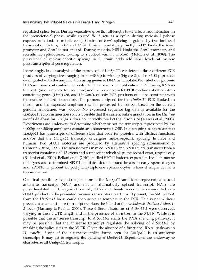
Investigating Host Induced Meiosis in a Fungal Plant Pathogen 441
regulated splice form. During vegetative growth, full-length Rem1 affects recombination in the premeiotic S phase, while spliced Rem1 acts as a cyclin during meiosis I (whose expression is toxic in mitotic cells). Control of Rem1 splicing is guided by two forkhead transcription factors, Fkh2 and Mei4. During vegetative growth, FKH2 binds the Rem1 promoter and Rem1 is not spliced. During meiosis, MEI4 binds the Rem1 promoter, and recruits the spliceosome, leading to a spliced variant of Rem1 (Moldon et al., 2008). The prevalence of meiosis-specific splicing in S. pombe adds additional levels of meiotic posttranscriptional gene regulation.
Interestingly, in our analysis of the expression of UmSpo11, we detected three different PCR products of varying sizes ranging from ~400bp to ~600bp (Figure 2a). The ~600bp product co-migrated with the amplification using genomic DNA as template. We ruled out genomic DNA as a source of contamination due to the absence of amplification in PCR using RNA as template (minus reverse transcriptase) and the presence, in RT-PCR reactions of other intron containing genes (Um01426, and UmGapd), of only PCR products of a size consistent with the mature (spliced) transcripts. The primers designed for the UmSpo11 PCR flanked an intron, and the expected amplicon size for processed transcripts, based on the current genome annotation, was ~350bp. No expressed sequence tag data is available for the UmSpo11 region in question so it is possible that the current online annotation in the Ustilago
maydis database for UmSpo11 does not correctly predict the intron size (Mewes et al., 2008). Experiments are underway to determine whether or not the transcripts represented by the ~400bp or ~500bp amplicons contain an uninterrupted ORF. It is tempting to speculate that UmSpo11 has transcripts of different sizes that code for proteins with distinct functions, and/or that the UmSpo11 transcript undergoes meiosis-specific splicing. In mice and humans, two SPO11 isoforms are produced by alternative splicing (Romanienko & Camerini-Otero, 1999). The two isoforms in mice, SPO11┚ and SPO11┙, are translated from a transcript containing all 13 exons and a transcript which skips the second exon, respectively (Bellani et al., 2010). Bellani et al. (2010) studied SPO11 isoform expression levels in mouse meiocytes and determined SPO11┚ initiates double strand breaks in early spermatocytes and SPO11┙ is present in pachytene/diplotene spermatocytes where it might act as a topoisomerase.
One final possibility is that one, or more of the UmSpo11 amplicons represents a natural antisense transcript (NAT) and not an alternatively spliced transcript. NATs are polyadenylated in U. maydis (Ho et al., 2007) and therefore could be represented as a cDNA product in the presented reverse transcriptase reactions. If present, the NAT cDNA from the UmSpo11 locus could then serve as template in the PCR. This is not without precedent as an antisense transcript overlaps the 3' end of the Arabidopsis thaliana AtSpo11-
2 locus (Hartung & Puchta, 2000). Three different isoforms of AtSpo11-2 were observed, varying in their 3'UTR length and in the presence of an intron in the 3'UTR. While it is possible that the antisense transcript to AtSpo11-2 elicits the RNA silencing pathway, it may be possible that the antisense transcript regulates the splicing of AtSpo11-2 by masking the splice sites in the 3'UTR. Given the absence of a functional RNAi pathway in U. maydis, if one of the alternative splice forms seen for UmSpo11 is an antisense transcript, it may act to regulate the splicing of UmSpo11. Experiments are underway to characterize all UmSpo11 transcripts.
www.intechopen.com
Meiosis - Molecular Mechanisms and Cytogenetic Diversity 442
It is noteworthy that Heimel et al. (2010) recently showed that UmCib1, a transcription factor required for pathogenic development, is predominantly unspliced during saprophytic growth, but undergoes splicing during biotrophic growth. Therefore, it is highly likely that other biological roles for alternate splicing in U. maydis remain to be discovered.
6. A regulatory role for fungal noncoding RNAs in meiosis
This section will begin with a broad overview of noncoding RNA (ncRNA) function in eukaryotes. There is very limited information on ncRNAs in other smuts and rusts; therefore, we will highlight the prevalence of meiosis-specific ncRNAs in S. cerevisiae and S. pombe, including putative functional roles for their expression. We will review specific examples of how ncRNAs function in controlling gene expression and conclude with knowledge that our laboratory is accumulating on ncRNAs and long natural antisense transcripts (NATs) in U. maydis.
6.1 Introduction to noncoding RNAs in eukaryotes
Recent estimates are that >90% of eukaryotic genomes are transcribed, but protein-coding transcripts only account for ~2-3% of eukaryotic genome transcription (reviewed in Costa, 2010). The difference is comprised of noncoding RNAs, some of which have a role in controlling gene expression. Thus, at least a portion of these so-called ncRNAs actually have a function that adds to the complexity of gene expression. Noncoding RNAs are divided into classes depending on their size and origin. For example, short ncRNAs (~20-31nt) include microRNAs (miRNAs), piwi-interacting RNAs (piRNAs), and small interfering RNAs (siRNAs). Long ncRNAs (>200nt) include, but are not limited to, long intergenic transcripts, and natural antisense transcripts (NATs). NATs are RNA molecules transcribed from the DNA strand complementary to that which codes the mRNA (reviewed in Costa, 2010; Faghihi & Wahlestedt 2009; Tisseur et al., 2011). Noncoding RNAs exert their function at the transcriptional or post-transcriptional level using a wide range of regulatory mechanisms including: RNA interference (RNAi), transcriptional interference, RNA masking (affecting mRNA-splicing, -transport, -polyadenylation, -translation, or -stability), RNA editing, X chromosome inactivation, imprinting, and chromatin remodelling (Costa, 2005; reviewed in Lavorgna et al., 2004; Munroe & Zhu, 2005). Notably, while most plants, animal and fungi contain functional RNA silencing machinery, phylogenetic analyses have revealed select ascomycetes (S. cerevisiae and Candida lusitaniae) and U. maydis lack the canonical RNAi machinery. The expression of ncRNAs, especially NATs in fungi that do not contain the canonical RNAi machinery sets the stage for discovering new mechanisms of controlling meiotic gene expression.
6.2 Global expression of meiosis-specific ncRNAs in S. cerevisiae and S. pombe
Yassour et al. (2011) created a strand-specific cDNA library using RNA isolated from S. cerevisiae cells during mid-log growth in rich media. Next-generation (Illumina) sequencing revealed transcript units that had an antisense orientation to 1,103 annotated transcripts. It was concluded that these antisense transcripts may be functional, given there was little evidence they arose from unterminated transcription, bi-directional transcription initiated from divergent promoters, or potential nucleosome-free regions (Yassour et al., 2010). A
www.intechopen.com
Investigating Host Induced Meiosis in a Fungal Plant Pathogen 443
subset of sense-antisense transcript pairs was examined using strand-specific quantitative real-time PCR (qRT-PCR) and their expression patterns were inversely related, supporting a model where antisense transcripts interfere with sense transcript expression (Yassour et al., 2010). Additionally, the expression levels of six sense-antisense transcript pairs were studied across five yeast species using qRT-PCR. In some instances, both antisense transcript presence and differential expression were conserved, further supporting a functional role for antisense transcripts in S. cerevisiae (Yassour et al., 2010). 85 genes expressed at 8 hours post induction of sporulation were associated with antisense transcripts during S. cerevisiae mid-log phase growth. For example, the meiosis genes encoding Ime4, Ndt80, Rec102, Gas2, Sps19, Slz1, Rim9, and Smk1 were all associated with long antisense transcripts. Overall, antisense units were prevalent in processes repressed during mid-log phase growth, leading Yassour et al. (2010) to hypothesize that NAT expression in S. cerevisiae may be involved in a global repression of stress, stationary phase, and meiosis genes when cells are grown in rich conditions. As a whole, in S. cerevisiae, and other yeast species, differentially expressed sense-antisense transcript pairs provide a means of controlling gene expression in response to environmental conditions and in some cases, enable the transition from mid-log phase- to meiotic-growth conditions.
Meiosis-specific genes were identified by T. Watanabe et al. (2001) using a subtractive cDNA library enriched for meiotic gene expression in heterozygous S. pombe diploid cells grown in nitrogen starvation conditions. This approach identified 31 Meu (meiotic expression upregulated) transcripts, 5 of which were ncRNAs. Notably, Meu16 encodes an antisense ncRNA overlapping Mde6 (a Mei4-dependent protein). The function of this antisense is not known. Additionally, individual RACE experiments uncovered ncRNAs at the Rec7 (required for early steps of meiotic recombination) and Spo6 (required for progression of meiosis II and sporulation) loci (Molnar et al., 2001; Nakamura et al., 2000). Specifically, three antisense transcripts (Tos1, Tos2 and Tos3), of varying lengths, were discovered at the Rec7 locus. It has been suggested that the Tos RNAs may indicate the location of dsDNA break formation (DSB; see below) at the Rec7 locus (Wahls et al., 2008). Spo6-L, encodes a constitutively expressed bidirectional transcript in reverse orientation to the meiosis-specific Spo6 gene. The function of Spo6-L remains unknown (Nakamura et al., 2000).
Subsequently, Wilhelm et al. (2008) studied the transcriptome of S. pombe from cells grown in rich media and from five stages of meiotic development under nitrogen limiting conditions.
Their investigation yielded 426 previously unannotated ncRNAs, including 58 ncRNAs (34 of which overlapped known genes in antisense orientation) upregulated under meiotic growth conditions. Many of the ncRNAs appear to be expressed in waves similar to meiosis-specific gene expression in S. pombe (Mata et al., 2002, 2007). Ni et al. (2010) created a strand-specific cDNA library using RNA isolated from S. pombe cells grown in rich media with or without heat shock. Next generation (Illumina) sequencing revealed ncRNA transcript units that did not overlap with previously defined protein-coding genes. Some loci in S. pombe, absent of protein-coding genes, had a comparable number of ncRNA transcripts on both strands, indicating that unlike S. cerevisiae, ncRNAs in S. pombe may function via the formation of double-stranded RNA, possibly eliciting chromatin remodelling, or posttranscriptional gene silencing. Additional differences that Ni et al. (2010) found between
www.intechopen.com
Meiosis - Molecular Mechanisms and Cytogenetic Diversity 444
budding and fission yeast include: that antisense transcription is commonly driven through bidirectional promoters in S. pombe but not in S. cerevisiae and, while the majority of antisense transcripts in S. pombe are independently regulated, sense-antisense transcript levels are coordinated in S. cerevisiae. This may imply that antisense-mediated gene regulation in S. pombe occurs at the posttranscriptional level, or in trans, without affecting the relative levels of the sense transcript (Ni et al., 2010). It was observed that differentially expressed genes had a higher abundance of antisense transcripts than constitutively expressed genes, indicating that antisense RNAs may be involved in a targeted control of gene expression (Ni et al., 2010). In total, 2,409 S. pombe genes had overlapping antisense transcripts under normal or heat shock conditions. Gene ontology analysis revealed, like S.
cerevisiae, S. pombe is enriched for antisense transcription at 68 loci involved in meiosis, meiotic chromosome segregation, meiotic recombination and ascospore formation. It was hypothesized that antisense transcripts may repress “leaky” meiotic genes under vegetative conditions at the transcriptional or posttranscriptional level (Ni et al., 2010). Using RNA-Seq, the transcriptomes of the fission yeasts S. pombe, S. octosporus, S. cryophilus, and S.
japonicus were compared under growth conditions including log-phase, glucose starvation, early stationary phase, and heat shock (Rhind et al., 2011). This comparison identified conserved antisense transcripts among some of the fission yeasts. Additionally, for meiotic genes with antisense transcripts, the level of the antisense transcript was higher than the sense transcript (Rhind et al., 2011). For example, meiosis-upregulated genes (Mug5, Mug7, Mug27, Mug28, Mug97), MEI4 dependent genes (Mde2, Mde3, Mde4, Mde7), genes involved in meiotic recombination (Rec7, Rec15, Rec24, Rec27) and sporulation-specific genes (Spo4, Spo6) had higher levels of antisense transcript than sense transcript (Rhind et al., 2011, Supporting online material S21). Notably, antisense transcripts detected towards the core eukaryotic meiosis genes Hop1 and Dmc1, were higher than sense transcript levels, while an antisense transcript level lower than that of the sense transcript was observed for Rec8. Strand-specific northern blotting was used to detect relative levels of S. pombe Spo4, Spo6, Mde2, Mde7, and Mug8 sense-antisense transcript pairs. This revealed that antisense transcription of meiotic genes did not share an inverse relationship to sense transcription during log-phase growth, reinforcing the findings of Ni et al. (2010). Taken together, these findings suggest that antisense transcription during log-phase growth does not directly inhibit the transcription of sense transcripts as in S. cerevisiae, but elicits the formation of double-stranded RNA to recruit RNAi machinery to destroy "leaky" meiotic transcripts during mitotic growth (Rhind et al., 2011).
6.3 Rrp6 controls global expression of meiosis-specific ncRNAs in S. cerevisiae
Additional mechanisms of regulating the timing of ncRNA expression during meiosis in S.
cerevisiae have been discovered. Degradation of ncRNAs in S. cerevisiae was linked to RRP6, a key component of the nuclear exosome complex responsible for RNA processing and degradation (Wyers et al., 2005). Deep transcriptome analyses identified cryptic unstable transcripts (CUTs) that accumulate in ΔRrp6 vegetative cells (Neil et al., 2009; Xu et al., 2009). Separately, high-resolution oligonucleotide tiling arrays identified meiotic unannotated transcripts (MUTs), ncRNAs that accumulate in meiotic but not fermenting or respiring cells, and rsSUTs, ncRNAs exhibiting peak expression during respiring or
www.intechopen.com
Investigating Host Induced Meiosis in a Fungal Plant Pathogen 445
sporulating MAT a/┙ diploids (Lardenois et al., 2011). MUTs and rsSUTs were observed to overlap sense mRNAs in an antisense orientation, known promoter regions, or autonomously replicating sequences (ARSs). Lardenois et al. (2011) found that a subset of MUTs and rsSUTs, previously characterized as CUTs (Neil et al., 2009; Xu et al., 2009), are targeted by the nuclear exosome for degradation during vegetative growth conditions. Moreover, Rrp6 expression was observed across all cell-types; but RRP6 levels dropped dramatically as cells switched from mitotic to meiotic growth. During this transition, the decrease in RRP6 levels paralleled the increase in MUTs. Additionally, ∆Rrp6 cells were unable to proceed with premeiotic DNA replication or to undergo meiosis and spore formation. The progression through meiosis may be facilitated by the accumulation of MUTs transcribed at mitotically active loci (such as Chs2, Cln3, and Hug1); MUT expression would interfere with the transcription of some mitotic genes during meiosis (Lardenois et al., 2011). Yassour et al. (2011) observed an increased level of antisense transcription in ∆Rrp6 mutants, associated with a small decrease in complementary sense mRNA levels. Additionally, the transcription of the MUT, itself, may interfere with promoter regions required for proper transcription of select mRNAs or ARS elements required for DNA replication during specific cell-stages. For example, MUT expression was detected towards the Cln2 promoter region. CLN2 is a repressor of IME1; therefore, Cln2 levels decrease during meiosis, possibly facilitated through a promoter-interference mechanism where MUT transcription hinders promoter activity (Lardenois et al., 2011).
6.4 S. cerevisiae IME4 and ZIP1 ncRNA functions
S. cerevisiae MAT a/┙ diploids require full activation of IME1 which is mediated by IME4 expression. Therefore, Ime4 expression during nutrient starvation is pivotal in determining whether or not the cell initiates meiosis. Hongay et al. (2006) observed cell-type specific sense and antisense transcription at the Ime4 locus. Haploids expressed an antisense transcript to Ime4, called Rme2 (regulator of meiosis 2; Gelfand et al., 2011), and MAT a/┙ diploids expressed Ime4 sense RNA. In MAT a/┙ diploids, the a1-┙2 protein heterodimer silences expression of Rme2, enabling the expression of Ime4, full expression of Ime1, and entry into meiosis. Their findings were consistent with a transcriptional interference mechanism, since transcription of Rme2 in haploid cells only interfered with Ime4 sense expression in cis (Hongay et al., 2006). Similarly, Zip2, a meiosis-specific protein involved in synaptonemal complex formation, shows cell-type specific sense and antisense expression (Gelfand et al., 2011). Haploids express an antisense transcript to Zip2, called Rme3
(regulator of meiosis 3) and MAT a/┙ diploids express Zip2 sense RNA. The a1-┙2 protein heterodimer silences expression of Rme3 in MAT a/┙ diploids, enabling expression of Zip2. Gelfand et al. (2011) expanded on previous research to show that Rme2 extension through the Ime4 promoter region was not required for Ime4 repression and Rme3 does not extend through the entire Zip2 ORF, indicating that both Rme2 and Rme3 do not interfere with TATA-binding proteins or polymerase binding in the promoter regions. Additionally, a 450 bp region within Ime4 was essential for Rme2-mediated repression. This suggested that the 450 bp region may only be transcribed in a single direction at one time, or that extension of the transcript is terminated by specific protein complexes which bind this region, or chromatin remodelling occurs at this site. Overall, transcriptional interference may be
www.intechopen.com
Meiosis - Molecular Mechanisms and Cytogenetic Diversity 446
prevalent in controlling yeast gene expression (Gelfand et al., 2011). Such mechanisms may be useful in fine-tuning condition-specific gene expression, especially when "leaky" expression of certain genes may be harmful to the cell.
6.5 S. pombe meiRNA function: The mei2-meiRNA complex
S. pombe MEI2 is an RNA-binding protein required for premeiotic DNA replication and the initiation of meiosis I (Y. Watanabe & Yamamoto, 1994; reviewed in Yamamoto et al., 2010). This protein has been discussed in detail in “4.2.2 Commitment and Continuation”. In the context of this section, the role of a 0.5kb ncRNA transcribed at the Sme2 locus (dubbed meiRNA) will be discussed. meiRNA binds to MEI2 and this ncRNA-protein complex is transported to the nucleus (Y. Watanabe & Yamamoto, 1994). Two scenarios have been described whereby meiRNA determines the nuclear localization of MEI2; meiRNA either interferes with MEI2 export from the nucleus, or facilitates MEI2 import into the nucleus (Sato et al., 2001). This nuclear MEI2-meiRNA complex binds MMI1, a protein that targets meiotic transcripts containing DSR motifs for degradation, during mitosis. This interaction interferes with MMI1, stabilizing transcripts required for the progression of S. pombe cells through meiosis I (Yamamoto et al., 2010). From this overview one can see that ncRNAs control the mitosis-meiosis switch in the divergent S. pombe and S. cerevisiae; however, they do so by very different mechanisms.
6.6 S. pombe meiotic hotspots
In S. pombe, the dsDNA breaks (DSBs) that initiate recombination, cluster to 194 prominent and 159 weak DSB peaks that favour intergenic regions (IGRs, Cromie et al., 2007). T. Watanabe et al. (2002) identified 68 polyadenylated ncRNAs from a random sampling of cDNAs originating from S. pombe cells in mitotic or meiotic growth phase. By cross referencing the aforementioned studies, it was determined that 24 polyadenylated ncRNAs were located entirely within a DSB peak (Wahls et al., 2008). Overall, Wahls et al. (2008) concluded that meiotic DSB hotspots preferentially form at loci that express long polyadenylated ncRNAs, many of which are expressed solely during meiosis. There are two mechanisms by which ncRNAs may guide meiotic recombination proteins to DSB hotspots: 1) the ncRNAs make DNA accessible to DSB formation through meiotically induced chromatin remodelling, and 2) ncRNA-DNA hybrids (R-loops) guide the meiotic recombination machinery to the DSB sites (Wahls et al., 2008).
6.7 U. maydis ncRNA/antisense transcription
RNA-seq has not been performed on U. maydis cell-types, but limited strand-specific expression data is available from the creation of EST libraries from various cell-types and nutritional conditions. To help facilitate U. maydis genome annotation, cDNA libraries were constructed from cell-types, including: germinating and dormant teliospores (Sacadura & Saville, 2003; Ho et al., 2007), filamentous diploids (Nugent et al., 2004) and dikaryons (Morrison et al., in preparation). Recall that teliospore formation and germination are temporally linked to meiosis in U. maydis (reviewed in Donaldson & Saville, 2008). Of the 319 uniESTs that did not match an annotated gene model in the U. maydis genome, 108 uniquely represented RNAs expressed in the dormant or germinating teliospores. This
www.intechopen.com
Investigating Host Induced Meiosis in a Fungal Plant Pathogen 447
corresponds to 34% of the identified ncRNAs, while these two cDNA libraries account for only 17% of the total ESTs from all cell-types (Saville, unpublished). In total, ~250 NATs have been identified in U. maydis, including NATs expressed in the dormant and germinating teliospores (55 and 12, respectively). The function of the NATs and ncRNAs in U. maydis is under investigation. Ten teliospore-specific NATs, annotated during analysis of the dormant teliospore cDNA library, have been verified as teliospore-specific, using strand-specific RT-PCR (Ho et al., 2010). Unlike S. cerevisiae and S. pombe, the U. maydis NATs expressed in the dormant and germinating teliospore are not enriched for mitosis-specific genes and the NATs expressed during vegetative growth are not enriched for meiosis-specific genes. Additionally, inverse expression patterns have not been observed for sense-antisense transcript pairs; precluding transcriptional interference as the principal mechanism of action for NATs in U. maydis. Therefore, their function in the dormant and germinating teliospores appears to be unique to U. maydis.
7. Conclusion
This chapter provides an overview of meiotic events in the model fungal species Saccharomyces cerevisiae, Schizosaccharomyces pombe and Coprinopsis cinerea as a means of providing context for an exploration of meiosis in the model plant pathogen Ustilago maydis. Like the yeast fungi, U. maydis has set genetic requirements for entry into meiosis; it must be diploid and contain complementary alleles at the b mating type locus. With this genetic background, the fungus is able to accept an environmental signal that triggers entry into meiosis. This signal comes from the plant host and an exploration of the stages of pathogenic development led us to hypothesize that the stage before hyphal fragmentation, and after the cells become embedded in a mucilaginous matrix, is the time it must receive a signal from the plant to trigger entry into premeiotic S phase and undergo karyogamy. We uncover similarities between the role of the MAPK and cAMP/PKA pathways in mating and meiosis initiation in yeasts and the mating and pathogenesis signal transduction pathways in U. maydis. This is very relevant because of the requirement for growth within the host for U. maydis to become meiotically competent. These comparisons emphasized that the U. maydis genes Crk1 and Prf1, which are orthologs of the major meiosis control genes Ime2 in S. cerevisiae and Ste11 in S. pombe respectively, provide a means whereby mating type and environmental signals could be transduced to influence meiosis. This led to a model of how meiosis is triggered in U. maydis and its linkage with teliospore development. We present an overview of waves in transcription in the yeasts and present evidence for potential waves of transcription in U. maydis. The identification of U. maydis meiosis genes by bioinformatic analyses is updated with an identification of the conserved absence of core meiosis genes in plant pathogenic fungi. We also present data that identifies UmNdt80 as the first gene known to be required for meiosis completion in U. maydis. The timing of expression of six core meiosis genes in U. maydis is followed during in planta development. This uncovered support for the model that U. maydis enters meiosis very soon after karyogamy and then arrests during pachytene, when the teliospore matures and enters a dormant state. This information also identified transcriptional and posttranscriptional control of Spo11 as potential key transitions in U. maydis meiotic progression. The final portion of the chapter highlights new data on the bioinformatic discovery of ncRNAs and NATs in U. maydis and their overrepresentation among ESTs in the teliospore and
www.intechopen.com
Meiosis - Molecular Mechanisms and Cytogenetic Diversity 448
germinating teliospore libraries. In the context of the emerging role for these RNAs in controlling aspects of meiosis in S. cerevisiae and S. pombe, the discovery and confirmation of these RNAs in U. maydis is compelling. This chapter identifies several areas where further research will provide tremendous insight regarding meiosis initiation and progression in U.
maydis.
During proofing, gametogenesis initiation (van Werven & Amon, 2011) and RNAi-independent roles for antisense transcripts in controlling meiotic genes (Chen & Neiman, 2011) in budding and fission yeasts were reviewed.
8. Acknowledgement
We would like to acknowledge Natalie Pearson for her excellent artwork in Figure 1, thank Laura Peers for fact finding and organizational support, and thank Christine Russell for assistance in proofing this manuscript. We also acknowledge funding from NSERC of Canada, and the Ontario Ministry of Research and Innovation’s Ontario Research Fund-Research Excellence program.
9. References
Abe, H. & Shimoda, C. (2000). Autoregulated expression of Schizosaccharomyces pombe meiosis-specific transcription factor Mei4 and a genome-wide search for its target genes. Genetics, Vol.154, No.4, (April 2000), pp. 1497-1508, ISSN 0016-6731
Ahmed, N.T., Bungard, D., Shin, M.E., Moore, M. & Winter, E. (2009). The Ime2 protein kinase enhances the disassociation of the Sum1 repressor from middle meiotic promoters. Molecular and cellular biology, Vol.29, No.16, (August 2009), pp. 4352-4362, ISSN 1098-5549
Allen, A., Islamovic, E., Kaur, J., Gold, S., Shah, D. & Smith, T.J. (2011). Transgenic maize plants expressing the Totivirus antifungal protein, KP4, are highly resistant to corn smut. Plant biotechnology journal, (February 2011), ISSN 1467-7652
Alvarez, B. & Moreno, S. (2006). Fission yeast Tor2 promotes cell growth and represses cell differentiation. Journal of cell science, Vol.119, No.Pt 21, (November 2006), pp. 4475-4485, ISSN 0021-9533
Averbeck, N., Sunder, S., Sample, N., Wise, J.A. & Leatherwood, J. (2005). Negative control contributes to an extensive program of meiotic splicing in fission yeast. Molecular cell, Vol.18, No.4, (May 2005), pp. 491-498, ISSN 1097-2765
Banuett, F. (2010). Ustilago maydis and Maize: a Delightful Interaction, In: Cellular and Molecular Biology of Filamentous Fungi, K.A. Borkovich & D.J. Ebbole, (Ed.), 622-644, ASM Press, ISSN 978-1-55581-473-1, Washington, USA
Banuett, F. (2002). Pathogenic development in Ustilago maydis: A progression of morphological transitions that results in tumor formation and teliospore production, In: Molecular biology of fungal development, H.D. Osiewacz, (Ed.), 349-398, Marcel Dekker, ISSN 0824707443, New York, USA
Banuett, F. & Herskowitz, I. (1996). Discrete developmental stages during teliospore formation in the corn smut fungus, Ustilago maydis. Development, Vol.122, No.10, (October 1996), pp. 2965-2976, ISSN 0950-1991
Banuett, F. (1995).Genetics of Ustilago maydis, a fungal pathogen that induces tumors in maize.Annual Review of Genetics, Vol.29, (n.d.), pp. 179-208, ISSN 0066-4197
www.intechopen.com
Investigating Host Induced Meiosis in a Fungal Plant Pathogen 449
Banuett, F. & Herskowitz, I. (1989). Different a alleles of Ustilago maydis are necessary for maintenance of filamentous growth but not for meiosis. Proceedings of the National Academy of Sciences of the United States of America, Vol.86, No.15, (August 1989), pp. 5878-5882, ISSN 0027-8424
Barnes, C.W., Szabo, L.J., May, G. & Groth, J.V. (2004). Inbreeding levels of two Ustilago maydis populations. Mycologia, Vol.96, No.6, (November 2004), pp. 1236-1244, ISSN 0027-5514
Bauman, P., Cheng, Q.C. & Albright, C.F. (1998). The Byr2 kinase translocates to the plasma membrane in a Ras1-dependent manner. Biochemical and biophysical research communications, Vol.244, No.2, (March 1998), pp. 468-474, ISSN 0006-291X
Bellani, M.A., Boateng, K.A., McLeod, D. & Camerini-Otero, R.D. (2010). The expression profile of the major mouse SPO11 isoforms indicates that SPO11beta introduces double strand breaks and suggests that SPO11alpha has an additional role in prophase in both spermatocytes and oocytes. Molecular and cellular biology, Vol.30, No.18, (September 2010), pp. 4391-4403, ISSN 1098-5549
Boehm, E.W.A., Wenstrom, J.C., McLaughlin, D.J., Szabo, L.J., Roelfs, A.P. & Bushnell, W.R. (1992). An ultrastructural pachytene karyotype for Puccinia graminis f. sp. tritici. Canadian Journal of Botany, Vol.70, (n.d.), pp. 401-4113, ISSN 0008-4026
Borkovich, K.A., Alex, L.A., Yarden, O., Freitag, M., Turner, G.E., Read, N.D., Seiler, S., Bell-Pedersen, D., Paietta, J., Plesofsky, N., Plamann, M., Goodrich-Tanrikulu, M., Schulte, U., Mannhaupt, G., Nargang, F.E., Radford, A., Selitrennikoff, C., Galagan, J.E., Dunlap, J.C., Loros, J.J., Catcheside, D., Inoue, H., Aramayo, R., Polymenis, M., Selker, E.U., Sachs, M.S., Marzluf, G.A., Paulsen, I., Davis, R., Ebbole, D.J., Zelter, A., Kalkman, E.R., O'Rourke, R., Bowring, F., Yeadon, J., Ishii, C., Suzuki, K., Sakai, W. & Pratt, R. (2004). Lessons from the genome sequence of Neurospora crassa: tracing the path from genomic blueprint to multicellular organism. Microbiology and molecular biology reviews, Vol.68, No.1, (March 2004), pp. 1-108, ISSN 1092-2172
Brefort, T., Doehlemann, G., Mendoza-Mendoza, A., Reissmann, S., Djamei, A. & Kahmann, R. (2009). Ustilago maydis as a Pathogen. Annual Review of Phytopathology, Vol.47, (n.d.), pp. 423-445, ISSN 0066-4286
Burdon, J.J. & Roelfs, A.P. (1985). The effect of sexual and asexual reproduction on the isozyme structure of populations of Puccinia graminis. Phytopathology, Vol.75, (n.d.), pp. 1068-1073, ISSN 0031-949X
Burns, C., Pukkila, P.J. & Miriam, Z.E. (2010a). Meiosis, In: Cellular and Molecular Biology of Filamentous Fungi, K.A. Borkovich & D.J. Ebbole, (Ed.), 81-95, ASM Press, ISSN 978-1-55581-473-1, Washington, USA
Burns, C., Stajich, J.E., Rechtsteiner, A., Casselton, L., Hanlon, S.E., Wilke, S.K., Savytskyy, O.P., Gathman, A.C., Lilly, W.W., Lieb, J.D., Zolan, M.E. & Pukkila, P.J. (2010b). Analysis of the Basidiomycete Coprinopsis cinerea reveals conservation of the core meiotic expression program over half a billion years of evolution. PLoS genetics, Vol.6, No.9, (September 2010), pp. e1001135, ISSN 1553-7404
Butler, G., Rasmussen, M.D., Lin, M.F., Santos, M.A., Sakthikumar, S., Munro, C.A., Rheinbay, E., Grabherr, M., Forche, A., Reedy, J.L., Agrafioti, I., Arnaud, M.B., Bates, S., Brown, A.J., Brunke, S., Costanzo, M.C., Fitzpatrick, D.A., de Groot, P.W., Harris, D., Hoyer, L.L., Hube, B., Klis, F.M., Kodira, C., Lennard, N., Logue, M.E., Martin, R., Neiman, A.M., Nikolaou, E., Quail, M.A., Quinn, J., Santos, M.C., Schmitzberger, F.F., Sherlock, G., Shah, P., Silverstein, K.A., Skrzypek, M.S., Soll, D., Staggs, R., Stansfield, I., Stumpf, M.P., Sudbery, P.E., Srikantha, T., Zeng, Q., Berman, J., Berriman, M., Heitman, J., Gow, N.A., Lorenz, M.C., Birren, B.W., Kellis,
www.intechopen.com
Meiosis - Molecular Mechanisms and Cytogenetic Diversity 450
M. & Cuomo, C.A. (2009). Evolution of pathogenicity and sexual reproduction in eight Candida genomes. Nature, Vol.459, No.7247, (June 2009), pp. 657-662, ISSN 1476-4687
Capitol Commodity Services. (February, 2011). U.S. crop values jumped 22% in 2010, 04.09.2011, Available from <http://www.ccstrade.com/futures-news/grains/story/u-s-crop-values-jumped-22-in-2010-800410457/>
Celerin, M., Merino, S.T., Stone, J.E., Menzie, A.M. & Zolan, M.E. (2000). Multiple roles of Spo11 in meiotic chromosome behavior. The EMBO journal, Vol.19, No.11, (June 2000), pp. 2739-2750, ISSN 0261-4189
Chen, H.M. & Neiman, A.M. (2011). A conserved regulatory role for antisense RNA in meiotic gene expression in yeast. Current opinion in microbiology, Vol.14, No.6, (December 2011), pp. 655-659, ISSN 1879-0364
Chu, S., DeRisi, J., Eisen, M., Mulholland, J., Botstein, D., Brown, P.O. & Herskowitz, I. (1998). The transcriptional program of sporulation in budding yeast. Science, Vol.282, No.5389, (October 1998), pp. 699-705, ISSN 0036-8075
Chu, S. & Herskowitz, I. (1998). Gametogenesis in yeast is regulated by a transcriptional cascade dependent on Ndt80. Molecular cell, Vol.1, No.5, (April 1998), pp. 685-696, ISSN 1097-2765
Chung, K.S., Won, M., Lee, S.B., Jang, Y.J., Hoe, K.L., Kim, D.U., Lee, J.W., Kim, K.W. & Yoo, H.S. (2001). Isolation of a novel gene from Schizosaccharomyces pombe: stm1+ encoding a seven-transmembrane loop protein that may couple with the heterotrimeric Galpha 2 protein, Gpa2. The Journal of biological chemistry, Vol.276, No.43, (October 2001), pp. 40190-40201, ISSN 0021-9258
Colomina, N., Gari, E., Gallego, C., Herrero, E. & Aldea, M. (1999). G1 cyclins block the Ime1 pathway to make mitosis and meiosis incompatible in budding yeast. The EMBO journal, Vol.18, No.2, (January 1999), pp. 320-329, ISSN 0261-4189
Cooper, K.F. & Strich, R. (2002). Saccharomyces cerevisiae C-type cyclin Ume3p/Srb11p is required for efficient induction and execution of meiotic development. Eukaryotic cell, Vol.1, No.1, (February 2002), pp. 66-74, ISSN 1535-9778
Costa, F.F. (2010). Non-coding RNAs: Meet thy masters. BioEssays: news and reviews in molecular, cellular and developmental biology, Vol.32, No.7, (July 2010), pp. 599-608, ISSN 1521-1878
Costa, F.F. (2005). Non-coding RNAs: new players in eukaryotic biology. Gene, Vol.357, No.2, (September 2005), pp. 83-94, ISSN 0378-1119
Covitz, P.A. & Mitchell, A.P. (1993). Repression by the yeast meiotic inhibitor RME1. Genes & development, Vol.7, No.8, (August 1993), pp. 1598-1608, ISSN 0890-9369
Covitz, P.A., Herskowitz, I. & Mitchell, A.P. (1991). The yeast RME1 gene encodes a putative zinc finger protein that is directly repressed by a1-alpha 2. Genes & development, Vol.5, No.11, (November 1991), pp. 1982-1989, ISSN 0890-9369
Cromie, G.A., Hyppa, R.W., Cam, H.P., Farah, J.A., Grewal, S.I. & Smith, G.R. (2007). A discrete class of intergenic DNA dictates meiotic DNA break hotspots in fission yeast. PLoS genetics, Vol.3, No.8, (August 2007), pp. e141, ISSN 1553-7404
Donaldson, M.E. & Saville, B.J. (2008). Bioinformatic identification of Ustilago maydis meiosis genes. Fungal genetics and biology, Vol.45, Suppl 1, (August 2008), pp. S47-53, ISSN 1096-0937
Donzeau, M. & Bandlow, W. (1999). The yeast trimeric guanine nucleotide-binding protein alpha subunit, Gpa2p, controls the meiosis-specific kinase Ime2p activity in response to nutrients. Molecular and cellular biology, Vol.19, No.9, (September 1999), pp. 6110-6119, ISSN 0270-7306
www.intechopen.com
Investigating Host Induced Meiosis in a Fungal Plant Pathogen 451
Doyle, C.E., Donaldson, M.E., Morrison, E.N. & Saville, B.J. (2011). Ustilago maydis transcript features identified through full-length cDNA analysis. Molecular genetics and genomics, Vol.286, No.2, (August 2011), pp. 143-159, ISSN 1617-4623
Faghihi, M.A. & Wahlestedt, C. (2009). Regulatory roles of natural antisense transcripts. Nature reviews molecular cell biology, Vol.10, No.9, (September 2009), pp. 637-643, ISSN 1471-0080
Feldbrugge, M., Kamper, J., Steinberg, G. & Kahmann, R. (2004). Regulation of mating and pathogenic development in Ustilago maydis.Current opinion in microbiology, Vol.7, No.6, (December 2004), pp. 666-672, ISSN 1369-5274
Fletcher, H.L. (1981). A search for synaptonemal complexes in Ustilago maydis. Journal of cell science, Vol.50, (August 1981), pp. 171-180, ISSN 0021-9533
Gallego, C., Gari, E., Colomina, N., Herrero, E. & Aldea, M. (1997). The Cln3 cyclin is down-regulated by translational repression and degradation during the G1 arrest caused by nitrogen deprivation in budding yeast. The EMBO journal, Vol.16, No.23, (December 1997), pp. 7196-7206, ISSN 0261-4189
Garrido, E., Voss, U., Muller, P., Castillo-Lluva, S., Kahmann, R. & Perez-Martin, J. (2004). The induction of sexual development and virulence in the smut fungus Ustilago maydis depends on Crk1, a novel MAPK protein. Genes & development, Vol.18, No.24, (December 2004), pp. 3117-3130, ISSN 0890-9369
Garrido, E. & Perez-Martin, J. (2003). The crk1 gene encodes an Ime2-related protein that is required for morphogenesis in the plant pathogen Ustilago maydis. Molecular microbiology, Vol.47, No.3, (February 2003), pp. 729-743, ISSN 0950-382X
Gelfand, B., Mead, J., Bruning, A., Apostolopoulos, N., Tadigotla, V., Nagaraj, V., Sengupta, A.M. & Vershon, A.K. (2011). Regulated antisense transcription controls expression of cell-type-specific genes in yeast. Molecular and cellular biology, Vol.31, No.8, (April 2011), pp. 1701-1709, ISSN 1098-5549
Gold, S.E., Brogdon, S.M., Mayorga, M.E. & Kronstad, J.W. (1997). The Ustilago maydis regulatory subunit of a cAMP-dependent protein kinase is required for gall formation in maize. The Plant cell, Vol.9, No.9, (September 1997), pp. 1585-1594, ISSN 1040-4651
Govin, J. & Berger, S.L. (2009). Genome reprogramming during sporulation. The International journal of developmental biology, Vol.53, No.2-3, (n.d.), pp. 425-432, ISSN 1696-3547
Guttmann-Raviv, N., Martin, S. & Kassir, Y. (2002). Ime2, a meiosis-specific kinase in yeast, is required for destabilization of its transcriptional activator, Ime1. Molecular and cellular biology, Vol.22, No.7, (April 2002), pp. 2047-2056, ISSN 0270-7306
Harigaya, Y. & Yamamoto, M. (2007). Molecular mechanisms underlying the mitosis-meiosis decision. Chromosome research: an international journal on the molecular, supramolecular and evolutionary aspects of chromosome biology, Vol.15, No.5, (n.d.), pp. 523-537, ISSN 0967-3849
Harigaya, Y., Tanaka, H., Yamanaka, S., Tanaka, K., Watanabe, Y., Tsutsumi, C., Chikashige, Y., Hiraoka, Y., Yamashita, A. & Yamamoto, M. (2006). Selective elimination of messenger RNA prevents an incidence of untimely meiosis. Nature, Vol.442, No.7098, (July 2006), pp. 45-50, ISSN 1476-4687
Hartmann, H.A., Kruger, J., Lottspeich, F. & Kahmann, R. (1999). Environmental signals controlling sexual development of the corn Smut fungus Ustilago maydis through the transcriptional regulator Prf1. The Plant cell, Vol.11, No.7, (July 1999), pp. 1293-1306, ISSN 1040-4651
www.intechopen.com
Meiosis - Molecular Mechanisms and Cytogenetic Diversity 452
Hartmann, H.A., Kahmann, R. & Bolker, M. (1996). The pheromone response factor coordinates filamentous growth and pathogenicity in Ustilago maydis. The EMBO journal, Vol.15, No.7, (April 1996), pp. 1632-1641, ISSN 0261-4189
Hartung, F. & Puchta, H. (2000). Molecular characterisation of two paralogous SPO11 homologues in Arabidopsis thaliana. Nucleic acids research, Vol.28, No.7, (April 2000), pp. 1548-1554, ISSN 1362-4962
Heimel, K., Scherer, M., Schuler, D. & Kamper, J. (2010). The Ustilago maydis Clp1 protein orchestrates pheromone and b-dependent signaling pathways to coordinate the cell cycle and pathogenic development. The Plant cell, Vol.22, No.8, (August 2010), pp. 2908-2922, ISSN 1532-298X
Hepworth, S.R., Friesen, H. & Segall, J. (1998). NDT80 and the meiotic recombination checkpoint regulate expression of middle sporulation-specific genes in Saccharomyces cerevisiae. Molecular and cellular biology, Vol.18, No.10, (October 1998), pp. 5750-5761, ISSN 0270-7306
Higuchi, T., Watanabe, Y. & Yamamoto, M. (2002). Protein kinase A regulates sexual development and gluconeogenesis through phosphorylation of the Zn finger transcriptional activator Rst2p in fission yeast. Molecular and cellular biology, Vol.22, No.1, (January 2002), pp. 1-11, ISSN 0270-7306
Ho, E.C., Donaldson, M.E. & Saville, B.J. (2010). Detection of antisense RNA transcripts by strand-specific RT-PCR, In: RT-PCR Protocols,2nd Edition, N. King, (Ed.), 125-138, Humana Press, ISSN 1940-6029, New York, USA
Ho, E.C., Cahill, M.J. & Saville, B.J. (2007). Gene discovery and transcript analyses in the corn smut pathogen Ustilago maydis: expressed sequence tag and genome sequence comparison. BMC genomics, Vol.8, No.1, (September 2007), pp. 334, ISSN 1471-2164
Holloman, W.K., Schirawski, J. & Holliday, R. (2008). The homologous recombination system of Ustilago maydis. Fungal genetics and biology, Vol.45, Suppl 1, (August 2008), pp. S31-9, ISSN 1096-0937
Hongay, C.F., Grisafi, P.L., Galitski, T. & Fink, G.R. (2006). Antisense transcription controls cell fate in Saccharomyces cerevisiae. Cell, Vol.127, No.4, (November 2006), pp. 735-745, ISSN 0092-8674
Honigberg, S.M. & Purnapatre, K. (2003). Signal pathway integration in the switch from the mitotic cell cycle to meiosis in yeast. Journal of cell science, Vol.116, No.Pt 11, (June 2003), pp. 2137-2147, ISSN 0021-9533
Honigberg, S.M. & Lee, R.H. (1998). Snf1 kinase connects nutritional pathways controlling meiosis in Saccharomyces cerevisiae. Molecular and cellular biology, Vol.18, No.8, (August 1998), pp. 4548-4555, ISSN 0270-7306
Horie, S., Watanabe, Y., Tanaka, K., Nishiwaki, S., Fujioka, H., Abe, H., Yamamoto, M. & Shimoda, C. (1998). The Schizosaccharomyces pombe mei4+ gene encodes a meiosis-specific transcription factor containing a forkhead DNA-binding domain. Molecular and cellular biology, Vol.18, No.4, (April 1998), pp. 2118-2129, ISSN 0270-7306
Horst, R.J., Doehlemann, G., Wahl, R., Hofmann, J., Schmiedl, A., Kahmann, R., Kamper, J., Sonnewald, U. & Voll, L.M. (2010a). Ustilago maydis infection strongly alters organic nitrogen allocation in maize and stimulates productivity of systemic source leaves. Plant physiology, Vol.152, No.1, (January 2010), pp. 293-308, ISSN 1532-2548
Horst, R.J., Doehlemann, G., Wahl, R., Hofmann, J., Schmiedl, A., Kahmann, R., Kamper, J. & Voll, L.M. (2010b). A model of Ustilago maydis leaf tumor metabolism. Plant signaling & behavior, Vol.5, No.11, (November 2010), pp. 1446-1449, ISSN 1559-2324
www.intechopen.com
Investigating Host Induced Meiosis in a Fungal Plant Pathogen 453
Huerta-Espino, J., Singh, R.P., German, S., McCallum, B.D., Park, R.F., Chen, W.Q., Bhardway, S.C. & Goyeau, H. (2011). Global status of wheat leaf rust caused by Puccinia triticina. Euphytica, Vol.179, (n.d.), pp. 143-160, ISSN 0014-2336
Isshiki, T., Mochizuki, N., Maeda, T. & Yamamoto, M. (1992). Characterization of a fission yeast gene, gpa2, that encodes a G alpha subunit involved in the monitoring of nutrition. Genes & development, Vol.6, No.12B, (December 1992), pp. 2455-2462, ISSN 0890-9369
Jin, Y. (2011). Role of Berberis spp. as alternate hosts in generating new races of Puccinia graminis and P. striiformis. Euphytica, Vol.179, (n.d.), pp. 105-108, ISSN 0014-2336
Juneau, K., Palm, C., Miranda, M. & Davis, R.W. (2007). High-density yeast-tiling array reveals previously undiscovered introns and extensive regulation of meiotic splicing. Proceedings of the National Academy of Sciences of the United States of America, Vol.104, No.5, (January 2007), pp. 1522-1527, ISSN 0027-8424
Kaffarnik, F., Muller, P., Leibundgut, M., Kahmann, R. & Feldbrugge, M. (2003). PKA and MAPK phosphorylation of Prf1 allows promoter discrimination in Ustilago maydis. The EMBO journal, Vol.22, No.21, (November 2003), pp. 5817-5826, ISSN 0261-4189
Kahmann, R. & Kamper, J. (2004). Ustilago maydis: how its biology relates to pathogenic development. New Phytologist, Vol.164, (n.d.), pp. 31-42, ISSN 1469-8137
Kamper, J., Kahmann, R., Bolker, M., Ma, L.J., Brefort, T., Saville, B.J., Banuett, F., Kronstad, J.W., Gold, S.E., Muller, O., Perlin, M.H., Wosten, H.A., de Vries, R., Ruiz-Herrera, J., Reynaga-Pena, C.G., Snetselaar, K., McCann, M., Perez-Martin, J., Feldbrugge, M., Basse, C.W., Steinberg, G., Ibeas, J.I., Holloman, W., Guzman, P., Farman, M., Stajich, J.E., Sentandreu, R., Gonzalez-Prieto, J.M., Kennell, J.C., Molina, L., Schirawski, J., Mendoza-Mendoza, A., Greilinger, D., Munch, K., Rossel, N., Scherer, M., Vranes, M., Ladendorf, O., Vincon, V., Fuchs, U., Sandrock, B., Meng, S., Ho, E.C., Cahill, M.J., Boyce, K.J., Klose, J., Klosterman, S.J., Deelstra, H.J., Ortiz-Castellanos, L., Li, W., Sanchez-Alonso, P., Schreier, P.H., Hauser-Hahn, I., Vaupel, M., Koopmann, E., Friedrich, G., Voss, H., Schluter, T., Margolis, J., Platt, D., Swimmer, C., Gnirke, A., Chen, F., Vysotskaia, V., Mannhaupt, G., Guldener, U., Munsterkotter, M., Haase, D., Oesterheld, M., Mewes, H.W., Mauceli, E.W., DeCaprio, D., Wade, C.M., Butler, J., Young, S., Jaffe, D.B., Calvo, S., Nusbaum, C., Galagan, J. & Birren, B.W. (2006). Insights from the genome of the biotrophic fungal plant pathogen Ustilago maydis. Nature, Vol.444, No.7115, (November 2006), pp. 97-101, ISSN 1476-4687
Kassir, Y., Adir, N., Boger-Nadjar, E., Raviv, N.G., Rubin-Bejerano, I., Sagee, S. & Shenhar, G. (2003).Transcriptional regulation of meiosis in budding yeast. International review of cytology, Vol.224, (n.d.), pp. 111-171, ISSN 0074-7696
Kato, T.,Jr, Okazaki, K., Murakami, H., Stettler, S., Fantes, P.A. & Okayama, H. (1996). Stress signal, mediated by a Hog1-like MAP kinase, controls sexual development in fission yeast. FEBS letters, Vol.378, No.3, (January 1996), pp. 207-212, ISSN 0014-5793
Kjaerulff, S., Andersen, N.R., Borup, M.T. & Nielsen, O. (2007). Cdk phosphorylation of the Ste11 transcription factor constrains differentiation-specific transcription to G1. Genes & development, Vol.21, No.3, (February 2007), pp. 347-359, ISSN 0890-9369
Klosterman, S.J., Perlin, M.H., Garcia-Pedrajas, M., Covert, S.F. & Gold, S.E. (2007). Genetics of morphogenesis and pathogenic development of Ustilago maydis. Advances in Genetics, Vol.57, (n.d.), pp. 1-47, ISSN 0065-2660
www.intechopen.com
Meiosis - Molecular Mechanisms and Cytogenetic Diversity 454
Kojic, M., Kostrub, C.F., Buchman, A.R. & Holloman, W.K. (2002). BRCA2 homolog required for proficiency in DNA repair, recombination, and genome stability in Ustilago maydis. Molecular cell, Vol.10, No.3, (September 2002), pp. 683-691, ISSN 1097-2765
Kronstad, J.W. (2008). Mining the genome of the biotrophic fungal pathogen Ustilago maydis. Fungal genetics and biology, Vol.45 Suppl 1, (August 2008), pp. S1-2, ISSN 1096-0937
Kruger, J., Loubradou, G., Wanner, G., Regenfelder, E., Feldbrugge, M. & Kahmann, R. (2000). Activation of the cAMP pathway in Ustilago maydis reduces fungal proliferation and teliospore formation in plant tumors. Molecular plant-microbe interactions, Vol.13, No.10, (October 2000), pp. 1034-1040, ISSN 0894-0282
Kues, U. (2000). Life history and developmental processes in the basidiomycete Coprinus cinereus. Microbiology and molecular biology reviews, Vol.64, No.2, (June 2000), pp. 316-353, ISSN 1092-2172
Kunitomo, H., Higuchi, T., Iino, Y. & Yamamoto, M. (2000). A zinc-finger protein, Rst2p, regulates transcription of the fission yeast ste11(+) gene, which encodes a pivotal transcription factor for sexual development. Molecular biology of the cell, Vol.11, No.9, (September 2000), pp. 3205-3217, ISSN 1059-1524
Lardenois, A., Liu, Y., Walther, T., Chalmel, F., Evrard, B., Granovskaia, M., Chu, A., Davis, R.W., Steinmetz, L.M. & Primig, M. (2011). Execution of the meiotic noncoding RNA expression program and the onset of gametogenesis in yeast require the conserved exosome subunit Rrp6. Proceedings of the National Academy of Sciences of the United States of America, Vol.108, No.3, (January 2011), pp. 1058-1063, ISSN 1091-6490
Lavorgna, G., Dahary, D., Lehner, B., Sorek, R., Sanderson, C.M. & Casari, G. (2004). In search of antisense. Trends in biochemical sciences, Vol.29, No.2, (February 2004), pp. 88-94, ISSN 0968-0004
Lee, N., D'Souza, C.A. & Kronstad, J.W. (2003). Of smuts, blasts, mildews, and blights: cAMP signaling in phytopathogenic fungi. Annual Review of Phytopathology, Vol.41, (n.d.), pp. 399-427, ISSN 0066-4286
Loidl, J. (2006). S. pombe linear elements: the modest cousins of synaptonemal complexes. Chromosoma, Vol.115, No.3, (June 2006), pp. 260-271, ISSN 0009-5915
Lu, B.C. (2000). The control of meiosis progression in the fungus Coprinus cinereus by light/dark cycles. Fungal genetics and biology, Vol.31, No.1, (October 2000), pp. 33-41, ISSN 1087-1845
Malik, S.B., Pightling, A.W., Stefaniak, L.M., Schurko, A.M. & Logsdon, J.M.,Jr. (2007). An expanded inventory of conserved meiotic genes provides evidence for sex in Trichomonas vaginalis. PloS one, Vol.3, No.8, (August 2007), pp. e2879, ISSN 1932-6203
Marston, A.L. & Amon, A. (2004). Meiosis: cell-cycle controls shuffle and deal. Nature reviews. Molecular cell biology, Vol.5, No.12, (December 2004), pp. 983-997, ISSN 1471-0072
Martinez-Espinoza, A.D., Garcia-Pedrajas, M.D. & Gold, S.E. (2002). The Ustilaginales as plant pests and model systems. Fungal genetics and biology, Vol.35, No.1, (February 2002), pp. 1-20, ISSN 1087-1845
Mata, J., Wilbrey, A. & Bahler, J. (2007).Transcriptional regulatory network for sexual differentiation in fission yeast. Genome biology, Vol.8, No.10, (October 2007), pp. R217, ISSN 1465-6914
www.intechopen.com
Investigating Host Induced Meiosis in a Fungal Plant Pathogen 455
Mata, J. & Bahler, J. (2003). Correlations between gene expression and gene conservation in fission yeast. Genome research, Vol.13, No.12, (December 2003), pp. 2686-2690, ISSN 1088-9051
Mata, J., Lyne, R., Burns, G. & Bahler, J. (2002). The transcriptional program of meiosis and sporulation in fission yeast. Nature genetics, Vol.32, No.1, (September 2002), pp. 143-147, ISSN 1061-4036
Matsuo, T., Otsubo, Y., Urano, J., Tamanoi, F. & Yamamoto, M. (2007). Loss of the TOR kinase Tor2 mimics nitrogen starvation and activates the sexual development pathway in fission yeast. Molecular and cellular biology, Vol.27, No.8, (April 2007), pp. 3154-3164, ISSN 0270-7306
Mewes, H.W., Dietmann, S., Frishman, D., Gregory, R., Mannhaupt, G., Mayer, K.F., Munsterkotter, M., Ruepp, A., Spannagl, M., Stumpflen, V. & Rattei, T. (2008). MIPS: analysis and annotation of genome information in 2007. Nucleic acids research, Vol.36, No.Database issue, (January 2008), pp. D196-201, ISSN 1362-4962
Mims, C.W. & Richardson, E.A. (2005). Light and electron microscopy of teliospores and teliospore germination in the rust fungus Coleosporium ipomoeae. Canadian Journal of Botany, Vol.83, No.5, (May 2005), pp. 451-458, ISSN 0008-4026
Mitchell, A.P. (1994). Control of meiotic gene expression in Saccharomyces cerevisiae. Microbiological reviews, Vol.58, No.1, (March 1994), pp. 56-70, ISSN 0146-0749
Mitchell, A.P., Driscoll, S.E. & Smith, H.E. (1990). Positive control of sporulation-specific genes by the IME1 and IME2 products in Saccharomyces cerevisiae. Molecular and cellular biology, Vol.10, No.5, (May 1990), pp. 2104-2110, ISSN 0270-7306
Mochizuki, N. & Yamamoto, M. (1992). Reduction in the intracellular cAMP level triggers initiation of sexual development in fission yeast. Molecular & general genetics, Vol.233, No.1-2, (May 1992), pp. 17-24, ISSN 0026-8925
Moldon, A., Malapeira, J., Gabrielli, N., Gogol, M., Gomez-Escoda, B., Ivanova, T., Seidel, C. & Ayte, J. (2008). Promoter-driven splicing regulation in fission yeast. Nature, Vol.455, No.7215, (October 2008), pp. 997-1000, ISSN 1476-4687
Molnar, M., Parisi, S., Kakihara, Y., Nojima, H., Yamamoto, A., Hiraoka, Y., Bozsik, A., Sipiczki, M. & Kohli, J. (2001). Characterization of rec7, an early meiotic recombination gene in Schizosaccharomyces pombe. Genetics, Vol.157, No.2, (February 2001), pp. 519-532, ISSN 0016-6731
Muller, P., Leibbrandt, A., Teunissen, H., Cubasch, S., Aichinger, C. & Kahmann, R. (2004). The Gbeta-subunit-encoding gene bpp1 controls cyclic-AMP signaling in Ustilago maydis. Eukaryotic cell, Vol.3, No.3, (June 2004), pp. 806-814, ISSN 1535-9778
Muller, P., Weinzierl, G., Brachmann, A., Feldbrugge, M. & Kahmann, R. (2003). Mating and pathogenic development of the Smut fungus Ustilago maydis are regulated by one mitogen-activated protein kinase cascade. Eukaryotic cell, Vol.2, No.6, (December 2003), pp. 1187-1199, ISSN 1535-9778
Munroe, S.H. & Zhu, J. (2006). Overlapping transcripts, double-stranded RNA and antisense regulation: a genomic perspective. Cellular and molecular life sciences, Vol.63, No.18, (September 2006), pp. 2102-2118, ISSN 1420-682X
Nakamura, T., Kishida, M. & Shimoda, C. (2000). The Schizosaccharomyces pombe spo6+ gene encoding a nuclear protein with sequence similarity to budding yeast Dbf4 is required for meiotic second division and sporulation. Genes to cells: devoted to molecular & cellular mechanisms, Vol.5, No.6, (June 2000), pp. 463-479, ISSN 1356-9597
www.intechopen.com
Meiosis - Molecular Mechanisms and Cytogenetic Diversity 456
Neale, M.J. & Keeney, S. (2006). Clarifying the mechanics of DNA strand exchange in meiotic recombination. Nature, Vol.442, No.7099, (July 2006), pp. 153-158, ISSN 1476-4687
Neil, H., Malabat, C., d'Aubenton-Carafa, Y., Xu, Z., Steinmetz, L.M. & Jacquier, A. (2009). Widespread bidirectional promoters are the major source of cryptic transcripts in yeast. Nature, Vol.457, No.7232, (February 2009), pp. 1038-1042, ISSN 1476-4687
Ni, T., Tu, K., Wang, Z., Song, S., Wu, H., Xie, B., Scott, K.C., Grewal, S.I., Gao, Y. & Zhu, J. (2010). The prevalence and regulation of antisense transcripts in Schizosaccharomyces pombe. PloS one, Vol.5, No.12, (December 2010), pp. e15271, ISSN 1932-6203
Nielsen, O. (1993). Signal transduction during mating and meiosis in S. pombe. Trends in cell biology, Vol.3, No.2, (February 1993), pp. 60-65, ISSN 0962-8924
Nugent, K.G., Choffe, K. & Saville, B.J. (2004). Gene expression during Ustilago maydis diploid filamentous growth: EST library creation and analyses. Fungal genetics and biology, Vol.41, No.3, (March 2004), pp. 349-360, ISSN 1087-1845
Obara, T., Nakafuku, M., Yamamoto, M. & Kaziro, Y. (1991). Isolation and characterization of a gene encoding a G-protein alpha subunit from Schizosaccharomyces pombe: involvement in mating and sporulation pathways. Proceedings of the National Academy of Sciences of the United States of America, Vol.88, No.13, (July 1991), pp. 5877-5881, ISSN 0027-8424
O'Donnell, K.L. & McLaughlin, D.J. (1984). Ultrastructure of Meiosis in Ustilago maydis. Mycologia, Vol.76, No.3, (May 1984), pp. 468-485, ISSN 0027-5514
Ordonez, M.E. & Kolmer, J.A. (2009). Differentiation of molecular genotypes and virulence phenotypes of Puccinia triticina from common wheat in North America. Phytopathology, Vol.99, No.6, (June 2009), pp. 750-758, ISSN 0031-949X
Pak, J. & Segall, J. (2002a). Regulation of the premiddle and middle phases of expression of the NDT80 gene during sporulation of Saccharomyces cerevisiae. Molecular and cellular biology, Vol.22, No.18, (September 2002), pp. 6417-6429, ISSN 0270-7306
Pak, J. & Segall, J. (2002b). Role of Ndt80, Sum1, and Swe1 as targets of the meiotic recombination checkpoint that control exit from pachytene and spore formation in Saccharomyces cerevisiae. Molecular and cellular biology, Vol.22, No.18, (September 2002), pp. 6430-6440, ISSN 0270-7306
Pawlowski, W.P., Sheehan, M.J. & Ronceret, A. (2007). In the beginning: the initiation of meiosis. BioEssays: news and reviews in molecular, cellular and developmental biology, Vol.29, No.6, (June 2007), pp. 511-514, ISSN 0265-9247
Peng, Z., Wang, W., Schettino, A., Leung, B. & McLeod, M. (2003). Inactivation of Ran1/Pat1 kinase bypasses the requirement for high-level expression of mei2 during fission yeast meiosis. Current genetics, Vol.43, No.3, (June 2003), pp. 178-185, ISSN 0172-8083
Piekarska, I., Rytka, J. & Rempola, B. (2010). Regulation of sporulation in the yeast Saccharomyces cerevisiae. Acta Biochimica Polonica, Vol.57, No.3, (n.d.), pp. 241-250, ISSN 1734-154X
Pierce, M., Benjamin, K.R., Montano, S.P., Georgiadis, M.M., Winter, E. & Vershon, A.K. (2003). Sum1 and Ndt80 proteins compete for binding to middle sporulation element sequences that control meiotic gene expression. Molecular and cellular biology, Vol.23, No.14, (July 2003), pp. 4814-4825, ISSN 0270-7306
Primig, M., Williams, R.M., Winzeler, E.A., Tevzadze, G.G., Conway, A.R., Hwang, S.Y., Davis, R.W. & Esposito, R.E. (2000). The core meiotic transcriptome in budding yeasts. Nature genetics, Vol.26, No.4, (December 2000), pp. 415-423, ISSN 1061-4036
www.intechopen.com
Investigating Host Induced Meiosis in a Fungal Plant Pathogen 457
Pukkila, P.J., Yashar, B.M. & Binninger, D.M. (1984). Analysis of meiotic development in Coprinus cinereus. Symposia of the Society for Experimental Biology, Vol.38, (n.d.), pp. 177-194, ISSN 0081-1386
Purnapatre, K., Gray, M., Piccirillo, S. & Honigberg, S.M. (2005). Glucose inhibits meiotic DNA replication through SCFGrr1p-dependent destruction of Ime2p kinase. Molecular and cellular biology, Vol.25, No.1, (January 2005), pp. 440-450, ISSN 0270-7306
Ramberg, J.E. & McLaughlin, D.J. (1980). Ultrastructural study of promycelial development and basidiospore initiation in Ustilago maydis. Canadian Journal of Botany, Vol.58, No.14, (n.d.), pp. 1548-1561, ISSN 0008-4026
Ramesh, M.A., Malik, S.B. & Logsdon, J.M.,Jr. (2005). A phylogenomic inventory of meiotic genes; evidence for sex in Giardia and an early eukaryotic origin of meiosis. Current biology, Vol.15, No.2, (January 2005), pp. 185-191, ISSN 0960-9822
Regenfelder, E., Spellig, T., Hartmann, A., Lauenstein, S., Bolker, M. & Kahmann, R. (1997). G proteins in Ustilago maydis: transmission of multiple signals? The EMBO journal, Vol.16, No.8, (April 1997), pp. 1934-1942, ISSN 0261-4189
Rhind, N., Chen, Z., Yassour, M., Thompson, D.A., Haas, B.J., Habib, N., Wapinski, I., Roy, S., Lin, M.F., Heiman, D.I., Young, S.K., Furuya, K., Guo, Y., Pidoux, A., Chen, H.M., Robbertse, B., Goldberg, J.M., Aoki, K., Bayne, E.H., Berlin, A.M., Desjardins, C.A., Dobbs, E., Dukaj, L., Fan, L., FitzGerald, M.G., French, C., Gujja, S., Hansen, K., Keifenheim, D., Levin, J.Z., Mosher, R.A., Muller, C.A., Pfiffner, J., Priest, M., Russ, C., Smialowska, A., Swoboda, P., Sykes, S.M., Vaughn, M., Vengrova, S., Yoder, R., Zeng, Q., Allshire, R., Baulcombe, D., Birren, B.W., Brown, W., Ekwall, K., Kellis, M., Leatherwood, J., Levin, H., Margalit, H., Martienssen, R., Nieduszynski, C.A., Spatafora, J.W., Friedman, N., Dalgaard, J.Z., Baumann, P., Niki, H., Regev, A. & Nusbaum, C. (2011). Comparative functional genomics of the fission yeasts. Science, Vol.332, No.6032, (May 2011), pp. 930-936, ISSN 1095-9203
Roeder, G.S. & Bailis, J.M. (2000). The pachytene checkpoint. Trends in genetics, Vol.16, No.9, (September 2000), pp. 395-403, ISSN 0168-9525
Roelfs, A.P. (1982). Effects of barberry eradication on stem rust in the United States. Plant Disease, Vol.66, No.2, (n.d.), pp. 177-181, ISSN 0191-2917
Romanienko, P.J. & Camerini-Otero, R.D. (1999). Cloning, characterization, and localization of mouse and human SPO11. Genomics, Vol.61, No.2, (October 1999), pp. 156-169, ISSN 0888-7543
Rubin-Bejerano, I., Mandel, S., Robzyk, K. & Kassir, Y. (1996). Induction of meiosis in Saccharomyces cerevisiae depends on conversion of the transcriptional represssor Ume6 to a positive regulator by its regulated association with the transcriptional activator Ime1. Molecular and cellular biology, Vol.16, No.5, (May 1996), pp. 2518-2526, ISSN 0270-7306
Sacadura, N.T. & Saville, B.J. (2003). Gene expression and EST analyses of Ustilago maydis germinating teliospores. Fungal genetics and biology, Vol.40, No.1, (October 2003), pp. 47-64, ISSN 1087-1845
Saccharomyces Genome Database. (August, 2011). Saccharomyces Genome Database, 17.08.2011, Available from <http://www.yeastgenome.org/>
Sagee, S., Sherman, A., Shenhar, G., Robzyk, K., Ben-Doy, N., Simchen, G. & Kassir, Y. (1998). Multiple and distinct activation and repression sequences mediate the regulated transcription of IME1, a transcriptional activator of meiosis-specific genes in Saccharomyces cerevisiae. Molecular and cellular biology, Vol.18, No.4, (April 1998), pp. 1985-1995, ISSN 0270-7306
www.intechopen.com
Meiosis - Molecular Mechanisms and Cytogenetic Diversity 458
Sato, M., Shinozaki-Yabana, S., Yamashita, A., Watanabe, Y. & Yamamoto, M. (2001). The fission yeast meiotic regulator Mei2p undergoes nucleocytoplasmic shuttling. FEBS letters, Vol.499, No.3, (June 2001), pp. 251-255, ISSN 0014-5793
Schirawski, J., Mannhaupt, G., Munch, K., Brefort, T., Schipper, K., Doehlemann, G., Di Stasio, M., Rossel, N., Mendoza-Mendoza, A., Pester, D., Muller, O., Winterberg, B., Meyer, E., Ghareeb, H., Wollenberg, T., Munsterkotter, M., Wong, P., Walter, M., Stukenbrock, E., Guldener, U. & Kahmann, R. (2010). Pathogenicity determinants in smut fungi revealed by genome comparison. Science, Vol.330, No.6010, (December 2010), pp. 1546-1548, ISSN 1095-9203
Schlecht, U. & Primig, M. (2003). Mining meiosis and gametogenesis with DNA microarrays. Reproduction, Vol.125, No.4, (April 2003), pp. 447-456, ISSN 1470-1626
Schumann, G.L. & D’Arcy, J.D. (2009). What Are the Causes of Plant Diseases?, In: Essential Plant Pathology, Second Edition, pp. 21-39, American Phytopathological Society Press, ISSN 978-0-89054-381-8, Minnesota, USA
Shah, J.C. & Clancy, M.J. (1992). IME4, a gene that mediates MAT and nutritional control of meiosis in Saccharomyces cerevisiae. Molecular and cellular biology, Vol.12, No.3, (March 1992), pp. 1078-1086, ISSN 0270-7306
Shenhar, G. & Kassir, Y. (2001). A positive regulator of mitosis, Sok2, functions as a negative regulator of meiosis in Saccharomyces cerevisiae. Molecular and cellular biology, Vol.21, No.5, (March 2001), pp. 1603-1612, ISSN 0270-7306
Shin, M.E., Skokotas, A. & Winter, E. (2010). The Cdk1 and Ime2 protein kinases trigger exit from meiotic prophase in Saccharomyces cerevisiae by inhibiting the Sum1 transcriptional repressor. Molecular and cellular biology, Vol.30, No.12, (June 2010), pp. 2996-3003, ISSN 1098-5549
Shiozaki, K. & Russell, P. (1996). Conjugation, meiosis, and the osmotic stress response are regulated by Spc1 kinase through Atf1 transcription factor in fission yeast. Genes & development, Vol.10, No.18, (September 1996), pp. 2276-2288, ISSN 0890-9369
Shuster, E.O. & Byers, B. (1989). Pachytene arrest and other meiotic effects of the start mutations in Saccharomyces cerevisiae. Genetics, Vol.123, No.1, (September 1989), pp. 29-43, ISSN 0016-6731
Simchen, G. (2009). Commitment to meiosis: what determines the mode of division in budding yeast? BioEssays: news and reviews in molecular, cellular and developmental biology, Vol.31, No.2, (February 2009), pp. 169-177, ISSN 1521-1878
Singh, R.P., Hodson, D.P., Huerta-Espino, J., Jin, Y., Bhavani, S., Njau, P., Herrera-Foessel, S., Singh, P.K., Singh, S. & Govindan, V. (2011). The Emergence of Ug99 Races of the Stem Rust Fungus is a Threat to World Wheat Production. Annual Review of Phytopathology, Vol.49, (September 2011), pp. 465-481, ISSN 0066-4286
Snetselaar, K.M. & Mims, C.W. (1992). Sporidial fusion and infection of maize seedlings by the smut fungus Ustilago maydis. Mycologia, Vol.84, (n.d.), pp. 193-203, ISSN 0027-5514
Sugimoto, A., Iino, Y., Maeda, T., Watanabe, Y. & Yamamoto, M. (1991). Schizosaccharomyces pombe ste11+ encodes a transcription factor with an HMG motif that is a critical regulator of sexual development. Genes & development, Vol.5, No.11, (November 1991), pp. 1990-1999, ISSN 0890-9369
Takeda, T., Toda, T., Kominami, K., Kohnosu, A., Yanagida, M. & Jones, N. (1995). Schizosaccharomyces pombe atf1+ encodes a transcription factor required for sexual development and entry into stationary phase. The EMBO journal, Vol.14, No.24, (December 1995), pp. 6193-6208, ISSN 0261-4189
www.intechopen.com
Investigating Host Induced Meiosis in a Fungal Plant Pathogen 459
Thevelein, J.M. & de Winde, J.H. (1999). Novel sensing mechanisms and targets for the cAMP-protein kinase A pathway in the yeast Saccharomyces cerevisiae. Molecular microbiology, Vol.33, No.5, (September 1999), pp. 904-918, ISSN 0950-382X
Tisseur, M., Kwapisz, M. & Morillon, A. (2011). Pervasive transcription - Lessons from yeast. Biochimie, Vol.93, No.11, (November 2011), pp. 1889-1896, ISSN 1638-6183
Tripathi, R.K. & Gottlieb, D. (1974). Sequential biosynthesis of ribonucleic acids during germination of teliospores of Ustilago maydis. Mycologia, Vol.66, No.3, (May-Jun 1974), pp. 413-421, ISSN 0027-5514
Tung, K.S., Hong, E.J. & Roeder, G.S. (2000). The pachytene checkpoint prevents accumulation and phosphorylation of the meiosis-specific transcription factor Ndt80. Proceedings of the National Academy of Sciences of the United States of America, Vol.97, No.22, (October 2000), pp. 12187-12192, ISSN 0027-8424
U.S. Department of Agriculture. (August, 2011). Wheat Data: Yearbook Tables, World and U.S. wheat production, exports, and ending stocks, 06.09.2011, Available from <http://www.ers.usda.gov/data/wheat/YBtable04.asp>
U.S. Grains Council.(November, 2010). Corn, 06.09.2011, Available from <http://www.grains.org/corn>
Valbuena, N. & Moreno, S. (2010). TOR and PKA pathways synergize at the level of the Ste11 transcription factor to prevent mating and meiosis in fission yeast. PloS one, Vol.5, No.7, (July 2010), pp. e11514, ISSN 1932-6203
van Werven, F.J. & Amon, A. (2011). Regulation of entry into gametogenesis. Philosophical Transactions of the Royal Society B: Biological Sciences, Vol.366, No.1584, (December 2011), pp. 3521-3531, ISSN 0962-8436
Vershon, A.K. & Pierce, M. (2000). Transcriptional regulation of meiosis in yeast. Current opinion in cell biology, Vol.12, No.3, (June 2000), pp. 334-339, ISSN 0955-0674
Villeneuve, A.M. & Hillers, K.J. (2001). Whence meiosis? Cell, Vol.106, No.6, (September 2001), pp. 647-650, ISSN 0092-8674
Voegele, R.T. & Mendgen, K. (2003). Rust haustoria: nutrient uptake and beyond. New Phytologist, Vol.159, (n.d.), pp. 93-100, ISSN 1469-8137
Wahls, W.P., Siegel, E.R. & Davidson, M.K. (2008). Meiotic recombination hotspots of fission yeast are directed to loci that express non-coding RNA. PloS one, Vol.3, No.8, (August 2008), pp. e2887, ISSN 1932-6203
Watanabe, T., Miyashita, K., Saito, T.T., Nabeshima, K. & Nojima, H. (2002). Abundant poly(A)-bearing RNAs that lack open reading frames in Schizosaccharomyces pombe. DNA research: an international journal for rapid publication of reports on genes and genomes, Vol.9, No.6, (December 2002), pp. 209-215, ISSN 1340-2838
Watanabe, T., Miyashita, K., Saito, T.T., Yoneki, T., Kakihara, Y., Nabeshima, K., Kishi, Y.A., Shimoda, C. & Nojima, H. (2001). Comprehensive isolation of meiosis-specific genes identifies novel proteins and unusual non-coding transcripts in Schizosaccharomyces pombe. Nucleic acids research, Vol.29, No.11, (June 2001), pp. 2327-2337, ISSN 1362-4962
Wilhelm, B.T., Marguerat, S., Watt, S., Schubert, F., Wood, V., Goodhead, I., Penkett, C.J., Rogers, J. & Bahler, J. (2008). Dynamic repertoire of a eukaryotic transcriptome surveyed at single-nucleotide resolution. Nature, Vol.453, No.7199, (June 2008), pp. 1239-1243, ISSN 1476-4687
Watanabe, Y. & Yamamoto, M. (1994). S. pombe mei2+ encodes an RNA-binding protein essential for premeiotic DNA synthesis and meiosis I, which cooperates with a novel RNA species meiRNA. Cell, Vol.78, No.3, (August 1994), pp. 487-498, ISSN 0092-8674
www.intechopen.com
Meiosis - Molecular Mechanisms and Cytogenetic Diversity 460
Watanabe, Y., Lino, Y., Furuhata, K., Shimoda, C. & Yamamoto, M. (1988). The S. pombe mei2 gene encoding a crucial molecule for commitment to meiosis is under the regulation of cAMP. The EMBO journal, Vol.7, No.3, (March 1988), pp. 761-767, ISSN 0261-4189
Wilkinson, M.G., Samuels, M., Takeda, T., Toone, W.M., Shieh, J.C., Toda, T., Millar, J.B. & Jones, N. (1996). The Atf1 transcription factor is a target for the Sty1 stress-activated MAP kinase pathway in fission yeast. Genes & development, Vol.10, No.18, (September 1996), pp. 2289-2301, ISSN 0890-9369
Willer, M., Hoffmann, L., Styrkarsdottir, U., Egel, R., Davey, J. & Nielsen, O. (1995). Two-step activation of meiosis by the mat1 locus in Schizosaccharomyces pombe. Molecular and cellular biology, Vol.15, No.9, (September 1995), pp. 4964-4970, ISSN 0270-7306
Wyers, F., Rougemaille, M., Badis, G., Rousselle, J.C., Dufour, M.E., Boulay, J., Regnault, B., Devaux, F., Namane, A., Seraphin, B., Libri, D. & Jacquier, A. (2005). Cryptic pol II transcripts are degraded by a nuclear quality control pathway involving a new poly(A) polymerase. Cell, Vol.121, No.5, (June 2005), pp. 725-737, ISSN 0092-8674
Xiao, Y. & Mitchell, A.P. (2000). Shared roles of yeast glycogen synthase kinase 3 family members in nitrogen-responsive phosphorylation of meiotic regulator Ume6p. Molecular and cellular biology, Vol.20, No.15, (August 2000), pp. 5447-5453, ISSN 0270-7306
Xu, Z., Wei, W., Gagneur, J., Perocchi, F., Clauder-Munster, S., Camblong, J., Guffanti, E., Stutz, F., Huber, W. & Steinmetz, L.M. (2009). Bidirectional promoters generate pervasive transcription in yeast. Nature, Vol.457, No.7232, (February 2009), pp. 1033-1037, ISSN 1476-4687
Yamamoto, M. (2010). The selective elimination of messenger RNA underlies the mitosis-meiosis switch in fission yeast. Proceedings of the Japan Academy:Series B, Physical and biological sciences, Vol.86, No.8, (n.d.), pp. 788-797, ISSN 1349-2896
Yamamoto, M. (1996a). The molecular control mechanisms of meiosis in fission yeast. Trends in biochemical sciences, Vol.21, No.1, (January 1996), pp. 18-22, ISSN 0968-0004
Yamamoto, M. (1996b). Regulation of meiosis in fission yeast. Cell structure and function, Vol.21, No.5, (October 1996), pp. 431-436, ISSN 0386-7196
Yamashita, A., Watanabe, Y., Nukina, N. & Yamamoto, M. (1998). RNA-assisted nuclear transport of the meiotic regulator Mei2p in fission yeast. Cell, Vol.95, No.1, (October 1998), pp. 115-123, ISSN 0092-8674
Yassour, M., Pfiffner, J., Levin, J.Z., Adiconis, X., Gnirke, A., Nusbaum, C., Thompson, D.A., Friedman, N. & Regev, A. (2010). Strand-specific RNA sequencing reveals extensive regulated long antisense transcripts that are conserved across yeast species. Genome biology, Vol.11, No.8, (n.d.), pp. R87, ISSN 1465-6914
Zahiri, A.R., Babu, M.R. & Saville, B.J. (2005). Differential gene expression during teliospore germination in Ustilago maydis. Molecular genetics and genomics, Vol.273, No.5, (June 2005), pp. 394-403, ISSN 1617-4615
Zaman, S., Lippman, S.I., Zhao, X. & Broach, J.R. (2008). How Saccharomyces responds to nutrients. Annual Review of Genetics, Vol.42, (n.d.), pp. 27-81, ISSN 0066-4197
www.intechopen.com
Meiosis - Molecular Mechanisms and Cytogenetic DiversityEdited by Dr. Andrew Swan
ISBN 978-953-51-0118-5Hard cover, 472 pagesPublisher InTechPublished online 29, February, 2012Published in print edition February, 2012
InTech EuropeUniversity Campus STeP Ri Slavka Krautzeka 83/A 51000 Rijeka, Croatia Phone: +385 (51) 770 447 Fax: +385 (51) 686 166www.intechopen.com
InTech ChinaUnit 405, Office Block, Hotel Equatorial Shanghai No.65, Yan An Road (West), Shanghai, 200040, China
Phone: +86-21-62489820 Fax: +86-21-62489821
Meiosis, the process of forming gametes in preparation for sexual reproduction, has long been a focus ofintense study. Meiosis has been studied at the cytological, genetic, molecular and cellular levels. Studies inmodel systems have revealed common underlying mechanisms while in parallel, studies in diverse organismshave revealed the incredible variation in meiotic mechanisms. This book brings together many of the diversestrands of investigation into this fascinating and challenging field of biology.
How to referenceIn order to correctly reference this scholarly work, feel free to copy and paste the following:
B. J. Saville, M. E. Donaldson and C. E. Doyle (2012). Investigating Host Induced Meiosis in a Fungal PlantPathogen, Meiosis - Molecular Mechanisms and Cytogenetic Diversity, Dr. Andrew Swan (Ed.), ISBN: 978-953-51-0118-5, InTech, Available from: http://www.intechopen.com/books/meiosis-molecular-mechanisms-and-cytogenetic-diversity/investigating-host-induced-meiosis-in-a-fungal-plant-pathogen-

© 2012 The Author(s). Licensee IntechOpen. This is an open access articledistributed under the terms of the Creative Commons Attribution 3.0License, which permits unrestricted use, distribution, and reproduction inany medium, provided the original work is properly cited.


![Biological control of the soil-borne fungal pathogen ... · Ascomycota)] is an ubiquitous pathogen of many plants be-longing to the families Solanaceae, Cruciferae, Umbelliferae,](https://img.pdfslide.us/doc/110x75/5f200e8bffe8627bf6168c28/biological-control-of-the-soil-borne-fungal-pathogen-ascomycota-is-an-ubiquitous.jpg)