Embed Size (px)
Citation preview
C H A P T E R
29
In Search of the Engram in the Honeybee BrainRandolf Menzel
Freie Universitat Berlin, Berlin, Germany
THE CONCEPT OF THE ENGRAM
Memories exist in multiple forms and have multiplefunctions. They may be categorized according to theircell-physiological substrates along a timescale definedas short-term, mid-term, and long-term memory (STM,MTM, and LTM, respectively). Ongoing neural activityserves as the storage device for STM; intracellular sig-naling cascades lead to MTM; and gene activation,protein synthesis, and new structures underlie LTM.The physiological substrates of these memory stages orphases can be sequential or in parallel, indicating thatmemory systems are highly dynamic. But what exactlyis processed and stored in these different phases ofmemory formation? Memory is about something,namely objects, events, and relations between objectsin the external world as well as internal body statessuch as hunger and satiety. Thus, memory storesinformation about the meaning of multiple signals,external and internal; in other words, it has content.Stimuli and actions are evaluated by the nervous sys-tem according to expected outcomes, and it is this loopinto the future that defines the core of memory con-tent. The ultimate goal of memory research is touncover the neural mechanisms that allow the contentof memory to be encoded, stored, processed, andretrieved. The content of memory is usually consideredto be encoded as an engram or memory trace. Lashley1
referred to the engram in the title of his famous paperand asked questions concerning the location of theengram(s) in different parts of the mammalian brain(cortex). Localization is indeed a major feature ofmemory, and in the mammalian brain memory locali-zations can be categorized according to the types ofmemory that are processed—for example, proceduralmemory (e.g., cerebellum), episodic memory (hippo-campus and prefrontal cortex), and emotional memory
(amygdala)—but the content of each of these memoriesinvolves many more parts of the brain. Another char-acter of memory is content-sensitive processing.2 Anyretrieval from the memory store changes its contentdue to the updating process in working memory, aprocess referred to as ‘reconsolidation.’ It is this updat-ing process that may reveal rules underlying generali-zation, categorization, and implicit (and explicit inhumans) forms of abstraction. However, both localiza-tion and content-specific processing tell us little aboutthe mechanisms of how content is encoded and storedin the nervous system, although knowledge of bothaspects of the engram is requirements for hypothesis-driven research.
Cognitive psychology has struggled with the ques-tion of whether the engram or memory trace ‘exists’if it is not retrieved. “Where is the memory tracewhen we are not remembering? . . . The hunt for theengram (the physical manifestation of the memorytrace that is independent of the operations needed torecover it) may prove to be fruitless as the huntingof the Snark”.3 Indeed, the engram is not yet a mem-ory if it is not activated, but it is the necessary physi-cal condition for memory to emerge through thereadout process in the nervous system. In this sense,the engram, together with the neural processes ofactivating it and combining it with the informationprovided by the retrieval process, leads to memory.The informational content of the engram is thereforerather elusive,4 and we characterize our efforts betterby saying that we aim to uncover necessary physicalcomponents that we hope will define (at least tosome extent) the informational content of the traceleading to the engram. These attempts will be verylimited because in reading these physical componentsas separate entities, the emerging properties from theinteraction of multiple components necessary to
397Invertebrate Learning and Memory.
DOI: http://dx.doi.org/10.1016/B978-0-12-415823-8.00029-0 © 2013 Elsevier B.V. All rights reserved.
convert the isolated traces to the engram will beextremely difficult to discover.
Memory content requires ubiquitous molecules andchains of cellular reaction cascades but is certainly notencoded and stored in such elemental processes.Rather, memory content is expected to result from thespatial distribution of learning-dependent changes ofneural activity and synaptic transmission resulting inreorganized neural nets. Such a view follows Ramon yCajal’s5 view that the engram is expressed in novelbrain structures. Addressing “Hebb’s dream,” asNicolelis and co-workers6 called it, the uncovering ofthe engram thus requires capturing the changing struc-tures and the dynamic processes hidden in learning-dependent changes of whole neural nets. Ideally, onewould like to elucidate all these components of neuralnets with subcellular resolution, a demanding task forhigher organisms but possibly not out of reach forinvertebrate brains.
In this chapter, I discuss attempts to follow this lineof argument by characterizing the olfactory engram inthe honeybee brain that develops in the course ofodor/reward learning. Early in my research career,I would have liked to address this question for visual(color) learning in bees because my major interestwas studying behavior in bees. Because bees do notlearn colors well (or do not reveal their visual learn-ing of the proboscis response) under conditions thatallow brain recordings, I needed to shift to olfaction,a perceptual modality that makes stimulus quantifi-cation much more difficult. Furthermore, at the timethese efforts started (1985), very little was knownabout primary sensory coding of odors in the insectbrain (and in other brains). In hindsight, this forceddetour was favorable because it led us to think aboutmethods that allow simultaneous measurements ofneural excitation in neural nets under conditions inwhich an animal learns, encodes, stores, retrieves,and prepares for actions on the basis of a memorytrace.
THE OLFACTORY LEARNING PARADIGM
Learning takes many forms and plays an essentialrole in honeybee behavior. Latent (observational) andassociative learning (operant and classical condition-ing) interact in natural behavior. On their first flightsout of the colony, honeybees explore the environmentand learn the spatial relations of landscape structuresrelative to their hive location within a reference sys-tem, the sun compass, by relating the sun azimuthtime function to extended landmarks.7 They attend towaggle dances, receive the information about dis-tance and direction of the outbound flight toward
the indicated location, and apply this informationwithin the frame of the experienced landscape.Olfactory cues sensed during dance recruitment arelearned and searched for when localizing the com-municated place.8 At a feeding site, they associatethe signals (odors, colors, shape, manipulatory com-ponents, spatial location relative to nearby land-marks, and time of the day) with the quality andquantity of reward, nectar and pollen. Multiple visitsto the feeding site allow them to extract featuressuch as the change of reward quantity over time9
and to the reliable components of the signals such assymmetry10,11 or the consistent components withinvariable odor mixtures.11
The memory traces resulting from such rich formsof learning store not just the stimulus-response asso-ciations but also derived representations characterizingthe where, when, and what components of evaluatedexperiences (see Chapter 3 for further arguments infavor of a cognitive interpretation of learned repre-sentations in honeybees). It is also important to rec-ognize the dynamic structure of the memory trace.Four memory stages (in addition to a sensory mem-ory in the seconds range) can be distinguisheddepending on the respective time courses controllingbehavior, sensitivity to interference, and the molecu-lar cascades involved,12 resembling the general struc-ture of memory dynamics in other invertebrates andmammals.13 Consolidation from STM to MTM requiresongoing neural activity in the minutes range and acti-vation of protein kinase M (PKM; mediated by theproteolytic cleavage of PKC in the hours range,14
whereas consolidation of STM to LTM requires theactivity of the cAMP-dependent PKA in the antennallobes.15 Early long-term memory (eLTM) depends ontranslation (1 to 2 days after conditioning) and latelong-term memory (lLTM) on transcription andtranslation (.3 days after conditioning). Both formsof LTM develop in parallel.16 LTM lasts for the life-time of a bee, which may be more than 6 months inoverwintering animals.17 Formation of LTM requiresmultiple (three or more) learning trials, whereasSTM and MTM can be formed after one learningtrial.18
Control of ongoing behavior at any given momentis supervised by working memory, a “limitedcapacity system for maintaining and manipulatinginformation . . . allowing for complex and flexible cogni-tion”.19 The span of working memory in freely behav-ing honeybees has been uncovered by severalexperimental procedures. Short-term working memory(in the seconds range) was observed in maze learn-ing,20 matching-to-sample tests,21 and serial learningtasks.22 Longer term working memory (in the range ofseveral minutes) was reported in tests in which the
398 29. IN SEARCH OF THE ENGRAM IN THE HONEYBEE BRAIN
4. MECHANISMS FROM THE MOST IMPORTANT SYSTEMS
quantitative reward conditions were made contingenton the animals’ own behavior.23 Very long workingmemory (in the range of hours to days) was found intests that involved learning of incentive gradients.9 Inthe latter two cases, working memory is characterizedby the retrieval of context-specific memory that isused to evaluate and update current experience.Directed attention, a characteristic component ofworking memory, has yet to be addressed in honey-bee research.
The search for the neural correlates of memory callsfor experimental conditions in which a bee learns toassociate a stimulus with reinforcement and forms alasting memory trace. Olfactory reward conditioningof the proboscis extension response (PER) is a robustparadigm that allows combining behavioral with neu-ral studies. Foraging bees are collected at the hiveentrance, cooled, and harnessed in a tube so that theantennae and mouthparts are freely moving but thelegs, wings, and abdomen are encased. The condi-tioned stimulus (CS1; odor, mechanical stimuli, CO2,humidity, and temperature) is applied to the antennaeand subsequently rewarded with sucrose reward(unconditioned stimulus (US). Hungry bees extend theproboscis when the sucrose receptors on the antennaeare stimulated. The proboscis is then allowed to licksucrose solution for a few seconds. The optimal timeinterval between onset of CS1 and US is 2�4 sec. Beesacquire the conditioned response to CS1 within a fewtrials and retain it after several training trials as longas they can be kept alive under these conditions (up to1 week if fed to satiation every evening). Backwardconditioning (first US and then CS1; optimal interval,20 sec24) leads to inhibitory learning, as indicated byresistance to subsequent acquisition. Multiple condi-tioning procedures have been tested, including trialspacing effects, second-order conditioning, conditionedinhibition, extinction learning and spontaneous recov-ery from extinction, compound processing, and occa-sion setting (25; see also Chapter 33). Performancevalues are usually group-average learning curves;however, such group effects do not adequately repre-sent the behavior of individual animals, an importantfinding because correlations between behavior andneural correlates need to be established on the basisof individuals. Individual behavior is characterizedby a rapid and stable acquisition of the conditionedresponse (CR), as well as by a rapid andstable cessation of the CR following unrewardedstimuli (extinction). Two processes interact duringclassical conditioning—a gradual and an all-or-nonelearning process. Thus, individual behavior is a mean-ingful predictor for the internal state of a honey-bee irrespective of the group-average behavioralperformance.26
THE OLFACTORY PATHWAY IN THEBEE BRAIN AND POTENTIALLOCATIONS OF THE ENGRAM
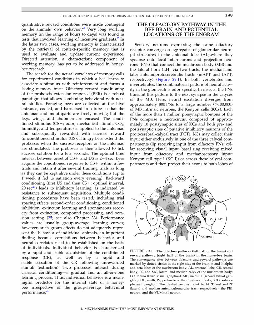
Sensory neurons expressing the same olfactoryreceptor converge on aggregates of glomerular neuro-pil structures in the antennal lobe (AL),where theysynapse onto local interneurons and projection neu-rons (PNs) that connect the mushroom body (MB) andthe lateral horn (LH) via two tracts, the median andlater antennoprotocerebralis tracts (mAPT and lAPT,respectively) (Figure 29.1). In both vertebrates andinvertebrates, the combinatorial pattern of neural activ-ity in the glomeruli is odor specific. In insects, the PNstransmit this pattern to the next synapse in the calycesof the MB. Here, neural excitation diverges fromapproximately 800 PNs to a large number (.100,000)of MB intrinsic neurons, the Kenyon cells (KCs). Eachof the more than 1 million presynaptic boutons of thePNs comprise a microcircuit composed of approxi-mately 10 postsynaptic sites of KCs and both pre- andpostsynaptic sites of putative inhibitory neurons of theprotocerebral-calycal tract (PCT). KCs may collect theirinput either exclusively in one of the three calycal com-partments (lip receiving input from olfactory PNs, col-lar receiving visual input, basal ring receiving mixedinput from olfactory and mechanosensory input;Kenyon cell type I (KC I)) or across these calycal com-partments and then project their axons to both lobes of
FIGURE 29.1 The olfactory pathway (left half of the brain) andreward pathway (right half of the brain) in the honeybee brain.
The convergence sites between olfactory and reward pathways aremarked by dotted circles in the right side of the brain. α and β, alphaand beta lobes of the mushroom body; AL, antennal lobe; CB, centralbody; LC and MC, lateral and median calyx of the mushroom body;LO, lobula (third visual ganglion); ME, medulla (second visual gan-glion); OC, ocelli; Pe, peduncle of the mushroom body; SOG, subeso-phageal ganglion. The dashed arrows point to lAPT and mAPT(lateral and median antennoglomerular tract, respectively), the PE1neuron, and the VUMmx1 neuron.
399THE OLFACTORY PATHWAY IN THE BEE BRAIN AND POTENTIAL LOCATIONS OF THE ENGRAM
4. MECHANISMS FROM THE MOST IMPORTANT SYSTEMS
the MB (α and β lobes; Kenyon cell type II (KC II)).The axons of both types form collaterals halfway alongthe peduncle and project to the α and β lobes. KC Iproject one collateral to the dorsal α lobe and the otherto the caudal part of the β lobe. KC II project one tothe ventral part of the α lobe and the other to the prox-imal part of the β lobe.27 The large number of KCs con-verge on a rather small number (B400) of MB extrinsicneurons (ENs), leaving the α lobe at three prominentexit points—the lateral, ventral, and ventromedian exitpoints.28 These ENs are divided into seven subgroups(A1�A7) depending on the localization of theirsomata. Most of them are postsynaptic to KCs, asjudged by their spiny-like structure and in some casesby electron microscopic evidence, but post- and pre-synaptic structures are known to occur in close vicin-ity. ENs project to many parts of the brain—some ofthem (e.g., the identified neurons PE1 and the A4 neu-rons) to different subregions of the LH, where theyconverge directly or indirectly with collaterals ofmAPT and lAPT. Other ENs (A3 neurons) project via arecurrent pathway back to the calyx of the same MBalong the GABA-immunoreactive (GABA-ir) PCT.Multiple ENs connect the ring neuropil around the αlobe (A1, A2, A4, and A7) with the MB on the ipsi- orcontralateral side (A6 and A7) or with other protocer-ebral areas on the ipsi- or contralateral side (A4, A5,and A7). A single neuron has been identified thatappears to project back to the ipsilateral AL (the AL1).Dendrites of ENs are often restricted within the α lobeto one of the horizontal bands, suggesting that theyreceive sensory modality-specific input via KCs.Others distribute their dendrites across the bandedstructure of the α and β lobes. The structural diversityof ENs reflects a multiplicity of functions concerningthe readout from the MB and the information flow toother parts of the brain.
Neurons containing the neuromodulators octopa-mine (OA) and dopamine are related to the reinforcingfunctions during conditioning both in Drosophila (seeChapters 5 and 27) and in the bee. One OA immunore-active neuron, the VUMmx1, was identified in areward substitution experiment to be sufficient for thereward function of sucrose in olfactory conditioning inthe bee.29 VUMmx1 receives its input in the subeso-phageal ganglion and converges with the olfactorypathway at three pairs of symmetrical sites—the ALs,the LHs, and the lip regions of the MB calyces, respec-tively (Figure 29.1). Thus, it has been hypothesizedthat these convergence sites may constitute localiza-tions of the olfactory engram as it develops in rewardlearning,30 and therefore recordings of learning-relatedneural plasticity focused on the neurons and their syn-aptic connections so far on two of these three sites (ALand MB calyx). A functional MB was previously found
to be required for the consolidation of olfactory STMinto MTM.31 The VUMmx1 neuron responds tosucrose and to many other stimuli. In the course ofconditioning, it enhances its response to the forwardpaired conditioned olfactory stimulus (CS1) andreduces its response to the backward paired stimulus(CS2) (see Figure 29.10). Interestingly, regarding thenotion of expectation and anticipation, the octopami-nergic neuron VUMmx1 exhibits activity reflecting theanimal’s expectation: It responds to unexpected sucrosepresentations but not to expected ones.29 Althoughthere is evidence in vertebrates of how this reduction inthe error signal may be implemented biologically, suchevidence is still lacking in invertebrates.
THE ANTENNAL LOBE
The AL of the honeybee is believed to constitute acomponent of a distributed network storing olfactoryinformation, but evidence is controversial. As notedpreviously, convergence of the olfactory and rewardpathway in the AL suggests a memory trace to beformed in the AL. Indeed, substituting reward in olfac-tory PER conditioning by local injection of octopamine(the putative transmitter of VUMmx1) into the ALleads to learning of the forward but not the backwardpaired odor.32 Accordingly, blocking octopaminereceptors in the AL with RNAi reduces olfactorylearning.33 Additional arguments in favor of a memorytrace in the AL concern (1) the role of the AL in mem-ory consolidation and (2) neural correlates of a mem-ory trace. First, memory consolidation induced by asingle learning trial was found to be blocked if the ALis cooled during the minute following the trial,whereas cooling even immediately after the last ofmultiple learning trials does not impair memory con-solidation, suggesting that the AL possibly in connec-tion with other brain parts stores a short-lastingmemory trace necessary for consolidation.31 Localuncaging of cAMP in the AL (cAMP promotes thetransfer from STM to LTM in bees) shifts STM to LTMwhen it is uncaged soon after a single learning trial.15
So far, it has not been possible to test directly whethera more permanent memory trace is stored in the ALbecause blocking neural activity in the AL duringretrieval tests interferes with the processing of olfac-tory coding. Second, neural correlates of olfactorylearning were collected with two methods—Ca21
imaging of glomerular activity and extracellularrecordings from PNs. In the first case, the Ca21 signalscame either predominantly from the presynaptic term-inals of olfactory receptor neurons (and possibly alsofrom glia cells) in the glomeruli or from the postsynap-tic elements, the PNs. Presynaptic signals increased for
400 29. IN SEARCH OF THE ENGRAM IN THE HONEYBEE BRAIN
4. MECHANISMS FROM THE MOST IMPORTANT SYSTEMS
the CS1 odor.34 Controversial data exist for associativeplasticity in PNs. Peele and co-workers35 found noconsistent changes for CS1 or CS2 during differentialPER conditioning, whereas Weidert and co-workers36
did. These inconsistencies may be resolved on thebasis of the data from multiunit recordings of PNs.In line with the interpretation that PNs undergo asso-ciative change is the finding by Fernandez andco-workers37 that binary mixtures of odors are codedmore differently for learned odors and this effect corre-lates with changes of neural responses as seen in Ca21-imaging. Rath and co-workers38 reported that PNsundergo associative plasticity in differential PER con-ditioning depending on their response level to therespective CS1 and CS2 before conditioning:Glomeruli responding to CS1 before conditioningenhanced their CS1 responses, those that responded toCS2 did not change their responses, and those thatresponded before both CS1 and CS2 either reduced orenhanced their respective odor responses depending onthe strength of their responses (weak responses wereenhanced, and strong responses were reduced). Themodel derived from these studies assumes two types ofplastic synapses in the glomeruli: (1) synapses between
olfactory receptor neurons and PNs and (2) synapsesbetween olfactory receptor neurons and local interneur-ons. Taken together, these results indicate that odorlearning improves spatial representations of the learnedodors and facilitates their discrimination—forms ofspecified memory traces that contribute importantcomponents to corresponding memory traces storedsomewhere else.
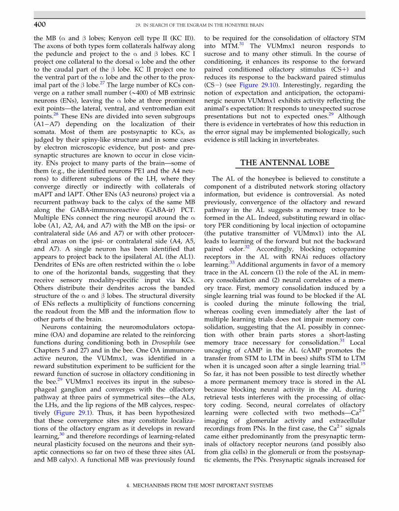
Multiunit extracellular recordings from PNs docu-mented both increases and decreases in rate changesto the reinforced (CS1) and the specifically not rein-forced (CS2) odor (Figure 29.2),39 but it is unknownhow these effects relate to the differential associativechanges seen in Ca21 imaging. If such associativelyup- and downregulated PNs receive their inputswithin the same glomerulus (e.g., for the CS1), it isnot surprising that the overall associative changes seenby Ca21 imaging of whole glomeruli may cancel eachother out. A model implementing these findings andmaking specific assumptions about spike timing-dependent plasticity induced by activity of the rewardneuron VUMmx1 on the connection between localinterneurons and PN predicts asymmetric changes ofPN responses to the rewarded neuron.36
FIGURE 29.2 Changes in LFP power of projection neurons in the course of differential conditioning as recorded by multiple extracel-lular electrodes. (A) Time-resolved power spectra for CS1 tests before (left; Pre) and after (right; Post) differential conditioning averagedacross three test trials for all animals. Dashed white lines indicate stimulus onset and offset; power is indicated in color scale. The white circlesindicate a decrease of power in the high-frequency band, and the dark circles indicate an increase in the low-frequency band. (B) Averagepower change during the on-response resolved by individual frequency bands for CS1 (left), CS2 (middle), and a control odor, Ctrl (right).Before averaging across animals, the differences between power before and after conditioning were calculated. Error bars (62.5%) wereobtained using 1000 bias-corrected standard bootstraps. Source: After Denker et al.39
401THE ANTENNAL LOBE
4. MECHANISMS FROM THE MOST IMPORTANT SYSTEMS

The high temporal resolution of spike recordingsallows for the analysis of the ensemble activity usingodor-induced local field potentials (LFPs) and theirrelation to single-unit activity. The largest learning-related difference was found for CS1. LFP powerincreases for CS1 in the 15- to 40-Hz frequency bandand decreases for frequencies higher than 45 Hz39
(Figure 29.2). This learning-related power change cor-relates with the size of the neuronal ensemble that isphase-locked to the particular frequency: After learn-ing, less units are entrained to the higher frequencyband, and more units are entrained to the lowerband. These results reflect associative plasticity inthe AL resulting from a restructured odor codingnetwork.
The memory trace in the AL as seen by opto- andelectrophysiological recordings results from multipletraining trials, suggesting that it represents a lastingtrace. It optimizes primary odor coding both at thespatial and at the temporal domain. It is unknownwhether other neuropils or neural tracks (e.g.,feedback neurons from the MB) are required forits formation and readout and whether it containsinformation about the specifically learned odors.To test for this, it will be necessary to manipulateselectively the contribution of neural subsets withinthe AL separately for learning, consolidation, andretrieval.
INTRINSIC NEURONS OF THEMUSHROOM BODY: KENYON CELLS
MBs are expected to house the engram of insects.In 1896, Kenyon40 stated the following:
Ever since Dujardin41 discovered the mushroom bodiesand pointed out the relation between their size and the devel-opment of insect intelligence, nearly every writer on the sub-ject of the hexapod brain who has referred to the matter ofintelligence has recognized the fact. (p. 161)
However, even with the brilliant work in Drosophila,direct evidence is rather scarce. A first hint in favor ofthe idea that the MB is involved in memory storagecame from the finding that the time course of retro-grade amnesia induced by cooling the honeybeecalyces matches the time course of cooling the wholeanimal.31 Olfactory memory is expected to be locatedin the lips of the calyces because they comprise thesecond-order convergence sites of the olfactory path-way with the reward pathway, suggesting associativeprocessing at the MB input. PNs are presynaptic toKCs in discrete microcircuits, the microglomeruli, com-posed of one large presynaptic bouton of PNs, 6�12postsynaptic KC spines, and usually one GABA-ir pro-file, the presynaptic site of A3-v neurons of the PCT,and frequently a profile with dense core vesicles mostlikely from the OA-ir VUMmx1 neuron (Figure 29.3).
FIGURE 29.3 Synaptic organiza-
tion of the microglomerulus in the lip
region of the MB. (Top right) Theterminals of two projection neurons(one in yellow and one in blue) in thelip region of the calyx with their multi-ple presynaptic swellings (boutons).(Left) Electron microscopic view: Thelarge presynaptic bouton of a projectionneuron (PN; surrounded by a yellowline) comprises the center of the micro-circuit. It is presynaptic to multiplespines of Kenyon cells (KC) and post-synaptic to GABA-ir profiles (inh. N;blue) of the A3-v neurons. The boutonalso receives input from profiles withdense core vesicles (DG; pink) inter-preted to represent presynaptic sitesof the reward pathway (VUMmx1).(Bottom right) The schematic represen-tation of the microcircuit indicatesdirections of synaptic contacts andassumes modulatory input (mod. N) toall three partners of the circuit. Source:After Ganeshina et al.42
402 29. IN SEARCH OF THE ENGRAM IN THE HONEYBEE BRAIN
4. MECHANISMS FROM THE MOST IMPORTANT SYSTEMS
The density of these microglomeruli depends on theage and experience of the animals and increases dur-ing protein synthesis-dependent consolidation intoolfactory LTM.43 This latter finding was interpreted todocument a structural correlate of the olfactory mem-ory trace based on the growth of new synapses—anintriguing interpretation that will become even moreconvincing if it becomes possible to documentstimulus-specific changes of microglomerulus patterns.
KCs feature a sparse odor code in a twofold manner:An odor activates a small proportion of highly odor-specific KCs, and in contrast to the presynaptic PNs,KCs respond with brief and phasic responses oftencombined with off-responses,44 corroborating findingsin the locust.45 Stimuli of different modalities inducequalitatively similar responses, activating small subsetsof KCs. Theoretically, temporal and population sparse-ness makes KCs potentially well suited as a memorystore because the organization of the calyx can be con-ceptualized as an associative matrix comparable to thenetwork of the hippocampus or cortex.46 The memorytrace as stored in such an associative matrix is charac-terized by features such as partial overlap betweenclosely related traces, an optimal number of changesper trace (1�5% of the total number of synaptic
contacts), and the ability to reconstruct the full patterneven if only part of the trace is activated.
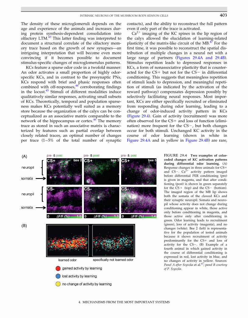
Ca21 imaging of the KC spines in the lip region ofthe calyx allowed the elucidation of learning-relatedplasticity of the matrix-like circuit of the MB.47 For thefirst time, it was possible to reconstruct the spatial dis-tribution of multiple changes in a neural net with alarge range of partners (Figures 29.4A and 29.4B).Stimulus repetition leads to depressed responses inKCs, a form of nonassociative plasticity that is counter-acted for the CS1 but not for the CS2 in differentialconditioning. This suggests that meaningless repetitionof stimuli leads to depression, and meaningful repeti-tion of stimuli (as indicated by the activation of thereward pathway) compensates depression possibly byselectively facilitating neural responses. Most impor-tant, KCs are either specifically recruited or eliminatedfrom responding during odor learning, leading to achange of odor-induced activity pattern in KCs(Figure 29.4). Gain of activity (recruitment) was moreoften observed for the CS1 and loss of function (elimi-nation) more frequent for the CS2, but both changesoccur for both stimuli. Unchanged KC activity in thecourse of odor learning (shown in white inFigure 29.4A and in yellow in Figure 29.4B) are rare,
FIGURE 29.4 Two examples of color-
coded changes of KC activation patterns
during differential odor learning. (A)Response changes in three animals for CS1and CS2. Ca21 activity pattern imagedbefore differential PER conditioning (pre)is given in magenta, and that after condi-tioning (post) is shown in green separatelyfor the CS1 (top) and the CS2 (bottom).The imaged region of the MB lip showsboth the somata of the clawed KCs andtheir synaptic neuropil. Somata and neuro-pil whose activity does not change duringconditioning appear in white, those activeonly before conditioning in magenta, andthose active only after conditioning ingreen. Odor learning leads to recruitment(green), loss of activity (magenta), and nochanges (white). Bee 2 (left) is representa-tive for the population of tested animalsbecause it shows recruitment of activitypredominantly for the CS1 and loss ofactivity for the CS2. (B) Example of afourth animal in which gained activity inthe course of differential conditioning isexpressed in red, lost activity in blue, andno changes of activity in yellow. Sources:Panel A after Szyszka et al.47; panel B courtesyof P. Szyszka.
403INTRINSIC NEURONS OF THE MUSHROOM BODY: KENYON CELLS
4. MECHANISMS FROM THE MOST IMPORTANT SYSTEMS
indicating that learning leads to a drastic rearrange-ment of odor representation in the MB input. Becauseodors activate primarily non-overlapping KC ensem-bles, the parallel representations of multiple odortraces allow for an effective and robust memory trace.In the future, it will be necessary to compare patterns ofchanges for odors generalized more or less.Furthermore, it will be necessary to show that multi-modal stimuli lead to a more precise KC activity patternrather than to a higher number of activated KCs.
In addition to these changes in activity patterns,KCs also undergo dynamic changes. Before condition-ing, their odor responses are short, even during long-lasting odor stimulation.43 During odor�sucrosepairing, the odor-activated KCs become reactivated,leading to coincident activity in odor coding andreward coding neurons, a possible mechanism fordelayed and trace conditioning.48 The picture emerg-ing from Ca21 imaging studies assumes an intracellu-lar trace for the CS1, possibly in the form of a lastingincrease in Ca2149 that is associatively paired with adelayed OA input from VUMmx1 leading to anenhancement of KC activity. A reduced response toCS1 in KCs may result from a similar mechanism ininhibitory inputs to KCs—for example, via A3 neuronsof the PCT, a mechanism suggested by the close appo-sition of OA-ir profiles and GABA-ir profiles in micro-glomeruli of the calyx lip (Figure 29.3). Taken together,it is conceivable that the olfactory engram in the MBlip comprises a combinatorial pattern of predomi-nantly enhanced synaptic transmission to KCs but alsoreduced transmission, leading to the conclusion thatKCs store a memory trace in stimulus-specific sparseactivation patterns.
As noted previously, KC I receive their input selec-tively via small dendritic fields from lip, collar, orbasal ring and project one axon collateral to the dorsalhalf of the α lobe and the other to the caudal part ofthe β lobe. KC II, to which the imaged clawed KCsbelong, collect input across the calyx, thus receivinginput from multiple sensory modalities via elaborateand clawed dendritic fields, and project one of theiraxons to the ventral part of the α lobe and the other tothe proximal part of the β lobe. KC II converge on asmall number of MB ENs, whereas KC I serve moreENs. One would expect the two types of KCs to pro-cess high-order sensory input and value signals differ-ently, possibly leading to two parallel coding, storing,and retrieval schemes within the MB—a concept ofhigh relevance in Drosophila MB function50 (seeChapter 27 for further discussion of controversialdata). Unfortunately, electrophysiological recordingsfrom KCs of either type have been unsuccessful so fardespite intensive efforts, and imaging experimentshave not been performed in KC I. It will be an
important task for the future to unravel the specificcoding schemes in the two KC types as well as eluci-date the potentially different roles of the median andlateral calyces with their KC projections to the innerand outer part of the lobes, respectively.
Although the MB in bees is large in comparisonto the whole brain, its total volume (253 106 μm351) issmall given the high number of densely packed KCs.Witthoft’s52 estimate of 170,000 KCs needs revision to alower number, but even 100,000�150,000 KCs asderived from a comparison between the volume of sin-gle KCs and total MB volume (see Chapter 4) is a veryhigh number. Obviously, MB intrinsic circuitry isdesigned to take advantage of small neuron size (lessmaterial, lower energy consumption) and particularlyeffective interneuron cross talk via short connections.It thus can be concluded that the miniaturized MB cir-cuitry pushes information processing capacity (IPC) toan upper limit with the lowest possible volume of neu-ral tissue, highest efficiency of use of material and met-abolic energy, and shortest interneuron connections.However, axon diameters well below 0.5 μm causeproblems of reliable transfer of action potentialsbecause reduced numbers of ion channels in suchsmall membrane areas lead to a decline in signal-to-noise ratio.53 One important component in optimiza-tion of IPC in the MB may be related to the lowspiking activity in KCs, a phenomenon well documen-ted in the MB of locusts45 but only indirectly assumedin the honeybee MB. IPC, rather than absolute or rela-tive brain volumes, is considered to be the major deter-mining factor in brain�intelligence relations.54 The MBappears to optimize this factor by its dense packing ofKCs. It will be exciting in future work to unravel thephysiological and anatomical measures of IPC in KCs.
EXTRINSIC NEURONS OF THEMUSHROOM BODY
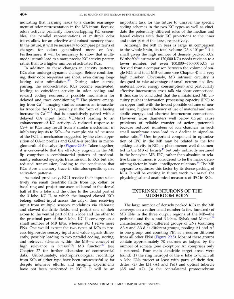
The large number of densely packed KCs in the MBconverge on a rather small number (a few hundred) ofMB ENs in the three output regions of the MB—thepeduncle and the α and β lobes. Rybak and Menzel28
characterized eight different groups of ENs (countingA3-v and A3-d as different groups, pooling A1 and A2in one group, and counting PE1 as a neuron differentfrom all other ENs) (Figure 29.5). Most of these groupscontain approximately 70 neurons as judged by thenumber of somata (one exception: A5 comprises only4 neurons). Four main dendritic target areas werefound: (1) the ring neuropil of the α lobe to which allα lobe ENs project at least with parts of their den-drites, (2) the LH (A4 and Pe1) and optical tubercle(A5 and A7), (3) the contralateral protocerebrum
404 29. IN SEARCH OF THE ENGRAM IN THE HONEYBEE BRAIN
4. MECHANISMS FROM THE MOST IMPORTANT SYSTEMS
(A6 and A7), and (4) the feedback neurons to the calyx(A3). The multiplicity of connections established bythese ENs makes it very likely that each group servesa different function. To date, these differences have notbeen able to be interpreted because we are ignorantabout the functional characteristics of many of the tar-get areas. In particular, we do not know the functionalproperties of the ring neuropil around the α lobe andthe various subregions of the unstructured lateral pro-tocerebrum. In any case, this structural multiplicitybetween EN groups and the number of neurons pergroup suggests forms of combinatorial coding of neuralprocessing categories that are defined by the respectiveinput and output regions. What are these categories?
Because many of these ENs receive input across themodality-specific regions of the MB, it is not surprisingthat they respond to a large range of sensory stimuli,indicating a different coding scheme than the highlyspecific combinatorial sensory code at the input of theMB. One large EN, the PE1, offers the unique possibility
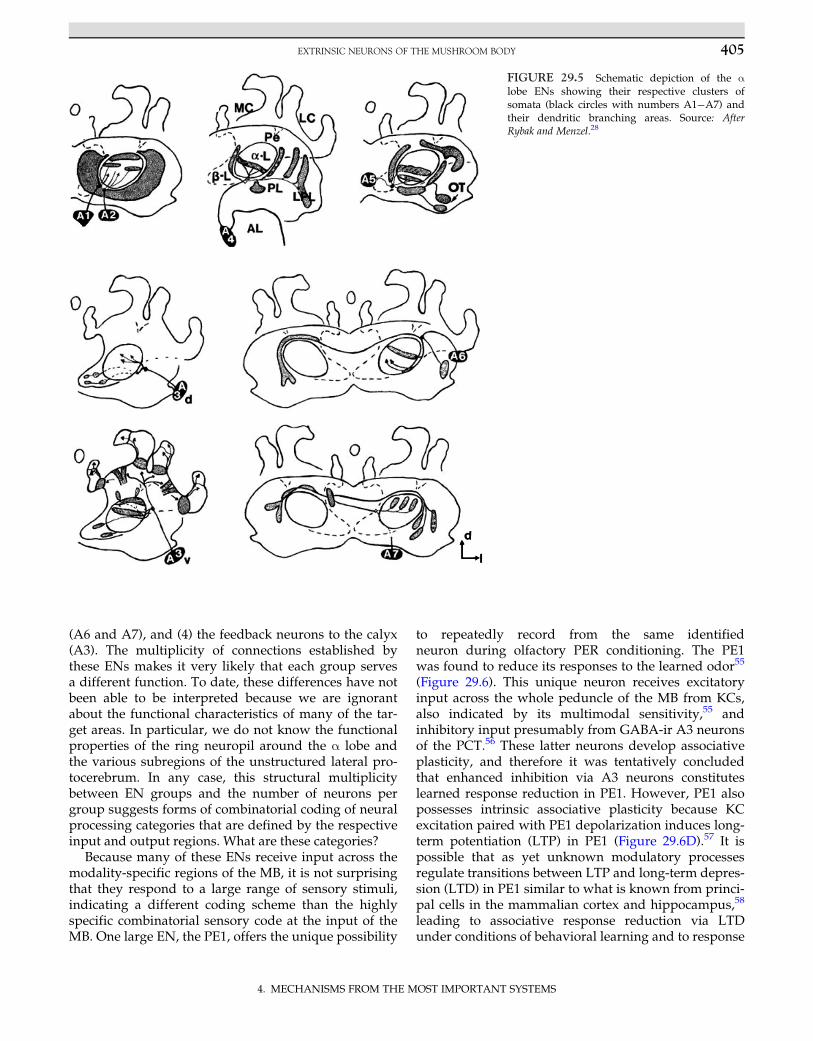
to repeatedly record from the same identifiedneuron during olfactory PER conditioning. The PE1was found to reduce its responses to the learned odor55
(Figure 29.6). This unique neuron receives excitatoryinput across the whole peduncle of the MB from KCs,also indicated by its multimodal sensitivity,55 andinhibitory input presumably from GABA-ir A3 neuronsof the PCT.56 These latter neurons develop associativeplasticity, and therefore it was tentatively concludedthat enhanced inhibition via A3 neurons constituteslearned response reduction in PE1. However, PE1 alsopossesses intrinsic associative plasticity because KCexcitation paired with PE1 depolarization induces long-term potentiation (LTP) in PE1 (Figure 29.6D).57 It ispossible that as yet unknown modulatory processesregulate transitions between LTP and long-term depres-sion (LTD) in PE1 similar to what is known from princi-pal cells in the mammalian cortex and hippocampus,58
leading to associative response reduction via LTDunder conditions of behavioral learning and to response
FIGURE 29.5 Schematic depiction of the αlobe ENs showing their respective clusters ofsomata (black circles with numbers A1�A7) andtheir dendritic branching areas. Source: AfterRybak and Menzel.28
405EXTRINSIC NEURONS OF THE MUSHROOM BODY
4. MECHANISMS FROM THE MOST IMPORTANT SYSTEMS
enhancement via LTP under conditions of tetanic KCstimulation as used in the study by Menzel andManz.57 In such a scenario, associative response reduc-tion to the CS1 would not reflect enhanced inhibitionvia A3 neurons but would represent an additional PE1-specific associative mechanism. Associative LTP andLTD in PE1 could also reflect spike timing-dependentplasticity (STDP), leading to either enhancement orreduction of synaptic efficiency, depending on the pre-cise timing of spikes from KCs—a mechanism modeledfor associative plasticity in the antennal lobe andreported for ENs in the locust.59 However, STDP hasyet to be proven to be related to behavioral learning inan insect.
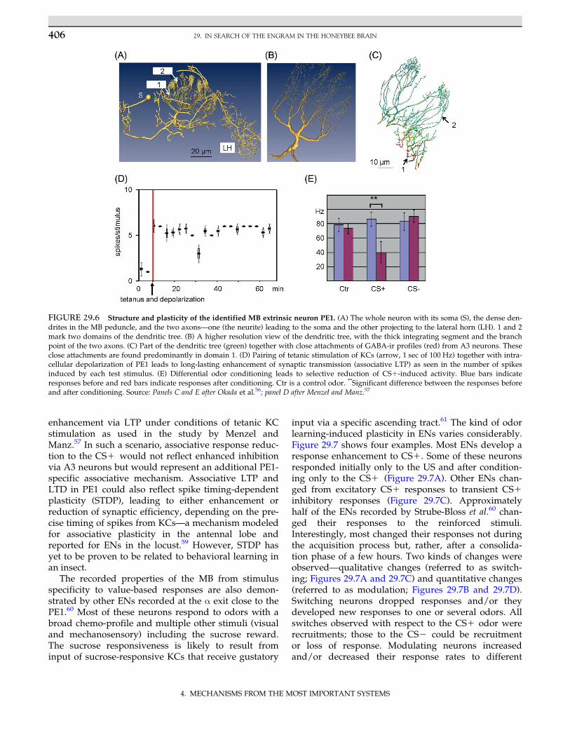
The recorded properties of the MB from stimulusspecificity to value-based responses are also demon-strated by other ENs recorded at the α exit close to thePE1.60 Most of these neurons respond to odors with abroad chemo-profile and multiple other stimuli (visualand mechanosensory) including the sucrose reward.The sucrose responsiveness is likely to result frominput of sucrose-responsive KCs that receive gustatory
input via a specific ascending tract.61 The kind of odorlearning-induced plasticity in ENs varies considerably.Figure 29.7 shows four examples. Most ENs develop aresponse enhancement to CS1. Some of these neuronsresponded initially only to the US and after condition-ing only to the CS1 (Figure 29.7A). Other ENs chan-ged from excitatory CS1 responses to transient CS1inhibitory responses (Figure 29.7C). Approximatelyhalf of the ENs recorded by Strube-Bloss et al.60 chan-ged their responses to the reinforced stimuli.Interestingly, most changed their responses not duringthe acquisition process but, rather, after a consolida-tion phase of a few hours. Two kinds of changes wereobserved—qualitative changes (referred to as switch-ing; Figures 29.7A and 29.7C) and quantitative changes(referred to as modulation; Figures 29.7B and 29.7D).Switching neurons dropped responses and/or theydeveloped new responses to one or several odors. Allswitches observed with respect to the CS1 odor wererecruitments; those to the CS2 could be recruitmentor loss of response. Modulating neurons increasedand/or decreased their response rates to different
FIGURE 29.6 Structure and plasticity of the identified MB extrinsic neuron PE1. (A) The whole neuron with its soma (S), the dense den-drites in the MB peduncle, and the two axons—one (the neurite) leading to the soma and the other projecting to the lateral horn (LH). 1 and 2mark two domains of the dendritic tree. (B) A higher resolution view of the dendritic tree, with the thick integrating segment and the branchpoint of the two axons. (C) Part of the dendritic tree (green) together with close attachments of GABA-ir profiles (red) from A3 neurons. Theseclose attachments are found predominantly in domain 1. (D) Pairing of tetanic stimulation of KCs (arrow, 1 sec of 100 Hz) together with intra-cellular depolarization of PE1 leads to long-lasting enhancement of synaptic transmission (associative LTP) as seen in the number of spikesinduced by each test stimulus. (E) Differential odor conditioning leads to selective reduction of CS1-induced activity. Blue bars indicateresponses before and red bars indicate responses after conditioning. Ctr is a control odor. **Significant difference between the responses beforeand after conditioning. Source: Panels C and E after Okada et al.56; panel D after Menzel and Manz.57
406 29. IN SEARCH OF THE ENGRAM IN THE HONEYBEE BRAIN
4. MECHANISMS FROM THE MOST IMPORTANT SYSTEMS
odors: CS1 always provoked increased responses,whereas CS2 and control odors (the latter were usedto test generalization phenomena) decreased orincreased responses in approximately equal propor-tions. It was argued that the dichotomy of ‘switching’and ‘modulating’ neurons may result from morpholog-ically distinct ENs because switched and modulatedneurons were rarely observed in the same recording.The delayed expression of associative plasticity in theswitched and modulated ENs could reflect memoryconsolidation that depends on prolonged neural activi-ties because consolidation in the MB can be blocked bycooling.
ENs appear to reflect both KC-related and ownendogenous plasticity. The multiplicity of responsechanges during and after associative learning in several
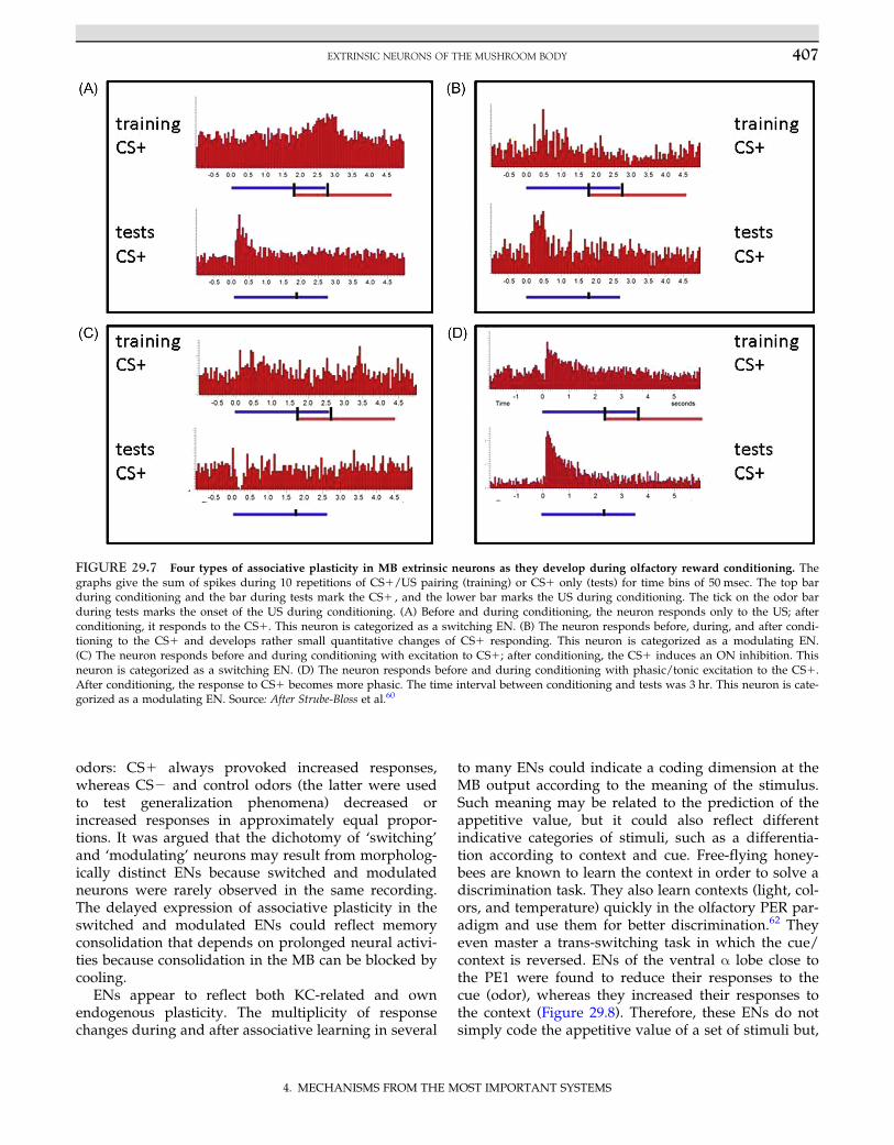
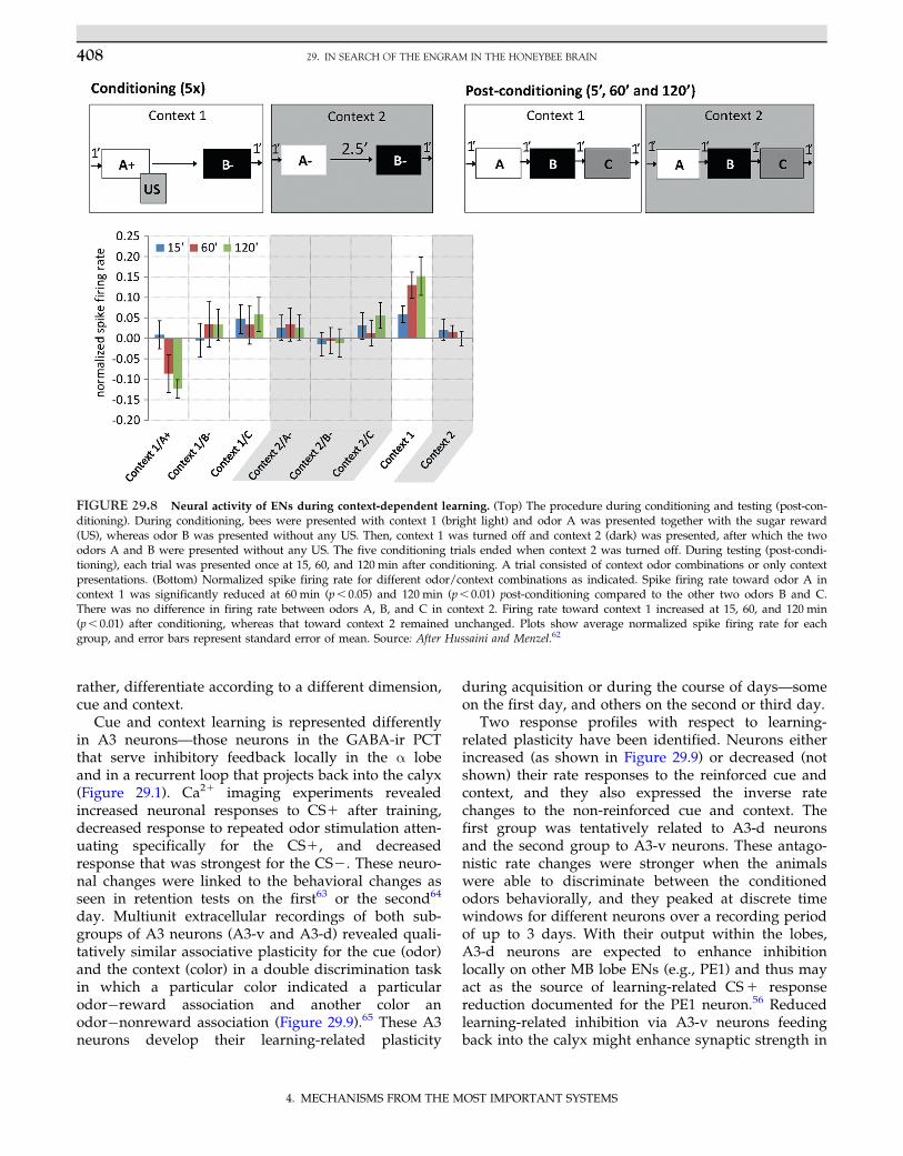
to many ENs could indicate a coding dimension at theMB output according to the meaning of the stimulus.Such meaning may be related to the prediction of theappetitive value, but it could also reflect differentindicative categories of stimuli, such as a differentia-tion according to context and cue. Free-flying honey-bees are known to learn the context in order to solve adiscrimination task. They also learn contexts (light, col-ors, and temperature) quickly in the olfactory PER par-adigm and use them for better discrimination.62 Theyeven master a trans-switching task in which the cue/context is reversed. ENs of the ventral α lobe close tothe PE1 were found to reduce their responses to thecue (odor), whereas they increased their responses tothe context (Figure 29.8). Therefore, these ENs do notsimply code the appetitive value of a set of stimuli but,
FIGURE 29.7 Four types of associative plasticity in MB extrinsic neurons as they develop during olfactory reward conditioning. Thegraphs give the sum of spikes during 10 repetitions of CS1/US pairing (training) or CS1 only (tests) for time bins of 50 msec. The top barduring conditioning and the bar during tests mark the CS1 , and the lower bar marks the US during conditioning. The tick on the odor barduring tests marks the onset of the US during conditioning. (A) Before and during conditioning, the neuron responds only to the US; afterconditioning, it responds to the CS1. This neuron is categorized as a switching EN. (B) The neuron responds before, during, and after condi-tioning to the CS1 and develops rather small quantitative changes of CS1 responding. This neuron is categorized as a modulating EN.(C) The neuron responds before and during conditioning with excitation to CS1; after conditioning, the CS1 induces an ON inhibition. Thisneuron is categorized as a switching EN. (D) The neuron responds before and during conditioning with phasic/tonic excitation to the CS1.After conditioning, the response to CS1 becomes more phasic. The time interval between conditioning and tests was 3 hr. This neuron is cate-gorized as a modulating EN. Source: After Strube-Bloss et al.60
407EXTRINSIC NEURONS OF THE MUSHROOM BODY
4. MECHANISMS FROM THE MOST IMPORTANT SYSTEMS
rather, differentiate according to a different dimension,cue and context.
Cue and context learning is represented differentlyin A3 neurons—those neurons in the GABA-ir PCTthat serve inhibitory feedback locally in the α lobeand in a recurrent loop that projects back into the calyx(Figure 29.1). Ca21 imaging experiments revealedincreased neuronal responses to CS1 after training,decreased response to repeated odor stimulation atten-uating specifically for the CS1, and decreasedresponse that was strongest for the CS2. These neuro-nal changes were linked to the behavioral changes asseen in retention tests on the first63 or the second64
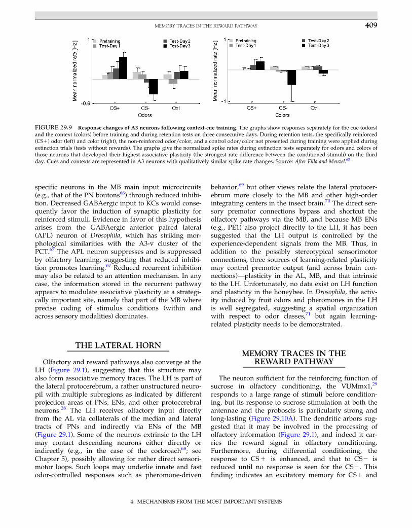
day. Multiunit extracellular recordings of both sub-groups of A3 neurons (A3-v and A3-d) revealed quali-tatively similar associative plasticity for the cue (odor)and the context (color) in a double discrimination taskin which a particular color indicated a particularodor�reward association and another color anodor�nonreward association (Figure 29.9).65 These A3neurons develop their learning-related plasticity
during acquisition or during the course of days—someon the first day, and others on the second or third day.
Two response profiles with respect to learning-related plasticity have been identified. Neurons eitherincreased (as shown in Figure 29.9) or decreased (notshown) their rate responses to the reinforced cue andcontext, and they also expressed the inverse ratechanges to the non-reinforced cue and context. Thefirst group was tentatively related to A3-d neuronsand the second group to A3-v neurons. These antago-nistic rate changes were stronger when the animalswere able to discriminate between the conditionedodors behaviorally, and they peaked at discrete timewindows for different neurons over a recording periodof up to 3 days. With their output within the lobes,A3-d neurons are expected to enhance inhibitionlocally on other MB lobe ENs (e.g., PE1) and thus mayact as the source of learning-related CS1 responsereduction documented for the PE1 neuron.56 Reducedlearning-related inhibition via A3-v neurons feedingback into the calyx might enhance synaptic strength in
FIGURE 29.8 Neural activity of ENs during context-dependent learning. (Top) The procedure during conditioning and testing (post-con-ditioning). During conditioning, bees were presented with context 1 (bright light) and odor A was presented together with the sugar reward(US), whereas odor B was presented without any US. Then, context 1 was turned off and context 2 (dark) was presented, after which the twoodors A and B were presented without any US. The five conditioning trials ended when context 2 was turned off. During testing (post-condi-tioning), each trial was presented once at 15, 60, and 120 min after conditioning. A trial consisted of context odor combinations or only contextpresentations. (Bottom) Normalized spike firing rate for different odor/context combinations as indicated. Spike firing rate toward odor A incontext 1 was significantly reduced at 60 min (p, 0.05) and 120 min (p, 0.01) post-conditioning compared to the other two odors B and C.There was no difference in firing rate between odors A, B, and C in context 2. Firing rate toward context 1 increased at 15, 60, and 120 min(p, 0.01) after conditioning, whereas that toward context 2 remained unchanged. Plots show average normalized spike firing rate for eachgroup, and error bars represent standard error of mean. Source: After Hussaini and Menzel.62
408 29. IN SEARCH OF THE ENGRAM IN THE HONEYBEE BRAIN
4. MECHANISMS FROM THE MOST IMPORTANT SYSTEMS
specific neurons in the MB main input microcircuits(e.g., that of the PN boutons66) through reduced inhibi-tion. Decreased GABAergic input to KCs would conse-quently favor the induction of synaptic plasticity forreinforced stimuli. Evidence in favor of this hypothesisarises from the GABAergic anterior paired lateral(APL) neuron of Drosophila, which has striking mor-phological similarities with the A3-v cluster of thePCT.67 The APL neuron suppresses and is suppressedby olfactory learning, suggesting that reduced inhibi-tion promotes learning.67 Reduced recurrent inhibitionmay also be related to an attention mechanism. In anycase, the information stored in the recurrent pathwayappears to modulate associative plasticity at a strategi-cally important site, namely that part of the MB whereprecise coding of stimulus conditions (within andacross sensory modalities) dominates.
THE LATERAL HORN
Olfactory and reward pathways also converge at theLH (Figure 29.1), suggesting that this structure mayalso form associative memory traces. The LH is part ofthe lateral protocerebrum, a rather unstructured neuro-pil with multiple subregions as indicated by differentprojection areas of PNs, ENs, and other protocerebralneurons.28 The LH receives olfactory input directlyfrom the AL via collaterals of the median and lateraltracts of PNs and indirectly via ENs of the MB(Figure 29.1). Some of the neurons extrinsic to the LHmay contact descending neurons either directly orindirectly (e.g., in the case of the cockroach68; seeChapter 5), possibly allowing for rather direct sensori-motor loops. Such loops may underlie innate and fastodor-controlled responses such as pheromone-driven
behavior,69 but other views relate the lateral protocer-ebrum more closely to the MB and other high-orderintegrating centers in the insect brain.70 The direct sen-sory premotor connections bypass and shortcut theolfactory pathways via the MB, and because MB ENs(e.g., PE1) also project directly to the LH, it has beensuggested that the LH output is controlled by theexperience-dependent signals from the MB. Thus, inaddition to the possibly stereotypical sensorimotorconnections, three sources of learning-related plasticitymay control premotor output (and across brain con-nections)—plasticity in the AL, MB, and that intrinsicto the LH. Unfortunately, no data exist on LH functionand plasticity in the honeybee. In Drosophila, the activ-ity induced by fruit odors and pheromones in the LHis well segregated, suggesting a spatial organizationwith respect to odor classes,71 but again learning-related plasticity needs to be demonstrated.
MEMORY TRACES IN THEREWARD PATHWAY
The neuron sufficient for the reinforcing function ofsucrose in olfactory conditioning, the VUMmx1,29
responds to a large range of stimuli before condition-ing, but its response to sucrose stimulation at both theantennae and the proboscis is particularly strong andlong-lasting (Figure 29.10A). The dendritic arbors sug-gested that it may be involved in the processing ofolfactory information (Figure 29.1), and indeed it car-ries the reward signal in olfactory conditioning.Furthermore, during differential conditioning, theresponse to CS1 is enhanced, and that to CS2 isreduced until no response is seen for the CS2. Thisfinding indicates an excitatory memory for CS1 and
FIGURE 29.9 Response changes of A3 neurons following context-cue training. The graphs show responses separately for the cue (odors)and the context (colors) before training and during retention tests on three consecutive days. During retention tests, the specifically reinforced(CS1) odor (left) and color (right), the non-reinforced odor/color, and a control odor/color not presented during training were applied duringextinction trials (tests without rewards). The graphs give the normalized spike rates during extinction tests separately for odors and colors ofthose neurons that developed their highest associative plasticity (the strongest rate difference between the conditioned stimuli) on the thirdday. Cues and contexts are represented in A3 neurons with qualitatively similar spike rate changes. Source: After Filla and Menzel.65
409MEMORY TRACES IN THE REWARD PATHWAY
4. MECHANISMS FROM THE MOST IMPORTANT SYSTEMS
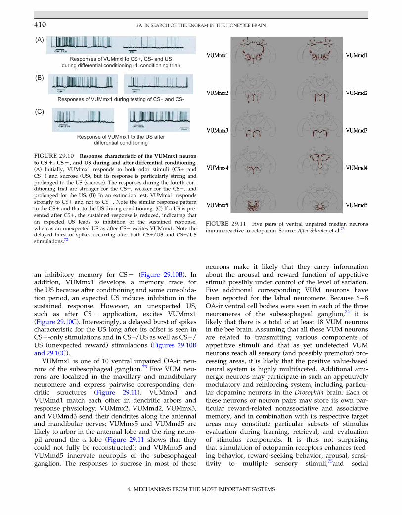
an inhibitory memory for CS2 (Figure 29.10B). Inaddition, VUMmx1 develops a memory trace forthe US because after conditioning and some consolida-tion period, an expected US induces inhibition in thesustained response. However, an unexpected US,such as after CS2 application, excites VUMmx1(Figure 29.10C). Interestingly, a delayed burst of spikescharacteristic for the US long after its offset is seen inCS1-only stimulations and in CS1/US as well as CS2/US (unexpected reward) stimulations (Figures 29.10Band 29.10C).
VUMmx1 is one of 10 ventral unpaired OA-ir neu-rons of the subesophageal ganglion.73 Five VUM neu-rons are localized in the maxillary and mandibularyneuromere and express pairwise corresponding den-dritic structures (Figure 29.11). VUMmx1 andVUMmd1 match each other in dendritic arbors andresponse physiology; VUMmx2, VUMmd2, VUMmx3,and VUMmd3 send their dendrites along the antennaland mandibular nerves; VUMmx5 and VUMmd5 arelikely to arbor in the antennal lobe and the ring neuro-pil around the α lobe (Figure 29.11 shows that theycould not fully be reconstructed); and VUMmx5 andVUMmd5 innervate neuropils of the subesophagealganglion. The responses to sucrose in most of these
neurons make it likely that they carry informationabout the arousal and reward function of appetitivestimuli possibly under control of the level of satiation.Five additional corresponding VUM neurons havebeen reported for the labial neuromere. Because 6�8OA-ir ventral cell bodies were seen in each of the threeneuromeres of the subesophageal ganglion,74 it islikely that there is a total of at least 18 VUM neuronsin the bee brain. Assuming that all these VUM neuronsare related to transmitting various components ofappetitive stimuli and that as yet undetected VUMneurons reach all sensory (and possibly premotor) pro-cessing areas, it is likely that the positive value-basedneural system is highly multifaceted. Additional ami-nergic neurons may participate in such an appetitivelymodulatory and reinforcing system, including particu-lar dopamine neurons in the Drosophila brain. Each ofthese neurons or neuron pairs may store its own par-ticular reward-related nonassociative and associativememory, and in combination with its respective targetareas may constitute particular subsets of stimulusevaluation during learning, retrieval, and evaluationof stimulus compounds. It is thus not surprisingthat stimulation of octopamin receptors enhances feed-ing behavior, reward-seeking behavior, arousal, sensi-tivity to multiple sensory stimuli,75and social
Responses of VUMmxl to CS+, CS- and USduring differential conditioning (4. conditioning trial)
Responses of VUMmx1 during testing of CS+ and CS-
Response of VUMmx1 to the US afterdifferential conditioning
(A)
(B)
(C)
FIGURE 29.10 Response characteristic of the VUMmx1 neuronto CS1 , CS2 , and US during and after differential conditioning.
(A) Initially, VUMmx1 responds to both odor stimuli (CS1 andCS2) and sucrose (US), but its response is particularly strong andprolonged to the US (sucrose). The responses during the fourth con-ditioning trial are stronger for the CS1, weaker for the CS2, andprolonged for the US. (B) In an extinction test, VUMmx1 respondsstrongly to CS1 and not to CS2. Note the similar response patternto the CS1 and that to the US during conditioning. (C) If a US is pre-sented after CS1, the sustained response is reduced, indicating thatan expected US leads to inhibition of the sustained response,whereas an unexpected US as after CS2 excites VUMmx1. Note thedelayed burst of spikes occurring after both CS1/US and CS2/USstimulations.72
FIGURE 29.11 Five pairs of ventral unpaired median neuronsimmunoreactive to octopamin. Source: After Schroter et al.73
410 29. IN SEARCH OF THE ENGRAM IN THE HONEYBEE BRAIN
4. MECHANISMS FROM THE MOST IMPORTANT SYSTEMS
interactions,76,77 whereas blocking octopamin receptorsreduces appetitive arousal, learning, and retrieval.33,78
THE DISTRIBUTED NATUREOF THE ENGRAM
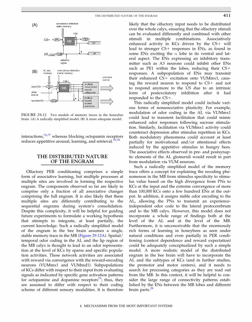
Olfactory PER conditioning comprises a simpleform of associative learning, but multiple processes atmultiple sites are involved in forming the respectiveengram. The components observed so far are likely tocomprise only a fraction of all associative changescomprising the full engram. It is also likely that thesemultiple sites are differently contributing to thesequential engrams during system’s consolidation.Despite this complexity, it will be helpful for guidingfuture experiments to formulate a working hypothesisthat attempts to integrate, at least partially, thecurrent knowledge. Such a radically simplified modelof the engram in the bee brain assumes a single,stable memory trace in the MB (Figure 29.12A). Spatial/temporal odor coding in the AL and the lip region ofthe MB calyx is thought to lead to an odor representa-tion at the level of KCs by sparse and specific popula-tion activities. These network activities are associatedwith reward via convergence with the reward-encodingneurons (VUMmx1 and VUMmd1). Subpopulationsof KCs differ with respect to their input from evaluatingsignals as indicated by specific gene activation patternsfor octopamine and dopamine receptors79; thus, theyare assumed to differ with respect to their codingscheme of different sensory modalities. It is therefore
likely that the olfactory input needs to be distributedover the whole calyx, ensuring that the olfactory stimulican be evaluated differently and combined with otherstimuli in multiple combinations. Associativelyenhanced activity in KCs driven by the CS1 willlead to stronger CS1 responses in ENs, as found insome ENs exciting the α lobe in its ventral and lat-eral aspect. The ENs expressing an inhibitory trans-mitter such as A3 neurons could inhibit other ENssuch as PE1 within the lobes, reducing their CS1responses. A subpopulation of ENs may transmittheir enhanced CS1 excitation onto VUMmx1, caus-ing the reward neuron to respond to CS1 and notto respond anymore to the US due to an intrinsicform of postexcitatory inhibition after it hadresponded to the CS1.
This radically simplified model could include vari-ous forms of nonassociative plasticity. For example,modulation of odor coding in the AL via VUMmx1could lead to transient facilitation that could mimicenhanced odor responses following sucrose stimula-tion. Similarly, facilitation via VUMmx1 activity couldcounteract depression after stimulus repetition in KCs.Both modulatory phenomena could account at leastpartially for motivational and/or attentional effectsinduced by the appetitive stimulus in hungry bees.The associative effects observed in pre- and postsynap-tic elements of the AL glomeruli would result in partfrom modulation via VUM neurons.
Such a radically simplified model of the memorytrace offers a concept for explaining the recoding phe-nomenon in the MB from stimulus specificity to stimu-lus value based on the high divergence from PNs toKCs at the input and the extreme convergence of morethan 100,000 KCs onto a few hundred ENs at the out-put. In addition, it assigns stable odor encoding to theAL, allowing the PNs to transmit an experience-independent odor code to the lateral protocerebrumand to the MB calyx. However, this model does notincorporate a whole range of findings both at thelevel of the AL and at the level of the MB.Furthermore, it is unconceivable that the enormouslyrich forms of learning in honeybees as seen undernatural conditions and even partially in PER condi-tioning (context dependence and reward expectation)could be adequately conceptualized by such a simplemodel. A more realistic model of the distributedengram in the bee brain will have to incorporate theAL and the subtypes of KCs (and in further studies,the premotor and motor centers), and it needs tosearch for processing categories as they are read outfrom the MB. In this context, it will be helpful to con-sider the large range of connectivity patterns estab-lished by the ENs between the MB lobes and differentbrain parts.28
FIGURE 29.12 Two models of memory traces in the honeybeebrain. (A) A radically simplified model. (B) A more adequate model.
411THE DISTRIBUTED NATURE OF THE ENGRAM
4. MECHANISMS FROM THE MOST IMPORTANT SYSTEMS
Evidence is strong for an independent memory tracein the AL. Although the stronger and more synchro-nized responses of PNs to the learned odor couldresult from feed-forward loops to the AL carryinginformation about the learned odor—for example, viathe reward pathway (VUMmx1) or/and via inputsfrom the MB—additional assumptions are necessary toinclude the different forms of associative plasticity inglomeruli as seen in both Ca21 imagining and multi-electrode recordings. PNs are either up- or downregu-lated for the CS1 depending on whether or not theyresponded to the CS1 before learning. It is unlikelythat such plasticity could result from VUMmx1 modu-latory signals or from a single forward loop from theMB. The stable enhancement of odor mixture codingafter learning requires plasticity of the odor codingnetwork in the AL and cannot be provided by generalmodulatory signals. Additional arguments in favor ofan independent olfactory memory trace in the ALcome from US substitution experiments by local injec-tion of octopamine into the AL and the finding thatblocking of octopamin receptors in the AL interfereswith olfactory learning. In addition, it was shown thatthe transition from short- to long-term memory can befacilitated by activating cAMP-dependent PKC in theAL. Taken together, both the MB calyx and the LHappear to receive odor signals from the AL that encodeexperience with the particular odors and counteractthe concept that the AL codes odors in a stable andexperience-independent way. However, what exactlyis transmitted about experience is unclear. It may wellbe that it is limited to enhanced attentional effectsrather than to indexing a specific odor.
The simplified model assigns the stimulus-specificmemory trace to the associative matrix of divergentPNs onto KCs in the lip of the MB calyx and, moregenerally, to all neurons reaching the calyx and feed-ing the more than 100,000 KCs of the MB. The matrixmemory could indeed store the rich content of thememory trace, including all relevant combinations ofexternal (cues and contexts) and internal stimuli,because formally it could possess the necessary intrin-sic properties—divergent and convergent connectivity,sparse population and temporal coding, and highthresholds in KCs—making them respond only to con-vergent input. Neuroanatomical evidence supports theassumption that all sensory inputs more or less pro-cessed converge in the MB calyx onto KCs. A tinyglimpse into such a storage device is given inFigure 29.4 for a specific subtype of KCs (clawed KCsof KC II) receiving input across the modality-specificregions (lip, collar, and basal ring) of the calyx. Asmentioned previously, the different types of KCs com-bine different subsets of inputs, some of which keepthe sensory modalities apart and others combine them.
Modulating and evaluating pathways reach the calyx(VUMmx1 and VUMmd1) or the peduncle (neuronsimmunoreactive to dopamine and serotonin). Theirpattern of convergence with the different KC types isunknown except for the fact that the two VUM neu-rons reach only the olfactory input (lip region of thecalyx) and not other sensory inputs. It will be neces-sary to demonstrate how visual and other modalitiesare evaluated by reward and whether such an associa-tive matrix-based model also applies to these sensorymodalities. It is also not known how aversive stimuliare evaluated by the MB, except for the hint that dopa-mine neurons are likely to be involved,80 which is alsocorroborated by findings in Drosophila.81 Because theexpression of aminergic receptor genes differs betweengroups of KCs, this indicates that subsets may be selec-tively involved in coding appetitive and aversiveforms of learning.79 These authors also provide evi-dence in favor of different subpopulations of KCs tostore short- and long-term olfactory memory.
Important neural components of the calycal matrixmemory also include the presynaptic sites—for exam-ple, the boutons of the PNs for olfactory memory.Because these microcircuits can be easily quantifiedhistologically, their structural plasticity in the course ofnatural life history and olfactory learning is well docu-mented, indicating a structural substrate of the lastingmemory trace at the MB input site. Surprisingly, Ca21
imaging during learning reveals rather small associa-tive effects in the presynaptic boutons of PNs(Yamagata, personal communication). Hypothetically,protein synthesis-dependent restructuring of PN bou-tons as seen after olfactory conditioning43 may beorchestrated by postsynaptic effects of KCs, and short-term associative effects may therefore not be seen. Ifthis interpretation is correct, different patterns ofchange could correspond to short- and long-termmemory traces storing the same content—a conceptsupported for the MB of Drosophila.50
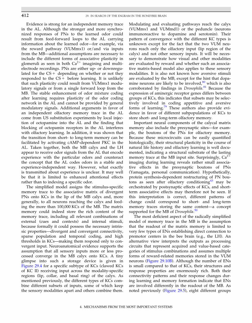
The most deficient aspect of the radically simplifiedmodel of memory trace in the MB is the assumptionthat the readout of the matrix memory is limited tovery few types of ENs establishing direct connection topremotor centers in the bee brain (e.g., the LH). Analternative view interprets the outputs as processingcircuits that represent acquired and value-based cate-gories of stimulus combinations and assumes multipleforms of reward-related memories stored in the VUMneurons (Figure 29.10B). Although the number of ENsis small compared to that of KCs, their structures andresponse properties are enormously rich. Both theirconnectivity patterns and their response changes dur-ing learning and memory formation indicate that ENsare involved differently in the readout of the MB. Asnoted previously (Figure 29.5), eight different groups
412 29. IN SEARCH OF THE ENGRAM IN THE HONEYBEE BRAIN
4. MECHANISMS FROM THE MOST IMPORTANT SYSTEMS
of ENs have been characterized on the basis of theirsomata loci and their arborizations patterns. The richstructural variability makes it likely that ENs of differ-ent groups serve different functions. Although thesedifferences cannot be interpreted yet, the structuralmultiplicity suggests forms of combinatorial coding ofneural processing categories that are defined by therespective input and output regions. What are thesecategories?
A common property of these categories could bethat they represent acquired and value-based informa-tion. The following value-based categories (VBCs)come to mind:
1. Detection of novel versus already learned stimulusconditions
2. Distinction between appetitively and aversivelylearned stimulus conditions
3. Separation between cues and contexts4. Separation between self-generated stimuli as
experienced during active exploration and passivelyexperienced stimuli as during classical conditioning
5. Storing stimulus traces for later learning,particularly under latent learning conditions as innavigation
6. Recognition and learning of symmetrical inputs topaired sense organs across sensory modality
7. Activation of specific memory traces forconsolidation, such as during sleep or other formsof neural self-organization
These and other VBCs may define higher order neu-ral processes brought about by the cooperation andcombination of the lower level neural processes occur-ring in other parts of the brain, including (1) definingglobal context conditions (within the social context vs.acting individually; foraging for food vs. foraging forinformation during exploration), (2) working memoryas a neural platform for the evaluation of expected out-comes, and (3) wakefulness versus sleep.
Although the assumption of defined VBCs is specu-lative, it may help in future work to relate some of thegroups of ENs to these or other VBCs on the basis oftheir morphology. Consider several examples. First,A3-v projecting back from the lobes to the calyx couldbe involved in the distinction between novel andlearned stimulus conditions and may be involved infacilitating the respective stimuli according to theresponse strength of the KCs. (2) ENs of groupsA1�A4 are characterized by their branches in the ringneuropil of the same MB from which they receiveinput. Although we know nothing about the functionof the ring neuropil, it could be that it houses long-term memory outside the active circuits of the MB,and neurons communicating between lobes and ringneuropil may be involved in memory consolidation
and memory retrieval. Third, ENs connecting the twoMBs in the two hemispheres of the brain (A6 and A7)may help to detect symmetrical stimuli acrosssensory modalities, transfer memory content from oneMB to the other as proposed by Sandoz and Menzel,82
and/or coordinate consolidation processes betweenthe two MBs.
CONCLUSION
The engram of olfactory stimuli in the bee brain ischaracterized by its distributed nature with differentprevailing processing categories at different sites.I conclude from the limited existing data that the tracein the AL relates predominantly to attention-generating properties, the matrix trace in the calyx tohigh-order combinatorics of all sensory inputs, the sys-tem of VUM neurons to appetitive internal states ofthe animal controlling nonassociative and associativetraces, and the ENs of the MB to multiple processingcategories that represent the acquired values and pro-vide neural commands for goal-directed behavior anddecision making. Although speculative, this frame-work offers a structure for experimental and modelingapproaches and prevents us from believing that theproperties of the memory trace can be captured bysimply assuming flexible and experience-dependentsensory�interneuron�motor connections. Rather, wehave to search for the coding/recoding, evaluating,and predicting processes involved in storing the con-tents of memory, the engram.
Gerber and co-workers83 asked whether it is possi-ble to localize a memory trace to a subset of cells inthe brain. According to them, it needs to be shownthat (1) neuronal plasticity occurs in the respectivecells, (2) neuronal plasticity in these cells is sufficientfor memory recall, (3) neuronal plasticity in these cellsis necessary for memory formation, (4) memory con-tent is lost if these cells do not function duringretrieval tests, (5) and memory formation is abolishedif these cells do not receive input during learning. Thislist of requirements, although difficult to meet experi-mentally (possibly only in Drosophila so far), is notcomplete and suffers from the focus on processesinvolved in neural plasticity rather than asking whereand how the content of memory, the engram, is stored.The engram will not be found in a single type of neu-ron. It results from distributed network properties thatadd their respective contents when memory is formed,processed (consolidated), and retrieved. In otherwords, the engram is not a property of particular neu-rons but, rather, that of highly interacting networks ofneurons. This form of interaction is different duringmemory formation, consolidation, and retrieval,
413CONCLUSION
4. MECHANISMS FROM THE MOST IMPORTANT SYSTEMS
meaning that different engrams (for the same content)exist depending on what happens to them and forwhat they are used. In this way, the engram does not‘exist’ but develops over time and in relation to actionsof the brain as mirrored in incorporating new contentsinto existing ones, in consulting different contents dur-ing decision making and planning, and during execu-tion of behavioral acts.
References
1. Lashley KS. In search of the engram. Symp Soc exp Biol.1950;4:454�482.
2. Dudai Y. The neurobiology of consolidations, or, how stable isthe engram? Annu Rev Psychol. 2004;55:51�86.
3. Craik FI. Levels of processing: past, present. and future?Memory. 2002;10(5-6):305�318.
4. Moscovitch M. Memory: why the engram is elusive.In: Roediger HL, Dudai Y, Fristzpatrick SM, eds. Science ofMemory: Concepts. Oxford: Oxford University Press;2007:17�21.
5. Ramon Y, Cajal S. Einige hypothesen uber den anatomischenmechanismus der ideenbildung, der association und der auf-merksamkeit. Archiv fur Anatomie und Physiologie. 1895;25:367�378.
6. Nicolelis MAL, Fanselow EE, Ghazanfar AA. Hebb’s dream: theresurgence of cell assemblies. Neuron. 1997;19:219�221.
7. von Frisch K. The Dance Language and Orientation of Bees.Cambridge, MA: Harvard University Press; 1967.
8. Farina WM, Gruter C, Acosta L, Mc CS. Honeybees learn floralodors while receiving nectar from foragers within the hive.Naturwissenschaften. 2007;94:55�60.
9. Gil M, De Marco RJ, Menzel R. Learning reward expectations inhoneybees. Learn Mem. 2007;14(491):496.
10. Giurfa M, Eichmann B, Menzel R. Symmetry as a perceptual cat-egory in honeybee vision. In: Elsner N, Menzel R, eds. Learningand Memory. Proceedings of the 23rd Gottingen NeurobiologyConference. Stuttgart: G. Thieme Verlag; 1995:423.
11. Wright GA, Skinner BD, Smith BH. Ability of honeybee, Apismellifera, to detect and discriminate odors of varieties of canola(Brassica rapa and Brassica napus) and snapdragon flowers(Antirrhinum majus). J Chem Ecol. 2002;28(4):721�740.
12. Menzel R. Memory dynamics in the honeybee. J Comp Physiol A.1999;185:323�340.
13. Davis RL. Traces of Drosophila memory. Neuron. 2011;70(1):8�19.14. Grunbaum L, Muller U. Induction of a specific olfactory mem-
ory leads to a long-lasting activation of protein kinase C in theantennal lobe of the honeybee. J Neurosci. 1998;18:4384�4392.
15. Muller U. Prolonged activation of cAMP-dependent proteinkinase during conditioning induces long-term memory in honey-bees. Neuron. 2000;27:159�168.
16. Friedrich A, Thomas U, Muller U. Learning at different satiationlevels reveals parallel functions for the cAMP-protein kinase acascade in formation of long-term memory. J Neurosci. 2004;24(18):4460�4468.
17. Lindauer M. Allgemeine sinnesphysiologie. Orientierung imraum. Fortschr Zool. 1963;16:58�140.
18. Menzel R. Das gedachtnis der honigbiene fur spektralfarben. I.Kurzzeitiges und langzeitiges behalten. Z vergl Physiol.1968;60:82�102.
19. Baddeley AD. Working Memory, Thought and Action. Oxford:Oxford University Press; 2007.
20. Zhang SW, Bartsch K, Srinivasan MV. Maze learning by honey-bees. Neurobiol Learn Mem. 1996;66:267�282.
21. Dacke M, Srinivasan MV. Evidence for counting in insects. AnimCogn. 2008;11(4):683�689.
22. Menzel R. Serial position learning in honeybees. PLoS ONE.2009;4(3):e4694�e4701.
23. Greggers U, Menzel R. Memory dynamics and foraging strate-gies of honeybees. Behav Ecol Sociobiol. 1993;32:17�29.
24. Hellstern F, Malaka R, Hammer M. Backward inhibitory learn-ing in honeybees: a behavioral analysis of reinforcement proces-sing. Learn Mem. 1998;4:429�444.
25. Menzel R, Giurfa M. Dimensions of cognition in an insect, thehoneybee. Behav Cogn Neurosci Rev. 2006;5:24�40.
26. Pamir E, Chakroborty NK, Stollhoff N, Gehring KB, AntemannV, Morgenstern L, et al. Average group behavior does not repre-sent individual behavior in classical conditioning of the honey-bee. Learn Mem. 2011;18(11):733�741.
27. Mobbs PG. The brain of the honeybee Apis mellifera: I. The con-nections and spatial organization of the mushroom bodies. PhilTrans R Soc Lond B. 1982;298:309�354.
28. Rybak J, Menzel R. Anatomy of the mushroom bodies in thehoney bee brain: the neuronal connections of the alpha-lobe.J Comp Neurol. 1993;334(3):444�465.
29. Hammer M. An identified neuron mediates the unconditionedstimulus in associative olfactory learning in honeybees. Nature.1993;366:59�63.
30. Hammer M, Menzel R. Learning and memory in the honeybee.J Neurosci. 1995;15(3):1617�1630.
31. Menzel R, Erber J, Masuhr T. Learning and memory in the hon-eybee. In: Barton-Browne L, ed. Experimental Analysis of InsectBehaviour. Berlin: Springer; 1974:195�217.
32. Hammer M, Menzel R. Multiple sites of associative odor learn-ing as revealed by local brain microinjections of octopamine inhoneybees. Learn Mem. 1998;5:146�156.
33. Farooqui T, Vaessin H, Smith BH. Octopamine receptors in thehoneybee (Apis mellifera) brain and their disruption by RNA-mediated interference. J Insect Physiol. 2004;50(8):701�713.
34. Faber T, Joerges J, Menzel R. Associative learning modifies neu-ral representations of odors in the insect brain. NatureNeuroscience. 1999;2(1):74�78.
35. Peele P, Ditzen M, Menzel R, Galizia CG. Appetitive odor learn-ing does not change olfactory coding in a subpopulation of hon-eybee antennal lobe neurons. J Comp Physiol A Neuroethol SensoryNeural Behav Physiol. 2006;192(10):1083�1103.
36. Schmuker M, Weidert M, Menzel R. A network model forlearning-induced changes in odor representation in the antennallobe. In: Laurent UP, Emmanuel D, eds. Proceedings of the SecondFrench Conference on Computational Neuroscience. Marseille,France; 2008.
37. Fernandez PC, Locatelli FF, Person-Rennell N, Deleo G, SmithBH. Associative conditioning tunes transient dynamics ofearly olfactory processing. J Neurosci. 2009;29(33):10191�10202.
38. Rath L, Giovanni GC, Szyszka P. Multiple memory traces afterassociative learning in the honey bee antennal lobe. Eur JNeurosci. 2011;34(2):352�360.
39. Denker M, Finke R, Schaupp F, Grun S, Menzel R. Neural corre-lates of odor learning in the honeybee antennal lobe. Eur JNeurosci. 2010;31(1):119�133.
40. Kenyon FC. The brain of the bee: a preliminary contribution tothe morphology of the nervous system of the arthropoda. J CompNeurol. 1896;6:134�210.
41. Dujardin J. Memoire sur le systeme nerveux des insects. Ann SciNat Zool. 1850;14:196�206.
42. Ganeshina OT, Vorobyev MV, Menzel R. Synaptogenesis in themushroom body calyx during metamorphosis in the honeybee
414 29. IN SEARCH OF THE ENGRAM IN THE HONEYBEE BRAIN
4. MECHANISMS FROM THE MOST IMPORTANT SYSTEMS
Apis mellifera: an electron microscopic study. J Comp Neurol.2006;497(6):876�897.
43. Hourcade B, Muenz TS, Sandoz JC, Rossler W, Devaud JM.Long-term memory leads to synaptic reorganization in themushroom bodies: a memory trace in the insect brain. J Neurosci.2010;30(18):6461�6465.
44. Szyszka P, Ditzen M, Galkin A, Galizia CG, Menzel R.Sparsening and temporal sharpening of olfactory representationsin the honeybee mushroom bodies. J Neurophysiol. 2005;94(5):3303�3313.
45. Perez-Orive J, Mazor O, Turner GC, Cassenaer S, Wilson RI,Laurent G. Oscillations and sparsening of odor representationsin the mushroom body. Science. 2002;297(5580):359�365.
46. Rolls ET. An attractor network in the hippocampus: theory andneurophysiology. Learn Mem. 2007;14(11):714�731.
47. Szyszka P, Galkin A, Menzel R. Associative and non-associativeplasticity in Kenyon cells of the honeybee mushroom body.Front Syst Neurosci. 2008;2:1�10.
48. Szyszka P, Demmler C, Oemisch M, Sommer L, Biergans S,Birnbach B, et al. Mind the gap: olfactory trace conditioning inhoneybees. J Neurosci. 2011;31(20):7229�7239.
49. Grunewald B, Wersing A, Wustenberg D. Learning channels:cellular physiology of odor processing neurons within the hon-eybee brain. Acta Biol Hungarica. 2004;55(1-4):53�63.
50. Pascual A, Preat T. Localization of long-term memory within theDrosophila mushroom body. Science. 2001;294(5544):1115�1117.
51. Brandt R, Rohlfing T, Rybak J, Krofczik S, Maye A, WesterhoffM, et al. A three-dimensional average-shape atlas of the hon-eybee brain and its applications. J Comp Neurol. 2005;492(1):1�19.
52. Witthoft W. Absolute anzahl und verteilung der zellen im hirnder honigbiene. Z Morph Tiere. 1967;61:160�184.
53. White JA, Rubinstein JT, Kay AR. Channel noise in neurons.Trends Neurosci. 2000;23(3):131�137.
54. Roth G, Dicke U. Evolution of the brain and intelligence. TrendsCogn Sci. 2005;9(5):250�257.
55. Mauelshagen J. Neural correlates of olfactory learning in anidentified neuron in the honey bee brain. J Neurophysiol.1993;69:609�625.
56. Okada R, Rybak J, Manz G, Menzel R. Learning-related plastic-ity in PE1 and other mushroom body-extrinsic neurons in thehoneybee brain. J Neurosci. 2007;27(43):11736�11747.
57. Menzel R, Manz G. Neural plasticity of mushroom body-extrinsic neurons in the honeybee brain. J Exp Biol. 2005;208(Pt22):4317�4332.
58. Bear MF, Malenka RC. Synaptic plasticity: LTP and LTD. CurrOpin Neurobiol. 1994;4(3):389�399.
59. Cassenaer S, Laurent G. Hebbian STDP in mushroom bodiesfacilitates the synchronous flow of olfactory information inlocusts. Nature. 2007;448(7154):709�713.
60. Strube-Bloss MF, Nawrot MP, Menzel R. Mushroom body out-put neurons encode odor reward associations. J Neurosci. 2011;31(8):3129�3140.
61. Schroter U, Menzel RA. New ascending sensory tract to the caly-ces of the honeybee mushroom body, the subesophageal-calycaltract. J Comp Neurol. 2003;465:168�178.
62. Hussaini SA, Menzel R. Mushroom body extrinsic neurons in thehoneybee brain encode cues and contexts differently. J Neurosci.2012, in press.
63. Haehnel M, Menzel R. Sensory representation and learning-related plasticity in mushroom body extrinsic feedback neuronsof the protocerebral tract. Front Neurosci. 2010;4:1�16.
64. Haehnel M, Menzel R. Long-term memory and response gener-alization in mushroom body extrinsic neurons in the honeybeeApis mellifera. J Exp Biol. 2012;215(559):565.
65. Filla I, Menzel R. Visual and olfactory associative plasticity in aninhibitory local and recurrent pathway in the honeybee. 2012[In revision.]
66. Ganeshina OT, Menzel R. GABA-immunoreactive neurons in themushroom bodies of the honeybee: an electron microscopicstudy. J Comp Neurol. 2001;437(3):335�349.
67. Liu X, Davis RL. The GABAergic anterior paired lateral neuronsuppresses and is suppressed by olfactory learning. NatNeurosci. 2009;12(1):53�59.
68. Okada R, Sakura M, Mizunami M. Distribution of dendrites ofdescending neurons and its implications for the basic organiza-tion of the cockroach brain. J Comp Neurol. 2003;459(3):158�174.
69. Gerber B, Stocker RF. The Drosophila larva as a model for study-ing chemosensation and chemosensory learning: a review. ChemSenses. 2006;32(1):65�89.
70. Li Y, Strausfeld NJ. Multimodal efferent and recurrent neuronsin the medial lobes of cockroach mushroom bodies. J CompNeurol. 1999;409(4):647�663.
71. Jefferis GS, Potter CJ, Chan AM, Marin EC, Rohlfing T, MaurerJr. CR, et al. Comprehensive maps of Drosophila higher olfactorycenters: spatially segregated fruit and pheromone representa-tion. Cell. 2007;128(6):1187�1203.
72. Hammer M. The neural basis of associative reward learning inhoneybees. Trends Neurosci. 1997;20(6):245�252.
73. Schroter U, Malun D, Menzel R. Innervation pattern of suboeso-phageal VUM neurons in the honeybee brain. Cell Tissue Res.2006;326(3):647�667.
74. Kreissl S, Eichmuller S, Bicker G, Rapus J, Eckert M.Octopamine-like immunoreactivity in the brain and suboesopha-geal ganglion of the honeybee. J Comp Neurol. 1994;348:583�595.
75. Bicker G, Menzel R. Chemical codes for the control of behaviourin arthropods. Nature. 1989;337:33�39.
76. Barron AB, Maleszka R, Vander Meer RK, Robinson GE.Octopamine modulates honey bee dance behavior. Proc NatlAcad Sci USA. 2007;104(5):1703�1707.
77. Johnson RN, Oldroyd BP, Barron AB, Crozier RH. Genetic con-trol of the honey bee (Apis mellifera) dance language: segregatingdance forms in a backcrossed colony. J Hered. 2002;93(3):170�173.
78. Farooqui T, Robinson K, Vaessin H, Smith BH. Modulation ofearly olfactory processing by an octopaminergic reinforcementpathway in the honeybee. J Neurosci. 2003;23(12):5370�5380.
79. McQuillan HJ, Nakagawa S, Mercer AR. Mushroom bodies ofthe honeybee brain show cell population-specific plasticity inexpression of amine-receptor genes. Learn Mem. 2012;19(4):151�158.
80. Vergoz V, Roussel E, Sandoz JC, Giurfa M. Aversive learning inhoneybees revealed by the olfactory conditioning of the stingextension reflex. PLoS ONE. 2007;2:e288.
81. Schwaerzel M, Monastirioti M, Scholz H, Friggi-Grelin F,Birman S, Heisenberg M. Dopamine and octopamine differenti-ate between aversive and appetitive olfactory memories inDrosophila. J Neurosci. 2003;23(33):10495�10502.
82. Sandoz J-C, Menzel R. Side-specificity of olfactory learning inthe honeybee: generalization between odors and sides. LearnMem. 2001;8:286�294.
83. Gerber B, Tanimoto H, Heisenberg M. An engram found?Evaluating the evidence from fruit flies. Curr Opin Neurobiol.2004;14(6):737�744.
415REFERENCES
4. MECHANISMS FROM THE MOST IMPORTANT SYSTEMS