Embed Size (px)
Citation preview
Invention and early history of telepathology(1985-2000)Ronald S. Weinstein, University of ArizonaMichael J. Holcomb, University of ArizonaElizabeth Anne Krupinski, Emory University
Journal Title: Journal of Pathology InformaticsVolume: Volume 10, Number 1Publisher: Medknow Publications | 2019-01-01, Pages 1-1Type of Work: Article | Final Publisher PDFPublisher DOI: 10.4103/jpi.jpi_71_18Permanent URL: https://pid.emory.edu/ark:/25593/tnntd
Final published version: http://dx.doi.org/10.4103/jpi.jpi_71_18
Copyright information:© 2019 Medknow. All Rights Reserved.This is an Open Access work distributed under the terms of the CreativeCommons Attribution-NonCommercial-ShareAlike 4.0 International License(http://creativecommons.org/licenses/by-nc-sa/4.0/).
Accessed January 17, 2022 6:33 AM EST
© 2019 Journal of Pathology Informatics | Published by Wolters Kluwer - Medknow 1
Review Article
IntroductIon
Telepathology is the diagnosis of surgical pathology cases at a distance using real-time video imaging or store-and-forward digitized images.[1-11] The American Telemedicine Association clinical guidelines for telepathology define telepathology as: “A form of communication between medical professionals that includes the transmission of pathology images and associated clinical information for the purpose of various clinical applications including, but not limited to, primary diagnoses, rapid cytology interpretation, intraoperative and second opinion consultations, ancillary study review, archiving, and quality activities.”[10]
In this review of the early history of telepathology, we identify and discuss interconnected factors that help explain how robotic-dynamic telepathology became the technology‑of‑choice for the first sustainable telepathology programs in the United States, Canada, and Europe. The initial
driver for the invention was a crisis in a National Cancer Institute (NCI)-funded National Organ Site Cancer Program, the National Bladder Cancer Project (NBCP), with the National Bladder Cancer Group (NBCG) the clinical trial arm of NBCP.
Why invent dynamic-robotic telepathology? For logistical reasons, the NBCP collaboratives’ “analytical diagnoses,” the diagnoses used for data analysis at the end of a clinical trial, were generated by the NBCG’s own Central Pathology Laboratory (CPL) located in Chicago, IL. These bladder cancer diagnoses, including re-staging and re-grading of
This narrative‑based paper provides a first‑person account of the early history of telepathology (1985–2000) by the field’s inventor, Ronald S. Weinstein, M. D. During the 1980s, Dr. Weinstein, a Massachusetts General Hospital‑trained pathologist, was director of the Central Pathology Laboratory (CPL) for the National Cancer Institute‑funded National Bladder Cancer Project, located at Rush Medical College in Chicago, IL. The CPL did post therapy revalidations of surgical pathology and cytopathology diagnoses before outcomes of the completed clinical trials were published. The CPL reported that interobserver variability was invalidating inclusion of dozens of treated bladder cancer patients in published reports on treatment outcomes. This problem seemed ripe for a technology-assisted solution. In an effort to solve the interobserver variability problem, Dr. Weinstein devised a novel solution, dynamic-robotic telepathology, that would potentially enable CPL uropathologists to consult on distant uropathology cases in real‑time before their assignment to urinary bladder cancer, tumor stage, and grade‑specific clinical trials. During the same period, universities were ramping up their support for faculty entrepreneurism and creating in-house technology transfer organizations. Dr. Weinstein recognized telepathology as a potential growth industry. He and his sister, Beth Newburger, were a successful brother–sister entrepreneur team. Their PC‑based education software business, OWLCAT™, had just been acquired by Digital Research Inc., a leading software company, located in California. With funding from the COMSAT Corporation, a publically traded satellite communications company, the Weinstein-Newburger team brought the earliest dynamic-robotic telepathology systems to market. Dynamic-robotic telepathology became a dominant telepathology technology in the late 1990s. Dr. Weinstein, a serial entrepreneur, continued to innovate and, with a team of optical scientists at The University of Arizona’s College of Optical Sciences, developed the first sub‑1‑min whole‑slide imaging system, the DMetrix DX-40 scanner, in the early 2000s.
Keywords: Digital pathology, innovation, medical education, pathology, telepathology, The University of Arizona, virtual pathology
Address for correspondence: Dr. Ronald S. Weinstein, Department of Pathology, The University of Arizona, 1501 North Campbell
Avenue, Tucson, Arizona 85724, USA. E‑mail: [email protected]
Access this article online
Quick Response Code:Website: www.jpathinformatics.org
DOI: 10.4103/jpi.jpi_71_18
This is an open access journal, and articles are distributed under the terms of the Creative Commons Attribution‑NonCommercial‑ShareAlike 4.0 License, which allows others to remix, tweak, and build upon the work non‑commercially, as long as appropriate credit is given and the new creations are licensed under the identical terms.
For reprints contact: [email protected]
How to cite this article: Weinstein RS, Holcomb MJ, Krupinski EA. Invention and early history of telepathology (1985‑2000). J Pathol Inform 2019;10:1.Available FREE in open access from: http://www.jpathinformatics.org/text.asp?2019/10/1/1/250755
Invention and Early History of Telepathology (1985-2000)Ronald S. Weinstein1, Michael J. Holcomb1, Elizabeth A. Krupinski2
1Department of Pathology, College of Medicine, University of Arizona, Tucson, Arizona, USA, 2Department of Radiology and Imaging Sciences, Emory University, Atlanta, Georgia, USA
Abstract
Received: 26 September 2018 Accepted: 19 November 2018 Published: 24 January 2019

Journal of Pathology Informatics2
J Pathol Inform 2019, 1:1 http://www.jpathinformatics.org/content/10/1/1
cancers, were generated retrospectively, often after the clinical trial protocol was completed. It turned out that interobserver variability, comparing the originating institution’s surgical pathology and cytopathology diagnoses and the CPL’s diagnoses, was a significant problem, especially for some contributing institutions.[12‑15] Not infrequently, microscopic field selection, or up-and-down focusing of the light microscope, were at issue. Theoretically, dynamic-robotic telepathology (not yet invented) could provide a remote up-and-down focusing solution. There was general agreement that the ideal solution would be to introduce prospective CPL re-reviews of surgical pathology and cytopathology cases before the initiation of therapy in individual cases. This was not practical for logistical reasons in the 1970s and early 1980s. There was no technology solution that could provide immediate access to CPL uropathologists within the narrow window of time between the harvesting of tissue at surgery and histopathology and cytopathology sign-outs by pathologists and cytopathologists, and there was no infrastructure and environment to provide immediate telepathology services. This was all in the future.
To put the expenditure of resources for the creation of robotic-dynamic telepathology in some perspective, both in terms of time and money, it is stressed that this effort to reduce the interobserver variability challenge in urinary bladder cancer evaluations was in reaction to a very challenging situation. Urinary bladder cancer staging and grading are unusually challenging for pathologists, although many pathologists are quite unaware of that fact.[16] Of the multiple organ‑site‑specific cancer trial programs funded by the NCI in the 1970s and 1980s, the NBCP was the only National Organ Site Cancer Program that could justify having a full-time NCI-funded CPL of its own.[17]
The NBCP’s leadership successfully argued that this reflected the unusual difficulty and complexity of rendering reproducible urinary bladder cancer diagnoses. Various efforts to improve the reproducibility of surgical pathology diagnoses for urinary bladder cancers were unsuccessful. Annually, the NBCG uropathologists from 8 collaborating institutions (e.g., MGH, Roswell Park, etc.) would meet in Sarasota, FL, sit at multiheaded light microscopes, and try to
work out their differences in the diagnoses they had rendered on “difficult” cases. Anecdotal evidence suggests that there were perhaps beneficial outcomes from these group meetings, but a change in practice by the surgical pathologists was not sustainable (R. S. Weinstein, unpublished observations, 1984). Interestingly, it is noteworthy that recent data collected for a different reason, gaining approval of the United States Food and Drug Administration for rendering primary diagnosis with whole‑slide imaging (WSI), verified the relative difficulty of classifying bladder cancer patients in comparison with rendering cancer diagnosis in other organs.[16]
The NBCG completed 15 clinical trials between 1974 and 1990, when the NBCG’s federal funding ended. CPL pathologists had re‑reviewed 15,000 surgical pathology cases and 17,000 cytopathology cases. The interobserver variability, comparing the pathology diagnoses of the originating institution with the CPL diagnosis, was unacceptably high for some collaborating institutions. This resulted in post therapy rejection of urinary bladder cancer cases from inclusion in the analysis of the outcomes of certain urinary bladder cancer clinical trials.
Figure 1 and Table 1 show unpublished interobserver variability data for the 8 institutions contributing urinary bladder cancer cases to the NBCG’s consortium studies during the 1983–1984 academic year. It can be said that this data set triggered the invention of dynamic‑robotic telepathology (R. S. Weinstein, unpublished data, 1984). In the 1983–1984 time frame, major discrepancies were defined, by the CPL, as a one level or more difference in tumor stage or two levels of variance in tumor grade (R. S. Weinstein, unpublished data, 1984).
The bar graph in Figure 1 shows the major and minor discrepancies in the staging and grading of urinary bladder cancer cases at the 8 academic medical centers participating in NCI‑sponsored clinical trials for the 1983–1984 academic year. The data supporting the bar graph in Figure 1 are shown in Table 1 (R. S. Weinstein, unpublished data, 1984). There was a statistically significant difference between institutions for major discrepancies (χ2 = 47.13, P < 0.0001), minor discrepancies (χ2 = 14.24, P = 0.0472), and total number of cases (χ2 = 17.83, P = 0.0128).
Table 1: Concordance Rates at the National Bladder Cancer Groups’ Central Pathology Laboratorya
Institution number
Total number cases
Major discordances
number of cases
Major discordances % of cases
Minor discordances
number of cases
Minor discordances % of cases
Total number of discordant
cases
Total discordances % of cases
1 186 9 4.84 17 9.14 26 13.982 118 4 3.39 17 14.40 21 17.803 75 14 18.67 19 23.33 33 44.004 154 18 11.69 12 7.79 30 19.485 185 38 20.54 7 3.78 45 24.326 58 12 20.68 15 25.86 27 46.557 70 13 18.57 10 14.29 23 32.558 70 13 18.57 5 7.14 18 25.71Total 916 121 102 223aR.S. Weinstein, previously unpublished data, 1984

Journal of Pathology Informatics 3
J Pathol Inform 2019, 1:1 http://www.jpathinformatics.org/content/10/1/1
CPL case re-review diagnoses served as the standard for comparison. In Figure 1, each column represents one of the 8 individual participating institutions. Major discrepancies (orange) ranged from 4% (Institution #1) to 23% (Institution #6). Major discrepancies resulted in disqualifying a patient’s data from inclusion in the analysis of the outcomes of clinical trials. Minor discrepancies (yellow) most often represented one level of difference in tumor grading and did not affect the data analysis for a clinical trial. Numbers at the top of each column represent the total number of cases accessioned into clinical protocol analysis in the 1983–1984 academic year.
Based on concerns over the variability of the performances of pathologists, as compared to CPL retrospective diagnosis, especially at institutions 3, 6, and 8, telepathology was conceived of as a possible solution to this interobserver variability challenge in these bladder cancer clinical trials (R. S. Weinstein, unpublished data, 1984). The idea was that by centralizing the diagnoses of the urinary bladder cancers at the CPL, with immediate readouts by telepathology, and then utilizing these CPL initial surgical pathology diagnoses for assigning bladder cancer patients to disease-specific therapeutic trial protocols, the interobserver variability in diagnoses might be minimized.
InventIon of dynamIc‑robotIc telepathology
Dr. Weinstein was encouraged by NBCP leaders to create the means for CPL uropathologists to analyze the NBCG’s patients’ urinary bladder cancers before their entry into clinical trials.[15,16] In 1985, he co‑founded Corabi International Telemetrics, Inc., the first company to bring dynamic‑robotic microscopy to market. He invented and patented the first dynamic robotic telepathology system while he was Director of the NBCP’s CPL, located at Rush‑Presbyterian St. Luke’s Medical Center (1982–1990).[18‑27] The first customer for a Corabi International Telemetrics system was Grady Memorial Hospital, in Atlanta, Georgia. It was linked to Emory University
Medical Center 4 miles away using a bi-directional microwave telecommunications system. The system became operational in 1989. Tragically, the entire National Organ Site Cancer Program, which supported the NBCP and other individual Organ Site Cancer Programs, was defunded by the NCI the same year, for reasons unrelated to the NBCP and its CPL. Ironically, the primary reason for inventing dynamic-robotic telepathology, to provide a means for the rapid re-review of urinary bladder cancer surgical pathology diagnosis by the CPL, before therapy became moot, at least for the time being.
IntroductIon of telepathology to the health‑care Industry and the general publIc
In 1986, Corabi International Telemetrics, Inc. developed a four-pronged product rollout and marketing strategy for robotic-dynamic telepathology systems. These activities were envisioned at a strategic planning meeting in Bethesda, MD, in late 1985. They were executed in parallel and in highly coordinated fashion for maximum market impact and to attract investors.[28‑30]
The Corabi strategic plan was to (1) introduce the scientific term “telepathology” into the English language; (2) organize the first national demonstration of satellite‑enabled robotic telepathology, working with the US Department of Defense and COMSAT Corporation, for the demonstration; (3) have Dr. Weinstein author and submit the first telepathology US patent application; and (4) create an academic literature on telepathology.[20-27,31]
Figure 1: Unpublished data that launched the creation of dynamic‑robotic telepathology in 1984
Figure 2: Still pictures taken from a video recording of the “Armed Forces Institute of Pathology’s Col. Earle Ash Lecture: May 22, 1986.” Introduction of the word “telepathology” into the English language. (a) Col. Robert McMeekam, Armed Forces Institute of Pathology Director, welcomes the invited audience to Dr. Weinstein’s lecture. (b) Fred W. Stewart, M. D., Chief of Surgical Pathology at Memorial Sloan Kettering Cancer Center in New York City introduces the Col. Earle Ash Lecturer. (c) Dr. Weinstein, at the very moment in the Col. Earle Ash Lecture, that he used the word “telepathology” for the first time in public. (d) Recreation of the Kodachrome slide being shown at the moment Dr. Weinstein used the word “telepathology”
dc
ba
Journal of Pathology Informatics4
J Pathol Inform 2019, 1:1 http://www.jpathinformatics.org/content/10/1/1
The academic and commercial rollouts of telepathology were carefully orchestrated, starting with the development of a Rush University technology transfer roadmap. The public event at which the announcement of the invention of robotic-dynamic telepathology by Dr. Weinstein would take place was linked to the simultaneous publication of a Human Pathology editorial entitled “Prospects for Telepathology.”[20] Dr. Weinstein’s public announcement of the concept of telepathology and the naming of the field was done at a formal, black-tie “Ash Lecture” lecture at the Armed Forces Institute of Pathology (AFIP) on the Walter Reed Army Medical Center Campus, in Washington, DC, also in May 1986 [Figure 2]. The first public demonstration of robotic‑dynamic telepathology was staged in August 1986 in Washington, DC [Figure 3].[31,32] In 1987, Dr. Weinstein submitted his first US patent application for telepathology. Eventually, the US Patent and Trademark Office acknowledged that the Weinstein patent application was so rich in original ideas that it deserved to be divided into two patents (called a “divisional” patent), each titled “Telepathology Diagnostics Networks,” and issued in 1993 and 1994, respectively.[25,26]
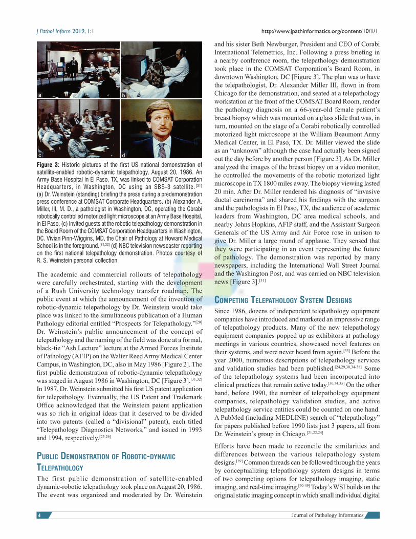
publIc demonstratIon of robotIc‑dynamIc telepathology
The first public demonstration of satellite-enabled dynamic‑robotic telepathology took place on August 20, 1986. The event was organized and moderated by Dr. Weinstein
and his sister Beth Newburger, President and CEO of Corabi International Telemetrics, Inc. Following a press briefing in a nearby conference room, the telepathology demonstration took place in the COMSAT Corporation’s Board Room, in downtown Washington, DC [Figure 3]. The plan was to have the telepathologist, Dr. Alexander Miller III, flown in from Chicago for the demonstration, and seated at a telepathology workstation at the front of the COMSAT Board Room, render the pathology diagnosis on a 66‑year‑old female patient’s breast biopsy which was mounted on a glass slide that was, in turn, mounted on the stage of a Corabi robotically controlled motorized light microscope at the William Beaumont Army Medical Center, in El Paso, TX. Dr. Miller viewed the slide as an “unknown” although the case had actually been signed out the day before by another person [Figure 3]. As Dr. Miller analyzed the images of the breast biopsy on a video monitor, he controlled the movements of the robotic motorized light microscope in TX 1800 miles away. The biopsy viewing lasted 20 min. After Dr. Miller rendered his diagnosis of “invasive ductal carcinoma” and shared his findings with the surgeon and the pathologists in El Paso, TX, the audience of academic leaders from Washington, DC area medical schools, and nearby Johns Hopkins, AFIP staff, and the Assistant Surgeon Generals of the US Army and Air Force rose in unison to give Dr. Miller a large round of applause. They sensed that they were participating in an event representing the future of pathology. The demonstration was reported by many newspapers, including the International Wall Street Journal and the Washington Post, and was carried on NBC television news [Figure 3].[31]
competIng telepathology system desIgns
Since 1986, dozens of independent telepathology equipment companies have introduced and marketed an impressive range of telepathology products. Many of the new telepathology equipment companies popped up as exhibitors at pathology meetings in various countries, showcased novel features on their systems, and were never heard from again.[33] Before the year 2000, numerous descriptions of telepathology services and validation studies had been published.[24,29,30,34‑38] Some of the telepathology systems had been incorporated into clinical practices that remain active today.[30,34,35] On the other hand, before 1990, the number of telepathology equipment companies, telepathology validation studies, and active telepathology service entities could be counted on one hand. A PubMed (including MEDLINE) search of “telepathology” for papers published before 1990 lists just 3 papers, all from Dr. Weinstein’s group in Chicago.[21,22,24]
Efforts have been made to reconcile the similarities and differences between the various telepathology system designs.[39] Common threads can be followed through the years by conceptualizing telepathology system designs in terms of two competing options for telepathology imaging, static imaging, and real-time imaging.[40-49] Today’s WSI builds on the original static imaging concept in which small individual digital
Figure 3: Historic pictures of the first US national demonstration of satellite‑enabled robotic‑dynamic telepathology, August 20, 1986. An Army Base Hospital in El Paso, TX, was linked to COMSAT Corporation Headquar ters, in Washington, DC using an SBS‑3 satellite. [31] (a) Dr. Weinstein (standing) briefing the press during a predemonstration press conference at COMSAT Corporate Headquarters. (b) Alexander A. Miller, III, M. D., a pathologist in Washington, DC, operating the Corabi robotically controlled motorized light microscope at an Army Base Hospital, in El Paso. (c) Invited guests at the robotic telepathology demonstration in the Board Room of the COMSAT Corporation Headquarters in Washington, DC. Vivian Pinn‑Wiggins, MD, the Chair of Pathology at Howard Medical School is in the foreground. [31,32] (d) NBC television newscaster reporting on the first national telepathology demonstration. Photos courtesy of R. S. Weinstein personal collection
dc
ba

Journal of Pathology Informatics 5
J Pathol Inform 2019, 1:1 http://www.jpathinformatics.org/content/10/1/1
images were electronically stitched together into progressively larger composite static images until an entire histopathology or cytopathology WSI was represented in one giant digital pathology image.[22,24,40-44] Dynamic-robotic telepathology is based on a different concept. Whereas WSI telepathology can be performed asynchronously as a giant store-and-forward image modality, dynamic-robotic telepathology is performed in real time with the specimen/glass slide on the motorized stage of the remote light microscope.
reconcIlIatIon of telepathology system desIgns
Both dynamic-robotic telepathology and WSI (virtual slides) telepathology evolved out of the work of a single medical imaging research group active at Rush Presbyterian St. Luke’s Medical Center and Rush Medical College in the late 1970s and early 1980s. James A. Bacus, PhD, was a research professor in Rush’s Section of Hematology in the Department of Medicine. His collaborator, Dr. Weinstein, was Chair of the Department of Pathology, Director of the CPL of the NBCG, and founder of Corabi International Telemetrics, Inc. They shared interests in pathology imaging but from different perspectives and had earlier collaborated on one of the last NIH grants to develop an automated PAP smear screening device.[50] Dr. Bacus founded Cell Analysis Systems (CAS) which produced an automated blood cell analyzer, which was acquired by Beckton Dickinson, Inc. after CAS brought their digital scanner to market. Dr. Weinstein’s Corabi patents were licensed to Apollo Telemedicine, Inc., another family-owned company, which marketed robotic-dynamic telepathology systems in the United States and Canada. Drs. Weinstein and Bacus became stiff competitors once they relocated their respective high-tech startup companies to the Chicago’s West Side Technology Park.[51]
The focus and priorities of Dr. Weinstein’s companies and the Bacus companies were different and produced highly differentiated products.[46,51‑53] Dr. Weinstein’s goal was to improve the diagnostic accuracy and reproducibility of surgical pathology results, especially with respect to the inadequate up-and-down focusing issue that was a possible source of interobserver variability. Dr. Bacus was less interested in the focusing issue and more interested in imaging mono-layers of blood cells on glass slides. Dr. Weinstein’s instruments would be robotically controlled by a telepathologist at a distant location and Dr. Bacus’s products would be totally automated and without requiring a pathologist-operator.
What is interesting from the prospective of university technology transfer is that as soon as Dr. Weinstein and Dr. Bacus set up their companies, their scientific collaboration at Rush Medical College came to an abrupt halt. Although their companies were tenants of the same innovation building in the West Side Technology Park and they frequently saw one another in Chicago and at national meetings, they never spoke to one another again.[51] Participating in university spin-out companies can be highly polarizing. Information
sharing may be nonexistent. The principals of the companies find themselves competing on many levels: for attention at their home university, for claims on intellectual property and patents, investors, and for local “bragging” rights.[51‑53]
sustaInable deployments of dynamIc robotIc telepathology before the year 2000In the 1990s, static image telepathology’s main use was in education, as medical schools began to replace their student light microscopy laboratories with WSI. In addition, there were dozens of relatively short-lived static-image telepathology services worldwide by the year 2000 [Figure 4c].[3,19,28,36‑38, 41,54,57] There were, however, noteworthy implementations of dynamic-robotic telepathology for rendering provisional primary surgical pathology diagnoses.[8,29,30,34,35] Two well-documented, sustainable, dynamic-robotic telepathology programs were located in Tromsø, Norway, and Milwaukee, WI.
In 1991, a landmark paper was published in Human Pathology, by a group of Norwegians, including Ivar Nordrum, MD, a pathology resident, and Tor Eide, MD, Professor, and Chair of Pathology at the University of Tromsø.[34,35] The Norwegian telepathology frozen section service remained in operation for decades. Following the publication of the 1991 Norwegian paper, Human Pathology became a “go to” journal for publishing telepathology papers for the next two decades.[5,8,9,11,20,27,34,37,43,45,49]
Figure 4: Public recognition of telepathology. (a) Dr. Weinstein being honored at Armed Forces Institute of Pathology for his pioneering work in creating the field telepathology, on May 22, 1986. (b) American and European pioneers in robotic telepathology meet for the first time at the University of Tromsø, in Tromsø, Norway, in 1993. L to R ‑ R. S. Weinstein, K. J. Bloom (United States), and I. Nordrum (standing) and T. Ide, M. D. (seated) (Norway). (c) Drs. A. Bhattacharyya and R. S. Weinstein, in Tucson, AZ, rendering the first USA‑China static‑image surgical telepathology diagnosis in Hangzhou, China, on October 3, 1993. [37,38, 54] (d) Bruce E. Dunn, M. D., in Milwaukee, WI (3rd from the left, white lab coat) in 1996 receiving a US Department of Veterans Affairs “Hammer Award” for innovation in telepathology from US Vice President Al Gore (5th from the left)
dc
ba

Journal of Pathology Informatics6
J Pathol Inform 2019, 1:1 http://www.jpathinformatics.org/content/10/1/1
A 1995 implementation of robotic‑dynamic telepathology, linking a small Veterans Administration (VA) Hospital in Iron Mountain, MI, 240 miles north of the large VA Medical Center in Milwaukee, WI, was credited with keeping the Iron Mountain facility open (B. E. Dunn, personal communication, 1998). This dynamic‑robotic telepathology system remained in operation for two decades. It was used to render a primary, provisional diagnosis, for over 15,000 surgical pathology cases [Figure 4d].[8,29,30]. It used a telepathology system based, in part, on Dr. Weinstein’s original patents.[25,26]
By the year 2000, telepathology had existed for over a dozen years and received significant public recognition [Figure 4]. Telepathology sessions were available at meetings in a number of countries.[33] Telepathology was on the World Health Organization’s radar screen. Nevertheless, telepathology remained somewhat of a fringe activity.[1,42,54‑60] In the United States, overarching issues included the need for standards for telepathology systems, lack of equipment interoperability, slow glass slide digitization throughput for WSI, and Z-stack (focus) requirements.[39] Furthermore, by the year 2000, there was a confusing array of telepathology systems on the market. It became apparent to some authorities in the telepathology field that WSI would not become a practical solution in most surgical pathology laboratory environments until the throughput rate was significantly improved. The lack of a consensus on the ideal target for glass slide digitization throughput rates of slide digitizer was hampering manufacturers [Figure 5]. This became an agenda item for the International Kyoto Forum for Telepathology and Regional Medicine, held in 2000 in Japan [Figure 5].
Table 2 summarizes the telepathology advances in telepathology since the early 1950s.[61] Between 1952 and 1968, television
microscopy was introduced and found its first clinical application, its use for the remote evaluation of blood smears, and urine sediments mounted on glass slides.
the year 2000 and beyond
Around the year 2000, which we arbitrarily designate as the “end” of the “early history of telepathology,” the number of telepathology equipment companies appeared to be nearing a peak. There was already a sorting out of the various system designs and system options driven by market forces. At the Kyoto International Forum for Telepathology and Regional Medicine, there was a sense that the key product missing from the arsenal of available WSI glass slide digital scanners was a 1-min slide digitizer. Lack of a “1-minute slide scanner” emerged as the critical bottleneck in laboratory slide digitization.
Upon return from Kyoto Japan, Dr. Weinstein and a group of optical engineers at The University of Arizona, College of Optical Sciences, invented and produced a novel array microscope-equipped ultrarapid glass slide digitizer that drastically reduced the WSI glass slide digitizing throughput times, from 30 to 45 min, to under 1 min.[11] Basically, they devised an optical array light microscope system that increased the functional “field‑of‑view” for WSI light microscopy, at ×20 magnification, from 1 mm to 2 cm. This improved glass slide digitizer throughput time by over an order of magnitude. Other organizations, such as Philips, approached the problem a different way. Apparently, they increased the efficiency of the CMOS-enabled digital cameras in their glass slide scanners while retaining a single optical pathway layout for their lens
Figure 5: International leaders in telepathology meeting in Kyoto, Japan, October 21, 2000, Front Row, L ‑ R: T. Sawai (Japan) J. Szymas (Poland), K. Kayser (Germany), Y. Tsuchihashi (Japan), R. S. Weinstein (United States), and J. McGee (England). J Gilbertson (United States) is the tall attendee near the center in the back row. Missing from the photo: Y. Yagi (Japan and the United States)
Table 2 Innovations in telepathology system designsa
Television microscopy and Telepathology Systems YearTelevision microscopy system assembly and testingb 1952Research applications of television microscopy 1955Clinical applications of television microscopy 1968Static image telepathology 1985Dynamic-robotic telepathology (incorporating static-digital image gross tissue section mapping for the systems’ remote light microscope slide navigation system)
1986
Hybrid dynamic‑robotic telepathology/Static image telepathologyc
1989
Automated WSI (WSI) telepathology 1991Integrated automated and operator-directed virtual slide processor
1994
Ultrarapid WSI glass slide processord 2004Dual Dynamic-robotic/Static imaging telepathology + WSI telepathologye
2011
aModified from Krupinski EA et al., reference 61, with permission. bRCA/David Sarnoff Research Laboratories, Princeton, NJ. c“Hybrid” indicates that the system houses two independent microscopy imaging modes, dynamic robotic telepathology and static image telepathology. dSub‑1‑minute scanning time (×20 objective lens) for digital imaging of a 1.5cm2 histopathology tissue section.[11] e“Dual” indicates the simultaneous use of multiple imaging modes, for example, using WSI telepathology and dynamic robotic telepathology, in different layers, even toggling back-and-forth in a single diagnostic session

Journal of Pathology Informatics 7
J Pathol Inform 2019, 1:1 http://www.jpathinformatics.org/content/10/1/1
systems. This was in comparison to the 80‑miniaturized lens system approach introduced by The University of Arizona scientists. The Arizona group and their spin-out company, DMetrix, Inc., were first to digitize 1000 glass slides, the entire glass slide production workload of the typical histopathology laboratory, in a single day. This became the aspirational glass slide digitizer throughput target for laboratory histopathology slide digitizing equipment.[11] After 2004, the number of WSI glass slide digitizer manufacturing companies appeared to decline. Some consolidation of the industry took place through acquisitions of smaller companies by the larger companies, such as Roche and Ventana. Today, the glass slide digitizer market is dominated by a handful of large companies. Market leaders offer ultrarapid WSI equipment with a dual dynamic-robotic/static imaging telepathology option for their top-of-the-line instruments. Some hospital-based laboratories are going entirely digital, based on this hybrid technology.
Financial support and sponsorshipNil.
Conflicts of interestThere are no conflicts of interest.
references1. Pantanowitz L, Valenstein PN, Evans AJ, Kaplan KJ, Pfeifer JD,
Wilbur DC, et al. Review of the current state of whole slide imaging inpathology. J Pathol Inform 2011;2:36.
2. Kayser K, Borkenfeld S, Djenouni A, Kayser G. History and structures of telecommunication in pathology, focusing on open access platforms.Diagn Pathol 2011;6:110.
3. Kaplan KJ, Weinstein RS, Pantanowitz L. “Telepathology”. In: Pantanowitz L, Balis U, Tuthill M., eds. Pathology Informatics: ModernPractice and Theory for Clinical Laboratory Computing. Chicago, IL.American Society for Clinical Pathology Press 2012. p. 257‑72.
4. Pantanowitz L, Sharma A, Carter AB, Kurc T, Sussman A, Saltz J.Twenty years of digital pathology: An overview of the road travelled,what is on the horizon, and the emergence of vendor-neutral archives. JPathol Inform 2018;9:40.
5. Weinstein RS, Bhattacharyya AK, Graham AR, Davis JR. Telepathology: A ten‑year progress report. Hum Pathol 1997;28:1‑7.
6. Telepathology. Available from: https://www.en.wikipedia.org/wiki/Tehttps://en.wikipedia.org/wiki/Telepathologylepathology. [Lastaccessed on 2018 Oct 26].
7. Weinstein RS. Available from: https://www.en.wikipedia.org/wiki/Ronald_S._Weinstein. [Last accessed on 2018 Oct 26].
8. Weinstein RS, Descour MR, Liang C, Bhattacharyya AK, Graham AR, Davis JR, et al. Telepathology overview: From concept toimplementation. Hum Pathol 2001;32:1283‑99.
9. Weinstein RS, Graham AR, Richter LC, Barker GP, Krupinski EA, Lopez AM, et al. Overview of telepathology, virtual microscopy,and whole slide imaging: Prospects for the future. Hum Pathol2009;40:1057‑69.
10. Pantanowitz L, Dickinson K, Evans AJ, Hassell LA, Henricks WH,Lennerz JK, et al. American telemedicine association clinical guidelines for telepathology. J Pathol Inform 2014;5:39.
11. Weinstein RS, Descour MR, Liang C, Barker G, Scott KM, Richter L, et al. An array microscope for ultrarapid virtual slide processing andtelepathology. Design, fabrication, and validation study. Hum Pathol 2004;35:1303‑14.
12. Weinstein RS, Gardner WA, Jr., editors. Pathology and Pathobiology of the Urinary Bladder and Prostate. Baltimore, M.D: William & Wilkins,Co.; 1992. p. 1-221.
13. Weinstein RS, Coon JS, Pauli BU. Characterization of urinary bladder
cancer cells. In: Smith PH, Prout GR Jr., editors. Bladder Cancer: Butterworth International Medical Reviews. Vol. I. Kent, England: Butterworth & Co.; 1984. p. 12‑31.
14. Mostofi K, Ito N, Weinstein RS, Barlebo H, Bultinck J. Basic aspects of disease in clinical trials: Pathology. In: Denis L, Niijima T, Prout G Jr.,Schrader FH, editors. Developments in Bladder Cancer. New York: AlanLiss, Inc.; 1986. p. 67‑83.
15. Heney NM, Koontz WW, Barton B, Soloway M, Trump DL, Hazra T,et al. Intravesical thiotepa versus mitomycin C in patients with ta, T1 and TIS transitional cell carcinoma of the bladder: A phase III prospectiverandomized study. J Urol 1988;140:1390‑3.
16. Mukhopadhyay S, Feldman MD, Abels E, Ashfaq R, Beltaifa S, Cacciabeve NG, et al. Whole slide imaging versus microscopy forprimary diagnosis in surgical pathology: A Multicenter blinded randomized noninferiority study of 1992 cases (Pivotal study). Am JSurg Pathol 2018;42:39‑52.
17. King TJ. The organ site programs and the national cancer program.Cancer Res 1977;37:2743‑4.
18. Bashshur RL, Shannon GW. History of Telemedicine. Evolution,Context, and Transformation. New Rochelle, NY: Mary Ann Liebert,Inc.; 2009. p. 165‑72.
19. Kayser K, Szymas J, Weinstein R. Telepathology. Telecommunication,Electronic Education and Publication in Pathology. Berlin: Springer;1999. p. 113‑25.
20. Weinstein RS. Prospects for telepathology. Hum Pathol 1986;17:433‑4.21. Weinstein RS, Bloom KJ, Rozek LS. Telepathology and the
networking of pathology diagnostic services. Arch Pathol Lab Med1987;111:646‑52.
22. Weinstein RS, Bloom KJ, Rozek LS. Telepathology: System design and specifications. SPIE Proc Vis Commun Image Process 1987b; 845:404.
23. Weinstein RS, Bloom KJ, Rozek LS. Telepathology. Long‑distancediagnosis. Am J Clin Pathol 1989;91:S39‑42.
24. Weinstein RS, Bloom KJ, Rozek LS. Static and dynamic imaging inpathology. IEEE Proc Image Manage Commun 1990;1:77‑85.
25. Weinstein RS. Telepathology Diagnostic Network. U.S. Patent No. 5,216,596; July 1993.
26. Weinstein RS. Telepathology Diagnostic Network. U.S. Patent No. 5,297,034; March 1994.
27. Weinstein RS. On being a pathologist: A pathway to pathology practice;the added value of supplemental vocational training and mentoring incollege and medical school. Hum Pathol 2018:82;10‑19.
28. Shimosato Y, Yagi Y, Yamagishi K, Mukai K, Hirohashi S, Matsumoto T,et al. Experience and present status of telepathology in the nationalcancer center hospital, Tokyo. Zentralbl Pathol 1992;138:413‑7.
29. Dunn BE, Chejfec G, Weinstein RS. Progress toward development of afull‑service, telepathology‑based laboratory. Cell Vis 1996;3:463‑6.
30. Dunn BE, Choi H, Recla DL, Kerr SE, Wagenman BL. Robotic surgical telepathology between the Iron Mountain and Milwaukee departmentof veterans affairs medical centers: A 12‑year experience. Hum Pathol2009;40:1092‑9.
31. Colburn D. The Next Best Thing to Being. Washington DC: Washington Post; 1986.
32. Pinn VW. Perspectives for a pathologist: My journey on the path to women’s health research, sex and gender policy, and practiceimplications. Annu Rev Pathol Mech Dis 2018;13:1‑25.
33. Kayser K. Travels on Conferences. Evolution of Digital Pathology.Berlin: Schaefermueller Publishing; 2015.
34. Nordrum I, Engum B, Rinde E, Finseth A, Ericsson H, Kearney M, et al. Remote frozen section service: A telepathology project in NorthernNorway. Hum Pathol 1991;22:514‑8.
35. Nordrum I, Eide TJ. Remote frozen section service in Norway. ArchAnat Cytol Pathol 1995;43:253‑6.
36. Martin E, Dusserre P, Fages A, Hauri P, Vieillefond A, Bastien H, et al. Telepathology: A new tool of pathology? Presentation of a Frenchnational network. Zentralbl Pathol 1992;138:419‑23.
37. Halliday BE, Bhattacharyya AK, Graham AR, Davis JR, Leavitt SA, Nagle RB, et al. Diagnostic accuracy of an international static-imagingtelepathology consultation service. Hum Pathol 1997;28:17‑21.
38. Bhattacharyya AK, Davis JR, Halliday BE, Graham AR, Leavitt SA, Martinez R, et al. Case triage model for the practice of telepathology.

Journal of Pathology Informatics8
J Pathol Inform 2019, 1:1 http://www.jpathinformatics.org/content/10/1/1
Telemed J 1995;1:9‑17.39. Weinstein RS, Graham AR, Lian F, Braunhut BL, Barker GR,
Krupinski EA, et al. Reconciliation of diverse telepathology system designs. Historic issues and implications for emerging markets and new applications. APMIS 2012;120:256‑75.
40. Ferreira R, Moon B, Humphries J, Sussman A, Saltz J, Miller R, et al. The virtual microscope. Proc AMIA Annu Fall Symp 1997; p. 449‑53.
41. Tsuchihashi Y, Mazaki T, Nakasato K, Morishima M, Nagata H,Tofukuji I, et al. The basic diagnostic approaches used in robotic still‑image telepathology. J Telemed Telecare 1999;5 Suppl 1:S115‑7.
42. Kayser K, Beyer M, Blum S, Kayser G. Recent developments and present status of telepathology. Anal Cell Pathol 2000;21:101‑6.
43. Zhou J, Hogarth MA, Walters RF, Green R, Nesbitt TS. Hybrid systemfor telepathology. Hum Pathol 2000;31:829‑33.
44. Rojo MG, García GB, Mateos CP, García JG, Vicente MC. Critical comparison of 31 commercially available digital slide systems in pathology. Int J Surg Pathol 2006;14:285‑305.
45. Frierson HF Jr., Galgano MT. Frozen‑section diagnosis by wireless telepathology and ultra portable computer: Use in pathology resident/faculty consultation. Hum Pathol 2007;38:1330‑4.
46. Cucoranu IC, Parwani AV, Vepa S, Weinstein RS, Pantanowitz L. Digitalpathology: A systematic evaluation of the patent landscape. J Pathol Inform 2014;5:16.
47. Della Mea V, Cataldi P, Pertoldi B, Beltrami CA. Combiningdynamic‑ and static‑robotic techniques for real‑time telepathology. In: Kumar S, Dunn BE, editors. Telepathology. Vol. 8. Berlin: Springer; 2009. p. 79‑89.
48. Della Mea V, Beltrami CA. Current experiences with internet telepathology and possible evolution in the next generation of internet services. Anal Cell Pathol 2000;21:127‑34.
49. Evans AJ, Chetty R, Clarke BA, Croul S, Ghazarian DM, Kiehl TR, et al. Primary frozen section diagnosis by robotic microscopy and virtual slide telepathology: The university health network experience. Hum Pathol 2009;40:1070‑81.
50. Bacus JW, Wiley EL, Galbraith W, Marshall PN, Wilbanks GD, Weinstein RS, et al. Malignant cell detection and cervical cancerscreening. Anal Quant Cytol 1984;6:121‑30.
51. “Dry Spell Is Ending for High‑Tech Oasis”. Tribunedigital‑Chicagotribune. Available from: https://www.chicagotribune.com/news/ct‑xpm‑1987‑04‑19‑8701300232‑story.html. [Last accessed on 2018 Oct 26].
52. Weinstein RS. The S‑curve framework: Predicting the future of anatomicpathology. Arch Pathol Lab Med 2008;132:739‑42.
53. Weinstein RS. Risks and rewards of pathology innovation: The academic pathology department as a business incubator. Arch Pathol Lab Med2009;133:580‑6.
54. Weinstein RS, Bhattacharyya A, Yu YP, Davis JR, Byers JM, Graham AR, et al. Pathology consultation services via the Arizona-InternationalTelemedicine Network. Arch Anat Cytol Pathol 1995;43:219‑26.
55. Bloom KJ, Rozek LS, Weinstein RS. ROC curve analysis of super high-resolution video for histopathology. SPIE Proc Vis CommunImage Proc 1987;845:408‑12.
56. Weinstein RS, Bloom KJ, Krupinski EA, Rozek LS. Human performancestudies of the video microscopy component of a dynamic telepathologysystem. Zentralbl Pathol 1992;138:399‑403.
57. Cross SS, Dennis T, Start RD. Telepathology: Current status and futureprospects in diagnostic histopathology. Histopathology 2002;41:91‑109.
58. Kumar S. Introduction to telepathology. In: Kumar S, Dunn BE, editors. Telepathology. Vol. 1. Berlin: Springer; 2009. p. 1‑4.
59. Della Mea V 25 years of telepathology research: A bibliometric analysis. Diagn Pathol 2011;6 Suppl 1:S26.
60. Sinard JH. Practical Pathology Informatics. New York: Springer; 2006.p. 265‑86.
61. Krupinski EA, Bhattacharyya AK, Weinstein RS. Telepathology anddigital pathology research. In: Kaplan K, Rao LK, editors. DigitalPathology: Historical Perspectives, Current Contents, and Future Applications. New York: Springer Science; 2016. p. 41‑54.