Embed Size (px)
Citation preview
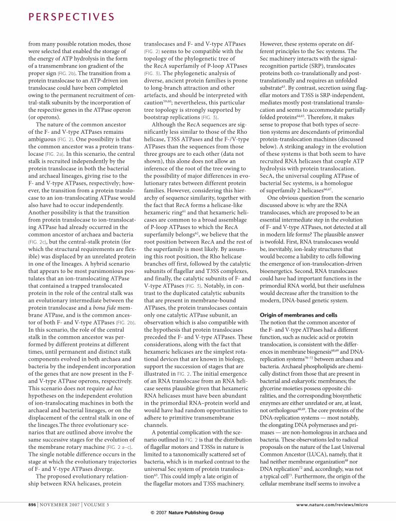
F-type and V-type reversible ATPases are membrane-associated molecular machines that couple the transfer of protons or sodium cations across the membrane with ATP hydrolysis or synthesis1–8 (FIG. 1). These enzymes represent the cornerstone of cel-lular bioenergetics and are ubiquitous to all three domains of life (bacteria, archaea and eukaryotes). The F-type F0F1 ATPases are found in the mitochondria and chloroplasts of all eukaryotic cells and in most bacteria, and have a range of structural features that distinguish them from the V (vacuolar)-type ATPases. V-type ATPases occur in eukaryotic cytoplasmic membranes (in particular, vacuoles), archaea and in a small, although important, number of bacteria. In rooted phylogenetic trees, eukaryotic, archaeal and bacterial V-type ATPases invariably cluster together, and separately, from the F-type ATPases9. The F-type ATPase is thought to be the ancestral bacterial cation-translocating ATPase, and conversely, the V-type ATPase is thought to be the ancestral archaeal form. Accordingly, the presence of V-type ATPases in several bacterial lineages, and the presence of F-type ATPases in two species of the archaeal genus Methanosarcina, is thought to be a conse-quence of the extensive horizontal transfer of the respective genes between the two domains10–12.
Recent structural studies have provided new insights into conserved and distinct features of F- and V-type ATPases2,13–20. Both
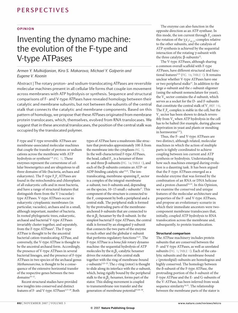
types of ATPase have a mushroom-like struc-ture that protrudes approximately 100 Å from the membrane into the cytoplasm (FIG. 1). In the well-characterized F-type ATPases, the head, called F1, is a hexamer of three α- and three β-subunits (FIG. 1a; TABLE 1), and each of the β-subunits contains an ATP- or ADP-binding catalytic site13,21. The ion-translocating, membrane-spanning F0 sector is a complex of the integral-membrane a-subunit, two b-subunits and, depending on the species, 10–15 small c-subunits17. This component of the structure is connected to the F1 component by both a peripheral and a central stalk. The peripheral stalk is formed by the protruding parts of the membrane-anchored b-subunits that are connected to the α3β3-hexamer by the δ-subunit. In the simplest bacterial F-type ATPases, the central stalk is formed by an elongated γ-subunit that connects the two parts of the enzyme to each other and the globular ε-subunit that performs regulatory functions13,21. The F-type ATPase is a bona fide rotary dynamo machine: the sequential hydrolysis of ATP molecules by the α3β3-catalytic hexamer drives the rotation of the central stalk together with the ring of membrane-bound c-subunits5,22–25. The c-ring (rotor) is thought to slide along its interface with the a-subunit, which, being rigidly bound by the peripheral stalk to the α3β3-hexamer, forms part of the stator. This sliding movement is coupled to transmembrane-ion transfer and the generation of membrane potential23,26–30.
The enzyme can also function in the opposite direction as an ATP synthase. In this mode, the ion current through F0 causes the rotation of the γ1ε1c10–15-complex relative to the other subunits, and the catalysis of ATP synthesis is achieved by the sequential interaction of the rotating γ-subunit with the three catalytic β-subunits22.
The V-type ATPases, although sharing a common overall scaffold with F-type ATPases, have different structural and func-tional features2,31 (FIG. 1b; TABLE 1). It remains unclear whether V-type ATPases have one or two peripheral stalks15. In addition to the large a-subunit and the c-subunit oligomer (using the subunit nomenclature for yeast), the V0 sector contains the d-subunit, which serves as a socket for the D- and F-subunits that constitute the central stalk of V1 (REF. 14). The F1F0 complex is stable in the cell, but the V1 sector has been shown to detach revers-ibly from V0 when ATP hydrolysis in the cell has to be halted (for example, during glucose deprivation in yeast and plants or moulting in hornworms2,31).
Thus, the F- and V-type ATPases are two distinct, although related, molecular machines in which the action of multiple parts is tightly coordinated to achieve coupling between ion current and ATP synthesis or hydrolysis. Understanding how such machines emerged during evolu-tion is a daunting task. It has been argued that the F-type ATPases emerged as a modular enzyme that was formed by the combination of an RNA or DNA helicase and a proton channel32,33. In this Opinion, we examine the conserved and unique features of the molecular architecture and properties of the F- and V-type ATPases, and propose an evolutionary scenario in which their immediate ancestors were two-component membrane translocases that, initially, coupled ATP hydrolysis to RNA translocation across the membrane and, subsequently, to protein translocation.
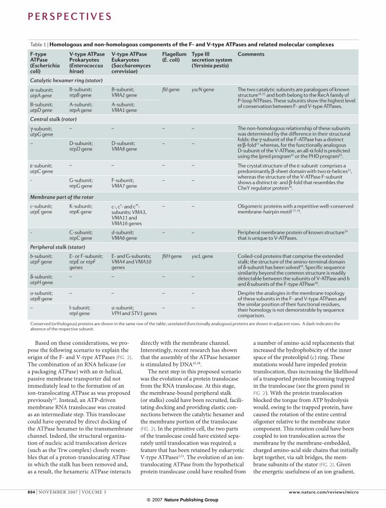
Structural comparisonThe ATPase machinery includes protein subunits that are conserved between the F- and V-type ATPases, as well as unrelated subunits (FIG. 1; TABLE 1). Each of the cata-lytic subunits and the membrane-bound c (proteolipid)-subunits are homologous and highly conserved. The homology between the δ-subunit of the F-type ATPase, the protruding portion of the b-subunit of the F-type ATPase and the E- and G-subunits of the V-ATPase, has been inferred from weak sequence similarity34,35. The relationship between the membrane parts of the stator is
o p i n i o n
Inventing the dynamo machine: the evolution of the F‑type and V‑type ATPasesArmen Y. Mulkidjanian, Kira S. Makarova, Michael Y. Galperin and Eugene V. Koonin
Abstract | The rotary proton- and sodium-translocating ATPases are reversible molecular machines present in all cellular life forms that couple ion movement across membranes with ATP hydrolysis or synthesis. Sequence and structural comparisons of F- and V-type ATPases have revealed homology between their catalytic and membrane subunits, but not between the subunits of the central stalk that connects the catalytic and membrane components. Based on this pattern of homology, we propose that these ATPases originated from membrane protein translocases, which, themselves, evolved from RNA translocases. We suggest that in these ancestral translocases, the position of the central stalk was occupied by the translocated polymer.
P e r s P e c t i v e s
892 | NOVEmBER 2007 | VOlUmE 5 www.nature.com/reviews/micro
© 2007 Nature Publishing Group
a more complex issue. Although there is no significant sequence similarity between the a-subunits of the F- and V-type ATPases, there are structural and mechanistic analo-gies7,36. Both types of a-subunit contain a strictly conserved arginine residue in the middle of the penultimate transmembrane helix. The sequential electrostatic interaction of this arginine with the conserved carboxy-late moieties that are contained within the rotating c-subunits is crucial for ion trans-port37,38. In terms of domain organization, the a-subunit of the V-type ATPase, which consists of a bundle of hydrophobic trans-membrane helices and a hydrophilic amino terminus, resembles a fusion of the F-type ATPase a-subunit (five transmembrane heli-ces) with a b-subunit (one transmembrane helix and a hydrophilic amino terminus). Whether the functional and structural similarity between the a-subunits of F- and V-type ATPases reflects a common origin remains uncertain.
By contrast, the subunits of the central stalk, which connects the head structure with the membrane moiety, are not homolo-gous or even structurally similar. This is clear not only from a lack of significant sequence similarity but also from the pres-ence of distinct folds in these subunits2 (FIG. 1; TABLE 1). Specifically, the crystal structure of the γ-subunit of the F-type ATPase has revealed a distinct α/β-fold that resembles the Rossmann fold21. Although the structure of the functionally analogous
D-subunit of the V-type ATPase has not been solved, a 100% α-fold has been con-fidently predicted for this protein (TABLE 1). The section of the F-type ATPase γ-subunit that protrudes into the catalytic hexamer is formed by two long amino-terminal and carboxy-terminal α-helices that bracket the domain that contains the Rossmann-like fold. The sequences of these helices have no detectable similarity to any portion of the D-subunit of the V-type ATPase, and their different placement within the γ-subunit sequence argues against the possibility that at least one domain of the F-type ATPase γ-subunit is homologous to the D-subunit of the V-type ATPase.
The presence of conserved and unrelated subunits within the structures of F- and V-type ATPases is non-random and requires an explanation. In the following sections we propose a scenario for the evolution of the two classes of membrane-bound ATPases.
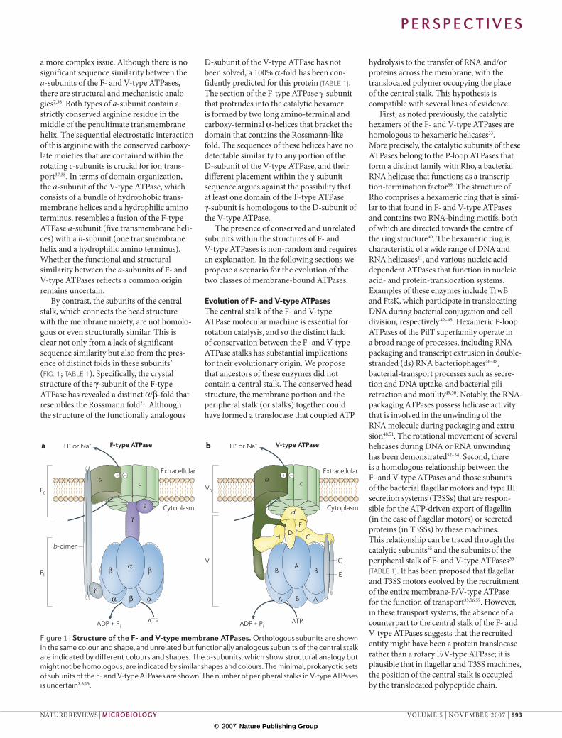
Evolution of F‑ and V‑type ATpasesThe central stalk of the F- and V-type ATPase molecular machine is essential for rotation catalysis, and so the distinct lack of conservation between the F- and V-type ATPase stalks has substantial implications for their evolutionary origin. We propose that ancestors of these enzymes did not contain a central stalk. The conserved head structure, the membrane portion and the peripheral stalk (or stalks) together could have formed a translocase that coupled ATP
hydrolysis to the transfer of RNA and/or proteins across the membrane, with the translocated polymer occupying the place of the central stalk. This hypothesis is compatible with several lines of evidence.
First, as noted previously, the catalytic hexamers of the F- and V-type ATPases are homologous to hexameric helicases33. more precisely, the catalytic subunits of these ATPases belong to the P-loop ATPases that form a distinct family with Rho, a bacterial RNA helicase that functions as a transcrip-tion-termination factor39. The structure of Rho comprises a hexameric ring that is simi-lar to that found in F- and V-type ATPases and contains two RNA-binding motifs, both of which are directed towards the centre of the ring structure40. The hexameric ring is characteristic of a wide range of DNA and RNA helicases41, and various nucleic acid-dependent ATPases that function in nucleic acid- and protein-translocation systems. Examples of these enzymes include TrwB and FtsK, which participate in translocating DNA during bacterial conjugation and cell division, respectively42–45. Hexameric P-loop ATPases of the PilT superfamily operate in a broad range of processes, including RNA packaging and transcript extrusion in double-stranded (ds) RNA bacteriophages46–48, bacterial-transport processes such as secre-tion and DNA uptake, and bacterial pili retraction and motility49,50. Notably, the RNA-packaging ATPases possess helicase activity that is involved in the unwinding of the RNA molecule during packaging and extru-sion48,51. The rotational movement of several helicases during DNA or RNA unwinding has been demonstrated52–54. Second, there is a homologous relationship between the F- and V-type ATPases and those subunits of the bacterial flagellar motors and type III secretion systems (T3SSs) that are respon-sible for the ATP-driven export of flagellin (in the case of flagellar motors) or secreted proteins (in T3SSs) by these machines. This relationship can be traced through the catalytic subunits55 and the subunits of the peripheral stalk of F- and V-type ATPases35 (TABLE 1). It has been proposed that flagellar and T3SS motors evolved by the recruitment of the entire membrane-F/V-type ATPase for the function of transport35,56,57. However, in these transport systems, the absence of a counterpart to the central stalk of the F- and V-type ATPases suggests that the recruited entity might have been a protein translocase rather than a rotary F/V-type ATPase; it is plausible that in flagellar and T3SS machines, the position of the central stalk is occupied by the translocated polypeptide chain.
Nature Reviews | Microbiology
B AA
BG
H+ or Na+H+ or Na+ V-type ATPase
E
ADP + PiATP
d
c
F
BA
a
H C
β αα
β
Cytoplasm
Extracellular
Cytoplasm
Extracellular
a bF-type ATPase
ADP + PiATP
γ
ε
c
β α
a + – + –
δ
F0
F1
V0
V1
D
b-dimer
Figure 1 | Structure of the F‑ and V‑type membrane ATPases. Orthologous subunits are shown in the same colour and shape, and unrelated but functionally analogous subunits of the central stalk are indicated by different colours and shapes. The a-subunits, which show structural analogy but might not be homologous, are indicated by similar shapes and colours. The minimal, prokaryotic sets of subunits of the F- and V-type ATPases are shown. The number of peripheral stalks in V-type ATPases is uncertain2,8,15.
P e r s P e c t i v e s
NATURE REVIEWS | microbiology VOlUmE 5 | NOVEmBER 2007 | 893
© 2007 Nature Publishing Group
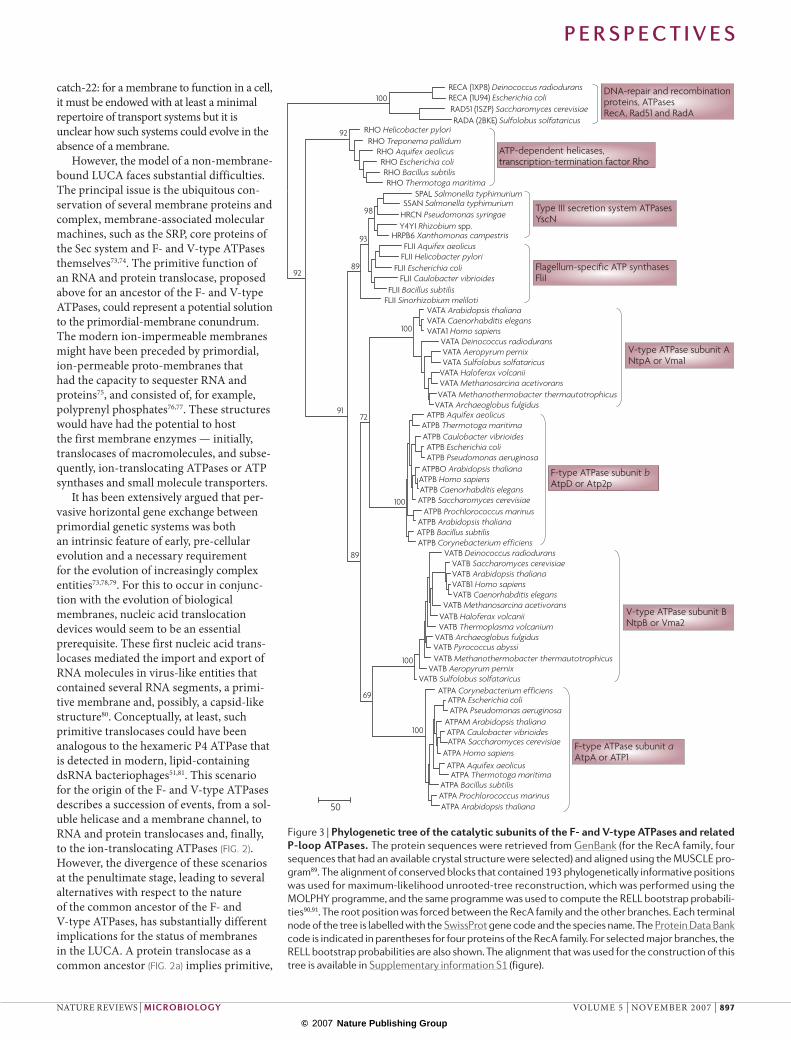
Based on these considerations, we pro-pose the following scenario to explain the origin of the F- and V-type ATPases (FIG. 2). The combination of an RNA helicase (or a packaging ATPase) with an α-helical, passive membrane transporter did not immediately lead to the formation of an ion-translocating ATPase as was proposed previously33. Instead, an ATP-driven membrane RNA translocase was created as an intermediate step. This translocase could have operated by direct docking of the ATPase hexamer to the transmembrane channel. Indeed, the structural organiza-tion of nucleic acid translocation devices (such as the Trw complex) closely resem-bles that of a proton-translocating ATPase in which the stalk has been removed and, as a result, the hexameric ATPase interacts
directly with the membrane channel. Interestingly, recent research has shown that the assembly of the ATPase hexamer is stimulated by DNA43,58.
The next step in this proposed scenario was the evolution of a protein translocase from the RNA translocase. At this stage, the membrane-bound peripheral stalk (or stalks) could have been recruited, facili-tating docking and providing elastic con-nections between the catalytic hexamer and the membrane portion of the translocase (FIG. 2). In the primitive cell, the two parts of the translocase could have existed sepa-rately until translocation was required; a feature that has been retained by eukaryotic V-type ATPases2,31. The evolution of an ion- translocating ATPase from the hypothetical protein translocase could have resulted from
a number of amino-acid replacements that increased the hydrophobicity of the inner space of the proteolipid (c) ring. These mutations would have impeded protein translocation, thus increasing the likelihood of a transported protein becoming trapped in the translocase (see the green panel in FIG. 2). With the protein translocation blocked the torque from ATP hydrolysis would, owing to the trapped protein, have caused the rotation of the entire central oligomer relative to the membrane stator component. This rotation could have been coupled to ion translocation across the membrane by the membrane-embedded, charged amino-acid side chains that initially kept together, via salt bridges, the mem-brane subunits of the stator (FIG. 2). Given the energetic usefulness of an ion gradient,
Table 1 | Homologous and non‑homologous components of the F‑ and V‑type ATpases and related molecular complexes
F‑type ATPase (Escherichia coli)
V‑type ATPase Prokaryotes (Enterococcus hirae)
V‑type ATPase Eukaryotes (Saccharomyces cerevisiae)
Flagellum (E. coli)
Type iii secretion system (Yersinia pestis)
comments
Catalytic hexamer ring (stator)
α-subunit; atpA gene
B-subunit; ntpB gene
B-subunit; VMA2 gene
fliI gene yscN gene The two catalytic subunits are paralogues of known structure19, 21 and both belong to the RecA family of P-loop NTPases. These subunits show the highest level of conservation between F- and V-type ATPases. B-subunit;
atpD geneA-subunit; ntpA gene
A-subunit; VMA1 gene
Central stalk (rotor)
γ-subunit; atpG gene
– – – – The non-homologous relationship of these subunits was determined by the difference in their structural folds: the γ-subunit of the F-ATPase has a distinct α/β-fold21 whereas, for the functionally analogous D-subunit of the V-ATPase, an all-α fold is predicted using the Jpred program92 or the PHD program93.
– D-subunit; ntpD gene
D-subunit; VMA8 gene
– –
ε-subunit; atpC gene
– – – – The crystal structure of the ε-subunit comprises a predominantly β-sheet domain with two α-helices21, whereas the structure of the V-ATPase F-subunit shows a distinct α- and β-fold that resembles the CheY regulator protein16.
- G-subunit; ntpG gene
F-subunit; VMA7 gene
– –
Membrane part of the rotor
c-subunit; atpE gene
K-subunit; ntpK gene
c-, c′- and c′′-subunits; VMA3, VMA11 and VMA16 genes
– – Oligomeric proteins with a repetitive well-conserved membrane-hairpin motif 17, 18.
- C-subunit; ntpC gene
d-subunit; VMA6 gene
– – Peripheral membrane protein of known structure14 that is unique to V-ATPases.
Peripheral stalk (stator)
b-subunit; atpF gene
E- or F-subunit; ntpE or ntpF genes
E- and G-subunits; VMA4 and VMA10 genes
fliH gene yscL gene Coiled-coil proteins that comprise the extended stalk; the structure of the amino-terminal domain of δ-subunit has been solved20. Specific sequence similarity beyond the common structure is readily detectable between the subunits of V-ATPase and b and δ subunits of the F-type ATPase30.
δ-subunit; atpH gene
– – – –
a-subunit; atpB gene
– – – – Despite the analogies in the membrane topology of these subunits in the F- and V-type ATPases and the similar position of their functional residues, their homology is not demonstrable by sequence comparison.
– I-subunit; ntpI gene
a-subunit; VPH and STV1 genes
– –
Conserved (orthologous) proteins are shown in the same row of the table; unrelated (functionally analogous) proteins are shown in adjacent rows. A dash indicates the absence of the respective subunit.
P e r s P e c t i v e s
894 | NOVEmBER 2007 | VOlUmE 5 www.nature.com/reviews/micro
© 2007 Nature Publishing Group
Nature Reviews | Microbiology
Membrane channel RNA helicase RNA translocase Protein
translocase
a b c
T3SS translocase
FliH-dimer
V-type ATPase
Common ancestor of F- and V-type ATPases
F-type ATPase
– + –
+
+ –
+
+
Flil
H+ or Na+ H+ or Na+
+ – + –
Figure 2 | Proposed evolution of the F‑ and V‑type membrane ATPases. Hypothetical, ancestral semi-permeable membranes are shown by empty circles and light curved lines, and modern-type, ion-tight membranes are shown by filled circles and dense curved lines. The presence of two peripheral stalks in the primordial protein translocase and the flagellar motor and type III secretion systems (T3SSs) is based on the assumption that a translocation system with only one peripheral stalk would be unstable in the absence of the translocated substrate. The involvement of two FliH subunits in each peripheral stalk is based on the ability of FliH dimers to form a complex with one FliI subunit87,88. The light-green panel in the centre shows the three consecutive evolutionary intermediates (a–c), each of which is considered to be the common ancestor of the F- and V-type ATPases in the three alternative evolutionary scenarios described in the main text. The purple tubes denote the translocated, partially unfolded proteins. The red tube denotes a translocated protein that is trapped in the channel of the membrane translocase. The names of subunits are indicated only for the flagellar motor and T3SS machines.
P e r s P e c t i v e s
NATURE REVIEWS | microbiology VOlUmE 5 | NOVEmBER 2007 | 895
© 2007 Nature Publishing Group
from many possible rotation modes, those were selected that enabled the storage of the energy of ATP hydrolysis in the form of a transmembrane ion gradient of the proper sign (FIG. 2b). The transition from a protein translocase to an ATP-driven ion translocase could have been completed owing to the permanent recruitment of cen-tral-stalk subunits by the incorporation of the respective genes in the ATPase operon (or operons).
The nature of the common ancestor of the F- and V-type ATPases remains ambiguous (FIG. 2). One possibility is that the common ancestor was a protein trans-locase (FIG. 2a). In this scenario, the central stalk is recruited independently by the protein translocase in both the bacterial and archaeal lineages, giving rise to the F- and V-type ATPases, respectively; how-ever, the transition from a protein translo-case to an ion-translocating ATPase would also have had to occur independently. Another possibility is that the transition from protein translocase to ion-translocat-ing ATPase had already occurred in the common ancestor of archaea and bacteria (FIG. 2c), but the central-stalk protein (for which the structural requirements are flex-ible) was displaced by an unrelated protein in one of the lineages. A hybrid scenario that appears to be most parsimonious pos-tulates that an ion-translocating ATPase that contained a trapped translocated protein in the role of the central stalk was an evolutionary intermediate between the protein translocase and a bona fide mem-brane ATPase, and is the common ances-tor of both F- and V-type ATPases (FIG. 2b). In this scenario, the role of the central stalk in the common ancestor was per-formed by different proteins at different times, until permanent and distinct stalk components evolved in both archaea and bacteria by the independent incorporation of the genes that are now present in the F- and V-type ATPase operons, respectively. This scenario does not require ad hoc hypotheses on the independent evolution of ion-translocating machines in both the archaeal and bacterial lineages, or on the displacement of the central stalk in one of the lineages.The three evolutionary sce-narios that are outlined above involve the same successive stages for the evolution of the membrane rotary machine (FIG. 2 a–c). The single notable difference occurs in the stage at which the evolutionary trajectories of F- and V-type ATPases diverge.
The proposed evolutionary relation-ship between RNA helicases, protein
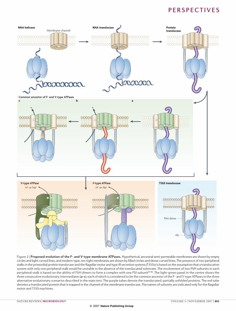
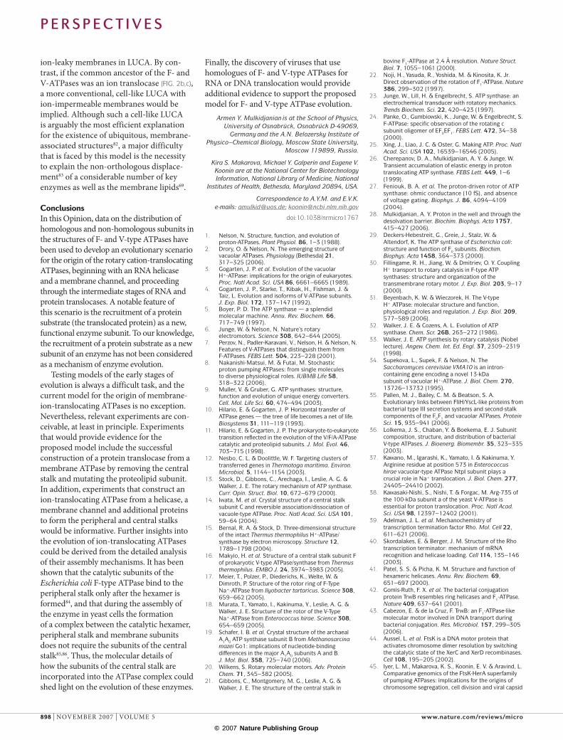
translocases and F- and V-type ATPases (FIG. 2) seems to be compatible with the topology of the phylogenetic tree of the RecA superfamily of P-loop ATPases (FIG. 3). The phylogenetic analysis of diverse, ancient protein families is prone to long-branch attraction and other artefacts, and should be interpreted with caution59,60; nevertheless, this particular tree topology is strongly supported by bootstrap replications (FIG. 3).
Although the RecA sequences are sig-nificantly less similar to those of the Rho helicase, T3SS ATPases and the F-/V-type ATPases than the sequences from those three groups are to each other (data not shown), this alone does not allow an inference of the root of the tree owing to the possibility of major differences in evo-lutionary rates between different protein families. However, considering this hier-archy of sequence similarity, together with the fact that RecA forms a helicase-like hexameric ring61 and that hexameric heli-cases are common to a broad assemblage of P-loop ATPases to which the RecA superfamily belongs62, we believe that the root position between RecA and the rest of the superfamily is most likely. By assum-ing this root position, the Rho helicase branches off first, followed by the catalytic subunits of flagellar and T3SS complexes, and finally, the catalytic subunits of F- and V-type ATPases (FIG. 3). Notably, in con-trast to the duplicated catalytic subunits that are present in membrane-bound ATPases, the protein translocases contain only one catalytic ATPase subunit, an observation which is also compatible with the hypothesis that protein translocases preceded the F- and V-type ATPases. These considerations, along with the fact that hexameric helicases are the simplest rota-tional devices that are known in biology, support the succession of stages that are illustrated in FIG. 2. The initial emergence of an RNA translocase from an RNA heli-case seems plausible given that hexameric RNA helicases must have been abundant in the primordial RNA–protein world and would have had random opportunities to adhere to primitive transmembrane channels.
A potential complication with the sce-nario outlined in FIG. 2 is that the distribution of flagellar motors and T3SSs in nature is limited to a taxonomically scattered set of bacteria, which is in marked contrast to the universal Sec system of protein transloca-tion63. This could imply a late origin of the flagellar motors and T3SS machinery.
However, these systems operate on dif-ferent principles to the Sec systems. The Sec machinery interacts with the signal-recognition particle (SRP), translocates proteins both co-translationally and post-translationally and requires an unfolded substrate63. By contrast, secretion using flag-ellar motors and T3SS is SRP-independent, mediates mostly post-translational translo-cation and seems to accommodate partially folded proteins64,65. Therefore, it makes sense to propose that both types of secre-tion systems are descendants of primordial protein-translocation machines (discussed below). A striking analogy in the evolution of these systems is that both seem to have recruited RNA helicases that couple ATP hydrolysis with protein translocation. SecA, the universal coupling ATPase of bacterial Sec systems, is a homologue of superfamily 2 helicases66,67.
One obvious question from the scenario discussed above is: why are the RNA translocases, which are proposed to be an essential intermediate step in the evolution of F- and V-type ATPases, not detected at all in modern life forms? The plausible answer is twofold. First, RNA translocases would be, inevitably, ion-leaky structures that would become a liability to cells following the emergence of ion-translocation-driven bioenergetics. Second, RNA translocases could have had important functions in the primordial RNA world, but their usefulness would decrease after the transition to the modern, DNA-based genetic system.
origin of membranes and cellsThe notion that the common ancestor of the F- and V-type ATPases had a different function, such as nucleic acid or protein translocation, is consistent with the differ-ences in membrane biogenesis68,69 and DNA-replication systems70–72 between archaea and bacteria. Archaeal phospholipids are chemi-cally distinct from those that are present in bacterial and eukaryotic membranes; the glycerine moieties possess opposite chi-ralities, and the corresponding biosynthetic enzymes are either unrelated or are, at least, not orthologous68,69. The core proteins of the DNA-replication systems — most notably, the elongating DNA polymerases and pri-mases — are non-homologous in archaea and bacteria. These observations led to radical proposals on the nature of the last Universal Common Ancestor (lUCA), namely, that it had neither membrane organization68 nor DNA replication72 and, accordingly, was not a typical cell73. Furthermore, the origin of the cellular membrane itself seems to involve a
P e r s P e c t i v e s
896 | NOVEmBER 2007 | VOlUmE 5 www.nature.com/reviews/micro
© 2007 Nature Publishing Group
catch-22: for a membrane to function in a cell, it must be endowed with at least a minimal repertoire of transport systems but it is unclear how such systems could evolve in the absence of a membrane.
However, the model of a non-membrane-bound lUCA faces substantial difficulties. The principal issue is the ubiquitous con-servation of several membrane proteins and complex, membrane-associated molecular machines, such as the SRP, core proteins of the Sec system and F- and V-type ATPases themselves73,74. The primitive function of an RNA and protein translocase, proposed above for an ancestor of the F- and V-type ATPases, could represent a potential solution to the primordial-membrane conundrum. The modern ion-impermeable membranes might have been preceded by primordial, ion-permeable proto-membranes that had the capacity to sequester RNA and proteins75, and consisted of, for example, polyprenyl phosphates76,77. These structures would have had the potential to host the first membrane enzymes — initially, translocases of macromolecules, and subse-quently, ion-translocating ATPases or ATP synthases and small molecule transporters.
It has been extensively argued that per-vasive horizontal gene exchange between primordial genetic systems was both an intrinsic feature of early, pre-cellular evolution and a necessary requirement for the evolution of increasingly complex entities73,78,79. For this to occur in conjunc-tion with the evolution of biological membranes, nucleic acid translocation devices would seem to be an essential prerequisite. These first nucleic acid trans-locases mediated the import and export of RNA molecules in virus-like entities that contained several RNA segments, a primi-tive membrane and, possibly, a capsid-like structure80. Conceptually, at least, such primitive translocases could have been analogous to the hexameric P4 ATPase that is detected in modern, lipid-containing dsRNA bacteriophages51,81. This scenario for the origin of the F- and V-type ATPases describes a succession of events, from a sol-uble helicase and a membrane channel, to RNA and protein translocases and, finally, to the ion-translocating ATPases (FIG. 2). However, the divergence of these scenarios at the penultimate stage, leading to several alternatives with respect to the nature of the common ancestor of the F- and V-type ATPases, has substantially different implications for the status of membranes in the lUCA. A protein translocase as a common ancestor (FIG. 2a) implies primitive,
50
RECA (1XP8) Deinococcus radiodurans RECA (1U94) Escherichia coli RAD51 (1SZP) Saccharomyces cerevisiae RADA (2BKE) Sulfolobus solfataricus
DNA-repair and recombination proteins, ATPasesRecA, Rad51 and RadA
ATP-dependent helicases, transcription-termination factor Rho
Type III secretion system ATPasesYscN
Flagellum-specific ATP synthases FliI
V-type ATPase subunit ANtpA or Vma1
V-type ATPase subunit BNtpB or Vma2
F-type ATPase subunit bAtpD or Atp2p
F-type ATPase subunit aAtpA or ATP1
RHO Helicobacter pylori RHO Treponema pallidum
RHO Aquifex aeolicus RHO Escherichia coli RHO Bacillus subtilis RHO Thermotoga maritima
SPAL Salmonella typhimurium SSAN Salmonella typhimurium
HRCN Pseudomonas syringae Y4YI Rhizobium spp.
HRPB6 Xanthomonas campestris FLII Aquifex aeolicus
FLII Helicobacter pylori FLII Escherichia coli
FLII Caulobacter vibrioides FLII Bacillus subtilis
FLII Sinorhizobium meliloti VATA Arabidopsis thaliana VATA Caenorhabditis elegans VATA1 Homo sapiens
VATA Deinococcus radiodurans VATA Aeropyrum pernix VATA Sulfolobus solfataricus
VATA Haloferax volcanii VATA Methanosarcina acetivorans VATA Methanothermobacter thermautotrophicus
VATA Archaeoglobus fulgidus ATPB Aquifex aeolicus
ATPB Thermotoga maritima ATPB Caulobacter vibrioides ATPB Escherichia coli ATPB Pseudomonas aeruginosa
ATPBO Arabidopsis thaliana ATPB Homo sapiens ATPB Caenorhabditis elegans ATPB Saccharomyces cerevisiae
ATPB Prochlorococcus marinus ATPB Arabidopsis thaliana ATPB Bacillus subtilis ATPB Corynebacterium efficiens
VATB Deinococcus radiodurans VATB Saccharomyces cerevisiae VATB Arabidopsis thaliana VATB1 Homo sapiens VATB Caenorhabditis elegans
VATB Methanosarcina acetivorans VATB Haloferax volcanii VATB Thermoplasma volcanium
VATB Archaeoglobus fulgidus VATB Pyrococcus abyssi VATB Methanothermobacter thermautotrophicus
VATB Aeropyrum pernix VATB Sulfolobus solfataricus
ATPA Corynebacterium efficiens ATPA Escherichia coli ATPA Pseudomonas aeruginosa
ATPAM Arabidopsis thaliana ATPA Caulobacter vibrioides ATPA Saccharomyces cerevisiae
ATPA Homo sapiens ATPA Aquifex aeolicus ATPA Thermotoga maritima
ATPA Bacillus subtilis ATPA Prochlorococcus marinus ATPA Arabidopsis thaliana
100
100
100
100
100
72
69
89
92
92
98
93
89
91
Nature Reviews | Microbiology Figure 3 | Phylogenetic tree of the catalytic subunits of the F‑ and V‑type ATPases and related P‑loop ATPases. The protein sequences were retrieved from GenBank (for the RecA family, four sequences that had an available crystal structure were selected) and aligned using the MUSCLE pro-gram89. The alignment of conserved blocks that contained 193 phylogenetically informative positions was used for maximum-likelihood unrooted-tree reconstruction, which was performed using the MOLPHY programme, and the same programme was used to compute the RELL bootstrap probabili-ties90,91. The root position was forced between the RecA family and the other branches. Each terminal node of the tree is labelled with the SwissProt gene code and the species name. The Protein Data Bank code is indicated in parentheses for four proteins of the RecA family. For selected major branches, the RELL bootstrap probabilities are also shown. The alignment that was used for the construction of this tree is available in Supplementary information S1 (figure).
P e r s P e c t i v e s
NATURE REVIEWS | microbiology VOlUmE 5 | NOVEmBER 2007 | 897
© 2007 Nature Publishing Group
ion-leaky membranes in lUCA. By con-trast, if the common ancestor of the F- and V-ATPases was an ion translocase (FIG. 2b,c), a more conventional, cell-like lUCA with ion-impermeable membranes would be implied. Although such a cell-like lUCA is arguably the most efficient explanation for the existence of ubiquitous, membrane-associated structures82, a major difficulty that is faced by this model is the necessity to explain the non-orthologous displace-ment83 of a considerable number of key enzymes as well as the membrane lipids69.
ConclusionsIn this Opinion, data on the distribution of homologous and non-homologous subunits in the structures of F- and V-type ATPases have been used to develop an evolutionary scenario for the origin of the rotary cation-translocating ATPases, beginning with an RNA helicase and a membrane channel, and proceeding through the intermediate stages of RNA and protein translocases. A notable feature of this scenario is the recruitment of a protein substrate (the translocated protein) as a new, functional enzyme subunit. To our knowledge, the recruitment of a protein substrate as a new subunit of an enzyme has not been considered as a mechanism of enzyme evolution.
Testing models of the early stages of evolution is always a difficult task, and the current model for the origin of membrane-ion-translocating ATPases is no exception. Nevertheless, relevant experiments are con-ceivable, at least in principle. Experiments that would provide evidence for the proposed model include the successful construction of a protein translocase from a membrane ATPase by removing the central stalk and mutating the proteolipid subunit. In addition, experiments that construct an ion-translocating ATPase from a helicase, a membrane channel and additional proteins to form the peripheral and central stalks would be informative. Further insights into the evolution of ion-translocating ATPases could be derived from the detailed analysis of their assembly mechanisms. It has been shown that the catalytic subunits of the Escherichia coli F-type ATPase bind to the peripheral stalk only after the hexamer is formed84, and that during the assembly of the enzyme in yeast cells the formation of a complex between the catalytic hexamer, peripheral stalk and membrane subunits does not require the subunits of the central stalk85,86. Thus, the molecular details of how the subunits of the central stalk are incorporated into the ATPase complex could shed light on the evolution of these enzymes.
Finally, the discovery of viruses that use homologues of F- and V-type ATPases for RNA or DNA translocation would provide additional evidence to support the proposed model for F- and V-type ATPase evolution.
Armen Y. Mulkidjanian is at the School of Physics, University of Osnabrück, Osnabrück D‑49069,
Germany and the A.N. Belozersky Institute of Physico–Chemical Biology, Moscow State University,
Moscow 119899, Russia.
Kira S. Makarova, Michael Y. Galperin and Eugene V. Koonin are at the National Center for Biotechnology Information, National Library of Medicine, National
Institutes of Health, Bethesda, Maryland 20894, USA.
Correspondence to A.Y.M. and E.V.K. e‑mails: [email protected]; [email protected]
doi:10.1038/nrmicro1767
1. Nelson, N. Structure, function, and evolution of proton‑ATPases. Plant Physiol. 86, 1–3 (1988).
2. Drory, O. & Nelson, N. The emerging structure of vacuolar ATPases. Physiology (Bethesda) 21, 317–325 (2006).
3. Gogarten, J. P. et al. Evolution of the vacuolar H+‑ATPase: implications for the origin of eukaryotes. Proc. Natl Acad. Sci. USA 86, 6661–6665 (1989).
4. Gogarten, J. P., Starke, T., Kibak, H., Fishman, J. & Taiz, L. Evolution and isoforms of V‑ATPase subunits. J. Exp. Biol. 172, 137–147 (1992).
5. Boyer, P. D. The ATP synthase — a splendid molecular machine. Annu. Rev. Biochem. 66, 717–749 (1997).
6. Junge, W. & Nelson, N. Nature’s rotary electromotors. Science 308, 642–644 (2005).
7. Perzov, N., Padler‑Karavani, V., Nelson, H. & Nelson, N. Features of V‑ATPases that distinguish them from F‑ATPases. FEBS Lett. 504, 223–228 (2001).
8. Nakanishi‑Matsui, M. & Futai, M. Stochastic proton pumping ATPases: from single molecules to diverse physiological roles. IUBMB Life 58, 318–322 (2006).
9. Muller, V. & Gruber, G. ATP synthases: structure, function and evolution of unique energy converters. Cell. Mol. Life Sci. 60, 474–494 (2003).
10. Hilario, E. & Gogarten, J. P. Horizontal transfer of ATPase genes — the tree of life becomes a net of life. Biosystems 31, 111–119 (1993).
11. Hilario, E. & Gogarten, J. P. The prokaryote‑to‑eukaryote transition reflected in the evolution of the V/F/A‑ATPase catalytic and proteolipid subunits. J. Mol. Evol. 46, 703–715 (1998).
12. Nesbo, C. L. & Doolittle, W. F. Targeting clusters of transferred genes in Thermotoga maritima. Environ. Microbiol. 5, 1144–1154 (2003).
13. Stock, D., Gibbons, C., Arechaga, I., Leslie, A. G. & Walker, J. E. The rotary mechanism of ATP synthase. Curr. Opin. Struct. Biol. 10, 672–679 (2000).
14. Iwata, M. et al. Crystal structure of a central stalk subunit C and reversible association/dissociation of vacuole‑type ATPase. Proc. Natl Acad. Sci. USA 101, 59–64 (2004).
15. Bernal, R. A. & Stock, D. Three‑dimensional structure of the intact Thermus thermophilus H+‑ATPase/synthase by electron microscopy. Structure 12, 1789–1798 (2004).
16. Makyio, H. et al. Structure of a central stalk subunit F of prokaryotic V‑type ATPase/synthase from Thermus thermophilus. EMBO J. 24, 3974–3983 (2005).
17. Meier, T., Polzer, P., Diederichs, K., Welte, W. & Dimroth, P. Structure of the rotor ring of F‑Type Na+‑ATPase from Ilyobacter tartaricus. Science 308, 659–662 (2005).
18. Murata, T., Yamato, I., Kakinuma, Y., Leslie, A. G. & Walker, J. E. Structure of the rotor of the V‑Type Na+‑ATPase from Enterococcus hirae. Science 308, 654–659 (2005).
19. Schafer, I. B. et al. Crystal structure of the archaeal A1A0 ATP synthase subunit B from Methanosarcina mazei Go1: implications of nucleotide‑binding differences in the major A1A0 subunits A and B. J. Mol. Biol. 358, 725–740 (2006).
20. Wilkens, S. Rotary molecular motors. Adv. Protein Chem. 71, 345–382 (2005).
21. Gibbons, C., Montgomery, M. G., Leslie, A. G. & Walker, J. E. The structure of the central stalk in
bovine F1‑ATPase at 2.4 Å resolution. Nature Struct. Biol. 7, 1055–1061 (2000).
22. Noji, H., Yasuda, R., Yoshida, M. & Kinosita, K. Jr. Direct observation of the rotation of F1‑ATPase. Nature 386, 299–302 (1997).
23. Junge, W., Lill, H. & Engelbrecht, S. ATP synthase: an electrochemical transducer with rotatory mechanics. Trends Biochem. Sci. 22, 420–423 (1997).
24. Panke, O., Gumbiowski, K., Junge, W. & Engelbrecht, S. F‑ATPase: specific observation of the rotating c subunit oligomer of EF0EF1. FEBS Lett. 472, 34–38 (2000).
25. Xing, J., Liao, J. C. & Oster, G. Making ATP. Proc. Natl Acad. Sci. USA 102, 16539–16546 (2005).
26. Cherepanov, D. A., Mulkidjanian, A. Y. & Junge, W. Transient accumulation of elastic energy in proton translocating ATP synthase. FEBS Lett. 449, 1–6 (1999).
27. Feniouk, B. A. et al. The proton‑driven rotor of ATP synthase: ohmic conductance (10 fS), and absence of voltage gating. Biophys. J. 86, 4094–4109 (2004).
28. Mulkidjanian, A. Y. Proton in the well and through the desolvation barrier. Biochim. Biophys. Acta 1757, 415–427 (2006).
29. Deckers‑Hebestreit, G., Greie, J., Stalz, W. & Altendorf, K. The ATP synthase of Escherichia coli: structure and function of F0 subunits. Biochim. Biophys. Acta 1458, 364–373 (2000).
30. Fillingame, R. H., Jiang, W. & Dmitriev, O. Y. Coupling H+ transport to rotary catalysis in F‑type ATP synthases: structure and organization of the transmembrane rotary motor. J. Exp. Biol. 203, 9–17 (2000).
31. Beyenbach, K. W. & Wieczorek, H. The V‑type H+ ATPase: molecular structure and function, physiological roles and regulation. J. Exp. Biol. 209, 577–589 (2006).
32. Walker, J. E. & Cozens, A. L. Evolution of ATP synthase. Chem. Scr. 26B, 263–272 (1986).
33. Walker, J. E. ATP synthesis by rotary catalysis (Nobel lecture). Angew. Chem. Int. Ed. Engl. 37, 2309–2319 (1998).
34. Supekova, L., Supek, F. & Nelson, N. The Saccharomyces cerevisiae VMA10 is an intron‑containing gene encoding a novel 13‑kDa subunit of vacuolar H+‑ATPase. J. Biol. Chem. 270, 13726–13732 (1995).
35. Pallen, M. J., Bailey, C. M. & Beatson, S. A. Evolutionary links between FliH/YscL‑like proteins from bacterial type III secretion systems and second‑stalk components of the F0F1 and vacuolar ATPases. Protein Sci. 15, 935–941 (2006).
36. Lolkema, J. S., Chaban, Y. & Boekema, E. J. Subunit composition, structure, and distribution of bacterial V‑type ATPases. J. Bioenerg. Biomembr. 35, 323–335 (2003).
37. Kawano, M., Igarashi, K., Yamato, I. & Kakinuma, Y. Arginine residue at position 573 in Enterococcus hirae vacuolar‑type ATPase NtpI subunit plays a crucial role in Na+ translocation. J. Biol. Chem. 277, 24405–24410 (2002).
38. Kawasaki‑Nishi, S., Nishi, T. & Forgac, M. Arg‑735 of the 100‑kDa subunit a of the yeast V‑ATPase is essential for proton translocation. Proc. Natl Acad. Sci. USA 98, 12397–12402 (2001).
39. Adelman, J. L. et al. Mechanochemistry of transcription termination factor Rho. Mol. Cell 22, 611–621 (2006).
40. Skordalakes, E. & Berger, J. M. Structure of the Rho transcription terminator: mechanism of mRNA recognition and helicase loading. Cell 114, 135–146 (2003).
41. Patel, S. S. & Picha, K. M. Structure and function of hexameric helicases. Annu. Rev. Biochem. 69, 651–697 (2000).
42. Gomis‑Ruth, F. X. et al. The bacterial conjugation protein TrwB resembles ring helicases and F1‑ATPase. Nature 409, 637–641 (2001).
43. Cabezon, E. & de la Cruz, F. TrwB: an F1‑ATPase‑like molecular motor involved in DNA transport during bacterial conjugation. Res. Microbiol. 157, 299–305 (2006).
44. Aussel, L. et al. FtsK is a DNA motor protein that activates chromosome dimer resolution by switching the catalytic state of the XerC and XerD recombinases. Cell 108, 195–205 (2002).
45. Iyer, L. M., Makarova, K. S., Koonin, E. V. & Aravind, L. Comparative genomics of the FtsK‑HerA superfamily of pumping ATPases: implications for the origins of chromosome segregation, cell division and viral capsid
P e r s P e c t i v e s
898 | NOVEmBER 2007 | VOlUmE 5 www.nature.com/reviews/micro
© 2007 Nature Publishing Group
packaging. Nucleic Acids Res. 32, 5260–5279 (2004).
46. Juuti, J. T., Bamford, D. H., Tuma, R. & Thomas, G. J. Jr. Structure and NTPase activity of the RNA‑translocating protein (P4) of bacteriophage phi 6. J. Mol. Biol. 279, 347–359 (1998).
47. Pirttimaa, M. J., Paatero, A. O., Frilander, M. J. & Bamford, D. H. Nonspecific nucleoside triphosphatase P4 of double‑stranded RNA bacteriophage phi6 is required for single‑stranded RNA packaging and transcription. J. Virol. 76, 10122–10127 (2002).
48. Kainov, D. E. et al. RNA packaging device of double‑stranded RNA bacteriophages, possibly as simple as hexamer of P4 protein. J. Biol. Chem. 278, 48084–48091 (2003).
49. Wall, D. & Kaiser, D. Type IV pili and cell motility. Mol. Microbiol. 32, 1–10 (1999).
50. Merz, A. J., So, M. & Sheetz, M. P. Pilus retraction powers bacterial twitching motility. Nature 407, 98–102 (2000).
51. Kainov, D. E., Tuma, R. & Mancini, E. J. Hexameric molecular motors: P4 packaging ATPase unravels the mechanism. Cell. Mol. Life Sci. 63, 1095–1105 (2006).
52. Laskey, R. A. & Madine, M. A. A rotary pumping model for helicase function of MCM proteins at a distance from replication forks. EMBO Rep. 4, 26–30 (2003).
53. Lee, J. Y. & Yang, W. UvrD helicase unwinds DNA one base pair at a time by a two‑part power stroke. Cell 127, 1349–1360 (2006).
54. Skordalakes, E. & Berger, J. M. Structural insights into RNA‑dependent ring closure and ATPase activation by the Rho termination factor. Cell 127, 553–564 (2006).
55. Vogler, A. P., Homma, M., Irikura, V. M. & Macnab, R. M. Salmonella typhimurium mutants defective in flagellar filament regrowth and sequence similarity of FliI to F0F1, vacuolar, and archaebacterial ATPase subunits. J. Bacteriol. 173, 3564–3572 (1991).
56. Aizawa, S. I. Bacterial flagella and type III secretion systems. FEMS Microbiol. Lett. 202, 157–164 (2001).
57. Blocker, A., Komoriya, K. & Aizawa, S. Type III secretion systems and bacterial flagella: insights into their function from structural similarities. Proc. Natl Acad. Sci. USA 100, 3027–3030 (2003).
58. Tato, I., Zunzunegui, S., de la Cruz, F. & Cabezon, E. TrwB, the coupling protein involved in DNA transport during bacterial conjugation, is a DNA‑dependent ATPase. Proc. Natl Acad. Sci. USA 102, 8156–8161 (2005).
59. Philippe, H. & Laurent, J. How good are deep phylogenetic trees? Curr. Opin. Genet. Dev. 8, 616–623 (1998).
60. Gribaldo, S. & Philippe, H. Ancient phylogenetic relationships. Theor. Popul. Biol. 61, 391–408 (2002).
61. Yu, X. & Egelman, E. H. The RecA hexamer is a structural homologue of ring helicases. Nature Struct. Biol. 4, 101–104 (1997).
62. Iyer, L. M., Leipe, D. D., Koonin, E. V. & Aravind, L. Evolutionary history and higher order classification of AAA+ ATPases. J. Struct. Biol. 146, 11–31 (2004).
63. Pohlschroder, M., Hartmann, E., Hand, N. J., Dilks, K. & Haddad, A. Diversity and evolution of protein translocation. Annu. Rev. Microbiol. 59, 91–111 (2005).
64. Wilharm, G., Dittmann, S., Schmid, A. & Heesemann, J. On the role of specific chaperones, the specific ATPase, and the proton motive force in type III secretion. Int. J. Med. Microbiol. 297, 27–36 (2007).
65. Wilharm, G., Lehmann, V., Neumayer, W., Trcek, J. & Heesemann, J. Yersinia enterocolitica type III secretion: evidence for the ability to transport proteins that are folded prior to secretion. BMC Microbiol. 4, 27 (2004).
66. Koonin, E. V. & Gorbalenya, A. E. Autogenous translation regulation by Escherichia coli ATPase SecA may be mediated by an intrinsic RNA helicase activity of this protein. FEBS Lett. 298, 6–8 (1992).
67. Keramisanou, D. et al. Disorder‑order folding transitions underlie catalysis in the helicase motor of SecA. Nature Struct. Mol. Biol. 13, 594–602 (2006).
68. Martin, W. & Russell, M. J. On the origins of cells: a hypothesis for the evolutionary transitions from abiotic geochemistry to chemoautotrophic prokaryotes, and from prokaryotes to nucleated cells. Phil. Trans. R. Soc. Lond. B 358, 59–85 (2003).
69. Pereto, J., Lopez‑Garcia, P. & Moreira, D. Ancestral lipid biosynthesis and early membrane evolution. Trends Biochem. Sci. 29, 469–477 (2004).
70. Mushegian, A. R. & Koonin, E. V. A minimal gene set for cellular life derived by comparison of complete bacterial genomes. Proc. Natl Acad. Sci. USA 93, 10268–10273 (1996).
71. Edgell, D. R. & Doolittle, W. F. Archaea and the origin(s) of DNA replication proteins. Cell 89, 995–998 (1997).
72. Leipe, D. D., Aravind, L. & Koonin, E. V. Did DNA replication evolve twice independently? Nucleic Acids Res. 27, 3389–3401 (1999).
73. Koonin, E. V. & Martin, W. On the origin of genomes and cells within inorganic compartments. Trends Genet. 21, 647–654 (2005).
74. Jekely, G. Did the last common ancestor have a biological membrane? Biol. Direct 1, 35 (2006).
75. Deamer, D. W. The first living systems: a bioenergetic perspective. Microbiol. Mol. Biol. Rev. 61, 239–261 (1997).
76. Ourisson, G. & Nakatani, Y. The terpenoid theory of the origin of cellular life: the evolution of terpenoids to cholesterol. Chem. Biol. 1, 11–23 (1994).
77. Gotoh, M. et al. Membrane properties of branched polyprenyl phosphates, postulated as primitive membrane constituents. Chem. Biodivers. 3, 434–455 (2006).
78. Woese, C. R. On the evolution of cells. Proc. Natl Acad. Sci. USA 99, 8742–8747 (2002).
79. Vetsigian, K., Woese, C. & Goldenfeld, N. Collective evolution and the genetic code. Proc. Natl Acad. Sci. USA 103, 10696–10701 (2006).
80. Koonin, E. V., Senkevich, T. G. & Dolja, V. V. The ancient virus world and evolution of cells. Biol. Direct 1, 29 (2006).
81. Kainov, D. E., Lisal, J., Bamford, D. H. & Tuma, R. Packaging motor from double‑stranded RNA bacteriophage phi12 acts as an obligatory passive conduit during transcription. Nucleic Acids Res. 32, 3515–3521 (2004).
82. Ouzounis, C. A., Kunin, V., Darzentas, N. & Goldovsky, L. A minimal estimate for the gene content of the last universal common ancestor — exobiology from a terrestrial perspective. Res. Microbiol. 157, 57–68 (2006).
83. Koonin, E. V., Mushegian, A. R. & Bork, P. Non‑orthologous gene displacement. Trends Genet. 12, 334–336 (1996).
84. Senior, A. E., Muharemagic, A. & Wilke‑Mounts, S. Assembly of the stator in Escherichia coli ATP synthase. Complexation of α subunit with other F1 subunits is prerequisite for δ subunit binding to the N‑terminal region of α. Biochemistry 45, 15893–15902 (2006).
85. Mueller, D. M. Partial assembly of the yeast mitochondrial ATP synthase. J. Bioenerg. Biomembr. 32, 391–400 (2000).
86. Puri, N., Lai‑Zhang, J., Meier, S. & Mueller, D. M. Expression of bovine F1‑ATPase with functional complementation in yeast Saccharomyces cerevisiae. J. Biol. Chem. 280, 22418–22424 (2005).
87. Minamino, T. & Namba, K. Self‑assembly and type III protein export of the bacterial flagellum. J. Mol. Microbiol. Biotechnol. 7, 5–17 (2004).
88. Imada, K., Minamino, T., Tahara, A. & Namba, K. Structural similarity between the flagellar type III ATPase FliI and F1‑ATPase subunits. Proc. Natl Acad. Sci. USA 104, 485–490 (2007).
89. Edgar, R. C. MUSCLE: multiple sequence alignment with high accuracy and high throughput. Nucleic Acids Res. 32, 1792–1797 (2004).
90. Adachi, J. & Hasegawa, M. MOLPHY: programs for Molecular Phylogenetics (Institute of Statistical Mathematics, Tokyo, 1992).
91. Hasegawa, M., Kishino, H. & Saitou, N. On the maximum likelihood method in molecular phylogenetics. J. Mol. Evol. 32, 443–445.
92. Cuff, J. A., Clamp, M. E., Siddiqui, A. S., Finlay, M. & Barton, G. J. J Pred: a consensus secondary structure prediction server. Bioinformatics 14, 892–893 (1998).
93. Rost, B., Yachdav, G. & Liu, J. The PredictProtein server. Nucleic Acids Res. 32, W321–326 (2004).
AcknowledgementsWe thank D. Cherepanov, M. Forgac, M. Huss and W. Junge for helpful discussions, and V. Dolja and T. Senkevich for criti‑cal reading of the manuscript. This study was supported by grants to KSM, MYG and EVK from INTAS and Deutsche Forschungsgemeinschaft (AYM), and the Intramural Research Program of the National Library of Medicine at the National Institutes of Health.
FURTHER inFoRMATioneugene v. Koonin’s homepage: http://www.ncbi.nlm.nih.gov/CBBresearch/Koonin/GenBank: http://www.psc.edu/general/software/packages/genbank/genbank.htmlProtein Data Bank: http://www.rcsb.org/pdb/home/home.doswissProt: http://expasy.org/people/swissprot.html
SUppLEMEnTARY inFoRMATionsee online article: S1 (figure)
All linkS ArE AcTiVE in ThE onlinE PdF
P e r s P e c t i v e s
NATURE REVIEWS | microbiology VOlUmE 5 | NOVEmBER 2007 | 899
© 2007 Nature Publishing Group