Embed Size (px)
Citation preview

1
3/17/2017
Invenia™ ABUS
AutomatedBreastUltrasound
Won-Mean LeeSenior Software Engineer
Sunnyvale, CA
New Orleans, LA
Unmet Clinical Need
Grade 2 Invasive Lobular CarcinomaCourtesy of László Tabár, MD
53/17/2017
JB47015XX
1. Kolb et al. Radiology. October 2002.
2. Richard J. Santen, M.D., and Robert Mansel, M.D., Ph.D.: Benign Breast Disorders. N Engl J Med 2005; 353:275-285.
3. Boyd, et al. NEJM Jan 2007.
4. Tabár L, et al. Swedish two‐county trial: impact of mammographic screening on breast cancer mortality during 3 decades. Radiology. 2011; 260:658‐63.
* Third party trademarks are the property of their respective owners
Ultrasound can find additional, mammographically-occult breast cancers
Low breast density High breast density
Mammo in fatty breasts
Mammo in dense breasts
The Clinical Need forSupplemental Screening
63/17/2017
JB47015XX
Different Tests for Different BreastsPatient Risk
• Dense breasts increase cancer risk 4-6x1 affecting >40% of US women2
• Each woman’s risk factors are different
- Personalized screening approach needed
Societal Impact
• Early detection enables4
- Less invasive treatment
- Lower morbidity
- Reduced treatment cost
• Awareness and density notification legislation is growing
Clinical Dilemma
• For every cancer found, a cancer is missed in extremely dense breast tissue3
• Supplemental screening finds more cancers, but needs reasonable callback and biopsy rate
• Dense tissue also is challenging for diagnostic and surveillance exams
1. Boyd, et al, NEJM Jan 2007. 2. Pisano et al. Diagnostic Performance of Digital versus Film Mammography for Breast –Cancer Screening. NEJM 2005;353:1773.
3. Kolb et al, Radiology, Oct 20024. Bluhmen H, Fitch K, Polkus V. Am Health Drug Benefits. 2016;9(1):21-30.

2
3/17/2017
73/17/2017
JB47015XX
Major Publications 2015-2016
SomoInsight Study (Brem, Radiology, March 2015)
• In asymptomatic screening, ABUS + Mammo detects significantly more cancers in patients with dense breasts than Mammo alone (55% relative sensitivity increase, or additional 1.9 per 1000)
EASY Study (Wilczek, EJR, June 2016)
• Adding ABUS to Mammo in women with BI-RADS densities III and IV found additional 2.4 cancers per 1000 without raising the recall rate significantly (57% relative sensitivity increase)
ABUS FDA PMA Reader Study (Giger, AJR, April 2016)
• Adding ABUS to Mammo increased sensitivity by 110% for cancers originally missed with Mammo alone in patients with no prior interventions
• Increase in sensitivity did not show a statistically significant decrease in specificity
83/17/2017
JB47015XX
Ongoing ABUS Research
Discovery
Technology development and
innovation;disease-focused,
exploratory concepts
Development
Product development;
safe & effective use;regulatory clearance
Acceptance
Evidence to support clinical use; adoption to practice guidelines and reimbursement
Expansion
Post-market expansion of
intended uses or indications for use
New Products Existing Products
GE sponsored projects in Discovery, Development and Expansion:• Product development testing &
feedback• Case collection and studies for
enhanced claims
Investigator sponsored projects in Acceptance:• Region-specific clinical evidence• Screening & diagnostic• Driving practice guidelines &
adoption
Regional Studies
• 2 active studies in China
• 2 active studies in USA
• Active studies in Korea, Thailand, Sweden
• Studies ramping up for Japan, Austria, Netherlands, Brazil and France
Areas of focus
• Value of adding ABUS to current screening practice
• Comparison of ABUS to DBT and HHUS
• Cancer detection, recall rate
• 2 view vs 3 view in small breast
• ABUS in BIRADS 0 / diagnostic
• ABUS adoption tools
• Influencing local guidelines
Invenia™ ABUS System
103/17/2017
JB47015XX
Image Acquisition with Invenia™ ABUS
113/17/2017
JB47015XX
Invenia™ ABUS Scan Station
Invenia™ ABUS Imaging Architecture
• Extraordinary image quality
• Operator-independence for reproducibility
• Screening environment analogous to mammography
• Year-over-year longitudinal fidelity
Reverse Curve™ Transducer
• Conforms to female anatomy
• Improved compression, user comfortand tissue contact
• 15 cm field of view with 6-15 MHz bandwidth
Intelligent Imaging Algorithms
• Single button optimization forreproducible image quality
123/17/2017
JB47015XX
Invenia™ ABUS Review Software
Tools for Streamlined Review
• Whole breast coronal view for quick orientationusing the nipple and chest wall
• Patented 2.0 mm coronal slice viewing for detectingabnormal terminal ductal lobular units (TDLUs) andarchitectural distortions in smaller invasive cancers
• Radial slice viewing for accurate lesioncharacteristics, including size, margins,spiculation, and shadows
• 3D volume visualization
• Customizable hanging protocols
• Separation of acquisition and interpretation
3
3/17/2017
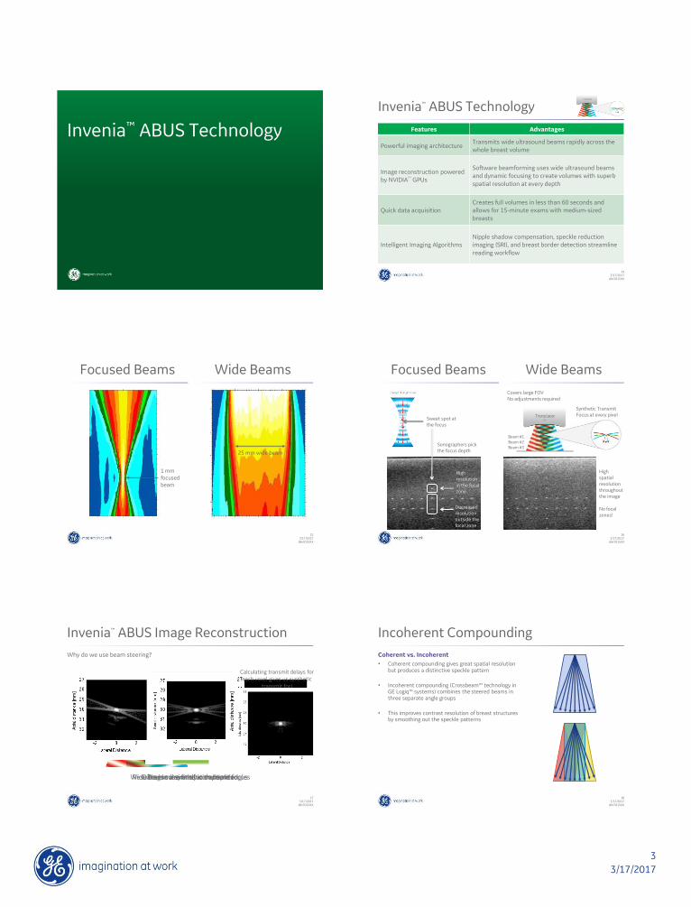
Invenia™ ABUS Technology
143/17/2017
JB47015XX
Invenia™ ABUS Technology
Features Advantages
Powerful imaging architectureTransmits wide ultrasound beams rapidly across the whole breast volume
Image reconstruction powered by NVIDIA
TM
GPUs
Software beamforming uses wide ultrasound beams and dynamic focusing to create volumes with superb spatial resolution at every depth
Quick data acquisitionCreates full volumes in less than 60 seconds and allows for 15-minute exams with medium-sized breasts
Intelligent Imaging AlgorithmsNipple shadow compensation, speckle reduction imaging (SRI), and breast border detection streamline reading workflow
153/17/2017
JB47015XX
Focused Beams Wide Beams
25 mm wide beam
1 mm focused beam
163/17/2017
JB47015XX
Focused Beams
High resolution in the focal zone
Decreased resolution outside the focal zone
High spatial resolution throughout the image
No focal zones!
Sonographers pick the focus depth
Sweet spot at the focus
Covers large FOVNo adjustments required
Synthetic Transmit Focus at every pixel
Beam #1Beam #2Beam #3
Wide Beams
173/17/2017
JB47015XX
Data is coherently compoundedResulting in a synthetic transmit focusTransmission of wide beams
2.5 cm
Why do we use beam steering?
Wide beams are fired at multiple angles
Invenia™ ABUS Image Reconstruction
Calculating transmit delays for each voxel gives us synthetic
transmit foci
183/17/2017
JB47015XX
Incoherent Compounding
Coherent vs. Incoherent
• Coherent compounding gives great spatial resolutionbut produces a distinctive speckle pattern
• Incoherent compounding (Crossbeam™ technology inGE Logiq™ systems) combines the steered beams inthree separate angle groups
• This improves contrast resolution of breast structuresby smoothing out the speckle patterns

4
3/17/2017
193/17/2017
JB47015XX
Invenia™ ABUS Imaging Architecture
768 elements, 15cm wide
Focused image created using compounded steered beams
Reverse Curve Transducer
Breast
Transverse images are generated as the
transducer sweeps the breast to create a full
frame
Active elements / aperture Wide beam acquisitions with multiple angle groups
= 768 x
Nipple Shadow Compensation
Minimizes nipple shadow and allows reading near the nipple
NSC off
NSC on
Speckle Reduction Imaging (SRI)
Decreases speckle noise
Minimizes distractions caused by noise
Speckle off Speckle level 1 Speckle level 2
Breast Border Detection
BBD off BBD on
• Estimates boundary between breast tissue and the background
• Removes out of breast areas and nearfield artifacts
• Reduces reading time spent on these areas
Invenia™ ABUSImage Quality Control
243/17/2017
JB47015XX
New System, Familiar Tests
B-mode Image Quality
• Despite advances in software image reconstruction and algorithms, the core technology in ABUS is the familiar pulse-echo ultrasound
• Similar image quality requirements as handheld ultrasound
• Image artifacts, uniformity, penetration, spatial resolution, contrast resolution, speckle noise
Invenia™ ABUS Image Quality Control (IQC)
• Largely follows the precedence set by GE Logiq™ systems
• Image uniformity and penetration is pre-optimized

5
3/17/2017
253/17/2017
JB47015XX
Invenia™ ABUS IQC
IQC Parameters
• Display monitor fidelity
• Image artifacts
• Electromagnetic noise
• Dead transducer elements (coronal lines)
• Spatial resolution
• Distance accuracy
• Contrast resolution
• Dynamic range
• Speckle
Recommended Phantom
• ATS Laboratories Model UC-551M Small Parts Phantom
• Urethane rubber phantom shaped to match the Invenia™ ABUS curvature
• 0.5 dB/cm/MHz
• 1450 m/s ±1% @ 23°C
263/17/2017
JB47015XX
IQC Principles
Display Monitor Fidelity
• Scan Station uses Planar PT1785P-BK
• Workstation uses NEC P242W-BK
• Verify that there are no visible damage or dead pixel(s)
• Verify that the monitor’s correct ICC profile is installed
• Backup any configuration prior to any changes
• Use the monitor’s on-screen display (OSD) to correctany color, brightness, or contrast configurations
• If needed, use Windows Color Calibration or a SMPTE image for adjustment
• Verify any changes with clinical images
273/17/2017
JB47015XX
IQC Principles
Image Artifacts
• Check for any electromagnetic noise using a uniform section of the test phantom
• Electromagnetic noise shows up as bright arcs in the image
• Note: ultrasound reflection off of the bottom of thetest phantom is expected
• Check for any dead transducer elementsby performing a full scan
• Any faulty element should be dark anddistinct in the coronal view
283/17/2017
JB47015XX
IQC Principles
Spatial Resolution
• Distance can be measured on the Workstation using the caliper tool
• Scan a test phantom and send the case to the Workstation
• Verify the distances between the linear line target (5 mm)
• Repeat for different depths and horizontal positions
• Verify the distances betweenthe targets in the axial-lateralresolution arrays
• Consider the transverse viewpixel spacing(axial 82 µm, lateral 200 µm)
• Verify voxel sizes inthe DICOM headers:(0018,0088) Spacing Between
Slices(0028,0030) Pixel Spacing
293/17/2017
JB47015XX
IQC Principles
Contrast Resolution
• Default dynamic range of the system is 80 dB prior to compression and dynamic gain compensation (DGC)
• Verify that the +15 dB and -15 dB gray scale target structures are visible
• Verify that the anechoic targetstructures are very dark,with minimal speckle and noise
Invenia™ ABUSImaging Demo

6
3/17/2017
Invenia™ ABUSExtra Slides
Mammo negative; Biopsy-proven IDC with multifocal disease Mammo negative; Biopsy-proven IDC with MRI
Biopsy-proven fibroadenoma
Invenia™ ABUS Imaging Architecture

7
3/17/2017
Invenia™ ABUS Imaging Architecture
Dense tissue; Normal duct
Invenia™ ABUS Imaging Architecture
Breast Density Classifications
PredominatelyFatty
BI-RADS A
Scattered Fibroglandular
BI-RADS B
HeterogeneouslyDense
BI-RADS C
ExtremelyDense
BI-RADS D
403/17/2017
JB47015XX
SomoInsight Study (Brem et al.1)
Detection of Cancer in Dense Breast Tissue
• A total of 15,318 women were included and classified as having breast density in BI-RADS category III (n=11,488, 75.0%) or IV (n=3,830, 25.0%)
• Breast cancer was diagnosed at screening in 112 women: 82 by full-field digital mammography (FFDM) and an additional 30 by ABUS
Adding ABUS to Mammo
• The addition of ABUS to FFDM yielded an additional 1.9 detected cancersper 1000 (95% CI 1.2, 2.7; p<0.001) screens
1. Brem RF, Tabár L, et.al. Assessing Improvement in Detection of Breast Cancer with Three-dimensional Automated Breast US in Women with Dense Breast Tissue: The SomoInsight Study. Radiology. 2015 Mar; 274(3): 663-73.
413/17/2017
JB47015XX
SomoInsight Study (Brem et al.1)
ABUS Screening
Screening with ABUS has a 55% relative increase in invasive breast cancers* identifiedusing supplemental ABUS and a 37% relative increase in cancer detection overallthan mammography alone.
* Increase in sensitivity was associated with a decrease in overall specificity1. Brem RF, Tabár L, et.al. Assessing Improvement in Detection of Breast Cancer with Three-dimensional Automated Breast US in Women with Dense Breast Tissue: The SomoInsight Study. Radiology. 2015 Mar; 274(3): 663-73.
Invenia™ ABUS is approved for diagnostic ultrasound imaging of the breast in symptomatic women in China
(CFDA Indication for Use)
423/17/2017
JB47015XX
EASY* Study (Wilczek et al.2)
Adding ABUS to Mammo
• The study included 1,668 women with no prior history of breast cancer
• 6.6 cancers per 1000 women screened by FFDSM + 3D ABUS
• 4.2 cancers per 1000 women screened by FFDSM alone
• ABUS produced a relative increase of 57%
• Sensitivity +36.4% and specificity -0.7%
Callback Rate
• 2.3% callback rate for FFDSM + 3D ABUS
• 2.1% callback rate for FFDSM alone
Conclusion
• FFDM + ABUS significantly increases the breast cancer detection ratewithout raising the recall rate significantly
* European Asymptomatic Screening Study
2. Wilczek B, Wilczek H E, Rasouliyan L and Leifland K. Adding 3D automated breast ultrasound to mammography screening in women with heterogeneously and extremely dense breasts: Report from a hospital-based, high-volume, single center breast cancer screening program”, European Journal of Radiology 2016;85: 1554-1563.

8
3/17/2017
433/17/2017
JB47015XX
FDA PMA Reader Study (Giger et al.3)
Enriched Reader Study
• ROC analysis
• 185 cases of which 52 were cancer and 133 were non-cancer
• 17 radiologists were presented with FFDM and then FFDM plus ABUS image sets
Adding ABUS to Mammo
• 110% sensitivity increase for cancers originally missed with FFDM and no prior interventions
• Overall, specificity decreased only slightly with the addition of ABUSfrom 78.1% to 76.2% which was not statistically significant
Conclusion
• “The addition of ABUS to screening mammography showeda significant increase in cancer detection with a nominal,insignificant decrease in specificity.”
3. Giger, ML, Inciardi, MF, et al. Automated Breast Ultrasound in Breast Cancer Screening of Women With Dense Breasts: Reader Study of Mammography-Negative and Mammography-Positive Cancers. AJR 2016; 206:1–10
Coverage in Aunt Minnie (June 9, 2016)
ABUS: An effective option for dense breast screeningBy Kate Madden Yee, Aunt Minnie staff writer
443/17/2017
JB47015XX
Reverse Curve™
Designed to Match a Woman’s Anatomy
• Uniform compression across the entire breast
• 15 cm wide field of view
• 6-15 MHz wide bandwidth
• Designed for patient comfort
Conforms to female anatomy < 1 minute scan time per view
Reverse Curve™
Linear Reverse Curve
463/17/2017
JB47015XX
Ergonomic and Intuitive
Icon Driven Touchscreen
• Intuitive icons
• Customizable workflow
• Touchscreen interface
Compression Assist
• Provides patient and operator comfort
• Multi-level compression with one-touch operation
• Promotes complete acquisition and study reproducibility
473/17/2017
JB47015XX
GE Healthcare Breast Portfolio